Article

Revealing a Third Dissolved-Phase Xenon-129 Resonance in Blood Caused by Hemoglobin Glycation

by
Lutosława Mikowska
1,†,
Vira Grynko
2,3,†,
Yurii Shepelytskyi
3,4,
Iullian C. Ruset
5,
Joseph Deschamps
6,
Hannah Aalto
6,
Marta Targosz-Korecka
1,
Dilip Balamore
7,
Hubert Harańczyk
1 and
Mitchell S. Albert
3,4,8,*
1
Faculty of Physics, Astronomy, and Applied Computer Science, Jagiellonian University, 30-348 Krakow, Poland
2
Chemistry and Material Science Program, Lakehead University, Thunder Bay, ON P7B 5E1, Canada
3
Thunder Bay Regional Health Research Institute, Thunder Bay, ON P7B 7A5, Canada
4
Chemistry Department, Lakehead University, Thunder Bay, ON P7B 5E1, Canada
5
Xemed LLC, Durham, NH 03824, USA
6
Applied Life Sciences Program, Lakehead University, Thunder Bay, ON P7B 5E1, Canada
7
Department of Engineering, Physics and Technology, Nassau Community College, New York, NY 11530, USA
8
Faculty of Medical Sciences, Northern Ontario School of Medicine University, Thunder Bay, ON P3E 2C6, Canada
*
Author to whom correspondence should be addressed.
These authors contributed equally to this work.
Int. J. Mol. Sci. 2023, 24(14), 11311; https://doi.org/10.3390/ijms241411311
Submission received: 27 May 2023 / Revised: 6 July 2023 / Accepted: 10 July 2023 / Published: 11 July 2023
(This article belongs to the Section Molecular Biophysics)

Abstract

:
Hyperpolarized (HP) xenon-129 (129Xe), when dissolved in blood, has two NMR resonances: one in red blood cells (RBC) and one in plasma. The impact of numerous blood components on these resonances, however, has not yet been investigated. This study evaluates the effects of elevated glucose levels on the chemical shift (CS) and  T 2 *  relaxation times of HP 129Xe dissolved in sterile citrated sheep blood for the first time. HP 129Xe was mixed with sheep blood samples premixed with a stock glucose solution using a liquid–gas exchange module. Magnetic resonance spectroscopy was performed on a 3T clinical MRI scanner using a custom-built quadrature dual-tuned 129Xe/1H coil. We observed an additional resonance for the RBCs (129Xe-RBC1) for the increased glucose levels. The CS of 129Xe-RBC1 and 129Xe-plasma peaks did not change with glucose levels, while the CS of 129Xe-RBC2 (original RBC resonance) increased linearly at a rate of 0.015 ± 0.002 ppm/mM with glucose level. 129Xe-RBC1  T 2 *  values increased nonlinearly from 1.58 ± 0.24 ms to 2.67 ± 0.40 ms. As a result of the increased glucose levels in blood samples, the novel additional HP 129Xe dissolved phase resonance was observed in blood and attributed to the 129Xe bound to glycated hemoglobin (HbA1c).

1. Introduction

Since its invention [1], hyperpolarized (HP) xenon-129 (129Xe) MRI has been primarily used for the functional imaging of the human lungs and for studying pulmonary disorders [2,3,4,5,6,7]. Over the past decade, the feasibility of HP 129Xe dissolved-phase imaging has been demonstrated for imaging pulmonary gas transfer [8,9,10,11,12], brain imaging [13,14,15,16,17,18], and kidney imaging [19,20]. HP 129Xe dissolved-phase imaging relies on the sufficiently long T1 relaxation time in blood (~3.4–7.8 s) [21,22,23,24,25] and the well-distinguished resonances of HP 129Xe nuclei dissolved in blood and various tissue compartments. Therefore, the chemical composition of blood should have a critical effect on HP 129Xe dissolved-phase imaging since it affects the T1 and chemical shift (CS) of the dissolved 129Xe resonances. A number of studies have previously evaluated the role of blood oxygenation and its effects on HP 129Xe MR spectra [21,22,24,26]. However, there is another vital component of blood chemical composition that has yet to be explored—the glucose level.
A high glucose concentration in blood leads to excessive non-enzymatic chemical interactions between glucose and proteins in the blood [27]. Hemoglobin is the protein most affected by glycation. Structurally, hemoglobin has four subunits, consisting of two α and two β chains, each of which carry an iron-containing heme group responsible for oxygen binding [28]. During the glycation process, a non-enzymatic reaction occurs between glucose and the α-amino groups of valine residues at the N-terminus of the β-chains [29]. The most abundant form of glycated hemoglobin, which is widely used in clinical practice for the evaluation of glycemia in diabetes mellitus, is hemoglobin A1c (HbA1c) [30]. Glycated hemoglobin is naturally present in healthy individuals (~4%), whereas glycation levels up to 20% have been reported in patients diagnosed with diabetes [31]. Elevated HbA1c levels (>6.5%) are routinely used for the diagnosis of diabetes mellitus [32]. The standard HbA1c test is used for the diagnosis of type 2 diabetes and prediabetes. It shows the amount of glycated hemoglobin and reflects the average blood glucose level over the past three months [33].
Hemoglobin glycation induces the formation of oxygen-derived free radicals, which are responsible for causing oxidative stress in erythrocytes [34], leading to an increase in membrane lipid peroxidation [35] and membrane damage in diverse cell types. The glycation of iron-containing heme proteins can cause the degradation of heme and further reactions with H2O2, leading to increased iron release, as well as ferryl myoglobin formation [27,36]. In addition, iron overload caused by high concentrations of glycated hemoglobin has been shown to be associated with changes in the structure of the red blood cells (RBCs) and increased thrombotic events [37]. Numerous studies have been conducted evaluating the effect of elevated levels of glycated hemoglobin on red blood cell distribution width (RDW); however, the data published has been inconclusive [38]. Some studies have also suggested that hyperglycemia may have some effect on erythropoiesis and RBC survival at concentrations of glucose higher than 40 mM [35,38].
It is known that the HP 129Xe signal from blood originates from Xe in plasma and the RBCs [1]. 129Xe binds to hemoglobin in hydrophobic cavities close to the external surface in both α and β chains [39]. Therefore, alternations in the structure of hemoglobin caused by glycation are expected to have a direct effect on the HP 129Xe magnetic environment and its dipole–dipole interaction with hemoglobin, potentially resulting in CS changes. Furthermore, the overall increase in free radical levels can also be anticipated to cause an effect on HP 129Xe CS. The destruction of heme groups accompanied by the release of iron would be expected to have potential effects on HP 129Xe-RBC CS, as well as on the  T 2 *  relaxation process.
The present lung studies with HP 129Xe exploit the measuring of the signal intensity of 129Xe bound to tissue and RBC [2,3,4,7,8]. However, the elevated glucose level in blood may have a considerable impact on the 129Xe spectroscopic properties and should be taken into account for patients with diabetes.
This work, for the first time, investigates the effect of glucose concentration in blood on the physical properties of dissolved HP 129Xe, such as CS and on the effective spin–spin relaxation time  T 2 * . Moreover, using non-linear curve fitting, we demonstrated that the spectrum of HP 129Xe in blood contains not two (as was conventionally thought) but three dissolved-phase resonances. We report the resonance frequencies,  T 2 *  relaxation times, and the CS dependance on glucose level of the three observed spectral components in sterile citrated sheep blood. In addition, we propose a potential mechanism for glycation-related changes in the physical parameters of the dissolved HP 129Xe. This research suggests that for a comprehensive understanding of dissolved-phase imaging, glucose levels in the blood should also be taken into account along with blood oxygenation, since both play distinct roles.

2. Results

2.1. CS Analysis Using a Conventional Three-Peak Model (3PM)

The measurements of the CS of the 129Xe dissolved in plasma (129Xe-plasma) and 129Xe dissolved in RBC (129Xe-RBC) resonances were conducted for the glucose concentration range of 0–55 mM. The acquired MRS spectra were first fitted using the conventional three-peak model (3PM): gas peak, plasma peak, and RBC peak. Figure 1A,B show MRS acquired for 10 mM and 45 mM glucose concentrations in the blood, respectively. The increase in glucose concentration did not cause a significant change in the 129Xe-plasma CS. Conversely, the 129Xe-RBC resonance shifted from 217.86 ppm (10 mM of glucose) to 219.07 ppm (45 mM). Figure 1C shows three representative cumulative Lorentzian 3PM fits for 10 mM, 20 mM, and 45 mM glucose concentrations. The downfield shift of the HP 129Xe-RBC resonance with respect to the gaseous 129Xe resonance can be clearly observed with increasing glucose concentration. The observed CS increased linearly with the increased glucose concentration (Figure 1D). The fit of the CS dependence of the 129Xe-RBC peak on glucose concentration revealed a (0.025 ± 0.004) ppm/mM 129Xe-RBC change rate. A strong positive correlation (r = 0.91) was observed between the 129Xe-RBC peak CS and the glucose concentration in the measured blood samples. It can be clearly seen that the 129Xe-plasma peak was completely unaffected by an increase in glucose concentration (r = 0.1 with a slope of the line equal to (0.001 ± 0.005) ppm/mM. The observed downfield shift of the HP 129Xe-RBC resonance is similar to the HP 129Xe-RBC resonance frequency change due to the increase in blood oxygenation, albeit with a total span of only ~1.25 ppm.

2.2. T 2 *  Relaxation Measurements

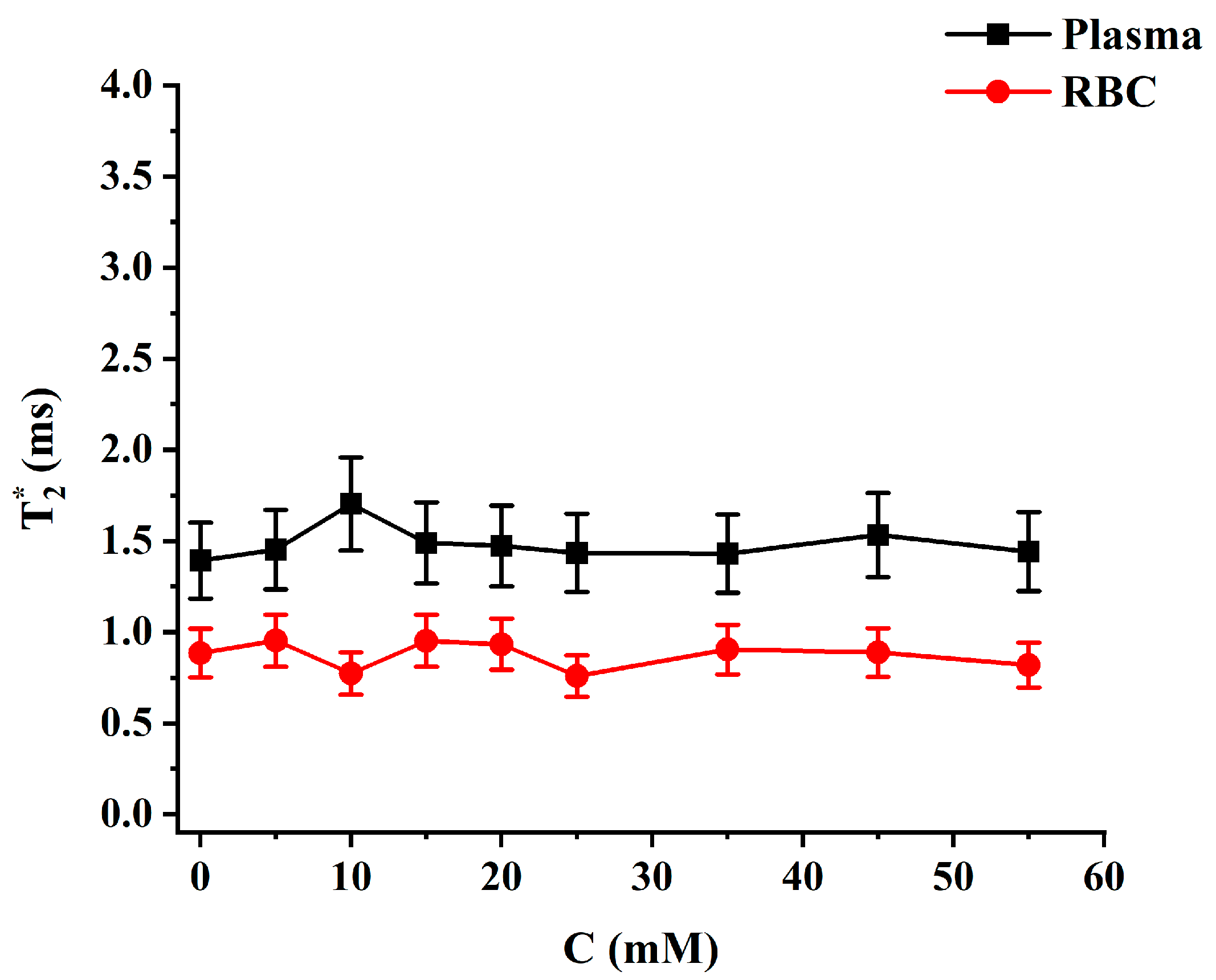
Following the measurements of CS,  T 2 *  relaxation was assessed based on Lorentzian fits of the measured spectra to 3PM. No significant change was observed in  T 2 *  values for HP 129Xe dissolved in both RBC and plasma pools with an increase in glucose content (Figure 2). The mean  T 2 *  values were equal to (1.48 ± 0.09) ms and (0.87 ± 0.07) ms for HP 129Xe dissolved in plasma and RBC, respectively.

2.3. Four-Peak Spectroscopic Model (4PM) Analysis

With the blood glucose level increase, the RBC resonance in the 129Xe spectrum became more asymmetrical and its linewidth increased slightly (Figure 3). In addition, a small splitting of the RBC peak was noted for higher glucose concentrations (red arrow on Figure 3). Considering the glycation process in RBCs, the observed asymmetry and splitting in the RBC peak can be interpreted by a four-peak spectroscopic model (4PM). In general, structural and functional changes to hemoglobin occur as a result of glycation [27]. Therefore, we can subdivide the RBC pool into HP 129Xe bound to non-glycated HbA0 and HP 129Xe bound to glycated HbA1c. These two pools can be characterized by their own HP 129Xe resonances. The suggested 4PM includes one gas phase resonance and three dissolved-phase resonances: 129Xe-plasma, 129Xe-RBC1, and 129Xe-RBC2.
The suggested 4PM was used to fit the experimental data (Figure 4A,B). The application of the 4PM did not affect the plasma peak position. The position of the HP 129Xe-RBC1 peak was barely affected by glucose: 216.08 ppm for the 10 mM solution and 215.92 ppm for the 45 mM solution. The 129Xe-RBC2 peak, however, shifted downfield with increasing glucose concentrations: 220.32 ppm for the 10 mM solution and 220.78 ppm for the 45 mM. The residual error for the 3PM, once plotted, showed distinct peaks at 216, 218, and 222 ppm for the 10 mM sample and at 216 ppm and 219 ppm for the 45 mM sample (Figure 4C,D). Once the spectrum is fitted to the 4PM, however, the residual error becomes flat at the RBC resonance position (between 210 ppm and 240 ppm). This indicates that 4PM fits the acquired HP 129Xe blood spectra more accurately compared to the conventional 3PM.
It should be noted that the 4PM fit worked better for glucose concentrations above 5 mM. This was indicated by slightly higher R2 values. In addition, small alterations in the peak position for the RBC resonances did not affect R2 significantly. Therefore, a lower accuracy of the recalculated spectral parameters can be anticipated for the pure blood and 5 mM samples compared to the samples with higher glucose levels. This is in accordance with our hypothesis that the second HP 129Xe RBC peak originates from HbA1c. There is no naturally occurring HbA1c in the pure blood and the concentration is expected to be low in a 5 mM glucose sample.
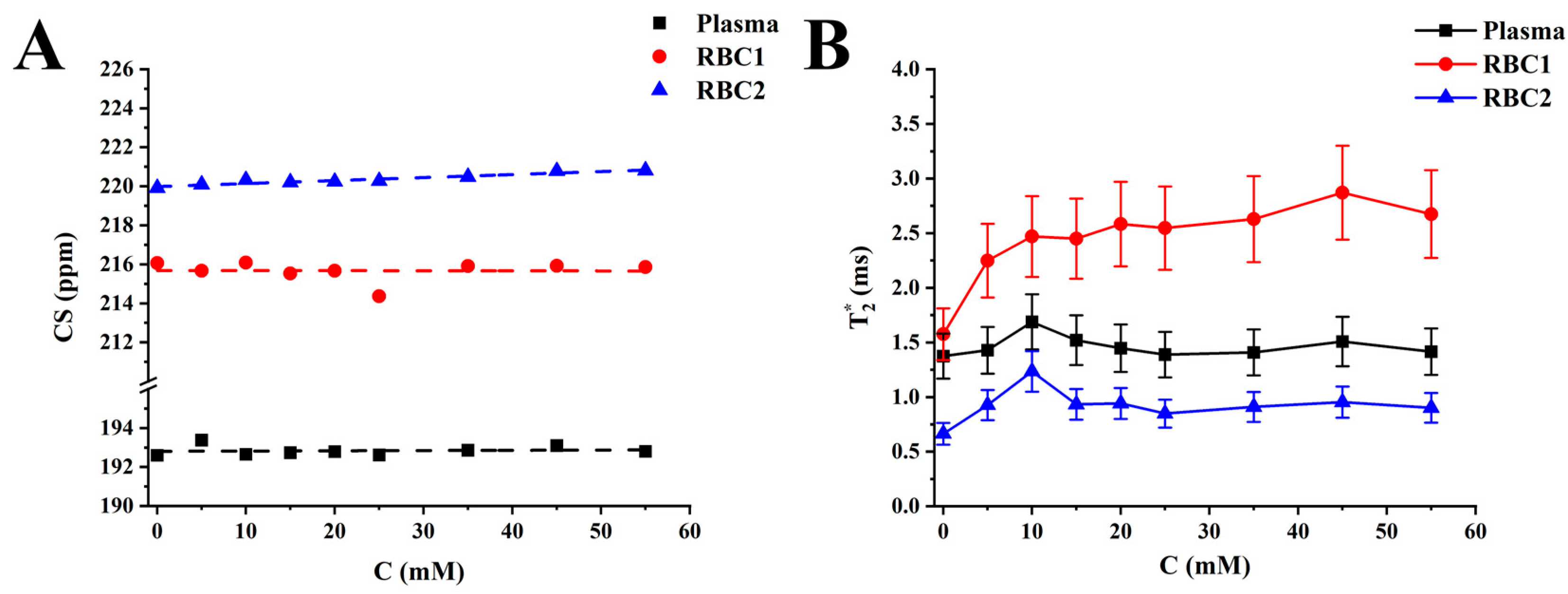
The proposed 4PM did not affect the CS of the HP 129Xe gas peak and 129Xe-plasma peak. The HP 129Xe-RBC1 CS change was not observed with a glucose level increase (Figure 5A). The HP 129Xe-RBC2 resonance frequency, however, increased with a rate of (0.015 ± 0.002) ppm/mM. A strong Pearson correlation was observed between the HP 129Xe-RBC2 peak position and the blood glucose level (r = 0.95). The  T 2 *  relaxation time for 129Xe-plasma did not depend on the glucose concentration in the sample (Figure 5B). Conversely, the 129Xe-RBC1  T 2 *  time increased non-linearly from (1.58 ± 0.24) ms up to (2.67 ± 0.40) ms over the studied range of glucose concentrations. The 129Xe-RBC2  T 2 *  relaxation time increased from (0.66 ± 0.10) ms to (1.23 ± 0.19) ms over a 0–10 mM glucose concentration range and leveled out at approximately (0.91 ± 0.03) ms at higher glucose levels.

3. Discussion

In normal physiological conditions, 95–98% of the hemoglobin in RBCs is present in the HbA0 state [31]. Once glucose levels elevate in the blood, HbA1c is generated as part of the glycation process [40]. In fact, the concentration of HbA1c hemoglobin increases linearly with the glucose concentration [30]. Watala et al. demonstrated that there are three major sites where structural changes occur in HbA1c: at the amino N-terminus of the β chain (~1/3 of the total amount of glucose binds to this site), α amino sites (~1/3 of the total glycation), and lysine residues (~40% of all glycation) [41]. Even if glucose is mostly linked to N-terminal valine, the glycation affects the spatial structure of the whole hemoglobin molecule. Ye at al. demonstrated that glycation transforms alpha-helices into beta-sheets, which results in conformation changes or the unfolding or even aggregation of hemoglobin [42]. Moreover, Sen at al. observed that the conformation caused by glycation increased the exposure of the hydrophobic tryptophan residues, like Trp14 in alpha-chains, which created one of the xenon-binding sites [27]. These structural changes result in significant alterations in hemoglobin conformation [27,41]. Glycation-induced conformational changes in hemoglobin weaken the heme-globing linkage in HbA1c and make it more thermolabile compared to HbA0 [27]. Thus, the global changes in spatial hemoglobin’s structure (tertiary and quaternary structure) may affect the shape of hydrophobic cavities or the binding sites of xenon, potentially resulting in a different chemical shift.
Savino et al. identified a total of twelve 129Xe binding sites per Hb4 tetramer: the α1 chain contains four binding sites; the α2 chain contains three; three more are located in the β2 chain; and two can be found in the β1 chain [43]. Considering the glycation-induced structural changes in HbA1c, it is possible and fairly likely that the number, size, and/or spatial location of 129Xe binding sites are different in HbA1c compared with HbA0. In addition, the glycation of hemoglobin results in the accumulation of free OH∙ radicals due to an iron-mediated Fenton’s reaction [27]. Increasing OH∙ concentrations are anticipated to result in the deshielding of the HP 129Xe nuclei. This hypothesis is supported by our experimental data, indicating the linear shift of the RBC HP 129Xe resonance downfield with respect to the resonance frequency of the gaseous HP 129Xe due to the increase in glucose concentration. The linear change in the HP 129Xe CS as a function of glucose level is in agreement with a known linear increase in HbA1c concentration caused by an increase in glucose concentration.
Considering our hypothesis that HP 129Xe binding sites are affected by glycation and are different in HbA1c compared to HbA0, it is reasonable to assume the existence of two HP 129Xe resonances—one from 129Xe bound to HbA0 and another originating from 129Xe bound to HbA1c. Therefore, instead of utilizing a conventional three peak model (gas phase resonance, plasma resonance, and RBC resonance) for spectroscopic data analysis, a four-peak model (gas phase resonance, plasma resonance, and two RBC resonances) can be used, according to our hypothesis. Spectral data were analyzed using both models and the residual analyses better supported the hypothesis of the 4PM. After 4PM Lorentzian fits, the residual error was smooth and almost completely flat in the region between 213 and 235 ppm. On the contrary, several residual peaks were observed in the same region after 3PM Lorentzian fits, indicating the presence of certain spectral component that remained unaccounted for by the 3PM. The residual analysis findings were further supported by the visual observation of asymmetry of the RBC peak and the presence of a small peak splitting for higher glucose concentrations.
It should also be mentioned that the 4PM Lorentzian fits of the spectroscopy data acquired for higher glucose concentrations were much better compared to the 4PM Lorentzian fits for the low glucose levels. For the pure blood samples as well as for the 5 mM glucose concentration, perturbations in 129Xe-RBC1 and 129Xe-RBC2 peak positions did not affect R2 significantly, if at all. Therefore, it can be concluded that the 4PM functions better for blood glucose levels above 5 mM. For concentration ranges between 0 mM and 5 mM, the 4PM can potentially result in some level of inaccuracy due to the four-peak fitting process. This can plausibly be explained by a low glycation level of the 0 mM and 5 mM samples.
The utilization of the 4PM demonstrated that the CS of one of HP 129Xe-hemoglobin peaks (RBC1) is independent of glucose level, whereas the resonances of the RBC2 peak experience a downfield shift linearly with an increase in the glucose level, and thereby, an increase in HbA1c concentration. Although the 4PM preserves the overall linear trend of the 129Xe CS evolution, the net span of CS changes was reduced from ~1.25 ppm (for 3PM) down to ~0.9 ppm. The CS change in HP 129Xe resonances due to the glycation of hemoglobin is much smaller than the previously reported changes due to the blood oxygenation by Norquay et al. [24]. It should be mentioned that the actual physiological range of glucose levels in the blood has an upper limit of ~35 mM. Therefore, the net expected physiologically relevant change in HP 129Xe RBC resonances due to HbA1c formation is limited to ~ (0.53 ± 0.07) ppm, which is one order of magnitude smaller compared to the blood oxygenation CS changes. The plasma resonance position was unaffected by the elevation of the blood glucose level.
The analysis of the  T 2 *  relaxation times indicated that the HP 129Xe-plasma resonance was unaffected by glucose level. Interestingly, the  T 2 *  values for the RBC1 peak increased non-linearly over the studied range of glucose concentrations. The  T 2 *  dynamics of the RBC2 peak were similar for glucose levels below 10 mM, albeit leveling out at higher concentrations.
Considering that the HP 129Xe-plasma resonance was completely unaffected by alternations in glucose concentration, the detected changes of HP 129Xe RBC relaxation should originate from internal alternations in RBCs’ magnetic susceptibilities and/or from changes in HP 129Xe spin–spin interactions with hemoglobin. There are two plausible mechanisms accounting for HP 129Xe transverse relaxation changes within the RBC. It was previously suggested that glycation induces iron release from heme pockets of hemoglobin [27] and, therefore, should reduce the spin–spin interaction between HP 129Xe enclosed in hemoglobin and the heme–iron atoms. Alternatively, conformational changes of HbA1c may occur in a way that the heme pockets become spatially distanced from HP 129Xe binding sites, thereby reducing the HP 129Xe—namely iron spin–spin interactions. Considering that iron release and conformational changes occur during the glycation process itself, it is possible that both mechanisms contribute to the observed changes in the  T 2 *  relaxation of HP 129Xe within the RBC.
Although a careful study of the position of the 129Xe binding sites in HbA1c is required for a proper explanation of the observed results, it is plausible that conformational changes in hemoglobin due to glycation occur in such a way that the dissociation constant of the HP 129Xe-HbA1c becomes substantially lower compared to the dissociation constant of the HP 129Xe-HbA0 complexes. Unfortunately, an accurate assessment of the relative changes in dipole–dipole interaction strength is not possible without knowledge of the location of 129Xe atoms in HbA1c.
Despite the fact that the knowledge of the 129Xe-HbA1c binding sites is of critical importance before attempting a quantitative description of the relaxation mechanisms responsible for the changes in HP 129Xe relaxation and the alternations of the CS, it should be possible to hypothesize the origin of the 129Xe-RBC1 and 129Xe-RBC2 peaks by combining our experimental results with previous knowledge on the glycation process and HbA1c. Due to the glycation process, iron is released from the heme pocket of hemoglobin [27]. In addition, hydrogen peroxide is produced by autoxidizing glucose, and its concentration builds up with time [44]. Hydrogen peroxide further initiates iron release from hemoglobin [45], affecting HbA1c much faster compared to HbA0 [27]. The iron released from heme pockets participates in an iron-dependent Fenton’s reaction that becomes a source of free OH∙ radicals within RBCs [27]. The charge of free radicals deshields the HP 129Xe nuclei, resulting in a downfield chemical shift detected via MRS. On the other hand, the release of iron may result in a decrease in the spin–spin interaction between the HP 129Xe bound to the hemoglobin. Since the iron release is much higher for HbA1c, it is plausible that the reduction in spin–spin interaction would be substantial for HP 129Xe enclosed within HbA1c. The  T 2 *  of HP 129Xe bond to HbA0 would be less affected due to the lower amount of iron released. Therefore, based on our results, we hypothesize that the RBC1 peak originates from HP 129Xe-HbA1c, while the RBC2 peak corresponds to the HP 129Xe-HbA0 signal. It is further plausible that the CS of the HP 129Xe-HbA1c signal is unaffected by the free OH∙ radical levels due to the conformational changes of the HbA1c and potential enclosure of HP 129Xe within the protein structure. On the other hand, the HP 129Xe-HbA0 signal experiences a linear downfield CS change due to the interaction with free radicals. It is also possible that the small change in  T 2 *  of HP 129Xe-HbA0 is a result of substantially slower iron extraction from HbA0.
Our study has several limitations. First, the initial incubation time of the blood samples with glucose solution was relatively short (only ~1 h). The glycation process is relatively slow, consisting of several steps and lasting a span of several days [44]. In the first step, the non-enzymatic Maillard early phase reaction takes place. This occurs after the binding of glucose transferred from the blood to the hemoglobin in RBCs and dictated by the interaction between the aldehyde group of the reducing glucose with the N-terminal amino base and ε-amino base of the lysine residue with the consequent formation of the Schiff base (almidine). This reaction is reversible—it is followed by an irreversible Amadori rearrangement, which results in the production of a stable form of HbA1c (ketoamine) [46]. The Amadori rearrangement is considered to be the limiting reaction in the glycation process [47,48]. The amount of HbA1c and synthetized H2O2, and therefore, the final concentration of free radicals, increases gradually over the glycation time course. Koga et al. used a 1 h incubation time in their study and confirmed the formation of the labile-glycated hemoglobin (aldimine); however, the formation of stable glycated hemoglobin (ketoamine) was not observed [46]. Thus, it is likely that we observed only the initial changes in the HP 129Xe CS and  T 2 *  relaxation time. A longer incubation time period will result in more pronounced changes in CS and in  T 2 *  relaxation time for HP 129Xe dissolved in RBCs. It should be noted that longer incubation times must be moderated by the fact that a small percentage of hemoglobin will be converted into methemoglobin in standing blood over time [49]. The formation of methemoglobin from hemoglobin within the red blood cells is an ongoing oxidative process that can result from the exposure of blood to the atmosphere in long standing blood samples. Methemoglobin forms when hemoglobin is oxidized to contain iron in the ferric [Fe3+] state rather than the normal ferrous [Fe2+] state [49]. Since methemoglobin is paramagnetic, this itself will cause additional changes to the CS and  T 2 *  relaxation time [23]. Therefore, longer incubation times are a tradeoff between allowing the full glycation of the blood samples versus limiting the formation of methemoglobin. In addition, all experiments were conducted on sterile citrated sheep blood. While it is not clear how the presence of citric acid affects protein glycation, the reproduction of these experiments may be performed in other types of animal blood.
Our work is a pioneering proof-of-concept study that should be further expanded by performing experiments with purified human hemoglobin, which is very low in the glycated form and separated RBCs from healthy and hyperglycemic subjects. This will allow us to study the effects of glucose on human RBCs. Consequently, the present study can be translated to the utilization of human blood drawn from healthy participants and hyperglycemic participants to observe how glycose-related changes are affected by the presence of other biological moieties from human blood. Furthermore, glycation level measurements are vital for obtaining reliable conclusions regarding the glucose effect on the 129Xe spectroscopic properties in blood. Additionally, no studies have yet been performed that investigate HP 129Xe binding to the glycated form of hemoglobin, which can be assessed in future X-ray diffraction (XRD) studies.

4. Materials and Methods

4.1. Sample Preparation

A 500 mM solution of glucose was prepared by dissolving 3.6 g of D-glucose (Sigma-Aldrich, St. Louis, MO, USA) in 40 mL of 1× phosphate-buffered saline (PBS) at pH 7.4 at room temperature. The mixture was stirred for complete dissolution and used as a stock solution for the preparation of the blood samples. Various volumes of the glucose solution in PBS were mixed with 20 mL of fresh citrated sheep blood (Cedarlane, Burlington, CA, USA) to create the following set of concentrations: 5, 10, 15, 20, 25, 35, 45, and 55 mM. The volume of glucose solution added to the blood was at least one order of magnitude smaller than the volume of the blood, in order to minimize blood dilution effects. A total of 20 mL of the pure citrated sheep blood without any additives served as a control sample. All blood samples were allowed to equilibrate to room temperature for approximately 1 h. The reported incubation times for glycation in the literature vary between 30 min up to hundreds of hours, depending on the incubation protocol [50]. Moreover, the vast majority of studies were able to confirm hemoglobin glycation for all of the incubation time periods. Therefore, successful hemoglobin glycation is expected within our experimental time frame.

4.2. Magnetic Resonance Spectroscopy (MRS)

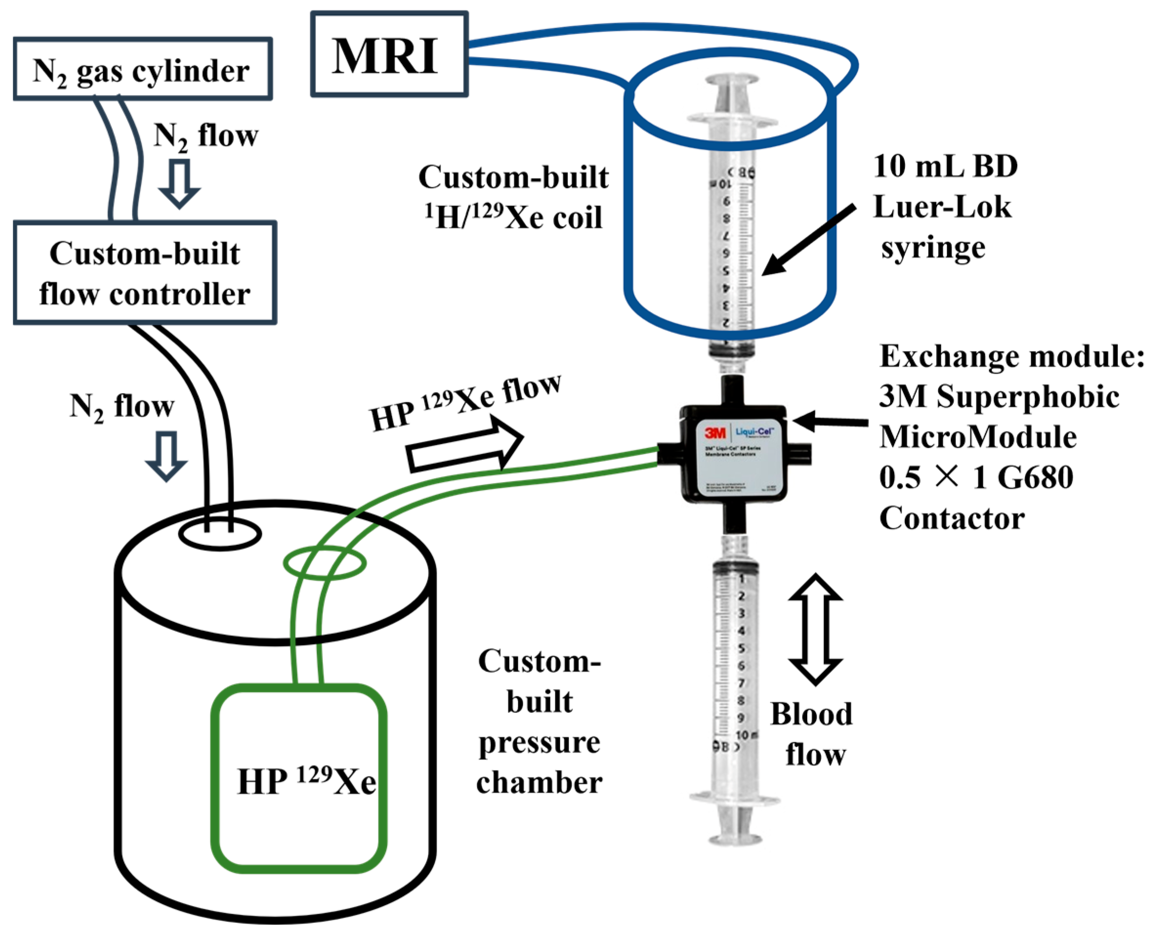
129Xe gas was polarized up to 56% using a XeBox-10E polarizer (Xemed, Durham, NH, USA) and disposed into 1 L Tedlar bags. The experimental setup (Figure 6) used for mixing HP 129Xe with blood was similar to that used by Norquay et al. [25]. Mixing was performed using an exchange module (Superphobic MicroModule 0.5 × 1 G680 Contactor; Membrana, Charlotte, NC, USA). A steady flow of HP 129Xe was set through the exchange module with the help of a pressure chamber pressurized with a continuous flow of N2 gas. The flow rate of N2 into the pressure chamber was controlled by a ventilator. The solution of sheep blood in the 10 mL syringe was connected to an exchange module and placed in a custom-built dual 1H/129Xe quadrature MRI birdcage coil, while the other empty syringe was connected to the other side of the exchange module. The blood was pumped back and forth manually through the exchange module perpendicular to the 129Xe flow for ~6 s. This technique allowed a sufficient amount of HP 129Xe to dissolve in the sheep blood and avoid the formation of gas bubbles in the blood.
A clinical Philips Achieva 3.0 T MRI scanner (Philips, Andover, MA, USA) equipped with a custom-built dual 1H/129Xe quadrature MRI birdcage coil was used for all 129Xe-blood spectroscopy acquisitions. The coil was tuned to 35.33 MHz, and the scanner was shimmed on the 1H signal from blood to correct for B1 inhomogeneities. Prior to taking any measurements, the coil was calibrated for the blood samples by applying a series of ten RF pulses with a s10° flip angle centered on the gas peak position of HP 129Xe resonance (for this, 1 mL syringes filled with HP 129Xe were placed into the coil near the blood sample).
To measure the effect of glucose concentration on the CS of the 129Xe dissolved in plasma (129Xe-plasma) and in RBC (129Xe-RBC) resonances, high-resolution single voxel spectroscopy was acquired for all samples. The receiver bandwidth was equal to 22 kHz and the number of samples was set to 4096, yielding a 0.15 ppm spectral resolution. A total of 0 ppm was set in between the peaks of 129Xe dissolved in plasma and the RBCs. A 90° rectangular excitation pulse was utilized. An FID spectrum was acquired with a TR of 189.6 ms and a TE of 0.25 ms. Measurements were repeated five times for each sample.

4.3. Data Reconstruction and Statistical Analysis

All data were initially analyzed using custom MatLab scripts in Matlab 2020b (Mathworks, Natick, MA, USA). The MRS spectra were postprocessed in Origin2021b (OriginLab, Northampton, MA, USA) for CS and  T 2 *  relaxation time assessment. HP 129Xe MRS spectra were fitted to either three (gas resonance, 129Xe-plasma resonance, and 129Xe-RBC resonance) or four (gas resonance, 129Xe-plasma resonance, and two 129Xe-RBC resonances) Lorentzian curves. The quality of fit was accessed via R2 value and through the residual fit error, which was automatically calculated at the end of the fitting algorithm. The comparison between the three and four peak model was performed based on the R2 and the residuals.
The full width half maximum (FWHM) of Lorentzian curves was utilized for calculations of the  T 2 *  of 129Xe dissolved in RBCs and plasma using the following equation [51]:
T 2 * = 1 π F W H M .
To evaluate statistical significance of the acquired results, a paired two-tailed t-test was used with a statistical significance level of 0.05. Pearson’s correlation coefficient was calculated between the CS changes of each HP 129Xe dissolved phase resonance and the glucose concentration. All statistical analysis was conducted using Origin2021b v.9.8.5.212 software.

5. Conclusions

In this work, for the first time, we demonstrated glucose-induced changes in CS and  T 2 *  relaxation of HP 129Xe dissolved in blood. By using a residual analysis, we observed the evidence for a third dissolved-phase resonance, which was attributed to HP 129Xe bound to HbA1c produced during HbA0 glycation. Our four-peak model suggests that one dissolved 129Xe resonance originates from plasma, whereas two other peaks originate from 129Xe encapsulated by HbA0 and HbA1c (the fourth peak stems from the gas phase resonance). We observed a linear dependence of the 129Xe-HbA0 resonance CS on the blood glucose levels. The 129Xe-HbA1c  T 2 *  changed non-linearly with an increase in glucose level. The HP 129Xe-plasma resonance was not affected by alternations in the glucose level.

Author Contributions

Conceptualization, L.M. and M.T.-K.; methodology, L.M., V.G., Y.S., M.S.A. and M.T.-K.; software, Y.S. and V.G.; validation, Y.S., M.S.A., I.C.R., M.T.-K. and H.H.; formal analysis, Y.S., V.G. and L.M.; investigation, L.M., V.G., Y.S., I.C.R., J.D., H.A. and M.S.A.; resources, M.S.A., L.M. and H.H.; data curation, M.S.A. and D.B.; writing—original draft preparation, V.G., L.M. and Y.S.; writing—review and editing, Y.S., V.G., L.M., M.S.A., H.H. and D.B.; visualization, V.G., Y.S. and L.M.; supervision, M.S.A., D.B., H.H. and M.T.-K.; project administration, Y.S., V.G. and M.S.A.; funding acquisition, L.M., V.G., Y.S. and M.S.A. All authors have read and agreed to the published version of the manuscript.

Funding

This research was funded by Natural Science and Engineering Research Council (NSERC) Discovery grant (RGPIN-2017-05359) and by the Priority Research Area SciMat under the program Excellence Initiative—Research University at the Jagiellonian University in Kraków (PSP: U1U/P05/NO/03.35). Y.S. was supported by the Mitacs Elevate Postdoctoral Fellowship (IT25574). V.G. was partially supported by an Ontario Trillium Scholarship and by Mitacs Accelerate Grant (IT31144). L.M. was supported by Jagiellonian Interdisciplinary Ph.D. Program.

Institutional Review Board Statement

This study was approved by the local biosafety committee.

Informed Consent Statement

Not applicable.

Data Availability Statement

The data that support the findings of this study are available from the corresponding author upon reasonable request.

Conflicts of Interest

Author I.C.R. was employed by the company Xemed LLC, Durham, NH, USA. The remaining authors declare that the research was conducted in the absence of any commercial or financial relationships that could be construed as a potential conflict of interest.

References

  1. Albert, M.S.; Cates, G.D.; Driehuys, B.; Happer, W.; Saam, B.; Springer, C.S.; Wishnia, A. Biological Magnetic Resonance Imaging Using Laser-Polarized 129Xe. Nature 1994, 370, 199–201. [Google Scholar] [CrossRef] [PubMed]
  2. Kruger, S.J.; Nagle, S.K.; Couch, M.J.; Ohno, Y.; Albert, M.; Fain, S.B. Functional Imaging of the Lungs with Gas Agents. J. Magn. Reson. Imaging 2016, 43, 295–315. [Google Scholar] [CrossRef] [PubMed] [Green Version]
  3. Patz, S.; Muradyan, I.; Hrovat, M.I.; Dabaghyan, M.; Washko, G.R.; Hatabu, H.; Butler, J.P. Diffusion of Hyperpolarized 129Xe in the Lung: A Simplified Model of 129Xe Septal Uptake and Experimental Results. New J. Phys. 2011, 13, 015009. [Google Scholar] [CrossRef]
  4. Ruppert, K.; Mata, J.F.; Brookeman, J.R.; Hagspiel, K.D.; Mugler, J.P. Exploring Lung Function with Hyperpolarized 129Xe Nuclear Magnetic Resonance. Magn. Reson. Med. 2004, 51, 676–687. [Google Scholar] [CrossRef] [PubMed]
  5. Santyr, G.; Fox, M.; Thind, K.; Hegarty, E.; Ouriadov, A.; Jensen, M.; Scholl, T.J.; Van Dyk, J.; Wong, E. Anatomical, Functional and Metabolic Imaging of Radiation-Induced Lung Injury Using Hyperpolarized MRI. NMR Biomed. 2014, 27, 1515–1524. [Google Scholar] [CrossRef] [Green Version]
  6. Svenningsen, S.; Kirby, M.; Starr, D.; Leary, D.; Wheatley, A.; Maksym, G.N.; McCormack, D.G.; Parraga, G. Hyperpolarized 3He and 129Xe MRI: Differences in Asthma before Bronchodilation. J. Magn. Reson. Imaging 2013, 38, 1521–1530. [Google Scholar] [CrossRef]
  7. Matheson, A.M.; Mcintosh, M.J.; Kooner, H.K.; Lee, J.; Desaigoudar, V.; Bier, E.; Driehuys, B.; Svenningsen, S.; Santyr, G.E.; Kirby, M.; et al. Persistent 129Xe MRI Pulmonary and CT Vascular Abnormalities in Symptomatic Individuals with Post-Acute COVID-19 Syndrome. Radiology 2022, 305, 466–476. [Google Scholar] [CrossRef]
  8. Rankine, L.J.; Wang, Z.; Wang, J.M.; He, M.; McAdams, H.P.; Mammarappallil, J.; Rackley, C.R.; Driehuys, B.; Tighe, R.M. 129xenon Gas Exchange Magnetic Resonance Imaging as a Potential Prognostic Marker for Progression of Idiopathic Pulmonary Fibrosis. Ann. Am. Thorac. Soc. 2020, 17, 121–125. [Google Scholar] [CrossRef]
  9. Grist, J.T.; Chen, M.; Collier, G.J.; Raman, B.; Abueid, G.; McIntyre, A.; Matthews, V.; Fraser, E.; Ho, L.P.; Wild, J.M.; et al. Hyperpolarized 129Xe MRI Abnormalities in Dyspneic Patients 3 Months after COVID-19 Pneumonia. Radiology 2021, 301, E353–E360. [Google Scholar] [CrossRef]
  10. Wang, Z.; Robertson, S.H.; Wang, J.; He, M.; Virgincar, R.S.; Schrank, G.M.; Bier, E.A.; Rajagopal, S.; Huang, Y.C.; O’Riordan, T.G.; et al. Quantitative Analysis of Hyperpolarized 129Xe Gas Transfer MRI. Med. Phys. 2017, 44, 2415–2428. [Google Scholar] [CrossRef] [Green Version]
  11. Willmering, M.M.; Cleveland, Z.I.; Walkup, L.L.; Woods, J.C. Removal of Off-Resonance Xenon Gas Artifacts in Pulmonary Gas-Transfer MRI. Magn. Reson. Med. 2021, 86, 907–915. [Google Scholar] [CrossRef] [PubMed]
  12. Niedbalski, P.J.; Hall, C.S.; Castro, M.; Eddy, R.L.; Rayment, J.H.; Svenningsen, S.; Parraga, G.; Zanette, B.; Santyr, G.E.; Thomen, R.P.; et al. Protocols for Multi-Site Trials Using Hyperpolarized 129Xe MRI for Imaging of Ventilation, Alveolar-Airspace Size, and Gas Exchange: A Position Paper from the 129Xe MRI Clinical Trials Consortium. Magn. Reson. Med. 2021, 86, 2966–2986. [Google Scholar] [CrossRef] [PubMed]
  13. Shepelytskyi, Y.; Grynko, V.; Rao, M.R.; Li, T.; Agostino, M.; Wild, J.M.; Albert, M.S. Hyperpolarized 129Xe Imaging of the Brain: Achievements and Future Challenges. Magn. Reson. Med. 2022, 88, 83–105. [Google Scholar] [CrossRef]
  14. Grynko, V.; Shepelytskyi, Y.; Li, T.; Hassan, A.; Granberg, K.; Albert, M.S. Hyperpolarized 129Xe Multi-Slice Imaging of the Human Brain Using a 3D Gradient Echo Pulse Sequence. Magn. Reson. Med. 2021, 86, 3175–3181. [Google Scholar] [CrossRef] [PubMed]
  15. Shepelytskyi, Y.; Grynko, V.; Li, T.; Hassan, A.; Granberg, K.; Albert, M.S. The Effects of an Initial Depolarization Pulse on Dissolved Phase Hyperpolarized 129Xe Brain MRI. Magn. Reson. Med. 2021, 86, 3147–3155. [Google Scholar] [CrossRef] [PubMed]
  16. Shepelytskyi, Y.; Hane, F.T.; Grynko, V.; Li, T.; Hassan, A.; Albert, M.S. Hyperpolarized 129Xe Time-of-Flight MR Imaging of Perfusion and Brain Function. Diagnostics 2020, 10, 630. [Google Scholar] [CrossRef] [PubMed]
  17. Rao, M.R.; Norquay, G.; Stewart, N.J.; Hoggard, N.; Griffiths, P.D.; Wild, J.M. Assessment of Brain Perfusion Using Hyperpolarized 129Xe MRI in a Subject with Established Stroke. J. Magn. Reson. Imaging 2019, 50, 1002–1004. [Google Scholar] [CrossRef]
  18. Rao, M.R.; Stewart, N.J.; Griffiths, P.D.; Norquay, G.; Wild, J.M. Imaging Human Brain Perfusion with Inhaled Hyperpolarized 129Xe MR Imaging. Radiology 2018, 286, 659–665. [Google Scholar] [CrossRef] [Green Version]
  19. Chacon-Caldera, J.; Maunder, A.; Rao, M.; Norquay, G.; Rodgers, O.I.; Clemence, M.; Puddu, C.; Schad, L.R.; Wild, J.M. Dissolved Hyperpolarized Xenon-129 MRI in Human Kidneys. Magn. Reson. Med. 2020, 83, 262–270. [Google Scholar] [CrossRef] [Green Version]
  20. Loza, L.A.; Kadlecek, S.J.; Pourfathi, M.; Hamedani, H.; Duncan, I.F.; Ruppert, K.; Rizi, R.R. Quantification of Ventilation and Gas Uptake in Free-Breathing Mice With Hyperpolarized 129Xe MRI. IEEE Trans. Med. Imaging 2019, 38, 2081–2091. [Google Scholar] [CrossRef]
  21. Albert, M.S.; Balamore, D.; Kacher, D.F.; Venkatesh, A.K.; Jolesz, F.A. Hyperpolarized 129XE T1 in Oxygenated and Deoxygenated Blood. NMR Biomed. 2000, 13, 407–414. [Google Scholar] [CrossRef] [PubMed]
  22. Albert, M.S.; Kacher, D.F.; Balamore, D.; Venkatesh, A.K.; Jolesz, F.A. T1 of 129Xe in Blood and the Role of Oxygenation. J. Magn. Reson. 1999, 140, 264–273. [Google Scholar] [CrossRef] [PubMed]
  23. Wolber, J.; Cherubini, A.; Dzik-Jurasz, A.S.K.K.; Leach, M.O.; Bifone, A. Spin-Lattice Relaxation of Laser-Polarized Xenon in Human Blood. Proc. Natl. Acad. Sci. USA 1999, 96, 3664–3669. [Google Scholar] [CrossRef] [PubMed]
  24. Norquay, G.; Leung, G.; Stewart, N.J.; Wolber, J.; Wild, J.M. 129Xe Chemical Shift in Human Blood and Pulmonary Blood Oxygenation Measurement in Humans Using Hyperpolarized 129Xe NMR. Magn. Reson. Med. 2017, 77, 1399–1408. [Google Scholar] [CrossRef] [PubMed] [Green Version]
  25. Norquay, G.; Leung, G.; Stewart, N.J.; Tozer, G.M.; Wolber, J.; Wild, J.M. Relaxation and Exchange Dynamics of Hyperpolarized 129Xe in Human Blood. Magn. Reson. Med. 2015, 74, 303–311. [Google Scholar] [CrossRef]
  26. Wolber, J.; Cherubini, A.; Leach, M.O.; Bifone, A. On the Oxygenation-Dependent 129Xe T 1 in Blood. NMR Biomed. 2000, 13, 234–237. [Google Scholar] [CrossRef] [PubMed]
  27. Sen, S.; Kar, M.; Roy, A.; Chakraborti, A.S. Effect of Nonenzymatic Glycation on Functional and Structural Properties of Hemoglobin. Biophys. Chem. 2005, 113, 289–298. [Google Scholar] [CrossRef]
  28. Giulivi, C.; Hochstein, P.; Davies, K.J.A. Hydrogen Peroxide Production by Red Blood Cells. Free Radic. Biol. Med. 1994, 16, 123–129. [Google Scholar] [CrossRef]
  29. De Rosa, M.C.; Sanna, M.T.; Messana, I.; Castagnola, M.; Galtieri, A.; Tellone, E.; Scatena, R.; Botta, B.; Botta, M.; Giardina, B. Glycated Human Hemoglobin HbA1c: Functional Characteristics and Molecular Modeling Studies. Biophys. Chem. 1998, 72, 323–335. [Google Scholar] [CrossRef]
  30. Sacks, D.B. Correlation between Hemoglobin A1c (HbA1c) and Average Blood Glucose: Can HbA1c Be Reported as Estimated Blood Glucose Concentration? J. Diabetes Sci. Technol. 2007, 1, 801–803. [Google Scholar] [CrossRef] [Green Version]
  31. Coletta, M.; Amiconi, G.; Bellellil, A.; Carsky, J.; Castagnola, M.; Condb, S.; Brunori, M. Alteration of T-State Binding Properties of Naturally Glycated. J. Mol. Biol. 1988, 203, 233–239. [Google Scholar] [CrossRef] [PubMed]
  32. Freeman, V.S. Glucose and Hemoglobin A1c. Lab. Med. 2014, 45, e21–e24. [Google Scholar] [CrossRef] [Green Version]
  33. Penttilä, I.; Penttilä, K.; Holm, P.; Laitinen, H.; Ranta, P.; Törrönen, J.; Rauramaa, R. Methods, Units and Quality Requirements for the Analysis of Haemoglobin A 1c in Diabetes Mellitus. World J. Methodol. 2016, 6, 133. [Google Scholar] [CrossRef] [PubMed]
  34. Inouye, M.; Mio, T.; Sumino, K. Glycated Hemoglobin and Lipid Peroxidation in Erythrocytes of Diabetic Patients. Metabolism 1999, 48, 205–209. [Google Scholar] [CrossRef]
  35. Viskupicova, J.; Blaskovic, D.; Galiniak, S.; Soszyński, M.; Bartosz, G.; Horakova, L.; Sadowska-Bartosz, I. Effect of High Glucose Concentrations on Human Erythrocytes in Vitro. Redox Biol. 2015, 5, 381–387. [Google Scholar] [CrossRef] [Green Version]
  36. Kar, M.; Chakraborti, A.S. Effect of Glucosylation on Iron-Mediated Free Radical Reactions of Haemoglobin. Curr. Sci. 2001, 80, 770–773. [Google Scholar]
  37. Buys, A.V.; Van Rooy, M.J.; Soma, P.; Van Papendorp, D.; Lipinski, B.; Pretorius, E. Changes in Red Blood Cell Membrane Structure in Type 2 Diabetes: A Scanning Electron and Atomic Force Microscopy Study. Cardiovasc. Diabetol. 2013, 12, 25. [Google Scholar] [CrossRef] [Green Version]
  38. Salvagno, G.L.; Sanchis-Gomar, F.; Picanza, A.; Lippi, G. Red Blood Cell Distribution Width: A Simple Parameter with Multiple Clinical Applications. Crit. Rev. Clin. Lab. Sci. 2015, 52, 86–105. [Google Scholar] [CrossRef]
  39. Schoenborn, B.P. Binding of Xenon to Horse Haemoglobin. Nature 1965, 208, 760–762. [Google Scholar] [CrossRef]
  40. Peterson, K.P.; Pavlovich, J.G.; Goldstein, D.; Little, R.; England, J.; Peterson, C.M. What Is Hemoglobin A1c? An Analysis of Glycated Hemoglobins by Electrospray Ionization Mass Spectrometry Endocrinology and Metabolism. Clin. Chem. 1998, 44, 1951–1958. [Google Scholar] [CrossRef] [Green Version]
  41. Watala, C.; Gwozdzinski, K.; Malek’, M. Direct Evidence for the Alterations in Protein Structure and Conformation upon In Vitro Nonenzymatic Glycosylation. J. Biochem. 1992, 24, 1295–1302. [Google Scholar] [CrossRef] [PubMed]
  42. Ye, S.; Ruan, P.; Yong, J.; Shen, H.; Liao, Z.; Dong, X. The Impact of the HbA1c Level of Type 2 Diabetics on the Structure of Haemoglobin. Sci. Rep. 2016, 6, 33352. [Google Scholar] [CrossRef] [PubMed]
  43. Savino, C.; Miele, A.E.; Draghi, F.; Johnson, K.A.; Sciara, G.; Brunori, M.; Vallone, B. Pattern of Cavities in Globins: The Case of Human Hemoglobin. Biopolym. Pept. Sci. Sect. 2009, 91, 1097–1107. [Google Scholar] [CrossRef] [PubMed]
  44. Wolff, S.P.; Jiang, Z.Y.; Huwr, J.V. Protein Glycation and Oxidative Stress in Diabetes Mellitus and Ageing. Free Radic. Biol. Med. 1991, 10, 339–352. [Google Scholar] [CrossRef] [PubMed]
  45. Halliwell, B.; Gutteridge, J.M.C. Role of Free Radicals and Catalytic Metal Ions in Human Disease: An Overview. Methods Enzymol. 1990, 186, 1–85. [Google Scholar] [CrossRef]
  46. Koga, M.; Inada, S.; Shimizu, S.; Hatazaki, M.; Umayahara, Y.; Nishihara, E. Aldimine Formation Reaction, the First Step of the Maillard Early-Phase Reaction, Might Be Enhanced in Variant Hemoglobin, Hb Himeji. Ann. Clin. Lab. Sci. 2015, 45, 643–649. [Google Scholar] [PubMed]
  47. Kerber, R.C. The A1c Blood Test: An Illustration of Principles from General and Organic Chemistry. J. Chem. Educ. 2007, 84, 1541–1545. [Google Scholar] [CrossRef]
  48. Lowrey, C.H.; Lyness, S.J.; Soeldner, J.S. The Effect of Hemoglobin Ligands on the Kinetics of Human Hemoglobin A(1c) Formation. J. Biol. Chem. 1985, 260, 11611–11618. [Google Scholar] [CrossRef]
  49. Umbreit, J. Methemoglobin—It’s Not Just Blue: A Concise Review. Am. J. Hematol. 2007, 82, 134–144. [Google Scholar] [CrossRef]
  50. da Silva, M.V.B.; Alet, A.I.; Castellini, H.V.; Riquelme, B.D. Methods: A New Protocol for in Vitro Red Blood Cell Glycation. Comp. Biochem. Physiol. Part A 2022, 264, 111109. [Google Scholar] [CrossRef]
  51. Shepelytskyi, Y.; Li, T.; Grynko, V.; Newman, C.; Hane, F.T.; Albert, M.S. Evaluation of Fluorine-19 Magnetic Resonance Imaging of the Lungs Using Octafluorocyclobutane in a Rat Model. Magn. Reson. Med. 2021, 85, 987–994. [Google Scholar] [CrossRef] [PubMed]
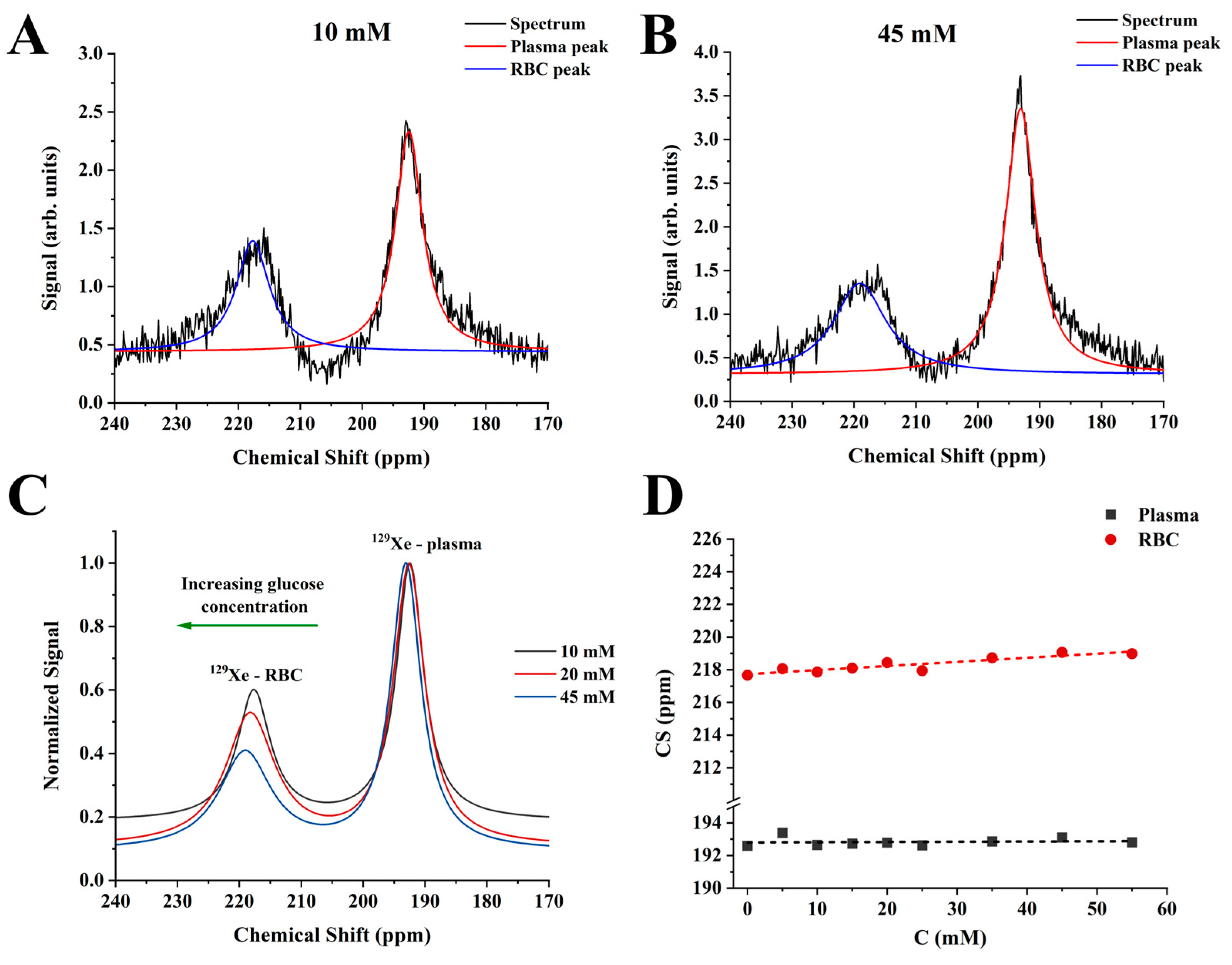
Figure 1. HP 129Xe MRS spectra of the 10 mM sample (A) and 45 mM sample (B) fitted to the conventional 3PM. The CS scale was selected to depict dissolved-phase resonances. The black line corresponds to the acquired HP 129Xe spectra. The red and blue lines are Lorentzian fits of the 129Xe-plasma and 129Xe-RBC peaks, respectively. (C) Three fitted HP 129Xe MRS spectra of 10 mM, 20 mM, and 45 mM samples demonstrate a noticeable downfield shift of the 129Xe-RBC resonance with a glucose concentration increase. The CS versus glucose concentration dependence of HP 129Xe-RBC peak appeared to be linear (D), whereas the CS of the HP 129Xe dissolved in plasma remained unaffected by blood glucose concentration.
Ijms 24 11311 g001
Figure 2. HP 129Xe  T 2 *  relaxation dependance on blood glucose level for 129Xe-plasma (black line) and 129Xe-RBC (red line).  T 2 *  values were not affected by the blood glucose level.
Ijms 24 11311 g002
Figure 3. HP 129Xe MRS spectra acquired for a pure blood sample (black line) and for a 25 mM blood glucose concentration (purple line). The RBC peak became asymmetrical and broader with the addition of glucose. A small splitting of the RBC peak can be seen (red arrow).
Ijms 24 11311 g003
Figure 4. HP 129Xe MRS spectra of the 10 mM sample (A) and 45 mM sample (B) fitted to the proposed 4PM. The CS scale was selected to depict dissolved-phase resonances. The black line corresponds to the acquired HP 129Xe spectra. The red line corresponds to the Lorentzian fit of the plasma resonance. The green and blue lines represent the Lorentzian fit of the 129Xe-RBC1 and 129Xe-RBC2 peaks, respectively. The residuals of 3PM (black lines) and 4PM (red line) were plotted as a function of chemical shift for the 10 mM sample (C) and 45 mM sample (D). It can be clearly seen that the conventional 3PM shows some residual signal present (green arrows). The proposed 4PM, however, results in a flat baseline with almost no residual signal at the RBC resonance position (between 210 ppm and 240 ppm) indicating a better fit of the RBC signal. Therefore, the proposed 4PM better suits the acquired HP 129Xe MRS spectra analysis compared to the conventional 3PM.
Ijms 24 11311 g004
Figure 5. HP 129Xe 4PM CS measurements and  T 2 *  relaxometry. (A) HP 129Xe CS dependences for 129Xe-plasma (black), 129Xe-RBC1 (red), and 129Xe-RBC2 (blue) as a function of blood glucose concentration. The dashed lines correspond to a linear fit of the measured CS dependances. A strong positive correlation can be observed between 129Xe-RBC2 CS and glucose level, whereas the CS of HP 129Xe-plasma and 129Xe-RBC1 remains unaffected by blood glucose. (B) HP 129Xe  T 2 *  relaxation dependence on blood glucose level for 129Xe-plasma (black line), 129Xe-RBC1 (red line), and 129Xe-RBC2 (blue line).  T 2 *  values of HP 129Xe in plasma were not affected by the blood glucose level, whereas the  T 2 *  relaxation time of 129Xe-RBC1 increased non-linearly with a glucose concentration increase.  T 2 *  values of the 129Xe-RBC2 peak increased slightly over the range of 0–10 mM and then leveled out.
Ijms 24 11311 g005
Figure 6. Setup used for mixing the blood with HP 129Xe and further MR signal acquisition. A steady flow of HP 129Xe from the 1 L bag was regulated by the continuous flow of N2 in the pressure chamber, forcing the HP 129Xe through the exchange module. The mixing of blood and HP 129Xe was performed by manually pumping the blood through the exchange module.
Ijms 24 11311 g006
Disclaimer/Publisher’s Note: The statements, opinions and data contained in all publications are solely those of the individual author(s) and contributor(s) and not of MDPI and/or the editor(s). MDPI and/or the editor(s) disclaim responsibility for any injury to people or property resulting from any ideas, methods, instructions or products referred to in the content.