Review

Endothelial Senescence and Its Impact on Angiogenesis in Alzheimer’s Disease

1
Institute of Biophysics and Biomedical Engineering, Bulgarian Academy of Sciences, Acad. George Bonchev, Str. Bl. 21, 1113 Sofia, Bulgaria
2
Institute of Neurobiology, Bulgarian Academy of Sciences, Acad. George Bonchev, Str. Bl. 23, 1113 Sofia, Bulgaria
3
Institute of Molecular Biology, Bulgarian Academy of Sciences, Acad. George Bonchev, Str. Bl. 21, 1113 Sofia, Bulgaria
*
Author to whom correspondence should be addressed.
Int. J. Mol. Sci. 2023, 24(14), 11344; https://doi.org/10.3390/ijms241411344
Submission received: 31 May 2023 / Revised: 7 July 2023 / Accepted: 10 July 2023 / Published: 12 July 2023
(This article belongs to the Special Issue Role of Apoptosis and Cellular Senescence in Cancer and Aging)

Abstract

:
Endothelial cells are constantly exposed to environmental stress factors that, above a certain threshold, trigger cellular senescence and apoptosis. The altered vascular function affects new vessel formation and endothelial fitness, contributing to the progression of age-related diseases. This narrative review highlights the complex interplay between senescence, oxidative stress, extracellular vesicles, and the extracellular matrix and emphasizes the crucial role of angiogenesis in aging and Alzheimer’s disease. The interaction between the vascular and nervous systems is essential for the development of a healthy brain, especially since neurons are exceptionally dependent on nutrients carried by the blood. Therefore, anomalies in the delicate balance between pro- and antiangiogenic factors and the consequences of disrupted angiogenesis, such as misalignment, vascular leakage and disturbed blood flow, are responsible for neurodegeneration. The implications of altered non-productive angiogenesis in Alzheimer’s disease due to dysregulated Delta-Notch and VEGF signaling are further explored. Additionally, potential therapeutic strategies such as exercise and caloric restriction to modulate angiogenesis and vascular aging and to mitigate the associated debilitating symptoms are discussed. Moreover, both the roles of extracellular vesicles in stress-induced senescence and as an early detection marker for Alzheimer’s disease are considered. The intricate relationship between endothelial senescence and angiogenesis provides valuable insights into the mechanisms underlying angiogenesis-related disorders and opens avenues for future research and therapeutic interventions.

1. Introduction

Angiogenesis is a complex biological process that involves the formation of new blood vessels from preexisting ones and should not be confused with vasculogenesis, in which blood vessels emerge de novo from endothelial progenitor cells [1]. It plays a crucial role during embryonic development and later in tissue growth and repair, wound healing, and reproduction. Still, it must be carefully regulated to avoid excessive or insufficient vascularization. New vessels emerge from sprouting endothelial cells (EC), the leading players, toward an angiogenic stimulus (sprouting angiogenesis) or by insertion into existing vessels and division into new ones (splitting angiogenesis) [2]. It is led by a tip cell that elongates and explores the environment while transmitting signals to the stalk cells that follow behind to proliferate and form tubular networks. The entire process is highly complex and difficult to imitate in vitro, highlighting the need for development of reliable models to study it [3,4,5]. Angiogenesis is governed by a strict balance between pro- and antiangiogenic factors, which, if broken, leads to uncontrolled cell proliferation (cancer, atherosclerosis, rheumatoid arthritis) or mitotic inhibition (aging and neurodegenerative diseases) [6,7,8]. Excessive angiogenesis can promote inflammation and tissue damage, while insufficient angiogenesis can lead to ischemia and cell death. Most studies are focused on the involvement of angiogenesis in cancer and cardiovascular diseases (CVD). Fewer examine its contribution to neurodegeneration, although it is correlated with the impairment of angiogenesis [9], endothelial senescence [7] and the occurrence of cerebrovascular angiopathy (a process in which small blood vessels burst and cause hemorrhages) [10,11]. The altered blood circulation in the elderly contributes to the lengthy process of wound healing and inadequate recovery of ischemic tissues due to the lack of response from aged ECs. Typically, ECs’ physiological functions are suppressed in time because of accumulated stress and induction of cellular senescence and apoptosis [12], leading to alterations in the regulation of angiogenesis and insufficient or excessive vascularization [6]. Age-related vasculature dysfunction has been implicated in the pathogenesis of various neurodegenerative diseases, including Alzheimer’s disease [13], Parkinson’s disease [14], and Huntington’s disease [15]. It may contribute to their progression by modulating the delivery of nutrients and oxygen and clearing of waste products from the brain.
Alzheimer’s disease (AD) is a debilitating condition characterized by progressive cognitive decline and behavioral changes that severely impact the daily lives of suffering individuals. Similarly, to other neurodegenerative diseases, aging is an essential factor contributing to its onset. There is overwhelming research aiming to find the causes, better ways for detection, treatment and, if possible, ways to avoid it altogether (reviewed elsewhere [16,17,18,19,20]). Factors involved in angiogenesis have roles in the birth of new neurons (neurogenesis), the prevention or mitigation of neuronal injury (neuroprotection), and the pathogenesis of stroke, AD and motor neuron disease [21]. Indeed, axon and blood vessel growth and migration are braided together via chemo-repulsive and attractive signals in which the vascular endothelial growth factor (VEGF) and the Delta-Notch signaling have a direct effect on both nervous and vascular systems [22], confirming that angiogenesis is closely related to neurodegeneration. AD patients exhibit changes in the number, diameter and density of blood vessels, which lead to decreased brain perfusion and BBB disruption. Here, we explore the current debate on the effect of the monomeric, oligomeric and plaque forms of amyloid-β on the efficacy of cerebral angiogenesis and blood flow.
This narrative review further explores the intricate relationships among senescence, oxidative stress, extracellular vesicles, and the extracellular matrix, highlighting their significance in the processes of angiogenesis, aging, and neurodegeneration. Additionally, it delves into potential therapeutic approaches aimed at modulation of angiogenesis and amelioration of disease progression.

2. The Dual Nature of Cellular Senescence

Cellular senescence is a fundamental process associated with tissue homeostasis during development, first described by Hayflick and Moorhead [23]. The authors observed a terminal pause in cell division of normal human fibroblasts after several cycles of passaging. They concluded that cultured cells cease to proliferate upon a finite number of doublings and, therefore, could be used as a model for aging. Today, this is referred to as the Hayflick limit. The processes of senescence and aging are intertwined in the sense that aging progresses with time and associates with increased numbers of senescent cells. Therefore, cellular senescence is also accepted as a hallmark of aging and a risk factor for age-related neurodegenerative diseases. However, senescence occurs during the full lifespan of an individual and is not restricted to later life stages. The resulting inability to divide is a consequence of irreversible cell cycle arrest, caused by the accumulation of various stress factors such as DNA damage, inflammation, telomere shortening, chromatin perturbations, and oncogene induction [12,24,25,26]. Senescence is believed to have evolved as a protective mechanism against cancer, but it also contributes to age-related physiological decline [27]. Additionally, loss of senescence during embryonic development allows the progression of unhealthy cells in embryos [28]. In contrast, while protecting against the propagation of mutated DNA, senescence harms long-living organisms, as it inhibits tissue renewal and function. These observations gave rise to the idea that there is a “right time to senesce”, arguing that the end goal of the fight against aging is not to completely eliminate senescent cells (SCs) but to learn how to tame them [29].

2.1. Hallmarks of Aging

In 2022, several new hallmarks of aging were introduced, stressing the complexity of the aging process [25]. They include compromised autophagy, impeded RNA processing, and changes in the microbiome and in the mechanical properties of both cells and extracellular matrix [25]. However, until recently the main focus was on the hallmarks of aging involving information loss (reviewed in [30]), telomere shortening [31] and endogenous reactive oxygen species (ROS) induced DNA damage [32] (Figure 1). The first is based on the fact that even though an organism shares the same genetic DNA among all cells, the epigenetic differences between them determine the cellular type. With age, epigenetic changes procured in response to DNA damage and p53 activation make information harder to read and trigger loss of cellular identity and function. This process is similar to reprogramming somatic cells to induced pluripotent stem cells (iPSC), achieved by the four Yamanaka factors, Oct4, Sox2, Klf4 and c-Myc [33]. A recent study supporting this theory utilized inducible epigenetic modifications to instigate premature aging in transgenic mice. The subsequent introduction of the Yamanaka factors reversed the “old” phenotype, hypothetically improving the animals’ quality of life [34]. Similar treatments were able to restore vision in aged mice [33], and short exposure to the same factors (for 13 days) rejuvenated fibroblasts and allowed them to maintain their original cell identity and improved their collagen secretion [35].
The other two hypotheses do not necessarily exclude epigenetic modifications. Instead, they focus on senescence as the cell’s response to life-long accumulation of stress-induced DNA damage by telomere shortening or oxidative stress (OS). The former is the result of the end-replication problem that causes dysfunctional telomeres and triggers the DNA-damage response (DDR) through ataxia-telangiectasia mutated protein kinase (ATM), checkpoint kinase 2 (CHK2), p53-binding protein 1 (53BP1) and γ-H2AX histone [36]. T cells can overcome this issue by elongating their telomeres, using telomeres from antigen-presenting cells (APCs), rather than increased telomerase activity. The intercellular transfer of telomeres via extracellular vesicles (EVs) rescues T cells from senescence and maintains their long-term immunological memory [37]. However, telomere extension can be overwhelmed by other senile factors, as telomerase activation cannot prevent senescence caused by OS-induced DNA damage in human fibroblasts but protects them against apoptosis and necrosis [38]. Furthermore, murine cells have significantly longer telomeres than human cells, but under standard culturing conditions, they senesce substantially faster due to high O2 levels and severe OS [39]. The induction of senescence in otherwise normal cells through exogenous factors such as chemicals [40], EVs generated from premature senile cells [41], septic shock [42] and OS is also known as stress-induced premature senescence [43].
In addition to its role in DDR, the protein kinase ATM functions as a sensor of redox homeostasis. It is oxidized and activated by hypoxia [44], resulting in ATM-mediated phosphorylation and stabilization of hypoxia-inducible factor (HIF)-1α [45], but it can also cause premature endothelial senescence and dysfunction [46]. In one study, OS stimulated ATM through the Akt/p53/p21 pathway, causing senescence in ECs, which was not the case for ATM-KO mice or upon treatment of HUVECs with ATM inhibitors [47]. Moreover, genetic or pharmacological ATM inhibition reduced cellular senescence and SASP expression [48]. ATM, Akt and the mammalian target of rapamycin (mTORC1) activation, as part of DDR, stimulates mitochondrial biogenesis and ROS-mediated DNA damage and senescence. Both of these processes are ameliorated by ATM or mTORC1 suppression [49].
DNA repair can be affected by numerous factors, including changes between anaerobic and aerobic carbohydrate metabolism that alter NAD+ levels. The molecule is used as a cofactor by multiple enzymes such as sirtuins (SIRTs)—NAD-dependent protein deacetylases involved in epigenetic modifications. SIRTs are known as “longevity genes” because their overexpression resulted in life extension in yeast, and they are depleted by insufficient NAD+ [50]. SIRT deficiency has been correlated with stress-induced premature endothelial [51] and hepatocyte [52] senescence. In a recent review by Charles Bennet [53], the author shares his disbelief in the correlation between lifespan, NAD+ and SIRT. He argues that the positive outcome of NMN and NAD+ supplementation is not due to the activation of SIRT but rather due to the antioxidant effect of these cofactors. Moreover, NAD+ is necessary for poly-(ADP-ribose) polymerase 1 (PARP1) activity in DDR [36] and, therefore, reduces the extent of DNA damage. Although PARP1 participates in one of the major DNA repair mechanisms, it is also involved in parthanatos (programmed cell death, independent from caspases, unlike apoptosis) [54]. Notably, OS-induced DNA damage and cell death can be avoided with PARP1 inhibitors or PARP1-KO [54], which also reduces PARP1-associated senescence-associated secretory phenotype (SASP) [55]. Meanwhile, SIRT1 has been associated with improved endothelial function [56] and increased microvascular density [57], whereas its knockout results in decreased angiogenesis [58]. A clearer picture of the interplay between NAD+- (SIRT1, PARP1) and redox sensors (ATM), and their contribution to endothelial senescence, could be immensely useful.

2.2. The Dose Makes the Poison

The cell’s choice between senescence or death depends on the level of accumulated stress and the subsequent activation of p53 [59]. Intermediate levels of p53 signal for senescence, and its hyper- or hypoactivation causes cell death or proliferation, respectively. To choose a path forward, the cell first undergoes a cell cycle arrest. If the experienced changes persist above a certain threshold, the cell proceeds with irreversible senescence, a process called geroconversion [60]. In the case of chronic stress, senescence can be triggered through either p53/p21 or retinoblastoma (Rb)/p16 pathways [26,59]. Therefore, if a cell is positive for either of these proteins and for senescence-associated β-galactosidase (SA-β-Gal), it can be considered senescent. Accumulation of SCs with chronological age varies depending on the cell and tissue types. The same applies to the expression and the activity of the factors that control the senescence signaling pathways.
SCs exhibit a hypersecretory phenotype known as SASP, which is used to alert the immune system to reduce local inflammation by eliminating them and, potentially, to direct tissue renewal. Removing SCs in this manner is essential because most of them are resistant to apoptosis. If SASP secretion is maintained for a short period, the consequential clearance of unhealthy cells can be very beneficial. On the other hand, prolonged SASP further increases intercellular stress and has the opposite effect. The SASP factors could stimulate nearby premalignant cells’ growth and angiogenic activity and, paradoxically, promote excessive angiogenesis and subsequent progression of cancer or neurodegenerative diseases [26,61]. Unfortunately, as the immune system weakens with age, its ability to clear SCs is reduced, and SASP evolves with a change from anti- to pro-inflammatory cytokine secretion [62]. Thus, the notion that temporally regulated mechanisms orchestrate the functions of SCs is probably the most coherent senescence concept so far. While all the beneficial roles of senescence share a transient profile, the deleterious functions of SCs are associated with their lingering persistence, namely chronic exposure to their SASP [29].

3. Endothelial Senescence

Aging and prolonged exposure to environmental factors, such as toxins, ROS, shear stress, and extracellular matrix (ECM) perturbations, induce senescence in ECs (Figure 1). Interestingly, unlike most SCs, senescent ECs (sen-ECs) remain susceptible to apoptosis [63], a mechanism most likely evolved to rearrange the microvasculature and counteract proliferation. Senescence in ECs is usually triggered by telomere shortening [26], which can be avoided by the exogenous introduction of telomerase [6]. Ionizing radiation can also geroconvert human microvascular cells in a time- and dose-dependent manner, predominantly by uncoupling Complex II of the mitochondrial respiratory chain [64], demonstrating ECs’ susceptibility to OS. In any case, the balance between senescence and angiogenesis becomes dysregulated during aging and neurodegenerative diseases, but the underlying mechanisms remain elusive. The negative consequences of vascular aging are apparent in older people in whom the regeneration of blood flow after ischemia or wounding is a slow and tedious process [65]. The accumulated stress over time reduces the proliferative capacity of ECs and modifies their interaction with the already altered ECM [66]. Furthermore, aging reduces the general expression of vascular endothelial growth factor (VEGF) [6] and promotes angiogenic incompetence in ECs, making them unable to respond to VEGF [7]. Some of the suggested reasons for the VEGF insensitivity are the age-related loss of VEGF receptor 2 (VEGFR2) [67], androgen resistance [68] and reduction in nitric oxide (NO) [6]. Furthermore, the SASP can directly inhibit angiogenesis by secreting factors that block endothelial cell proliferation and migration. At the same time, SCs can induce angiogenesis by secreting pro-inflammatory cytokines that promote neovascularization.

3.1. ECM Disruption Accelerates Vascular Aging

Aged vasculature is described with increased microvascular perfusion, susceptibility toward pro-inflammation and atherosclerosis, disrupted ECM interaction and altered secretory, barrier and transport functions [8,9,25,69]. The ECM comprises the natural scaffolding and framework on which ECs reside. The latter shape the vessel’s lumen, align to its length and curvature by attaching to the basal membrane and control the permeability, contractility and passage through the vessel [70]. The ECM consists primarily of collagen, elastin and fibrinogen, synthesized by ECs and fibroblasts and subjected to constant rearrangement by resident cells. It also mediates chemical cues that can alter the cell’s response and vice versa, creating the tissue microenvironment and enabling ECs to proliferate, migrate and stimulate vascular smooth muscle cells to form capillary networks and constrict/dilate fully formed vessels. One of the hallmarks of endothelial senescence and blood vessel aging is the stiffening of the ECM through glycation, aggregation and crosslinking [71]. Therefore, diseases occur not only when the cells are damaged but also when the ECM becomes impaired. For instance, elastin is renewed quite slowly; thus, changes in its structure tend to persist for longer periods of time. Meanwhile, collagen secretion increases, causing the stiffening of ECM in an NAD+-dependent manner [69]. Angiogenesis also relies heavily on the deposition and degradation of the ECM by metalloproteinases (MMPs), whose activity increases with age, further reducing the elasticity of the connective tissue and stimulating higher traction forces even in non-senescent cells [71]. This leads to endothelial dysfunction characterized by excess angiogenesis, leaky vasculature and low shear stress that cannot induce protective signaling pathways—a faulty process described as non-productive angiogenesis. Hence, by treating the age-related stiffening of the ECM, we could tackle endothelial dysfunction. One such example is the treatment of myocardial tissue with an optimized intravascular infusible ECM, which is able to fill gaps between the ECs, reduce vascular leakiness and improve vascular fitness [72].

3.2. Navigating the Currents: Shear Stress and Its Impact on Endothelial Cells’ Function

Naturally, endothelial cells are constantly exposed to shear stress in vivo from the movement of a non-Newtonian fluid, i.e., the blood. The wall shear stress (WSS) is described as the traction forces generated on the endothelial wall by a flow and depends on its velocity [73,74,75]. This type of stress activates the endothelial NO synthase (eNOS), aids in cellular alignment and protects against endothelial dysfunction [66,76]. In a comprehensive review, Yi-Shuan J. Li et al. summarized the effect of WSS on ECs and concluded that high shear stress inhibits apoptosis through PI3K/Akt-mediated activation of eNOS and increases migration [77]. It should be noted that values of high and low WSS can vary between studies and cell lines. In some cases, excessive proliferation is stimulated by disturbed (oscillating) flow that provokes local monolayer permeability (high turnover-leaky hypothesis [78]). This brings the question of whether the overall strength of the WSS or the local occurrence of such is the triggering force for these effects (Table 1). Moreover, could the increased proliferation aim to induce new vessel formation to dissipate the high pressure, especially in aortic ECs? In contrast, active cell division could be a substantial issue for brain blood vessels, as microvascular ECs must avoid uncontrolled proliferation and non-productive angiogenesis to maintain the blood–brain barrier (BBB). In agreement, human microvascular ECs (HMVECs) do not elongate in response to increased curvature and/or shear stress, presumably to minimize the length of tight junctions (per unit length of the capillary) and reduce the paracellular transport into the brain [79]. Human umbilical vein ECs (HUVECs), on the other hand, can migrate both with and against the flow, demonstrating the interplay between function and response to environmental signals [80].
The flow also helps to form the lumen of blood vessels and organizes the ECs during sprouting angiogenesis. Recent research describes the counteracting forces between the actin cytoskeleton of ECs and the hemodynamic forces of the flow, which are necessary to establish a fully functioning vessel [82]. It is unclear how sen-ECs respond to the flow regarding lumen formation. Since sen-ECs exhibit significant morphological changes and stronger focal adhesion compared to cell–cell contacts [83], it would be interesting to study whether senescent cells can withstand these forces.

3.3. Linking NO Signaling with Endothelial Senescence

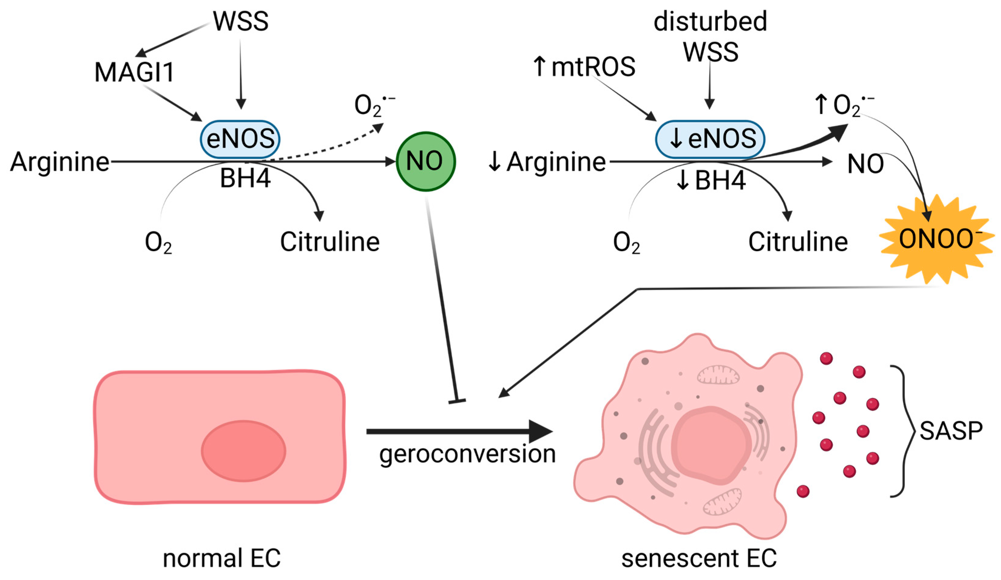
NO plays various roles beyond vasorelaxation, including influencing the maturation of endothelial progenitor cells, mitochondrial function, cell division, and inhibiting platelet aggregation and pro-inflammatory cytokine-induced signaling pathways. In short, NO protects against the factors contributing to endothelial senescence. It is generated from L-arginine by eNOS, which requires tetrahydrobiopterin (BH4) as a cofactor (Figure 2). The expression of eNOS is induced by shear stress [84], but its activity is inhibited by ROS or NG-nitro-L-arginine methyl ester (L-NAME). Furthermore, lack of BH4 or L-Arg can cause eNOS uncoupling and production of superoxide anion (O2•−) instead of NO—a major cause of endothelial senescence. A reaction between the superoxide anion and NO produces ONOO (peroxynitrite (PN)), further reducing NO’s bioavailability and promoting eNOS uncoupling and vascular dysfunction through OS [85]. PN also causes lipid peroxidation, protein oxidation and nitration, and LDL oxidation through Apolipoprotein E (ApoE). Inhibition of eNOS decreases the activity of human telomerase (hTERT) in HUVECs, making them susceptible to telomere-induced senescence [86]. Moreover, eNOS-KO mice experience premature cardiac aging and aortic stiffness, which is explained by increased calcium-dependent focal adhesion [87]. Although, PN-dependent Ca2+ influx in ECs leads to vascular dysfunction [88], when it is generated by shear stress, it activates eNOS through calcium-calmodulin complexes [89]. Undisturbed laminar flow also upregulates eNOS transcription in an ERK1/2- and NF-kB-dependent manner, contributing anti-inflammatory properties to NF-kB activation [90]. In addition, a novel eNOS modulator—MAGI1 (MAGUK with inverted domain structure-1), associated with VE-cadherin in cell-cell contacts—can support NO production under shear stress via PKA/AMPK-mediated mechanism [91]. Considering the vasoprotective properties of NO, eNOS activity under physiological flow can counteract endothelial dysfunction (Figure 2).

4. Unveiling the Interplay between Hypoxia and Oxidative Stress-Induced Endothelial Senescence

The main reason for O2’s negative manifestation is that it is responsible for the generation of reactive oxygen species (ROS), which cause DNA damage and induce senescence. It is still unclear whether there are different mechanisms of senescence activation, depending on the source of ROS and/or the place of accumulation [92]. For example, in CVD, mitochondrial dysfunction often triggers age-associated perturbations in the production of NO and VEGF [27,66], which can be mitigated by reduced mitochondrial oxidative phosphorylation in mammals [93]. On the other hand, mitochondrial ROS in the model organism Caenorhabditis elegans increases its longevity [94]. In addition, reduced mitochondrial mass and alterations in the electron transport chain (ETC) due to a decline in cytochrome C oxidase and Complex IV [95] and upregulated NADPH oxidases (NOX) increase OS and shorten telomeres [96]. The role of mitochondria in senescence was also confirmed by global transcriptomic analysis, where the expression of 38% of senescence-associated genes was reversed in mitochondrial-depleted fibroblasts [49]. A direct link between ROS, telomere shortening and senescence was evidenced by assessing the number of SA-β-Gal+ ECs after exposure to H2O2 or glutathione (GSH) peroxidase inhibitors (it should be noted that other senescence markers were not used) [96]. Since OS is a prominent contributor to endothelial senescence, it is natural to assume that low levels of O2 could prevent this process. Interestingly, low ROS delay DNA replication and cell cycle progression via a CDK2-dependent mechanism [97]. Therefore, lower ROS levels and prolonged cell division could potentially prevent replicative EC senescence due to excessive telomere shortening and reduced DNA damage. The following section further explores the interplay between hypoxia and OS-induced endothelial senescence.

4.1. HIF-1α in Angiogenesis

The excess O2 under typical in vitro experiments (20% pO2) generates a significant amount of ROS, making common culturing conditions hyperoxidative [92]. In contrast, human, bovine and murine fibroblasts grown under 5% pO2 increase their lifespan by 20%, 80% and up to 500%, respectively, due to significantly less OS [98,99,100]. Based on the available data, it is reasonable to consider that lower oxygen levels may reduce OS. This is not entirely true, because prolonged lack of O2 can seriously affect cellular metabolism and function, leading to tissue damage and organ failure if not treated promptly. This state is known as hypoxia and occurs when the body or a specific tissue or organ is deprived of adequate oxygen. However, short-term hypoxia can act as a hormetic stress (a short jump out of the individual’s comfort zone and subsequent quick recovery to homeostasis) and increase cell resilience (for a detailed review, see [101].)
Hypoxia orchestrates angiogenesis through the main pro-angiogenic factor—VEGF. Its expression is regulated by the three isoforms of HIF—1, 2 and 3α. They are under the control of prolyl-hydroxylases (PHDs), which target HIFs for proteasomal degradation but are sensitive to oxygen deprivation and are destroyed under hypoxic conditions. HIF-1α is ubiquitously expressed and responds to acute respiratory changes, whereas HIF-2α is responsible for chronic hypoxia and is localized in ECs and glial cells. Besides VEGF, other typical targets of HIF-1α and 2α are glucose transporter 1 (GLUT1) and lactate dehydrogenase A (LDHA). In addition, HIF-1α regulates the expression of erythropoietin (Epo) and Mmp-9, and HIF-2 that of Oct4 [102]. Besides acting as a transcription factor for VEGF, HIF-1α recruits endothelial progenitor cells from bone marrow and supports their differentiation into ECs, increases the expression of VEGF receptors (VEGFR1/2), stimulates the production and secretion of MMPs and recruits supporting cells to create mature and stable blood vessels [103]. Under hypoxic conditions, EVs carrying MMPs can also stimulate ECs to proliferate, migrate, and form capillary-like structures [104,105].

4.2. Hypoxia Is Essential for the Day–Night Cycle

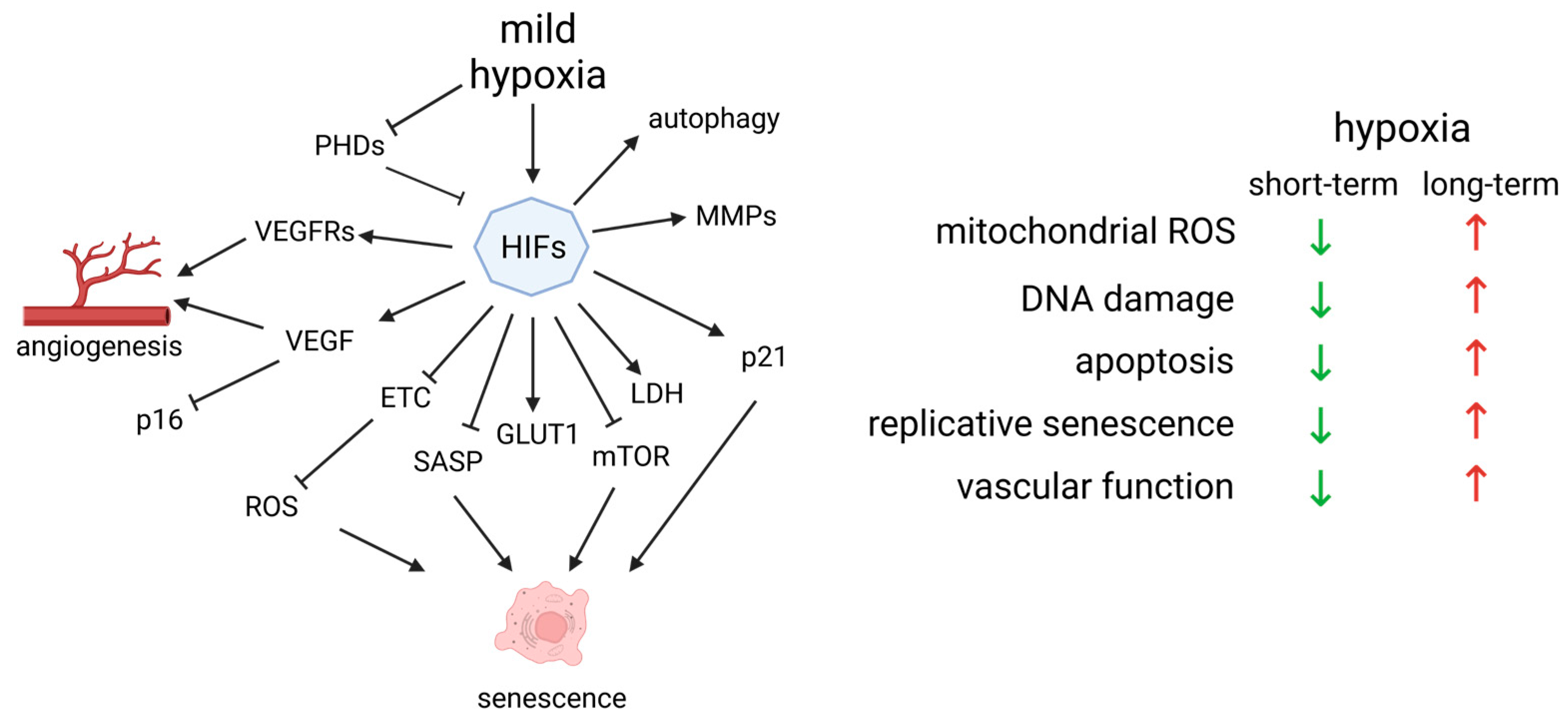
Although, HIF-1α is associated with poor prognosis in cancer and CVD, it works synergistically with one of the core regulators of the circadian rhythm—basic helix-loop-helix ARNT like 1 (BMAL1) [106]. In fact, hypoxia is so tightly related to the circadian clock that the incidence of heart attacks increases on the Monday following the daylight saving time transition [107]. Increase in HIF-1α during the day triggers the expression of the pro-angiogenic genes—Vegf, Epo and Glut1, but its persistence corresponds with the activation of pro-apoptotic genes—Bnip3 and Noxa1 in cells [106]. The circadian rhythm is also regulated by the hormone melatonin. It is synthesized predominantly by the pineal gland at night and is suppressed by bright light. Besides its role in the awake–sleep cycle, it is also one of the strongest known natural antioxidants. Melatonin supplementation can improve sleep and reduce jet lag, which is also observed with mild hypoxia [108]. Our recent studies demonstrated its ability to reduce OS and improve cognitive functions in a rat AD model [109]. Notably, short-term fluctuations in O2 activate autophagy, degrade damaged mitochondria and reduce mitochondrial ROS. Hence, there is an evolutionary pressure to adapt to moderately low O2 levels. In contrast, inhibition of autophagy during prolonged exposure to hypoxia increases the levels of ROS due to the uncoupling of complex III and generation of semiquinone (QH•). This results in O2•− formation and its conversion to H2O2 by superoxide dismutase (SOD) [110]. The generated H2O2 inhibits PDHs and indirectly stabilizes HIF-1α, causing chronic hypoxia [103] (Figure 3). Furthermore, inhibition of the ETC by hypoxia can lead to mitochondrial dysfunction and increased EV secretion, causing inflammation in many cell types [111].
During hypoxia, HIF-1α induces the expression of glycolytic enzymes, stimulates glycolysis and inhibits pyruvate dehydrogenase kinase 1 (PDK1), suppressing the pyruvate dehydrogenase complex (PDH) and the TCA cycle. As a result, mitochondrial respiration is reduced along with mitochondrial ROS and senescence [112]. However, disruption of the circadian clock impairs anaerobic glycolysis [113], causing acidification of the cells [114]. Lower pH redirects perinuclear lysosomes to the cytoplasm’s periphery, separating mTORC1 from its upstream activators, inhibiting its activity [114]. mTORC1 is an essential sensor for nutrients and feeding times, central to establishing the circadian rhythm. Hence, environmental factors such as the ATP/AMP ratio, NAD+ bioavailability and the overall redox state of the cell could prepare the organism for the light cycle as well as its metabolic response. Similarly, hypoxia can modulate the cell cycle arrest triggered by p16/Rb and provoke apoptosis resistance in SCs by elevating Bcl-2, Bcl-xL and p21 levels [60]. Even though p21 overexpression is connected with senescence, moderate levels promote cell survival, as p21-KO mice accumulate significant DNA damage and undergo apoptosis [115]. Hypoxia inhibits mTOR, which suppresses the conversion from p21-mediated cell cycle arrest to irreversible senescence [116], increases NO levels [117], inhibits NF-kB and decreases SASP [118], independent of p53 and HIF-1α. Inhibition of autophagy by mTOR can lead to insulin resistance, further increasing the concentration of glucose in blood plasma, subsequent OS and endothelial senescence. This demonstrates another possible mechanism through which mTOR inhibition can suppress aging [119] (Figure 3). Nevertheless, lower mitochondrial ROS failed to reduce senescence in hyperoxic conditions unless p53 and Rb were inhibited [120].

5. Exploring the Role of Extracellular Vesicles in Angiogenesis and Senescence

Legends about the infamous Hungarian Countess Elizabeth Bathory tell the story of her supposed anti-aging process of bathing in the blood of young girls. A similar idea governs the myths for vampires, which might not necessarily stay young, but become immortal by feeding on human blood. Surprisingly, there seems to be some truth in these myths, as recent studies showed that blood exchange from young to old mice rejuvenates them, but the opposite transfusion leads to senescence in the young [121,122,123]. The latter highlights the role of SASP in aging, which assists the immune response and, in the context of angiogenesis, influences new vessel formation. In addition to soluble factors, such as chemokines, inflammatory cytokines and growth factors, extracellular vesicles (EVs) are key components of SASP (reviewed in [124]). EVs are a very heterogeneous group of membranous structures, roughly categorized into three main groups based on size and origin: apoptotic bodies (ABs), microvesicles (MVs) that range from 50 to 5000 nm and are formed by outward budding and fission of the plasma membrane, and exosomes (30–100 nm) that are produced by the fusion of multivesicular endosomes with the plasma membrane, releasing intraluminal vesicles into the extracellular space. The EVs play important roles in intercellular communication, and their release is a strictly regulated process [125]. They are involved in both physiological and pathological processes and play a role in intercellular communication through the transfer of proteins, lipids, and nucleic acids [126,127]. EVs are implicated in cancer etiology due to their ability to promote cancer cell migration, transformation of non-malignant cells and pro-angiogenic activity [128]. While healthy cells release EVs as part of normal cellular homeostasis, senescent cells secrete EVs that have a significant role in angiogenesis and neurodegenerative disease progression. The presence of pro-angiogenic molecules like HIF-1α, VEGF, MMPs, and microRNAs in EVs [129] may lead to homeostasis disruption and non-productive angiogenesis. The role of EVs as key functional components of SASP is further highlighted by the observation that secretion of EVs is much higher in different types of senescent cells, including ECs, as compared to young ones [130,131]. A possible explanation for this is the observed upregulation of neutral sphingomyelinase and dysfunction of lysosomal activity in senescent cells [132]. One study even suggests that hypoxia prevents senescence by decreasing the SASP, rather than reducing the number of senescent cells [118].
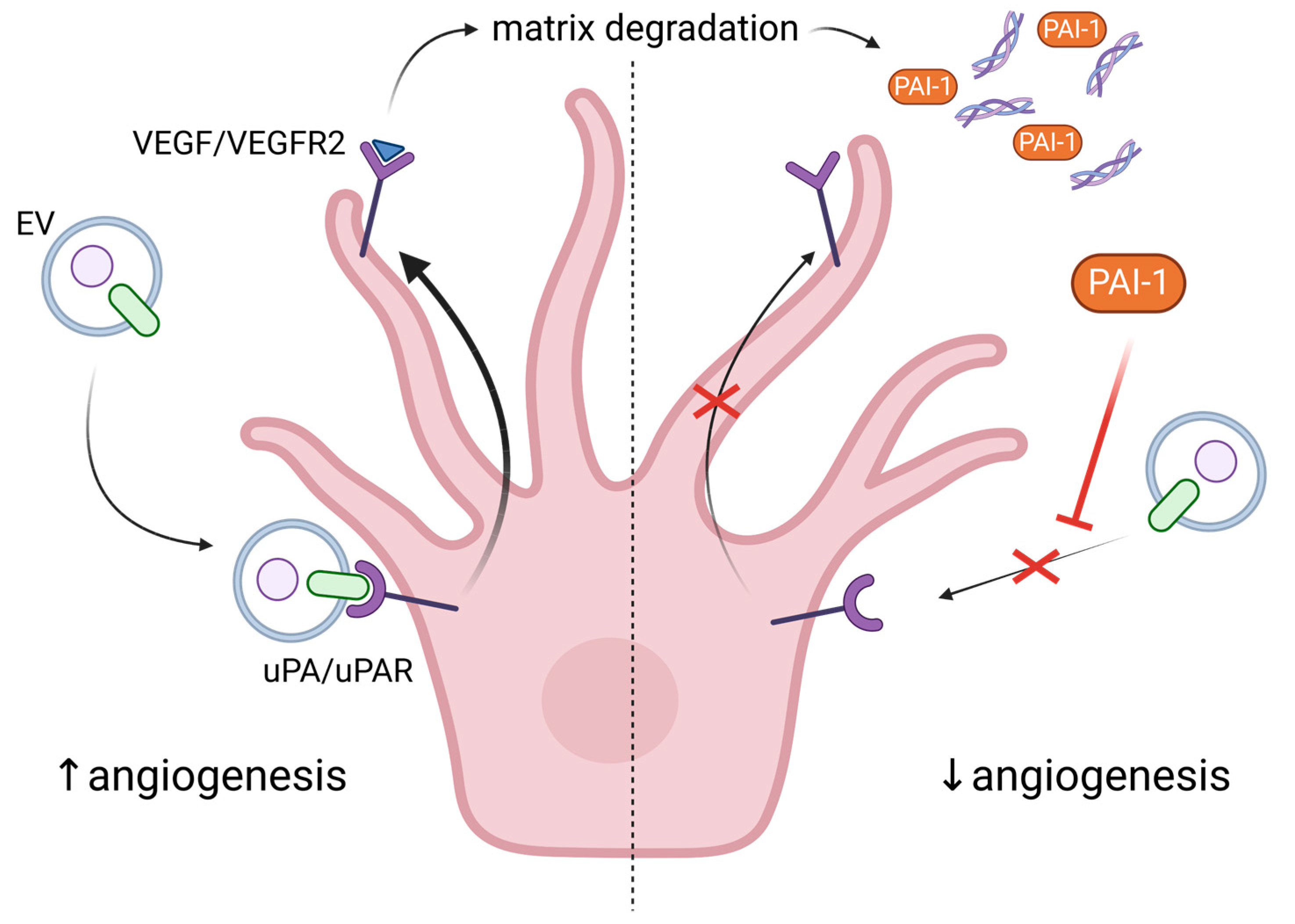
The important role of EVs from ECs, as well as other blood cell types, in angiogenesis is summarized here [125]. More specifically, EVs from ECs are rich in β1 integrins and metalloproteinases (MMP-2 and MMP-9), which allow them to penetrate the ECM, to remodel it and to form tubular capillary-like structures. Stimulation with VEGF and FGF-2 facilitates the association of the active and proenzyme forms of the MMPs with EC-derived vesicles [133]. EVs can also transport urokinase plasminogen activator/uPA receptor (uPA/uPAR), which are both pro-angiogenic. It was shown that uPAR modulates VEGF-induced EC migration by balancing the proteolysis of the ECM and the cell motility through integrin-associated focal adhesion (Figure 4). Revu Ann Alexander and colleagues demonstrated that VEGF causes endocytosis of αVβI integrin and activation of uPA/uPAR, resulting in matrix degradation [134]. Another active participant in this process is the inhibitor of uPA—plasminogen activator inhibitor (PAI-1), which is released from the degraded matrix and internalized, further directing the balance toward invasive cell migration, i.e., angiogenesis (Figure 4). Inhibition or deficiency of uPAR suppressed VEGF-induced angiogenesis in tumor cells [135] or in mice [136], respectively. Moreover, uPAR stimulated angiogenesis through VEGFR2, which upon internalization activates other pro-angiogenic stimuli [136]. In confluent ECs, the expression of uPAR is down-regulated compared to sub-confluent proliferative cells, thus preventing VEGF-activated signaling and angiogenesis [137]. In addition, levels of PAI-1 are elevated in senescent and aged ECs, making it a useful marker for senescence [138]. Besides inhibiting uPAR, PAI-1 also induces p53 and p21, activity that is suppressed by SIRT1 overexpression in endothelial cells. SIRT1 is also able to induce eNOS activity, protecting ECs from endothelial dysfunction [138]. The pro-angiogenic properties of exosomes from ECs may also be attributed to EV-associated micro RNAs such as miR-214 [139]. More specifically, the latter prevents senescence through silencing ATM in recipient cells.
Depending on their source and the specific experimental conditions, EVs may also have anti-angiogenic properties. For example, NO production and angiogenesis are impaired by EC-derived EVs under oxidative stress, via Src kinase- and NOX-dependent mechanisms [66,140,141]. Moreover, in contrast to EVs from young cells, those derived from senescent cells exert mostly negative effects on EC functions and angiogenesis. More specifically, senescent osteoblasts secrete EVs that induce senescence and apoptosis and decrease proliferation of ECs through transfer of miR-139-5p [142]. Likewise, senescent HUVEC cells secrete exosomes that interfere with cell growth and downregulate expression of adherent junction proteins, resulting in impaired endothelial migration of young ECs and endothelial barrier dysfunction [143].
Interestingly, the effect of EVs on angiogenesis may be swayed in opposite directions depending on the dose. Namely, it was found that a low dose of EVs exhibited pro-angiogenic activity, which was suppressed below control levels upon increasing the concentration of the EC-derived EVs [144]—an effect dependent on uPA activity. The inhibitory effect of EC-derived EVs on endothelial cell tube formation was confirmed by another study in which even higher concentrations of EVs were used, and it was shown that the inhibition was dependent on NF-kB signaling and eNOS pathway suppression [145]. EVs are carriers of damaged genomic DNA molecules whose concentration increases in EVs upon induction of senescence [130] and under pathological conditions [146]. Functioning as intercellular vectors, EVs may transfer their DNA into the cytoplasm of recipient cells, leading to activation of the cGAS-STING signaling and consequently EC senescence, eNOS suppression and endothelial dysfunction [147]. Therefore, the hormetic effect of EC-derived EVs on EC tube formation, as well as the inhibitory effect of EVs from senescent ECs on angiogenesis, may possibly be due to EV DNA-induced cGAS-STING activation. Shedding more light on these processes and mechanisms would be a particularly interesting direction for further studies.

6. The Non-Productive Angiogenesis in Alzheimer’s Disease

Currently, there are two main hypotheses for the development of AD—the accumulation of amyloid plaques (Aβ) due to an error in the metabolism of the amyloid precursor protein (APP); and the hyperphosphorylation of Tau (or p-Tau), resulting in microtubule polymerization catastrophe and formation of fibrils [16]. APP is a transmembrane glycoprotein separated into an intracellular C-terminal, Aβ transmembrane and N-terminal extracellular domains. Its primary function is interneuronal communication, and once it performs it, APP is degraded by α- and γ-secretase to a soluble, non-amyloid form, or by β- and γ-secretase to insoluble Aβ1–40 and Aβ1–42 isoforms [16]. In animal models, elevated levels of Aβ1–42 and p-Tau were correlated with cerebrovascular dysfunction, chronic hypoperfusion and worsened AD symptoms [148,149]. One of the most affected brain areas in AD is the hippocampus, which is normally able to continue with adult neurogenesis. Thus, a decline in neurogenesis could be used as a marker for AD progression in animal models [150]. In fact, we demonstrated worsened long-term memory and anxiety in a rat model of icvAβ1–42 concomitant with pinealectomy (AD with melatonin deficiency). These behaviors are controlled by the hippocampus and corresponded with increased OS in the structure [108].
Pro-inflammatory cytokines, such as interleukin-1β, become abundant during AD and induce the expression of VEGF, yielding new blood vessels [18]. Although angiogenesis is initiated around Aβ plaques, the process is non-productive, leading to the disassembly of Aβ plaque-associated blood vessels and the phagocytic activity of microglia [151]. However, there is conflicting evidence relating the cause of AD and whether there is an increase or decrease in blood vessel density [151,152,153,154,155,156,157,158] (Table 2).
Joe Steinman, Hong-Shuo Sun and Zhong-Ping Feng provide a reasonable explanation for the discrepancies—“An overall measure of vessel density may indicate loss of vessels due to holes [note: from plaque deposits], without accounting for the increase in vessels surrounding holes” [159]. Although angiogenesis might not be beneficial for AD’s progression, it seems to alleviate some of the cognitive disabilities. For instance, one study showed that AD patients and AD mouse models accumulated Aβ in arterioles and experienced apoptosis of ECs [157]. When the same mouse model TgCRND8 was treated with VEGF, the growth factor was able to rescue vascular loss. And, most importantly, it significantly improved the behavior and memory of the subjects [157]. However, this observation could not be repeated in vitro on Matrigel®, where Aβ maintained low vascular density regardless of VEGF in tube formation assays, demonstrating the inability to always correlate in vivo and in vitro studies. A natural way to suppress Aβ accumulation is through melatonin. Besides its function as a radical scavenger, research shows that melatonin disrupts amyloid fibril formation [20] and exhibits anti-angiogenic properties [160,161]. Thus, by hindering Aβ plaque formation and reducing OS, melatonin deflects their role in non-productive angiogenesis and endothelial senescence. Taken together, these observations support the use of the hormone as an adjuvant therapy in AD.

6.1. How Does Aβ Stimulate Cerebral Angiogenesis?

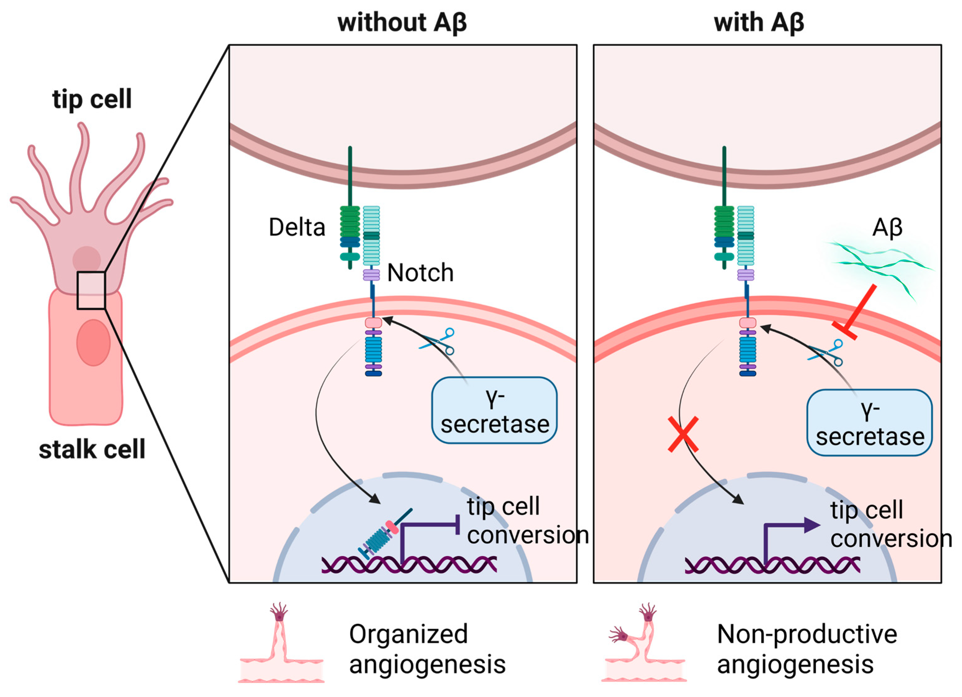
As Aβ is produced by β- and γ-secretase from APP, there have been attempts to reduce Aβ production via enzyme inhibition. Unfortunately, this has led to dense and highly branched blood vessels. Cameron et al. [151] demonstrated that treatment of HUVECs and zebrafish with Aβ monomers and γ-secretase inhibitors increases the number of tip ECs and suggested an alternative mechanism through Delta-Notch signaling. Tip cell formation is supported by the interaction between EV-associated Delta like 4 (Dll4) and cell membrane-localized Notch, which restricts excessive sprouting angiogenesis, ensuring that only a limited number of cells will identify as tip cells and initiate new vessel formation [162]. Here, NAD+’s role is to improve VEGF sensitivity of tip cells and stimulate their proliferation, migration and ECM invasion, while hampering the transcriptional activity of Notch in the nucleus of stalk cells. Therefore, the latter cannot assume the functions of a tip cell and produce unnecessary sprouts [163]. Upon monomer binding to the Notch extracellular domain (NEXT), the same is cleaved to Notch intracellular domain (NICD) by γ-secretase and suppresses tip cell formation and hypervascularization. In the case of AD, Aβ competes with NEXT for proteolysis and counteracts the inhibition of neovascularization (Figure 5).
Excessive angiogenesis is observed with γ-secretase inhibitors [151], while immunization against Aβ protects against amyloid-Notch-induced vessel formation [164]. This non-productive angiogenesis exposes AD patients to a significant risk of cerebral amyloid angiopathy (CAA) [10,11,159]. CAA is usually caused by Aβ accumulation in the small arteries and capillaries in the brain, leading to chronic hypoxia, microaneurysms and dementia. As a confirmation, a study using magnetic resonance angiography showed disturbed blood flow in an 11-month-old APP23 transgenic mouse AD model compared to 20-month-old WT mice [11]. The researchers could not detect Aβ accumulation in larger arteries and assumed that the blood vessel disruption was due to soluble Aβ monomers. On the other hand, Aβ monomers were shown to exhibit pro-angiogenic effects, while Aβ oligomers triggered senescence in ECs through the p53/p21 pathway [155]. Aβ1–42 monomers or fibrils had no effect on p21; instead, they increased VEGFR1 and decreased VEGFR2 expression. The overexpression of VEGFR1 readily induced senescence in brain MVs. At the same time, siRNA against VEGFR1 prevented upregulation of p21 upon Aβ1–42 oligomer treatment. While some studies have proposed that VEGF could be sequestered in Aβ plaques, Alvarez-Vergara et al. [153] observed high expression of VEGF in astrocytes surrounding Aβ plaques and an association between VEGF expression and the protrusion of filopodia from endothelial cells in mouse models. The integrin αVβIII was also concentrated around the plaques, indicating vascular remodeling. A similar conclusion was drawn from single-nucleus transcriptome analysis of AD patients, which showed an induction of a subpopulation of ECs with increased expression of growth factors and their receptors [165]. In addition, the examined ECs acted as antigen-presenting cells by MHC-I, which typically indicates viral infections. It is reasonable for Aβ deposits to induce a pro-inflammatory response, but why MHC-I is involved in this process remains unclear.
Cells or animals can be genetically engineered with mutated APP or Tau proteins to generate in vitro and in vivo models of AD. Alternatively, AD can be induced by exogenous treatment with purified Aβ peptides. Even though, these peptides can be of various lengths and modifications, once they are solubilized, they are not stable, and improper handling can negatively impact the acquired results. Initially, the peptides turn into monomers, which associate together, forming oligomers, and finally to amyloid plaques, and all of these transitions are spontaneous in a water solution. Thus, the type of Aβ used for treatment should be validated and explicitly stated in a study, because the cells’ response varies depending on the applied Aβ form. Moreover, tissue samples acquired postmortem from animal models or AD patients must be stored and prepared adequately as soon as possible since many macromolecules can deteriorate and give false data in later examinations.

6.2. EVs Can Be Used as Biomarkers for Early AD Detection

EVs, and more specifically—exosomes, isolated from all sorts of AD model systems are shown to carry Aβ (or APP) along with exosome markers such as Flotillin-1 and Alix [166,167,168,169]. Moreover, some studies demonstrate a prion-like toxic activity of Aβ-carrying exosomes, where shortly after being endocytosed, Aβ starts to propagate and induce cell death [166,167,170]. Along these lines, many studies have attempted to use EVs as biomarkers for early detection and prognosis of neurodegeneration. In a recent study, Gallart-Palau et al. used brain EVs in the progressive course of AD and performed a proteome-wide analysis [171]. They found damaged mitochondria, APP and prion proteins (PrP) in EVs from the temporal lobe of AD patients due to impaired autophagy. What is interesting is that they rebutted the hypothesis that PrP and APP together exhibit neurotoxicity. Instead, their results show a co-upregulation of both PrP and APP at the preclinical stage of the disease, where PrP binds with APP and helps to sequester it in brain EVs (in agreement with another study [172]). Unfortunately, this protective mechanism deteriorates and is inefficient at the clinical stage of AD [171]. Nevertheless, these and many other studies demonstrate the role of EVs in disease progression and their potential as biomarkers [168,173,174,175]. It is unclear whether the size of EVs determines their mode of distribution between neuronal cells. Gabrielli and colleagues propose a mechanism according to which small EVs are endocytosed and spread their pathologic cargo trans-synaptically, whereas larger EVs move along the surface of axons, jump between connected neurons, and finally activate a signal and/or become internalized at synaptic sites [174]. In AD, hypoxia impedes the autophagy in neurons, causing the release of EVs carrying dysfunctional mitochondria and APP. While these hypoxic EVs can exhibit pro-neurodegenerative function, they can also supply mediators of hypoxia adaptation, angiogenesis and protein quality control [176].

6.3. The Role of Endothelial Progenitor Cells (EPCs) as a Biomarker and Potential Therapeutic Target in AD

There is increasing evidence that points to the alteration and dysfunction of the cerebral vasculature as an important factor in assessing the pathophysiology of AD, and this process may contribute to the onset of neurodegeneration, inflammation, Aβ accumulation and tau phosphorylation [177,178]. The so called two-hit vascular hypothesis proposed by Zlokovic and co-workers [179] suggested that damage in the cerebral vasculature (hit one) induces the accumulation of Aβ in the brain (hit two). In this respect, endothelial progenitor cells (EPCs) appear as a possible biomarker for early detection of AD as well as a therapeutic target given their role in maintaining the vasculature. EPCs, which are a rare population of cells originated from the bone marrow [180], circulate in the peripheral blood and have a capacity to repair or replace the damaged vessels. The most characteristic surface markers of EPCs are CD34, VEGF receptor 2 (VEGFR-2), and CD133. There are two different types of EPCs: (a) early-outgrowth EPCs (e-EPCs), circulatory angiogenic cells, or colony-forming unit endothelial cells (CFU-EC), which take part in the process of network formation and the repair of injured endothelial cells in a paracrine way by secreting different angiogenic factors; and (b) late-outgrowth EPCs (l-EPCs), endothelial outgrowth cells, or endothelial colony-forming cells, which improve angiogenesis by differentiating into mature endothelial cells [181]. In addition to their different functionality, both types of EPCs can be recognized by their appearance in vitro. Whereas e-EPCs appear after a few days in culture and form colonies with spindle-shaped cells around them, l-EPCs appear after 2–3 weeks in culture and present a cobblestone shape [182].
EPCs as prognostic markers—The number of EPCs and their ability to form CFU-EC colonies has been proposed as a possible marker of vascular function in AD [183]. In a clinical study of AD patients, Kong and co-workers [184] observed reduced numbers of circulating EPCs compared to healthy patients and that lower numbers of EPCs correlated with greater cognitive impairment. In addition, EPCs from moderate and severe AD showed functional alterations in culture (such as reduced adhesion and migration capacity) compared to mild AD and controls [183,185].
EPCs as therapeutic target—Additionally to the analysis of the number of EPCs in AD, the therapeutic potential of these cells has also been suggested in different animal models. For instance, when e-EPCs were injected intravenously into repeated scopolamine (SCO)-induced cognitive impaired rats, it resulted in improved learning and memory, attenuation of Aβ plaque deposition, as well as suppression of Aβ and p-tau levels. Similarly, when l-EPCs were injected intravenously into APP/PS1 transgenic mice, researchers observed an enhanced penetration of exogenous EPCs into the brain compared to controls. Subsequently, if l-EPCs were injected directly into the hippocampus of the same transgenic mouse model [186], they could lead to up-regulation of tight junction proteins (such as zonula occludens-1, occludin, and claudin-5) in the BBB, increasing microvessel density and promoting angiogenesis in the hippocampus and cortex. In addition, EPCs also showed an anti-apoptotic effect, promoting neuronal survival in the hippocampus. Other effects of EPC transplantation were the inspected reduction in the area and intensity of Aβ plaques in the hippocampus and cerebral cortex and significantly improved learning and memory in AD mice (APP/PS1). Recently, transfected EPCs that release antibodies against Aβ and reduce its aggregation have been generated [187]. Therefore, EPCs are postulated as a good therapeutic option for pathologies that present BBB alterations.

7. Therapeutic Approaches to Endothelial Senescence and Dysfunction

Understanding the underlying mechanism of aging and neurodegenerative diseases will one day provide us with the means to treat them. Along with DNA damage, OS, and insufficient or disturbed blood flow, behavioral and social cues guiding unhealthy lifestyle choices also accelerate the aging process. It is urban knowledge that chronic stress with high cortisol levels, high-calorie food, lack of exercise, etc. worsens life quality and expectancy. As presented in this review, regular exposure to hormetic stress can substantially improve vascular fitness, while properly controlled angiogenesis could delay both aging and neurodegenerative processes. Indeed, many approaches entail exercise and caloric restriction (CR) as therapies for vascular health instead of drug treatment.

7.1. Exercise Improves CBF, Vascular Function and Cognitive Performance

Angiogenesis in the brain microvasculature can improve tissue oxygenation, but if done improperly, it can provoke vascular leakage and neurodegeneration. A way to ensure positive angiogenesis is exercise, which stimulates eNOS by increasing the CBF [188] and potentially reduces OS by hypoxia-mediated inhibition of oxidative phosphorylation. In addition, aerobic exercise increases energy consumption (mimicking CR), while alleviating basal membrane dysfunction [189] and age-related behavior changes [190].
In an eight-week comparative study between old sedentary and exercised male rats, moderate exercise decreased the mean arterial blood pressure in favor of the trained group [188]. It also improved CBF, VEGF, eNOS expression, capillary density and astrocyte growth [188,191,192]. Furthermore, malondialdehyde (MDA—a marker for lipid peroxidation) levels were reduced in the exercised aged group [188]. Exercise also reduced the levels of fibrin and fibrinogen in old mice, improving the activity of neurovascular units (microvascular ECs, basement membrane, pericytes and astrocytes) [189]. Increased CBF, by regular treadmill running, prevented the loss of BDNF, which usually leads to learning and memory deficiencies [190]. Aerobic running on a treadmill or cycling induces EV secretion before reaching an anaerobic state [193]. In contrast, Brahmer et al. collected EV samples of athletes before, during and after cycling to exhaustion [194]. They observed a significant increase in CD63+ EVs post exercise (at the highest lactate levels), with some also carrying CD105 and CD146 (markers for ECs). Thus, exercise itself rather than the intensity influences EV release. The EV release is very likely to be Ca2+-dependent, and since muscle activation leads to Ca2+ flux, this could be a potential cause of EV accumulation [195]. Meanwhile, Ca2+ signaling is impaired in senescent ECs and impedes the contraction of vascular smooth muscle cells in mesenteric arteries of aged (24–26 month old) mice [196]. Taken together, these findings support speculation that the increase in plasma Ca2+ due to exercise could improve the vasomotor control of the arteries. Furthermore, exercise-induced moderate hypoxia causes metabolic conversion to anaerobic glycolysis, securing NAD+ availability when the TCA cycle and the ETC are subdued. The resulting buildup of lactate provokes the expression of VEGFR2 in ECs [197] and stimulates reparative angiogenesis in ischemic tissues [198]. Furthermore, lactate secreted by skeletal muscle can travel through the blood and bind to the lactate receptor HCAR1, enriched in cells lining the brain’s blood vessels, inducing VEGF expression and cerebral angiogenesis [191]. This was positively impacted by high-intensity interval training (HIIT) or lactate injections and led to increased capillary density in the brain of WT mice and not in HCAR1-KO. The authors linked this effect with the activation of ERK1/2 and Akt, which are upstream positive regulators of VEGF [191]. In general, physical activity improves physical and cognitive function by enhancing CBF and reducing OS, neuroinflammation and vascular dysfunction, and positively impacts AD’s symptoms.

7.2. Caloric Restriction Reduces OS and Vascular Aging

Already, Ciceron has suggested that moderate eating and exercise are key factors for longevity. Therefore, caloric restriction (CR) could be beneficial for people, as it activates autophagy and triggers the cells to recycle and renew themselves [199]. Under CR, high temperatures or excessive competition, C. elegans undergoes a dramatic metamorphosis into a dauer form. In this state, the worms close their mouth apparatus, switch their metabolism from the TCA cycle to gluconeogenesis and seize their development until food becomes available. The incredible thing is that dauers live at least twice as long compared to adult worms [200]. This is one of the reasons why C. elegans is the go-to system when studying senescence. However, the restricted activity of mitochondria ultimately leads to their deterioration [94]. In a recent study, mice meeting their caloric needs but consuming less protein and branched fatty acids had lower adiposity, higher metabolic rates and lifespans [201]. The authors attributed this to lower activation of mTORC1 by amino and fatty acids, rather than CR itself. With aging, mTORC1 is upregulated, which correlates with eNOS uncoupling and O2•− generation, which are significantly reduced in senescent ECs treated with rapamycin (an mTOR inhibitor) [202] and in old mice under CR diet [203]. A detailed review by Christopher R. Martens and Douglas R. Seals describes other stress-induced cellular mechanisms inhibited in senile ECs—NO synthesis mediated by AMPK and SIRT1, autophagy (detailed review of autophagy factors promoting longevity [204]), and ECM stiffening through elastin proteolysis by MMP-9 and AGEs-induced inflammation of the arterial wall that can be ameliorated by CR [205]. Furthermore, the activity of SIRTs as histone deacetylases, hence, the epigenetic regulation of senescence and aging, is promoted by CR [178]. Although there is substantial evidence that CR can reduce and delay the deteriorating effects of aging and maintain our longevity, more controlled research is necessary to establish good CR protocols accounting for personal needs.

7.3. Role of Resveratrol in the Vascular Biology and Senescence Process

In general antioxidants such as reduced glutathione (GSH) and melatonin inhibit cell senescence by reducing reactive oxygen species (ROS) generation [206]. Resveratrol (3,5,40-trihydroxystilbene) (R), which is a non-flavonoid polyphenolic compound and derivative of stilbene, exhibits its pleotropic function also by decreasing ROS production and improving the antioxidant levels [207]. As mentioned above, EPCs are critical circulating components of the endothelium and are identified as key factors in endothelial repair. In this respect, resveratrol treatment can reverse EPC dysfunction by decreasing oxidative stress and increasing proliferation and capillary-like structure formation, and, by increasing the angiogenic factors like (NO), can reverse stress-induced senescence [208].

8. Conclusions

The study of angiogenesis in the context of endothelial senescence, aging and Alzheimer’s disease has revealed their intricate complexity and heterogeneity. Even though, senescent cells can trigger inflammation, they can also support tissue renewal in the adult organism. Their dual role depends on the time, place and degree of their accumulation. The induction and outcome of endothelial senescence can vary across different cell types, but it underlies vascular dysfunction and subsequent non-productive angiogenesis and vascular leakage. Short hormetic stress employed on blood vessels by hypoxia, metabolism switch or high shear stress can reduce OS and improve EC responsiveness to angiogenic stimuli and cognitive function. Identifying universal senescence markers remains a challenge, and careful selection and consideration of their limitations are crucial for accurate research conclusions. Furthermore, senile ECs secrete SASP factors that can accelerate aging and neurodegeneration through induced inflammation. As a key SASP component, EVs can be used as biomarkers for the early detection of AD, and the development of standardized repositories for SASP markers could enhance their application and reproducibility. Furthermore, studying neurodegenerative diseases and angiogenesis, researchers must choose suitable models and consider factors such as the type of Aβ peptide and endothelial cell line since the responses can vary significantly. Innovative in vitro and in vivo models could provide more physiologically relevant insights. Addressing these considerations will contribute to advancing our understanding of endothelial senescence and related processes.

Author Contributions

Conceptualization, I.G. and R.T.; investigation, I.G.; resources, I.G., D.I. and R.T.; writing—original draft preparation, I.G.; writing—review and editing, I.G., J.T., D.I. and R.T.; project administration, I.G. and R.T.; funding acquisition, I.G. and R.T. All authors have read and agreed to the published version of the manuscript.

Funding

This work was supported by the National Science Fund of Bulgaria (research grants # KП-06-H31/16 and KП-06-M61/5).

Institutional Review Board Statement

Not applicable.

Informed Consent Statement

Not applicable.

Data Availability Statement

Not applicable.

Acknowledgments

All figures were created with https://www.BioRender.com (accessed on 30 May 2023) and sublicensed for use in journal publications under the Academic License Terms.

Conflicts of Interest

The authors declare no conflict of interest.

Abbreviations

(R)AGE(Receptor for) Advanced Glycation End products
ADAlzheimer’s disease
APCsAntigen Presenting Cells
ApoEApolipoprotein E
APPAmyloid Precursor Protein
ATMAtaxia-telangiectasia mutated protein kinase
Beta-amyloid protein
BAECsBovine Aortic Endothelial Cells
BBBBlood-Brain Barrier
BH4Tetrahydrobiopterin
CAACerebral Amyloid Angiopathy
CBFCerebral Blood Flow
CRCaloric Restriction
CVDCardio Vascular Disease
DDRDNA Damage Response
Dll4Delta-like 4 protein
ECMExtracellular Matrix
ECsEndothelial cells
eNOSEndothelial nitric oxide synthase
EPOErythropoietin
ETCElectron Transport Chain
EVsExtracellular Vesicles
GLUT1Glucose Transporter 1
GSHGlutathione
H(B)MVECsHuman (Brain) Microvascular Endothelial Cells
HIFHypoxia-inducible factor
HUVECsHuman Umbilical Vein Endothelial Cells
KOKnock-out
LDHLactate Dehydrogenase
MMPMetalloproteinase
mTORMammalian Target of Rapamycin
NAD+Nicotinamide Adenine Dinucleotide
NEXTNotch extracellular domain
NF-kBNuclear Factor–kappa Beta
NICDNotch intracellular domain
NMNNicotinamide Mononucleotide
NONitric oxide
NOXNADPH oxidases
ONOO− (PN)Peroxynitrite
OSOxidative Stress
PAI-1plasminogen activator inhibitor
PARP1Poly (ADP-ribose) polymerase 1
PHDProlyl hydroxylases
PrPPrion protein
p-TauPhosphorylated-Tau
ROSReactive Oxygen Species
SASPSenescence-Associated Secretory Phenotype
SA-β-GalSenescence-Associated β-galactosidase
SCsSenescent cells
sen-ECsSenescent endothelial cells
SIRTSirtuins
SODSuperoxide dismutase
TCAThree Carboxylic Acid cycle
uPA(R)urokinase plasminogen activator/uPA receptor
VEGFVascular Endothelial Growth Factor
VEGFR1(2)Vascular Endothelial Growth Factor Receptor 1(2)
WSSWall Shear Stress
WTWild Type

References

  1. Patel-Hett, S.; D’Amore, P.A. Signal transduction in vasculogenesis and developmental angiogenesis. Int. J. Dev. Biol. 2011, 55, 353–363. [Google Scholar] [CrossRef] [PubMed] [Green Version]
  2. Adair, T.H.; Montani, J.-P. Overview of Angiogenesis. In Angiogenesis; Morgan & Claypool Life Sciences: San Rafael, CA, USA, 2010. [Google Scholar]
  3. Weinstein, N.; Mendoza, L.; Gitler, I.; Klapp, J. A network model to explore the effect of the micro-environment on endothelial cell behavior during angiogenesis. Front. Physiol. 2017, 8, 960. [Google Scholar] [CrossRef] [PubMed] [Green Version]
  4. Brassard-Jollive, N.; Monnot, C.; Muller, L.; Germain, S. In vitro 3D Systems to Model Tumor Angiogenesis and Interactions With Stromal Cells. Front. Cell Dev. Biol. 2020, 8, 594903. [Google Scholar] [CrossRef]
  5. Stryker, Z.I.; Rajabi, M.; Davis, P.J.; Mousa, S.A. Evaluation of angiogenesis assays. Biomedicines 2019, 7, 37. [Google Scholar] [CrossRef] [PubMed] [Green Version]
  6. Lähteenvuo, J.; Rosenzweig, A. Effects of aging on angiogenesis. Circ. Res. 2012, 110, 1252–1263. [Google Scholar] [CrossRef] [PubMed]
  7. Ungvari, Z.; Tarantini, S.; Kiss, T.; Wren, J.D.; Giles, C.B.; Griffin, C.T.; Murfee, W.L.; Pacher, P.; Csiszar, A. Endothelial dysfunction and angiogenesis impairment in the ageing vasculature. Nat. Rev. Cardiol. 2018, 15, 555–565. [Google Scholar] [CrossRef]
  8. Carmeliet, P.; Jain, R.K. Molecular mechanisms and clinical applications of angiogenesis. Nature 2011, 473, 298–307. [Google Scholar] [CrossRef] [Green Version]
  9. Ambrose, C.T. Pro-Angiogenesis Therapy and Aging: A Mini-Review. Gerontology 2017, 63, 393–400. [Google Scholar] [CrossRef] [Green Version]
  10. Vinters, H.V.; Gilbert, J.J. Cerebral amyloid angiopathy: Incidence and complications in the aging brain. II. The distribution of amyloid vascular changes. Stroke 1983, 14, 924–928. [Google Scholar] [CrossRef] [Green Version]
  11. Beckmann, N.; Schuler, A.; Mueggler, T.; Meyer, E.P.; Wiederhold, K.H.; Staufenbiel, M.; Krucker, T. Age-Dependent Cerebrovascular Abnormalities and Blood Flow Disturbances in APP23 Mice Modeling Alzheimer’s Disease. J. Neurosci. 2003, 23, 8453–8459. [Google Scholar] [CrossRef]
  12. Childs, B.G.; Baker, D.J.; Kirkland, J.L.; Campisi, J.; Van Deursen, J.M. Senescence and apoptosis: Dueling or complementary cell fates? EMBO Rep. 2014, 15, 1139–1153. [Google Scholar] [CrossRef] [Green Version]
  13. Singh, C.; Pfeifer, C.G.; Jefferies, W.A. Pathogenic Angiogenic Mechanisms in Alzheimer’s Disease. In Physiologic and Pathologic Angiogenesis-Signaling Mechanisms and Targeted Therapy; BoD–Books on Demand: Norderstedt, Germany, 2017. [Google Scholar] [CrossRef] [Green Version]
  14. Bradaric, B.D.; Patel, A.; Schneider, J.A.; Carvey, P.M.; Hendey, B. Evidence for Angiogenesis in Parkinson’s disease, Incidental Lewy Body disease, and Progressive Supranuclear Palsy. J. Neural Transm. 2012, 119, 59. [Google Scholar] [CrossRef] [Green Version]
  15. Ellison, S.M.; Trabalza, A.; Tisato, V.; Pazarentzos, E.; Lee, S.; Papadaki, V.; Goniotaki, D.; Morgan, S.; Mirzaei, N.; Mazarakis, N.D. Dose-dependent Neuroprotection of VEGF165 in Huntington’s DiseaseStriatum. Mol. Ther. 2013, 21, 1862. [Google Scholar] [CrossRef] [Green Version]
  16. Fontana, I.C.; Zimmer, A.R.; Rocha, A.S.; Gosmann, G.; Souza, D.O.; Lourenco, M.V.; Ferreira, S.T.; Zimmer, E.R. Amyloid-β oligomers in cellular models of Alzheimer’s disease. J. Neurochem. 2020, 155, 348–369. [Google Scholar] [CrossRef] [Green Version]
  17. Butterfield, D.A.; Halliwell, B. Oxidative stress, dysfunctional glucose metabolism and Alzheimer disease. Nat. Rev. Neurosci. 2019, 20, 148–160. [Google Scholar] [CrossRef]
  18. Jefferies, W.A.; Price, K.A.; Biron, K.E.; Fenninger, F.; Pfeifer, C.G.; Dickstein, D.L. Adjusting the compass: New insights into the role of angiogenesis in Alzheimer’s disease. Alzheimer’s Res. Ther. 2013, 5, 64–69. [Google Scholar] [CrossRef] [Green Version]
  19. Tchekalarova, J.; Tzoneva, R. Oxidative Stress and Aging as Risk Factors for Alzheimer’s Disease and Parkinson’s Disease: The Role of the Antioxidant Melatonin. Int. J. Mol. Sci. 2023, 24, 3022. [Google Scholar] [CrossRef]
  20. Tadokoro, K.; Ohta, Y.; Inufusa, H.; Loon, A.F.N.; Abe, K. Prevention of Cognitive Decline in Alzheimer’s Disease by Novel Antioxidative Supplements. Int. J. Mol. Sci. 2020, 21, 1974. [Google Scholar] [CrossRef] [Green Version]
  21. Greenberg, D.A.; Jin, K. From angiogenesis to neuropathology. Nature 2005, 438, 954–959. [Google Scholar] [CrossRef]
  22. Ribatti, D.; Guidolin, D. Morphogenesis of vascular and neuronal networks and the relationships between their remodeling processes. Brain Res. Bull. 2022, 186, 62–69. [Google Scholar] [CrossRef]
  23. Hayflick, L.; Moorhead, P.S. The serial cultivation of human diploid cell strains. Exp. Cell Res. 1961, 25, 585–621. [Google Scholar] [CrossRef] [PubMed]
  24. López-Otín, C.; Blasco, M.A.; Partridge, L.; Serrano, M.; Kroemer, G. The hallmarks of aging. Cell 2013, 153, 1194. [Google Scholar] [CrossRef] [PubMed] [Green Version]
  25. Schmauck-Medina, T.; Molière, A.; Lautrup, S.; Zhang, J.; Chlopicki, S.; Madsen, H.B.; Cao, S.; Soendenbroe, C.; Mansell, E.; Vestergaard, M.B.; et al. New hallmarks of ageing: A 2022 Copenhagen ageing meeting summary. Aging 2022, 14, 6829–6839. [Google Scholar] [CrossRef] [PubMed]
  26. Kumari, R.; Jat, P. Mechanisms of Cellular Senescence: Cell Cycle Arrest and Senescence Associated Secretory Phenotype. Front. Cell Dev. Biol. 2021, 9, 485. [Google Scholar] [CrossRef]
  27. Han, Y.; Kim, S.Y. Endothelial senescence in vascular diseases: Current understanding and future opportunities in senotherapeutics. Exp. Mol. Med. 2023, 55, 1–12. [Google Scholar] [CrossRef]
  28. Ramos-Ibeas, P.; Gimeno, I.; Cañón-Beltrán, K.; Gutiérrez-Adán, A.; Rizos, D.; Gómez, E. Senescence and Apoptosis During in vitro Embryo Development in a Bovine Model. Front. Cell Dev. Biol. 2020, 8, 1646. [Google Scholar] [CrossRef]
  29. De-Carvalho, D.P.; Jacinto, A.; Saúde, L. The right time for senescence. Elife 2021, 10, e72449. [Google Scholar] [CrossRef]
  30. Vujin, A.; Dick, K. The information theory of aging: Hacking immortality? Health Sci. Inq. 2020, 11, 148–154. [Google Scholar] [CrossRef]
  31. Harley, C.B.; Vaziri, H.; Counter, C.M.; Allsopp, R.C. The telomere hypothesis of cellular aging. Exp. Gerontol. 1992, 27, 375–382. [Google Scholar] [CrossRef]
  32. Harman, D. Aging: A theory based on free radical and radiation chemistry. J. Gerontol. 1956, 11, 298–300. [Google Scholar] [CrossRef] [Green Version]
  33. Lu, Y.; Brommer, B.; Tian, X.; Krishnan, A.; Meer, M.; Wang, C.; Vera, D.L.; Zeng, Q.; Yu, D.; Bonkowski, M.S.; et al. Reprogramming to recover youthful epigenetic information and restore vision. Nature 2020, 588, 124–129. [Google Scholar] [CrossRef]
  34. Yang, J.-H.; Hayano, M.; Griffin, P.T.; Amorim, J.A.; Bonkowski, M.S.; Apostolides, J.K.; Salfati, E.L.; Blanchette, M.; Munding, E.M.; Bhakta, M.; et al. Loss of epigenetic information as a cause of mammalian aging. Cell 2023, 186, 305–326.e27. [Google Scholar] [CrossRef]
  35. Gill, D.; Parry, A.; Santos, F.; Okkenhaug, H.; Todd, C.D.; Hernando-Herraez, I.; Stubbs, T.M.; Milagre, I.; Reik, W. Multi-omic rejuvenation of human cells by maturation phase transient reprogramming. Elife 2022, 11, e71624. [Google Scholar] [CrossRef]
  36. Aleksandrov, R.; Dotchev, A.; Poser, I.; Krastev, D.; Georgiev, G.; Panova, G.; Babukov, Y.; Danovski, G.; Dyankova, T.; Hubatsch, L.; et al. Protein Dynamics in Complex DNA Lesions. Mol. Cell 2018, 69, 1046–1061.e5. [Google Scholar] [CrossRef] [Green Version]
  37. Lanna, A.; Vaz, B.; D’Ambra, C.; Valvo, S.; Vuotto, C.; Chiurchiù, V.; Devine, O.; Sanchez, M.; Borsellino, G.; Akbar, A.N.; et al. An intercellular transfer of telomeres rescues T cells from senescence and promotes long-term immunological memory. Nat. Cell Biol. 2022, 24, 1461–1474. [Google Scholar] [CrossRef]
  38. Gorbunova, V.; Seluanov, A.; Pereira-Smith, O.M. Expression of human telomerase (hTERT) does not prevent stress-induced senescence in normal human fibroblasts but protects the cells from stress-induced apoptosis and necrosis. J. Biol. Chem. 2002, 277, 38540–38549. [Google Scholar] [CrossRef] [Green Version]
  39. Chen, Q.M.; Prowse, K.R.; Tu, V.C.; Purdom, S.; Linskens, M.H.K. Uncoupling the Senescent Phenotype from Telomere Shortening in Hydrogen Peroxide-Treated Fibroblasts. Exp. Cell Res. 2001, 265, 294–303. [Google Scholar] [CrossRef] [Green Version]
  40. Kirsch, V.; Ramge, J.M.; Schoppa, A.; Ignatius, A.; Riegger, J. In Vitro Characterization of Doxorubicin-Mediated Stress-Induced Premature Senescence in Human Chondrocytes. Cells 2022, 11, 1106. [Google Scholar] [CrossRef]
  41. Hettinger, Z.R.; Kargl, C.K.; Shannahan, J.H.; Kuang, S.; Gavin, T.P. Extracellular vesicles released from stress-induced prematurely senescent myoblasts impair endothelial function and proliferation. Exp. Physiol. 2021, 106, 2083–2095. [Google Scholar] [CrossRef]
  42. Merdji, H.; Kassem, M.; Chomel, L.; Clere-Jehl, R.; Helms, J.; Kurihara, K.; Chaker, A.B.; Auger, C.; Schini-Kerth, V.; Toti, F.; et al. Septic shock as a trigger of arterial stress-induced premature senescence: A new pathway involved in the post sepsis long-term cardiovascular complications. Vascul. Pharmacol. 2021, 141, 106922. [Google Scholar] [CrossRef]
  43. Debacq-Chainiaux, F.; Ben Ameur, R.; Bauwens, E.; Dumortier, E.; Toutfaire, M.; Toussaint, O. Stress-Induced (Premature) Senescence. In Healthy Ageing and Longevity; Springer: Cham, Switzerland, 2016; pp. 243–262. [Google Scholar]
  44. Xi, L.; Peng, M.; Liu, S.; Liu, Y.; Wan, X.; Hou, Y.; Qin, Y.; Yang, L.; Chen, S.; Zeng, H.; et al. Hypoxia-stimulated ATM activation regulates autophagy-associated exosome release from cancer-associated fibroblasts to promote cancer cell invasion. J. Extracell. Vesicles 2021, 10, e12146. [Google Scholar] [CrossRef] [PubMed]
  45. Olcina, M.M.; Grand, R.J.A.; Hammond, E.M. ATM activation in hypoxia-causes and consequences. Mol. Cell. Oncol. 2014, 1, e29903. [Google Scholar] [CrossRef] [PubMed] [Green Version]
  46. Stagni, V.; Ferri, A.; Cirotti, C.; Barilà, D. ATM Kinase-Dependent Regulation of Autophagy: A Key Player in Senescence? Front. Cell Dev. Biol. 2021, 8, 1582. [Google Scholar] [CrossRef] [PubMed]
  47. Zhan, H.; Suzuki, T.; Aizawa, K.; Miyagawa, K.; Nagai, R. Ataxia Telangiectasia Mutated (ATM)-mediated DNA damage response in oxidative stress-induced vascular endothelial cell senescence. J. Biol. Chem. 2010, 285, 29662–29670. [Google Scholar] [CrossRef] [PubMed] [Green Version]
  48. Zhao, J.; Zhang, L.; Lu, A.; Han, Y.; Colangelo, D.; Bukata, C.; Scibetta, A.; Yousefzadeh, M.J.; Li, X.; Gurkar, A.U.; et al. ATM is a key driver of NF-κB-dependent DNA-damage-induced senescence, stem cell dysfunction and aging. Aging 2020, 12, 4688. [Google Scholar] [CrossRef] [PubMed]
  49. Correia-Melo, C.; Marques, F.D.; Anderson, R.; Hewitt, G.; Hewitt, R.; Cole, J.; Carroll, B.M.; Miwa, S.; Birch, J.; Merz, A.; et al. Mitochondria are required for pro-ageing features of the senescent phenotype. EMBO J. 2016, 35, 724–742. [Google Scholar] [CrossRef]
  50. Kane, A.E.; Sinclair, D.A. Sirtuins and NAD+ in the Development and Treatment of Metabolic and Cardiovascular Diseases. Circ. Res. 2018, 123, 868–885. [Google Scholar] [CrossRef]
  51. Lee, O.H.; Woo, Y.M.; Moon, S.; Lee, J.; Park, H.; Jang, H.; Park, Y.Y.; Bae, S.K.; Park, K.H.; Heo, J.H.; et al. Sirtuin 6 deficiency induces endothelial cell senescence via downregulation of forkhead box M1 expression. Aging 2020, 12, 20946. [Google Scholar] [CrossRef]
  52. Duan, J.L.; Ruan, B.; Song, P.; Fang, Z.Q.; Yue, Z.S.; Liu, J.J.; Dou, G.R.; Han, H.; Wang, L. Shear stress–induced cellular senescence blunts liver regeneration through Notch–sirtuin 1–P21/P16 axis. Hepatology 2022, 75, 584–599. [Google Scholar] [CrossRef]
  53. Brenner, C. Sirtuins are not conserved longevity genes. Life Metab. 2022, 1, 122–133. [Google Scholar] [CrossRef]
  54. Huang, P.; Chen, G.; Jin, W.; Mao, K.; Wan, H.; He, Y. Molecular Mechanisms of Parthanatos and Its Role in Diverse Diseases. Int. J. Mol. Sci. 2022, 23, 7292. [Google Scholar] [CrossRef]
  55. Ohanna, M.; Giuliano, S.; Bonet, C.; Imbert, V.; Hofman, V.; Zangari, J.; Bille, K.; Robert, C.; Bressac-de Paillerets, B.; Hofman, P.; et al. Senescent cells develop a PARP-1 and nuclear factor-{kappa}B-associated secretome (PNAS). Genes Dev. 2011, 25, 1245–1261. [Google Scholar] [CrossRef] [Green Version]
  56. de Picciotto, N.E.; Gano, L.B.; Johnson, L.C.; Martens, C.R.; Sindler, A.L.; Mills, K.F.; Imai, S.; Seals, D.R. Nicotinamide mononucleotide supplementation reverses vascular dysfunction and oxidative stress with aging in mice. Aging Cell 2016, 15, 522–530. [Google Scholar] [CrossRef]
  57. Oomen, C.A.; Farkas, E.; Roman, V.; van der Beek, E.M.; Luiten, P.G.M.; Meerlo, P. Resveratrol Preserves Cerebrovascular Density and Cognitive Function in Aging Mice. Front. Aging Neurosci. 2009, 1, 4. [Google Scholar] [CrossRef] [Green Version]
  58. Maizel, J.; Xavier, S.; Chen, J.; Lin, C.H.S.; Vasko, R.; Goligorsky, M.S. Sirtuin 1 ablation in endothelial cells is associated with impaired angiogenesis and diastolic dysfunction. Am. J. Physiol. Heart Circ. Physiol. 2014, 307, H1691–H1704. [Google Scholar] [CrossRef] [Green Version]
  59. Sheekey, E.; Narita, M. p53 in senescence–it’s a marathon not a sprint. FEBS J. 2021, 290, 1212–1220. [Google Scholar] [CrossRef]
  60. Otero-Albiol, D.; Carnero, A. Cellular senescence or stemness: Hypoxia flips the coin. J. Exp. Clin. Cancer Res. 2021, 40, 243. [Google Scholar] [CrossRef]
  61. Gonzalez-Meljem, J.M.; Apps, J.R.; Fraser, H.C.; Martinez-Barbera, J.P. Paracrine roles of cellular senescence in promoting tumourigenesis. Br. J. Cancer 2018, 118, 1283–1288. [Google Scholar] [CrossRef] [Green Version]
  62. Ito, Y.; Hoare, M.; Narita, M. Spatial and Temporal Control of Senescence. Trends Cell Biol. 2017, 27, 820–832. [Google Scholar] [CrossRef] [Green Version]
  63. Wagner, M.; Hampel, B.; Bernhard, D.; Hala, M.; Zwerschke, W.; Jansen-Dürr, P. Replicative senescence of human endothelial cells in vitro involves G1 arrest, polyploidization and senescence-associated apoptosis. Exp. Gerontol. 2001, 36, 1327–1347. [Google Scholar] [CrossRef]
  64. Lafargue, A.; Degorre, C.; Corre, I.; Alves-Guerra, M.C.; Gaugler, M.H.; Vallette, F.; Pecqueur, C.; Paris, F. Ionizing radiation induces long-term senescence in endothelial cells through mitochondrial respiratory complex II dysfunction and superoxide generation. Free Radic. Biol. Med. 2017, 108, 750–759. [Google Scholar] [CrossRef] [PubMed] [Green Version]
  65. Nakae, I.; Fujita, M.; Miwa, K.; Hasegawa, K.; Kihara, Y.; Nohara, R.; Miyamoto, S.; Ueda, K.; Tamaki, S.I.; Sasayama, S. Age-dependent impairment of coronary collateral development in humans. Heart Vessels 2000, 15, 176–180. [Google Scholar] [CrossRef] [PubMed]
  66. Ungvari, Z.; Tarantini, S.; Donato, A.J.; Galvan, V.; Csiszar, A. Mechanisms of vascular aging. Circ. Res. 2018, 123, 849–867. [Google Scholar] [CrossRef] [PubMed]
  67. Baffert, F.; Thurston, G.; Rochon-Duck, M.; Le, T.; Brekken, R.; McDonald, D.M. Age-Related Changes in Vascular Endothelial Growth Factor Dependency and Angiopoietin-1-Induced Plasticity of Adult Blood Vessels. Circ. Res. 2004, 94, 984–992. [Google Scholar] [CrossRef]
  68. Lecce, L.; Lam, Y.T.; Lindsay, L.A.; Yuen, S.C.; Simpson, P.J.L.; Handelsman, D.J.; Ng, M.K.C. Aging Impairs VEGF-Mediated, Androgen-Dependent Regulation of Angiogenesis. Mol. Endocrinol. 2014, 28, 1487–1501. [Google Scholar] [CrossRef] [Green Version]
  69. Kohn, J.C.; Lampi, M.C.; Reinhart-King, C.A. Age-related vascular stiffening: Causes and consequences. Front. Genet. 2015, 6, 112. [Google Scholar] [CrossRef] [Green Version]
  70. Dessalles, C.A.; Leclech, C.; Castagnino, A.; Barakat, A.I. Integration of substrate- and flow-derived stresses in endothelial cell mechanobiology. Commun. Biol. 2021, 4, 764. [Google Scholar] [CrossRef]
  71. Bastounis, E.E.; Yeh, Y.T.; Theriot, J.A. Subendothelial stiffness alters endothelial cell traction force generation while exerting a minimal effect on the transcriptome. Sci. Rep. 2019, 9, 18209. [Google Scholar] [CrossRef] [Green Version]
  72. Spang, M.T.; Middleton, R.; Diaz, M.; Hunter, J.; Mesfin, J.; Banka, A.; Sullivan, H.; Wang, R.; Lazerson, T.S.; Bhatia, S.; et al. Intravascularly infused extracellular matrix as a biomaterial for targeting and treating inflamed tissues. Nat. Biomed. Eng. 2022, 7, 94–109. [Google Scholar] [CrossRef]
  73. Souilhol, C.; Serbanovic-Canic, J.; Fragiadaki, M.; Chico, T.J.; Ridger, V.; Roddie, H.; Evans, P.C. Endothelial responses to shear stress in atherosclerosis: A novel role for developmental genes. Nat. Rev. Cardiol. 2019, 17, 52–63. [Google Scholar] [CrossRef]
  74. White, C.R.; Haidekker, M.; Bao, X.; Frangos, J.A. Temporal Gradients in Shear, but Not Spatial Gradients, Stimulate Endothelial Cell Proliferation. Circulation 2001, 103, 2508–2513. [Google Scholar] [CrossRef] [Green Version]
  75. Lin, K.; Hsu, P.-P.; Chen, B.P.; Yuan, S.; Usami, S.; Shyy, J.Y.-J.; Li, Y.-S.; Chien, S. Molecular mechanism of endothelial growth arrest by laminar shear stress. Proc. Natl. Acad. Sci. USA 2000, 97, 9385–9389. [Google Scholar] [CrossRef]
  76. Warboys, C.M.; De Luca, A.; Amini, N.; Luong, L.; Duckles, H.; Hsiao, S.; White, A.; Biswas, S.; Khamis, R.; Chong, C.K.; et al. Disturbed flow promotes endothelial senescence via a p53-dependent pathway. Arterioscler. Thromb. Vasc. Biol. 2014, 34, 985–995. [Google Scholar] [CrossRef] [Green Version]
  77. Li, Y.S.J.; Haga, J.H.; Chien, S. Molecular basis of the effects of shear stress on vascular endothelial cells. J. Biomech. 2005, 38, 1949–1971. [Google Scholar] [CrossRef]
  78. Weinbaum, S.; Tzeghai, G.; Ganatos, P. Effect of cell turnover and leaky junctions on arterial macromolecular transport. Am. J. Physiol. Circ. Physiol. 1985, 248, H945–H960. [Google Scholar] [CrossRef]
  79. Ye, M.; Sanchez, H.M.; Hultz, M.; Yang, Z.; Bogorad, M.; Wong, A.D.; Searson, P.C. Brain microvascular endothelial cells resist elongation due to curvature and shear stress. Sci. Rep. 2014, 4, 4681. [Google Scholar] [CrossRef] [Green Version]
  80. Ostrowski, M.A.; Huang, N.F.; Walker, T.W.; Verwijlen, T.; Poplawski, C.; Khoo, A.S.; Cooke, J.P.; Fuller, G.G.; Dunn, A.R. Microvascular Endothelial Cells Migrate Upstream and Align Against the Shear Stress Field Created by Impinging Flow. Biophys. J. 2014, 106, 366–374. [Google Scholar] [CrossRef] [Green Version]
  81. Dolan, J.M.; Meng, H.; Singh, S.; Paluch, R.; Kolega, J. High Fluid Shear Stress and Spatial Shear Stress Gradients Affect Endothelial Proliferation, Survival, and Alignment. Ann. Biomed. Eng. 2011, 39, 1620. [Google Scholar] [CrossRef] [Green Version]
  82. Kondrychyn, I.; Kelly, D.J.; Carretero, N.T.; Nomori, A.; Kato, K.; Chong, J.; Nakajima, H.; Okuda, S.; Mochizuki, N.; Phng, L.K. Marcksl1 modulates endothelial cell mechanoresponse to haemodynamic forces to control blood vessel shape and size. Nat. Commun. 2020, 11, 5476. [Google Scholar] [CrossRef]
  83. Chala, N.; Moimas, S.; Giampietro, C.; Zhang, X.; Zambelli, T.; Exarchos, V.; Nazari-Shafti, T.Z.; Poulikakos, D.; Ferrari, A. Mechanical Fingerprint of Senescence in Endothelial Cells. Nano Lett. 2021, 21, 4911–4920. [Google Scholar] [CrossRef]
  84. Cheng, C.; Van Haperen, R.; De Waard, M.; Van Damme, L.C.A.; Tempel, D.; Hanemaaijer, L.; Van Cappellen, G.W.A.; Bos, J.; Slager, C.J.; Duncker, D.J.; et al. Shear stress affects the intracellular distribution of eNOS: Direct demonstration by a novel in vivo technique. Blood 2005, 106, 3691–3698. [Google Scholar] [CrossRef] [Green Version]
  85. Sena, C.M.; Leandro, A.; Azul, L.; Seiça, R.; Perry, G. Vascular oxidative stress: Impact and therapeutic approaches. Front. Physiol. 2018, 9, 1668. [Google Scholar] [CrossRef] [PubMed] [Green Version]
  86. Hayashi, T.; Matsui-Hirai, H.; Miyazaki-Akita, A.; Fukatsu, A.; Funami, J.; Ding, Q.F.; Kamalanathan, S.; Nation, Y.; Ignarro, L.J.; Iguchi, A. Endothelial cellular senescence is inhibited by nitric oxide: Implications in atherosclerosis associated with menopause and diabetes. Proc. Natl. Acad. Sci. USA 2006, 103, 17018–17023. [Google Scholar] [CrossRef] [PubMed]
  87. De Moudt, S.; Hendrickx, J.O.; De Meyer, G.R.Y.; Martinet, W.; Fransen, P. Basal Vascular Smooth Muscle Cell Tone in eNOS Knockout Mice Can Be Reversed by Cyclic Stretch and Is Independent of Age. Front. Physiol. 2022, 13, 882527. [Google Scholar] [CrossRef] [PubMed]
  88. Park, L.; Wang, G.; Moore, J.; Girouard, H.; Zhou, P.; Anrather, J.; Iadecola, C. The key role of transient receptor potential melastatin-2 channels in amyloid-β-induced neurovascular dysfunction. Nat. Commun. 2014, 5, 5318. [Google Scholar] [CrossRef] [Green Version]
  89. Sriram, K.; Laughlin, J.G.; Rangamani, P.; Tartakovsky, D.M. Shear-Induced Nitric Oxide Production by Endothelial Cells. Biophys. J. 2016, 111, 208–221. [Google Scholar] [CrossRef] [Green Version]
  90. Davis, M.E.; Grumbach, I.M.; Fukai, T.; Cutchins, A.; Harrison, D.G. Shear stress regulates endothelial nitric-oxide synthase promoter activity through nuclear factor kappaB binding. J. Biol. Chem. 2004, 279, 163–168. [Google Scholar] [CrossRef] [Green Version]
  91. Ghimire, K.; Zaric, J.; Alday-Parejo, B.; Seebach, J.; Bousquenaud, M.; Stalin, J.; Bieler, G.; Schnittler, H.J.; Rüegg, C. MAGI1 Mediates eNOS Activation and NO Production in Endothelial Cells in Response to Fluid Shear Stress. Cells 2019, 8, 388. [Google Scholar] [CrossRef] [Green Version]
  92. Welford, S.M.; Giaccia, A.J. Hypoxia and Senescence: The impact of oxygenation on tumor suppression. Mol. Cancer Res. 2011, 9, 538. [Google Scholar] [CrossRef] [Green Version]
  93. Donato, A.J.; Eskurza, I.; Silver, A.E.; Levy, A.S.; Pierce, G.L.; Gates, P.E.; Seals, D.R. Direct evidence of endothelial oxidative stress with aging in humans: Relation to impaired endothelium-dependent dilation and upregulation of nuclear factor-kappaB. Circ. Res. 2007, 100, 1659–1666. [Google Scholar] [CrossRef]
  94. Yang, W.; Hekimi, S. A Mitochondrial Superoxide Signal Triggers Increased Longevity in Caenorhabditis elegans. PLoS Biol. 2010, 8, 1000556. [Google Scholar] [CrossRef] [PubMed] [Green Version]
  95. Ungvari, Z.; Tucsek, Z.; Sosnowska, D.; Toth, P.; Gautam, T.; Podlutsky, A.; Csiszar, A.; Losonczy, G.; Valcarcel-Ares, M.N.; Sonntag, W.E.; et al. Aging-induced dysregulation of dicer1-dependent microRNA expression impairs angiogenic capacity of rat cerebromicrovascular endothelial cells. J. Gerontol. A Biol. Sci. Med. Sci. 2013, 68, 877–891. [Google Scholar] [CrossRef] [PubMed] [Green Version]
  96. Kurz, D.J.; Decary, S.; Hong, Y.; Trivier, E.; Akhmedov, A.; Erusalimsky, J.D. Chronic oxidative stress compromises telomere integrity and accelerates the onset of senescence in human endothelial cells. J. Cell Sci. 2004, 117, 2417–2426. [Google Scholar] [CrossRef] [Green Version]
  97. Kirova, D.G.; Judasova, K.; Vorhauser, J.; Zerjatke, T.; Leung, J.K.; Glauche, I.; Mansfeld, J. A ROS-dependent mechanism promotes CDK2 phosphorylation to drive progression through S phase. Dev. Cell 2022, 57, 1712–1727.e9. [Google Scholar] [CrossRef] [PubMed]
  98. Parrinello, S.; Samper, E.; Krtolica, A.; Goldstein, J.; Melov, S.; Campisi, J. Oxygen sensitivity severely limits the replicative lifespan of murine fibroblasts. Nat. Cell Biol. 2003, 5, 741–747. [Google Scholar] [CrossRef]
  99. Saito, H.; Hammond, A.T.; Moses, R.E. The effect of low oxygen tension on the in vitro-replicative life span of human diploid fibroblast cells and their transformed derivatives. Exp. Cell Res. 1995, 217, 272–279. [Google Scholar] [CrossRef]
  100. Packer, L.; Fuehr, K. Low oxygen concentration extends the lifespan of cultured human diploid cells. Nature 1977, 267, 423–425. [Google Scholar] [CrossRef]
  101. Epel, E.S. The geroscience agenda: Toxic stress, hormetic stress, and the rate of aging. Ageing Res. Rev. 2020, 63, 101167. [Google Scholar] [CrossRef]
  102. Franke, K.; Gassmann, M.; Wielockx, B. Erythrocytosis: The HIF pathway in control. Blood 2013, 122, 1122–1128. [Google Scholar] [CrossRef] [Green Version]
  103. Muz, B.; de la Puente, P.; Azab, F.; Azab, A.K. The role of hypoxia in cancer progression, angiogenesis, metastasis, and resistance to therapy. Hypoxia 2015, 83, 83–92. [Google Scholar] [CrossRef] [Green Version]
  104. Jiang, H.; Zhao, H.; Zhang, M.; He, Y.; Li, X.; Xu, Y.; Liu, X. Hypoxia Induced Changes of Exosome Cargo and Subsequent Biological Effects. Front. Immunol. 2022, 13, 1140. [Google Scholar] [CrossRef]
  105. Mao, Y.; Wang, Y.; Dong, L.; Zhang, Y.; Zhang, Y.; Wang, C.; Zhang, Q.; Yang, S.; Cao, L.; Zhang, X.; et al. Hypoxic exosomes facilitate angiogenesis and metastasis in esophageal squamous cell carcinoma through altering the phenotype and transcriptome of endothelial cells. J. Exp. Clin. Cancer Res. 2019, 38, 389. [Google Scholar] [CrossRef] [Green Version]
  106. Wu, Y.; Tang, D.; Liu, N.; Xiong, W.; Huang, H.; Li, Y.; Ma, Z.; Zhao, H.; Chen, P.; Qi, X.; et al. Reciprocal Regulation between the Circadian Clock and Hypoxia Signaling at the Genome Level in Mammals. Cell Metab. 2017, 25, 73–85. [Google Scholar] [CrossRef] [Green Version]
  107. Sipilä, J.O.T.; Ruuskanen, J.O.; Rautava, P.; Kytö, V. Changes in ischemic stroke occurrence following daylight saving time transitions. Sleep Med. 2016, 27–28, 20–24. [Google Scholar] [CrossRef]
  108. Adamovich, Y.; Ladeuix, B.; Golik, M.; Koeners, M.P.; Asher, G. Rhythmic Oxygen Levels Reset Circadian Clocks through HIF1α. Cell Metab. 2017, 25, 93–101. [Google Scholar] [CrossRef] [Green Version]
  109. Tzoneva, R.; Georgieva, I.; Ivanova, N.; Uzunova, V.; Nenchovska, Z.; Apostolova, S.; Stoyanova, T.; Tchekalarova, J. The Role of Melatonin on Behavioral Changes and Concomitant Oxidative Stress in icvA β 1-42 Rat Model with Pinealectomy. Int. J. Mol. Sci. 2021, 22, 12763. [Google Scholar] [CrossRef]
  110. Guzy, R.D.; Hoyos, B.; Robin, E.; Chen, H.; Liu, L.; Mansfield, K.D.; Simon, M.C.; Hammerling, U.; Schumacker, P.T. Mitochondrial complex III is required for hypoxia-induced ROS production and cellular oxygen sensing. Cell Metab. 2005, 1, 401–408. [Google Scholar] [CrossRef] [Green Version]
  111. Zhang, Y.; Tan, J.; Miao, Y.; Zhang, Q. The effect of extracellular vesicles on the regulation of mitochondria under hypoxia. Cell Death Dis. 2021, 12, 358. [Google Scholar] [CrossRef]
  112. Semba, H.; Takeda, N.; Isagawa, T.; Sugiura, Y.; Honda, K.; Wake, M.; Miyazawa, H.; Yamaguchi, Y.; Miura, M.; Jenkins, D.M.R.; et al. HIF-1α-PDK1 axis-induced active glycolysis plays an essential role in macrophage migratory capacity. Nat. Commun. 2016, 7, 11635. [Google Scholar] [CrossRef] [Green Version]
  113. Peek, C.B.; Levine, D.C.; Cedernaes, J.; Taguchi, A.; Kobayashi, Y.; Tsai, S.J.; Bonar, N.A.; McNulty, M.R.; Ramsey, K.M.; Bass, J. Circadian Clock Interaction with HIF1α Mediates Oxygenic Metabolism and Anaerobic Glycolysis in Skeletal Muscle. Cell Metab. 2017, 25, 86–92. [Google Scholar] [CrossRef] [Green Version]
  114. Walton, Z.E.; Patel, C.H.; Brooks, R.C.; Yu, Y.; Ibrahim-Hashim, A.; Riddle, M.; Porcu, A.; Jiang, T.; Ecker, B.L.; Tameire, F.; et al. Acid Suspends the Circadian Clock in Hypoxia through Inhibition of mTOR. Cell 2018, 174, 72–87.e32. [Google Scholar] [CrossRef] [PubMed] [Green Version]
  115. Jazvinšćak Jembrek, M.; Hof, P.R.; Šimić, G. Ceramides in Alzheimer’s Disease: Key Mediators of Neuronal Apoptosis Induced by Oxidative Stress and A β Accumulation. Oxid. Med. Cell. Longev. 2015, 2015, 346783. [Google Scholar] [CrossRef] [PubMed] [Green Version]
  116. Leontieva, O.V.; Natarajan, V.; Demidenko, Z.N.; Burdelya, L.G.; Gudkov, A.V.; Blagosklonny, M.V. Hypoxia suppresses conversion from proliferative arrest to cellular senescence. Proc. Natl. Acad. Sci. USA 2012, 109, 13314–13318. [Google Scholar] [CrossRef] [PubMed]
  117. Khor, E.S.; Wong, P.F. The roles of MTOR and miRNAs in endothelial cell senescence. Biogerontology 2020, 21, 517–530. [Google Scholar] [CrossRef]
  118. van Vliet, T.; Varela-Eirin, M.; Wang, B.; Borghesan, M.; Brandenburg, S.M.; Franzin, R.; Evangelou, K.; Seelen, M.; Gorgoulis, V.; Demaria, M. Physiological hypoxia restrains the senescence-associated secretory phenotype via AMPK-mediated mTOR suppression. Mol. Cell 2021, 81, 2041–2052.e6. [Google Scholar] [CrossRef]
  119. Tramutola, A.; Triplett, J.C.; Di Domenico, F.; Niedowicz, D.M.; Murphy, M.P.; Coccia, R.; Perluigi, M.; Allan Butterfield, D. Alteration of mTOR signaling occurs early in the progression of Alzheimer disease (AD): Analysis of brain from subjects with pre-clinical AD, amnestic mild cognitive impairment and late-stage AD. J. Neurochem. 2015, 133, 739–749. [Google Scholar] [CrossRef]
  120. Klimova, T.A.; Bell, E.L.; Shroff, E.H.; Weinberg, F.D.; Snyder, C.M.; Dimri, G.P.; Schumacker, P.T.; Scott Budinger, G.R.; Chandel, N.S. Hyperoxia-induced premature senescence requires p53 and pRb, but not mitochondrial matrix ROS. FASEB J. 2009, 23, 783–794. [Google Scholar] [CrossRef] [Green Version]
  121. Conboy, I.M.; Conboy, M.J.; Wagers, A.J.; Girma, E.R.; Weissman, I.L.; Rando, T.A. Conboy, 2005, Nature, Rejuvenecimento celular e nicho.pdf. Nature 2005, 433, 760–764. [Google Scholar] [CrossRef]
  122. Villeda, S.A.; Plambeck, K.E.; Middeldorp, J.; Castellano, J.M.; Mosher, K.I.; Luo, J.; Smith, L.K.; Bieri, G.; Lin, K.; Berdnik, D.; et al. Young blood reverses age-related impairments in cognitive function and synaptic plasticity in mice. Nat. Med. 2014, 20, 659–663. [Google Scholar] [CrossRef] [Green Version]
  123. Jeon, O.H.; Mehdipour, M.; Gil, T.H.; Kang, M.; Aguirre, N.W.; Robinson, Z.R.; Kato, C.; Etienne, J.; Lee, H.G.; Alimirah, F.; et al. Systemic induction of senescence in young mice after single heterochronic blood exchange. Nat. Metab. 2022, 4, 995–1006. [Google Scholar] [CrossRef]
  124. Oh, C.; Koh, D.; Jeon, H.B.; Kim, K.M. The Role of Extracellular Vesicles in Senescence. Mol. Cells 2022, 45, 603–609. [Google Scholar] [CrossRef]
  125. Todorova, D.; Simoncini, S.; Lacroix, R.; Sabatier, F.; Dignat-George, F. Extracellular Vesicles in Angiogenesis. Circ. Res. 2017, 120, 1658–1673. [Google Scholar] [CrossRef]
  126. Van Niel, G.; D’Angelo, G.; Raposo, G. Shedding light on the cell biology of extracellular vesicles. Nat. Rev. Mol. Cell Biol. 2018, 19, 213–228. [Google Scholar] [CrossRef]
  127. Raposo, G.; Stoorvogel, W. Extracellular vesicles: Exosomes, microvesicles, and friends. J. Cell Biol. 2013, 200, 373–383. [Google Scholar] [CrossRef] [Green Version]
  128. Kuriyama, N.; Yoshioka, Y.; Kikuchi, S.; Azuma, N.; Ochiya, T. Extracellular Vesicles Are Key Regulators of Tumor Neovasculature. Front. Cell Dev. Biol. 2020, 8, 611039. [Google Scholar] [CrossRef]
  129. Olejarz, W.; Kubiak-Tomaszewska, G.; Chrzanowska, A.; Lorenc, T. Exosomes in Angiogenesis and Anti-angiogenic Therapy in Cancers. Int. J. Mol. Sci. 2020, 21, 5840. [Google Scholar] [CrossRef]
  130. Takahashi, A.; Okada, R.; Nagao, K.; Kawamata, Y.; Hanyu, A.; Yoshimoto, S.; Takasugi, M.; Watanabe, S.; Kanemaki, M.T.; Obuse, C.; et al. Exosomes maintain cellular homeostasis by excreting harmful DNA from cells. Nat. Commun. 2017, 8, 15287. [Google Scholar] [CrossRef] [Green Version]
  131. Riquelme, J.A.; Takov, K.; Santiago-Fernández, C.; Rossello, X.; Lavandero, S.; Yellon, D.M.; Davidson, S.M. Increased production of functional small extracellular vesicles in senescent endothelial cells. J. Cell. Mol. Med. 2020, 24, 4871–4876. [Google Scholar] [CrossRef] [Green Version]
  132. Choi, E.J.; Kil, I.S.; Cho, E.G. Extracellular Vesicles Derived from Senescent Fibroblasts Attenuate the Dermal Effect on Keratinocyte Differentiation. Int. J. Mol. Sci. 2020, 21, 1022. [Google Scholar] [CrossRef] [Green Version]
  133. Taraboletti, G.; D’Ascenzo, S.; Borsotti, P.; Giavazzi, R.; Pavan, A.; Dolo, V. Shedding of the matrix metalloproteinases MMP-2, MMP-9, and MT1-MMP as membrane vesicle-associated components by endothelial cells. Am. J. Pathol. 2002, 160, 673–680. [Google Scholar] [CrossRef] [Green Version]
  134. Alexander, R.A.; Prager, G.W.; Mihaly-Bison, J.; Uhrin, P.; Sunzenauer, S.; Binder, B.R.; Schütz, G.J.; Freissmuth, M.; Breuss, J.M. VEGF-induced endothelial cell migration requires urokinase receptor (uPAR)-dependent integrin redistribution. Cardiovasc. Res. 2012, 94, 125–135. [Google Scholar] [CrossRef] [PubMed] [Green Version]
  135. Breuss, J.M.; Uhrin, P. VEGF-initiated angiogenesis and the uPA/uPAR system. Cell Adh. Migr. 2012, 6, 535. [Google Scholar] [CrossRef] [PubMed] [Green Version]
  136. Herkenne, S.; Paques, C.; Nivelles, O.; Lion, M.; Bajou, K.; Pollenus, T.; Fontaine, M.; Carmeliet, P.; Martial, J.A.; Nguyen, N.Q.N.; et al. The interaction of uPAR with VEGFR2 promotes VEGF-induced angiogenesis. Sci. Signal. 2015, 8, ra117. [Google Scholar] [CrossRef] [PubMed]
  137. Brunner, P.M.; Heier, P.C.; Mihaly-Bison, J.; Priglinger, U.; Binder, B.R.; Prager, G.W. Density enhanced phosphatase-1 down-regulates urokinase receptor surface expression in confluent endothelial cells. Blood 2011, 117, 4154–4161. [Google Scholar] [CrossRef] [PubMed] [Green Version]
  138. Vaughan, D.E.; Rai, R.; Khan, S.S.; Eren, M.; Ghosh, A.K. PAI-1 is a Marker and a Mediator of Senescence. Arterioscler. Thromb. Vasc. Biol. 2017, 37, 1446. [Google Scholar] [CrossRef] [Green Version]
  139. van Balkom, B.W.M.; de Jong, O.G.; Smits, M.; Brummelman, J.; den Ouden, K.; de Bree, P.M.; van Eijndhoven, M.A.J.; Pegtel, D.M.; Stoorvogel, W.; Würdinger, T.; et al. Endothelial cells require miR-214 to secrete exosomes that suppress senescence and induce angiogenesis in human and mouse endothelial cells. Blood 2013, 121, 3997–4006. [Google Scholar] [CrossRef] [Green Version]
  140. Ramakrishnan, D.P.; Hajj-Ali, R.A.; Chen, Y.; Silverstein, R.L. Extracellular vesicles activate a CD36-dependent signaling pathway to inhibit microvascular endothelial cell migration and tube formation. Arterioscler. Thromb. Vasc. Biol. 2016, 36, 534–544. [Google Scholar] [CrossRef] [Green Version]
  141. Mezentsev, A.; Merks, R.M.H.; O’Riordan, E.; Chen, J.; Mendelev, N.; Goligorsky, M.S.; Brodsky, S.V. Endothelial microparticles affect angiogenesis in vitro: Role of oxidative stress. Am. J. Physiol.-Heart Circ. Physiol. 2005, 289, 1106–1114. [Google Scholar] [CrossRef]
  142. Lu, Q.; Qin, H.; Tan, H.; Wei, C.; Yang, X.; He, J.; Liang, W.; Li, J. Senescence Osteoblast-Derived Exosome-Mediated miR-139-5p Regulates Endothelial Cell Functions. Biomed Res. Int. 2021, 2021, 5576023. [Google Scholar] [CrossRef]
  143. Wong, P.F.; Tong, K.L.; Jamal, J.; Khor, E.S.; Lai, S.L.; Mustafa, M.R. Senescent HUVECs-secreted exosomes trigger endothelial barrier dysfunction in young endothelial cells. Excli J. 2019, 18, 764–776. [Google Scholar] [CrossRef]
  144. Lacroix, R.; Sabatier, F.; Mialhe, A.; Basire, A.; Pannell, R.; Borghi, H.; Robert, S.; Lamy, E.; Plawinski, L.; Camoin-Jau, L.; et al. Activation of plasminogen into plasmin at the surface of endothelial microparticles: A mechanism that modulates angiogenic properties of endothelial progenitor cells in vitro. Blood 2007, 110, 2432–2439. [Google Scholar] [CrossRef] [Green Version]
  145. Ou, Z.J.; Chang, F.J.; Luo, D.; Liao, X.L.; Wang, Z.P.; Zhang, X.; Xu, Y.Q.; Ou, J.S. Endothelium-derived microparticles inhibit angiogenesis in the heart and enhance the inhibitory effects of hypercholesterolemia on angiogenesis. Am. J. Physiol. Endocrinol. Metab. 2011, 300, 661–668. [Google Scholar] [CrossRef] [Green Version]
  146. Li, Y.; Bax, C.; Patel, J.; Vazquez, T.; Ravishankar, A.; Bashir, M.M.; Grinnell, M.; Diaz, D.; Werth, V.P. Plasma-derived DNA containing-extracellular vesicles induce STING-mediated proinflammatory responses in dermatomyositis. Theranostics 2021, 11, 7144–7158. [Google Scholar] [CrossRef]
  147. Yu, H.; Liao, K.; Hu, Y.; Lv, D.; Luo, M.; Liu, Q.; Huang, L.; Luo, S. Role of the cGAS-STING Pathway in Aging-related Endothelial Dysfunction. Aging Dis. 2022, 13, 1901. [Google Scholar] [CrossRef]
  148. Park, J.H.; Hong, J.H.; Lee, S.W.; Ji, H.D.; Jung, J.A.; Yoon, K.W.; Lee, J.I.; Won, K.S.; Song, B.I.; Kim, H.W. The effect of chronic cerebral hypoperfusion on the pathology of Alzheimer’s disease: A positron emission tomography study in rats. Sci. Rep. 2019, 9, 14102. [Google Scholar] [CrossRef] [Green Version]
  149. Qiu, L.; Ng, G.; Tan, E.K.; Liao, P.; Kandiah, N.; Zeng, L. Chronic cerebral hypoperfusion enhances Tau hyperphosphorylation and reduces autophagy in Alzheimer’s disease mice. Sci. Rep. 2016, 6, 23964. [Google Scholar] [CrossRef] [Green Version]
  150. Babcock, K.R.; Page, J.S.; Fallon, J.R.; Webb, A.E. Adult hippocampal neurogenesis in aging and Alzheimer’s disease. Stem Cell Rep. 2021, 16, 681–693. [Google Scholar] [CrossRef]
  151. Cameron, D.J.; Galvin, C.; Alkam, T.; Sidhu, H.; Ellison, J.; Luna, S.; Ethell, D.W. Alzheimer’s-Related Peptide Amyloid-b Plays a Conserved Role in Angiogenesis. PLoS ONE 2012, 7, e39598. [Google Scholar] [CrossRef]
  152. Bennett, R.E.; Robbins, A.B.; Hu, M.; Cao, X.; Betensky, R.A.; Clark, T.; Das, S.; Hyman, B.T. Tau induces blood vessel abnormalities and angiogenesis-related gene expression in P301L transgenic mice and human Alzheimer’s disease. Proc. Natl. Acad. Sci. USA 2018, 115, E1289–E1298. [Google Scholar] [CrossRef] [Green Version]
  153. Alvarez-Vergara, M.I.; Rosales-Nieves, A.E.; March-Diaz, R.; Rodriguez-Perinan, G.; Lara-Ureña, N.; Ortega-de San Luis, C.; Sanchez-Garcia, M.A.; Martin-Bornez, M.; Gómez-Gálvez, P.; Vicente-Munuera, P.; et al. Non-productive angiogenesis disassembles Aß plaque-associated blood vessels. Nat. Commun. 2021, 12, 3098. [Google Scholar] [CrossRef]
  154. Tarkowski, E.; Issa, R.; Sjögren, M.; Wallin, A.; Blennow, K.; Tarkowski, A.; Kumar, P. Increased intrathecal levels of the angiogenic factors VEGF and TGF-β in Alzheimer’s disease and vascular dementia. Neurobiol. Aging 2002, 23, 237–243. [Google Scholar] [CrossRef] [PubMed]
  155. Angom, R.S.; Wang, Y.; Wang, E.; Pal, K.; Bhattacharya, S.; Watzlawik, J.O.; Rosenberry, T.L.; Das, P.; Mukhopadhyay, D. VEGF receptor-1 modulates amyloid β 1-42 oligomer-induced senescence in brain endothelial cells. FASEB J. 2019, 33, 4626. [Google Scholar] [CrossRef] [PubMed]
  156. Kouznetsova, E.; Klingner, M.; Sorger, D.; Sabri, O.; Großmann, U.; Steinbach, J.; Scheunemann, M.; Schliebs, R. Developmental and amyloid plaque-related changes in cerebral cortical capillaries in transgenic Tg2576 Alzheimer mice. Int. J. Dev. Neurosci. 2006, 24, 187–193. [Google Scholar] [CrossRef] [PubMed]
  157. Religa, P.; Cao, R.; Religa, D.; Xue, Y.; Bogdanovic, N.; Westaway, D.; Marti, H.H.; Winblad, B.; Cao, Y. VEGF significantly restores impaired memory behavior in Alzheimer’s mice by improvement of vascular survival. Sci. Rep. 2013, 3, srep02053. [Google Scholar] [CrossRef] [Green Version]
  158. Jullienne, A.; Quan, R.; Szu, J.I.; Trinh, M.V.; Behringer, E.J.; Obenaus, A. Progressive Vascular Abnormalities in the Aging 3xTg-AD Mouse Model of Alzheimer’s Disease. Biomedicines 2022, 10, 1967. [Google Scholar] [CrossRef]
  159. Steinman, J.; Sun, H.S.; Feng, Z.P. Microvascular Alterations in Alzheimer’s Disease. Front. Cell. Neurosci. 2021, 14, 472. [Google Scholar] [CrossRef]
  160. Rahbarghazi, A.; Siahkouhian, M.; Rahbarghazi, R.; Ahmadi, M.; Bolboli, L.; Keyhanmanesh, R.; Mahdipour, M.; Rajabi, H. Role of melatonin in the angiogenesis potential; highlights on the cardiovascular disease. J. Inflamm. 2021, 18, 4. [Google Scholar] [CrossRef]
  161. Goradel, N.H.; Asghari, M.H.; Moloudizargari, M.; Negahdari, B.; Haghi-Aminjan, H.; Abdollahi, M. Melatonin as an angiogenesis inhibitor to combat cancer: Mechanistic evidence. Toxicol. Appl. Pharmacol. 2017, 335, 56–63. [Google Scholar] [CrossRef]
  162. Lobov, I.B.; Renard, R.A.; Papadopoulos, N.; Gale, N.W.; Thurston, G.; Yancopoulos, G.D.; Wiegand, S.J. Delta-like ligand 4 (Dll4) is induced by VEGF as a negative regulator of angiogenic sprouting. Proc. Natl. Acad. Sci. USA 2007, 104, 3219–3224. [Google Scholar] [CrossRef]
  163. Das, A.; Huang, G.X.; Bonkowski, M.S.; Longchamp, A.; Li, C.; Schultz, M.B.; Kim, L.J.; Osborne, B.; Joshi, S.; Lu, Y.; et al. Impairment of an Endothelial NAD+-H2S Signaling Network Is a Reversible Cause of Vascular Aging. Cell 2018, 173, 74–89.e20. [Google Scholar] [CrossRef] [Green Version]
  164. Biron, K.E.; Dickstein, D.L.; Gopaul, R.; Fenninger, F.; Jefferies, W.A. Cessation of Neoangiogenesis in Alzheimer’s Disease Follows Amyloid-beta Immunization. Sci. Rep. 2013, 3, 1354. [Google Scholar] [CrossRef] [Green Version]
  165. Lau, S.; Cao, H.; Fu, A.K.Y.; Ip, N.Y. Single-nucleus transcriptome analysis reveals dysregulation of angiogenic endothelial cells and neuroprotective glia in Alzheimer’s disease. Proc. Natl. Acad. Sci. USA 2020, 117, 25800–25809. [Google Scholar] [CrossRef]
  166. Laulagnier, K.; Javalet, C.; Hemming, F.J.; Chivet, M.; Lachenal, G.; Blot, B.; Chatellard, C.; Sadoul, R. Amyloid precursor protein products concentrate in a subset of exosomes specifically endocytosed by neurons. Cell. Mol. Life Sci. 2018, 75, 757–773. [Google Scholar] [CrossRef]
  167. Sardar Sinha, M.; Ansell-Schultz, A.; Civitelli, L.; Hildesjö, C.; Larsson, M.; Lannfelt, L.; Ingelsson, M.; Hallbeck, M. Alzheimer’s disease pathology propagation by exosomes containing toxic amyloid-beta oligomers. Acta Neuropathol. 2018, 136, 41. [Google Scholar] [CrossRef] [Green Version]
  168. Vaz, M.; Martins, T.S.; Henriques, A.G. Extracellular vesicles in the study of Alzheimer’s and Parkinson’s diseases: Methodologies applied from cells to biofluids. J. Neurochem. 2022, 163, 266–309. [Google Scholar] [CrossRef]
  169. D’Anca, M.; Fenoglio, C.; Serpente, M.; Arosio, B.; Cesari, M.; Scarpini, E.A.; Galimberti, D. Exosome determinants of physiological aging and age-related neurodegenerative diseases. Front. Aging Neurosci. 2019, 11, 232. [Google Scholar] [CrossRef] [Green Version]
  170. Aulston, B.; Liu, Q.; Mante, M.; Florio, J.; Rissman, R.A.; Yuan, S.H. Extracellular Vesicles Isolated from Familial Alzheimer’s Disease Neuronal Cultures Induce Aberrant Tau Phosphorylation in the Wild-Type Mouse Brain. J. Alzheimers. Dis. 2019, 72, 575–585. [Google Scholar] [CrossRef]
  171. Gallart-Palau, X.; Guo, X.; Serra, A.; Sze, S.K. Alzheimer’s disease progression characterized by alterations in the molecular profiles and biogenesis of brain extracellular vesicles. Alzheimer’s Res. Ther. 2020, 12, 54. [Google Scholar] [CrossRef]
  172. Falker, C.; Hartmann, A.; Guett, I.; Dohler, F.; Altmeppen, H.; Betzel, C.; Schubert, R.; Thurm, D.; Wegwitz, F.; Joshi, P.; et al. Exosomal cellular prion protein drives fibrillization of amyloid beta and counteracts amyloid beta-mediated neurotoxicity. J. Neurochem. 2016, 137, 88–100. [Google Scholar] [CrossRef] [Green Version]
  173. Gabrielli, M.; Tozzi, F.; Verderio, C.; Origlia, N. Emerging Roles of Extracellular Vesicles in Alzheimer’s Disease: Focus on Synaptic Dysfunction and Vesicle–Neuron Interaction. Cells 2022, 12, 63. [Google Scholar] [CrossRef]
  174. Lee, S.; Mankhong, S.; Kang, J.H. Extracellular Vesicle as a Source of Alzheimer’s Biomarkers: Opportunities and Challenges. Int. J. Mol. Sci. 2019, 20, 1728. [Google Scholar] [CrossRef] [Green Version]
  175. Quiroz-Baez, R.; Hernández-Ortega, K.; Martínez-Martínez, E. Insights Into the Proteomic Profiling of Extracellular Vesicles for the Identification of Early Biomarkers of Neurodegeneration. Front. Neurol. 2020, 11, 1636. [Google Scholar] [CrossRef] [PubMed]
  176. Gallart-Palau, X.; Serra, A.; Hase, Y.; Tan, C.F.; Chen, C.P.; Kalaria, R.N.; Sze, S.K. Brain-derived and circulating vesicle profiles indicate neurovascular unit dysfunction in early Alzheimer’s disease. Brain Pathol. 2019, 29, 593–605. [Google Scholar] [CrossRef] [PubMed]
  177. Sagare, A.P.; Bell, R.D.; Zlokovic, B.V. Neurovascular Dysfunction and Faulty Amyloid β-Peptide Clearance in Alzheimer Disease. Cold Spring Harb. Perspect. Med. 2012, 2, a011452. [Google Scholar] [CrossRef] [PubMed] [Green Version]
  178. Custodia, A.; Ouro, A.; Romaus-Sanjurjo, D.; Pías-Peleteiro, J.M.; de Vries, H.E.; Castillo, J.; Sobrino, T. Endothelial Progenitor Cells and Vascular Alterations in Alzheimer’s Disease. Front. Aging Neurosci. 2022, 13, 811210. [Google Scholar] [CrossRef]
  179. Zlokovic, B.V. Neurovascular pathways to neurodegeneration in Alzheimer’s disease and other disorders. Nat. Rev. Neurosci. 2011, 12, 723–738. [Google Scholar] [CrossRef]
  180. Yoder, M.C. Human endothelial progenitor cells. Cold Spring Harb. Perspect. Med. 2012, 2, a006692. [Google Scholar] [CrossRef]
  181. Bauman, E.; Granja, P.L.; Barrias, C.C. Fetal bovine serum-free culture of endothelial progenitor cells-progress and challenges. J. Tissue Eng. Regen. Med. 2018, 12, 1567–1578. [Google Scholar] [CrossRef]
  182. Hur, J.; Yoon, C.H.; Kim, H.S.; Choi, J.H.; Kang, H.J.; Hwang, K.K.; Oh, B.H.; Lee, M.M.; Park, Y.B. Characterization of two types of endothelial progenitor cells and their different contributions to neovasculogenesis. Arterioscler. Thromb. Vasc. Biol. 2004, 24, 288–293. [Google Scholar] [CrossRef]
  183. Lee, S.T.; Chu, K.; Jung, K.H.; Park, H.K.; Kim, D.H.; Bahn, J.J.; Kim, J.H.; Oh, M.J.; Lee, S.K.; Kim, M.; et al. Reduced circulating angiogenic cells in Alzheimer disease. Neurology 2009, 72, 1858–1863. [Google Scholar] [CrossRef]
  184. Kong, X.; Zhang, Y.; Liu, L.; Sun, N.; Zhang, M.; Zhang, J. Endothelial progenitor cells with Alzheimer’s disease. Chin. Med. J. 2011, 124, 901–906. [Google Scholar] [CrossRef] [PubMed]
  185. Li, H.; Xu, X.; Liu, M.; Wei, L.; Gu, X.; Song, H.; Chen, Q.; Xu, J. Study of quantity and function of endothelial progenitor cells in peripheral blood of patients with Alzheimer’s disease. J. New Med. 2020, 51, 590. [Google Scholar] [CrossRef]
  186. Zhang, S.; Zhi, Y.; Li, F.; Huang, S.; Gao, H.; Han, Z.; Ge, X.; Li, D.; Chen, F.; Kong, X.; et al. Transplantation of in vitro cultured endothelial progenitor cells repairs the blood-brain barrier and improves cognitive function of APP/PS1 transgenic AD mice. J. Neurol. Sci. 2018, 387, 6–15. [Google Scholar] [CrossRef]
  187. Heller, L.; Thinard, R.; Chevalier, M.; Arpag, S.; Jing, Y.; Greferath, R.; Heller, R.; Nicolau, C. Secretion of proteins and antibody fragments from transiently transfected endothelial progenitor cells. J. Cell. Mol. Med. 2020, 24, 8772–8778. [Google Scholar] [CrossRef] [PubMed]
  188. Viboolvorakul, S.; Patumraj, S. Exercise training could improve age-related changes in cerebral blood flow and capillary vascularity through the upregulation of VEGF and eNOS. Biomed Res. Int. 2014, 2014, 230791. [Google Scholar] [CrossRef] [Green Version]
  189. Soto, I.; Graham, L.C.; Richter, H.J.; Simeone, S.N.; Radell, J.E.; Grabowska, W. APOE Stabilization by Exercise Prevents Aging Neurovascular Dysfunction and Complement Induction. PLoS Biol. 2015, 13, 1002279. [Google Scholar] [CrossRef] [Green Version]
  190. Archer, T. Physical exercise alleviates debilities of normal aging and Alzheimer Õ s disease. Acta Neurol. Scand. 2011, 123, 221–238. [Google Scholar] [CrossRef]
  191. Morland, C.; Andersson, K.A.; Haugen, Ø.P.; Hadzic, A.; Kleppa, L.; Gille, A.; Rinholm, J.E.; Palibrk, V.; Diget, E.H.; Kennedy, L.H.; et al. Exercise induces cerebral VEGF and angiogenesis via the lactate receptor HCAR1. Nat. Commun. 2017, 8, 15557. [Google Scholar] [CrossRef] [Green Version]
  192. Ding, Y.; Li, J.; Zhou, Y.; Rafols, J.A.; Clark, J.C.; Ding, Y. Cerebral Angiogenesis and Expression of Angiogenic Factors in Aging Rats after Exercise. Curr. Neurovascular Res. 2006, 3, 15–23. [Google Scholar] [CrossRef]
  193. Frühbeis, C.; Helmig, S.; Tug, S.; Simon, P.; Krämer-Albers, E.M. Physical exercise induces rapid release of small extracellular vesicles into the circulation. J. Extracell. Vesicles 2015, 4, 28239. [Google Scholar] [CrossRef] [Green Version]
  194. Brahmer, A.; Neuberger, E.; Esch-Heisser, L.; Haller, N.; Jorgensen, M.M.; Baek, R.; Möbius, W.; Simon, P.; Krämer-Albers, E.M. Platelets, endothelial cells and leukocytes contribute to the exercise-triggered release of extracellular vesicles into the circulation. J. Extracell. Vesicles 2019, 8, 1615820. [Google Scholar] [CrossRef]
  195. Nederveen, J.P.; Warnier, G.; Di Carlo, A.; Nilsson, M.I.; Tarnopolsky, M.A.; Mccarthy, J.J. Extracellular Vesicles and Exosomes: Insights From Exercise Science. Front. Physiol. 2021, 11, 604274. [Google Scholar] [CrossRef]
  196. Boerman, E.M.; Everhart, J.E.; Segal, S.S. Advanced age decreases local calcium signaling in endothelium of mouse mesenteric arteries in vivo. Am. J. Physiol.-Heart Circ. Physiol. 2016, 310, H1091–H1096. [Google Scholar] [CrossRef] [Green Version]
  197. Kumar, V.B.S.; Viji, R.I.; Kiran, M.S.; Sudhakaran, P.R. Endothelial cell response to lactate: Implication of PAR modification of VEGF. J. Cell. Physiol. 2007, 211, 477–485. [Google Scholar] [CrossRef]
  198. Porporato, P.E.; Payen, V.L.; De Saedeleer, C.J.; Préat, V.; Thissen, J.P.; Feron, O.; Sonveaux, P. Lactate stimulates angiogenesis and accelerates the healing of superficial and ischemic wounds in mice. Angiogenesis 2012, 15, 581–592. [Google Scholar] [CrossRef]
  199. Nencioni, A.; Caffa, I.; Cortellino, S.; Longo, V.D. Fasting and cancer: Molecular mechanisms and clinical application. Nat. Rev. Cancer 2018, 18, 707–719. [Google Scholar] [CrossRef]
  200. Ewald, C.Y.; Castillo-Quan, J.I.; Blackwell, T.K. Untangling longevity, dauer, and healthspan in Caenorhabditis elegans insulin/IGF-1-signalling. Gerontology 2018, 64, 96. [Google Scholar] [CrossRef]
  201. Solon-Biet, S.M.; McMahon, A.C.; Ballard, J.W.O.; Ruohonen, K.; Wu, L.E.; Cogger, V.C.; Warren, A.; Huang, X.; Pichaud, N.; Melvin, R.G.; et al. The ratio of macronutrients, not caloric intake, dictates cardiometabolic health, aging, and longevity in ad libitum-fed mice. Cell Metab. 2014, 19, 418–430. [Google Scholar] [CrossRef] [Green Version]
  202. Rajapakse, A.G.; Yepuri, G.; Carvas, J.M.; Stein, S.; Matter, C.M.; Scerri, I.; Ruffieux, J.; Montani, J.P.; Ming, X.F.; Yang, Z. Hyperactive S6K1 Mediates Oxidative Stress and Endothelial Dysfunction in Aging: Inhibition by Resveratrol. PLoS ONE 2011, 6, e19237. [Google Scholar] [CrossRef]
  203. Donato, A.J.; Walker, A.E.; Magerko, K.A.; Bramwell, R.C.; Black, A.D.; Henson, G.D.; Lawson, B.R.; Lesniewski, L.A.; Seals, D.R. Life-Long Caloric Restriction Reduces Oxidative Stress and Preserves Nitric Oxide Bioavailability and Function in Arteries of Old Mice. Aging Cell 2013, 12, 772. [Google Scholar] [CrossRef] [Green Version]
  204. Aman, Y.; Schmauck-Medina, T.; Hansen, M.; Morimoto, R.I.; Simon, A.K.; Bjedov, I.; Palikaras, K.; Simonsen, A.; Johansen, T.; Tavernarakis, N.; et al. Autophagy in healthy aging and disease. Nat. Aging 2021, 1, 634–650. [Google Scholar] [CrossRef] [PubMed]
  205. Martens, C.R.; Seals, D.R. Practical alternatives to chronic caloric restriction for optimizing vascular function with ageing. J. Physiol. 2016, 594, 7177–7195. [Google Scholar] [CrossRef] [PubMed] [Green Version]
  206. Liao, N.; Shi, Y.; Zhang, C.; Zheng, Y.; Wang, Y.; Zhao, B.; Zeng, Y.; Liu, X.; Liu, J. Antioxidants inhibit cell senescence and preserve stemness of adipose tissue-derived stem cells by reducing ROS generation during long-term in vitro expansion. Stem Cell Res. Ther. 2019, 10, 306. [Google Scholar] [CrossRef] [PubMed] [Green Version]
  207. Sovernigo, T.C.; Adona, P.R.; Monzani, P.S.; Guemra, S.; Barros, F.D.A.; Lopes, F.G.; Leal, C.L.V. Effects of supplementation of medium with different antioxidants during in vitro maturation of bovine oocytes on subsequent embryo production. Reprod. Domest. Anim. 2017, 52, 561–569. [Google Scholar] [CrossRef]
  208. Guillot, E.; Lemay, A.; Allouche, M.; Vitorino Silva, S.; Coppola, H.; Sabatier, F.; Dignat-George, F.; Sarre, A.; Peyter, A.C.; Simoncini, S.; et al. Resveratrol Reverses Endothelial Colony-Forming Cell Dysfunction in Adulthood in a Rat Model of Intrauterine Growth Restriction. Int. J. Mol. Sci. 2023, 24, 9747. [Google Scholar] [CrossRef]
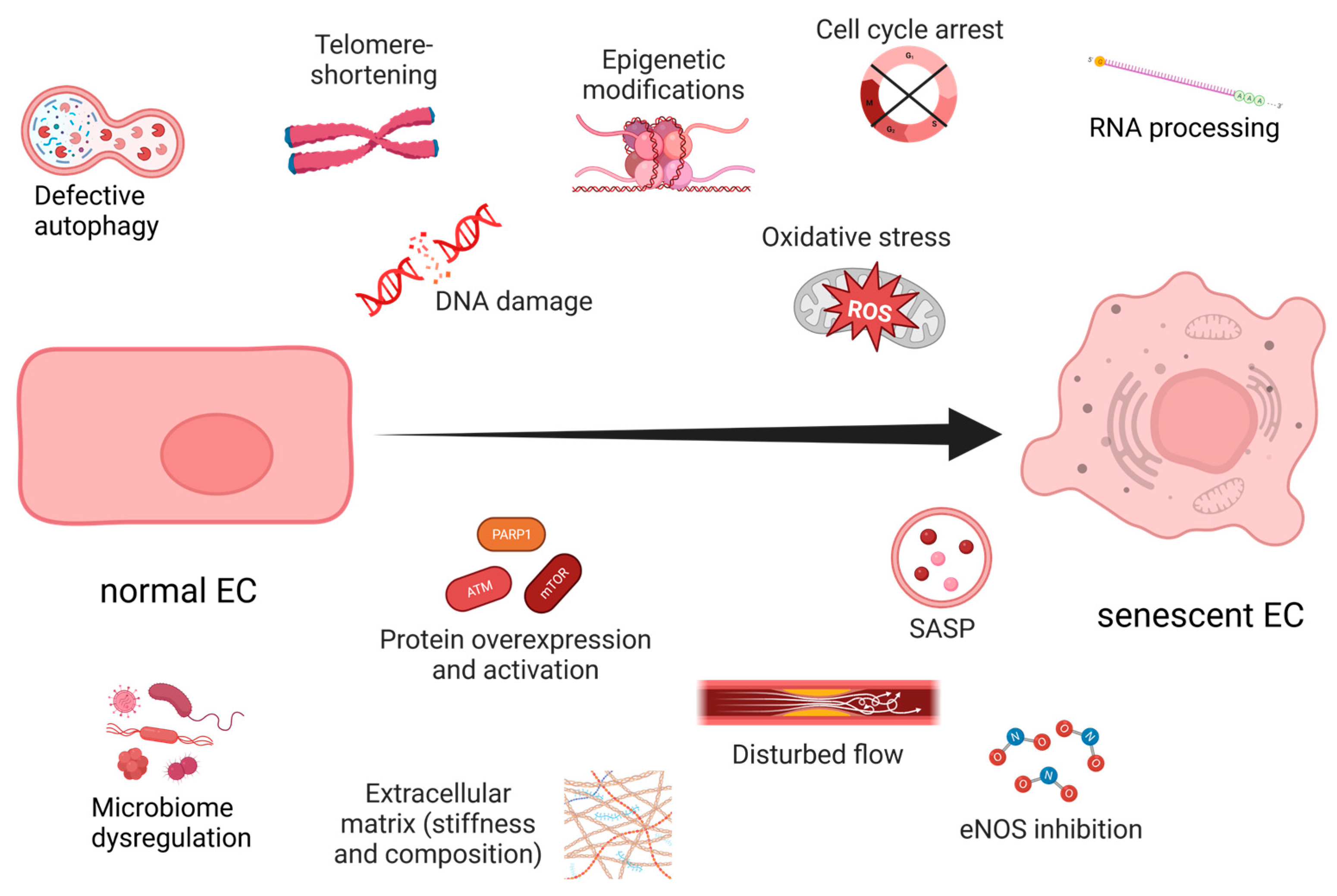
Figure 1. Hallmarks of endothelial senescence. Created with https://www.BioRender.com (accessed on 7 July 2023).
Ijms 24 11344 g001
Figure 2. The role of eNOS in endothelial senescence. Wall shear stress (WSS) and MAGUK with inverted domain structure-1 (MAGI1) activate eNOS, which produces NO from Arginine. The accumulated NO inhibits the geroconversion of ECs. When the levels of tetrahydrobiopterin (BH4) or Arginine are low, eNOS’s function is altered, resulting in its uncoupling and the increased production of superoxide anion (O2•−). The latter reacts with NO and yields ONOO (peroxynitrite), which causes senescence in ECs. eNOS is also negatively impacted by disturbed WSS and mitochondrial ROS. Senescent ECs generate SASP, which can further exacerbate endothelial dysfunction. Created with https://www.BioRender.com (accessed on 11 July 2023).
Ijms 24 11344 g002
Figure 3. Summary of the effects of short-term (mild) hypoxia on EC signaling. Short-term hypoxia reduces mitochondrial ROS, DNA damage, senescence and apoptosis, protecting the endothelium. During mild hypoxia, hypoxia-inducible factors (HIFs) are activated, unlike in normal oxygen levels when they are targeted by prolyl hydroxylases (PHDs) for degradation. HIFs stimulate VEGF and its receptors (VEGFRs), and metalloproteinases (MMPs), which stimulate the reorganization of the extracellular matrix and follow-up angiogenesis. The cell’s renewal through autophagy is also activated. HIFs induce anaerobic glycolysis by glucose transporter 1 (GLUT1) and lactate dehydrogenase (LDH), circumventing the ETC and further reducing OS. HIFs can cause overexpression of p21, which leads to senescence, but they inhibit multiple other pro-senescent factors such as p16, senescence-associated secretory phenotype (SASP), and mammalian target of rapamycin (mTOR). Red arrows indicate an increase in the corresponding processes, which have a negative senile effect on the cells. Green arrows indicate a decrease in said processes, which protect the cells from senescence. Created with https://www.BioRender.com (accessed on 30 May 2023).
Ijms 24 11344 g003
Figure 4. Regulation of angiogenesis by urokinase plasminogen activator and its receptor. uPA/uPAR are carried by extracellular vesicles (EVs) to ECs. Upon receptor binding, VEGF-mediated matrix degradation is stimulated via VEGFR2. The processed matrix releases the plasminogen activator inhibitor-1 (PAI-1), which inhibits uPA/uPAR recognition and subsequent VEGFR2 activation. This feedback loop prevents excessive angiogenesis. The red X depicts the obstruction of uPA/uPAR recognition under the influence of PAI-1 and the inability of uPA/uPAR to activate VEGFR2. Created with https://www.BioRender.com (accessed on 30 May 2023).
Ijms 24 11344 g004
Figure 5. Regulation of tip cell formation through Delta-Notch signaling. Under normal conditions, Delta-Notch signaling serves in intercellular communication. Upon their binding, Notch is cleaved by γ-secretase into an extracellular domain (NEXT) and an intracellular domain (NICD). The latter acts as a transcription factor and inhibits genes involved in tip cell conversion in stalk cells. The amyloid protein Aβ serves as a competitive inhibitor of Notch and distracts γ-secretase. As a result, NICD cannot inhibit tip cell conversion of stalk cells and triggers non-productive angiogenesis. Created with https://www.BioRender.com (accessed on 30 May 2023).
Ijms 24 11344 g005
Table 1. Effects of the type of wall shear stress (WSS) on EC function.
Type of WSSCell LineCell ResponseReference
Sudden, temporal, 10 dyne/cm2HUVECs↑ proliferation[74]
Steady, uniformHUVECsno effect[74]
Linear, physiological, 12 dyne/cm2 *BAECs↓ proliferation[75]
Gradient, <68 dyne/cm2HMVECsmigrate against flow; orient perpendicularly at highest WSS[80]
Linear, high, 284 dyne/cm2
Gradient, positive, 150–170 dyne/cm2
BAECs **↓ alignment;
↑ proliferation;
↑ apoptosis
[81]
Linear, low, 30 dyne/cm2
Gradient, negative, 170–150 dyne/cm2
BAECs↑ alignment;
↓ proliferation;
↓ apoptosis
[81]
* Physiological WSS 10–20 dyne/cm2; 10 dyne/cm2 = 1 N/m2 = 1 Pa. ** BAECs—bovine aortic endothelial cells. ↑—indicates an increase in the process; ↓—indicates a decrease in the process.
Table 2. The role of Aβ in cerebral blood vessels.
AD ModelBlood VesselsProtein ExpressionReferences
Aβ monomers in HUVEC and zebrafish↑ capillary density-[151]
Tau overexpressing mice; 15 months old↑ capillary density;
↑ angiogenesis;
↑ BBB permeability;
↓ CBF
↑ VEGF;
↑ uPAR;
↑ MMP-9;
↑ PAI-1
[152]
AD patients↑ VEGF;
↑ TGF-β
[153]
HMVECs + Aβ monomers↓ angiogenesis↑ VEGFR1-
↑ senescence
[154]
APP-PSEN1/+ mice↑ non-productive angiogenesis;
↓ capillary density around plaques
↑ VEGF[155]
Tg2576 mice↓ capillary density around plaques↓ GLUT1[156]
AD patients; APP695 mice↓ capillary densityVEGF supplementation improved cognitive function[157]
3xTG-AD mice↑ capillary density;
↓ junction density
[158]
↑—indicates an increase in the process; ↓—indicates a decrease in the process.
Disclaimer/Publisher’s Note: The statements, opinions and data contained in all publications are solely those of the individual author(s) and contributor(s) and not of MDPI and/or the editor(s). MDPI and/or the editor(s) disclaim responsibility for any injury to people or property resulting from any ideas, methods, instructions or products referred to in the content.