Article

κO-SrVIA Conopeptide, a Novel Inhibitor Peptide for Two Members of the Human EAG Potassium Channel Family

by
Luis Martínez-Hernández
1,
Estuardo López-Vera
2,*,
Manuel B. Aguilar
3,
Ximena C. Rodriguez-Ruiz
2 and
Mónica A. Ortíz-Arellano
4
1
Posgrado en Ciencias Biológicas, Instituto de Ciencias del Mar y Limnología, Universidad Nacional Autónoma de México, Ciudad de México 04510, Mexico
2
Laboratorio de Toxinología Marina, Unidad Académica de Ecología y Biodiversidad Acuática, Instituto de Ciencias del Mar y Limnología, Universidad Nacional Autónoma de México, Ciudad de México 04510, Mexico
3
Laboratorio de Neurofarmacología Marina, Departamento de Neurobiología Celular y Molecular, Instituto de Neurobiología, Universidad Nacional Autónoma de México, Juriquilla 76230, Mexico
4
Laboratorio de Malacología, Facultad de Ciencias del Mar, Universidad Autónoma de Sinaloa, Mazatlán 82000, Mexico
*
Author to whom correspondence should be addressed.
Int. J. Mol. Sci. 2023, 24(14), 11513; https://doi.org/10.3390/ijms241411513
Submission received: 23 June 2023 / Revised: 8 July 2023 / Accepted: 11 July 2023 / Published: 15 July 2023

Abstract

:
The first conotoxin affecting the voltage-gated potassium channels of the EAG family was identified and characterized from the venom of the vermivorous species Conus spurius from the Gulf of Mexico. This conopeptide, initially named Cs68 and later designated κO-SrVIA, is extremely hydrophobic and comprises 31 amino acid residues, including six Cysteines in the framework VI/VII, and a free C-terminus. It inhibits the currents mediated by two human EAG subtypes, Kv10.1 (IC50 = 1.88 ± 1.08 µM) and Kv11.1 (IC50 = 2.44 ± 1.06 µM), and also the human subtype Kv1.6 (IC50 = 3.6 ± 1.04 µM). Despite its clear effects on potassium channels, it shares a high sequence identity with δ-like-AtVIA and δ-TsVIA. Also, κO-SrVIA is the third conopeptide from the venom of C. spurius with effects on potassium channels, and the seventh conotoxin that blocks Kv1.6 channels.

1. Introduction

Marine snails belonging to the genus Conus are a group characterized mainly by the beauty of the color patterns on their shells and the great diversity of toxins contained in their venom apparatus, first detected by reports of poisoning in humans and the bioassays of the crude venom performed by Endean and Rudkin (1965) [1,2]. It is well known that these marine snails use the venom to capture their prey, worms, fishes, and other mollusks [3].
The venoms of Conus are generally composed of peptide molecules between 12 to 40 amino acid residues in length. These molecules are known as conopeptides and they have been documented to have remarkable affinity for a wide range of molecular targets such as voltage-gated ion channels (Nav+, Kv+, Cav2+), ligand-gated ion channels (nAChR, 5-HT3R, NMDAR), neurotransmitter transporters (for noradrenaline and norepinephrine) and G-protein-coupled receptors (for somatostatin) [4,5,6,7,8]. Of the more than 700 species that make up the genus, only thirteen conopeptides with activity on potassium channels from nine species have been characterized until now [9,10].
In mammals, the alpha subunits that form potassium channels are encoded by more than 90 genes [11]. In normal conditions, potassium channels mediate the electrical activity in those cells where they are expressed; for instance, they control the neuronal action potential (Kv1.1 and Kv1.2), the cardiac interval (Kv7.1 and Kv11.1) [12], and high-frequency stimulus trains in some brain regions (Kv10.1) [13]; indeed, they stop hyperexcitability upon traumatic nerve injury (Kv1.6) [14]. Under abnormal conditions, these subunits are related to diseases in humans called channelopathies. These channelopathies can be classified into two types: i) gain/loss of function and ii) ectopic expression. The first refers to mutations in the coding genes, which results in improper functioning of the channel; for example, a mutation in the KCNH2 gene that encodes the cardiac Kv11.1 channel induces an arrhythmia known as long QT syndrome [15]. The second refers to an expression where the channel is not normally expressed; an example of this is the KCNH1 gene that encodes the Kv10.1 channel, whose overexpression has been reported in a wide variety of tissues that present tumor growth, which is why it is considered a tumor marker [16].
Given the roles of the Kv10.1 channel, in the present work, we aimed to identify conotoxins affecting this subtype. Electrophysiological assays on Xenopus laevis oocytes allowed us to discover a new conopeptide called Cs68 (κO-SrVIA), which exerts inhibitory activity upon Kv10.1, Kv11.1, and Kv1.6 potassium channels, with a slightly higher affinity for the Kv10.1 channel.

2. Results

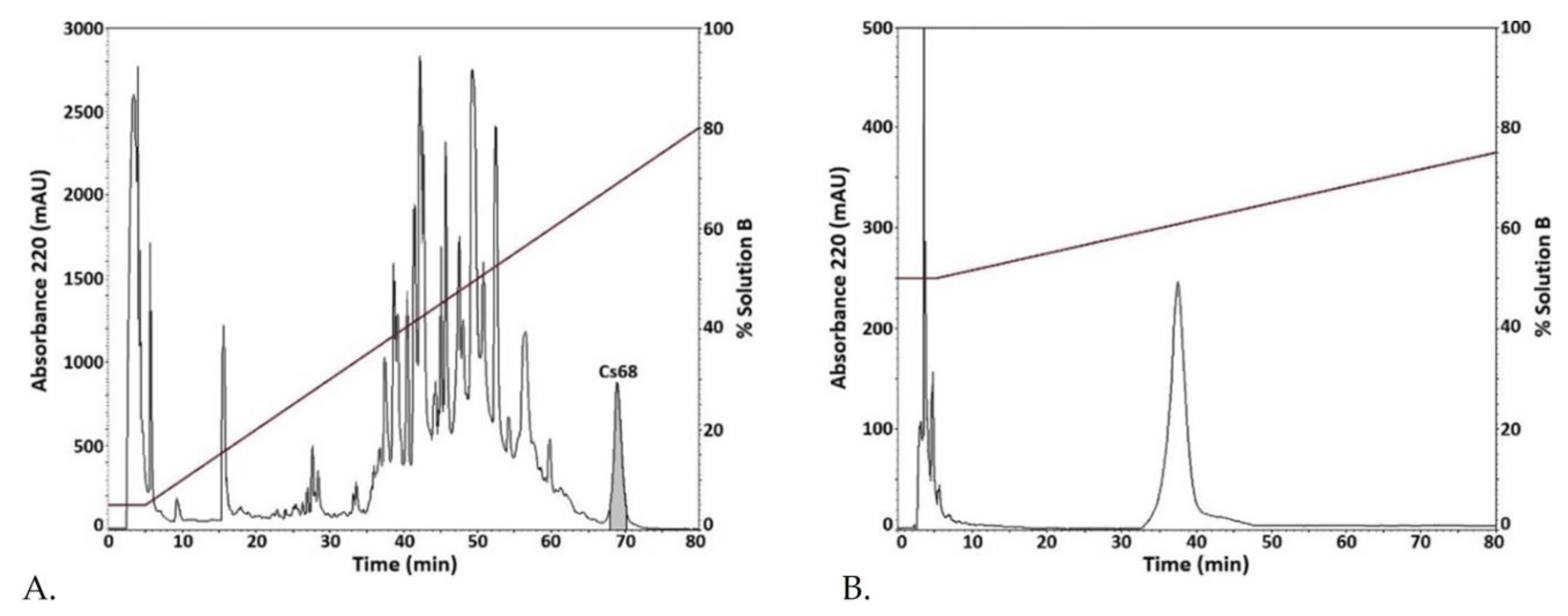
2.1. Conus spurius Venom Fractionation and Purification of Fraction Cs68 Subsection

In order to identify any component with inhibitory activity on the Kv10.1 potassium channel, electrophysiological evaluation of different fractions of C. spurius venom obtained by RP-HPLC was performed; the 24 apparently most abundant fractions were assayed at 0.041 µg/µL. Fraction Cs68, which gets its name because it elutes at minute 68 during fractionation, was the only fraction that had inhibitory activity on the Kv10.1 channel (Figure 1A). One additional step of purification by RP-HPLC was necessary to obtain a pure venom component (peptide Cs68) (Figure 1B).

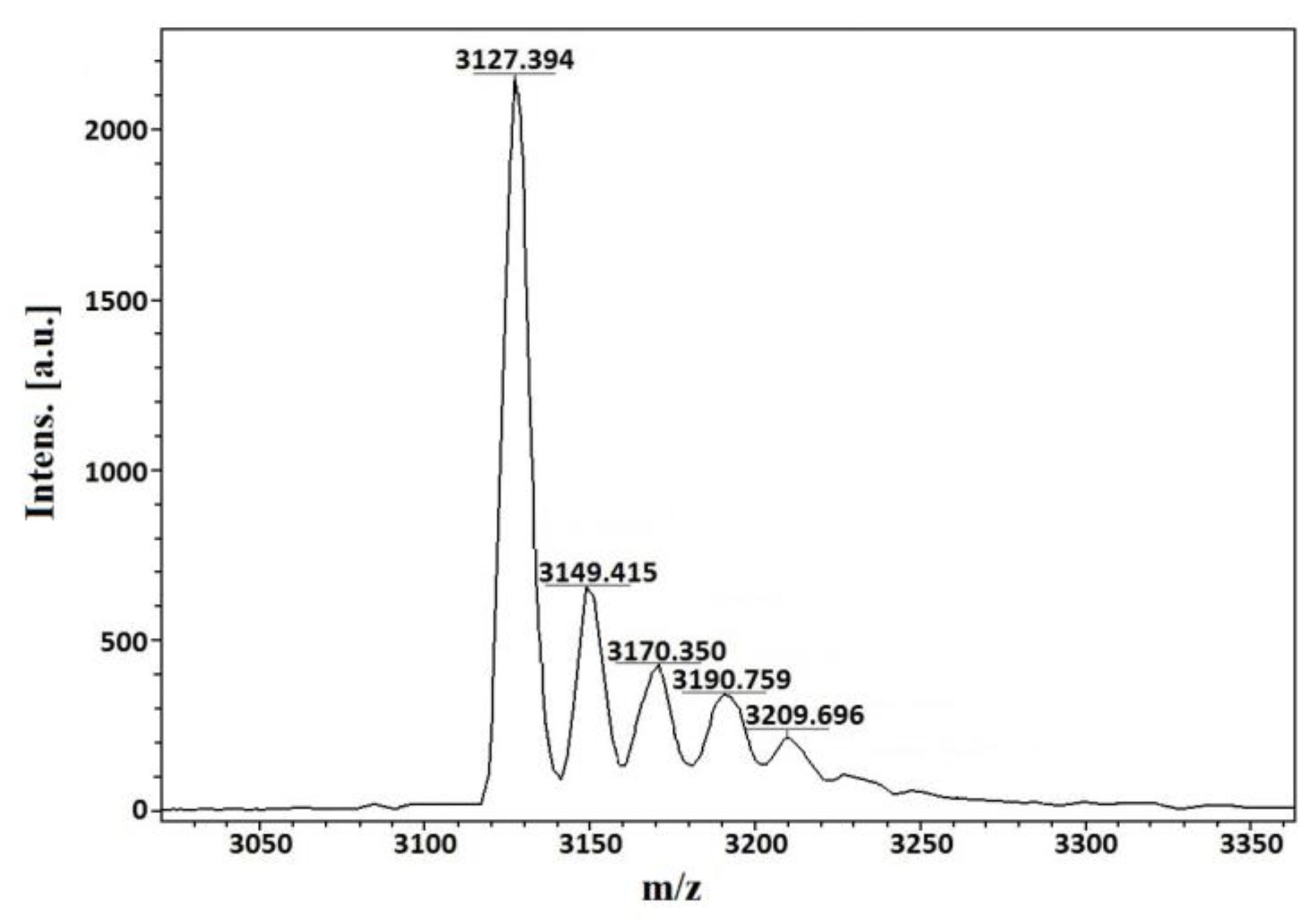
2.2. Molecular Mass and Amino Acid Sequence Analysis

The mass spectrum of peptide Cs68 displays a clean major m/z (z = +1) signal at 3127.394 (Figure 2), that corresponds to an average mass of 3126.386 Da. The signal at 3149.415 differs from the previous one by 22.02 m/z units and corresponds to a sodium adduct.
Thirty-two cycles of automated Edman degradations were performed and a sequence of 31 amino acids was obtained. Position 2, 9, 18, 19, 23, and 27 were blank cycles, which we assume correspond to cysteine residues. The theoretical average mass of the sequence, considering three disulfide bonds, is 3128.78 Da if a free C-terminus is presumed, and 3127.80 Da if a C-terminal amide is supposed. Due to the difference between the experimental and each theoretical values, we searched for the sequence of peptide Cs68 in the transcriptome of the venom gland of C. spurius, recently published [17]; the sequence is identical to the mature region predicted from Assembly/Isolate DN54709c1g1i1151/Sr6.O.04 by the ConoPrec tool of the ConoServer database [18]; in this precursor the C-terminus is a proline residue, which rules out the possibility of amidation (Figure 3).

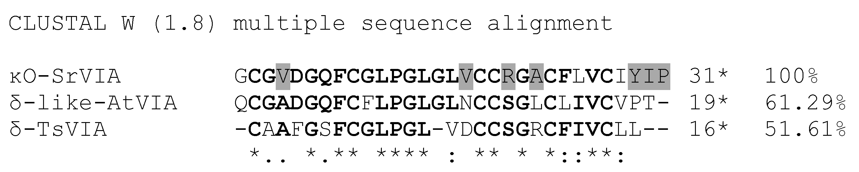
2.3. Similarity Search

BLASTp similarity search for the Cs68 sequence gave six hits with an E value better than 4 × 10⁻⁸, including two for conotoxins for which a target has been identified: δ-like-AtVIA from the worm hunter snail C. ateralbus (two hits with percent identity of 70.37% and query coverage of 87%) and δ-TsVIA (one hit with percent identity of 59.26% and query coverage of 87%) (Figure 3).

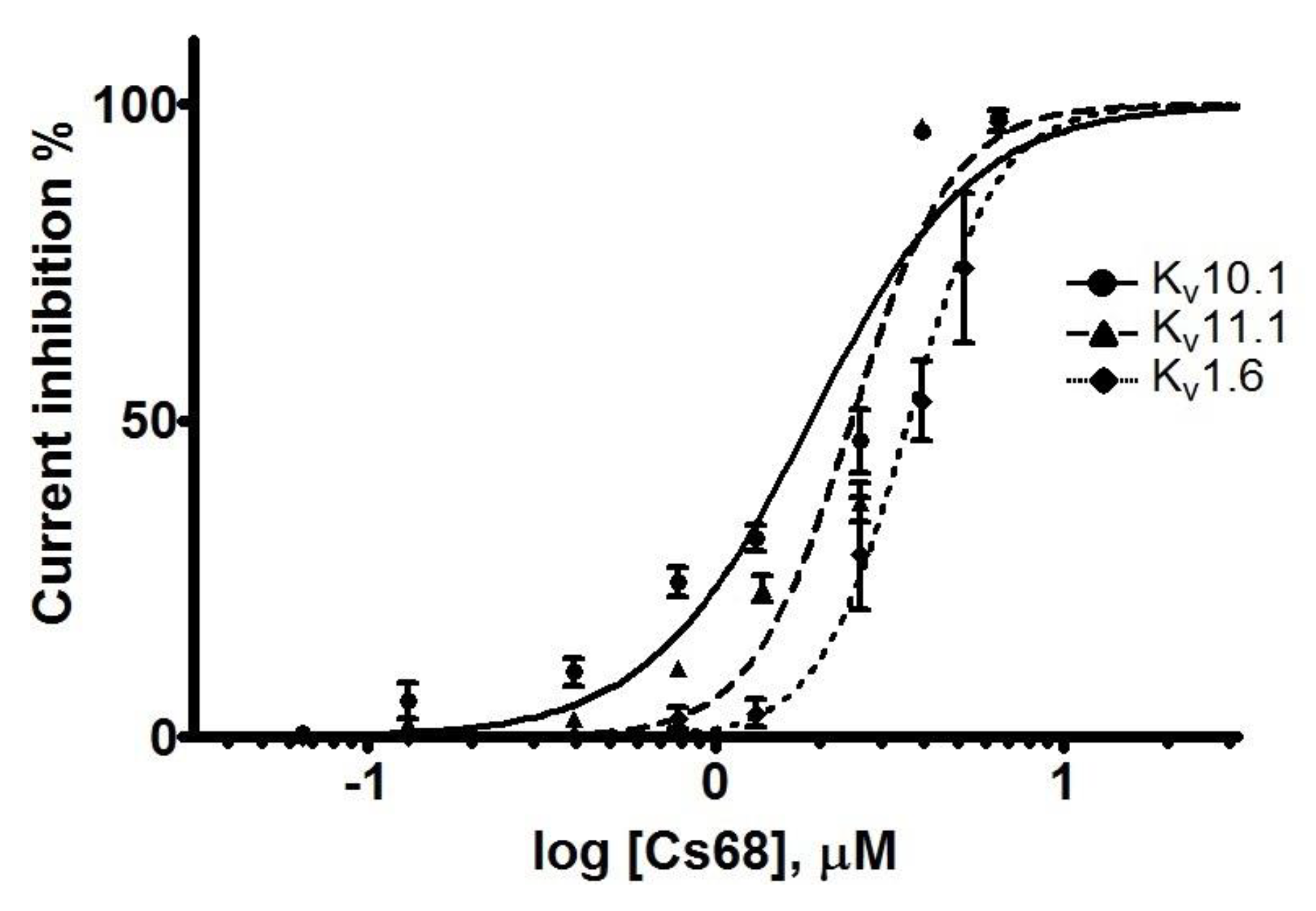
2.4. Electrophysiological Evaluation of Cs68 on Voltage-Gated Potassium Channels

Fraction Cs68 was initially evaluated by TEVC in X. laevis oocytes expressing the voltage-dependent K+ channel Kv10.1. Cs68 showed 100% inhibitory activity on the currents at 4 µM. Hence, we evaluated Cs68 on two more subtypes of K+ channels where other compounds with effects on Kv10.1 also have shown activity (Kv11.1 and Kv1.6) [9,19]. We found that at the low concentration of 0.4 µM the inhibitory activity was 10% on Kv10.1 and Kv11.1 (Figure 4A,B), but no effect on Kv1.6 was observed. However, at 4 µM the inhibition was less than 50% on Kv1.6 but 100% on Kv11.1 (Figure 4C). Figure 4D–F show the time course of inhibition of the 3 subtypes of K+ channels by Cs68. Then, concentration-response curves were generated for Kv10.1, Kv11.1, and Kv1.6 channels (Figure 5). Peptide Cs68 presented a slightly higher affinity for Kv10.1 (IC50 1.88 ± 1.08 µM) with respect to Kv11.1 (IC50 2.44 ± 1.06 µM) and Kv1.6 (IC50 3.6 ± 1.04 µM).

3. Discussion

In this work, we identified a new peptide, Cs68, from the venom of the vermivorous species Conus spurius; the amino acid sequence of the peptide consists of 31 amino acids with three disulfide bridges and a free C-terminus. Since Cs68 has activity on potassium channels, and, in accordance with Zamora-Bustillos et al. (2021), this conopeptide belongs to the superfamily O1 and presents the cysteine pattern VI/VII, we decided to name it κO-SrVIA following the proposed nomenclature for conotoxins [17]. The disulfide connectivity of this new conopeptide remains unknown; however, we assume that it could be similar to that supposed for δ-like-AtVIA (I–IV, II–V, III–VI) with which it shares 70.3% of its amino acid sequence [20].
Electrophysiological tests with κO-SrVIA revealed that it is able to inhibit the currents elicited by three subtypes of voltage-gated potassium channels, Kv1.6, Kv10.1, and Kv11.1, at different concentrations. To date (June 2023) there are only thirteen conopeptides with activities on potassium channels and six of them have effects on Kv1.6: κJ-PlXIVA, CPY-Fe1, CPY-Pl1, κM-RIIIJ, κ-SrXIA (peptide sr11a), and CNF-Sr3 [21,22,23,24,25]. Therefore, Kv1.6 is the most common potassium channel target subtype for conopeptides. In this sense, κO-SrVIA would be the seventh conopeptide reported with activity on this subtype, and the third one isolated from the venom of C. spurius that blocks it, but, nevertheless, the first one with inhibitory activity on subtypes Kv10.1 and Kv11.1.
The identification of a third component in the venom of C. spurius with inhibitory activity on potassium channels suggests ecological importance. In 1996, Terlau and collaborators proposed that the presence of more than one potassium channel blocker in the venom of a Conus species may be due to the fact that they are used during prey capture, specifically in the rapid immobilization component [26]. It is important to highlight that, despite the fact that this strategy was proposed for snails that feed on fish, it could be shared by worm hunting snails as are C. spurius (toxins κ-SrXIA, CNF-Sr3, and κO-SrVIA) and C. planorbis (toxins kJ-PIXIVA and CPY-Pl1).
The study of conopeptides with activity in potassium channels has gained interest since the characterization of κO-PVIIA and its selectivity for the Shaker subtype with respect to other subtypes (Kv1.1 and Kv1.4) [26]. Taking this into account, the identification of new conopeptides that could serve as molecular tools to study the functionality of potassium channels has been the subject of study, especially when specific subtypes are involved in the development of channelopathies such as the cases of Kv10.1 and Kv11.1, implicated in the advancement of childhood epilepsy, Temple-Baraitser syndrome [27], Zimmermann-Laband syndrome [28], tumor growth [16], and long QT syndrome [15].
Considering the above, κO-SrVIA (Cs68) could represent a promising molecule to study the Kv10.1 and Kv11.1 channels. However, its usefulness may be limited by the similar affinity it presents for these two subtypes of potassium channels. A problem that has been encountered when looking for molecules with specific activities on subtype Kv10.1 is that they also inhibit the Kv11.1 [19]. Nevertheless, to solve this problem a strategy that includes the design of analogs based on natural conopeptides with interesting properties, such as κ-RIIIK and κ-RIIIJ from C. radiatus, can be applied; these analogs are designed to determine which amino acids are responsible for the activity and thus promote selectivity for subtypes of potassium channels [23].
The importance of studying molecules contained in the venom of different animals that interact with Kv10.1 potassium channels rose after the finding by Dr. Pardo and collaborators in 1999 that inhibition of Kv10.1 channels causes reduced cell proliferation in different cancer cell lines [29]. At the moment, four toxins have been isolated that target Kv10.1: κ-hefutoxin (Heterometrus fulvipes), APETx4 (Anthopleura elegantissima), Aa1a (Avicularia aurantiaca), and Ap1a (Avicularia purpurea).
κ-hefutoxin has an IC50 of 26 µM on Kv10.1 [30] but inhibits Kv1.3 [31]; the IC50 of APETx4 for Kv10.1 is 1.01 µM and at a concentration slightly higher of 1.6 µM reduces the currents amplitude by 66% on Kv1.4 and by 50% on Nav1.4 channels [32]; Aa1A and Ap1A are the toxins that have higher affinities for Kv10.1 as their IC50 are 637 nM and 236 nM, respectively, with no activity on Kv11.1 until the concentration increases to 8 µM [33]. However, only Aa1a presents 100% of inhibition for Kv10.1 with a concentration of 20 µM; the same concentration for Ap1a produces less than 80% of inhibition [33]; a concentration of 80 µM of κ-hefutoxin reaches 90% of inhibition and APETx4 at 3 µM causes 88% of inhibition [32]. It is worth mentioning that, for Kv10.1, Ap1a and Aa1A have a Hill coefficient of 1.32 ± 0.13 and 1.03 ± 0.09 [33], respectively, while APETx4 has a coefficient of 3.8 ± 0.03 [32]; these values are close to those obtained with κO-SrVIA for the three subtypes of K+ channels. A Hill coefficient of ~1 normally indicates a single binding site while a higher Hill coefficient (~4 for APETx4) suggests possible cooperativity. Cooperativity might occur in the interaction between κO-SrVIA and the Kv10.1, Kv11.1, and Kv1.6 channels, but more experiments would be necessary to confirm this possibility.
In this sense, this work could contribute to the study of Kv10.1 due to the characteristics that κO-SrVIA has: it reaches 100% inhibition with a concentration of 4 µM (Figure 5), which is lower than the concentrations of the previously mentioned toxins; it has an IC50 similar to APETx4 (1.88 µM vs. 1.01 µM, respectively), and the length (31 amino acids) is similar to Aa1A and Ap1a. In other words, κO-SrVIA gathers all the characteristics of these toxins in one.

4. Materials and Methods

4.1. Isolation of Crude Venom Extract from Conus spurius

Six venom ducts from Conus spurius (stored at −70 °C) were homogenized in 10 mL of 40% (v/v) aqueous acetonitrile (ACN) and 2% (v/v) trifluoracetic acid (TFA) with a tissue homogenizer (BioSpec 985370 Tissue-Tearor; Bartlesville, OK, USA) at 4 °C. The resulting mix was centrifuged at 14,000× g for 15 min. The supernatant was decanted and the pellet was kept as the fraction rich in peptides. The protein content was quantified with a Nanodrop spectrophotometer at 280 nm (Nanodrop 2000, Thermo Fisher, Waltham, MA, USA).

4.2. Crude Venom Extract Fractionation by RP-HPLC

Aliquots of 7.5 mg of crude venom were dissolved with 1 mL of Solution A (aqueous solution with 0.1% (v/v) TFA) before fractionation by Reversed-Phase High Performance Liquid Chromatography (RP-HPLC) (Agilent Technologies, Infinity 1260 Series HPLC System; Santa Clara, CA, USA) using a Vydac Reverse-Phase Peptide and Protein C18 column (218TP54, 5 µm particle size, 4.6 mm × 250 mm) attached to a Vydac Reverse-Phase Peptide and Protein C18 Precolumn (218GK54, 5 µm particle size, 4.6 mm × 10 mm).
Fractions of the crude venom extract were eluted with an isocratic step of 5% Solution B (90% ACN in water containing 0.085% (v/v) TFA) for 5 min, followed by a linear gradient from 5% to 80% (v/v) of Solution B in 75 min, at a flow rate of 1 mL/min. Further purification of a selected fraction (Cs68) was performed by RP-HPLC using an isocratic step at 17% of Solution B for 5 min, followed by a linear gradient from 50% to 75% (v/v) of Solution B, over 75 min at a flow rate of 1 mL/min. The elution profiles were monitored at a wavelength of 220 nm. Each signal was collected manually.

4.3. Amino Acid Sequencing and Mass Spectrometry Analysis

The molecular mass of the Cs68 fraction was analyzed by MALDI-TOF with an α-cyano-4-hydroxycinnamic acid matrix at the facilities of the Chemistry Institute, UNAM.
To determine the amino acid sequence, one nmol of the peptide was analyzed by automated Edman degradation using a PPSQ-31A protein sequencer (Shimadzu Scientific Instruments; Tokyo, Japan) by Dr. Fernando Zamudio at the Biotechnology Institute, UNAM.
Theoretical masses were calculated using the Peptide Mass Calculator tool of the Ion Source-Mass Spectrometry Educational Resource website (https://www.ionsource.com/).

4.4. Preparation of Vectors Encoding Potassium Channels

cDNA of the clones for human Kv10.1, Kv11.1 and Kv1.6 channels in vector pSGEM, resistant to ampicillin, were linearized with SpHI, Sfil, and EcoRI, respectively. Then, the products were purified using a QIAquick PCR purification Kit (QIAGEN; Redwood City, CA, USA). cDNAs were transcribed in vitro with T7 polymerase (mMessage mMachine Kit; Ambion Inc., Austin, TX, USA). cRNA was purified using a Qiagen RNeasy kit (QIAGEN; Redwood City, CA, USA).

4.5. Expression of Voltage-Gated Potassium Channels

For heterologous expression, stage V-VI Xenopus laevis oocytes were isolated by microsurgery. Sexually mature Xenopus leavis female frogs were kept and handled according to the institutional bioethics committee requirements. The frogs were anesthetized by a 30 min submersion in 2% tricaine methane sulfonate (MS-222; Merck KGaA, Darmstadt, Alemania) before performing microsurgery. Isolated oocytes were defolliculated with 0.75 mg/mL collagenase A (Roche; San Jose, CA, USA; Cat. # 10103586001) and kept in ND96 extracellular solution (96.0 mM NaCl, 2.0 mM KCl, 1.0 mM MgCl2·6 H2O, 1.8 mM CaCl2·2 H2O, 5 mM HEPES, pH 7.3) supplemented with penicillin/streptomycin (100 U/100 µg)/mL and 100 µg/mL gentamicin at 15 °C.
Thirty nanograms of Kv1.6, 9.8 ng of Kv10.1, and 18 ng of Kv11.1 of cRNA were injected into each defolliculated oocyte using a micro-injector Nanoliter 2000 (World Precision Instruments; Sarasota, FL, USA).

4.6. Electrophysiological Recording

Whole-cell currents were recorded under two-electrode voltage clamp (TEVC) using an OC-725C clamp amplifier (Warner Instruments; Holliston, MA, USA) between 24 to 72 h after injections of the genetic material. Data acquisitions were done with the program LabView (National Instruments, Chihuahua, Mexico). The intracellular electrodes were filled with 3 M KCl and selected for resistance between 0.6–1 MΩ. Potassium currents were generated by depolarizations to +10 mV for 1 s every 10 s from a membrane potential held at −80 mV. The bath solution was ND96, consisting of 96 mM NaCl, 2.0 mM KCl, 1.8 mM CaCl2, 1.0 mM MgCl2, and 5 mM HEPES (pH 7.2) with pen/strep (100 U/100 µg)/mL and 100 µg/mL of gentamicin. To assay toxin Cs68, we stopped the flow and directly applied it (diluted in ND96, 3 µL) to the bath chamber (30 µL); once maximum inhibition was reached at a given concentration, the flow for toxin washout was restarted.
All electrophysiological measurements were performed at room temperature (≈22 °C). The duration of records was 1500 ms for Kv1.6 and Kv10.1, and 3000 ms for Kv11.1.
Measurements are reported as the mean ± standard error of the mean (SEM) of three independent experiments. The current inhibition % was calculated from the current amplitude values obtained with the ND96 extracellular solution (IControl) and those of each of the experimental conditions (I[Cs68]); therefore, Current inhibition % = [1 − (I[Cs68]/IControl)] × 100. Current plots were made with SigmaPlot version 10 (Systat Software, Inc., San Jose, CA, USA). The construction of the concentration–response curve and the determination of the mean inhibitory concentration (IC50) were obtained from the data of the current inhibition % as a function of the concentration (µM) of Cs68 expressed in logarithm (log[Cs68]) using GraphPad Prism version 9.5.1 (GraphPad Software, San Diego, CA, USA). Concentration–response curves were fit to the equation: Current inhibition % = 100/{1 + (log[Cs68]/IC50)nH}, where nH is the Hill coefficient.

4.7. Similarity Search and Sequence Comparison

The standard protein–protein Basic Local Alignment Search Tool (protein–protein BLAST, NCBI, 13 February 2023) was employed to search the non-redundant protein sequences database with organism Conus (taxid: 6490) using the default settings.
Sequence alignments were generated using Clustal W version 1.8 [34] at the PRABI-Gerland Rhone-Alpes Bioinformatic Pole Gerland Site, Institute of Biology and Protein Chemistry at https://npsa-prabi.ibcp.fr/cgi-bin/npsa_automat.pl?page=npsa_clustalw.html, with the default parameters [35].

5. Conclusions

Considering the pros and cons of the toxin characterized in this study compared with the other toxins mentioned above, we can say that κO-SrVIA is a promising molecule for the study of potassium channels and can act as a template for designing analogs that permit us to understand the mechanism underlying the function of subtypes Kv10.1 and Kv11.1.

6. Patents

Patent pending.

Author Contributions

Conceptualization, E.L.-V.; methodology, L.M.-H. and X.C.R.-R.; formal analysis, L.M.-H.; investigation, E.L.-V., L.M.-H., X.C.R.-R. and M.B.A.; resources, M.A.O.-A.; writing—original draft preparation, L.M.-H., M.B.A. and E.L.-V.; writing—review and editing, L.M.-H., M.B.A. and E.L.-V.; funding acquisition, E.L.-V. and M.B.A. All authors have read and agreed to the published version of the manuscript.

Funding

This research was funded by Programa de Apoyo a Proyectos de Investigación e Innovación Tecnológica-Universidad Nacional Autónoma de México (PAPIIT-UNAM) to E.L.-V. and M.B.A., grant number IG200220, Consejo Nacional de Ciencia y Tecnología (CONACYT) to E.L.-V., grant number 153915, and UNAM Institutional Budgets to the Laboratorio de Toxinología Marina (615), Instituto de Ciencias del Mar y Limnología, and Laboratorio de Neurofarmacología Marina (406), Instituto de Neurobiología.

Institutional Review Board Statement

Not applicable.

Informed Consent Statement

Not applicable.

Data Availability Statement

Not applicable.

Acknowledgments

This paper is part of the requirements for obtaining a Doctoral degree at the Posgrado en Ciencias Biológicas, UNAM. We thank the Consejo Nacional de Ciencia y Tecnología of Mexico (CONACYT) for a graduate scholarship (545550) to Luis Ángel Martínez Hernández. Collaborators: Luis Pardo (Max-Planck Institute), and Baldomero M. Olivera and Michael Sanguinetti (University of Utah) for donation of cDNA clones of Kv10.1, Kv11.1, and Kv1.6; Lourival D. Possani Postay and Fernando Zamudio for providing access to protein sequencer (Departamento de Medicina Molecular y Bioprocesos, Instituto de Biotecnología, UNAM) and Roberto Arreguín Espinosa de los Monteros (Instituto de Química, UNAM) for mass spectrometry. We thank Michael C. Jeziorski for editing the manuscript.

Conflicts of Interest

The authors declare no conflict of interest.

References

  1. Hermitte, L.C.D. Venomous marine molluscs of the genus. Conus. Trans. Roy. Soc. Trop. Med. Hyg. 1946, 39, 485–512. [Google Scholar] [CrossRef] [PubMed]
  2. Endean, R.; Rudkin, C. Further studies of the venoms of Conidae. Toxicon 1965, 2, 225–249. [Google Scholar] [CrossRef] [PubMed]
  3. Kohn, A.J. Food Specialization in Conus in Hawaii and California. Ecology 1966, 47, 1041–1043. [Google Scholar] [CrossRef]
  4. Teichert, R.W.; Jimenez, E.C.; Twede, V.; Watkins, M.; Hollmann, M.; Bulaj, G.; Olivera, B.M. Novel conantokins from Conus parius venom are specific antagonists of N-methyl-D-aspartate receptors. J. Biol. Chem. 2007, 282, 36905–36913. [Google Scholar] [CrossRef] [PubMed] [Green Version]
  5. Lewis, R.; Dutertre, S.; Vetter, I.; Christie, M. Conus venom peptide pharmacology. Pharmacol. Rev. 2012, 6, 259–298. [Google Scholar] [CrossRef] [PubMed]
  6. Sharpe, I.A.; Gehrmann, J.; Loughnan, M.L.; Thomas, L.; Adams, D.A.; Atkins, A.; Palant, E.; Craik, D.J.; Adams, D.; Alewood, P.F.; et al. Two new classes of conopeptides inhibit the α1-adrenoceptor and noradrenaline transporter. Nat. Neurosci. 2001, 4, 902–907. [Google Scholar] [CrossRef] [PubMed]
  7. Sharpe, I.; Palant, E.; Schroeder, C.; Kaye, D.; Adams, D.; Alewood, P.; Lewis, R. Inhibition of the norepinephrine transporter by the venom peptide χ-MrIA. J. Biol. Chem. 2003, 278, 40317–40323. [Google Scholar] [CrossRef] [Green Version]
  8. Petrel, C.; Hocking, H.G.; Reynaud, M.; Upert, G.; Favreau, P.; Biass, D.; Paolini-Bertrand, M.; Peigneur, S.; Tytgat, J.; Gilles, N.; et al. Identification, structural and pharmacological characterization of τ-CnVA, a conopeptide that selectively interacts with somatostatin sst3 receptor. Biochem. Pharmacol. 2013, 85, 1663–1671. [Google Scholar] [CrossRef]
  9. Finol-Urdaneta, R.K.; Belovanovic, A.; Micic-Vicovac, M.; Kinsella, G.K.; McArthur, J.R.; Al-Sabi, A. Marine toxins targeting Kv1 channels: Pharmacological tools and therapeutic scaffolds. Mar. Drugs 2020, 18, 173. [Google Scholar] [CrossRef] [Green Version]
  10. Tosti, E.; Boni, R.; Gallo, A. Pathophysiological responses to conotoxin modulation of voltage-gated ion currents. Mar. Drugs 2022, 20, 282. [Google Scholar] [CrossRef]
  11. Tian, C.; Zhu, R.; Zhu, L.; Qiu, T.; Cao, Z.; Kang, T. Potassium channels: Structures, diseases, and modulators. Chem. Biol. Drug Des. 2014, 83, 1–26. [Google Scholar] [CrossRef] [PubMed]
  12. Schmitt, N.; Grunnet, M.; Olesen, S.-P. Cardiac potassium channel subtypes: New roles in repolarization and arrhythmia. Physiol. Rev. 2014, 94, 609–653. [Google Scholar] [CrossRef] [PubMed] [Green Version]
  13. Mortensen, L.S.; Schmidt, H.; Farsi, Z.; Barrantes-Freer, A.; Rubio, M.E.; Ufartes, R.; Eilers, J.; Sakaba, T.; Stühmer, W.; Pardo, L.A. Kv10.1 opposes activity-dependent increase in Ca2+ influx into the presynaptic terminal of the parallel fibre–Purkinje cell synapse. J. Physiol. 2015, 593, 181–196. [Google Scholar] [CrossRef] [PubMed] [Green Version]
  14. Peck, L.J.; Patel, R.; Diaz, P.; Wintle, Y.M.; Dickenson, A.H.; Todd, A.J.; Calvo, M.; Bennett, D.L.H. Studying independent Kcna6 knock-out mice reveals toxicity of exogenous LacZ to central nociceptor terminals and differential effects of Kv1.6 on acute and neuropathic pain sensation. J. Neurosci. 2021, 41, 9141–9162. [Google Scholar] [CrossRef] [PubMed]
  15. Sanguinetti, M.C.; Tristani-Firouzi, M. hERG potassium channels and cardiac arrhythmia. Nature 2006, 440, 463–469. [Google Scholar] [CrossRef]
  16. Hemmerlein, B.; Weseloh, R.; de Queiroz, F.M.; Knötgen, H.; Sánchez, A.; Rubio, M.; Martin, S.; Schliephacke, T.; Jenke, M.; Heinz-Joachim-Radzun; et al. Overexpression of Eag1 potassium channels in clinical tumours. Mol. Cancer 2006, 5, 41. [Google Scholar] [CrossRef] [PubMed] [Green Version]
  17. Zamora-Bustillos, R.; Martínez-Núñez, M.A.; Aguilar, M.B.; Collí-Dula, R.C.; Brito-Domínguez, D.A. Identification of novel conotoxin precursors from the cone snail Conus spurius by high-throughput RNA sequencing. Mar. Drugs 2021, 19, 547. [Google Scholar] [CrossRef]
  18. Kaas, Q.; Yu, R.; Jin, A.-H.; Dutertre, S.; Craik, D.J. ConoServer: Updated content, knowledge, and discovery tools in the conopeptide database. Nucleic Acids Res. 2012, 40, D325–D330. [Google Scholar] [CrossRef]
  19. García-Ferreiro, R.E.; Kerschensteiner, D.; Major, F.; Monje, F.; Stühmer, W.; Pardo, L.A. Mechanism of block of hEag1 K+ channels by imipramine and astemizole. J. Gen. Physiol. 2004, 124, 301–317. [Google Scholar] [CrossRef] [Green Version]
  20. Neves, J.L.B.; Imperial, J.S.; Morgenstern, D.; Ueberheide, B.; Gajewiak, J.; Antunes, A.; Robinson, S.D.; Espino, S.; Watkins, M.; Vascoselos, V.; et al. Characterization of the first conotoxin from Conus ateralbus, a vermivorous cone snail from the Cabo Verde Archipielago. Mar. Drugs 2019, 17, 432. [Google Scholar] [CrossRef] [Green Version]
  21. Imperial, J.S.; Bansal, P.S.; Alewood, P.F.; Daly, N.L.; Craik, D.J.; Sporning, A.; Olivera, B.M. A novel conotoxin inhibitor of Kv1.6 channel and nAChR subtypes defines a new superfamily of conotoxins. Biochemistry 2006, 45, 8331–8340. [Google Scholar] [CrossRef] [PubMed]
  22. Imperial, J.; Chen, P.; Sporning, A.; Terlau, H.; Daly, N.; Craik, D.; Olivera, B.M. Tyrosine-rich conopeptides affect voltage-gated K+ channels. J. Biol. Chem. 2008, 283, 23026–23032. [Google Scholar] [CrossRef] [Green Version]
  23. Chen, P.; Dendorfer, A.; Finol-Urdaneta, R.K.; Terlau, H.; Olivera, B.M. Biochemical Characterization of κM-RIIIJ, a Kv1.2 channel blocker: Evaluation of cardioprotective effects of kM-conotoxins. J. Biol. Chem. 2010, 285, 14882–14889. [Google Scholar] [CrossRef] [PubMed] [Green Version]
  24. Aguilar, M.B.; Pérez-Reyes, L.; López, Z.; Heimer de la Cotera, E.; Falcón, A.; Ayala, C.; Galván, M.; Salvador, C.; Escobar, L. Peptide sr11a from Conus spurius is a novel peptide blocker for Kv1 potassium channels. Peptides 2010, 31, 1287–1291. [Google Scholar] [CrossRef] [PubMed]
  25. López-Vera, E.; Martínez-Hernández, L.; Aguilar, M.B.; Carrillo, E.; Gajewiak, J. Studies of conorfamide-Sr3 on human voltage-gated Kv1 potassium channel subtypes. Mar. Drugs 2020, 18, 425. [Google Scholar] [CrossRef] [PubMed]
  26. Terlau, H.; Shon, K.-J.; Grilley, M.; Stocker, M.; Stuhmer, W.; Olivera, B. Strategy for rapid immobilization of prey by a fish-hunting marine snail. Nature 1996, 381, 148–151. [Google Scholar] [CrossRef]
  27. Simons, C.; Rash, L.D.; Crawford, J.; Ma, L.; Cristofori-Armstrong, B.; Miller, D.; Ru, K.; Baillie, G.J.; Alanay, Y.; Jacquinet, A.; et al. Mutations in the voltage-gated potassium channel gene KCNH1 cause Temple-Baraitser syndrome and epilepsy. Nat. Genet. 2015, 47, 73–79. [Google Scholar] [CrossRef]
  28. Kortüm, F.; Caputo, V.; Bauer, C.K.; Stella, L.; Ciolfi, A.; Alawi, M.; Bocchinfuso, G.; Flex, E.; Paolacci, S.; Dentici, M.L.; et al. Mutations in KCNH1 and ATP6V1B2 cause Zimmermann-Laband syndrome. Nat. Genet. 2015, 47, 661–669. [Google Scholar] [CrossRef] [Green Version]
  29. Pardo, L.; Camino, D.; Sánchez, A.; Alves, F.; Brüggemann, A.; Beckh, S.; Stühmer, W. Oncogenic potential of EAG K+ channels. EMBO J. 1999, 18, 5540–5547. [Google Scholar] [CrossRef]
  30. Moreels, L.; Peigneur, S.; Yamaguchi, Y.; Vriens, K.; Waelkens, E.; Zhue, S.; Thevissen, K.; Cammue, B.; Sato, K.; Tytgat, J. Expanding the pharmacological profile of κ-hefutoxin 1 and analogues: A focus on the inhibitory effect on the oncogenic channel Kv10.1. Peptides 2017, 98, 43–50. [Google Scholar] [CrossRef]
  31. Peigneur, S.; Yamaguchi, Y.; Goto, H.; Srinivasan, K.N.; Gopalakrishnakone, P.; Tytgat, J.; Sato, K. Synthesis and characterization of amino acid deletion analogs of κ-hefutoxin 1, a scorpion toxin on potassium channels. Toxicon 2013, 71, 25–30. [Google Scholar] [CrossRef] [PubMed]
  32. Moreels, L.; Peigneur, S.; Galan, T.D.; De Pauw, E.; Béress, L.; Waelkens, E.; Pardo, A.L.; Quinton, L.; Tytgat, J. APETx4, a novel sea anemone toxin and a modulator of the cancer-relevant potassium channel Kv10.1. Mar. Drugs 2017, 15, 287. [Google Scholar] [CrossRef] [PubMed] [Green Version]
  33. Ma, L.; Chin, Y.; Dekan, Z.; Herzig, V.; Chow, C.Y.; Heighway, J.; Lam, S.W.; Guillemin, G.; Alewood, P.; King, G. Novel venom-derived inhibitors of the human EAG channel, a putative antiepileptic drug target. Biochem. Pharmacol. 2018, 158, 60–72. [Google Scholar] [CrossRef] [PubMed] [Green Version]
  34. Thompson, J.D.; Higgins, T.J.; Gibson, T.J. CLUSTAL W: Improving the sensitivity of progressive multiple sequence alignment through sequence weighting, positions-specific gap penalties and wight matrix choice. Nucleic Acids Res. 1994, 22, 4673–4680. [Google Scholar] [CrossRef] [Green Version]
  35. Combet, C.; Blanchet, C.; Geourjon, G.; Deléage, G. NPS@: Network protein sequence analysis. Trends Biochem. Sci. 2000, 25, 147–150. [Google Scholar] [CrossRef]
Figure 1. Purification of fraction Cs68 by RP-HPLC. (A) Chromatogram profile of the fractionation of the crude venom from C. spurius. After 5 min at 5% Solution B, a linear gradient of 5 to 80% Solution B in 75 min was applied at flow rate of 1 mL/min. (B) The fraction highlighted in (A) was subfractionated using an isocratic step of 50% Solution B for 5 min, followed by an elution gradient of 50 to 75% Solution B in 75 min at a flow rate of 1 mL/min.
Ijms 24 11513 g001
Figure 2. MALDI-TOF spectrum of purified fraction Cs68. The average m/z (z = +1) signal at 3127.394 corresponds to an average mass of 3126.386 Da.
Ijms 24 11513 g002
Figure 3. Amino acid sequence of peptide Cs68 (renamed as κO-SrVIA; see Section 3) and comparison with δ-like-AtVIA and δ-TsVIA. Asterisks indicate identical residues among all the sequences; colons denote conserved substitutions; dots indicate semiconserved substitutions. Identical residues in two or three sequences are highlighted in boldface, whereas residues in κO-SrVIA that have clearly different physicochemical properties with respect to the δ-conotoxins are highlighted in gray background. The figures followed by an asterisk are the numbers of identical residues compared to κO-SrVIA, whereas the numerals followed by a percent sign are the percents of identical residues compared to κO-SrVIA.
Ijms 24 11513 g003
Figure 4. Inhibition of K+ channels by Cs68. Representative recording showing control currents and in presence of different concentrations of Cs68 A on Kv10.1 (A), Kv11.1 (B), and Kv1.6 (C). Two electrode voltage clamp recordings were made from channels expressed in oocytes. Currents were evoked by voltage steps to +10 mV from a holding potential of −80 mV applied every 10 s. Time course of inhibition by Cs68 at 4 µM (static flow) from the normalized current (I/Imax) on Kv10.1 (D), Kv11.1 (E), and Kv1.6 (F).
Ijms 24 11513 g004
Figure 5. Concentration-response curves of Cs68 over different Kv channel subtypes. Cs68 inhibits all subtypes Kv10.1 (IC50 = 1.88 ± 1.08 µM, Hill coefficient 1.86 ± 0.25), Kv11.1 (IC50 = 2.44 ± 1.06 µM, Hill coefficient 2.85 ± 0.46), and Kv1.6 (IC50 = 3.6 ± 1.04 µM, Hill coefficient 3.35 ± 0.44). n = 3 for each concentration; error bars = standard error of the mean (SEM).
Ijms 24 11513 g005
Disclaimer/Publisher’s Note: The statements, opinions and data contained in all publications are solely those of the individual author(s) and contributor(s) and not of MDPI and/or the editor(s). MDPI and/or the editor(s) disclaim responsibility for any injury to people or property resulting from any ideas, methods, instructions or products referred to in the content.

