
Expression of Immune-Related and Inflammatory Markers and Their Prognostic Impact in Colorectal Cancer Patients
 , ,
, ,  , and
, and
Abstract
:1. Introduction
2. Results
2.1. Patient Characteristics
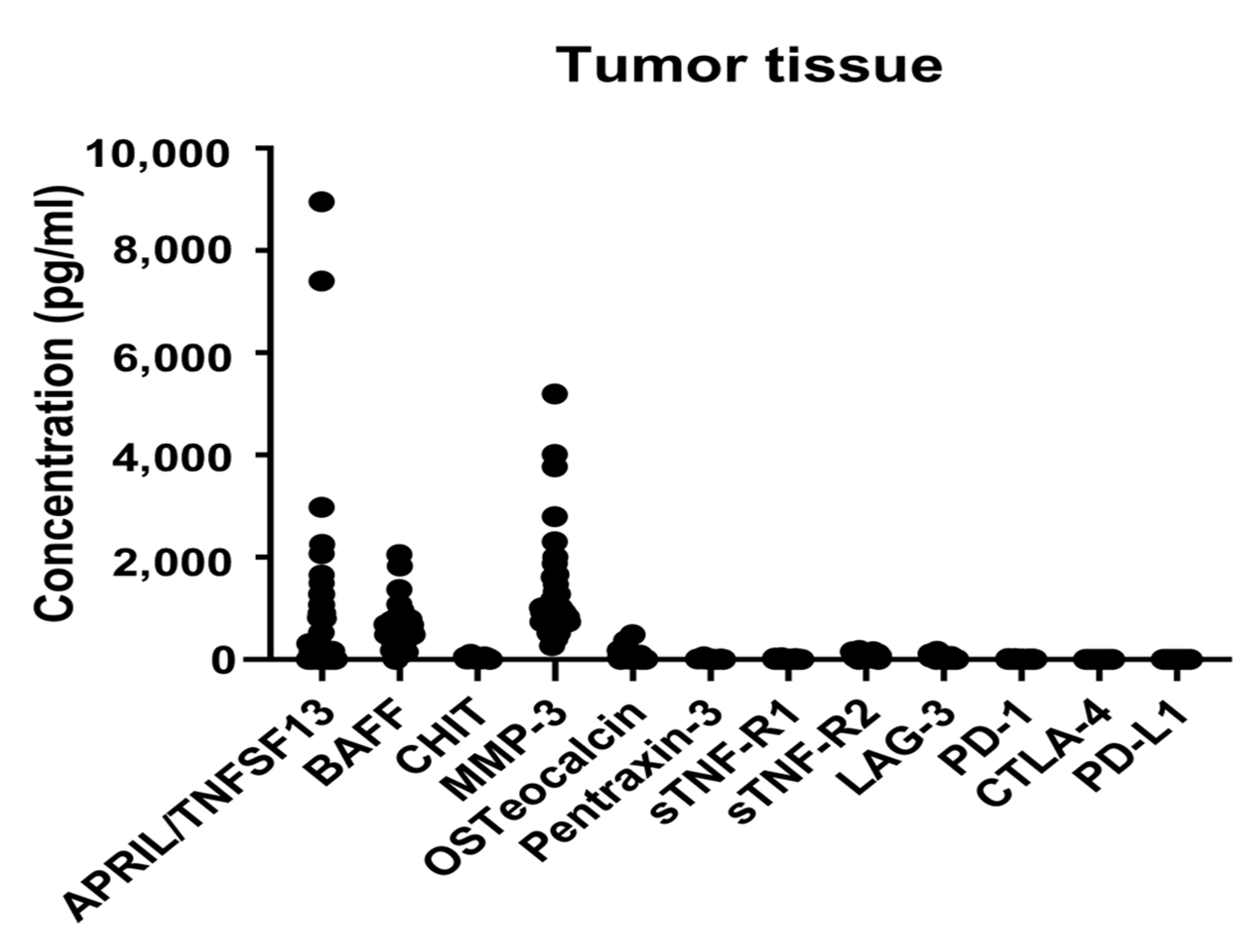
2.2. Immune-Related and Inflammatory Markers in the Tumor Tissues
2.3. Relationships between Immune-Related and Inflammatory Markers and Clinicopathologic Features
2.4. Relationships between Immune-Related and Inflammatory Markers and Long-Term Oncologic Outcomes
3. Discussion
4. Materials and Methods
4.1. Study Population
4.2. Tissue Sample Preparation
4.3. Selection of Immune-Related and Inflammatory Markers
4.4. Bio-Plex Multiplex Immunoassay System
4.5. Surgery and Pathological Examination
4.6. Statistical Analyses
5. Conclusions
Author Contributions
Funding
Institutional Review Board Statement
Informed Consent Statement
Data Availability Statement
Acknowledgments
Conflicts of Interest
References
- Sahin, I.H.; Akce, M.; Alese, O.; Shaib, W.; Lesinski, G.B.; El-Rayes, B.; Wu, C. Immune checkpoint inhibitors for the treatment of msi-h/mmr-d colorectal cancer and a perspective on resistance mechanisms. Br. J. Cancer 2019, 121, 809–818. [Google Scholar] [CrossRef] [PubMed]
- Boukouris, A.E.; Theochari, M.; Stefanou, D.; Papalambros, A.; Felekouras, E.; Gogas, H.; Ziogas, D.C. Latest evidence on immune checkpoint inhibitors in metastatic colorectal cancer: A 2022 update. Crit. Rev. Oncol. Hematol. 2022, 173, 103663. [Google Scholar] [CrossRef] [PubMed]
- Carlino, M.S.; Larkin, J.; Long, G.V. Immune checkpoint inhibitors in melanoma. Lancet 2021, 398, 1002–1014. [Google Scholar] [CrossRef] [PubMed]
- Venkatachalam, S.; McFarland, T.R.; Agarwal, N.; Swami, U. Immune checkpoint inhibitors in prostate cancer. Cancers 2021, 13, 2187. [Google Scholar] [CrossRef]
- Paz-Ares, L.; Ciuleanu, T.E.; Cobo, M.; Schenker, M.; Zurawski, B.; Menezes, J.; Richardet, E.; Bennouna, J.; Felip, E.; Juan-Vidal, O.; et al. First-line nivolumab plus ipilimumab combined with two cycles of chemotherapy in patients with non-small-cell lung cancer (checkmate 9la): An international, randomised, open-label, phase 3 trial. Lancet Oncol. 2021, 22, 198–211. [Google Scholar] [CrossRef]
- Hellmann, M.D.; Rizvi, N.A.; Goldman, J.W.; Gettinger, S.N.; Borghaei, H.; Brahmer, J.R.; Ready, N.E.; Gerber, D.E.; Chow, L.Q.; Juergens, R.A.; et al. Nivolumab plus ipilimumab as first-line treatment for advanced non-small-cell lung cancer (checkmate 012): Results of an open-label, phase 1, multicohort study. Lancet Oncol. 2017, 18, 31–41. [Google Scholar] [CrossRef] [Green Version]
- Le, D.T.; Uram, J.N.; Wang, H.; Bartlett, B.R.; Kemberling, H.; Eyring, A.D.; Skora, A.D.; Luber, B.S.; Azad, N.S.; Laheru, D.; et al. Pd-1 blockade in tumors with mismatch-repair deficiency. N. Engl. J. Med. 2015, 372, 2509–2520. [Google Scholar] [CrossRef] [Green Version]
- Overman, M.J.; McDermott, R.; Leach, J.L.; Lonardi, S.; Lenz, H.J.; Morse, M.A.; Desai, J.; Hill, A.; Axelson, M.; Moss, R.A.; et al. Nivolumab in patients with metastatic DNA mismatch repair-deficient or microsatellite instability-high colorectal cancer (checkmate 142): An open-label, multicentre, phase 2 study. Lancet Oncol. 2017, 18, 1182–1191. [Google Scholar] [CrossRef]
- San-Roman-Gil, M.; Torres-Jimenez, J.; Pozas, J.; Esteban-Villarrubia, J.; Albarran-Fernandez, V.; Alvarez-Ballesteros, P.; Chamorro-Perez, J.; Rosero-Rodriguez, D.; Orejana-Martin, I.; Martinez-Delfrade, I.; et al. Current landscape and potential challenges of immune checkpoint inhibitors in microsatellite stable metastatic colorectal carcinoma. Cancers 2023, 15, 863. [Google Scholar] [CrossRef] [PubMed]
- Gatalica, Z.; Vranic, S.; Xiu, J.; Swensen, J.; Reddy, S. High microsatellite instability (msi-h) colorectal carcinoma: A brief review of predictive biomarkers in the era of personalized medicine. Fam. Cancer 2016, 15, 405–412. [Google Scholar] [CrossRef] [PubMed] [Green Version]
- Mehdizadeh, S.; Bayatipoor, H.; Pashangzadeh, S.; Jafarpour, R.; Shojaei, Z.; Motallebnezhad, M. Immune checkpoints and cancer development: Therapeutic implications and future directions. Pathol.-Res. Pract. 2021, 223, 153485. [Google Scholar] [CrossRef]
- Deschoolmeester, V.; Baay, M.; Lardon, F.; Pauwels, P.; Peeters, M. Immune cells in colorectal cancer: Prognostic relevance and role of msi. Cancer Microenviron. 2011, 4, 377–392. [Google Scholar] [CrossRef] [Green Version]
- Islekel, H.; Oktay, G.; Terzi, C.; Canda, A.E.; Fuzun, M.; Kupelioglu, A. Matrix metalloproteinase-9,-3 and tissue inhibitor of matrix metalloproteinase-1 in colorectal cancer: Relationship to clinicopathological variables. Cell. Biochem. Funct. 2007, 25, 433–441. [Google Scholar] [CrossRef]
- Roeb, E.; Arndt, M.; Jansen, B.; Schumpelick, V.; Matern, S. Simultaneous determination of matrix metalloproteinase (mmp)-7, mmp-1, -3, and -13 gene expression by multiplex pcr in colorectal carcinomas. Int. J. Color. Dis. 2004, 19, 518–524. [Google Scholar] [CrossRef]
- Calu, V.; Ionescu, A.; Stanca, L.; Geicu, O.I.; Iordache, F.; Pisoschi, A.M.; Serban, A.I.; Bilteanu, L. Key biomarkers within the colorectal cancer related inflammatory microenvironment. Sci. Rep. 2021, 11, 7940. [Google Scholar] [CrossRef] [PubMed]
- Zou, W.; Wolchok, J.D.; Chen, L. Pd-l1 (b7-h1) and pd-1 pathway blockade for cancer therapy: Mechanisms, response biomarkers, and combinations. Sci. Transl. Med. 2016, 8, 328rv324. [Google Scholar] [CrossRef] [PubMed] [Green Version]
- Gialeli, C.; Theocharis, A.D.; Karamanos, N.K. Roles of matrix metalloproteinases in cancer progression and their pharmacological targeting. FEBS J. 2011, 278, 16–27. [Google Scholar] [CrossRef]
- Yu, J.; He, Z.; He, X.; Luo, Z.; Lian, L.; Wu, B.; Lan, P.; Chen, H. Comprehensive analysis of the expression and prognosis for mmps in human colorectal cancer. Front. Oncol. 2021, 11, 771099. [Google Scholar] [CrossRef] [PubMed]
- Baker, E.A.; Bergin, F.G.; Leaper, D.J. Matrix metalloproteinases, their tissue inhibitors and colorectal cancer staging. Br. J. Surg. 2000, 87, 1215–1221. [Google Scholar] [CrossRef] [PubMed]
- Gobin, E.; Bagwell, K.; Wagner, J.; Mysona, D.; Sandirasegarane, S.; Smith, N.; Bai, S.; Sharma, A.; Schleifer, R.; She, J.X. A pan-cancer perspective of matrix metalloproteases (mmp) gene expression profile and their diagnostic/prognostic potential. BMC Cancer 2019, 19, 581. [Google Scholar] [CrossRef] [Green Version]
- Nowacka, K.H.; Jabłońska, E. Role of the april molecule in solid tumors. Cytokine Growth Factor Rev. 2021, 61, 38–44. [Google Scholar] [CrossRef]
- Garcia-Castro, A.; Zonca, M.; Florindo-Pinheiro, D.; Carvalho-Pinto, C.E.; Cordero, A.; Gutierrez del Fernando, B.; Garcia-Grande, A.; Manes, S.; Hahne, M.; Gonzalez-Suarez, E.; et al. April promotes breast tumor growth and metastasis and is associated with aggressive basal breast cancer. Carcinogenesis 2015, 36, 574–584. [Google Scholar] [CrossRef]
- Zhi, X.; Tao, J.; Xiang, G.; Cao, H.; Liu, Z.; Yang, K.; Lv, C.; Ni, S. April induces cisplatin resistance in gastric cancer cells via activation of the nf-kappab pathway. Cell Physiol. Biochem. 2015, 35, 571–585. [Google Scholar] [CrossRef] [PubMed]
- Mhawech-Fauceglia, P.; Allal, A.; Odunsi, K.; Andrews, C.; Herrmann, F.R.; Huard, B. Role of the tumour necrosis family ligand april in solid tumour development: Retrospective studies in bladder, ovarian and head and neck carcinomas. Eur. J. Cancer 2008, 44, 2097–2100. [Google Scholar] [CrossRef]
- Bat-Erdene, U.; Quan, E.; Chan, K.; Lee, B.M.; Matook, W.; Lee, K.Y.; Rosales, J.L. Neutrophil tlr4 and pkr are targets of breast cancer cell glycosaminoglycans and effectors of glycosaminoglycan-induced april secretion. Oncogenesis 2018, 7, 45. [Google Scholar] [CrossRef]
- Lascano, V.; Hahne, M.; Papon, L.; Cameron, K.; Roeder, C.; Schafmayer, C.; Driessen, L.; van Eenennaam, H.; Kalthoff, H.; Medema, J.P. Circulating april levels are correlated with advanced disease and prognosis in rectal cancer patients. Oncogenesis 2015, 4, e136. [Google Scholar] [CrossRef] [PubMed]
- Wang, F.; Ding, W.; Wang, J.; Jing, R.; Wang, X.; Cong, H.; Wang, Y.; Ju, S.; Wang, H. Identification of microrna-target interaction in april-knockdown colorectal cancer cells. Cancer Gene Ther. 2011, 18, 500–509. [Google Scholar] [CrossRef]
- Ding, W.; Wang, J.; Wang, F.; Wang, G.; Wu, Q.; Ju, S.; Cong, H.; Wang, H. Serum sapril: A potential tumor-associated biomarker to colorectal cancer. Clin. Biochem. 2013, 46, 1590–1594. [Google Scholar] [CrossRef] [PubMed]
- Moreaux, J.; Veyrune, J.L.; De Vos, J.; Klein, B. April is overexpressed in cancer: Link with tumor progression. BMC Cancer 2009, 9, 83. [Google Scholar] [CrossRef] [PubMed]
- Pelekanou, V.; Notas, G.; Athanasouli, P.; Alexakis, K.; Kiagiadaki, F.; Peroulis, N.; Kalyvianaki, K.; Kampouri, E.; Polioudaki, H.; Theodoropoulos, P.; et al. Bcma (tnfrsf17) induces april and baff mediated breast cancer cell stemness. Front. Oncol. 2018, 8, 301. [Google Scholar] [CrossRef] [PubMed] [Green Version]
- Koizumi, M.; Hiasa, Y.; Kumagi, T.; Yamanishi, H.; Azemoto, N.; Kobata, T.; Matsuura, B.; Abe, M.; Onji, M. Increased b cell-activating factor promotes tumor invasion and metastasis in human pancreatic cancer. PLoS ONE 2013, 8, e71367. [Google Scholar] [CrossRef]
- Warakomska, M.; Tynecka, M.; Lemancewicz, D.; Grubczak, K.; Dzieciol, J.; Moniuszko, M.; Eljaszewicz, A.; Bolkun, L. The effects of baff and april signaling on non-small cell lung cancer cell proliferation and invasiveness. Oncol. Lett. 2021, 22, 728. [Google Scholar] [CrossRef]
- Wen, Y.; Zhang, X.; Li, X.; Tian, L.; Shen, S.; Ma, J.; Ai, F. Histone deacetylase (hdac) 11 inhibits matrix metalloproteinase (mmp) 3 expression to suppress colorectal cancer metastasis. J. Cancer 2022, 13, 1923–1932. [Google Scholar] [CrossRef] [PubMed]
- Wang, G.; Wang, F.; Ding, W.; Wang, J.; Jing, R.; Li, H.; Wang, X.; Wang, Y.; Ju, S.; Wang, H. April induces tumorigenesis and metastasis of colorectal cancer cells via activation of the pi3k/akt pathway. PLoS ONE 2013, 8, e55298. [Google Scholar] [CrossRef] [PubMed] [Green Version]
- Huang, W.; Luo, S.; Burgess, R.; Yi, Y.H.; Huang, G.F.; Huang, R.P. New insights into the tumor microenvironment utilizing protein array technology. Int. J. Mol. Sci. 2018, 19, 559. [Google Scholar] [CrossRef] [PubMed] [Green Version]
- Fu, Q.; Zhu, J.; Van Eyk, J.E. Comparison of multiplex immunoassay platforms. Clin. Chem. 2010, 56, 314–318. [Google Scholar] [CrossRef] [PubMed] [Green Version]
- Song, J.; Merbs, S.L.; Sokoll, L.J.; Chan, D.W.; Zhang, Z. A multiplex immunoassay of serum biomarkers for the detection of uveal melanoma. Clin. Proteomics 2019, 16, 10. [Google Scholar] [CrossRef] [Green Version]
- Doseeva, V.; Colpitts, T.; Gao, G.; Woodcock, J.; Knezevic, V. Performance of a multiplexed dual analyte immunoassay for the early detection of non-small cell lung cancer. J. Transl. Med. 2015, 13, 55. [Google Scholar] [CrossRef] [Green Version]
- Yurkovetsky, Z.; Skates, S.; Lomakin, A.; Nolen, B.; Pulsipher, T.; Modugno, F.; Marks, J.; Godwin, A.; Gorelik, E.; Jacobs, I.; et al. Development of a multimarker assay for early detection of ovarian cancer. J. Clin. Oncol. 2010, 28, 2159–2166. [Google Scholar] [CrossRef]
- Song, J.; Sokoll, L.J.; Pasay, J.J.; Rubin, A.L.; Li, H.; Bach, D.M.; Chan, D.W.; Zhang, Z. Identification of serum biomarker panels for the early detection of pancreatic cancer. Cancer Epidemiol. Biomark. Prev. 2019, 28, 174–182. [Google Scholar] [CrossRef] [Green Version]
- Lee, K.S.; Kwak, Y.; Ahn, S.; Shin, E.; Oh, H.K.; Kim, D.W.; Kang, S.B.; Choe, G.; Kim, W.H.; Lee, H.S. Prognostic implication of cd274 (pd-l1) protein expression in tumor-infiltrating immune cells for microsatellite unstable and stable colorectal cancer. Cancer Immunol. Immunother. 2017, 66, 927–939. [Google Scholar] [CrossRef]
- Lee, L.H.; Cavalcanti, M.S.; Segal, N.H.; Hechtman, J.F.; Weiser, M.R.; Smith, J.J.; Garcia-Aguilar, J.; Sadot, E.; Ntiamoah, P.; Markowitz, A.J.; et al. Patterns and prognostic relevance of pd-1 and pd-l1 expression in colorectal carcinoma. Mod. Pathol. 2016, 29, 1433–1442. [Google Scholar] [CrossRef] [PubMed] [Green Version]
- Martiniakova, M.; Mondockova, V.; Biro, R.; Kovacova, V.; Babikova, M.; Zemanova, N.; Ciernikova, S.; Omelka, R. The link between bone-derived factors osteocalcin, fibroblast growth factor 23, sclerostin, lipocalin 2 and tumor bone metastasis. Front. Endocrinol. 2023, 14, 1113547. [Google Scholar] [CrossRef] [PubMed]
- Takahashi, H.; Yoshimatsu, G.; Faustman, D.L. The roles of tnfr2 signaling in cancer cells and the tumor microenvironment and the potency of tnfr2 targeted therapy. Cells 2022, 11, 1952. [Google Scholar] [CrossRef]
- Liu, B.; Zhao, Y.; Guo, L. Increased serum pentraxin-3 level predicts poor prognosis in patients with colorectal cancer after curative surgery, a cohort study. Medicine 2018, 97, e11780. [Google Scholar] [CrossRef]
- Thein, M.S.; Kohli, A.; Ram, R.; Ingaramo, M.C.; Jain, A.; Fedarko, N.S. Chitotriosidase, a marker of innate immunity, is elevated in patients with primary breast cancer. Cancer Biomark. 2017, 19, 383–391. [Google Scholar] [CrossRef] [PubMed]
- Shi, L.; Tong, W.; Su, Z.; Han, T.; Han, J.; Puri, R.K.; Fang, H.; Frueh, F.W.; Goodsaid, F.M.; Guo, L.; et al. Microarray scanner calibration curves: Characteristics and implications. BMC Bioinform. 2005, 6 (Suppl. S2), S11. [Google Scholar] [CrossRef] [Green Version]
- Datar, I.; Sanmamed, M.F.; Wang, J.; Henick, B.S.; Choi, J.; Badri, T.; Dong, W.; Mani, N.; Toki, M.; Mejias, L.D.; et al. Expression analysis and significance of pd-1, lag-3, and tim-3 in human non-small cell lung cancer using spatially resolved and multiparametric single-cell analysis. Clin. Cancer Res. 2019, 25, 4663–4673. [Google Scholar] [CrossRef] [PubMed]
- Lee, S.J.; Jun, S.Y.; Lee, I.H.; Kang, B.W.; Park, S.Y.; Kim, H.J.; Park, J.S.; Choi, G.S.; Yoon, G.; Kim, J.G. Cd274, lag3, and ido1 expressions in tumor-infiltrating immune cells as prognostic biomarker for patients with msi-high colon cancer. J. Cancer Res. Clin. Oncol. 2018, 144, 1005–1014. [Google Scholar] [CrossRef]


| Number of Patients (n = 70) | Percentage (%) | |
|---|---|---|
| Age, mean ± SD | 69.6 ± 10.8 | |
| Gender | ||
| Male | 38 | 54.3 |
| Female | 32 | 45.7 |
| Body mass index, mean ± SD | 23.4 ± 3.5 | |
| ASA score | ||
| II | 35 | 50.0 |
| III | 35 | 50.0 |
| Medical history | ||
| None | 19 | 27.1 |
| One | 18 | 25.7 |
| Two or more | 33 | 47.1 |
| Tumor location | ||
| Right | 19 | 27.1 |
| Left | 27 | 38.6 |
| Rectum | 24 | 34.3 |
| CEA | ||
| <5 | 45 | 64.3 |
| ≥5 | 25 | 35.7 |
| Operation method | ||
| Open | 15 | 21.4 |
| MIS | 55 | 78.6 |
| T stage | ||
| Tis | 1 | 1.4 |
| 3 | 53 | 75.7 |
| 4 | 16 | 22.9 |
| N stage | ||
| 0 | 28 | 40.0 |
| 1 | 28 | 40.0 |
| 2 | 14 | 20.0 |
| M stage | ||
| 0 | 57 | 81.4 |
| 1 | 13 | 18.6 |
| TNM stage | ||
| 0 | 1 | 1.4 |
| 2 | 25 | 35.7 |
| 3 | 31 | 44.3 |
| 4 | 13 | 18.6 |
| Metastatic lymph node, mean ± SD | 2.2 ± 3.6 | |
| Harvested lymph node, mean ± SD | 24.8 ± 11.1 | |
| Tumor differentiation | ||
| Good differentiation | 13 | 18.8 |
| Moderate differentiation | 53 | 76.8 |
| Poor differentiation | 1 | 1.4 |
| Mucinous adenocarcinoma | 2 | 2.9 |
| Tumor size (cm), mean ± SD | 5.0 ± 2.0 | |
| Lymphatic invasion | ||
| Negative | 38 | 54.3 |
| Positive | 32 | 45.7 |
| Venous invasion | ||
| Negative | 63 | 90.0 |
| Positive | 7 | 10.0 |
| Perineural invasion | ||
| Negative | 50 | 71.4 |
| Positive | 20 | 28.6 |
| EGFR | ||
| Negative | 5 | 7.6 |
| Positive | 61 | 92.4 |
| MSI | ||
| MSS | 63 | 94.0 |
| MSI-H | 4 | 6.0 |
| KRAS | ||
| Wild | 39 | 58.2 |
| Mutant | 28) | 41.8 |
| NRAS | ||
| Wild | 47 | 92.2 |
| Mutant | 2 | 3.9 |
| BRAF | ||
| Wild | 62 | 95.4 |
| Mutant | 3 | 4.6 |
| Laboratory markers, mean ± SD | ||
| WBC (103/μL) | 7.6 ± 3.1 | |
| Hb (g/dL) | 11.9 ± 2.4 | |
| PLT (103/μL) | 288.0 ± 100.3 | |
| Neutrophil count (103/μL) | 5.4 ± 3.0 | |
| Lymphocyte count (103/μL) | 1.5 ± 0.6 | |
| NLR | 4.5 ± 4.9 | |
| C-reactive protein (mg/dL) | 1.7 ± 2.7 | |
| Albumin (g/dL) | 3.8 ± 0.6 | |
| Chemotherapy | ||
| No | 24 | 34.3 |
| Yes | 46 | 65.7 |
| Radiotherapy | ||
| No | 69 | 98.6 |
| Yes | 1 | 1.4 |
| Recurrence | ||
| No | 42 | 60.0 |
| Yes | 17 | 24.3 |
| Death | ||
| No | 45 | 64.3 |
| Yes | 5 | 7.1 |
| Median (IQR) | Range | |
|---|---|---|
| APRIL/TNFSF13 | 166.02 (0, 806.4) | 0~8954.58 |
| BAFF | 485.6 (355.3, 664.0) | 0~2053.2 |
| CHIT | 21.3 (10.24, 31.24) | 0~101.19 |
| MMP-3 | 905.1 (736.2, 1106.9) | 270.5~5198.8 |
| Osteocalcin | 16.33 (2.37, 41.34) | 0~487.48 |
| Pentraxin-3 | 8.98 (7.41, 12.19) | 3.23~57.43 |
| sTNF-R1 | 6.67 (5.43, 7.87) | 2.28~36.20 |
| sTNF-R2 | 60.99 (35.78, 106.93) | 0~177.87 |
| LAG-3 | 0 (0, 11.46) | 0~164.96 |
| PD-1 | 5.3 (5.30, 10.84) | 0~21.77 |
| PD-L1 | 0 (0, 0.43) | 0~4.49 |
| CTLA-4 | 0 (0, 0) | 0~3.1 |
| APRIL/TNFSF13 (806.4) | p | BAFF (664.0) | p | MMP-3 (736.2) | p | ||||
|---|---|---|---|---|---|---|---|---|---|
| Low (N = 51) | High (N = 19) | Low (N = 52) | High (N = 18) | Low (N = 23) | High (N = 47) | ||||
| Age, mean ± SD | 69.8 ± 10.7 | 69.2 ± 11.0 | 0.82 | 69.4 ± 11.1 | 70.3 ± 10.1 | 0.74 | 70.7 ± 11.4 | 69.1 ± 10.5 | 0.59 |
| Gender | |||||||||
| Male | 27 (52.9) | 11 (57.9) | 0.92 | 25 (48.1) | 13 (72.2) | 0.13 | 15 (65.2) | 23 (48.9) | 0.30 |
| Female | 24 (47.1) | 8 (42.1) | 27 (51.9) | 5 (27.8) | 8 (34.8) | 24 (51.1) | |||
| BMI | 23.5 ± 3.7 | 23.1 ± 3.1 | 0.70 | 23.4 ± 3.7 | 23.4 ± 3.0 | 0.96 | 23.3 ± 3.6 | 23.4 ± 3.5 | 0.90 |
| ASA score | |||||||||
| II | 28 (54.9) | 7 (36.8) | 0.28 | 29 (55.8) | 6 (33.3) | 0.17 | 13 (56.5) | 22 (46.8) | 0.61 |
| III | 23 (45.1) | 12 (63.2) | 23 (44.2) | 12 (66.7) | 10 (43.5) | 25 (53.2) | |||
| Medical history | |||||||||
| None | 14 (27.5) | 5 (26.3) | 0.95 * | 12 (23.1) | 7 (38.9) | 0.22 * | 6 (26.1) | 13 (27.7) | 0.81 |
| One | 14 (27.5) | 4 (21.1) | 16 (30.8) | 2 (11.1) | 5 (21.7) | 13 (27.7) | |||
| T or more | 23 (45.1) | 10 (52.6) | 24 (46.2) | 9 (50.0) | 12 (52.2) | 21 (44.7) | |||
| Tumor location | |||||||||
| Right | 15 (29.4) | 4 (21.1) | 0.65 | 17 (32.7) | 2 (11.1) | 0.13 * | 8 (34.8) | 11 (23.4) | 0.51 |
| Left | 20 (39.2) | 7 (36.8) | 20 (38.5) | 7 (38.9) | 7 (30.4) | 20 (42.6) | |||
| Rectum | 16 (31.4) | 8 (42.1) | 15 (28.8) | 9 (50.0) | 8 (34.8) | 16 (34.0) | |||
| CEA | |||||||||
| <5 | 34 (66.7) | 11 (57.9) | 0.68 | 34 (65.4) | 11 (61.1) | 0.96 | 19 (82.6) | 26 (55.3) | 0.04 |
| ≥5 | 17 (33.3) | 8 (42.1) | 18 (34.6) | 7 (38.9) | 4 (17.4) | 21 (44.7) | |||
| Operation method | |||||||||
| Open | 10 (19.6) | 5 (26.3) | 0.53 * | 11 (21.2) | 4 (22.2) | 1 * | 6 (26.1) | 9 (19.1) | 0.54 * |
| MIS | 41 (80.4) | 14 (73.7) | 41 (78.8) | 14 (77.8) | 17 (73.9) | 38 (80.9) | |||
| T stage | |||||||||
| Tis | 0 (0.0) | 1 (5.3) | 0.29 * | 0 (0.0) | 1 (5.6) | 0.09 * | 0 (0.0) | 1 (2.1) | 0.01 * |
| 3 | 40 (78.4) | 13 (68.4) | 42 (80.8) | 11 (61.1) | 22 (95.7) | 31 (66.0) | |||
| 4 | 11 (21.6) | 5 (26.3) | 10 (19.2) | 6 (33.3) | 1 (4.3) | 15 (31.9) | |||
| N stage | |||||||||
| 0 | 22 (43.1) | 6 (31.6) | 0.60 * | 20 (38.5) | 8 (44.4) | 0.81 * | 13 (56.5) | 15 (31.9) | 0.15 * |
| 1 | 20 (39.2) | 8 (42.1) | 22 (42.3) | 6 (33.3) | 6 (26.1) | 22 (46.8) | |||
| 2 | 9 (17.6) | 5 (26.3) | 10 (19.2) | 4 (22.2) | 4 (17.4) | 10 (21.3) | |||
| M stage | |||||||||
| 0 | 45 (88.2) | 12 (63.2) | 0.03 * | 45 (86.5) | 12 (66.7) | 0.08 * | 21 (91.3) | 36 (76.6) | 0.19 * |
| 1 | 6 (11.8) | 7 (36.8) | 7 (13.5) | 6 (33.3) | 2 (8.7) | 11 (23.4) | |||
| TNM stage | |||||||||
| 0 | 0 (0.0) | 1 (5.3) | 0.02 * | 0 (0.0) | 1 (5.6) | 0.06 * | 0 (0.0) | 1 (2.1) | 0.04 * |
| 2 | 21 (41.2) | 4 (21.1) | 19 (36.5) | 6 (33.3) | 13 (56.5) | 12 (25.5) | |||
| 3 | 24 (47.1) | 7 (36.8) | 26 (50.0) | 5 (27.8) | 8 (34.8) | 23 (48.9) | |||
| 4 | 6 (11.8) | 7 (36.8) | 7 (13.5) | 6 (33.3) | 2 (8.7) | 11 (23.4) | |||
| Metastatic lymph node | 1.8 ± 3.3 | 3.2 ± 4.3 | 0.23 | 2.0 ± 3.3 | 2.8 ± 4.4 | 0.45 | 1.1 ± 1.7 | 2.7 ± 4.2 | 0.02 |
| Harvested lymph node | 24.4 ± 9.4 | 25.9 ± 14.9 | 0.68 | 25.3 ± 9.8 | 23.4 ± 14.4 | 0.62 | 27.1 ± 13.1 | 23.7 ± 9.9 | 0.27 |
| Tumor differentiation | |||||||||
| WD | 10 (19.6) | 3 (16.7) | 0.79 * | 9 (17.3) | 4 (23.5) | 0.63 * | 4 (17.4) | 9 (19.6) | 0.91 * |
| MD | 39 (76.5) | 14 (77.8) | 41 (78.8) | 12 (70.6) | 19 (82.6) | 34 (73.9) | |||
| PD | 1 (2.0) | 0 (0.0) | 1 (1.9) | 0 (0.0) | 0 (0.0) | 1 (2.2) | |||
| Mucinous | 1 (2.0) | 1 (5.6) | 1 (1.9) | 1 (5.9) | 0 (0.0) | 2 (4.3) | |||
| Tumor size (cm), mean ± SD | 4.9 ± 2.2 | 5.2 ± 1.3 | 0.50 | 4.9 ± 2.2 | 5.1 ± 1.5 | 0.64 | 4.8 ± 2.0 | 5.0 ± 2.1 | 0.74 |
| Lymphatic invasion | |||||||||
| Negative | 28 (54.9) | 10 (52.6) | 1 | 28 (53.8) | 10 (55.6) | 1 | 11 (47.8) | 27 (57.4) | 0.61 |
| Positive | 23 (45.1) | 9 (47.4) | 24 (46.2) | 8 (44.4) | 12 (52.2) | 20 (42.6) | |||
| Venous invasion | |||||||||
| Negative | 47 (92.2) | 16 (84.2) | 0.37 * | 47 (90.4) | 16 (88.9) | 1 * | 21 (91.3) | 42 (89.4) | 1 * |
| Positive | 4 (7.8) | 3 (15.8) | 5 (9.6) | 2 (11.1) | 2 (8.7) | 5 (10.6) | |||
| Perineural invasion | |||||||||
| Negative | 40 (78.4) | 10 (52.6) | 0.06 | 40 (76.9) | 10 (55.6) | 0.15 | 19 (82.6) | 31 (66.0) | 0.24 |
| Positive | 11 (21.6) | 9 (47.4) | 12 (23.1) | 8 (44.4) | 4 (17.4) | 16 (34.0) | |||
| EGFR | |||||||||
| Negative | 1 (2.1) | 4 (22.2) | 0.01 * | 1 (2.0) | 4 (25.0) | 0.01 * | 0 (0.0) | 5 (11.6) | 0.15 * |
| Positive | 47 (97.9) | 14 (77.8) | 49 (98.0) | 12 (75.0) | 23 (100.0) | 38 (88.4) | |||
| MSI | |||||||||
| MSS | 46 (92.0) | 17 (100.0) | 0.56 * | 47 (92.2) | 16 (100.0) | 0.56 * | 21 (91.3) | 42 (95.5) | 0.60 * |
| MSI-H | 4 (8.0) | 0 (0.0) | 4 (7.8) | 0 (0.0) | 2 (8.7) | 2 (4.5) | |||
| KRAS | |||||||||
| Wild | 27 (56.2) | 12 (63.2) | 0.80 | 29 (59.2) | 10 (55.6) | 1 | 12 (57.1) | 27 (58.7) | 1 |
| Mutant | 21 (43.8) | 7 (36.8) | 20 (40.8) | 8 (44.4) | 9 (42.9) | 19 (41.3) | |||
| NRAS | |||||||||
| Wild | 33 (97.1) | 14 (93.3) | 0.52 * | 35 (97.2) | 12 (92.3) | 0.46 * | 16 (100.0) | 31 (93.9) | 1 * |
| Mutant | 1 (2.9) | 1 (6.7) | 1 (2.8) | 1 (7.7) | 0 (0.0) | 2 (6.1) | |||
| BRAF | |||||||||
| Wild | 45 (95.7) | 17 (94.4) | 1 * | 45 (93.8) | 17 (100.0) | 0.56 * | 19 (95.0) | 43 (95.6) | 1 * |
| Mutant | 2 (4.3) | 1 (5.6) | 3 (6.2) | 0 (0.0) | 1 (5.0) | 2 (4.4) | |||
| Laboratory markers, median [IQR] | |||||||||
| WBC (103/μL) | 6.6(5.4, 9.2) | 7.1 (6.5, 8.8) | 0.53 | 7.2 (5.5, 9.2) | 6.7 (5.9, 8.9) | 0.83 | 6.5 (4.9, 7.6) | 7.2 (5.9, 9.4) | 0.10 |
| Hb (g/dL) | 12.6 (10.4, 13.6) | 11.1 (9.7, 12.5) | 0.13 | 12.4 (10.2, 13.4) | 12.4 (9.8, 13.8) | 0.87 | 12.4 (10.1, 13.8) | 12.3 (10.2, 13.6) | 0.58 |
| PLT (103/μL) | 272.0 (209.5, 323.0) | 253.0 (231.0, 331.0) | 0.92 | 275.5 (212.2, 333.5) | 242.0 (224.5, 294.2) | 0.37 | 260.0 (193.0, 307.0) | 259.0 (222.5, 332.5) | 0.42 |
| Neutrophil (103/μL) | 4.7 (3.0, 6.4) | 5.1 (4.4, 7.1) | 0.17 | 4.7 (3.1, 6.9) | 4.9 (4.3, 6.8) | 0.38 | 3.6 (3.0, 5.8) | 4.9 (3.7, 7.1) | 0.08 |
| Lymphocyte (103/μL) | 1.6 (1.3, 1.9) | 1.3 (1.0, 1.8) | 0.20 | 1.5 (1.2, 1.9) | 1.3 (1.0, 1.8) | 0.49 | 1.4 (1.2, 1.7) | 1.6 (1.1, 1.9) | 0.48 |
| NLR | 2.7 (2.1, 4.2) | 4.1 (2.7, 6.0) | 0.04 | 2.7 (2.2, 4.4) | 3.9 (2.7, 5.4) | 0.15 | 2.5 (2.1, 4.1) | 3.6 (.5, 5.2) | 0.16 |
| CRP (mg/dL) | 0.4 (0.3, 1.6) | 1.0 (0.3, 2.3) | 0.25 | 0.5 (0.3, 1.8) | 0.7 (0.3, 1.3) | 0.97 | 0.6 (0.3, 1.3) | 0.7 (0.3, 1.8) | 0.99 |
| Albumin (g/dL) | 3.9 (3.6, 4.3) | 3.7 (3.2, 4.0) | 0.07 | 3.9 (3.6, 4.2) | 3.8 (3.3, 4.2) | 0.58 | 3.8 (3.2, 4.2) | 3.9 (3.5, 4.3 | 0.31 |
| Chemotherapy | |||||||||
| No | 17 (33.3) | 7 (36.8) | 1 | 16 (30.8) | 8 (44.4) | 0.44 | 9 (39.1) | 15 (31.9) | 0.74 |
| Yes | 34 (66.7) | 12 (63.2) | 36 (69.2) | 10 (55.6) | 14 (60.9) | 32 (68.1) | |||
| Radiotherapy | |||||||||
| No | 50 (98.0) | 19 (100.0) | 1 * | 51 (98.1) | 18 (100.0) | 1 * | 22 (95.7) | 47 (100.0) | 0.32 * |
| Yes | 1 (2.0) | 0 (0.0) | 1 (1.9) | 0 (0.0) | 1 (4.3) | 0 (0.0) | |||
| Recurrence | |||||||||
| No | 34 (66.7) | 8 (42.1) | 0.06 * | 35 (67.3) | 7 (38.9) | 0.03 * | 16 (69.6) | 26 (55.3) | 0.08 * |
| Yes | 12 (23.5) | 5 (26.3) | 12 (23.1) | 5 (27.8) | 2 (8.7) | 15 (31.9) | |||
| Death | |||||||||
| No | 36 (70.6) | 9 (47.4) | 0.11 * | 37 (71.2) | 8 (44.4) | 0.08 * | 16 (69.6) | 29 (61.7) | 0.78 * |
| Yes | 4 (7.8) | 1 (5.3) | 3 (5.8) | 2 (11.1) | 1 (4.3) | 4 (8.5) | |||
Disclaimer/Publisher’s Note: The statements, opinions and data contained in all publications are solely those of the individual author(s) and contributor(s) and not of MDPI and/or the editor(s). MDPI and/or the editor(s) disclaim responsibility for any injury to people or property resulting from any ideas, methods, instructions or products referred to in the content. |
© 2023 by the authors. Licensee MDPI, Basel, Switzerland. This article is an open access article distributed under the terms and conditions of the Creative Commons Attribution (CC BY) license (https://creativecommons.org/licenses/by/4.0/).
Share and Cite
An, S.; Kim, S.-K.; Kwon, H.Y.; Kim, C.S.; Bang, H.-J.; Do, H.; Kim, B.; Kim, K.; Kim, Y. Expression of Immune-Related and Inflammatory Markers and Their Prognostic Impact in Colorectal Cancer Patients. Int. J. Mol. Sci. 2023, 24, 11579. https://doi.org/10.3390/ijms241411579
An S, Kim S-K, Kwon HY, Kim CS, Bang H-J, Do H, Kim B, Kim K, Kim Y. Expression of Immune-Related and Inflammatory Markers and Their Prognostic Impact in Colorectal Cancer Patients. International Journal of Molecular Sciences. 2023; 24(14):11579. https://doi.org/10.3390/ijms241411579
Chicago/Turabian StyleAn, Sanghyun, Soo-Ki Kim, Hye Youn Kwon, Cheol Su Kim, Hui-Jae Bang, Hyejin Do, BoRa Kim, Kwangmin Kim, and Youngwan Kim. 2023. "Expression of Immune-Related and Inflammatory Markers and Their Prognostic Impact in Colorectal Cancer Patients" International Journal of Molecular Sciences 24, no. 14: 11579. https://doi.org/10.3390/ijms241411579