Article

Actions of a Novel Bacterial Topoisomerase Inhibitor against Neisseria gonorrhoeae Gyrase and Topoisomerase IV: Enhancement of Double-Stranded DNA Breaks

1
Department of Biochemistry, Vanderbilt University School of Medicine, Nashville, TN 37232, USA
2
Division of Medicinal Chemistry and Pharmacognosy, College of Pharmacy, The Ohio State University, Columbus, OH 43210, USA
3
Division of Pharmaceutics and Pharmacology, College of Pharmacy, The Ohio State University, Columbus, OH 42310, USA
4
Department of Medicine (Hematology/Oncology), Vanderbilt University School of Medicine, Nashville, TN 37232, USA
5
VA Tennessee Valley Healthcare System, Nashville, TN 37212, USA
*
Author to whom correspondence should be addressed.
These authors contributed equally to this work.
Int. J. Mol. Sci. 2023, 24(15), 12107; https://doi.org/10.3390/ijms241512107
Submission received: 28 June 2023 / Revised: 18 July 2023 / Accepted: 25 July 2023 / Published: 28 July 2023

Abstract

:
Novel bacterial topoisomerase inhibitors (NBTIs) are an emerging class of antibacterials that target gyrase and topoisomerase IV. A hallmark of NBTIs is their ability to induce gyrase/topoisomerase IV-mediated single-stranded DNA breaks and suppress the generation of double-stranded breaks. However, a previous study reported that some dioxane-linked amide NBTIs induced double-stranded DNA breaks mediated by Staphylococcus aureus gyrase. To further explore the ability of this NBTI subclass to increase double-stranded DNA breaks, we examined the effects of OSUAB-185 on DNA cleavage mediated by Neisseria gonorrhoeae gyrase and topoisomerase IV. OSUAB-185 induced single-stranded and suppressed double-stranded DNA breaks mediated by N. gonorrhoeae gyrase. However, the compound stabilized both single- and double-stranded DNA breaks mediated by topoisomerase IV. The induction of double-stranded breaks does not appear to correlate with the binding of a second OSUAB-185 molecule and extends to fluoroquinolone-resistant N. gonorrhoeae topoisomerase IV, as well as type II enzymes from other bacteria and humans. The double-stranded DNA cleavage activity of OSUAB-185 and other dioxane-linked NBTIs represents a paradigm shift in a hallmark characteristic of NBTIs and suggests that some members of this subclass may have alternative binding motifs in the cleavage complex.

1. Introduction

Fluoroquinolones, such as ciprofloxacin (Figure 1) and moxifloxacin, are among the most efficacious and broad-spectrum oral antibacterials in clinical use [1,2,3]. The World Health Organization (WHO) lists fluoroquinolones in their five “Highest Priority Critically Important Antimicrobials”, and these drugs are heavily used worldwide [1,3].
There is a growing crisis in antibacterial resistance. Unfortunately, fluoroquinolone resistance is becoming prevalent and is impeding the clinical efficacy of this important drug class [2,4,5,6,7]. For example, ciprofloxacin was used as frontline treatment for gonorrheal infections in humans starting in 1993 [8]. By 2003, >40% of cases of gonorrhea were treated with fluoroquinolones [9]. However, their use as routine therapy was discontinued in 2006 due to the high incidence of resistance (>30% of gonorrhea cases in the United States are currently resistant to fluoroquinolones) [10,11]. Drug-resistant Neisseria gonorrhoeae, the causative agent of gonorrhea, is categorized by the Centers for Disease Control (CDC) as one of the five “urgent level” antibiotic resistance threats in the United States [12]. It is a major cause of pelvic inflammatory disease and infertility and appears to facilitate the transmission of HIV [13,14]. Current estimates suggest new cases of gonorrhea exceed 82 million annually, and the WHO has issued dire warnings that gonorrhea has the potential to join herpes and HIV/AIDS as the third “incurable” sexually transmitted disease [15,16].
The targets for fluoroquinolones are the bacterial type II topoisomerases, gyrase and topoisomerase IV [4,5,6,7]. These enzymes modulate the superhelical state of the bacterial chromosome, remove positive supercoils that accumulate ahead of replication forks and transcription complexes, and remove tangles and knots from the genome [17,18,19,20,21,22,23]. Gyrase and topoisomerase IV regulate DNA topology by passing an intact double helix through a transient break that they generate in a second DNA segment [19,21,24,25,26]. To maintain genomic integrity while the DNA is cut, the enzymes form covalent bonds between active-site tyrosine residues and the newly generated 5′-DNA termini. These covalent enzyme-cleaved DNA complexes are known as “cleavage complexes” [4,19,21,23,25,26]. Fluoroquinolones kill bacterial cells by inserting into the cleaved DNA scissile bonds on both strands of the double helix (one drug molecule per strand), thereby stabilizing cleavage complexes, inhibiting enzyme function, and increasing levels of gyrase/topoisomerase IV-mediated double-stranded DNA breaks [2,4,5,6,7,27]. When stabilized cleavage complexes are approached by replication forks, transcription complexes, and other systems that require opening of the double helix, they disrupt the ability of the enzymes to ligate the DNA [6,25,28]. This requires the breaks to be resected via recombination/repair pathways [6,7,25,28]. When these breaks reach a critical level, they activate SOS pathways and ultimately lead to cell death [29,30,31].
The most important mechanism of fluoroquinolone resistance is target-mediated and is caused by mutations in a highly conserved serine residue (originally described as Ser83 in the GyrA subunit of Escherichia coli gyrase) and an acidic residue four amino acids away at position 87 [2,4,5,6,7,32]. These residues interact with two water molecules that are coordinated by a metal ion that is chelated to the fluoroquinolone C3-C4 keto acid group [4,6,27,33,34]. This “water-metal ion bridge” is the main conduit for interactions between fluoroquinolones and gyrase/topoisomerase IV [4,6,27,33,34].
To address fluoroquinolone resistance, new classes of compounds are under development that also target gyrase/topoisomerase IV but interact with different amino acid residues in the enzymes [2,6,35,36,37,38]. The most clinically advanced class of new compounds are Novel Bacterial Topoisomerase Inhibitors (NBTIs) [2,6,39,40,41,42,43]. Phase 3 clinical trials for the treatment of uncomplicated urinary tract infections (caused by uropathogens, including E. coli) with a member of this class were stopped early because of demonstrated efficacy [39,41,43,44]. An NBTI is also in phase 3 trials for uncomplicated urogenital gonorrhea (N. gonorrhoeae) [40,42]. In contrast to fluoroquinolones, only a single NBTI molecule binds per cleavage complex [35,37,45]. The left-hand side of the molecule sits in a pocket in the DNA on the two-fold axis of the complex, midway between the two DNA cleavage sites, and the right-hand side sits in a pocket on the two-fold axis between the two GyrA/ParC subunits [35,37,45]. While fluoroquinolones induce gyrase/topoisomerase IV-mediated double-stranded breaks [4,6,7,27,46,47], a hallmark of NBTIs is the generation of enzyme-mediated single-stranded DNA breaks and the suppression of double-stranded breaks [37,48,49]. However, some dioxane-linked amide members of the NBTI class have been shown to induce double-stranded and single-stranded DNA breaks mediated by Staphylococcus aureus gyrase [50].
To further explore the ability of NBTIs to induce double-stranded DNA breaks mediated by bacterial type II topoisomerases, we examined the effects of OSUAB-185 (Figure 1) on the DNA cleavage activity of N. gonorrhoeae gyrase and topoisomerase IV. Although a member of the NBTI class, the substituents on this dioxane-linked amide NBTI differ considerably from those of canonical members. Similar to other NBTIs, OSUAB-185 induced single-stranded and suppressed double-stranded DNA breaks mediated by N. gonorrhoeae gyrase. However, in contrast to NBTIs such as GSK126 [49], OSUAB-185 stabilized both single- and double-stranded DNA breaks mediated by topoisomerase IV. It does not appear that the induction of double-stranded breaks is due to the binding of a second NBTI molecule. The ability to induce double-stranded DNA breaks extends to fluoroquinolone-resistant N. gonorrhoeae topoisomerase IV, as well as type II enzymes from other species. Together with previous work on the dioxane-linked amide NBTIs [50], the double-stranded DNA cleavage activity of OSUAB-185 represents a paradigm shift in the hallmark characteristic of NBTIs and suggests that some members of this class may have alternative binding motifs in the cleavage complex.

2. Results

2.1. OSUAB-185 Induces Double-Stranded DNA Cleavage Mediated by N. gonorrhoeae Topoisomerase IV

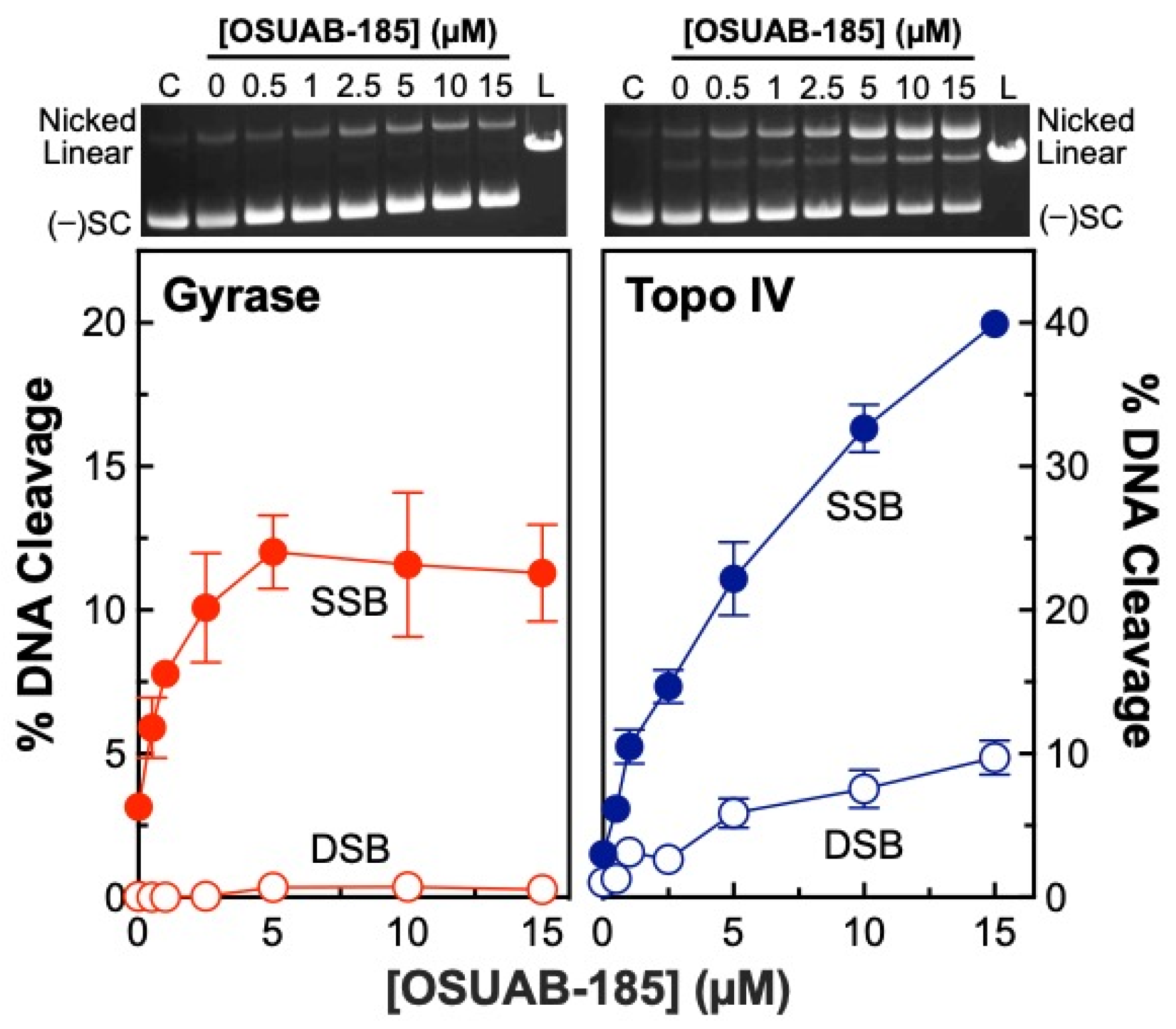
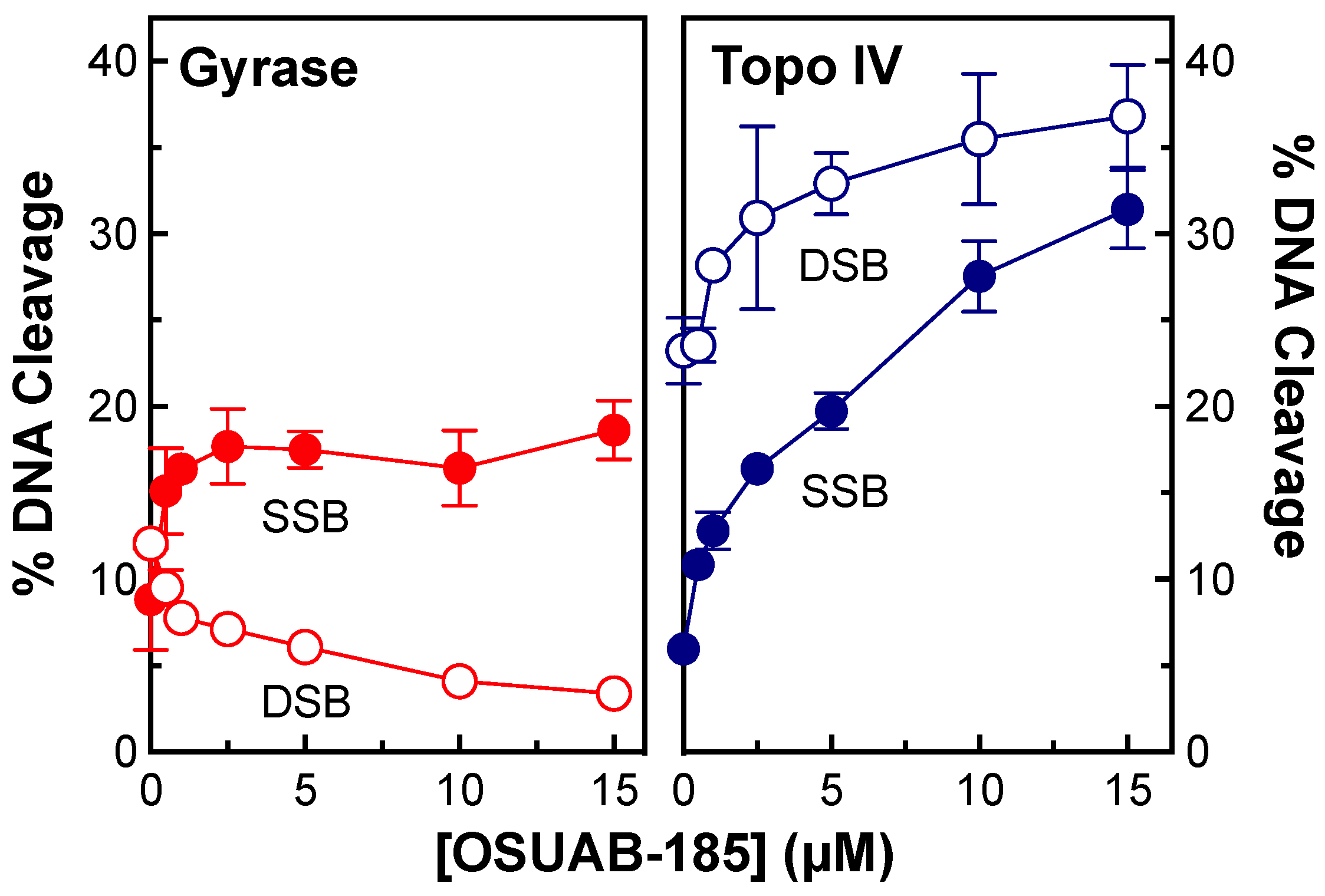
A previous study demonstrated that some dioxane-linked NBTIs were able to induce double-stranded (in addition to single-stranded) DNA breaks mediated by S. aureus gyrase [50]. Therefore, to further explore the ability of this NBTI subclass to induce double-stranded DNA breaks generated by bacterial type II topoisomerases and to address the basis for this activity, we assessed the effects of OSUAB-185 on DNA cleavage mediated by N. gonorrhoeae gyrase and topoisomerase IV (Figure 2). The NBTI displayed moderate activity against gyrase, increasing single-stranded DNA breaks to ~12.0% at 5 µM compound (compared to a baseline level of ~3.1%) with a CC50 value (concentration of compound required to induce 50% of maximal DNA cleavage) of ~0.9 µM. As reported previously for other NBTIs [37,48,49], induction of double-stranded DNA cleavage was not observed. Although OSUAB-185 was less potent against topoisomerase IV (CC50 ≈ 4.8 µM), it was considerably more efficacious, inducing ~40% single-stranded DNA breaks at 15 µM compound (compared to a baseline level of ~3.0%). Strikingly, OSUAB-185 also increased double-stranded DNA breaks mediated by topoisomerase IV. Approximately 10% double-stranded DNA breaks were observed at 15 µM compound (compared to a baseline level of ~1%) with a CC50 value of ~5.7 µM. The increase in double-stranded breaks is contrary to the hallmark of NBTIs, which is the induction of single-stranded DNA breaks by gyrase/topoisomerase IV and the suppression of enzyme-mediated double-stranded DNA breaks [37,48,49].
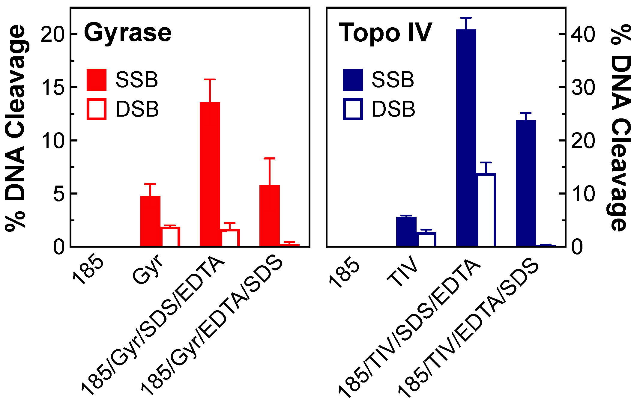
To ensure that the DNA breaks induced by OSUAB-185 were being generated by gyrase and topoisomerase IV, two controls were carried out (Figure 3). First, no single- or double-stranded DNA cleavage was observed in the presence of 10 µM compound in the absence of enzyme. Second, when DNA cleavage reactions were terminated by the addition of EDTA prior to SDS, DNA cleavage levels dropped for both enzymes. EDTA chelates the required catalytic Mg2+ ion, but only when the DNA is ligated [51]. Therefore, the decrease in cleaved DNA following EDTA treatment is inconsistent with a non-enzymatic reaction. These data provide strong evidence that the DNA breaks observed in the presence of OSUAB-185 were generated by gyrase and topoisomerase IV. It is notable that all of the NBTI-induced double-stranded breaks generated by topoisomerase IV disappeared in the presence of EDTA. This finding suggests that they may be less stable than the single-stranded DNA breaks generated in the presence of the compound.
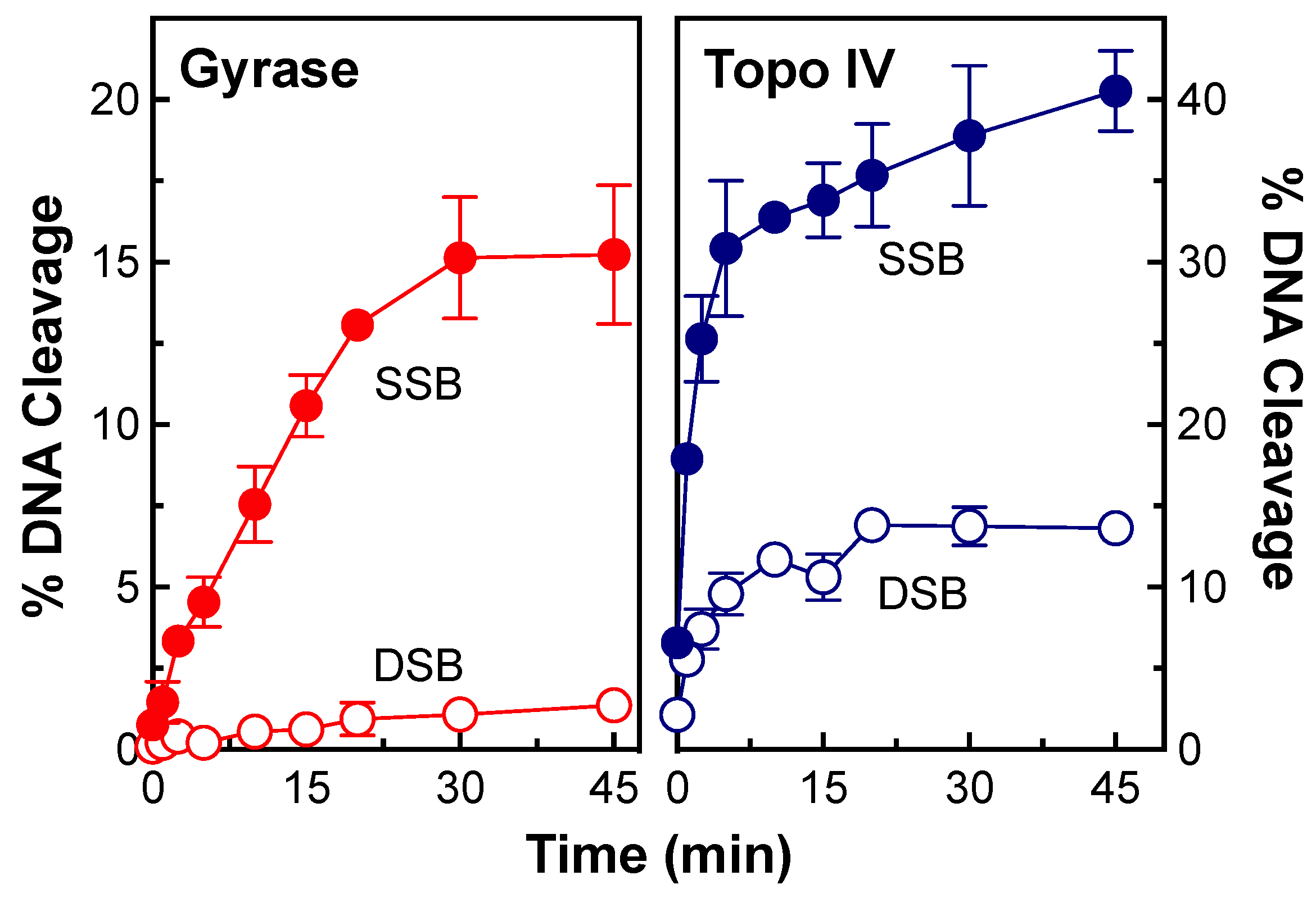
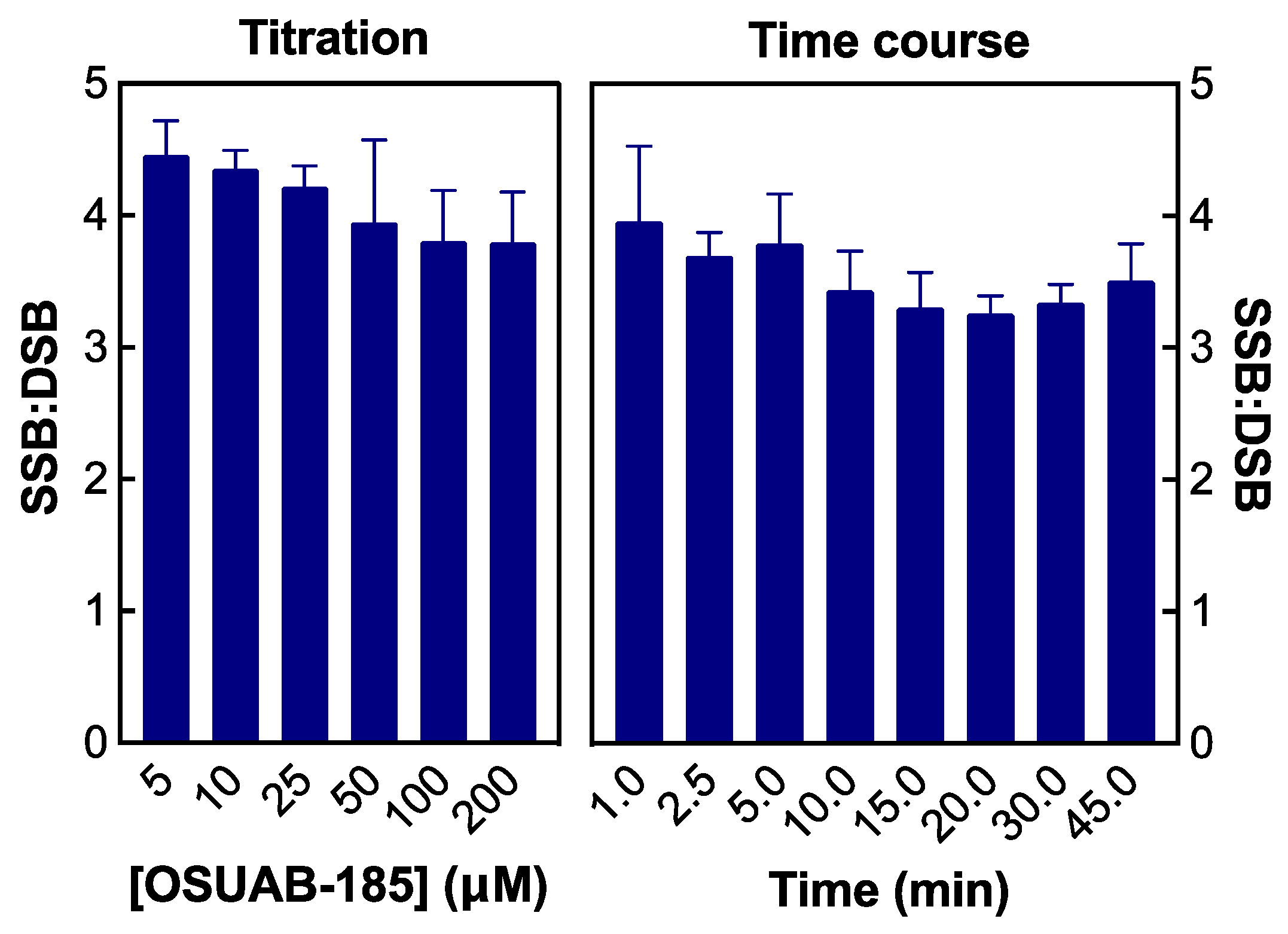
To determine whether the relative levels of topoisomerase IV-mediated double- vs. single-stranded DNA breaks change with time, a time course for DNA scission with both enzymes was monitored (Figure 4). As with the NBTI titration, no appreciable double-stranded breaks were observed with gyrase (Figure 4, left panel). In contrast, double- and single-stranded DNA breaks were observed with topoisomerase IV and appeared to be generated coordinately (Figure 4, right panel). To analyze the formation of double-stranded DNA breaks by topoisomerase IV in greater detail, data from the [OSUAB-185] titration (Figure 2, as well as NBTI concentrations up to 200 µM) and the time course for cleavage (Figure 4) were converted to ratios of single-stranded:double-stranded DNA breaks (Figure 5, left and right panel, respectively). The single-stranded:double-stranded DNA break ratios remained constant over a wide range of NBTI concentrations and cleavage reaction times. These findings provide further evidence that the single- and double-stranded DNA breaks generated by N. gonorrhoeae topoisomerase IV in the presence of OSUAB-185 were generated coordinately. This result argues against a second NBTI molecule entering the active site of topoisomerase IV at higher concentrations of OSUAB-185 or over longer reaction times. It also argues against a change in the binding conformation of the NBTI over time.
As a final demonstration that the induction of topoisomerase IV-mediated double-stranded DNA breaks by OSUAB-185 represents a novel mechanism of action, a titration of the NBTI was carried out in cleavage assays that replaced MgCl2 with CaCl2 (Figure 6). With some enzymes, Ca2+ dramatically raises baseline levels of single- and double-stranded DNA breaks, which affords a more ready assessment of NBTI effects on DNA cleavage [34,48,49,52]. Similar to previous results with other NBTIs, OSUAB-185 induced single-stranded DNA breaks and suppressed double-stranded breaks mediated by N. gonorrhoeae gyrase (Figure 6, left panel). However, in marked contrast, the NBTI enhanced both single- and double-stranded DNA cleavage mediated by topoisomerase IV (Figure 6, right panel). This finding provides strong evidence that OSUAB-185 induces double-stranded DNA breaks mediated by N. gonorrhoeae topoisomerase IV.

2.2. Stability of Single- and Double-Stranded DNA Breaks Induced by OSUAB-185

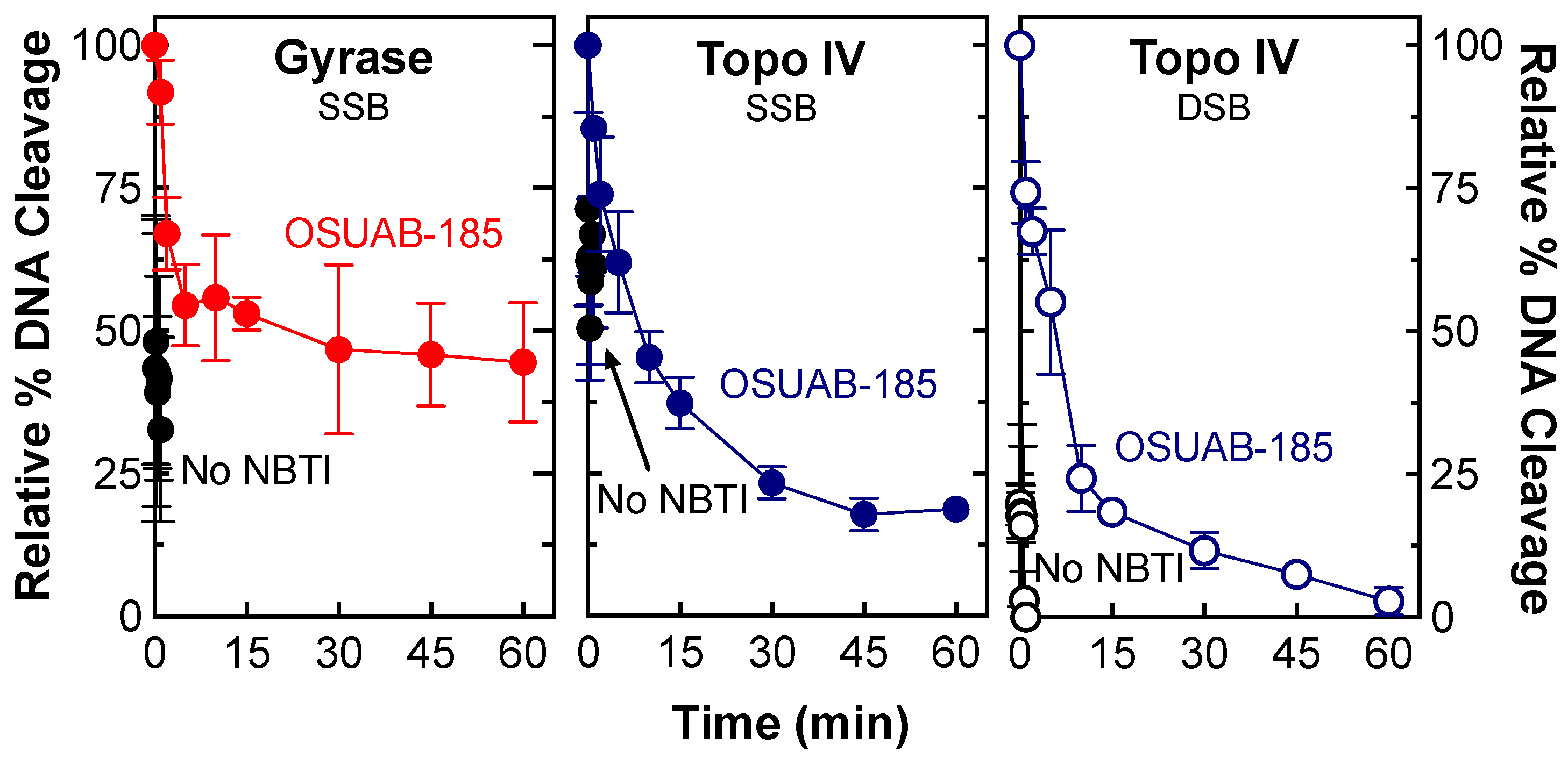
Other things being equal, drugs that generate the most stable cleavage complexes appear to be the most lethal in cells [53]. Therefore, two approaches were utilized to assess the effects of OSUAB-185 on the stability of cleavage complexes formed by N. gonorrhoeae gyrase and topoisomerase IV. In the first approach, the effects of OSUAB-185 on the persistence of cleavage complexes were determined. In this assay, cleavage complexes were formed in the presence of high concentrations of enzyme and DNA, and the lifetimes of cleavage complexes were monitored following 20-fold dilution into reaction buffer that lacked the catalytic divalent metal ion. While the shift in condition does not alter the DNA cleavage–ligation equilibrium in established cleavage complexes, complexes that disassociate are unlikely to reform. As seen in Figure 7, cleavage complexes formed with gyrase (monitoring single-stranded DNA cleavage, left panel) or topoisomerase IV (monitoring single-stranded DNA cleavage, middle panel, or double-stranded DNA cleavage, right panel) in the absence of the NBTI were highly unstable and rapidly disassociated following dilution. Complexes became considerably more stable in the presence of OSUAB-185. Consistent with the data from Figure 3, NBTI-induced double-stranded DNA breaks (t1/2 ≈ 4 min) were less stable than single-stranded DNA breaks (t1/2 ≈ 8 min) with topoisomerase IV.
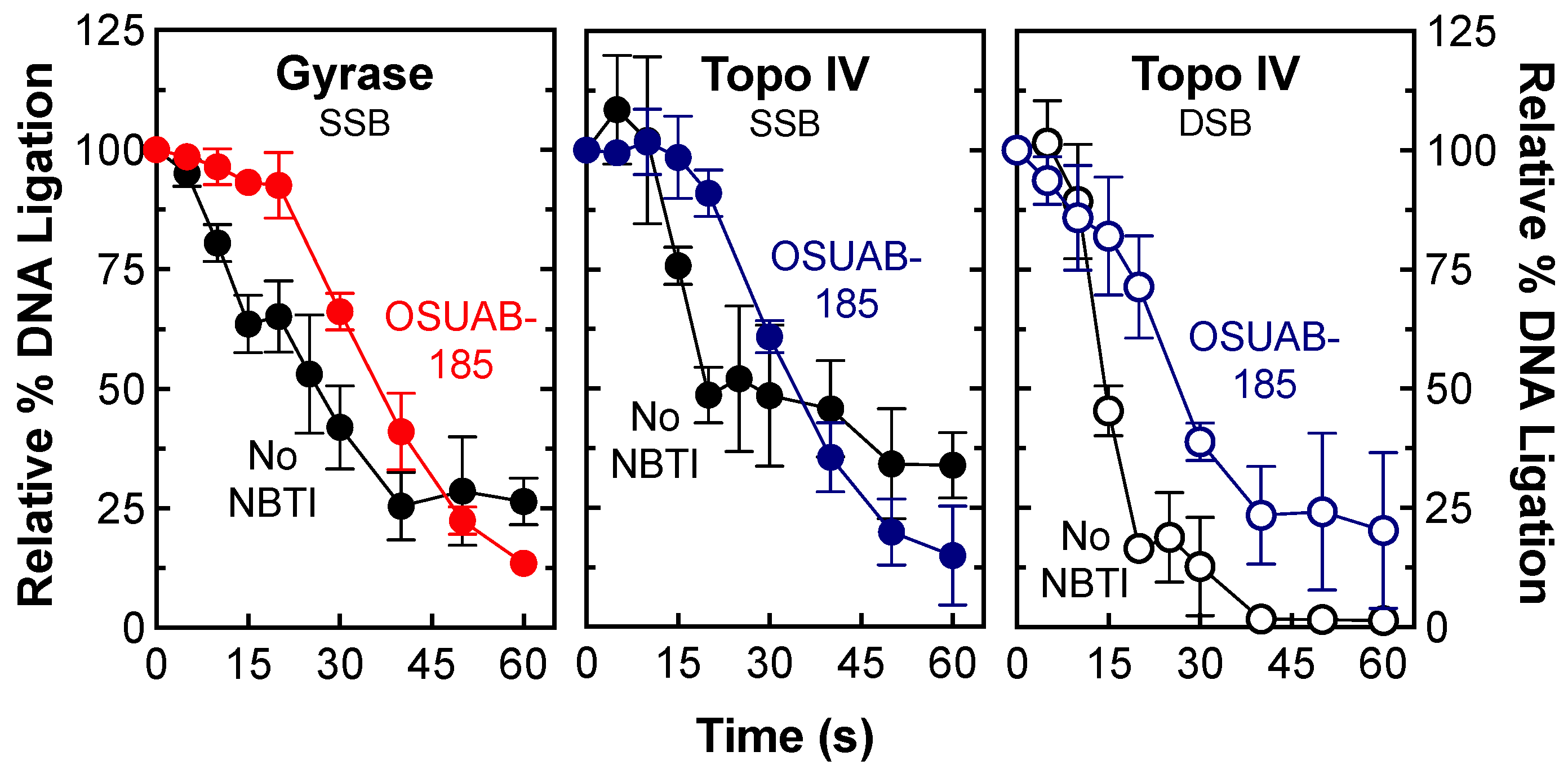
In the second approach, the effects of OSUAB-185 on the rate of gyrase/topoisomerase IV-mediated DNA ligation were monitored by shifting cleavage complexes from 37 °C to 65 °C (a temperature that allows ligation but not cleavage of the DNA) [54]. As seen in Figure 8, OSUAB-185 had a modest effect on rates of ligation for single-stranded DNA breaks with gyrase (left panel) and topoisomerase IV (middle panel) and double-stranded breaks with topoisomerase IV (right panel). The NBTI generated slightly more stable single-stranded (t1/2 ≈ 35 s) DNA cleavage complexes than double-stranded (t1/2 ≈ 27 s) DNA cleavage complexes with topoisomerase IV.

2.3. Effects of OSUAB-185 on Fluoroquinolone-Resistant N. gonorrhoeae Gyrase and Topoisomerase IV

To determine whether OSUAB-185 is able to overcome fluoroquinolone resistance in N. gonorrhoeae type II enzymes, the effects of the NBTI on gyrase that contained GyrAS91F and topoisomerase IV that contained ParCS87N were examined. These two amino acid substitutions represent the most prevalent fluoroquinolone-resistant mutations in N. gonorrhoeae gyrase and topoisomerase IV, respectively (Figure 9) [55,56]. Levels of DNA cleavage with GyrAS91F gyrase (top left panel) were similar to those obtained with the wild-type enzyme and only a single-stranded DNA cleavage was induced by OSUAB-185. However, the NBTI was ~4-fold less potent against the GyrAS91F mutant. In contrast, levels of NBTI-induced DNA scission with ParCS87N topoisomerase IV (top right panel) were ~3 to 4-fold lower than observed for the wild-type enzyme, and OSUAB-185 maintained its potency. Once again, the NBTI induced single- and double-stranded DNA scission with fluoroquinolone-resistant topoisomerase IV. These findings predict that the NBTI would retain at least some activity against fluoroquinolone-resistant N. gonorrhoeae cells harboring common mutations in gyrase/topoisomerase IV. They also suggest that the fluoroquinolone-resistant mutations do not alter the mechanism of action of OSUAB-185 against bacterial type II topoisomerases.

2.4. Effects of OSUAB-185 on Gyrase and Topoisomerase IV from E. coli and S. aureus

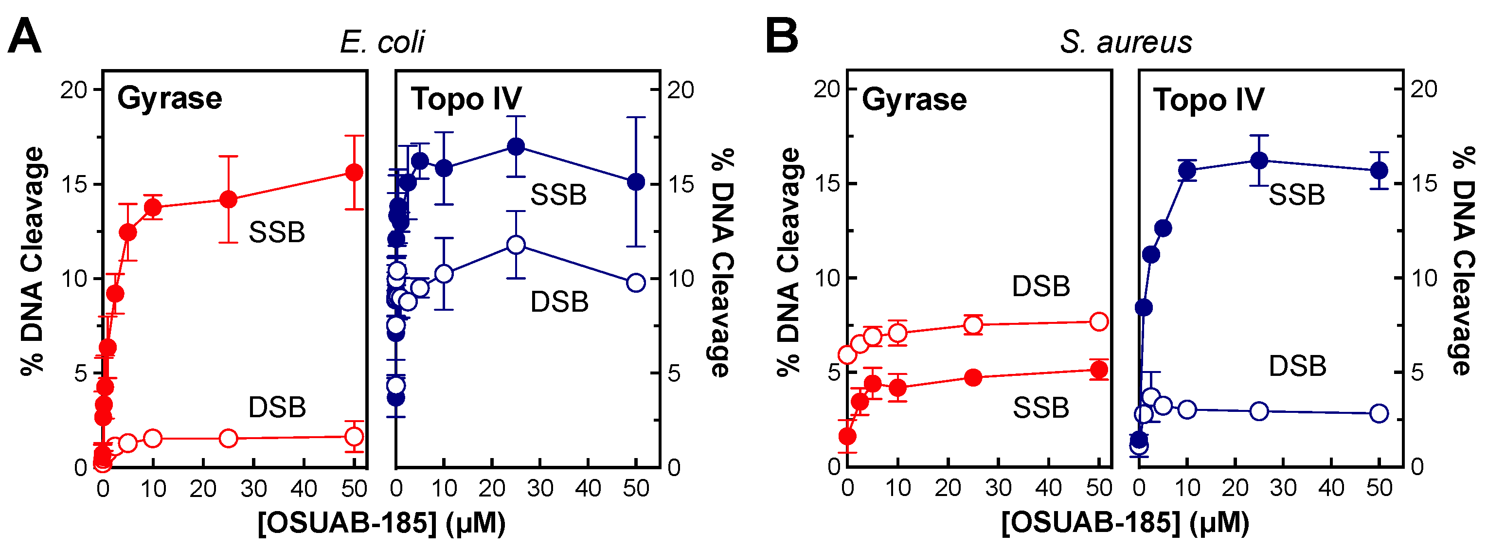
To determine whether OSUAB-185 is able to induce double-stranded DNA breaks with type II topoisomerases from other species (Figure 10), the effects of the NBTI on DNA cleavage mediated by gyrase (left panels) and topoisomerase IV (right panels) from E. coli (Figure 10A) and S. aureus (Figure 10B) were assessed. Note that the induction of double-stranded DNA breaks by OSUAB-185 and S. aureus gyrase has been reported previously [50]. To at least some extent, OSUAB-185 induced double-stranded breaks with all four of the enzymes. Double-stranded DNA breaks were especially prominent with E. coli topoisomerase IV. These data indicate that the ability of the NBTI to generate enzyme-mediated double-stranded DNA breaks is not confined to N. gonorrhoeae or either type II topoisomerase.

2.5. Effects of OSUAB-185 on Human Topoisomerase IIα

Very little is known about the interaction of NBTIs with human type II topoisomerases [57,58,59]. Therefore, the effects of OSUAB-185 on DNA cleavage mediated by human topoisomerase IIα was determined (Figure 11). The NBTI displayed reasonable activity against the human type II enzyme, with CC50 values in the low µM range. In addition, similar to the results with some of the bacterial enzymes, OSUAB-185 induced moderate levels of double-stranded DNA breaks with the human enzyme (maximal levels of cleavage of ~11% and ~8.5% for single- and double-stranded DNA breaks, respectively). This last finding provides further evidence that some members of the NBTI class are capable of generating enzyme-mediated double-stranded DNA breaks.

3. Discussion

NBTIs are an emerging class of compounds with antibacterial activity. In contrast to fluoroquinolones, a hallmark of most NBTIs is their ability to induce gyrase/topoisomerase IV-mediated single-stranded breaks in DNA and suppress the formation of double-stranded breaks [37,48,49]. Together with a previous study, the present work provides strong evidence that select dioxane-linked amide NBTIs are also capable of generating double-stranded DNA breaks mediated by type II topoisomerases from a variety of bacterial species, as well as humans [50].
Fluoroquinolones induce double-stranded DNA breaks because two drug molecules bind in the active site of gyrase and topoisomerase IV, with one molecule stabilizing a cleaved scissile bond on each strand of the double helix [27,46,47]. In contrast, only a single NBTI molecule binds in the active site of the bacterial type II topoisomerases, with the molecule binding midway between the two scissile bonds [35,37,45]. It is believed that canonical NBTIs stabilize single-stranded and suppress double-stranded DNA breaks by distorting the active site of gyrase/topoisomerase IV in a manner that inhibits ligation of the first strand but does not allow cleavage of the second [35,37]. It is not known how OSUAB-185 and potentially other dioxane-linked amide NBTIs generate double-stranded breaks. Results from OSUAB-185 titrations (Figure 2 and Figure 5) and time course experiments (Figure 4 and Figure 5) strongly suggest that it is not due to the binding of a second molecule of compound. Potentially, this NBTI has two mutually exclusive binding configurations that distort the active site of the enzyme differently. In the first, the NBTI acts as a canonical member of this class and induces distortion after one strand is cut to inhibit ligation and prevent cleavage of the second strand (thus enhancing single-stranded DNA breaks). In the second, the NBTI acts in a non-canonical manner and only induces the dramatic active site distortion after both DNA strands are cleaved (thus enhancing double-stranded DNA breaks). Future structural studies are needed to determine if this is the case.
The activity of OSUAB-185 against topoisomerase IIα suggests that this member of this NBTI subclass might cross over to human systems if used to treat infections. However, our results raise an interesting possibility. The double-stranded DNA breaks generated by human type II topoisomerases during transcription have the potential to trigger secondary leukemias in a small percentage of patients treated with topoisomerase II-targeted anticancer drugs [25,60,61,62,63]. If a drug was developed that induced only single-stranded DNA breaks with human type II topoisomerases, it is possible that it could overcome the secondary leukemias observed with current drugs. Although OSUAB-185 induced both single- and double-stranded DNA breaks mediated by human topoisomerase IIα, our findings open the door for future work on NBTIs and human type II topoisomerases.

4. Materials and Methods

4.1. Enzymes and Materials

Wild-type N. gonorrhoeae gyrase (GyrA, GyrB) and topoisomerase IV (ParC, ParE) subunits were prepared as described previously [35,46]. Fluoroquinolone-resistant N. gonorrhoeae GyrAS91F was a gift from Dr. Pan Chan (GlaxoSmithKline, Brentford, UK) and fluoroquinolone-resistant N. gonorrhoeae ParCS87N was generated using a QuickChange II XL site-directed mutagenesis kit (Agilent Technologies, Santa Clara, CA, USA) with custom primers for the desired mutations. Mutant N. gonorrhoeae ParCS87N was expressed and purified as described by Ashley et al. [64] with the following modifications to optimize protein expression and lysis: (1) ParCS87N was expressed for 3 h before harvesting, and (2) cells were lysed by sonication using a digital sonifier (Branson, Danbury, CT, USA). Wild-type E. coli gyrase (GyrA, GyrB) subunits were expressed and purified as described by Chan et al. [46], and wild-type E. coli topoisomerase IV (ParC, ParE, a gift from Dr. Keir Neumann, NHLBI), subunits were expressed and purified as described by Peng and Marians [65]. Wild-type S. aureus gyrase (GyrA, GyrB) and topoisomerase IV (GrlA and GrlB) subunits were ordered from GenScript and expressed and purified as described previously [37,46]. Recombinant human wild-type topoisomerase IIα was expressed in Saccharomyces cerevisiae and purified as described previously [66,67]. The identities of all constructs were confirmed by DNA sequencing, and all enzymes were stored at −80 °C.
Negatively supercoiled pBR322 DNA was prepared from E. coli using a Plasmid Mega Kit (Qiagen, Hilden, Germany) as described by the manufacturer. OSUAB-185 was synthesized as described previously [50] (shown as compound 23) and stored at 4 °C as a 20 mM stock solution in 100% DMSO. All other chemicals were of analytical reagent grade.

4.2. DNA Cleavage

DNA cleavage reactions were performed according to the procedure of Aldred et al. [33] (gyrase and topoisomerase IV) or Fortune and Osheroff [68] (human topoisomerase IIα). Reactions were performed in the absence of an NBTI or in the presence of increasing concentrations of OSUAB-185. Unless stated otherwise, assay mixtures contained 10 nM pBR322 and 100 nM wild-type or mutant (GyrAS91F) N. gonorrhoeae gyrase, 100 nM wild-type or mutant (ParCS87N) N. gonorrhoeae topoisomerase IV, 100 nM wild-type E. coli gyrase, 20 nM wild-type E. coli topoisomerase IV, 100 nM wild-type S. aureus gyrase, 20 nM S. aureus topoisomerase IV, or 300 nM wild-type human topoisomerase IIα in a total volume of 20 µL of cleavage buffer: 40 mM Tris-HCl (pH 7.9), 50 mM NaCl, 2.5% (w/v) glycerol, and 5 mM or 10 mM MgCl2 for E. coli or N. gonorrhoeae type II topoisomerases, respectively; 50 mM Tris-HCl (pH 7.5), 100 mM KGlu, 5 mM MgCl2, 5 mM dithiothreitol (DTT), and 50 μg/mL BSA for S. aureus gyrase; 40 mM Tris-HCl (pH 7.5), 6 mM MgCl2, 20 mM NaCl, 10 mM DTT, and 50 μg/mL BSA for S. aureus topoisomerase IV; 50 mM Tris-HCl (pH 7.9), 0.5 mM EDTA (pH 8.0), 500 mM KCl, 25 mM MgCl2, and 12.5% (w/v) glycerol for human topoisomerase IIα. In some cases, MgCl2 in the cleavage buffer was replaced with an equivalent concentration of CaCl2. Reactions were incubated at 37 °C for 30 min for wild-type and mutant (GyrAS91F) N. gonorrhoeae gyrase, mutant (ParCS87N) N. gonorrhoeae topoisomerase IV, and wild-type S. aureus gyrase; 20 min with wild-type E. coli gyrase; and 10 min for all other enzymes. Enzyme–DNA cleavage complexes were trapped by adding 2 µL of 4% SDS, followed by 2 µL of 250 mM EDTA (pH 8.0). Proteinase K was added (2 µL of a 0.8 mg/mL solution), and reaction mixtures were incubated for 30 min at 45 °C to digest the enzyme. Samples were mixed with 2 µL of agarose loading buffer [60% sucrose, 10 mM Tris-HCl (pH 7.9), 0.5% bromophenol blue, and 0.5% xylene cyanol FF] and heated for 2 min at 45 °C prior to electrophoresis in 1% agarose gels in 40 mM Tris-acetate (pH 8.3), and 2 mM EDTA containing 0.5 µg/mL ethidium bromide. DNA bands were visualized by mid-range ultraviolet light and quantified using an Alpha Innotech digital imaging system (Protein Simple, San Jose, CA, USA). DNA cleavage was monitored by the conversion of negatively supercoiled plasmid DNA to nicked (single-stranded break) or linear (double-stranded break) molecules. CC50 values (the concentration of compound that induced 50% maximal DNA cleavage complex formation) were calculated on GraphPad Prism Version 9.5.1 using a non-linear regression analysis with 95% confidence intervals.

4.3. Persistence of Cleavage Complexes

The persistence of gyrase/topoisomerase IV–DNA cleavage complexes was determined as described previously [33]. Cleavage complexes were formed by combining 50 nM pBR322 and 200 nM N. gonorrhoeae gyrase or 100 nM N. gonorrhoeae topoisomerase IV in the presence of 10 µM OSUAB-185 in a total volume of 20 µL of a cleavage buffer. Parallel control experiments were conducted to assess cleavage complexes formed in the absence of the NBTI by combining 50 nM pBR322 and 500 nM N. gonorrhoeae gyrase/topoisomerase IV in a 20 µL cleavage buffer. Reactions were incubated at 37 °C until the DNA cleavage/ligation equilibria were reached (30 min with gyrase and 10 min with topoisomerase IV) and diluted 20-fold in a DNA cleavage buffer lacking Mg2+. Samples (20 µL) were removed at time points ranging from 0 to 60 min, and reactions were stopped with 2 µL of 4% SDS, followed by 2 µL of 250 mM EDTA (pH 8.0). Proteinase K was added (2 µL of a 0.8 mg/mL solution), and the reactions mixtures were incubated for 30 min at 45 °C. Samples were mixed with 2 µL of agarose gel loading buffer and processed and analyzed as described above. Levels of DNA cleavage were set to 100% at time zero, and the persistence of cleavage complexes were determined by the decay of nicked (single-stranded breaks) or linear (double-stranded breaks) reaction products over time. Cleavage complex stability (half-life, t1/2) was calculated on GraphPad Prism Version 9.5.1 using non-linear regression analysis with 95% confidence intervals.

4.4. DNA Ligation

DNA ligation mediated by N. gonorrhoeae gyrase and topoisomerase IV was monitored using the procedure of Aldred et al. [33]. Initial reactions contained 10 nM pBR322 and 100 nM wild-type N. gonorrhoeae gyrase or topoisomerase IV in the absence or presence of 10 µM OSUAB-185. In order to better visualize levels of DNA breaks mediated by gyrase or topoisomerase IV in the absence of the NBTI, 10 mM MgCl2 was replaced by 10 mM CaCl2. DNA cleavage/ligation equilibria were established for 30 min with gyrase and 10 min with topoisomerase IV at 37 °C. DNA ligation was initiated by shifting samples from 37 °C to 65 °C, which allows enzyme-mediated ligation but prevents new rounds of DNA cleavage from occurring [54]. This results in a unidirectional sealing of the cleaved DNA. Reactions were stopped at time points ranging from 0 to 60 s by the addition of 4% SDS, followed by 2 µL 250 mM EDTA (pH 8.0). Proteinase K was added (2 µL of a 0.8 mg/mL solution), and reaction mixtures were incubated for 30 min at 45 °C. Samples were mixed with 2 µL of agarose loading buffer and processed and analyzed as described above. Nicked and linear DNA cleavage products at time zero were set to 100% to allow direct comparison between different conditions, and DNA ligation of single- or double-stranded breaks was monitored by the loss of nicked or linear DNA, respectively. The rate of DNA ligation (t1/2) was calculated on GraphPad Prism Version 9.5.1 using non-linear regression analysis with 95% confidence intervals.

4.5. Accession Codes

N. gonorrhoeae Gyrase, GyrA: UniProtKB P48371, GyrB: UniProtKB P22118; N. gonorrhoeae Topoisomerase IV, ParC: UniProtKB P48374, ParE: UniProtKB A0A8D9YA86; E. coli Gyrase, GyrA: UniProtKB P0AES4, GyrB: UniProtKB P0AES6; E. coli Topoisomerase IV, ParC: UniProtKB P0AFI2, ParE: UniProtKB P20083; B. anthracis Gyrase, GyrA: UniProtKB A0A0F7R8R3, GyrB: UniProtKB Q9X3Y6; B. anthracis Topoisomerase IV, GrlA: UniProtKB A0A6H3AG07, GrlB: UniProtKB A0A2B0YAF3; S. aureus Gyrase, GyrA: UniProtKB Q2FKQ0, GyrB: UniProtKB P0A0K8; S. aureus Topoisomerase IV, GrlA: UniProtKB P0C1U9, GrlB: UniProtKB P0C1S7; Human Topoisomerase IIα: UniProtKB P11388.

Author Contributions

Conceptualization, J.C.Y., M.J.M.-F. and N.O.; methodology, investigation, and analysis, S.E.D., J.A.C. and J.A.W.B.; visualization, S.E.D. and J.A.C.; resources, Y.L., J.C.Y., M.J.M.-F. and N.O.; funding acquisition, M.J.M.-F. and N.O.; writing—original draft preparation, S.E.D., J.A.C. and N.O.; writing—review and editing, all authors. All authors have read and agreed to the published version of the manuscript.

Funding

This research was funded by US Veterans Administration Merit Review Award I01 Bx002198 (N.O.), Dr. Ralph and Marian Fak Medical Research Trust (Transformational Award) (M.J.M-F.), and National Institutes of Health grants R01 GM126363 (N.O.), R01 AI170546 (N.O.), and R01 AI173072 (M.J.M-F.). S.E.D. was supported in part by the Aspirnaut Undergraduate Discovery Science Experience in Renal Biology and Disease (R25 DK096999).

Institutional Review Board Statement

Not applicable.

Informed Consent Statement

Not applicable.

Data Availability Statement

All data are contained within the article.

Acknowledgments

We thank Jeffrey Y. Jian, Jillian F. Armenia, and Samika S. Joshi for their critical reading of the manuscript.

Conflicts of Interest

The authors declare no conflict of interest.

References

  1. Centers for Disease Control and Prevention. Outpatient Antibiotic Prescriptions—United States, 2017; Centers for Disease Control and Prevention: Atlanta, GA, USA, 2017.
  2. Basarab, G.S. Four ways to skin a cat: Inhibition of bacterial topoisomerases leading to the clinic. In Antibacterials: Volume I; Fisher, J.F., Mobashery, S., Miller, M.J., Eds.; Springer International Publishing: Cham, Switzerland, 2018; pp. 165–188. [Google Scholar]
  3. World Health Organization. Critically Important Antimicrobials for Human Medicine; World Health Organization: Geneva, Switzerland, 2019. [Google Scholar]
  4. Aldred, K.J.; Kerns, R.J.; Osheroff, N. Mechanism of quinolone action and resistance. Biochemistry 2014, 53, 1565–1574. [Google Scholar] [CrossRef] [PubMed]
  5. Hooper, D.C.; Jacoby, G.A. Mechanisms of drug resistance: Quinolone resistance. Ann. N. Y. Acad. Sci. 2015, 1354, 12–31. [Google Scholar] [PubMed] [Green Version]
  6. Gibson, E.G.; Ashley, R.E.; Kerns, R.J.; Osheroff, N. Fluoroquinolone interactions with bacterial type II topoisomerases and target-mediated drug resistance. In Antimicrobial Resistance and Implications for the 21st Century; Drlica, K., Shlaes, D., Fong, I.W., Eds.; Springer: New York, NY, USA, 2018; pp. 507–529. [Google Scholar]
  7. Bush, N.G.; Diez-Santos, I.; Abbott, L.R.; Maxwell, A. Quinolones: Mechanism, lethality and their contributions to antibiotic resistance. Molecules 2020, 25, 5662. [Google Scholar] [CrossRef]
  8. Unemo, M.; Del Rio, C.; Shafer, W.M. Antimicrobial resistance expressed by Neisseria gonorrhoeae: A major global public health problem in the 21st century. Microbiol. Spectr. 2016, 4, 213–237. [Google Scholar]
  9. Kirkcaldy, R.D.; Harvey, A.; Papp, J.R.; Del Rio, C.; Soge, O.O.; Holmes, K.K.; Hook, E.W., 3rd; Kubin, G.; Riedel, S.; Zenilman, J.; et al. Neisseria gonorrhoeae antimicrobial susceptibility surveillance—The gonococcal isolate surveillance project, 27 sites, United States, 2014. MMWR Surveill. Summ. 2016, 65, 1–19. [Google Scholar] [CrossRef] [Green Version]
  10. CDC. Update to CDC’s sexually transmitted diseases treatment guidelines, 2006: Fluoroquinolones no longer recommended for treatment of gonococcal infections. MMWR Morb. Mortal. Wkly. Rep. 2007, 56, 332–336. [Google Scholar]
  11. Sexually Transmitted Disease Surveillance 2021; Centers for Disease Control and Prevention: Atlanta, GA, USA, 2021.
  12. CDC. Antibiotic Resistance Threats in the United States, 2019; U.S. Department of Health and Human Services, CDC: Atlanta, GA, USA, 2019.
  13. Galvin, S.R.; Cohen, M.S. The role of sexually transmitted diseases in HIV transmission. Nat. Rev. Microbiol. 2004, 2, 33–42. [Google Scholar] [CrossRef]
  14. Morgan, M.K.; Decker, C.F. Gonorrhea. Dis. Mon. 2016, 62, 260–268. [Google Scholar] [CrossRef]
  15. Koebler, J. World Health Organization Warns Gonorrhea Could Join HIV as ‘Uncurable’. U.S. News and World Report. 2012. Available online: https://www.usnews.com/news/articles/2012/06/06/world-health-organization-warns-gonorrhea-could-join-hiv-as-uncurable- (accessed on 20 June 2023).
  16. World Health Organization. Multi-Drug Resistant Gonorrhoea; World Health Organization: Geneva, Switzerland, 2022. [Google Scholar]
  17. Liu, Z.; Deibler, R.W.; Chan, H.S.; Zechiedrich, L. The why and how of DNA unlinking. Nucleic Acids Res. 2009, 37, 661–671. [Google Scholar] [CrossRef] [Green Version]
  18. Vos, S.M.; Tretter, E.M.; Schmidt, B.H.; Berger, J.M. All tangled up: How cells direct, manage and exploit topoisomerase function. Nat. Rev. Mol. Cell Biol. 2011, 12, 827–841. [Google Scholar]
  19. Chen, S.H.; Chan, N.L.; Hsieh, T.S. New mechanistic and functional insights into DNA topoisomerases. Annu. Rev. Biochem. 2013, 82, 139–170. [Google Scholar] [CrossRef] [PubMed]
  20. Bush, N.G.; Evans-Roberts, K.; Maxwell, A. DNA topoisomerases. EcoSal Plus 2015, 6, 1–34. [Google Scholar] [CrossRef]
  21. Dalvie, E.D.; Osheroff, N. DNA topoisomerases: Type II. In The Encyclopedia of Biological Chemistry, 3rd ed.; Allewell, N.M., Ed.; Academic Press: Waltham, MA, USA, 2021; Volume 3, pp. 479–486. [Google Scholar]
  22. McKie, S.J.; Neuman, K.C.; Maxwell, A. DNA topoisomerases: Advances in understanding of cellular roles and multi-protein complexes via structure-function analysis. Bioessays 2021, 43, 2000286. [Google Scholar] [CrossRef] [PubMed]
  23. Ashley, R.E.; Osheroff, N. Regulation of DNA by topoisomerases: Mathematics at the molecular level. In Knots, Low-Dimensional Topology and Applications; Adams, C., Gordon, C.M., Jones, V., Kauffman, L., Lambropoulou, S., Millet, K., Przytycki, J., Ricca, R., Sazdanovic, R., Eds.; Springer International Publishing: Cham, Switzerland, 2019. [Google Scholar]
  24. Dong, K.C.; Berger, J.M. Structural basis for gate-DNA recognition and bending by type IIA topoisomerases. Nature 2007, 450, 1201–1205. [Google Scholar] [CrossRef] [PubMed]
  25. Deweese, J.E.; Osheroff, N. The DNA cleavage reaction of topoisomerase II: Wolf in sheep’s clothing. Nucleic Acids Res. 2009, 37, 738–749. [Google Scholar] [CrossRef] [Green Version]
  26. Laponogov, I.; Pan, X.S.; Veselkov, D.A.; McAuley, K.E.; Fisher, L.M.; Sanderson, M.R. Structural basis of gate-DNA breakage and resealing by type II topoisomerases. PLoS ONE 2010, 5, e11338. [Google Scholar] [CrossRef]
  27. Wohlkonig, A.; Chan, P.F.; Fosberry, A.P.; Homes, P.; Huang, J.; Kranz, M.; Leydon, V.R.; Miles, T.J.; Pearson, N.D.; Perera, R.L.; et al. Structural basis of quinolone inhibition of type IIA topoisomerases and target-mediated resistance. Nat. Struct. Mol. Biol. 2010, 17, 1152–1153. [Google Scholar] [CrossRef] [PubMed]
  28. Vann, K.R.; Oviatt, A.A.; Osheroff, N. Topoisomerase II poisons: Converting essential enzymes into molecular scissors. Biochemistry 2021, 60, 1630–1641. [Google Scholar] [CrossRef]
  29. Michel, B. After 30 years of study, the bacterial SOS response still surprises us. PLoS Biol. 2005, 3, e255. [Google Scholar] [CrossRef]
  30. Drlica, K.; Malik, M.; Kerns, R.J.; Zhao, X. Quinolone-mediated bacterial death. Antimicrob. Agents Chemother. 2008, 52, 385–392. [Google Scholar] [CrossRef] [Green Version]
  31. Schroder, W.; Goerke, C.; Wolz, C. Opposing effects of aminocoumarins and fluoroquinolones on the SOS response and adaptability in Staphylococcus aureus. J. Antimicrob. Chemother. 2013, 68, 529–538. [Google Scholar] [CrossRef] [Green Version]
  32. Heisig, P.; Schedletzky, H.; Falkensteinpaul, H. Mutations in the gyrA gene of a highly fluoroquinolone-resistant clinical isolate of Escherichia coli. Antimicrob. Agents Chemother. 1993, 37, 696–701. [Google Scholar] [CrossRef] [Green Version]
  33. Aldred, K.J.; McPherson, S.A.; Wang, P.; Kerns, R.J.; Graves, D.E.; Turnbough, C.L.; Osheroff, N. Drug interactions with Bacillus anthracis topoisomerase IV: Biochemical basis for quinolone action and resistance. Biochemistry 2012, 51, 370–381. [Google Scholar]
  34. Aldred, K.J.; McPherson, S.A.; Turnbough, C.L.; Kerns, R.J.; Osheroff, N. Topoisomerase IV-quinolone interactions are mediated through a water-metal ion bridge: Mechanistic basis of quinolone resistance. Nucleic Acids Res. 2013, 41, 4628–4639. [Google Scholar] [CrossRef] [PubMed]
  35. Bax, B.D.; Chan, P.F.; Eggleston, D.S.; Fosberry, A.; Gentry, D.R.; Gorrec, F.; Giordano, I.; Hann, M.M.; Hennessy, A.; Hibbs, M.; et al. Type IIA topoisomerase inhibition by a new class of antibacterial agents. Nature 2010, 466, 935–940. [Google Scholar] [CrossRef]
  36. Basarab, G.S.; Kern, G.H.; McNulty, J.; Mueller, J.P.; Lawrence, K.; Vishwanathan, K.; Alm, R.A.; Barvian, K.; Doig, P.; Galullo, V.; et al. Responding to the challenge of untreatable gonorrhea: ETX0914, a first-in-class agent with a distinct mechanism-of-action against bacterial type II topoisomerases. Sci. Rep. 2015, 5, 11827. [Google Scholar] [CrossRef] [PubMed] [Green Version]
  37. Gibson, E.G.; Bax, B.; Chan, P.F.; Osheroff, N. Mechanistic and structural basis for the actions of the antibacterial gepotidacin against Staphylococcus aureus gyrase. ACS Infect. Dis. 2019, 5, 570–581. [Google Scholar] [CrossRef] [PubMed]
  38. Bax, B.D.; Murshudov, G.; Maxwell, A.; Germe, T. DNA topoisomerase inhibitors: Trapping a DNA-cleaving machine in motion. J. Mol. Biol. 2019, 431, 3427–3449. [Google Scholar] [CrossRef] [PubMed]
  39. GlaxoSmithKline. A Phase III, Randomized, Multicenter, Parallel-Group, Double-Blind, Double-Dummy Study in Adolescent and Adult Female Participants Comparing the Efficacy and Safety of Gepotidacin to Nitrofurantoin in the Treatment of Uncomplicated Urinary Tract Infection (Acute Cystitis); National Library of Medicine: Bethesda, MD, USA, 2021.
  40. GlaxoSmithKline. A Phase III, Randomized, Multicenter, Open-Label Study in Adolescent and Adult Participants Comparing the Efficacy and Safety of Gepotidacin to Ceftriaxone Plus Azithromycin in the Treatment of Uncomplicated Urogenital Gonorrhea Caused by Neisseria gonorrhoeae; National Library of Medicine: Bethesda, MD, USA, 2022.
  41. Perry, C.; Hossain, M.; Powell, M.; Raychaudhuri, A.; Scangarella-Oman, N.; Tiffany, C.; Xu, S.; Dumont, E.; Janmohamed, S. Design of two phase III, randomized, multicenter studies comparing gepotidacin with nitrofurantoin for the treatment of uncomplicated urinary tract infection in female participants. Infect. Dis. Ther. 2022, 11, 2297–2310. [Google Scholar] [CrossRef]
  42. Scangarella-Oman, N.E.; Hossain, M.; Perry, C.R.; Tiffany, C.; Powell, M.; Swift, B.; Dumont, E.F. Dose selection for a phase III study evaluating gepotidacin (GSK2140944) in the treatment of uncomplicated urogenital gonorrhoea. Sex. Transm. Infect. 2023, 99, 64–69. [Google Scholar] [CrossRef]
  43. Watkins, R.R.; Thapaliya, D.; Lemonovich, T.L.; Bonomo, R.A. Gepotidacin: A novel, oral, ‘first-in-class’ triazaacenaphthylene antibiotic for the treatment of uncomplicated urinary tract infections and urogenital gonorrhoea. J. Antimicrob. Chemother. 2023, 78, 1137–1142. [Google Scholar] [CrossRef] [PubMed]
  44. GlaxoSmithKline. EAGLE-2 and EAGLE-3 Phase III Trials for Gepotidacin Stopped Early for Efficacy Following Pre-Planned Interim Analysis by Independent Data Monitoring Committee; GlaxoSmithKline: Brentford, UK, 2022. [Google Scholar]
  45. Vanden Broeck, A.; Lotz, C.; Ortiz, J.; Lamour, V. Cryo-EM structure of the complete E. coli DNA gyrase nucleoprotein complex. Nat. Commun. 2019, 10, 4935. [Google Scholar] [CrossRef] [PubMed] [Green Version]
  46. Chan, P.F.; Srikannathasan, V.; Huang, J.; Cui, H.; Fosberry, A.P.; Gu, M.; Hann, M.M.; Hibbs, M.; Homes, P.; Ingraham, K.; et al. Structural basis of DNA gyrase inhibition by antibacterial QPT-1, anticancer drug etoposide and moxifloxacin. Nat. Commun. 2015, 6, 10048. [Google Scholar] [CrossRef] [PubMed] [Green Version]
  47. Blower, T.R.; Williamson, B.H.; Kerns, R.J.; Berger, J.M. Crystal structure and stability of gyrase-fluoroquinolone cleaved complexes from Mycobacterium tuberculosis. Proc. Natl. Acad. Sci. USA 2016, 113, 1706–1713. [Google Scholar] [CrossRef] [PubMed]
  48. Gibson, E.G.; Blower, T.R.; Cacho, M.; Bax, B.; Berger, J.M.; Osheroff, N. Mechanism of action of Mycobacterium tuberculosis gyrase inhibitors: A novel class of gyrase poisons. ACS Infect. Dis. 2018, 4, 1211–1222. [Google Scholar] [CrossRef] [Green Version]
  49. Gibson, E.G.; Oviatt, A.A.; Cacho, M.; Neuman, K.C.; Chan, P.F.; Osheroff, N. Bimodal actions of a naphthyridone/aminopiperidine-based antibacterial that targets gyrase and topoisomerase IV. Biochemistry 2019, 58, 4447–4455. [Google Scholar] [CrossRef]
  50. Lu, Y.; Papa, J.L.; Nolan, S.; English, A.; Seffernick, J.T.; Shkolnikov, N.; Powell, J.; Lindert, S.; Wozniak, D.J.; Yalowich, J.; et al. Dioxane-linked amide derivatives as novel bacterial topoisomerase inhibitors against gram-positive Staphylococcus aureus. ACS Med. Chem. Lett. 2020, 11, 2446–2454. [Google Scholar] [CrossRef]
  51. Osheroff, N. Role of the divalent cation in topoisomerase II mediated reactions. Biochemistry 1987, 26, 6402–6406. [Google Scholar] [CrossRef]
  52. Osheroff, N.; Zechiedrich, E.L. Calcium-promoted DNA cleavage by eukaryotic topoisomerase II: Trapping the covalent enzyme-DNA complex in an active form. Biochemistry 1987, 26, 4303–4309. [Google Scholar] [CrossRef]
  53. Bandele, O.J.; Osheroff, N. The efficacy of topoisomerase II-targeted anticancer agents reflects the persistence of drug-induced cleavage complexes in cells. Biochemistry 2008, 47, 11900–11908. [Google Scholar] [CrossRef] [Green Version]
  54. Anderson, V.E.; Zaniewski, R.P.; Kaczmarek, F.S.; Gootz, T.D.; Osheroff, N. Quinolones inhibit DNA religation mediated by Staphylococcus aureus topoisomerase IV: Changes in drug mechanism across evolutionary boundaries. J. Biol. Chem. 1999, 274, 35927–35932. [Google Scholar] [PubMed] [Green Version]
  55. Hall, C.L.; Harrison, M.A.; Pond, M.J.; Chow, C.; Harding-Esch, E.M.; Sadiq, S.T. Genotypic determinants of fluoroquinolone and macrolide resistance in Neisseria gonorrhoeae. Sex. Health 2019, 16, 479–487. [Google Scholar] [CrossRef] [PubMed] [Green Version]
  56. Dan, M. The use of fluoroquinolones in gonorrhoea: The increasing problem of resistance. Expert Opin. Pharmacother. 2004, 5, 829–854. [Google Scholar] [CrossRef] [PubMed]
  57. Mitton-Fry, M.J.; Brickner, S.J.; Hamel, J.C.; Brennan, L.; Casavant, J.M.; Chen, M.; Chen, T.; Ding, X.Y.; Driscoll, J.; Hardink, J.; et al. Novel quinoline derivatives as inhibitors of bacterial DNA gyrase and topoisomerase IV. Bioorg. Med. Chem. Lett. 2013, 23, 2955–2961. [Google Scholar] [CrossRef] [PubMed]
  58. Li, L.S.; Okumu, A.; Dellos-Nolan, S.; Li, Z.; Karmahapatra, S.; English, A.; Yalowich, J.C.; Wozniak, D.J.; Mitton-Fry, M.J. Synthesis and anti-staphylococcal activity of novel bacterial topoisomerase inhibitors with a 5-amino-1,3-dioxane linker moiety. Bioorg. Med. Chem. Lett. 2018, 28, 2477–2480. [Google Scholar] [CrossRef] [PubMed]
  59. Li, L.; Okumu, A.A.; Nolan, S.; English, A.; Vibhute, S.; Lu, Y.; Hervert-Thomas, K.; Seffernick, J.T.; Azap, L.; Cole, S.L.; et al. 1,3-dioxane-linked bacterial topoisomerase inhibitors with enhanced antibacterial activity and reduced hERG inhibition. ACS Infect. Dis. 2019, 5, 1115–1128. [Google Scholar] [CrossRef]
  60. Felix, C.A.; Kolaris, C.P.; Osheroff, N. Topoisomerase II and the etiology of chromosomal translocations. DNA Repair 2006, 5, 1093–1108. [Google Scholar] [CrossRef]
  61. Joannides, M.; Grimwade, D. Molecular biology of therapy-related leukaemias. Clin. Transl. Oncol. 2010, 12, 8–14. [Google Scholar] [CrossRef]
  62. Cowell, I.G.; Austin, C.A. Mechanism of generation of therapy related leukemia in response to anti-topoisomerase II agents. Int. J. Environ. Res. Public Health 2012, 9, 2075–2091. [Google Scholar] [CrossRef]
  63. Pendleton, M.; Lindsey, R.H., Jr.; Felix, C.A.; Grimwade, D.; Osheroff, N. Topoisomerase II and leukemia. Ann. N. Y. Acad. Sci. 2014, 1310, 98–110. [Google Scholar] [CrossRef] [Green Version]
  64. Ashley, R.E.; Dittmore, A.; McPherson, S.A.; Turnbough, C.L., Jr.; Neuman, K.C.; Osheroff, N. Activities of gyrase and topoisomerase IV on positively supercoiled DNA. Nucleic Acids Res. 2017, 45, 9611–9624. [Google Scholar] [CrossRef] [Green Version]
  65. Peng, H.; Marians, K.J. Escherichia coli topoisomerase IV. Purification, characterization, subunit structure, and subunit interactions. J. Biol. Chem. 1993, 268, 24481–24490. [Google Scholar] [CrossRef] [PubMed]
  66. Worland, S.T.; Wang, J.C. Inducible overexpression, purification, and active site mapping of DNA topoisomerase II from the yeast Saccharomyces cerevisiae. J. Biol. Chem. 1989, 264, 4412–4416. [Google Scholar] [CrossRef] [PubMed]
  67. Kingma, P.S.; Greider, C.A.; Osheroff, N. Spontaneous DNA lesions poison human topoisomerase IIα and stimulate cleavage proximal to leukemic 11q23 chromosomal breakpoints. Biochemistry 1997, 36, 5934–5939. [Google Scholar] [CrossRef] [PubMed]
  68. Fortune, J.M.; Osheroff, N. Merbarone inhibits the catalytic activity of human topoisomerase IIα by blocking DNA cleavage. J. Biol. Chem. 1998, 273, 17643–17650. [Google Scholar] [CrossRef] [PubMed] [Green Version]
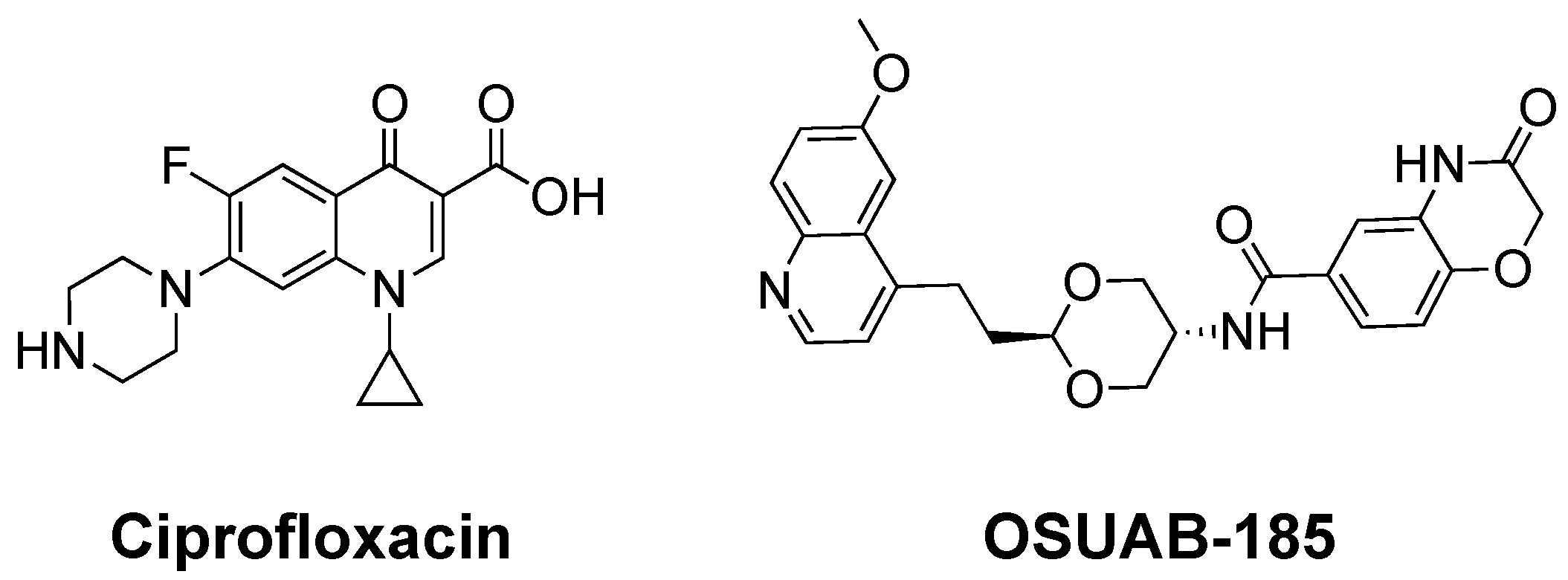
Figure 1. Structures of the fluoroquinolone ciprofloxacin and the NBTI OSUAB-185.
Ijms 24 12107 g001
Figure 2. OSUAB-185 enhances DNA cleavage mediated by N. gonorrhoeae gyrase and topoisomerase IV. The graphs show the effects of OSUAB-185 on single-stranded (SSB, closed circle) and double-stranded (DSB, opened circle) DNA cleavage mediated by N. gonorrhoeae gyrase (red, (left)) and topoisomerase IV (Topo IV, blue, (right)). Note that the scaling of percent DNA cleavage on the y-axis differs between gyrase and topoisomerase IV. Error bars represent SDs for at least three independent experiments. Representative agarose gels for DNA cleavage assays with gyrase (left) and topoisomerase IV (right) are shown above the graphs. Control DNA (C) in the absence of enzyme and a linear DNA standard (L) are indicated. The positions of negatively supercoiled [(−)SC], nicked, and linear plasmid are shown.
Ijms 24 12107 g002
Figure 3. DNA cleavage induced by OSUAB-185 is medicated by N. gonorrhoeae gyrase and topoisomerase IV. Bar graphs show single-stranded (SSB, filled bars) and double-stranded (DSB, open bars) DNA cleavage mediated by N. gonorrhoeae gyrase (red, (left)) and topoisomerase IV (Topo IV, blue, (right)). Reactions contained negatively supercoiled DNA, with 10 μM OSUAB-185 in the absence of enzyme (185), gyrase (Gyr) or topoisomerase IV (TIV) in the absence of OSUAB-185, or complete reaction mixtures that contained enzyme, DNA, and the NBTI (185/Gyr/SDS/EDTA and 185/TIV/SDS/EDTA). All of these reactions were stopped with SDS prior to the addition of EDTA. Alternatively, reactions that contained enzyme, DNA, and the NBTI were treated with 2 µL of 250 mM EDTA for 10 min prior to the addition of SDS (185/Gyr/EDTA/SDS and 185/TIV/EDTA/SDS). Note that the scaling of percent DNA cleavage on the y-axis differs between gyrase and topoisomerase IV and that gyrase reactions contained 200 nM enzyme, which is twice the concentration used in other cleavage assays, to increase baseline levels of DNA cleavage. Error bars represent SDs for at least three independent experiments.
Ijms 24 12107 g003
Figure 4. Time courses for DNA cleavage mediated by N. gonorrhoeae gyrase and topoisomerase IV. Time courses for single-stranded (SSB, closed circle) and double-stranded (DSB, opened circle) DNA cleavage mediated by gyrase (red, (left)) and topoisomerase IV (Topo IV, blue, (right)) in the presence of 10 μM OSUAB-185 are shown. Error bars represent SDs for at least three independent experiments.
Ijms 24 12107 g004
Figure 5. Ratios of NBTI-induced single-stranded to double-stranded DNA cleavage mediated by N. gonorrhoeae topoisomerase IV are maintained over OSUAB-185 concentrations and reaction time. The ratios of single-stranded to double-stranded (SSB:DSB) DNA breaks over a concentration range of OSUAB-185 ((left) panel) or a time course of DNA cleavage ((right) panel) induced by 10 µM NBTI with topoisomerase IV are shown. Error bars represent SDs for at least three independent experiments.
Ijms 24 12107 g005
Figure 6. OSUAB-185 suppresses levels of double-stranded DNA cleavage mediated by N. gonorrhoeae gyrase and increases levels of double-stranded DNA breaks generated by topoisomerase IV. DNA cleavage reactions contained CaCl2 in place of MgCl2 to raise baseline levels of DNA scission. Graphs show the effects of OSUAB-185 on single-stranded (SSB, closed circle) and double-stranded (DSB, opened circle) DNA cleavage mediated by gyrase (red, (left)) and topoisomerase IV (Topo IV, blue, (right)). Error bars represent SDs for at least three independent experiments.
Ijms 24 12107 g006
Figure 7. OSUAB-185 induces stable DNA cleavage complexes with N. gonorrhoeae gyrase and topoisomerase IV. Persistence reactions were allowed to reach cleavage–ligation equilibrium before dilution in reaction buffer that lacked MgCl2. The subsequent stability of cleavage complexes was monitored. Persistence of single-stranded DNA (SSB, closed circle) cleavage complexes by gyrase (red, (left) panel) and topoisomerase IV (blue, (middle) panel) and double-stranded DNA (DSB, open circle) cleavage complexes by topoisomerase IV (blue, (right) panel) formed in the presence of 10 μM OSUAB-185 are shown. Persistence reactions carried out in the absence of the NBTI (No NBTI) are shown in black (SSB, closed circle; DSB, open circle). DNA cleavage prior to dilution of cleavage complexes was set to 100%. Error bars represent SDs of three independent experiments.
Ijms 24 12107 g007
Figure 8. OSUAB-185 inhibits DNA ligation mediated by N. gonorrhoeae gyrase and topoisomerase IV. Ligation of single-stranded DNA (SSB, closed circle) cleavage complexes by gyrase (red, (left) panel) and topoisomerase IV (blue, (middle) panel) and double-stranded DNA (DSB, open circle) cleavage complexes by topoisomerase IV (blue, (right) panel) formed in the presence of 10 μM OSUAB-185 are shown. DNA ligation was also monitored in the absence of the NBTI (No NBTI, black, SSB, closed circle; DSB, open circle). Levels of single- and double-stranded DNA cleavage prior to the induction of ligation were set to 100%. Error bars represent SDs for at least three independent experiments.
Ijms 24 12107 g008
Figure 9. Activity of OSUAB-185 against N. gonorrhoeae gyrase and topoisomerase IV that contain fluoroquinolone-resistance mutations. The effects of OSUAB-185 on single-stranded (SSB, closed circle) and double-stranded (DSB, open circle) DNA cleavage mediated by fluoroquinolone-resistant GyrAS91F gyrase (S91F GyrA; (top left), red) and ParCS87N topoisomerase IV (S87N ParC; (top right), blue) are shown. Error bars represent SDs of three independent experiments. A summary table of DNA cleavage mediated by WT and mutant N. gonorrhoeae gyrase and topoisomerase IV is shown at the bottom. CC50 values are indicated for the enhancement of single-stranded and double-stranded DNA cleavage. Maximal levels of NBTI-induced DNA scission (Max %) are also shown. Significant levels of double-stranded DNA breaks were not observed with WT or GyrAS91F gyrase. The ratio of single-stranded to double-stranded DNA breaks (SSB:DSB) were calculated at maximal levels of DNA cleavage for WT and ParCS87N topoisomerase IV.
Ijms 24 12107 g009
Figure 10. OSUAB-185 induces double-stranded DNA breaks mediated by gyrase and topoisomerase IV from E. coli and S. aureus. The effects of OSUAB-185 on single-stranded (SSB, closed circles) and double-stranded (DSB, open circles) DNA cleavage mediated by gyrase (red, left panels) and topoisomerase IV (blue, right panels) from E. coli (A) and S. aureus (B) are shown. Error bars represent SDs of three independent experiments.
Ijms 24 12107 g010
Figure 11. Effects of OSUAB-185 on DNA cleavage mediated by human topoisomerase IIα. The graph shows the effects of OSUAB-185 on single-stranded (SSB, purple, closed circle) and double-stranded (DSB, purple, open circle) DNA cleavage mediated by human topoisomerase IIα. Error bars represent standard error of the mean of two independent experiments.
Ijms 24 12107 g011
Disclaimer/Publisher’s Note: The statements, opinions and data contained in all publications are solely those of the individual author(s) and contributor(s) and not of MDPI and/or the editor(s). MDPI and/or the editor(s) disclaim responsibility for any injury to people or property resulting from any ideas, methods, instructions or products referred to in the content.