Article

Pentylenetetrazole-Induced Seizures Are Increased after Kindling, Exhibiting Vitamin-Responsive Correlations to the Post-Seizures Behavior, Amino Acids Metabolism and Key Metabolic Regulators in the Rat Brain

by
Vasily A. Aleshin
1,2,
Anastasia V. Graf
3,4,
Artem V. Artiukhov
1,2,
Alexander L. Ksenofontov
1,
Lev G. Zavileyskiy
5,
Maria V. Maslova
3 and
Victoria I. Bunik
1,2,5,*
1
A.N. Belozersky Institute of Physicochemical Biology, Lomonosov Moscow State University, 119991 Moscow, Russia
2
Department of Biochemistry, Sechenov University, Trubetskaya, 8, Bld. 2, 119991 Moscow, Russia
3
Faculty of Biology, Lomonosov Moscow State University, 119234 Moscow, Russia
4
Faculty of Nano-, Bio-, Informational, Cognitive and Socio-Humanistic Sciences and Technologies at Moscow Institute of Physics and Technology, Maximova Street 4, 123098 Moscow, Russia
5
Faculty of Bioengineering and Bioinformatics, Lomonosov Moscow State University, 119991 Moscow, Russia
*
Author to whom correspondence should be addressed.
Int. J. Mol. Sci. 2023, 24(15), 12405; https://doi.org/10.3390/ijms241512405
Submission received: 20 July 2023 / Revised: 30 July 2023 / Accepted: 2 August 2023 / Published: 3 August 2023

Abstract

:
Epilepsy is characterized by recurrent seizures due to a perturbed balance between glutamate and GABA neurotransmission. Our goal is to reveal the molecular mechanisms of the changes upon repeated challenges of this balance, suggesting knowledge-based neuroprotection. To address this goal, a set of metabolic indicators in the post-seizure rat brain cortex is compared before and after pharmacological kindling with pentylenetetrazole (PTZ). Vitamins B1 and B6 supporting energy and neurotransmitter metabolism are studied as neuroprotectors. PTZ kindling increases the seizure severity (1.3 fold, p < 0.01), elevating post-seizure rearings (1.5 fold, p = 0.03) and steps out of the walls (2 fold, p = 0.01). In the kindled vs. non-kindled rats, the post-seizure p53 level is increased 1.3 fold (p = 0.03), reciprocating a 1.4-fold (p = 0.02) decrease in the activity of 2-oxoglutarate dehydrogenase complex (OGDHC) controlling the glutamate degradation. Further, decreased expression of deacylases SIRT3 (1.4 fold, p = 0.01) and SIRT5 (1.5 fold, p = 0.01) reciprocates increased acetylation of 15 kDa proteins 1.5 fold (p < 0.01). Finally, the kindling abrogates the stress response to multiple saline injections in the control animals, manifested in the increased activities of the pyruvate dehydrogenase complex, malic enzyme, glutamine synthetase and decreased malate dehydrogenase activity. Post-seizure animals demonstrate correlations of p53 expression to the levels of glutamate (r = 0.79, p = 0.05). The correlations of the seizure severity and duration to the levels of GABA (r = 0.59, p = 0.05) and glutamate dehydrogenase activity (r = 0.58, p = 0.02), respectively, are substituted by the correlation of the seizure latency with the OGDHC activity (r = 0.69, p < 0.01) after the vitamins administration, testifying to the vitamins-dependent impact of the kindling on glutamate/GABA metabolism. The vitamins also abrogate the correlations of behavioral parameters with seizure duration (r 0.53–0.59, p < 0.03). Thus, increased seizures and modified post-seizure behavior in rats after PTZ kindling are associated with multiple changes in the vitamin-dependent brain metabolism of amino acids, linked to key metabolic regulators: p53, OGDHC, SIRT3 and SIRT5.

1. Introduction

Epilepsy is a common, predominantly polygenic, disease characterized by recurrent seizures due to a perturbed balance between glutamate and GABA neurotransmission [1,2,3,4,5]. It is known that repeated sub-convulsive challenges of this balance cumulate into heightened seizure activity. This process, known as kindling, may be modelled by repeated administration of seizures-inducing chemicals [6,7,8,9], or electrical stimulation of certain brain area [10,11]. An advantage of the chemical kindling is that the affected protein targets are usually well defined. In particular, the imbalance between excitatory and inhibitory neurotransmission due to administration of the widely used epileptogenic compound pentylenetetrazole (PTZ) [12,13,14,15] involves its binding to the GABA-A receptor [14].
Independent studies point to the kindling-induced perturbations in brain neurotransmission, followed by neuronal damage. In particular, kindling is associated with cognitive dysfunctions due to the neuronal damage in the hippocampus [16]. Kindling also increases the expression and membrane translocation of rho kinase (ROCK II), whose inhibition decreases sensitivity to epileptogenic factors, probably due to the kinase association with activation of glutamatergic signaling [17]. In many brain regions, density of A1 adenosine receptors, NMDA and kainate glutamate receptors, as well as expression of heat shock proteins is increased by repeated PTZ injections more than by a single PTZ injection [18,19,20].
Perturbed glutamatergic neurotransmission plays a crucial role in increased generation of free radical species and neuronal cell death in the PTZ-kindled rats [21]. In various areas of the brain of rats and mice, including cerebral cortex, PTZ kindling increases oxidative stress. This is evident from elevated levels of H2O2, O2−•, nitrite, malonic aldehyde, 4-hydroxynonenal, accompanied by increased activities of the antioxidant enzymes superoxide dismutase and catalase and decreased levels of reduced glutathione (GSH) [22,23,24,25,26]. Remarkably, brain GSH level correlates negatively with the Racine seizure score and positively—with the locomotor activity and recognition memory after seizures [22].
Despite cerebral metabolism is tightly linked to the kindling-affected redox balance, neurotransmission and damage response, the kindling-associated metabolic changes are characterized in a rather fragmentary way. Compared to a single PTZ-induced seizure, the PTZ kindling is known to elevate the brain activities of alkaline phosphatase, glutamate-oxaloacetate transaminase (GOT) and lactate dehydrogenase (LDH), decreasing that of creatine kinase [27]. In addition, ATP hydrolysis in synaptosomes from the kindled animals is increased, compared to the single PTZ-induced seizures [28]. Different changes in post-translational modifications (PTM) of the brain proteins after the seizures induced without and after the PTZ kindling are revealed in our recent work employing mass spectrometry [29], suggesting the regulation of metabolic proteins by PTM to be involved in the kindling process. In particular, compared to the control brain of animals without seizures, the seizures after kindling are associated with decreased levels of malonylation of dihydrolipoamide acetyltrasferase (ODP2) or glutarylation of hexokinase, that is not observed after the seizures without the kindling. In contrast, the acetylation and glutarylation of the brain lactate dehydrogenase (LDHA) is not changed after the kindling, but is decreased vs. the control level after single seizures [29]. The different responses of different proteins to the specific pathological states, such as those observed post-seizures in the kindled or non-kindled animals, suggest biphasic metabolic regulation by the PTM in response to increasing perturbation induced by the kindling. That is, the acylations of lactate dehydrogenase are involved into the brain response to single seizures, returning to the control levels after seizures in the kindled animals. In contrast, acylations of ODP2 and hexokinase respond to seizures only in the kindled animals, remaining unchanged vs. control state after single seizures. It is also worth noting that the PTM level is changed in the metabolic proteins involved in transformation of a key branch point metabolite pyruvate, connecting glycolysis to mitochondrial energy production. Thus, PTM of these proteins may participate in the kindling-associated reprogramming of metabolic fluxes through these pathways. Mutual regulation of these fluxes depends on transcriptional factor p53, a master regulator of energy metabolism, that is known to control perturbations of neuronal activity, particularly in epileptic seizures and upon kindling [30,31,32,33,34]. In its turn, such PTM as phosphorylation strongly regulates the function and stability of p53 [35,36,37,38]. However, specific significance of the p53 phosphorylation in epilepsy is not studied as extensively as it is in cancer [35,37].
In humans, epileptic seizures arise upon disturbed metabolism of vitamins B1 or B6 [39,40,41,42]. In seizures-inducing encephalopathies, administration of B1 and B6 vitamins may be neuroprotective [43], while vitamin B1 and glucose are recommended in hypoglycemic patients with epilepsy [44]. These data stress the medical significance of the brain metabolism dependent on B1 and B6 vitamins. However, the molecular mechanisms of their action in epileptic seizures are not systematically studied.
The goal of the current work is to characterize the link between the central brain metabolism and pathophysiology in a rat model of PTZ-induced seizures, revealing the impact of the PTZ kindling on the brain metabolism. Our characterization of the seizures, behavior and ECG is therefore accompanied by simultaneous assessment of a number of key biochemical parameters of the brain, that may be relevant for the pathogenesis. These parameters include the levels of p53 and its regulatory phosphorylation, the functions of enzymes participating in the brain metabolism of neurotransmitters and energy production, the levels of the NAD+-dependent deacylases linked to the functions of these enzymes, and the levels of metabolites of the considered pathways. Potential involvement in the PTZ kindling of epigenetic mechanisms through different types of acylation of histones [45] is assessed by estimation of the kindling role in the acetylation and succinylation of the brain proteins of the low molecular masses corresponding to histones. The acylations of the brain proteins is also detected along with the protein expression of the corresponding deacylases SIRT3 and SIRT5. In view of the critical dependence of the brain energy and neurotransmitters metabolism on vitamins B1 and B6, both exhibiting neurotropic action underlying potential therapeutic significance in neurological disorders including epilepsy [46,47,48], we also evaluate the metabolic and physiological effects of supplementation of vitamins B1 and B6 on the seizures without or after the PTZ kindling.

2. Results

To reveal the molecular mechanisms of PTZ kindling and associated action of the vitamins B1 and B6, the seizures and post-seizures parameters are characterized in the two experimental settings, shown in Figure 1.
The protocol in Figure 1A (further the animal group SA throughout the text) shows the induction of a single seizures by PTZ, whereas the protocol in Figure 1B (further the animal group SB throughout the text) shows the induction of the seizures after the PTZ kindling. To compare the seizures-exposed animals at the same age (10–11 weeks), the kindling procedure starts at the age of 7–8 weeks.
As the treatment conditions, required for the PTZ kindling, include multiple injections, the injections-associated stress may have physiological and/or biochemical effects per se. Hence, to discriminate the effects of the PTZ kindling and those of the associated injections, the differences between the experimental SB and SA groups are analyzed in view of the potential stress effects in the corresponding control groups receiving the injections of saline without the groups-assigned substances according to the protocol B (CB group, Figure 1B) or protocol A (CA group, Figure 1A).
The further details of animal experiments are described in Methods.

2.1. PTZ Kindling Increases Severety of Seizures

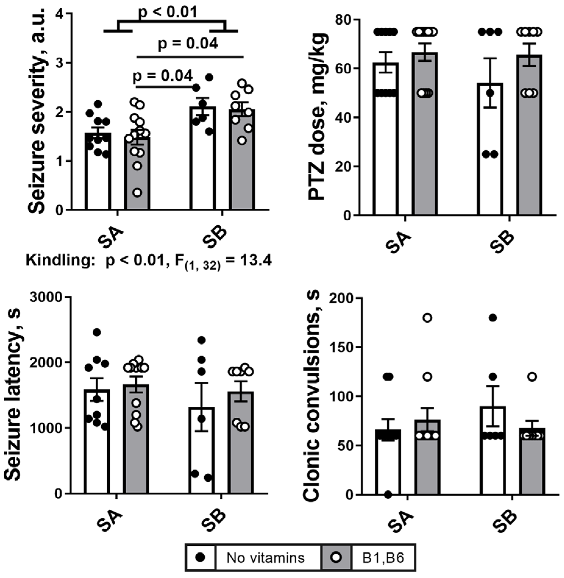
Sensitivity of the male rats to the seizure inducer PTZ is characterized by an average PTZ dose required to induce seizures, and pathophysiological parameters of the seizures, shown in Figure 2. Comparison of the two protocols of the seizure induction by PTZ (Figure 1) indicates that the seizure severity score is significantly lower in the single seizure model (group SA), compared to the seizures after PTZ kindling (group SB). The scores are 1.6 ± 0.1 and 2.1 ± 0.2 mg/kg, for SA and SB, respectively (Figure 2). The difference is observed between the combined groups of animals, i.e., both with and without vitamin administration, although the significance is more pronounced in the vitamin-treated than non-treated animals. Otherwise, the administration of vitamins B1 and B6 24 h before the induction of seizures by PTZ does not significantly affect the parameters of the seizures, either without or with the PTZ kindling.
Other parameters of seizures do not show statistically significant effects of the PTZ kindling, yet the observed differences in the three indicated parameters, i.e., the seizure latency, average PTZ doses to induce seizures and the duration of clonic seizures exhibit consistent changes. As seen in Figure 2, after the PTZ kindling, the PTZ dose (62.5 ± 4 mg/kg in SA group vs. 54.2 ± 10 mg/kg in SB group) and seizure latency (1587.0 ± 174.1 s in SA group vs. 1320.0 ± 369.2 s in SB group) decrease, while the convulsions time increases (66.0 ± 10.8 s in SA group vs. 90.0 ± 20.5 s in SB group).

2.2. Effects of the PTZ Kindling and the Administration of Vitamins B1 and B6 on Behavioral and ECG Parameters after the Seizures

Behavioral parameters presented in Figure 3A show that the PTZ kindling significantly increases the number of rearing acts, that is more pronounced in the animals receiving vitamins, and the steps out of the wall. These effects are specific to the PTZ kindling, as they are absent in the control animals (Figure 3B). While the control animals do not show any significant behavioral effects of the multiple injections mimicking the kindling procedure, they exhibit more reactivity to the vitamins (manifested in the steps out of the wall), whose action may also interact with the injections (manifested in the grooming acts and freezing time). These effects of the vitamins in the control animals are absent in the PTZ-kindled animals. Other assessed behavioral parameters, showing no significant effects, are presented in Supplementary Figure S6.
ECG parameters do not reveal any significant differences between the groups as a result of PTZ kindling (Figure 3C). However, in the control animals the multiple injections in the group CB decrease the relaxation index RMSSD, compared to the group CA (Figure 3D). The effect is not observed in the seizure groups.

2.3. PTZ Kindling Increases Expression of Transcriptional Factor p53 in the Brain

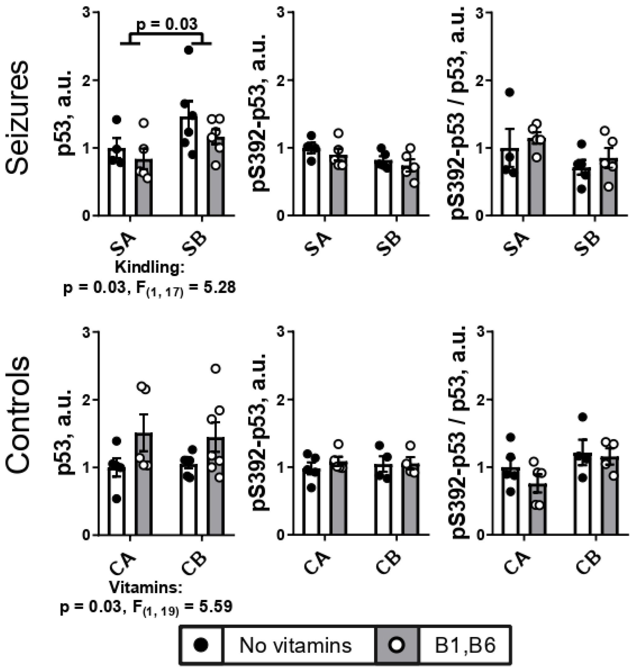
Upregulated in response to different types of cellular stresses, transcription factor p53 is known to have an important dual role in the neuronal damage, particularly the damage induced by seizures [32]. Figure 4 shows that the seizures after the PTZ kindling are associated with increased expression of the brain p53, compared to the animals undergoing seizures without the kindling. In average, an 1.3-fold increase is observed after the kindling. No significant effects of the kindling and vitamins are found when the Ser392-phosphorylated p53 is assessed. However, the changes in the p53 expression and phosphorylation in each animal of the studied groups appear to be roughly proportional, as the ratio of the p53 phosphorylation at Ser392 to the p53 expression, averaged after determination in each animal, is not changed by seizures with or without kindling (Figure 4).
The role of the repeated injections of PTZ in the p53 upregulation is confirmed by no changes in the p53 expression due to the different injection protocols in the control groups CA and CB. Interestingly, in these control animals, administration of the vitamins is a factor upregulating the p53 expression, in contrast to the seizures-exposed animals. The p53 phosphorylation or its ratio to the p53 expression are not significantly affected by the kindling, or injections, or vitamins (Figure 4).
Thus, p53 expression is increased after seizures in the PTZ-kindled vs. non-kindled animals, that is not observed after the multiple injections of saline, mimicking the kindling procedure.
As p53 is a master regulator of energy metabolism greatly relying on the B1- and B6-dependent metabolic pathways [49] and the TCA-cycle-limiting OGDHC in particular [50], we next examined a representative set of the B1,B6-dependent and related activities of the brain homogenate enzymes, working at the cross-roads of the energy production and glutamate/GABA biosynthesis.

2.4. Effect of PTZ Kindling and Administration of Vitamins B1 and B6 on the Activities of the B1- and B6-Dependent and Related Enzymes in the Animal Brain after Seizures

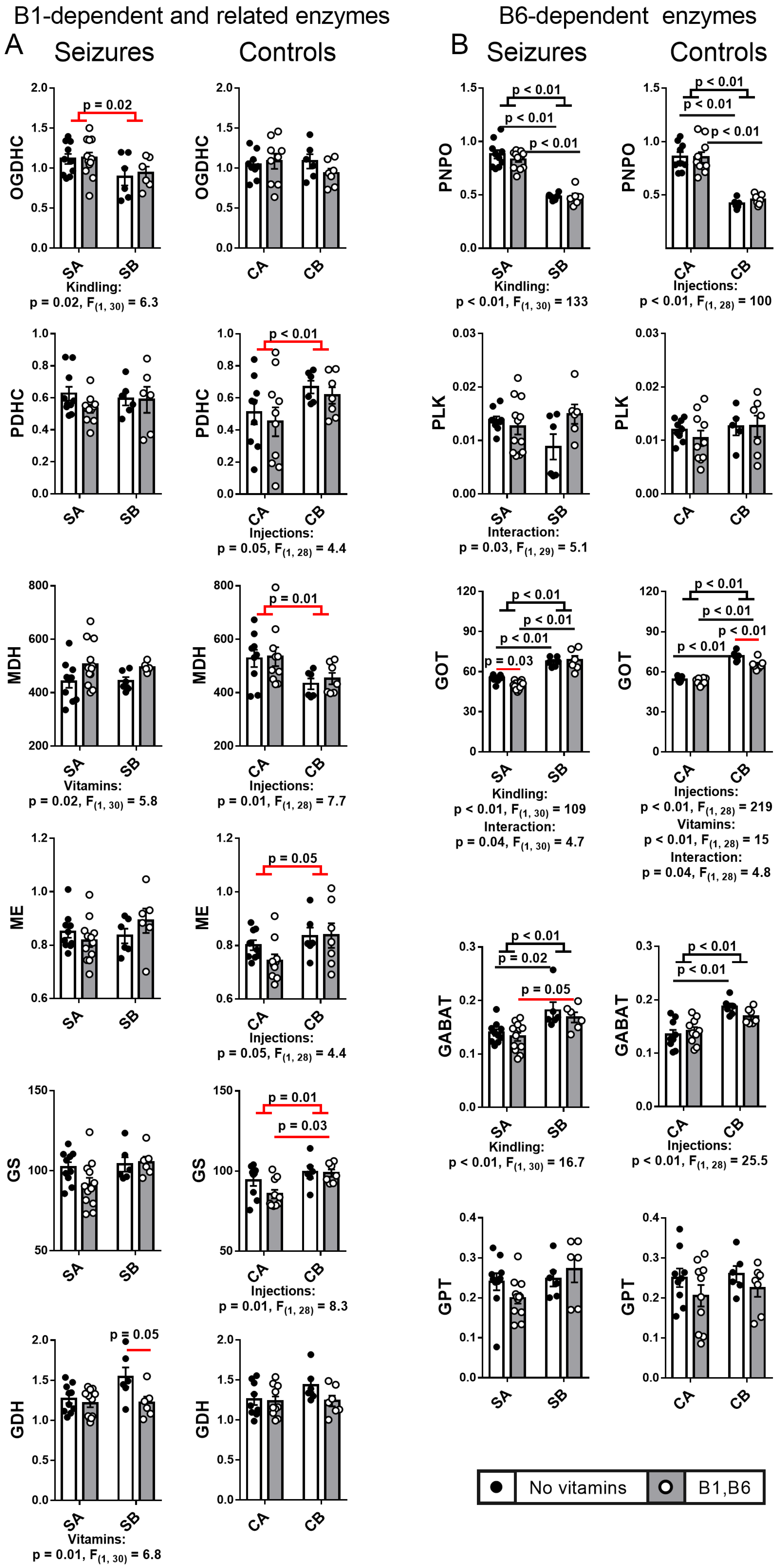
Complex changes in the activities of central metabolic brain enzymes are observed in the employed experimental models. The presentation of the data in Figure 5 helps discriminating the after-seizures effects of the PTZ kindling and the stress response to multiple injections associated with the kindling model.
Independent of the vitamins, the PTZ kindling causes a seizure-induced decrease in the brain OGDHC activity, that is not observed in the control animals (Figure 5), reciprocating the effect of the PTZ kindling on p53 (Figure 4). In contrast, in response to the equivalent number of saline injections, mimicking the PTZ kindling and seizure induction procedures, the control (without seizures) animals demonstrate a strong coupled effect of the injections on the brain activities of PDHC, MDH, ME and GS. That is, independent of vitamins administration, the PDHC, ME and GS activities increase, while the MDH activity decreases (Figure 5, B1-dependent and related enzymes in Control panel). This stress response of the brain enzymes is not observed in the animals after seizures.
Thus, the brain effects of the PTZ kindling vs. single PTZ-induced seizure include not only a decrease in the brain activity of OGDHC, but also attenuation of the brain stress response, inherent in the control animals.

2.5. Effect of PTZ Kindling and Administration of Vitamins B1 and B6 on the Brain Protein Acylation System

Perturbed function of the producers of acetyl-CoA and succinyl-CoA, i.e., PDHC and OGDHC, may be manifested in the protein acylation [51,52]. Hence, we studied how PTZ kindling would affect expression of the brain components of the acylation system.

2.5.1. Brain Protein Acetylation

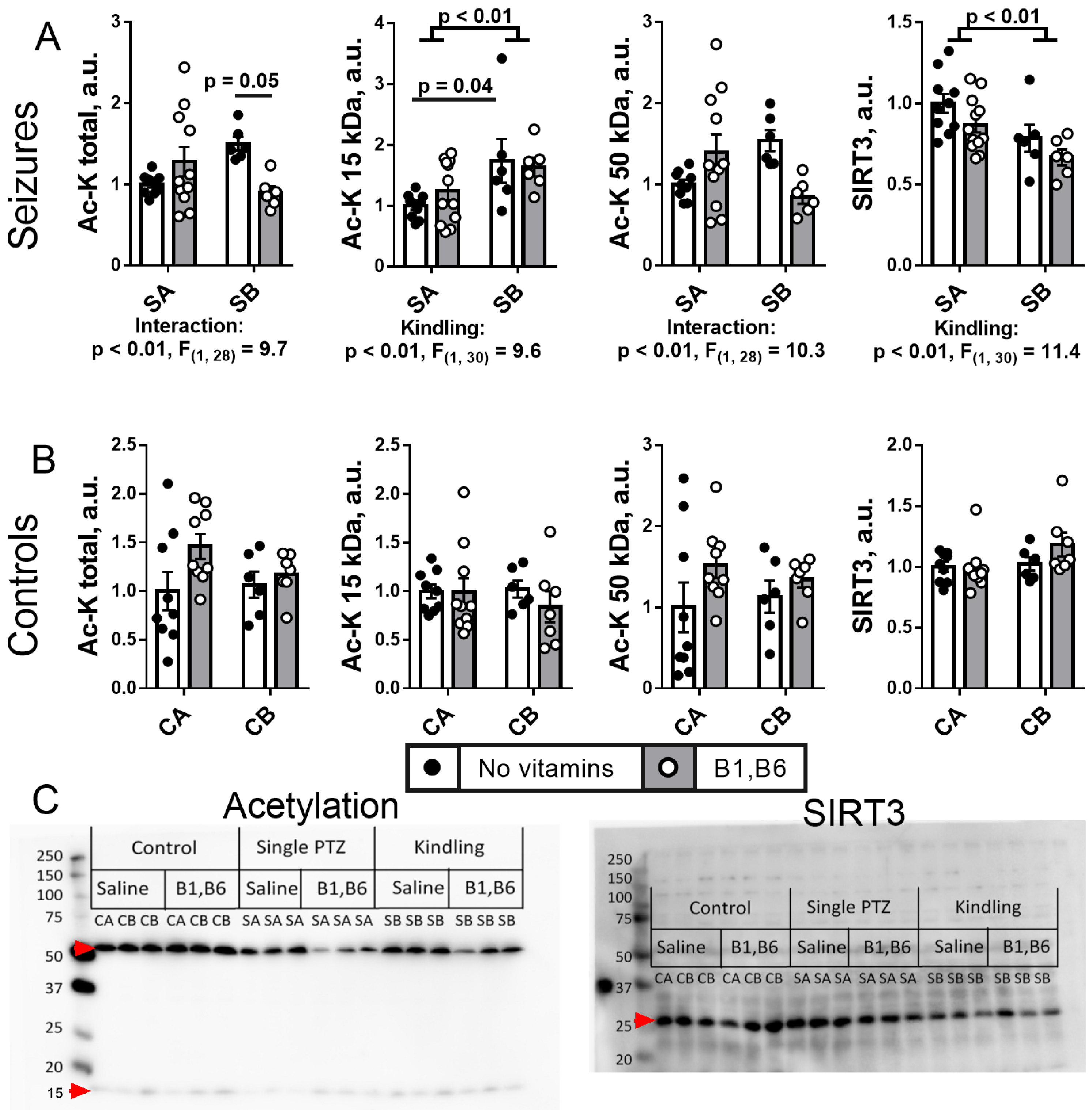
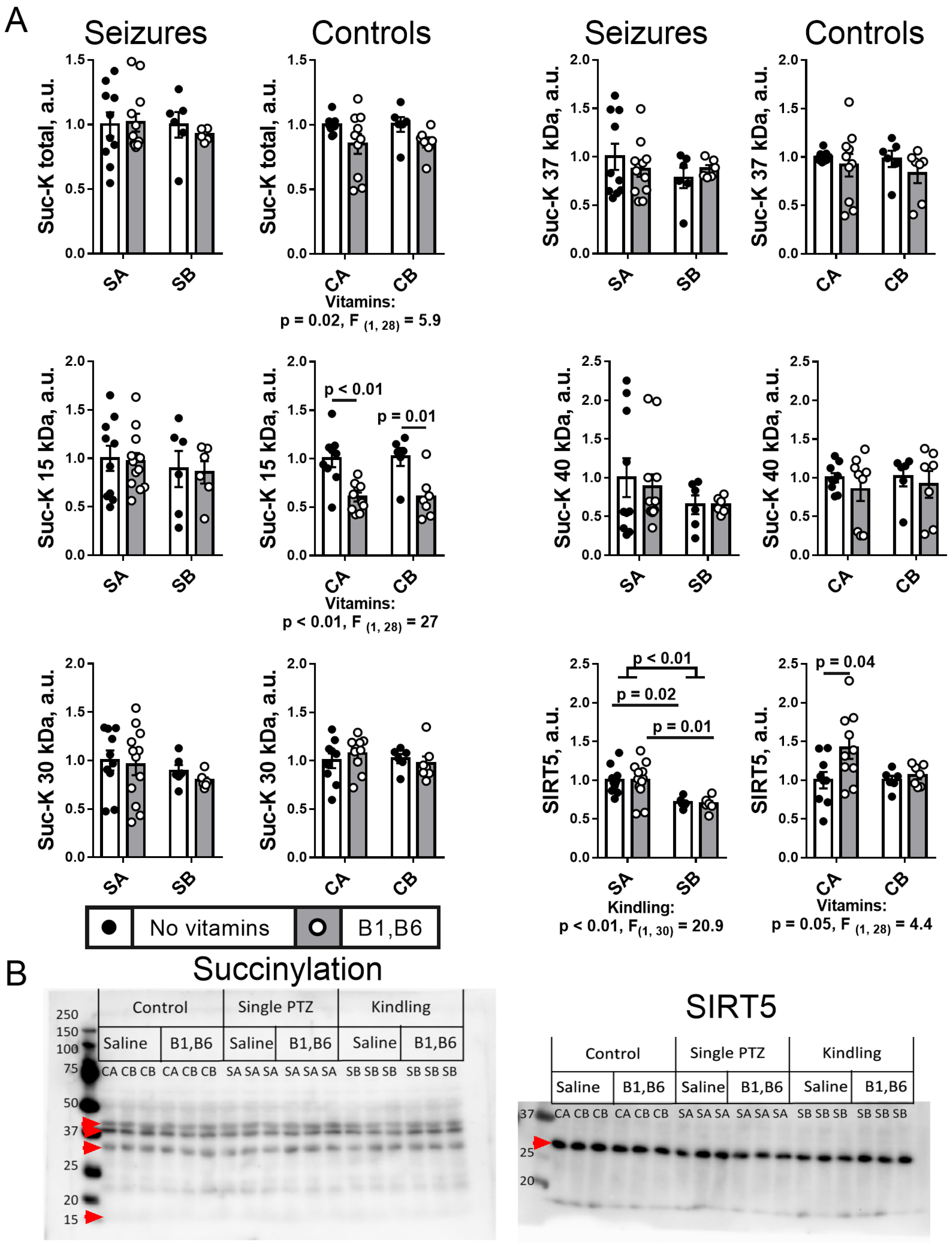
The protein acetylation system is affected by kindling compared to single seizures, as can be seen from Figure 6A, while no effect of multiple injections or vitamins is observed without seizures (Figure 6B). Of the two most expressed bands of acetylated proteins (Figure 6C) with the apparent masses of 50 and 15 kDa, inherent in tubulins and histones, respectively, the kindling predominantly influences acetylation of 15 kDa fraction including histones (p < 0.01 of “kindling” ANOVA factor). Acetylation of these proteins is increased by kindling (Figure 6A). The effect is more pronounced in the animal groups without administration of vitamins (p = 0.04) than after the vitamins administration (p > 0.05). Total acetylation of proteins significantly (p = 0.05) decreases upon vitamins administration to kindled rats (Figure 6A), revealing an interaction between the kindling and vitamins factors of ANOVA (p < 0.01). A higher histone acetylation after kindling is observed simultaneously with a decreased (p < 0.01) level of the mitochondrial deacetylase sirtuin 3 (Figure 6A), that is also reported to deacetylate histones in the nucleus [53,54].

2.5.2. Brain Protein Succinylation

Unlike acetylation, the protein succinylation is not changed by the PTZ kindling that, however, decreases the expression of the protein desuccinylase SIRT5 (Figure 7). In contrast, a significant decrease in succinylation of 15 kDa proteins including histones, is observed after the vitamins administration to the control animals of both CA ad CB groups (Figure 7). The effect reciprocates the vitamins-induced increase in p53 expression in these animals (Figure 4). In the vitamin-treated CA group, this succinylation decrease is accompanied by a significant (p = 0.05) upregulation of the desuccinylase SIRT5, with the effect not observed after the multiple saline injections in CB group (Figure 7).
Thus, PTZ kindling decreases expression of SIRT5. The vitamins-promoted desuccinylation of 15 kDa proteins in the control animals, is blocked by seizures, independent of kindling.

2.6. Effects of PTZ Kindling on the Levels of Metabolites

2.6.1. Redox Indicators

A single episode of the PTZ-induced seizures decreases cerebral NAD+ level (Figure 8) from 14.1 nmol/g FW (CA group) to 7.1 nmol/g FW (SA group) (p = 0.08 according to the Mann–Whitney test). There is no further decrease in NAD+ by PTZ kindling (Figure 8, Seizures). In the control animals, the injection factor is significant for the differences between the groups CA ad CB (Figure 8, Controls Panel). In these control groups, NAD+ is more significantly decreased without vitamin supplementation, compared to those vitamins-supplemented (Figure 8, Controls). In the PTZ-treated rats, vitamins significantly elevate the NAD+ level (Figure 8, Seizures).
In view of our simultaneous quantification of multiple proteins and metabolites, no specific tissue treatment to preserve the glutathione redox state could be employed. In this case, content of total glutathione is chosen as a measure of the brain antioxidant potential, excluding potential contribution of the storage of the oxidatively compromised post-seizure tissue to the glutathione redox status. Figure 8 shows that the total glutathione level is reduced both by the PTZ kindling and multiple saline injections.

2.6.2. Free Amino Acids and Related Compounds

Figure 9A shows the major amino acids neurotransmitters and their closest metabolic partners. While glutamate level does not change in the studied groups, its amidation product glutamine is decreased by PTZ kindling only (Figure 9A). The levels of the glutamate decarboxylation product GABA and the glutamate transamination co-product aspartate are increased by both the PTZ kindling and multiple saline injections (Figure 9A). These changes confirm the stress-perturbed metabolism of the amino acids in the brain, complementing the data of the enzymatic assays, but do not discriminate specific actions of PTZ or injections (Figure 5). The pyruvate transamination product alanine, however, is affected by multiple injections of saline only, with the effect abrogated by PTZ injections (Figure 9B), similar to the PDHC activity (Figure 5).
Compared to the short-term action of a single PTZ injection, the long-term procedure of PTZ kindling increases the neurotransmitter precursor phenylalanine, neuromodulator glycine (Figure 9C), and the branched-chain amino isoleucine supporting glutamate neurotransmission via the glutamate-regenerating transamination (Figure 9D). The PTZ kindling decreases taurine (Figure 9E)—a redox-active molecule with cytoprotective properties [55,56]. From the amino acids related to NO-dependent signaling, only lysine shows significant increase by the PTZ kindling.
The administration of vitamins B1 and B6 regulating activities of the amino-acids-related enzymes (Figure 5A,B), modifies the cerebral metabolism of amino acids (Figure 9). In many cases, stronger effects of the vitamins are observed in the animals with single PTZ-induced seizures than after the PTZ kindling. In particular, compared to the single episode of seizures without vitamins, the vitamins administration increases the levels of glutamate (Figure 9A), phenylalanine and tyrosine upon the single seizures (Figure 9C). In the PTZ-kindled group (SB) the vitamins increase the level of taurine (Figure 9E). In other cases, administration of vitamins has minor opposite effects in the SA and SB or CA and CB groups, obvious from decreased statistical significance of the kindling and/or injection stress effects (e.g., tyrosine, glycine, lysine, Figure 9).
Thus, specific PTZ- or injection-stress-induced changes in the brain levels of the amino acids neurotransmitters, their precursors and neuromodulators (Figure 9) complement the changes in the involved enzymes (Figure 5). Administration of the vitamins B1 and B6, providing the coenzymes for some of these enzymes, affect the levels of the brain amino acids, with many of the effects dependent on the animal state. In particular, the vitamins prevent the kindling-induced changes in a number of amino acids by differently affecting the two seizures groups (SA and SB).

2.7. Administration of Vitamins B1 and B6 Affects Correlations between the Assessed Parameters in the Post-Seizures and Control Rats

2.7.1. Vitamins Regulate the Seizures-Induced Metabolic and Physiological Changes

To elucidate the combined effect of the B1 and B6 vitamins administration, the correlations between the most relevant biochemical and physiological parameters are analyzed in the pooled seizure groups (SA + SB) without (Table 1, top right) and with (Table 1, bottom left) the vitamins. Remarkably, in the seizure groups (Table 1, top right), a positive correlation between the levels of p53 and neurotoxic glutamate is observed. Although less significant (p = 0.07), this correlation is also observed in the vitamins-treated rats. This finding is consistent with the leading role of the seizures-associated glutamate neurotoxicity in the post-seizures p53 induction.
The seizures-relevant effects of the B1 and B6 vitamins are implied by a number of correlations that strongly change in the animals undergoing seizures without the vitamins administration, compared to those with the vitamins. These include the correlations of p53 with the ECG parameters RMSSD and SI, characterizing the autonomous heart regulation; correlations of the seizures duration with physiological parameter and the activity of GDH which in the brain functions in the direction of oxidative deamination of glutamate [57]; correlation of the seizures severity with the brain levels of GABA. All these correlations are observed in the animals without the vitamins supplementation, disappearing after the supplementation. In contrast, the vitamins-supplemented group exhibits a positive correlation of the OGDHC activity with the seizures latency (Table 1, bottom left), absent in the animals undergoing seizures without the vitamins. As a result, the correlation analysis reveals an interplay between the behavioral/ECG changes and the seizures duration, with the vitamins abrogating these relationships along with affecting the correlations of the enzymes (OGDHC, GDH) and metabolites (Glu, GABA), involved in the Glu/GABA balance.

2.7.2. Regulation of Metabolism and Physiology by the Vitamins Administration in the Control Rats

In control rats treated with injections of saline instead of PTZ, a negative correlation of p53 with RMSSD is preserved, but the positive correlation of p53 with SI is not observed (Supplementary Table S1, top right). Thus, unlike the correlation of p53 with RMSSD, the correlation of p53 with SI is specifically related to the post-seizures state. This is consistent with the SI correlation to the seizures duration (Table 1, top right).
Negative correlations of GABA with grooming and rearing, consistent with the GABA action as an inhibitory neurotransmitter, are observed along with a positive correlation of the GABA levels and the GDH activity in the control rats (Supplementary Table S1, top right). Remarkably, in the vitamins-treated control rats, this GABA-GDH correlation disappears. Simultaneously, grooming and rearing become correlated with the activity of OGDHC (Supplementary Table S1, bottom left). The correlation analysis thus indicates that the behavioral effects of the vitamins are mediated by OGDHC, consistent with the essential role of this thiamine-diphosphate-dependent complex in determining the glutamate flux through different pathways [58].
As a result, the correlation analysis reveals that vitamins B1 and B6 affect metabolic relationships between the components supporting the Glu/GABA balance both in the control animals and those exposed to seizures.

3. Discussion

In this work, we characterize the multitude of the PTZ-kindling-induced changes in the rat pathophysiology and brain biochemistry. The strength of epileptic seizures, the behavioral and ECG parameters the day after the seizures are measured along with assaying a set of enzymes critical for the brain metabolism of glucose, glutamate and GABA. The set includes the multienzyme complexes of pyruvate dehydrogenase (PDHC) and 2-oxoglutarate dehydrogenase (OGDHC), GDH, GS, transaminases of aspartate (GOT), alanine (GPT) and GABA (GABAT), and the enzymes providing the transaminases with their coenzyme pyridoxal-5′-phosphate, i.e., PLK and PNPO. Finding the correspondence between the biochemical and pathophysiological parameters helps to reveal the brain metabolic pathways with the pathophysiological significance, that is required to discover novel pharmacological targets.
To reveal the changes induced by the PTZ kindling, we compare the parameters of the PTZ-induced seizures and the seizures-affected brain metabolism in the animals undergoing seizures without and after the kindling. The effects are considered to be specifically induced by the PTZ kindling, if they are not observed after the multiple saline injections mimicking the kindling procedure. In this way, we separate the effects of the PTZ kindling from the stress effects in response to multiple injections, required for the kindling. As a result of this analysis, one may conclude that most of the changes in the B6-dependent enzymes are induced by the injection stress, that also affects the B1-dependent enzymes with their network partners (Figure 5). However, the latter enzyme group is specifically affected by the PTZ kindling, either explicitly (OGDHC), or by decreased reactivity to the injection stress (PDHC, MDH, ME, GS), or by the different response to vitamins (GDH).
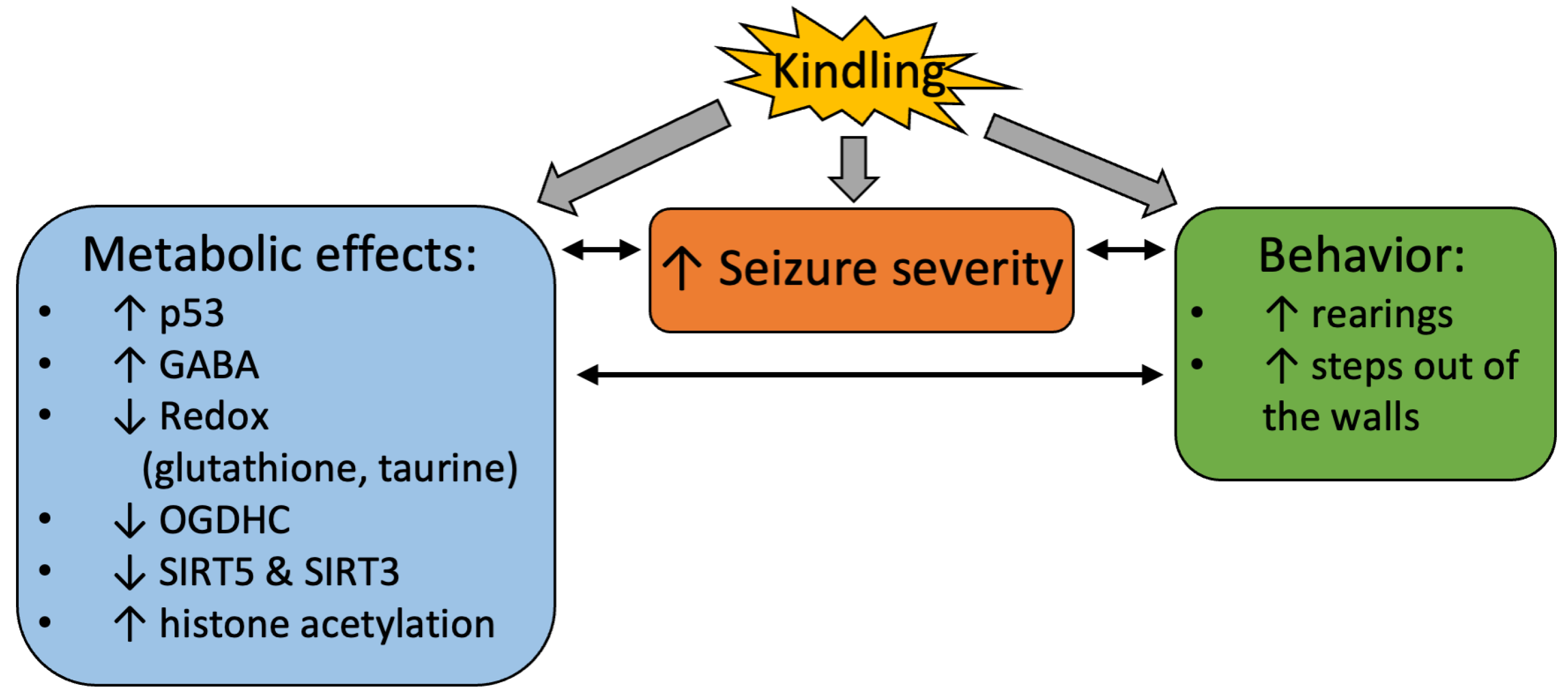
Remarkably, the PTZ kindling decreases the activity of the rate-limiting enzyme complex of the TCA cycle, OGDHC (Figure 5 and Figure 10), known to be critical for the brain function through its involvement in both the energy and Glu/GABA metabolism [58,59,60]. A wealth of evidence, including the patients’ data, indicate that OGDHC is strongly involved with glutamate excitotoxicity and associated production of reactive oxygen species (ROS) [61,62,63]. On the other hand, in different systems, OGDHC function and/or its saturation with the coenzyme thiamine diphosphate, the diphosphorylated vitamin B1, is known to be tightly linked to p53-dependent regulation of redox metabolism [49,50,64,65,66]. Additionally, in our epilepsy model, PTZ kindling increases expression of p53 (Figure 4 and Figure 10) concomitantly with downregulation of the OGDHC function (Figure 5 and Figure 10), both changes accompanying an increase in the severity of seizures after the kindling (Figure 2 and Figure 10). Thus, repeated challenge of the balance of Glu/GABA neurotransmission by PTZ blockade of the GABA-A receptors deteriorates seizures simultaneously with the increased expression of p53 and decreased flux through the TCA-cycle-limiting OGDHC, where the neurotoxic glutamate may be utilized [63]. Transcriptional control, induced by the PTZ kindling, is evident not only from the p53 upregulation, but also from the decreased expressions of SIRT3 and SIRT5 (Figure 6, Figure 7 and Figure 10), and the kindling-increased acetylation of the brain proteins with high mobility in the SDS-gel electrophoresis, inherent in histones (apparent molecular mass 15 kDa, Figure 6 and Figure 10).
The kindling-induced p53 upregulation at the level of the protein expression (Figure 4 and Figure 10) may be added by the functional activation of p53 through the simultaneous decrease in the SIRT5 expression (Figure 7 and Figure 10), as desuccinylation of Lys120 in p53 by SIRT5 suppresses the transcriptional activity of p53 upon the DNA damage response [67]. This mechanism may explain the neuroprotective action of SIRT5 known from earlier studies [68]. Deacetylation of p53 by SIRT3 may also be involved in the p53-dependent metabolic control. This regulation is more studied in aging and cancer [69]. In particular, SIRT3 suppresses the p53-mediated ferroptosis in cancer cells [70]. Thus, in addition to the increased expression of p53 (Figure 4 and Figure 10) the decreases in expression of SIRT3 and SIRT5 after the PTZ kindling (Figure 6, Figure 7 and Figure 10), may stimulate the p53 transcriptional activity after the kindling through post-translational acylations. Although regulatory phosphorylation of p53 at Ser392 in the protein C-terminus does not exhibit a significant change after the PTZ kindling, the ratio of the phosphorylated p53 to its expression level does not change (Figure 4), suggesting the p53 phosphorylation to increase proportionally to the protein expression.
In the control groups, we observe a stress response to multiple injections of saline, that is clearly different from the effects of the PTZ kindling in the seizure groups. Mimicking the PTZ kindling protocol, but not the PTZ action as such, the injections of saline result in changes in the brain enzymatic activities linked to the pyruvate levels (Figure 5). Obviously, the stress affects the pyruvate flux distribution through the TCA cycle and affiliated pathways. PDHC activity is increased along with a decrease in the activity of MDH. As less oxaloacetate—a product of the MDH reaction—may thus be condensed with the PDHC-produced acetyl-CoA, the observed changes in the PDHC and MDH activities manifest a regulation directed to decreased consumption of the acetyl-CoA in the TCA cycle. Moreover, production of pyruvate by the oxaloacetate-decarboxylating malic enzyme increases, supporting the stress-elevated PDHC activity by a higher pyruvate supply. The higher supply is also evident from the increase in the pyruvate transamination sibling alanine (Figure 9B). Finally, increased activity of the brain GS may point to increased degradation of the amino acids through the TCA cycle [71]. Feeding the cycle at different entry points, the amino acids degradation may compensate for the insufficient acetyl-CoA flux from PDHC to the TCA cycle. However, the degradation produces an excess of ammonia, whose utilization by GS not only attenuates the ammonia toxicity to the brain, but also preserves the nitrogen in the form of glutamine. Alternative to the oxidation in the TCA cycle, the increased production of acetyl-CoA by PDHC, supported by increased activity of malic enzyme, may be used for the increased biosynthetic needs, e.g., of acetylcholine. Remarkably, the adjustments of the brain enzymatic activities in response to the injection stress do not result in perturbed acylation of the brain proteins (Figure 6 and Figure 7), as occurs upon the PTZ kindling (Figure 6 and Figure 7) or a short-term PDHC inhibition [51,72].
The PTZ kindling affects mitochondrial metabolism not only in our experimental design where the effects of the kindling are characterized in the animals exposed to seizures, but also in other studies, where the effects of PTZ kindling are revealed by comparison to the control group [73]. Coincidence of the data in different models stresses the role of mitochondria in the kindling-induced changes. In particular, compared to the control animals, daily administration of PTZ to rats elevates expression of nitric oxide synthase (NOS) in cerebellar neurons and increases the NO generation, with the NO content positively correlated to the severity of seizures [74]. Accordingly, administration of arginine is shown to potentiate the PTZ-induced seizures, whereas NOS inhibitors or NOS knockout reduces the seizures [26,74,75]. The release of NO is observed upon stimulation of glutamatergic neurons [76,77], resulting in predominance of excitatory over inhibitory signals, similarly to the action of PTZ. Here, we do not measure NO, but assess potential changes in NO metabolism after the PTZ kindling through the levels of relevant amino acids (Figure 9E). Such changes are implicated by increased level of lysine in the PTZ-kindled rats vs. the rats undergoing seizures without the kindling (Figure 9E). In the reaction with arginine, producing ornithine, lysine generates an alternative source of NO, homoarginine (reviewed in [78]). Using our data (Figure 9D) for the pair-wise comparison of the kindled and control rats by the Mann–Whitney test, the levels of lysine (Figure 9D, SA vs. CA, p = 0.03) and ornithine (Figure 9D, SA vs. CA, p = 0.05) are reduced after a single seizure episode, and re-established after the PTZ kindling. Thus, our data add to the independent studies suggesting that effects of seizures on NO metabolism in the brain depend on the PTZ kindling.
It is worth noting that, compared to a single seizure episode, the PTZ kindling increases the level of glycine (Figure 9C), whose brain metabolism and signaling are known to be involved into epileptic seizures. For instance, microperfusion of glycine in hippocampus of rats with changed membrane distribution of the NMDA receptors induces seizures in 75% of the rats [79]. Glycine levels are known to be increased in a rat model of the kindling by electrical stimulation of amygdala [80]. The number of glycine-binding sites of the NMDA receptors is increased one month after the kindling [81]. Inhibition of these strychnine-insensitive glycine receptors by 7-chlorokynurenic acid suppresses the kindling effect [82], as does a downregulation of hippocampal glycine transporter 1, whose expression is increased in patients and rodent models of temporal lobe epilepsy [83]. The pro-convulsive action of glycine in these models is in good accord with our results, where the PTZ-kindling-induced increase in the brain glycine level, compared to a single PTZ-induced seizure (Figure 9C), accompanies an increase in the severity of seizures (Figure 2A). Remarkably, Figure 9C shows that the kindling-induced elevation in the brain glycine levels (p = 0.01, Tukey’s post hoc test, in the rats receiving no vitamins), is not significant after the supplementation of vitamins B1 and B6 (p > 0.05, Tukey’s post hoc test). Stimulation of the B6-dependent glycine decarboxylase may be involved in the effect. Significance of this enzyme for the brain levels of glycine, and the pro-convulsive action of glycine are evident from epileptic seizures induced by impairments of glycine decarboxylase causing glycine accumulation [84].
The PTZ kindling also perturbs antioxidant potential of the brain. Although accurate determination of redox state of glutathione in our tissue samples is complicated by its oxidation into GSSG during storage [85], our finding of the kindling-induced decrease in total glutathione (Figure 8) confirms the perturbed antioxidant power of the brain after the seizures. The antioxidant taurine is also decreased after the kindling (Figure 9E). The kindling-induced decrease in taurine, observed in our model, has also been observed in another study, along with increase in GABA [86], observed by us as well (Figure 9A).
Similar to increasing the seizure severity score (Figure 2), the PTZ kindling procedure induces an increase in rearings and steps out of the walls. The link between these behavioral parameters and seizures is substantiated by significant positive correlations of both rearings and steps out of the walls with the duration of clonic seizures in the PTZ-treated animals (Supplementary Table S1). Remarkably, after the seizures, the two parameters do not correlate to each other, as they do, independent of the vitamins administration, in the control animals. However, the vitamin administration to the seizure groups re-establishes the correlation between the rearings and steps out of the walls, testifying to a normalizing effect of the vitamins on the brain metabolism and behavior. It may be speculated, that the seizures-associated increases in rearings and steps out of the walls are related to the PTZ blocking the inhibitory GABA-A receptors [14]. It is also worth noting in this regard that several reports indicate an increase in hippocampal neurogenesis in PTZ-kindled animals [87,88], supposed to play a role in the pathophysiology or represent a compensatory process in epilepsy [89]. This effect of PTZ kindling on neurogenesis may contribute to the increases in the abovementioned parameters of exploratory activity, observed in our PTZ-kindling model (Figure 2).
Thus, our study reveals significant metabolic changes in the post-seizures brain exposed to the PTZ kindling, which may be alleviated by the administration of vitamins B1 and B6.

4. Materials and Methods

Animal experimental employ the reagents from the following manufacturers: thiamine (#36020.02)—«Serva», Heidelberg, Germany; pyridoxal (#A0960)—«PanReac AppliChem», Barcelona, Spain, NAD+ (#1013)—“Gerbu”, Heidelberg, Germany; malate dehydrogenase—«Reanal», Budapest, Hungary; other reagents for biochemical procedures as well as PTZ (#P6500) —«Sigma-Aldrich», St. Louis, MO, USA. Pyridoxine-5′-phosphate is synthesized from pyridoxal-5′-phosphate (#P9255) («Sigma-Aldrich», St. Louis, MO, USA) as described previously [90] and kindly provided by Dr. Martino di Salvo (Sapienza University, Rome, Italy). The solutions are prepared in Milli-Q deionized water, the salts used are of the highest purity available.

4.1. Animal Models

Our study uses Wistar male rats obtained from the Russian Federation State Research Center Institute of Biomedical Problems RAS (IBMP). The animals are kept under standard conditions in cages with free access to water and food and a light/dark cycle of 12/12 h (the light turned on at 9 a.m.). The adaptation period to the husbandry conditions is two weeks. Animals are distributed randomly between experimental groups. The animal experiments are approved by Bioethics Committee of Lomonosov Moscow State University (protocols 69-o from 9 June 2016 for single PTZ-induced seizure model and 139-a from 11 November 2021 for the PTZ kindling model). In both models, the age of the animals at the time of the seizure induction by PTZ is 10–11 weeks (weight 320 ± 20 g).

4.1.1. Model of Single Episode of PTZ-Induced Seizures

Single episode of seizures (animal group SA) is induced in the 10–11-week-old male rats by intraperitoneal administration of PTZ in saline at 25 mg/kg dose according to the protocol in Figure 1A.
After PTZ administration, the severity of seizures is visually assessed according to the modified Racine scale developed for the employed protocol of PTZ administration [91] (Table 2) for 15 min in individual cages (OpenScience, Moscow, Russia).
The total time of the seizure observation is 45 min.
During the visual assessment, there are no more than 4 rats in individual cages (2 with PTZ and 2 with saline (0.9% NaCl) injections). This arrangement allows one to observe each of the rats. Scores are registered every minute of the observation, and an epileptic seizure of maximum score is noted for the given minute of observation. If the stages 4–5 on the modified Racine scale (tonic or tonic-clonic seizures, Table 2) do not develop within 15 min, PTZ is re-injected at the same dose of 25 mg/kg. The procedure is repeated no more than three times, and the total PTZ dose thus does not exceed 75 mg/kg. The “seizure severity” score is calculated as the average score of the severity of an epileptic seizure over the entire observation period from the moment of administration of the first PTZ dose.

4.1.2. Model of PTZ-Induced Seizures after the PTZ Kindling

As we aim at comparison of the consequences of the acute PTZ-induced seizures occurring at the same age, the rats entering the kindling experiment are younger (Figure 1B), than those in the single PTZ-induced seizure episode (Figure 1A). Taking into account 19 days required for the PTZ kindling according to our previously published protocol [29], the kindling is started with the 7–8-week-old male rats of 140 ± 10 g. This kindling model of epilepsy (animal group SB) is shown in Figure 1B. The scheme is selected to minimize mortality, simultaneously achieving a strong manifestation of the clonic seizures according to the Racine scale (see Table 2). The rats are injected PTZ i.p. at an originally sub-convulsive dose of 37.5 mg/kg 3 times a week for 3 weeks, resulting in the total 9 injections of PTZ and its total dose of 337.5 mg/kg during the PTZ kindling. Each of the injections is followed by the visual assessment of the animals according to Table 2, as described above. A week after the last injection of 37.5 mg/kg PTZ in the kindling procedure, the PTZ administration to induce seizures is performed exactly as in the model of a single seizure episode (Figure 1). 24 h post-seizures the rats are physiologically tested and sacrificed by decapitation, as shown in the flow-chart in Figure 1. The procedure is performed exactly as described before [94].

4.1.3. Administration of Vitamins

Vitamins B1 (thiamine, 100 mg per kg body weight) and B6 (pyridoxal, 100 mg per kg body weight) are administered intraperitoneally twice, first time 24 h before the first administration of 25 mg/kg PTZ and second time after completion of a 45 min follow-up of the PTZ-induced seizures, either in the single seizure model or following PTZ kindling (Figure 1). This vitamin regimen takes into account the results of previous studies on the potential protective effect of high doses of vitamins before [95] or after [65] the exposure to stress, when increased availability of vitamins may provide, respectively, the better stabilization or normalization of the metabolic state [96]. The injections with equivalent volumes of saline (0.9% NaCl) instead of the vitamins are given to the reference group.
According to the formula recommended by the US Food and Drug Administration (http://www.fda.gov/downloads/Drugs/GuidanceComplianceRegulatoryInformation/Guidances/ucm078932.pdf (accessed on 5 May 2023), the doses of vitamins B1 and B6 used in this study on rats (100 mg/kg each of the vitamins per day) correspond to a dose of 16 mg/kg, or 1 g for an average weight of 60 kg, in humans. These doses are within the interval of the doses used in the vitamin therapy [46,48,97,98,99].

4.1.4. Animal Survival

The data on the total number of animals in our experiments and their survival is summarized in Table 3.

4.1.5. Control Animals to Reveal the Injections-Related Stress Effects

As the kindling protocol is associated with multiple injections, the effects of PTZ kindling are discriminated from the effects of injections without the substances in the control animals CA (protocol of Figure 1A) or CB (protocol of Figure 1B) receiving saline (0.9% NaCl) or saline with vitamins according to the group assignment, at the times and in the volumes, equal to those of the PTZ and/or vitamins solutions.

4.2. Assessment of Physiological Parmeters after Seizures and in the Corresponding Control Groups

To assess the spontaneous behavior of animals in an unfamiliar environment, the «Open Field» test («OpenScience», Moscow, Russia) is used [100]. To quantify the animal behavior, the animals are tested for 3 min in complete silence under the light of a 15 W red lamp as described before [46,52]. Locomotor activity is estimated by the number of line crossings. Anxiety level is determined, registering defecation acts, duration of freezing, the duration and number of grooming acts. Exploratory activity is assessed by the number of rearing acts, steps out of the walls and entries to the center.
ECG is recorded for 3 min using non-invasive electrodes as previously described [46]. Balance of the heart autonomous regulation is assessed by the following parameters of ECG: an average R-R-interval (R-R interval, ms), standard deviation of an average R-R (SD, ms), a range of R-R interval values, i.e., a difference between the maximal and minimal values (dX, ms), root mean square of successive differences in R-R intervals (RMSSD, ms), and stress index (SI, arbitrary units).

4.3. Preparation of Homogenates of the Rat Cerebral Cortex

The procedure is performed exactly as described before [94]. After the animal decapitation, the brain is excised and transferred onto ice, where the cerebral cortex is separated for freezing in liquid nitrogen within 60–90 s after decapitation. The cortices are stored at −70 °C. Homogenization of the tissue and sonication of homogenates is carried out according to the previously published protocol [94].

4.4. Measurement of Enzymatic Activities

The activities of oxoglutarate dehydrogenase complex (OGDHC), pyruvate dehydrogenase complex (PDHC), glutamate dehydrogenase (GDH), malate dehydrogenase (MDH), glutamine synthetase (GS) and NADP+-dependent malic enzyme (ME) are measured in cerebral cortex homogenates as described previously [71,101]. Prior to the activity assays, except for that of GS, the homogenates are sonicated, following by the addition of one volume of solubilization buffer (50 mM Tris-HCl, pH 7.4, 600 mM NaCl, 4 mM EDTA, 1% sodium deoxycholate, and 4% NP-40) to the three volumes of sonicated homogenate as described before [94].
Activity of the GABA transaminase (GABAT) is measured by the published assay [102]. Briefly, 10 µL of sonicated cerebral cortex homogenate per microplate well is used and the reaction is started by addition of reaction mixture comprising 100 mM Tris-HCl, pH 8.1, with 2 mM 2-oxoglutarate, 1 mM NAD+, 20 mM 2-mercaptoethanol, and 3 mM GABA.
The activities of GOT and glutamate pyruvate transaminase (GPT) are measured using a coupled reaction with MDH or LDH, respectively. Briefly, the GPT is measured using 2 µL of sonicated homogenate added per well, and the reaction is started by addition of reaction mixture comprising 100 mM NaKHPO4 buffer with 0.1 mM NADH, 2 mM 2-oxoglutarate, 40 mM DL-alanine and 4 U/mL LDH, pH 7.4. Similarly, GOT is measured using 3 µL of sonicated homogenate diluted 25 times in 50 mM MOPS buffer pH 7.0, added per well. The reaction is started by addition of reaction mixture comprising 100 mM NaKHPO4 buffer with 0.1 mM NADH, 2 mM 2-oxoglutarate, 40 mM DL-aspartate and 5 µg/mL MDH, pH 7.4.
The activities of pyridoxal kinase (PLK) and pyridoxine-5′-phosphate oxidase (PNPO) are measured in black 96-well microplates using the fluorescence mode of a CLARIOstar Plus plate reader (BMG LABTECH, Ortenberg, Germany) according to the published protocol [46]. All other activities are assayed spectrophotometrically in transparent 96-well microplates, measuring absorbance at 500 (PDHC), 540 (GS) or 340 (all the rest) nm by a Sunrise plate reader (Tecan, Grödig, Austria).

4.5. Western Blotting

Cerebral cortex homogenates are diluted in Laemmli buffer and subjected to SDS-PAGE. The resulting gels are used for the assessment of protein expression via 2,2,2-tricholoroethanol staining as described before [65]. Total protein succinylation is assessed using primary antibodies #PTM-1151 from PTM-Biolabs (Chicago, IL, USA), expression of sirtuins 3 and 5 and total protein acetylation—#5490, #8782 and #9841, respectively, from Cell Signaling Technology (Danvers, MA, USA). These primary antibodies were used at 1:2000 dilution. The expression of p53 protein and its phosphorylated form (pS392-p53) were assessed using primary antibodies #MA5-12453 and #44-640G from Invitrogen (Waltham, MA, USA) at 1:350 and 1:500 dilution, respectively. Secondary HRP-conjugated anti-rabbit antibody #7074 from Cell Signaling Technology (Danvers, MA, USA) is used in 1:3000 dilution. Chemiluminescence is detected using ChemiDoc MP Imager (Bio-Rad, Hercules, CA, USA) and processed in Image Lab software v. 6.0.1 (Bio-Rad, Hercules, CA, USA). The intensities of the quantified parameters are normalized to total protein in the lane and shown as a ratio to the parameter intensities in the comparison groups, i.e., to the averaged values of animals receiving no vitamins in control (C) or seizure (S) groups from protocol A (Figure 1). Protein intensities from different membranes are compared across all the membranes through normalization to the intensities of several common samples repeated on all the membranes. The original images of membranes and gels with visualized protein bands are presented in Supplementary Figures S1–S5.

4.6. Preparation of Tissue Extracts and Quantifications of Metabolites

Methanol-acetate extraction of metabolites from the rat cerebral cortex and quantification of its amino acids and urea are performed as described before [103,104]. Briefly, frozen cerebral cortices are homogenized in ice-cold methanol, then acetic acid solution is added and the proteins are precipitated. The sodium-citrate buffer system with a Hitachi L-8800 amino acid analyzer is used according to the published protocol [103]. NAD+ quantification in methanol-acetic extracts of rat cortices is performed using recombinant formate dehydrogenase according to the previously published protocol [105]. Reduced (GSH) and oxidized (GSSG) glutathione content is measured using fluorometric assay with o-phthalaldehyde [106] as described before [101]. Since no specific techniques have been employed to minimize glutathione oxidation during the storage of extracts, only changes in the total glutathione (GSH + 2 × GSSG) are analyzed.

4.7. Statistical Analysis and Data Presentation

Data are analyzed using the GraphPad Prism 7.0 software (GraphPad Software Inc., La Jolla, CA, USA). Individual values of the parameters for each animal, their mean and SEM are shown on the graphs. D’Agostino-Pearson omnibus normality testing and ROUT test for outliers are used. Statistical significance of differences upon comparison of four experimental groups differing in two factors such as “vitamins” and “kindling” or “vitamins” and “injections”, is assessed using two-way analysis of variance (ANOVA) with Tukey’s post hoc test. In some additional comparisons of specific two groups, mentioned in the data discussion, the Mann–Whitney U-test is employed. The statistical methods are indicated in the text and/or figure legends. The statistical significances at p ≤ 0.05 are shown on the graphs. The ANOVA results for the significant (p ≤ 0.05) factors and their interaction are also shown below the figures together with the corresponding F-statistics.

5. Conclusions

The PTZ-kindling-induced changes in the rat pathophysiology and brain biochemistry are characterized, revealing significant relationships between the post-seizures brain metabolism and behavior with the seizures parameters. In particular, expression of p53 in the post-seizures brain correlates to the brain level of glutamate and stress index of the heart autonomous regulation. The correlations are absent in the control animal group and in the animals exposed to seizures after administration of vitamins B1 and B6, pointing to the alleviation of the post-seizures effects by the vitamins. The seizures-modified behavior correlates to the seizures duration. Absence of the correlations in the vitamins-treated group substantiates the positive action of vitamins on the post-seizures brain. Indeed, the vitamins are shown to affect the metabolic enzymes and levels of the amino acids neurotransmitters and their precursors, dependent on the seizures and kindling exposures. The vitamins also increase the brain levels of NAD+ in the animals exposed to seizures. The sensitivity of the relationships between the post-seizure behavior and seizures parameters to vitamins B1 and B6, characterized in this work, provides a basis for future human studies of the neuroprotective effects of the vitamins in repeated epileptic seizures.

Supplementary Materials

The following supporting information can be downloaded at: https://www.mdpi.com/article/10.3390/ijms241512405/s1.

Author Contributions

Conceptualization, V.I.B.; methodology, validation, formal analysis, investigation, resources, data curation—all authors; writing—original draft preparation, V.A.A., A.V.A. and V.I.B.; writing—review and editing, V.I.B.; visualization, V.A.A., A.V.A. and A.V.G.; supervision, V.I.B.; project administration, V.I.B. and A.V.G.; funding acquisition, V.I.B. and A.V.G. All authors have read and agreed to the published version of the manuscript.

Funding

This work was supported by the RFBR grant Number 20-54-7804.

Institutional Review Board Statement

All animal experiments were performed according to the guidelines of the Declaration of Helsinki and were approved by Bioethics Committee of Lomonosov Moscow State University (protocol numbers 69-o from 9 June 2016 and 139-a from 11 November 2021).

Data Availability Statement

The data presented in this study are available in this article (summarized in figures and Tables, including Supplementary Data). The raw data are available on request from the corresponding author.

Acknowledgments

The authors acknowledge the kind donation of the pyridoxine-5′-phosphate by M. di Salvo (Sapienza University, Rome, Italy) for the PNPO assays.

Conflicts of Interest

The authors declare no conflict of interest. The funding sponsors had no role in the design of the study, as well as in the collection, analyses, or interpretation of data, writing of the manuscript, or in the decision to publish the results.

Abbreviations

PTZpentylenetetrazole
OGDHC2-oxoglutarate dehydrogenase complex
PDHCpyruvate dehydrogenase complex
GDHglutamate dehydrogenase
MDHmalate dehydrogenase
PLKpyridoxal kinase
PNPOpyridoxal 5′-phosphate oxidase;
GSglutamine synthetase
MEmalic enzyme
GOTglutamate-oxaloacetate transaminase
GPTglutamate-pyruvate transaminase
GABAgamma-aminobutyric acid
GABATGABA transaminase
NOSnitric oxide synthase

References

  1. Treiman, D.M. GABAergic Mechanisms in Epilepsy. Epilepsia 2001, 42, 8–12. [Google Scholar] [CrossRef]
  2. Kim, A.Y.; Baik, E.J. Glutamate Dehydrogenase as a Neuroprotective Target Against Neurodegeneration. Neurochem. Res. 2018, 44, 147–153. [Google Scholar] [CrossRef] [PubMed]
  3. Eid, T.; Tu, N.; Lee, T.-S.W.; Lai, J.C.K. Regulation of astrocyte glutamine synthetase in epilepsy. Neurochem. Int. 2013, 63, 670–681. [Google Scholar] [CrossRef] [Green Version]
  4. Storici, P.; Capitani, G.; De Biase, D.; Moser, M.; John, R.A.; Jansonius, J.N.; Schirmer, T. Crystal Structure of GABA-Aminotransferase, a Target for Antiepileptic Drug Therapy. Biochemistry 1999, 38, 8628–8634. [Google Scholar] [CrossRef] [PubMed]
  5. Su, C.; Liang, X.-J.; Li, W.-J.; Wu, D.; Liu, M.; Cao, B.-Y.; Chen, J.-J.; Qin, M.; Meng, X.; Gong, C.-X. Clinical and Molecular Spectrum of Glutamate Dehydrogenase Gene Defects in 26 Chinese Congenital Hyperinsulinemia Patients. J. Diabetes Res. 2018, 2018, 2802540. [Google Scholar] [CrossRef] [PubMed] [Green Version]
  6. Shimada, T.; Yamagata, K. Pentylenetetrazole-Induced Kindling Mouse Model. J. Vis. Exp. 2018, 136, e56573. [Google Scholar] [CrossRef] [Green Version]
  7. Dhir, A. Pentylenetetrazol (PTZ) Kindling Model of Epilepsy. Curr. Protoc. Neurosci. 2012, 58, 9–37. [Google Scholar] [CrossRef]
  8. Schmoll, H.; Badan, I.; Grecksch, G.; Walker, L.; Kessler, C.; Popa-Wagner, A. Kindling Status in Sprague-Dawley Rats Induced by Pentylenetetrazole. Am. J. Pathol. 2003, 162, 1027–1034. [Google Scholar] [CrossRef]
  9. Hoeller, A.; de Carvalho, C.; Franco, P.; Formolo, D.; Imthon, A.; dos Santos, H.; Eidt, I.; Souza, G.; Constantino, L.; Ferreira, C.; et al. Behavioral and Neurochemical Consequences of Pentylenetetrazol-Induced Kindling in Young and Middle-Aged Rats. Pharmaceuticals 2017, 10, 75. [Google Scholar] [CrossRef] [Green Version]
  10. Postma, T.; Krupp, E.; Li, X.L.; Post, R.M.; Weiss, S.R. Lamotrigine treatment during amygdala-kindled seizure development fails to inhibit seizures and diminishes subsequent anticonvulsant efficacy. Epilepsia 2000, 41, 1514–1521. [Google Scholar] [CrossRef]
  11. Kelly, M.E.; McIntyre, D.C. Hippocampal kindling protects several structures from the neuronal damage resulting from kainic acid-induced status epilepticus. Brain Res. 1994, 634, 245–256. [Google Scholar] [CrossRef] [PubMed]
  12. Velisek, L.; Kubova, H.; Pohl, M.; Stankova, L.; Mareš, P.; Schickerova, R. Pentylenetetrazol-induced seizures in rats: An ontogenetic study. Naunyn-Schmiedebergs Arch. Pharmacol. 1992, 346, 588–591. [Google Scholar] [CrossRef] [PubMed]
  13. Squires, R.F.; Saederup, E.; Crawley, J.N.; Skolnick, P.; Paul, S.M. Convulsant potencies of tetrazoles are highly correlated with actions on GABA/benzodiazepine/picrotoxin receptor complexes in brain. Life Sci. 1984, 35, 1439–1444. [Google Scholar] [CrossRef]
  14. Huang, R.Q.; Bell-Horner, C.L.; Dibas, M.I.; Covey, D.F.; Drewe, J.A.; Dillon, G.H. Pentylenetetrazole-induced inhibition of recombinant gamma-aminobutyric acid type A (GABA(A)) receptors: Mechanism and site of action. J. Pharmacol. Exp. Ther. 2001, 298, 986–995. [Google Scholar]
  15. Kalueff, A.V. Mapping convulsants’ binding to the GABA-A receptor chloride ionophore: A proposed model for channel binding sites. Neurochem. Int. 2007, 50, 61–68. [Google Scholar] [CrossRef] [PubMed] [Green Version]
  16. Cavazos, J.E.; Sutula, T.P. Progressive neuronal loss induced by kindling: A possible mechanism for mossy fiber synaptic reorganization and hippocampal sclerosis. Brain Res. 1990, 527, 1–6. [Google Scholar] [CrossRef]
  17. Inan, S.; Buyukafsar, K. Antiepileptic effects of two Rho-kinase inhibitors, Y-27632 and fasudil, in mice. Br. J. Pharmacol. 2008, 155, 44–51. [Google Scholar] [CrossRef] [Green Version]
  18. Angelatou, F.; Pagonopoulou, O.; Kostopoulos, G. Alterations of A1 adenosine receptors in different mouse brain areas after pentylentetrazol-induced seizures, but not in the epileptic mutant mouse ‘tottering’. Brain Res. 1990, 534, 251–256. [Google Scholar] [CrossRef] [PubMed]
  19. Cremer, C.M.; Palomero-Gallagher, N.; Bidmon, H.J.; Schleicher, A.; Speckmann, E.J.; Zilles, K. Pentylenetetrazole-induced seizures affect binding site densities for GABA, glutamate and adenosine receptors in the rat brain. Neuroscience 2009, 163, 490–499. [Google Scholar] [CrossRef] [PubMed]
  20. Bidmon, H.J.; Gorg, B.; Palomero-Gallagher, N.; Schliess, F.; Gorji, A.; Speckmann, E.J.; Zilles, K. Bilateral, vascular and perivascular glial upregulation of heat shock protein-27 after repeated epileptic seizures. J. Chem. Neuroanat. 2005, 30, 1–16. [Google Scholar] [CrossRef]
  21. Rauca, C.; Zerbe, R.; Jantze, H. Formation of free hydroxyl radicals after pentylenetetrazol-induced seizure and kindling. Brain Res. 1999, 847, 347–351. [Google Scholar] [CrossRef]
  22. de Souza, A.G.; Chaves Filho, A.J.M.; Souza Oliveira, J.V.; de Souza, D.A.A.; Lopes, I.S.; de Carvalho, M.A.J.; de Lima, K.A.; Florenco Sousa, F.C.; Mendes Vasconcelos, S.M.; Macedo, D.; et al. Prevention of pentylenetetrazole-induced kindling and behavioral comorbidities in mice by levetiracetam combined with the GLP-1 agonist liraglutide: Involvement of brain antioxidant and BDNF upregulating properties. Biomed. Pharmacother. 2019, 109, 429–439. [Google Scholar] [CrossRef] [PubMed]
  23. Ngoupaye, G.T.; Adassi, M.B.; Foutsop, A.F.; Yassi, F.B.; Ngo Bum, E. Pentylenetetrazole kindling-induced epilepsy rat models: Insight on the severity state, a comparative study. IBRO Neurosci. Rep. 2022, 13, 164–176. [Google Scholar] [CrossRef] [PubMed]
  24. Lazzarotto, L.; Pfluger, P.; Regner, G.G.; Santos, F.M.; Aguirre, D.G.; Brito, V.B.; Moura, D.J.; Dos Santos, N.M.; Picada, J.N.; Parmeggiani, B.; et al. Lacosamide improves biochemical, genotoxic, and mitochondrial parameters after PTZ-kindling model in mice. Fundam. Clin. Pharmacol. 2021, 35, 351–363. [Google Scholar] [CrossRef] [PubMed]
  25. Bhardwaj, M.; Kumar, A. Neuroprotective mechanism of Coenzyme Q10 (CoQ10) against PTZ induced kindling and associated cognitive dysfunction: Possible role of microglia inhibition. Pharmacol. Rep. 2016, 68, 1301–1311. [Google Scholar] [CrossRef] [PubMed]
  26. Zhu, X.; Dong, J.; Han, B.; Huang, R.; Zhang, A.; Xia, Z.; Chang, H.; Chao, J.; Yao, H. Neuronal Nitric Oxide Synthase Contributes to PTZ Kindling Epilepsy-Induced Hippocampal Endoplasmic Reticulum Stress and Oxidative Damage. Front. Cell Neurosci. 2017, 11, 377. [Google Scholar] [CrossRef] [Green Version]
  27. Erakovic, V.; Zupan, G.; Varljen, J.; Laginja, J.; Simonic, A. Altered activities of rat brain metabolic enzymes caused by pentylenetetrazol kindling and pentylenetetrazol--induced seizures. Epilepsy Res. 2001, 43, 165–173. [Google Scholar] [CrossRef]
  28. Bonan, C.D.; Amaral, O.B.; Rockenbach, I.C.; Walz, R.; Battastini, A.M.; Izquierdo, I.; Sarkis, J.J. Altered ATP hydrolysis induced by pentylenetetrazol kindling in rat brain synaptosomes. Neurochem. Res. 2000, 25, 775–779. [Google Scholar] [CrossRef]
  29. Zavileyskiy, L.G.; Aleshin, V.A.; Kaehne, T.; Karlina, I.S.; Artiukhov, A.V.; Maslova, M.V.; Graf, A.V.; Bunik, V.I. The Brain Protein Acylation System Responds to Seizures in the Rat Model of PTZ-Induced Epilepsy. Int. J. Mol. Sci. 2022, 23, 12302. [Google Scholar] [CrossRef]
  30. Jewett, K.A.; Christian, C.A.; Bacos, J.T.; Lee, K.Y.; Zhu, J.; Tsai, N.P. Feedback modulation of neural network synchrony and seizure susceptibility by Mdm2-p53-Nedd4-2 signaling. Mol. Brain 2016, 9, 32. [Google Scholar] [CrossRef] [Green Version]
  31. Engel, T.; Murphy, B.M.; Schindler, C.K.; Henshall, D.C. Elevated p53 and lower MDM2 expression in hippocampus from patients with intractable temporal lobe epilepsy. Epilepsy Res. 2007, 77, 151–156. [Google Scholar] [CrossRef] [PubMed] [Green Version]
  32. Morrison, R.S.; Kinoshita, Y. The role of p53 in neuronal cell death. Cell Death Differ. 2000, 7, 868–879. [Google Scholar] [CrossRef] [PubMed] [Green Version]
  33. Ding, D.X.; Tian, F.F.; Guo, J.L.; Li, K.; He, J.X.; Song, M.Y.; Li, L.; Huang, X. Dynamic expression patterns of ATF3 and p53 in the hippocampus of a pentylenetetrazole-induced kindling model. Mol. Med. Rep. 2014, 10, 645–651. [Google Scholar] [CrossRef] [Green Version]
  34. Engel, T.; Sanz-Rodgriguez, A.; Jimenez-Mateos, E.M.; Concannon, C.G.; Jimenez-Pacheco, A.; Moran, C.; Mesuret, G.; Petit, E.; Delanty, N.; Farrell, M.A.; et al. CHOP regulates the p53-MDM2 axis and is required for neuronal survival after seizures. Brain J. Neurol. 2013, 136, 577–592. [Google Scholar] [CrossRef] [Green Version]
  35. Ghate, N.B.; Kim, S.; Mehmood, R.; Shin, Y.; Kim, K.; An, W. VprBP/DCAF1 regulates p53 function and stability through site-specific phosphorylation. Oncogene 2023, 42, 1405–1416. [Google Scholar] [CrossRef]
  36. Zavileyskiy, L.; Bunik, V. Regulation of p53 Function by Formation of Non-Nuclear Heterologous Protein Complexes. Biomolecules 2022, 12, 327. [Google Scholar] [CrossRef] [PubMed]
  37. Bode, A.M.; Dong, Z. Post-translational modification of p53 in tumorigenesis. Nat. Rev. Cancer 2004, 4, 793–805. [Google Scholar] [CrossRef]
  38. Ashcroft, M.; Kubbutat, M.H.; Vousden, K.H. Regulation of p53 function and stability by phosphorylation. Mol. Cell. Biol. 1999, 19, 1751–1758. [Google Scholar] [CrossRef] [Green Version]
  39. Accorsi, P.; Cellini, E.; Paolantonio, C.D.; Panzarino, G.; Verrotti, A.; Giordano, L. Pyridoxine responsiveness in pyridox(am)ine-5-phosphate oxidase deficiency: The importance of early treatment. Clin. Neurol. Neurosurg. 2017, 163, 90–93. [Google Scholar] [CrossRef]
  40. Barile, A.; Mills, P.; di Salvo, M.L.; Graziani, C.; Bunik, V.; Clayton, P.; Contestabile, R.; Tramonti, A. Characterization of Novel Pathogenic Variants Causing Pyridox(am)ine 5′-Phosphate Oxidase-Dependent Epilepsy. Int. J. Mol. Sci. 2021, 22, 12013. [Google Scholar] [CrossRef]
  41. Barile, A.; Nogues, I.; di Salvo, M.L.; Bunik, V.; Contestabile, R.; Tramonti, A. Molecular characterization of pyridoxine 5′-phosphate oxidase and its pathogenic forms associated with neonatal epileptic encephalopathy. Sci. Rep. 2020, 10, 13621. [Google Scholar] [CrossRef]
  42. Keyser, A.; De Bruijn, S.F. Epileptic manifestations and vitamin B1 deficiency. Eur. Neurol. 1991, 31, 121–125. [Google Scholar] [CrossRef] [PubMed]
  43. Fukui, K.O.; Kubota, M.; Terashima, H.; Ishiguro, A.; Kashii, H. Early administration of vitamins B1 and B6 and l-carnitine prevents a second attack of acute encephalopathy with biphasic seizures and late reduced diffusion: A case control study. Brain Dev. 2019, 41, 618–624. [Google Scholar] [CrossRef] [PubMed]
  44. Crawshaw, A.A.; Cock, H.R. Medical management of status epilepticus: Emergency room to intensive care unit. Seizure 2020, 75, 145–152. [Google Scholar] [CrossRef] [PubMed]
  45. Xie, L.; Xiao, Y.; Meng, F.; Li, Y.; Shi, Z.; Qian, K. Functions and Mechanisms of Lysine Glutarylation in Eukaryotes. Front. Cell Dev. Biol. 2021, 9, 667684. [Google Scholar] [CrossRef]
  46. Aleshin, V.A.; Graf, A.V.; Artiukhov, A.V.; Boyko, A.I.; Ksenofontov, A.L.; Maslova, M.V.; Nogués, I.; di Salvo, M.L.; Bunik, V.I. Physiological and Biochemical Markers of the Sex-Specific Sensitivity to Epileptogenic Factors, Delayed Consequences of Seizures and Their Response to Vitamins B1 and B6 in a Rat Model. Pharmaceuticals 2021, 14, 737. [Google Scholar] [CrossRef]
  47. Yang, M.T.; Chou, I.C.; Wang, H.S. Role of vitamins in epilepsy. Epilepsy Behav. 2023, 139, 109062. [Google Scholar] [CrossRef]
  48. Bunik, V. The Therapeutic Potential of Vitamins B1, B3 and B6 in Charcot–Marie–Tooth Disease with the Compromised Status of Vitamin-Dependent Processes. Biology 2023, 12, 897. [Google Scholar] [CrossRef]
  49. Bunik, V.I.; Aleshin, V.A.; Zhou, X.; Krishnan, S.; Karlsson, A. Regulation of Thiamine (Vitamin B1)-Dependent Metabolism in Mammals by p53. Biochem. Biokhimiia 2020, 85, 801–807. [Google Scholar] [CrossRef]
  50. Bunik, V.I.; Aleshin, V.A.; Zhou, X.; Tabakov, V.Y.; Karlsson, A. Activation of Mitochondrial 2-Oxoglutarate Dehydrogenase by Cocarboxylase in Human Lung Adenocarcinoma Cells A549 Is p53/p21-Dependent and Impairs Cellular Redox State, Mimicking the Cisplatin Action. Int. J. Mol. Sci. 2020, 21, 3759. [Google Scholar] [CrossRef]
  51. Aleshin, V.A.; Sibiryakina, D.A.; Kazantsev, A.V.; Graf, A.V.; Bunik, V.I. Acylation of the Rat Brain Proteins is Affected by the Inhibition of Pyruvate Dehydrogenase in vivo. Biochemistry 2023, 88, 105–118. [Google Scholar] [CrossRef]
  52. Graf, A.V.; Maslova, M.V.; Artiukhov, A.V.; Ksenofontov, A.L.; Aleshin, V.A.; Bunik, V.I. Acute Prenatal Hypoxia in Rats Affects Physiology and Brain Metabolism in the Offspring, Dependent on Sex and Gestational Age. Int. J. Mol. Sci. 2022, 23, 2579. [Google Scholar] [CrossRef]
  53. Sengupta, A.; Haldar, D. Human sirtuin 3 (SIRT3) deacetylates histone H3 lysine 56 to promote nonhomologous end joining repair. DNA Repair 2018, 61, 1–16. [Google Scholar] [CrossRef] [PubMed]
  54. Scher, M.B.; Vaquero, A.; Reinberg, D. SirT3 is a nuclear NAD+-dependent histone deacetylase that translocates to the mitochondria upon cellular stress. Genes. Dev. 2007, 21, 920–928. [Google Scholar] [CrossRef] [PubMed] [Green Version]
  55. Baliou, S.; Adamaki, M.; Ioannou, P.; Pappa, A.; Panayiotidis, M.I.; Spandidos, D.A.; Christodoulou, I.; Kyriakopoulos, A.M.; Zoumpourlis, V. Protective role of taurine against oxidative stress (Review). Mol. Med. Rep. 2021, 24, 605. [Google Scholar] [CrossRef]
  56. Seidel, U.; Huebbe, P.; Rimbach, G. Taurine: A Regulator of Cellular Redox Homeostasis and Skeletal Muscle Function. Mol. Nutr. Food Res. 2019, 63, e1800569. [Google Scholar] [CrossRef]
  57. Bunik, V.; Artiukhov, A.; Aleshin, V.; Mkrtchyan, G. Multiple Forms of Glutamate Dehydrogenase in Animals: Structural Determinants and Physiological Implications. Biology 2016, 5, 53. [Google Scholar] [CrossRef] [PubMed] [Green Version]
  58. Mkrtchyan, G.V.; Graf, A.; Trofimova, L.; Ksenofontov, A.; Baratova, L.; Bunik, V. Positive correlation between rat brain glutamate concentrations and mitochondrial 2-oxoglutarate dehydrogenase activity. Anal. Biochem. 2018, 552, 100–109. [Google Scholar] [CrossRef]
  59. Bunik, V.I.; Fernie, A.R. Metabolic control exerted by the 2-oxoglutarate dehydrogenase reaction: A cross-kingdom comparison of the crossroad between energy production and nitrogen assimilation. Biochem. J. 2009, 422, 405–421. [Google Scholar] [CrossRef] [Green Version]
  60. Gibson, G.E.; Park, L.C.; Sheu, K.F.; Blass, J.P.; Calingasan, N.Y. The alpha-ketoglutarate dehydrogenase complex in neurodegeneration. Neurochem. Int. 2000, 36, 97–112. [Google Scholar] [CrossRef]
  61. Kabysheva, M.S.; Storozhevykh, T.P.; Pinelis, V.G.; Bunik, V.I. Synthetic regulators of the 2-oxoglutarate oxidative decarboxylation alleviate the glutamate excitotoxicity in cerebellar granule neurons. Biochem. Pharmacol. 2009, 77, 1531–1540. [Google Scholar] [CrossRef]
  62. Zundorf, G.; Kahlert, S.; Bunik, V.I.; Reiser, G. alpha-Ketoglutarate dehydrogenase contributes to production of reactive oxygen species in glutamate-stimulated hippocampal neurons in situ. Neuroscience 2009, 158, 610–616. [Google Scholar] [CrossRef]
  63. Weidinger, A.; Milivojev, N.; Hosmann, A.; Duvigneau, J.C.; Szabo, C.; Toro, G.; Rauter, L.; Vaglio-Garro, A.; Mkrtchyan, G.V.; Trofimova, L.; et al. Oxoglutarate dehydrogenase complex controls glutamate-mediated neuronal death. Redox Biol. 2023, 62, 102669. [Google Scholar] [CrossRef] [PubMed]
  64. Aleshin, V.A.; Zhou, X.; Krishnan, S.; Karlsson, A.; Bunik, V.I. Interplay between Thiamine and p53/p21 Axes Affects Antiproliferative Action of Cisplatin in Lung Adenocarcinoma Cells by Changing Metabolism of 2-Oxoglutarate/Glutamate. Front. Genet. 2021, 12, 658446. [Google Scholar] [CrossRef]
  65. Boyko, A.; Tsepkova, P.; Aleshin, V.; Artiukhov, A.; Mkrtchyan, G.; Ksenofontov, A.; Baratova, L.; Ryabov, S.; Graf, A.; Bunik, V. Severe Spinal Cord Injury in Rats Induces Chronic Changes in the Spinal Cord and Cerebral Cortex Metabolism, Adjusted by Thiamine That Improves Locomotor Performance. Front. Mol. Neurosci. 2021, 14, 620593. [Google Scholar] [CrossRef]
  66. Chornyy, S.; Parkhomenko, Y.; Chorna, N. Thiamine antagonists trigger p53-dependent apoptosis in differentiated SH-SY5Y cells. Sci. Rep. 2017, 7, 10632. [Google Scholar] [CrossRef] [PubMed] [Green Version]
  67. Liu, X.; Rong, F.; Tang, J.; Zhu, C.; Chen, X.; Jia, S.; Wang, Z.; Sun, X.; Deng, H.; Zha, H.; et al. Repression of p53 function by SIRT5-mediated desuccinylation at Lysine 120 in response to DNA damage. Cell Death Differ. 2022, 29, 722–736. [Google Scholar] [CrossRef] [PubMed]
  68. Morris-Blanco, K.C.; Dave, K.R.; Saul, I.; Koronowski, K.B.; Stradecki, H.M.; Perez-Pinzon, M.A. Protein Kinase C Epsilon Promotes Cerebral Ischemic Tolerance Via Modulation of Mitochondrial Sirt5. Sci. Rep. 2016, 6, 29790. [Google Scholar] [CrossRef] [Green Version]
  69. Chen, J.; Wang, A.; Chen, Q. SirT3 and p53 Deacetylation in Aging and Cancer. J. Cell Physiol. 2017, 232, 2308–2311. [Google Scholar] [CrossRef]
  70. Jin, Y.; Gu, W.; Chen, W. Sirt3 is critical for p53-mediated ferroptosis upon ROS-induced stress. J. Mol. Cell Biol. 2021, 13, 151–154. [Google Scholar] [CrossRef]
  71. Tsepkova, P.M.; Artiukhov, A.V.; Boyko, A.I.; Aleshin, V.A.; Mkrtchyan, G.V.; Zvyagintseva, M.A.; Ryabov, S.I.; Ksenofontov, A.L.; Baratova, L.A.; Graf, A.V.; et al. Thiamine Induces Long-Term Changes in Amino Acid Profiles and Activities of 2-Oxoglutarate and 2-Oxoadipate Dehydrogenases in Rat Brain. Biochemistry 2017, 82, 723–736. [Google Scholar] [CrossRef]
  72. Artiukhov, A.V.; Aleshin, V.A.; Karlina, I.S.; Kazantsev, A.V.; Sibiryakina, D.A.; Ksenofontov, A.L.; Lukashev, N.V.; Graf, A.V.; Bunik, V.I. Phosphonate Inhibitors of Pyruvate Dehydrogenase Perturb Homeostasis of Amino Acids and Protein Succinylation in the Brain. Int. J. Mol. Sci. 2022, 23, 13186. [Google Scholar] [CrossRef]
  73. Bhardwaj, M.; Kumar, A. Neuroprotective Effect of Lycopene Against PTZ-induced Kindling Seizures in Mice: Possible Behavioural, Biochemical and Mitochondrial Dysfunction. Phytother. Res. 2016, 30, 306–313. [Google Scholar] [CrossRef] [PubMed]
  74. Itoh, K.; Watanabe, M.; Yoshikawa, K.; Kanaho, Y.; Berliner, L.J.; Fujii, H. Magnetic resonance and biochemical studies during pentylenetetrazole-kindling development: The relationship between nitric oxide, neuronal nitric oxide synthase and seizures. Neuroscience 2004, 129, 757–766. [Google Scholar] [CrossRef]
  75. Singh, A.; Kumar, G.; Naidu, P.S.; Kulkarni, S.K. Protective effect of FK506 (tacrolimus) in pentylenetetrazol-induced kindling in mice. Pharmacol. Biochem. Behav. 2003, 75, 853–860. [Google Scholar] [CrossRef]
  76. Endoh, M.; Maiese, K.; Wagner, J. Expression of the inducible form of nitric oxide synthase by reactive astrocytes after transient global ischemia. Brain Res. 1994, 651, 92–100. [Google Scholar] [CrossRef] [PubMed] [Green Version]
  77. Garthwaite, J.; Boulton, C.L. Nitric oxide signaling in the central nervous system. Annu. Rev. Physiol. 1995, 57, 683–706. [Google Scholar] [CrossRef]
  78. Boyko, A.; Ksenofontov, A.; Ryabov, S.; Baratova, L.; Graf, A.; Bunik, V. Delayed Influence of Spinal Cord Injury on the Amino Acids of NO(*) Metabolism in Rat Cerebral Cortex Is Attenuated by Thiamine. Front. Med. 2017, 4, 249. [Google Scholar] [CrossRef] [Green Version]
  79. Vazquez-Lopez, A.; Sierra-Paredes, G.; Sierra-Marcuno, G. Seizures induced by microperfusion of glutamate and glycine in the hippocampus of rats pretreated with latrunculin A. Neurosci. Lett. 2005, 388, 81–85. [Google Scholar] [CrossRef]
  80. Loscher, W.; Horstermann, D.; Honack, D.; Rundfeldt, C.; Wahnschaffe, U. Transmitter amino acid levels in rat brain regions after amygdala-kindling or chronic electrode implantation without kindling: Evidence for a pro-kindling effect of prolonged electrode implantation. Neurochem. Res. 1993, 18, 775–781. [Google Scholar] [CrossRef] [PubMed]
  81. Yeh, G.C.; Bonhaus, D.W.; Nadler, J.V.; McNamara, J.O. N-methyl-D-aspartate receptor plasticity in kindling: Quantitative and qualitative alterations in the N-methyl-D-aspartate receptor-channel complex. Proc. Natl. Acad. Sci. USA 1989, 86, 8157–8160. [Google Scholar] [CrossRef] [PubMed]
  82. Namba, T.; Morimoto, K.; Yamada, N.; Otsuki, S. Antiepileptogenic action of 7-chlorokynurenic acid on amygdala kindling of rats. Pharmacol. Biochem. Behav. 1993, 46, 275–281. [Google Scholar] [CrossRef]
  83. Shen, H.Y.; van Vliet, E.A.; Bright, K.A.; Hanthorn, M.; Lytle, N.K.; Gorter, J.; Aronica, E.; Boison, D. Glycine transporter 1 is a target for the treatment of epilepsy. Neuropharmacology 2015, 99, 554–565. [Google Scholar] [CrossRef] [PubMed] [Green Version]
  84. Azize, N.A.; Ngah, W.Z.; Othman, Z.; Md Desa, N.; Chin, C.B.; Md Yunus, Z.; Mohan, A.; Hean, T.S.; Syed Zakaria, S.Z.; Lock-Hock, N. Mutation analysis of glycine decarboxylase, aminomethyltransferase and glycine cleavage system protein-H genes in 13 unrelated families with glycine encephalopathy. J. Hum. Genet. 2014, 59, 593–597. [Google Scholar] [CrossRef] [PubMed]
  85. McGill, M.R.; Jaeschke, H. A direct comparison of methods used to measure oxidized glutathione in biological samples: 2-vinylpyridine andN-ethylmaleimide. Toxicol. Mech. Methods 2015, 25, 589–595. [Google Scholar] [CrossRef] [Green Version]
  86. Carmody, S.; Brennan, L. Effects of pentylenetetrazole-induced seizures on metabolomic profiles of rat brain. Neurochem. Int. 2010, 56, 340–344. [Google Scholar] [CrossRef]
  87. Park, J.H.; Cho, H.; Kim, H.; Kim, K. Repeated brief epileptic seizures by pentylenetetrazole cause neurodegeneration and promote neurogenesis in discrete brain regions of freely moving adult rats. Neuroscience 2006, 140, 673–684. [Google Scholar] [CrossRef]
  88. Zhu, X.; Dong, J.; Shen, K.; Bai, Y.; Chao, J.; Yao, H. Neuronal nitric oxide synthase contributes to pentylenetetrazole-kindling-induced hippocampal neurogenesis. Brain Res. Bull. 2016, 121, 138–147. [Google Scholar] [CrossRef]
  89. Shahpari, M.; Aligholi, H.; Namavar, M.R.; Vafaee, F.; Emamghoreishi, M. Improved Stage Categorization of PTZ-Induced Kindling and Late Enhanced Neurogenesis in PTZ Kindled Mice. Galen. Med. J. 2019, 8, e1511. [Google Scholar] [CrossRef]
  90. Barile, A.; Tramonti, A.; di Salvo, M.L.; Nogués, I.; Nardella, C.; Malatesta, F.; Contestabile, R. Allosteric feedback inhibition of pyridoxine 5′-phosphate oxidase from Escherichia coli. J. Biol. Chem. 2019, 294, 15593–15603. [Google Scholar] [CrossRef]
  91. Racine, R.J. Modification of seizure activity by electrical stimulation: II. Motor seizure. Electroencephalogr. Clin. Neurophysiol. 1972, 32, 281–294. [Google Scholar] [CrossRef]
  92. Lüttjohann, A.; Fabene, P.F.; van Luijtelaar, G. A revised Racine’s scale for PTZ-induced seizures in rats. Physiol. Behav. 2009, 98, 579–586. [Google Scholar] [CrossRef]
  93. Zybina, A.; Anshakova, A.; Malinovskaya, J.; Melnikov, P.; Baklaushev, V.; Chekhonin, V.; Maksimenko, O.; Titov, S.; Balabanyan, V.; Kreuter, J.; et al. Nanoparticle-based delivery of carbamazepine: A promising approach for the treatment of refractory epilepsy. Int. J. Pharm. 2018, 547, 10–23. [Google Scholar] [CrossRef]
  94. Aleshin, V.A.; Mkrtchyan, G.V.; Kaehne, T.; Graf, A.V.; Maslova, M.V.; Bunik, V.I. Diurnal regulation of the function of the rat brain glutamate dehydrogenase by acetylation and its dependence on thiamine administration. J. Neurochem. 2020, 153, 80–102. [Google Scholar] [CrossRef]
  95. Mkrtchyan, G.V.; Ucal, M.; Mullebner, A.; Dumitrescu, S.; Kames, M.; Moldzio, R.; Molcanyi, M.; Schaefer, S.; Weidinger, A.; Schaefer, U.; et al. Thiamine preserves mitochondrial function in a rat model of traumatic brain injury, preventing inactivation of the 2-oxoglutarate dehydrogenase complex. Biochim. Biophys. Acta Bioenerg. 2018, 1859, 925–931. [Google Scholar] [CrossRef] [PubMed]
  96. Mkrtchyan, G.; Graf, A.; Bettendorff, L.; Bunik, V. Cellular thiamine status is coupled to function of mitochondrial 2-oxoglutarate dehydrogenase. Neurochem. Int. 2016, 101, 66–75. [Google Scholar] [CrossRef] [PubMed]
  97. Spinneker, A.; Sola, R.; Lemmen, V.; Castillo, M.J.; Pietrzik, K.; Gonzalez-Gross, M. Vitamin B6 status, deficiency and its consequences—An overview. Nutr. Hosp. 2007, 22, 7–24. [Google Scholar] [PubMed]
  98. Hawk, P.B.; Oser, B.L.; Summerson, W.H. Practical physiological chemistry. By Philip B. Hawk, Bernard L. Oser, and William H. Summerson. The Blakiston Co., Inc. New York, 1954. 13th ed. xvi + 1439 pp. 23.5 × 16 cm. Price $12. J. Am. Pharm. Assoc. 1955, 44, 62–63. [Google Scholar] [CrossRef]
  99. Galvin, R.; Brathen, G.; Ivashynka, A.; Hillbom, M.; Tanasescu, R.; Leone, M.A. EFNS guidelines for diagnosis, therapy and prevention of Wernicke encephalopathy. Eur. J. Neurol. 2010, 17, 1408–1418. [Google Scholar] [CrossRef]
  100. Gould, T.D.; Dao, D.T.; Kovacsics, C.E. The Open Field Test. In Mood and Anxiety Related Phenotypes in Mice; Humana Press: Totowa, NJ, USA, 2009; pp. 1–20. [Google Scholar] [CrossRef]
  101. Artiukhov, A.V.; Graf, A.V.; Kazantsev, A.V.; Boyko, A.I.; Aleshin, V.A.; Ksenofontov, A.L.; Bunik, V.I. Increasing Inhibition of the Rat Brain 2-Oxoglutarate Dehydrogenase Decreases Glutathione Redox State, Elevating Anxiety and Perturbing Stress Adaptation. Pharmaceuticals 2022, 15, 182. [Google Scholar] [CrossRef]
  102. Boer, T.D.; Bruinvels, J. Assay and Properties of 4-Aminobutyric-2-Oxoglutaric Acid Transaminase and Succinic Semialdehyde Dehydrogenase in Rat Brain Tissue. J. Neurochem. 1977, 28, 471–478. [Google Scholar] [CrossRef] [PubMed]
  103. Trofimova, L.; Ksenofontov, A.; Mkrtchyan, G.; Graf, A.; Baratova, L.; Bunik, V. Quantification of Rat Brain Amino Acids: Analysis of the Data Consistency. Curr. Anal. Chem. 2016, 12, 349–356. [Google Scholar] [CrossRef]
  104. Graf, A.; Trofimova, L.; Ksenofontov, A.; Baratova, L.; Bunik, V. Hypoxic Adaptation of Mitochondrial Metabolism in Rat Cerebellum Decreases in Pregnancy. Cells 2020, 9, 139. [Google Scholar] [CrossRef] [Green Version]
  105. Artiukhov, A.V.; Pometun, A.A.; Zubanova, S.A.; Tishkov, V.I.; Bunik, V.I. Advantages of formate dehydrogenase reaction for efficient NAD(+) quantification in biological samples. Anal. Biochem. 2020, 603, 113797. [Google Scholar] [CrossRef] [PubMed]
  106. Hissin, P.J.; Hilf, R. A fluorometric method for determination of oxidized and reduced glutathione in tissues. Anal. Biochem. 1976, 74, 214–226. [Google Scholar] [CrossRef] [PubMed]
Figure 1. The flowcharts of the two models of PTZ-induced seizures. (A)—The flowchart for the single episode of epileptic seizures induced by PTZ (SA group). The seizures are induced by up to three injections of 25 mg/kg PTZ, depending on the development of the seizure. Next day the “open field” test and ECG are performed, after that the rats are sacrificed, the cerebral cortices extracted and frozen in liquid nitrogen. (B)—The flowchart for the PTZ kindling (SB group). The seizures according to the protocol described in (A), are induced after 9 injections of 37.5 mg/kg PTZ on the indicated days within 20 days. In both protocols, the vitamins B1 and B6 (100 mg/kg each) are injected 24 h before the seizure induction and 45 min after the first PTZ injection. The effects of injections without the substances are tested on the control animals (CA or CB) receiving saline (0.9% NaCl) or saline and vitamins at the times and in the volumes, equal to those of PTZ and/or vitamins, according to the group assignment.
Ijms 24 12405 g001
Figure 2. Parameters of the PTZ-induced seizures observed without and after the PTZ kindling. Seizures (S) are induced according to the two protocols shown in Figure 1: SA defines the animals treated by the protocol in Figure 1A, i.e., without the PTZ kindling (n = 10 without vitamins; n = 12 with vitamins); SB defines the animals subjected to the PTZ kindling according to the protocol in Figure 1B (n = 6 without vitamins; n = 8 with vitamins). The data on animals receiving vitamins B1 and B6 are represented by grey bars, as shown in the legend. The seizures parameters are characterized as described in “Materials and methods”. Statistically significant (p < 0.05) differences between the experimental groups, determined using the two-way ANOVA with Tukey’s post hoc test, are shown on the graphs. Further, significant factors, determined by the two-way ANOVA, and their statistics, are indicated under the graphs.
Ijms 24 12405 g002
Figure 3. Effects of the PTZ kindling and vitamins on the behavioral (A,B) and ECG (C,D) parameters of rats after seizures, in comparison to the effects of the corresponding injections of physiological solution in the control animals. Seizures (S) are induced according to the two protocols shown in Figure 1: SA defines the animals treated by the protocol in Figure 1A, i.e., without the PTZ kindling (n = 10 without vitamins; n = 12 with vitamins); SB defines the animals subjected to the PTZ kindling according to the protocol in Figure 1B (n = 6 without vitamins; n = 6 with vitamins). In the control (C) animals, CA defines those receiving saline instead of vitamins and/or PTZ according to the protocol in Figure 1A (n = 9 without vitamins; n = 10 with vitamins); CB defines the analogous control animals additionally subjected to the repetitive injections of saline instead of PTZ according to the protocol in Figure 1B (n = 6 without vitamins; n = 7 with vitamins). The assayed parameters are indicated on the Y axes. Statistically significant (p < 0.05) differences between the experimental groups, determined using the two-way ANOVA with Tukey’s post hoc test, are shown on the graphs. Further, significant factors, determined by the two-way ANOVA, and their statistics, are indicated under the graphs.
Ijms 24 12405 g003
Figure 4. Effects of the PTZ kindling and vitamin administration on the brain expression of p53 protein and its phosphorylation after seizures, in comparison to the effects of the corresponding injections in the control rats. Seizures (S) are induced according to the two protocols shown in Figure 1: SA defines the animals treated by the protocol in Figure 1A, i.e., without the PTZ kindling (n = 4 without vitamins; n = 5 with vitamins); SB defines the animals subjected to the PTZ kindling according to the protocol in Figure 1B (n = 6 without vitamins; n = 6 with vitamins). In the control (C) animals, CA defines those receiving saline instead of vitamins and/or PTZ according to the protocol in Figure 1A (n = 5 without vitamins; n = 5 with vitamins); CB defines the analogous control animals additionally subjected to the repetitive injections of saline instead of PTZ according to the protocol in Figure 1B (n = 6 without vitamins; n = 7 with vitamins). Statistically significant (p < 0.05) differences between the experimental groups, determined using the two-way ANOVA with Tukey’s post hoc test, are shown on the graphs. Further, significant factors, determined by the two-way ANOVA, and their statistics, are indicated under the graphs. The levels of p53 or its phosphorylated at Ser392 form (pS392) are normalized to total protein in the lane and shown as a ratio to the levels in the comparison groups without vitamins (CA or SA) (see “Materials and Methods”). The original images of the blots are provided as Supplementary Figure S5.
Ijms 24 12405 g004
Figure 5. Effects of the PTZ kindling and vitamin administration on the activities of cerebral cortex enzymes related to vitamin B1 (A) and B6 (B), after seizures, in comparison to the effects of the corresponding injections in the control rats. Seizures (S) are induced according to the two protocols shown in Figure 1: SA defines the animals treated by the protocol in Figure 1A, i.e., without the PTZ kindling (n = 10 without vitamins; n = 12 with vitamins); SB defines the animals subjected to the PTZ kindling according to the protocol in Figure 1B (n = 6 without vitamins; n = 6 with vitamins). In the control (C) animals, CA defines those receiving saline instead of vitamins and/or PTZ according to the protocol in Figure 1A (n = 9 without vitamins; n = 10 with vitamins); CB defines the analogous control animals additionally subjected to the repetitive injections of saline instead of PTZ according to the protocol in Figure 1B (n = 6 without vitamins; n = 7 with vitamins). Statistically significant (p < 0.05) differences between the experimental groups, determined using the two-way ANOVA with Tukey’s post hoc test, are shown on the graphs. Further, significant factors, determined by the two-way ANOVA, and their statistics, are indicated under the graphs. Different effects in the control and seizures groups are indicated with red lines. The assayed parameters are indicated on the Y axes. GDH—glutamate dehydrogenase, GS—glutamine synthetase, OGDHC—2-oxoglutarate dehydrogenase complex, PDHC—pyruvate dehydrogenase complex, ME—malic enzyme, MDH—malate dehydrogenase, GOT—glutamate oxaloacetate transaminase, GABAT—GABA transaminase, GPT—glutamate pyruvate transaminase, PLK—pyridoxal kinase, and PNPO—pyridoxamine 5′-phosphate oxidase.
Ijms 24 12405 g005
Figure 6. Effects of the PTZ kindling and vitamins administration on the brain protein acetylation and mitochondrial deacetylase Sirtuin 3 after seizures (A), in comparison to the effects of the corresponding injections in the control rats (B). Representative images of the blots are shown in (C). Seizures (S) are induced according to the two protocols shown in Figure 1: SA defines the animals treated by the protocol in Figure 1A, i.e., without the PTZ kindling (n = 10 without vitamins; n = 12 with vitamins); SB defines the animals subjected to the PTZ kindling according to the protocol in Figure 1B (n = 6 without vitamins; n = 6 with vitamins). In the control (C) animals, CA defines those receiving saline instead of vitamins and/or PTZ according to the protocol in Figure 1A (n = 9 without vitamins; n = 10 with vitamins); CB defines the analogous control animals additionally subjected to the repetitive injections of saline instead of PTZ according to the protocol in Figure 1B (n = 6 without vitamins; n = 7 with vitamins). Statistically significant (p < 0.05) differences between the experimental groups, determined using the two-way ANOVA with Tukey’s post hoc test, are shown on the graphs. Besides, significant factors, determined by the two-way ANOVA, and their statistics, are indicated under the graphs. The assayed parameters are indicated on the Y axes. The amount of acetylated protein lysine residues and the level of SIRT3 are normalized to total protein and are shown to the levels in the comparison groups without vitamins (CA or SA) (see “Materials and Methods”). The main bands are quantified in addition to the total acylation levels. The original images of the blots are provided as Supplementary Figures S1 and S2.
Ijms 24 12405 g006
Figure 7. Effects of the PTZ kindling and vitamins administration on the brain protein succinylation and corresponding deacylase Sirtuin 5 (A) after seizures, in comparison to the effects of the corresponding injections in the control rats. Representative images of the blots are shown in (B). Seizures (S) are induced according to the two protocols shown in Figure 1: SA defines the animals treated by the protocol in Figure 1A, i.e., without the PTZ kindling (n = 10 without vitamins; n = 12 with vitamins); SB defines the animals subjected to the PTZ kindling according to the protocol in Figure 1B (n = 6 without vitamins; n = 6 with vitamins). In the control (C) animals, CA defines those receiving saline instead of vitamins and/or PTZ according to the protocol in Figure 1A (n = 9 without vitamins; n = 10 with vitamins); CB defines the analogous control animals additionally subjected to the repetitive injections of saline instead of PTZ according to the protocol in Figure 1B (n = 6 without vitamins; n = 7 with vitamins). Statistically significant (p < 0.05) differences between the experimental groups, determined using the two-way ANOVA with Tukey’s post hoc test, are shown on the graphs. Further, significant factors, determined by the two-way ANOVA, and their statistics, are indicated under the graphs. The assayed parameters are indicated on the Y axes. The amount of succinylated protein (protein lysine residues) and the level of SIRT5 are normalized to total protein and are shown to the levels in the comparison groups without vitamins (CA or SA) (see “Materials and Methods”). The main bands are quantified in addition to the total acylation levels. The original images of the blots are provided as Supplementary Figures S3 and S4.
Ijms 24 12405 g007
Figure 8. Effects of the PTZ kindling and vitamins administration on the brain NAD+ and total glutathione content after seizures, in comparison to the effects of the corresponding injections in the control rats. Seizures (S) are induced according to the two protocols shown in Figure 1: SA defines the animals treated by the protocol in Figure 1A, i.e., without the PTZ kindling (n = 5 without vitamins; n = 5 with vitamins); SB defines the animals subjected to the PTZ kindling according to the protocol in Figure 1B (n = 6 without vitamins; n = 6 with vitamins). In the control (C) animals, CA defines those receiving saline instead of vitamins and/or PTZ according to the protocol in Figure 1A (n = 5 without vitamins; n = 5 with vitamins); CB defines the analogous control animals additionally subjected to the repetitive injections of saline instead of PTZ according to the protocol in Figure 1B (n = 6 without vitamins; n = 7 with vitamins). Statistically significant (p < 0.05) differences between the experimental groups, determined using the two-way ANOVA with Tukey’s post hoc test, are shown on the graphs. Further, significant factors, determined by the two-way ANOVA, and their statistics, are indicated under the graphs. The assayed parameters are indicated on the Y axes.
Ijms 24 12405 g008
Figure 9. Effects of the PTZ kindling and vitamins administration on the post-seizures brain levels of amino acids and derivatives, in comparison to the effects of the corresponding injections in the control rats. The assayed amino acids (in the standard three-letter code) and their derivatives (Ph-Ser is phosphoserine) are indicated on the Y axes. Different effects in the control and seizures groups are indicated with red lines. The assayed metabolites are grouped according to their biological roles discussed in the text. (A)—Major excitatory (Glu) and inhibitory (GABA) neurotransmitters and the related abundant amino acids (Glu amidation product Gln and Glu transamination participant Asp). (B)—Amino acids related to pyruvate and pyruvate dehydrogenase complex. (C)—Neurotransmitter precursors (Phe, Tyr) and neuromodulators (Gly). (D)—Branched-chain amino acids supporting glutamate neurotransmission through the transamination. (E)—Amino acids and derivatives, related to the redox state. (F)—Other. Seizures (S) are induced according to the two protocols shown in Figure 1: SA defines the animals treated by the protocol in Figure 1A, i.e., without the PTZ kindling (n = 5 without vitamins; n = 5 with vitamins); SB defines the animals subjected to the PTZ kindling according to the protocol in Figure 1B (n = 6 without vitamins; n = 6 with vitamins). In the control (C) animals, CA defines those receiving saline instead of vitamins and/or PTZ according to the protocol in Figure 1A (n = 5 without vitamins; n = 5 with vitamins); CB defines the analogous control animals additionally subjected to the repetitive injections of saline instead of PTZ according to the protocol in Figure 1B (n = 6 without vitamins; n = 7 with vitamins). Statistically significant (p < 0.05) differences between the experimental groups, determined using the two-way ANOVA with Tukey’s post hoc test, are shown on the graphs. Further, significant factors, determined by the two-way ANOVA, and their statistics, are indicated under the graphs.
Ijms 24 12405 g009
Figure 10. Interconnected effects of the PTZ kindling on seizures, metabolism and behavior in the seizures-exposed rats. The arrows near the parameters indicate up- or downregulation of the parameter by kindling.
Ijms 24 12405 g010
Table 1. Correlations between the parameters of seizures and selected post-seizures biochemical and physiological parameters in dependence on the vitamins administration. The correlations for the rats subjected to seizures without (top right, n = 12–16) or with the administration of vitamins B1 and B6 (bottom left, n = 11–18) are built for the combined groups SA (seizures, model A, see Figure 1) and SB (seizures, model B, see Figure 1). Every cell contains a Spearman correlation coefficient (above) and its p-value (below). The cells with significant (p < 0.05) p-values are colored in red or blue, corresponding to the positive or negative correlation coefficients.
No VitaminsGDHOGDHCp53GluGABAGrooming ActsRearing ActsSteps OutRMSSDSISeizure
Latency
Seizure
Severity
Seizure
Duration
Vitamins
GDH −0.42
0.10
−0.26
0.47
0.26
0.42
0.34
0.28
0.31
0.23
0.20
0.45
0.80
0.00
0.08
0.78
−0.38
0.16
−0.11
0.69
0.30
0.26
0.58
0.02
OGDHC0.11
0.66
−0.42
0.23
−0.34
0.28
−0.32
0.31
−0.04
0.89
0.11
0.68
−0.40
0.13
0.28
0.31
−0.11
0.69
0.18
0.51
−0.23
0.39
−0.21
0.45
p53−0.21
0.54
−0.26
0.43
0.79
0.05
0.18
0.71
−0.08
0.83
−0.63
0.06
−0.46
0.18
−0.80
0.01
0.77
0.02
0.52
0.13
0.41
0.25
−0.36
0.30
Glu−0.07
0.84
0.37
0.26
0.75
0.07
0.38
0.22
0.19
0.54
−0.17
0.59
0.04
0.90
−0.45
0.17
0.37
0.26
0.45
0.15
0.36
0.25
−0.03
0.94
GABA0.33
0.33
−0.41
0.21
0.43
0.35
0.33
0.33
0.48
0.11
0.35
0.27
0.55
0.07
0.15
0.67
−0.11
0.75
−0.16
0.61
0.59
0.05
0.23
0.49
Grooming acts0.27
0.29
−0.33
0.18
0.01
0.98
−0.13
0.72
0.15
0.65
0.53
0.04
0.44
0.09
0.20
0.46
−0.12
0.67
0.14
0.61
0.07
0.81
0.57
0.02
Rearing acts0.33
0.18
−0.34
0.17
0.27
0.42
−0.26
0.44
0.57
0.07
0.19
0.44
0.42
0.11
0.34
0.22
−0.33
0.22
−0.28
0.29
−0.12
0.67
0.53
0.03
Steps out0.35
0.15
−0.30
0.22
0.20
0.55
−0.36
0.27
0.40
0.22
0.45
0.06
0.66
0.00
0.37
0.18
−0.37
0.17
−0.25
0.35
0.16
0.54
0.59
0.01
RMSSD−0.33
0.18
−0.21
0.41
0.15
0.67
0.14
0.69
0.25
0.47
−0.16
0.53
0.27
0.27
0.25
0.32
−0.70
0.00
−0.17
0.53
−0.28
0.32
0.23
0.41
SI0.25
0.31
0.37
0.13
−0.36
0.27
−0.10
0.78
−0.40
0.22
0.22
0.38
−0.27
0.28
−0.23
0.36
−0.70
0.00
0.32
0.25
−0.05
0.85
−0.59
0.02
Seizure latency0.29
0.24
0.69
0.00
−0.22
0.51
0.24
0.47
−0.07
0.83
−0.32
0.19
−0.05
0.84
−0.22
0.38
−0.05
0.83
0.19
0.46
0.37
0.15
−0.27
0.32
Seizure severity−0.16
0.53
−0.20
0.42
0.16
0.63
−0.49
0.13
0.33
0.33
−0.09
0.72
0.53
0.02
0.45
0.06
0.41
0.09
−0.51
0.03
0.20
0.42
0.15
0.60
Seizure duration−0.43
0.08
−0.21
0.40
−0.20
0.73
−0.42
0.20
−0.33
0.33
0.28
0.27
−0.23
0.35
0.07
0.77
−0.05
0.83
0.30
0.23
−0.34
0.17
−0.08
0.75
Table 2. Modified Racine scale for visual assessment of the severity of PTZ-induced seizures in rats [6,92,93].
ScoreBehavioral Manifestations of Seizures
0Normal behavior, no abnormality
1immobilization, lying on belly
2head nodding, facial, forelimb or hindlimb myoclonus
3myoclonic twitches, continuous whole-body myoclonus, tail held up stiffly
4clonic rearing, bilateral clonic seizure, falling down on a side
5tonic-clonic seizure, falling down on back, wild rushing and jumping
Table 3. Initial (n0) and final (n) number of animals, with their survival in each group. The seizures are induced by PTZ without (A) or with (B) PTZ kindling.
Seizure ModelGroupn0nSurvival, %
A. Single seizure episode
(up to 7 injections)
Seizure induction1010100
Seizure induction with B1,B61212100
B1,B6 (saline and vitamin injections)1010100
Control (saline injections)99100
B. Seizure episode after PTZ kindling (up to 16 injections)Kindling + seizure induction6 *6100
Kindling + seizure induction with B1,B68 *675
B1,B6 (saline and vitamin injections)77100
Control (saline injections)66100
* A total of 3 out of 17 rats in the kindling group died during kindling period (two after the 5th PTZ injection and one after the 8th one).
Disclaimer/Publisher’s Note: The statements, opinions and data contained in all publications are solely those of the individual author(s) and contributor(s) and not of MDPI and/or the editor(s). MDPI and/or the editor(s) disclaim responsibility for any injury to people or property resulting from any ideas, methods, instructions or products referred to in the content.