Review

Latest Research of Doped Hydroxyapatite for Bone Tissue Engineering

by
Diana-Elena Radulescu
1,
Otilia Ruxandra Vasile
1,2,3,
Ecaterina Andronescu
1,2,3,* and
Anton Ficai
1,2,3
1
Department of Science and Engineering of Oxide Materials and Nanomaterials, Faculty of Chemical Engineering and Biotechnologies, Bucharest National Polytechnic University of Science and Technology, 011061 Bucharest, Romania
2
National Research Center for Micro and Nanomaterials, Bucharest National Polytechnic University of Science and Technology, 060042 Bucharest, Romania
3
Romanian Academy of Scientists, 050045 Bucharest, Romania
*
Author to whom correspondence should be addressed.
Int. J. Mol. Sci. 2023, 24(17), 13157; https://doi.org/10.3390/ijms241713157
Submission received: 2 August 2023 / Revised: 17 August 2023 / Accepted: 20 August 2023 / Published: 24 August 2023
(This article belongs to the Section Materials Science)

Abstract

:
Bone tissue engineering has attracted great interest in the last few years, as the frequency of tissue-damaging or degenerative diseases has increased exponentially. To obtain an ideal treatment solution, researchers have focused on the development of optimum biomaterials to be applied for the enhancement of bioactivity and the regeneration process, which are necessary to support the proper healing process of osseous tissues. In this regard, hydroxyapatite (HA) has been the most widely used material in the biomedical field due to its great biocompatibility and similarity with the native apatite from the human bone. However, HA still presents some deficiencies related to its mechanical properties, which are essential for HA to be applied in load-bearing applications. Bioactivity is another vital property of HA and is necessary to further improve regeneration and antibacterial activity. These drawbacks can be solved by doping the material with trace elements, adapting the properties of the material, and, finally, sustaining bone regeneration without the occurrence of implant failure. Considering these aspects, in this review, we have presented some general information about HA properties, synthesis methods, applications, and the necessity for the addition of doping ions into its structure. Also, we have presented their influence on the properties of HA, as well as the latest applications of doped materials in the biomedical field.

1. Introduction

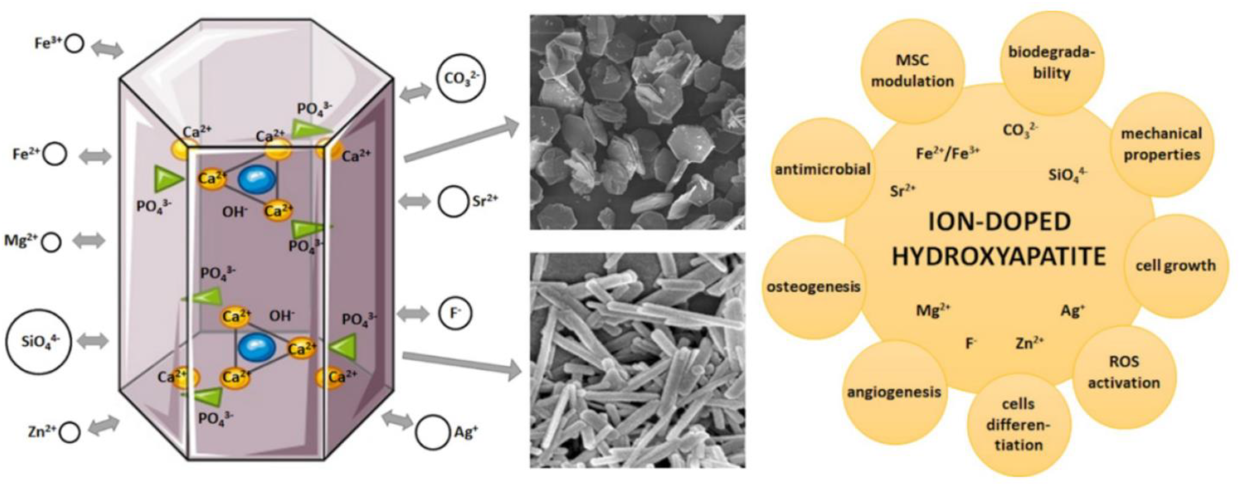
Over the past years, interest in the development and application of biomaterials for bone tissue engineering has increased extraordinarily. Since bone regeneration is considered the most complex and well-orchestrated physiological process, the main purpose of researchers has been to design biocompatible materials with well-defined biomechanical and physicochemical properties that can enhance bone formation and regeneration (Figure 1). Further, these biomaterials aim to initiate specific cellular responses at a molecular level, from cell interaction, proliferation, cell attachment, and differentiation to extracellular matrix synthesis [1,2].
Moreover, biomaterials are a distinct class of materials extensively used in bone tissue engineering. They have been comprehensively studied and applied in the biomedical field due to their similar microstructure with the osseous tissue and biologic-responsive properties, such as biocompatibility, osteoconductivity, osteoinductivity, osseointegration, and superior mechanical properties. Although they present numerous advantages, many drawbacks are still present, such as wear, corrosion, the release of toxic ions, stress-shielding limitations, and, in some cases, the necessity for the removal of non-degradable materials. To surpass these limitations, novel materials have been investigated, in which hydroxyapatite (HA) has been widely applied [4,5].
Thus, HA has been extensively studied by researchers in biomedical applications due to its osteogenic properties, biocompatibility, capacity to form a strong bond with the osseous tissues, and natural occurrence in the human body. This material has been mainly used in bone-healing applications. Even though synthetic HA presents excellent results in bone tissue engineering, the material has poor mechanical strength, low fracture toughness, and brittleness, which marks it unsuitable for load-carrying applications [6]. To surpass these disadvantages, several methods have been studied to increase its physicochemical and biological properties, mainly the osteoconductivity of synthesized HA. Hence, the optimum solution to obtain this goal was to alter the structure of HA by doping metal or non-metal ions, or even their composites. The inclusion of nanoscopic trace elements into HA can lead to modifications in crystallinity, lattice, and dispersal kinetics and improve mechanical properties and biological activity [7,8].
Another essential drawback of the applications of HA in bone tissue engineering is the presence of infections generated by pathogenic microorganisms. These microorganisms can easily infect the implantation site or the implant itself during surgical intervention. This issue may lead to increased treatment costs, as additional surgeries may be needed, or the rehabilitation time may be extended [9]. Moreover, the use of antibiotics does not ensure sufficient protection against bacterial strains resistant to multiple antibiotics. Hence, the doping of HA with different cations including zinc (Zn), silver (Ag), cerium (Ce), strontium (Sr), magnesium (Mg), copper (Cu), or titanium (Ti) ions is the ideal solution to enhance the antimicrobial activity at the implanted site, as they are not cytotoxic at low concentrations [10].
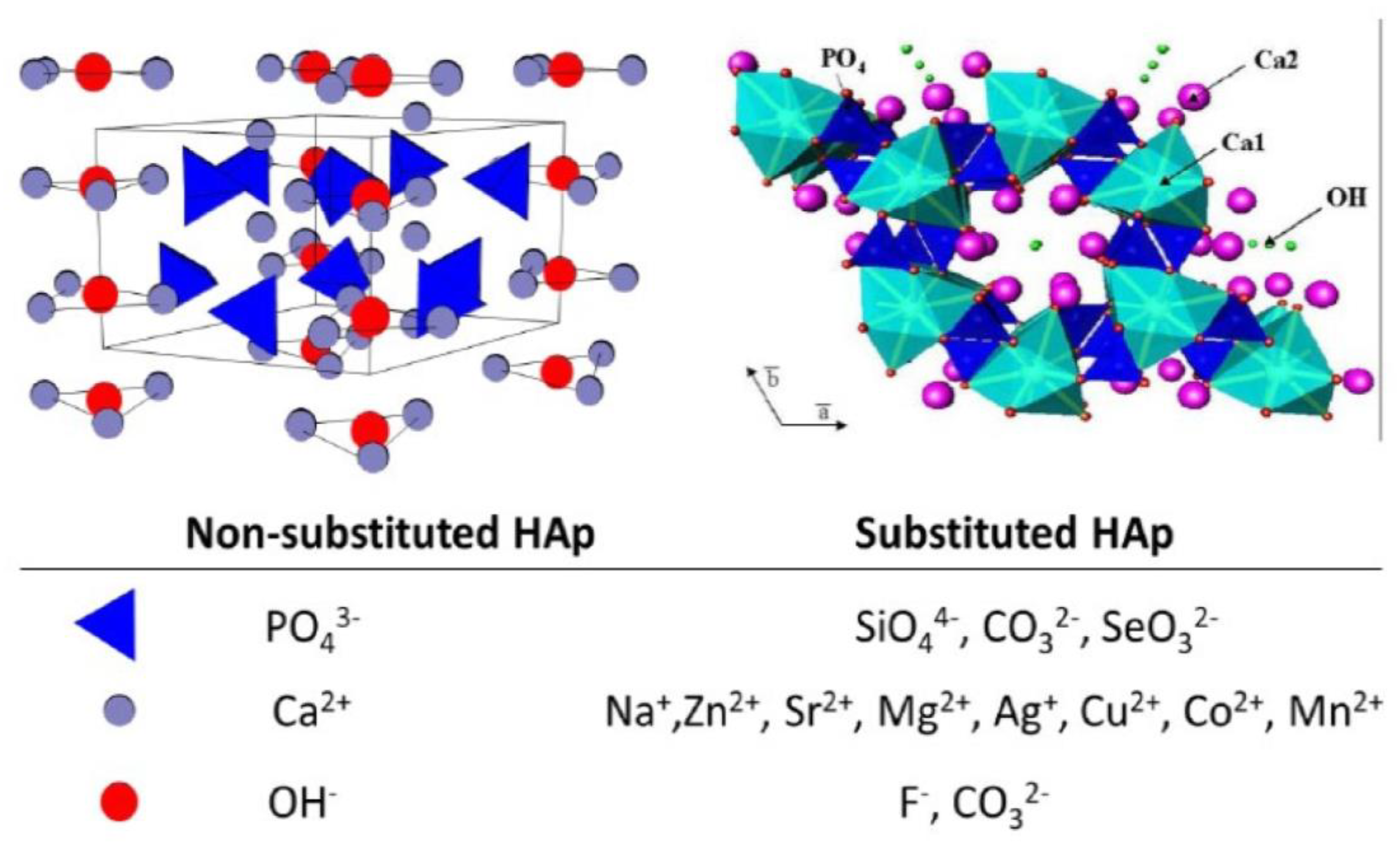
The atomic substitution or doping of HA (Figure 2) is the best approach to also improve the main weaknesses of implants based on HA, mainly the mechanical properties. By performing different types of substitutions, especially anionic (PO43−) and cationic (Ca2+) substitutions, all the drawbacks mentioned before can be solved [11].
In this regard, Bassyouni et al. reported that, by increasing the degree of crystallinity of HA and decreasing its grain size, the bioavailability of the material would be improved. Also, for the increase in dissolution rate, appropriate structural changes could be considered. The structure of HA can be modified by:
  • replacing the Ca2+ sites with Zn2+, Sr2+, Mg2+, and Na+ ions;
  • replacing the PO43− sites with CO32−, or SiO32− ions;
  • replacing the −OH group with Cl, F, and CO32− ions.
These examples of suggested ionic exchanges lead to the disturbance of crystallinity, surface charge, lattice parameters, and morphology, which can determine variations in the thermal stability, mechanic properties, solubility, and bioactivity of HA [13].
This review aims to present the main features of HA in the medical field, including their properties and synthesis methods, but also to present the importance of functionalization by doping HA. Moreover, we have summarized the influence of doping materials and the latest applications in biomedical engineering.

2. General Aspects of Hydroxyapatite Applied in Bone Tissue Engineering

2.1. Physical and Mechanical Properties

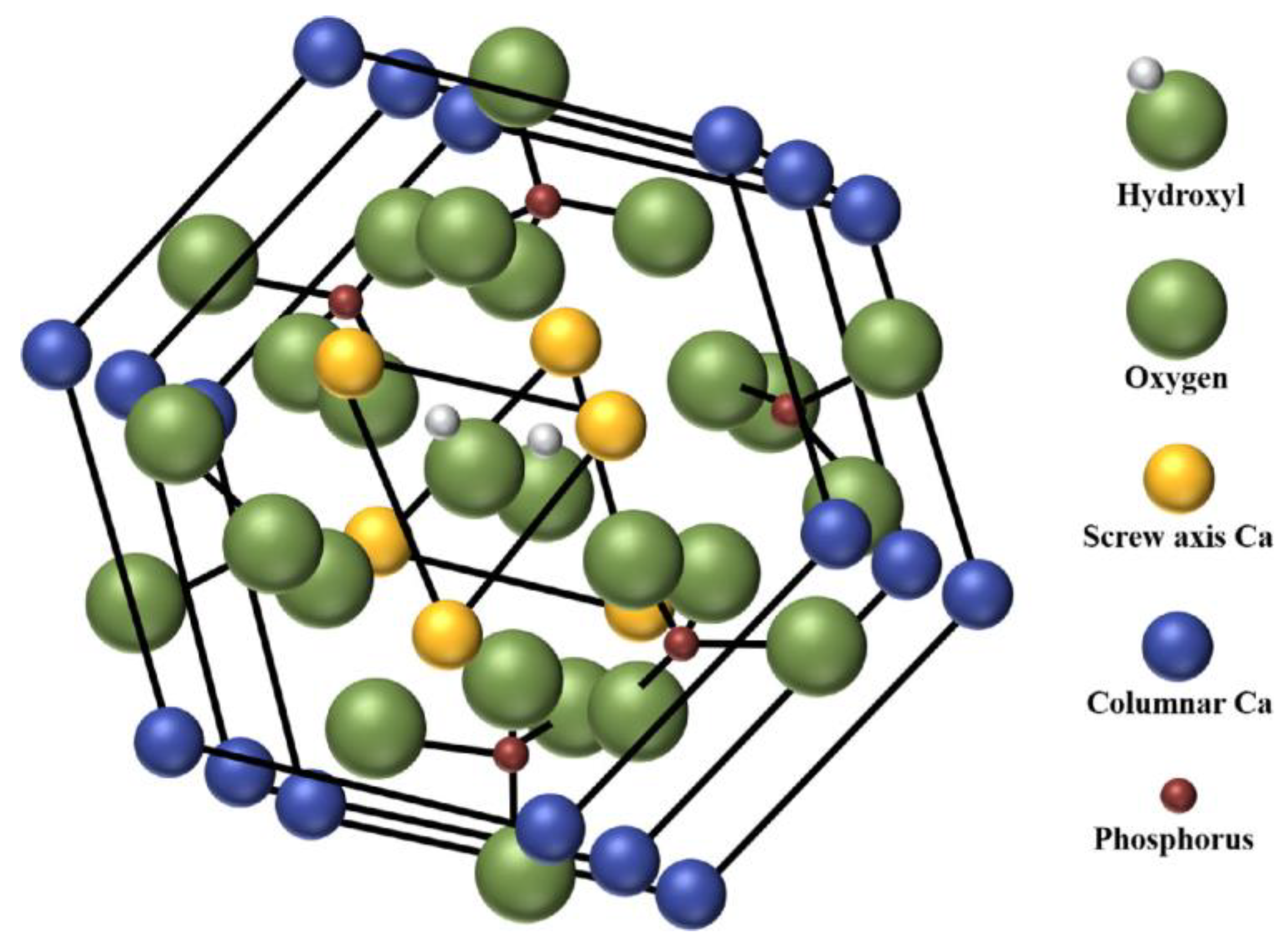
Ca10(PO4)6(OH)2 (HA) is part of the calcium phosphate (CP) ceramic group found in human osseous tissue bone (around 60–65%), a natural mineral compound. This material has been preferred to be applied as bone graft material due to its resemblance with the composition of the native hard tissue, with a stoichiometrically similar Ca/P ratio (1.67). Further, it is also well known for its good osteoconductivity, biocompatibility, and bioactivity. Nevertheless, synthesized HA (Figure 3) does not possess the specific trace ions which are normally found in the chemical structure of the osseous tissue that aids in bone formation, and, therefore, it possesses inferior bone regeneration [14,15].
The high purity and variable stoichiometry of HA have led to increased interest in its use in biomedical applications, mainly in bone tissue engineering. These materials, especially nanoparticles, present improved densification and sintering capabilities due to their high surface energy. Moreover, its enhanced wear resistance, chemical stability, and similar composition to the native tissue make this biomaterial more suitable compared to polymers or metals [17]. HA powder can be obtained through chemical synthesis routes at temperatures ranging between 60 and 85 °C by using precursors containing phosphate and calcium. The −OH groups increase the absorbance capacity for biomacromolecules and dielectric properties. Moreover, it increases osseointegration and bioactivity [18,19]. Also, its intrinsic osteoinduction, osteoconduction, and osteogenic properties are ideal for its use in the biomedical domain. Still, the use of synthetic HA is, at present, limited because of the insufficient mechanical and morphological limitations connected to cell interaction and infiltration [20].
Generally, the mechanical properties of the biomaterials applied in bone tissue engineering relate to their specific surface, pore size, pore distribution, total pore volume, and porosity. In addition, a ceramic with high porosity, inhomogeneous pore size distribution, superior total pore volume, large pore size, and large specific surface area will present low strength. Moreover, the bulk moduli and elastic constants of HA are mechanical properties that directly influence the strength of HA implants or scaffolds. For example, the porosity decreases the mechanical strength of the ceramic, thus reducing the cross-sectional area when a load is applied. Therefore, reducing the porosity, pore size, specific surface area, and total pore volume will improve the strength of the ceramic [21,22,23].
Essentially, the presence of porosity on the surface of the implant increases the depth, which corresponds to nano-HA accuracy in reducing calcium deficiency and surface roughness. This development is vital to the healing process. Moreover, increasing the HA microporosity may lead to an increase in the surface area in direct contact with body fluid, thus accelerating biodegradation and enhancing the deposition of the apatite on the surface of the material, improving the bioactivity of the material. By taking into consideration the aspect mentioned above, a proper balance between the porosity of the material and its mechanical properties is necessary [24,25].
The main requirement in the successful application of HA in bone tissue engineering is to provide sufficient mechanical strength until completed regeneration and new tissue formation, to offer suitable support. Furthermore, HA mechanical properties obtained by sintering are directly influenced by pore shape, material density, pore connection, and grain size. All these parameters can be attuned by the sintering temperature. In this direction, Trzaskowska et al. mentioned that, by increasing the sintering temperature, the compressive strength and density of HA are improved; meanwhile, the porosity and elastic deformation, according to Young’s modulus values, decrease [24]. Additionally, Benaqqa et al. reported that the application of HA sintered at high temperatures is not allowed, especially due to high brittleness. The obtained HA is very susceptible to the generation of small defects, slow crack growth, and, finally, eventual ceramic failure. Moreover, a sintering temperature higher than 900 °C led to the formation of large grains, reduced porosity, high crystallinity, low specific surface area, and high biocompatibility but is limited in clinical applications [26]. Although the main approach to obtaining HA is based on high sintering temperatures and high-pressure compaction, the obtained material has high porosity and loses uniformity with the generation of cracks. Mechanical properties are altered while the hydroxyl functional groups in HA are eliminated and the material is decomposed to α-tricalcium phosphate (α-TCP), β-tricalcium phosphate (β-TCP), and tetra calcium phosphate (TTCP); but, most importantly, the biocompatibility decreases [27].
Another important property of the material is represented by tensile strength. At the interaction with the osseous tissue, tensile forces are developed at the interface between the material and the tissue by the difference in the modulus of bone-material elasticity. HA fracture toughness also limits the applications of the material, as the required values need to be similar to the ones of the native tissue [28].
Crystallization is another property that has a great influence on the use of HA. While crystallization increases, the abrasion is decreased and reinforces a higher molecular weight of HA. This results in an improved tensile strength as the nanometer size of the bone improves. The extent to which the scaffold mimics the native osseous tissue also influences the crystallite size, increasing the abrasion resistance and reinforcing in vivo loading [25]. Defects such as dislocation and their density may also influence the bioactivity of HA crystals. The dislocations impact the processes that lead to bone apposition on bioactive materials. Moreover, dislocations also are a highly important factor in vivo dissolution processes and in the crystal maturation of biological cells/tissues. Additionally, the dislocations allow plastic deformations through slips. Researchers have proved that the hexagonal lattice of HA crystal shows highly anisotropic plastic deformation at increased temperatures [29,30].
On the other hand, the bioactivity of the material has also been thoroughly considered. This characteristic is represented by the capacity of the material to directly “bond” to the osseous tissue through chemical interactions. Commonly, when HA is implanted into the human body, a fiber-free and carbonated apatite with fiber-free layers is formed on the surface, improving the bonding and the fixation with the surrounding tissue [31]. The properties of the material surface are influenced by both chemical and topological aspects. The surface chemistry controls the dispersion of all charges and electric dipole moments at the interface between the material and tissue, generating electrostatic interactions in the aqueous environment with the proteins. In this regard, several factors must be considered when the surface of the material is modified. One of these factors is represented by the hydrophilicity of the surfaces, which promotes cellular adhesion [32].
Siddiqui et al. [33] demonstrated that the electrostatic interactions of the HA surface, with Ca2+ and PO43− ions encourage bone-like apatite formation. They reported that, when the HA surface is exposed to simulated body fluid (SBF), the surface displays a negative charge and interacts with the Ca2+ ions. In this process, an amorphous calcium phosphate layer rich in calcium is formed, altering the charge to become positively charged. The process continues with the electrostatic interaction with PO43− ions and leads to the formation of amorphous calcium phosphate that is deficient in calcium, which eventually matures into bone-like apatite.
Ultimately, HA presents poor fracture toughness, brittleness, undesirable tribological response, and low hardness. All recent research findings mentioned that, even though the chemical characteristics are similar, there is still a significant difference between the properties of osseous tissue and synthetic HA [34,35]. Nonetheless, the best feature of HA is represented by the capacity to take cationic and anionic substituents, improving their properties for numerous applications. In this regard, the properties of HA must be tailored in such a manner that it should improve cell proliferation and hinder prosthetic device failure for in vivo applications [36].

2.2. Synthesis Methods

It is well known that both synthetic and natural HA present superior biocompatibility, osteoconductivity, chemical support, and bioactivity, which aids bone tissue regeneration. These are the fundamental characteristics of scaffold applications or coatings used in tissue engineering [37]. Synthetic HA can be obtained through two principal methods: top-down and bottom-up approaches. The top-down method involves the successive cutting or slicing of bulk materials to obtain nanoparticles. On the other hand, the bottom-up method is represented by the build-up of material from the bottom, atom by atom, molecule by molecule, or cluster by cluster. The majority of the published research findings have been focused on the bottom-up method to obtain HA [38]. An abundance of reports has focused on the synthesis of HA powders through solid-state reactions, hydrothermal methods, sol–gel processes, microemulsion, emulsion, and also chemical precipitation. All these synthesis methods have been largely investigated by numerous researchers over many decades, as shown in Scheme 1, to improve their efficiency and functionality. These techniques have been preferred due to their simplicity and cost-effectiveness. Still, the obtained material might lack several essential ions, for example, Na, Sr, Mg, Si, K, and Fe [39].
The conventional solid-state technique is the preferred method to synthesize HA, as the developed nanoparticles preserve the morphology of the phosphate precursor particles. Alternatively, the chemical precipitation method is performed using precursor materials and an aqueous solution at low reaction temperatures, leading to the development of porous nanoparticles. The precipitation method is another chosen method for synthesizing nanosized HA with high purity due to its simplicity and low costs. The properties of the obtained HA depend on the technique used for synthesis, the selected precursors, and the processing parameters (reactant addition rate during the reaction, reaction temperature, and aging time) [51]. These aspects could affect their crystallinity, size, and shape (Table 1).
Another highly studied synthesis method is represented by hydrothermal synthesis. During this technique, the interaction is between the phosphate solution and the calcium precursor in the presence of water, an organic solvent, or a mixture of aqueous/organic. The reactions occur under the well-defined parameters of temperature or high pressure, which can be compared to those under autoclave pressure. Further, this technique generates chemical structures and develops nuclei that form HA with superior crystallinity [68]. Mushtaq et al. [69] mentioned that this technique is not only cost-effective and environmentally friendly, but that it can also easily vary in particle morphologies and sizes, which is a great advantage for biomedical applications. Likewise, the technique can be combined with additional methods, such as microwaves, hot-pressing, ultrasound, mechanochemistry, or electrochemistry. Moreover, Indira et al. [70] suggested that they can facilitate the synthesis time. For example, microwave irradiation efficiently increased the temperature and reaction kinetics compared to conventional methods. On the other side, microwave application leads to good homogeneity and increased fine particle purity.
Between those techniques, the sol–gel method presents many advantages, such as high purity, low synthesis temperature, excellent chemical homogeneity, and good mixing of the starting materials. The synthesis of HA using sol–gel demands that the Ca/P ratio is 1.67. However, depending on the experimental conditions (pH, temperature, aging time), different quantities of β-TCP can be formed [71]. Nellis et al. also confirmed that this route offers the capacity to obtain more versatile materials with good biocompatibility [72]. Furthermore, Ahmadi et al. mentioned that the sol–gel dip-coating approach is based on mixing phosphorus and calcium precursors. This technique was highlighted due to its advantages such as low cost, process temperature, accurate control of chemical composition, higher biocompatibility, and coating thickness. In the sol–gel process, numerous parameters (diluent type of HA solution, type of phosphorus and calcium precursors, temperature, calcination temperature, aging time) and the formation of inter-layer and residual stress in the coating have a substantial influence on the uniformity and quality of the coating [73].
Co-precipitation is another method that is highly studied in HA synthesis. This synthesis pathway aids the formation of the different morphologies of the particles with a low-crystalline nature [74]. Additionally, wet chemical precipitation represents one of the most used techniques due to its simplicity, cost, and low reaction temperature, in which high ratios of pure product can be obtained. As mentioned by numerous researchers, this technique depends on the mechanism of growth–agglomeration–nucleation–aggregation–agglomeration. The procedure beings with the preparation of alkaline (Ca source) and acid (P source) solutions, continued by the reaction between them. Next, there are the stages of precipitation, filtration, drying, and, finally, heat treatment, respectively [75]. The obtained materials through this method were proved to increase bone regeneration, surface absorption, and mechanical properties. On the other hand, more efficient synthesis methods with simplified preparation procedures and short duration are needed [76].
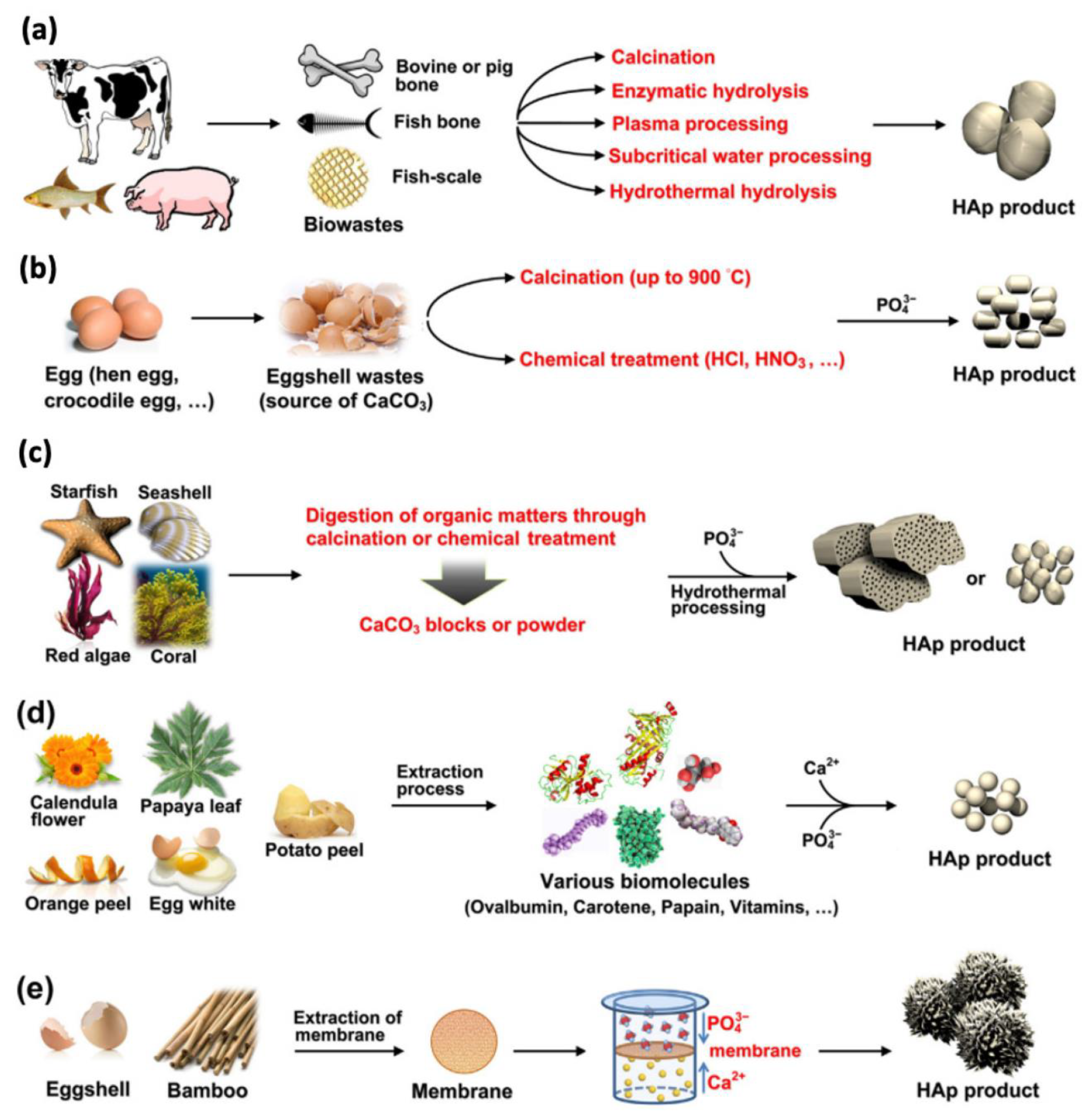
Nevertheless, conventional synthesis methods may generate toxicity and environmental damage, mainly air and water pollution, due to the usage of toxic solvents and harsh chemicals. Moreover, researchers have concluded that conventionally synthesized HA does not possess substantial biological properties. Considering this, eco-friendly synthesis methods of HA from natural resources (eggshells, fish bone, seashells, and cow bone) have been successfully developed due to their availability, nontoxicity, and lower cost. The extraction and purification of HA have been further studied in recent years to enhance their quality and expand the application domains [77,78,79]. Another advantage of this route is the capacity to retain some beneficial trace elements from the natural source of the synthesized material. Furthermore, mechanical and thermal processing can be also improved [80,81].
Studies on the naturally derived HA have indicated that these preparation methods presented trace elements such as Na, Mg, Sr, and K, but also carbonate anions, which are essential for bioactivity enhancement. Furko et al. mentioned that the most investigated preparation method is calcination, which can lead to obtaining HA powders with superior crystallinity in a fast and economical production process that requires less chemical consumption [82]. To obtain natural HA from eggshells, fish bones, or bovine bones, several chemical and physical methods have been employed. These include thermal treatment, microwave treatment, precipitation, a sol–gel technique, or hydrothermal procedures [83,84]. The attention to natural HAs for bone tissue engineering applications has been generated by their increased biocompatibility, osteoconductivity, and bioactivity [85].

2.3. Hydroxyapatite Applications

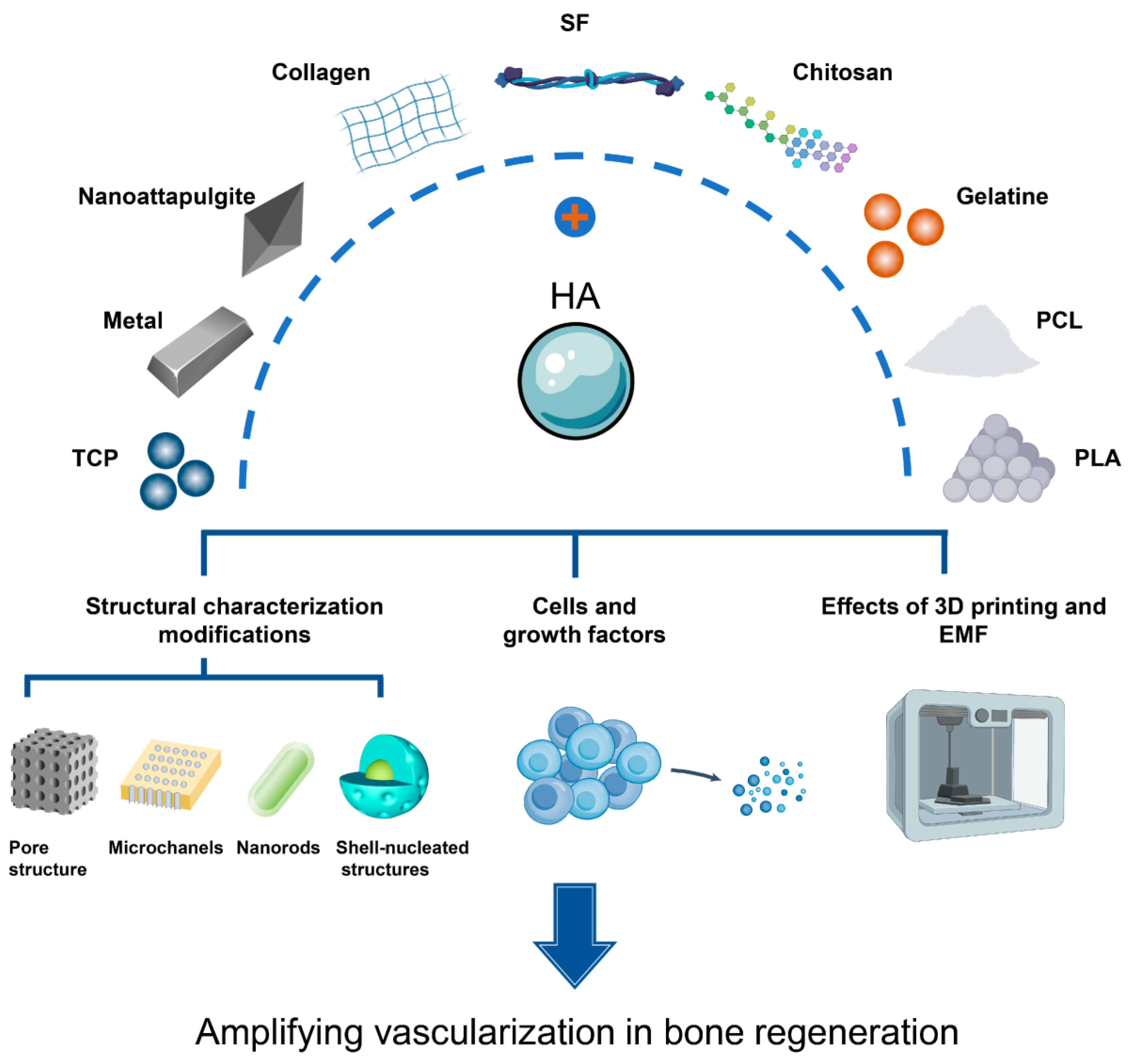
HA is a promising material for bone tissue engineering that has gained considerable interest in recent years. Its main applications in the biomedical industry have been widely explored due to its unique characteristics such as biocompatibility and enhancing cell proliferation and growth at the implantable site [86]. This material and its composites with polymeric matrices have proved to be ideal, mimicking materials for bone regeneration (Figure 4) due to their intrinsic advantages in osteoinduction, osseointegration, and osteoconduction. Also, HA presents the capacity of undifferentiated cells to render osteocytes and osteoblasts and facilitate the interaction between the osseous tissue and implant. All the characteristics must function properly for the appropriate performance of the application. In this regard, HA can be introduced in a biocompatible and biodegradable polymeric matrix that presents specific architectures and compositions to mimic the natural extracellular matrix. Also, they must possess good vascularization to provide the required transport of metabolic products, nutrients, and bioactive agents [87,88].
HA is the most used biomaterial in biomedical applications, offering support for osseous tissue formation and osteoconduction. Many studies have reported its efficiency in maxillofacial applications, orthopedics, infra-bony defects, hip and joint prosthesis, ridge augmentation, as well as in dental applications or coatings. Numerous properties influence their effectiveness, such as composition, size, structure, surface area, shape, and density, which can impact the kinetic process of new apatite formation on the HA surface [90]. HA is also well known for its hydroxyl function group. The groups generate chemical radicals that enhance implant adhesion and implant functionality. Additionally, its improved corrosion resistance has drawn the attention of researchers, as HA-coated metallic biomaterials increase strength and bioactivity. For this reason, numerous researchers have investigated the use of HA in drug delivery, tissue engineering, implants, coatings, and bone regeneration applications [91,92,93].
In dental applications, HA is frequently used for tooth fillers to enable bond formation among bone tissue and fillings. Zawisza et al. mentioned that even synthetic apatite, when applied as filling and dental implant coatings, can improve cell adhesion, proliferation, and differentiation, and that bacterial colonization cannot be surpassed [94]. Nano-HA presents several applications in dentistry due to its superior bioactivity and biocompatibility. For example, in cases of dentin hypersensitivity, HA was proven to induce the occlusion of dental tubules by functioning as a mineralizing agent. Likewise, nano-HA is preferred for implant coatings as it enhances bone-to-implant contact and encourages bone adhesion [95]. Another highly studied application involves coating the implant with biomaterials, namely HA, to increase the bioactivity of the implant. Because of its crystallographic composition, chemical structure, and similarity with the native tissue, HA forms a chemical connection between the osseous tissue and the surface of the biomedical implant. This encourages growth stimulation, and, therefore, the osseointegration process [96,97].
Nevertheless, stability in the timing of ceramic coatings affects the success of clinical applications and depends, therefore, on the deposition technique. For example, the deposition method controls the stability and uniformity of the HA bond to the metal substrate. Since CPs are difficult to deposit through direct physical methods, researchers have presented a combination of electrodeposition and 3D printing to avoid direct decomposition. Additive manufacturing techniques have been recently recommended for the incorporation of bioactive agents to adjust their deposition related to the high surface area of the coating material. This method facilitates the deposition of a coating in an efficient manner for drug delivery agents in bone tissue engineering by enhancing the osseointegration and reducing the formation of biofilm on the implant [98,99,100].
Moreover, HA has been also proposed for controlled drug delivery systems, cancer, and gene therapies, as well as bioimaging [101]. Controlled delivery systems can transport drug molecules at a controlled rate and time to increase the efficiency of drug uptake by the human body. Some scientists have proposed the combination of nano-HA with other biomaterials to develop composite bone scaffolds that incorporate drugs. These scaffolds have the capacity to release drugs slowly and locally, thus promoting the formation of new bone in the required zone, but also providing a new opportunity for the treatment of bone diseases. Moreover, they can provide appropriate structures for bone regeneration and support and also shelter the bioactive molecules from environmental degradation factors [102]. Asghar et al. also demonstrated the application of HA in drug delivery systems. They were successful in obtaining HA microporous structures and using them as drug delivery systems. The developed systems can transport bioactive molecules or drugs directly to the specific site [103].
Scaffolds containing HA for bone tissue engineering applications can also be considered a successful application in biomedical engineering, acting as an excellent temporary substrate. This substrate allows cell in-growth, proliferation, differentiation, and osseous tissue regeneration after in vivo implantation [104]. Hence, Hou et al. reported that HA with biomimetic micro-/nanostructured hybrid scaffolds possesses the superior adhesion, proliferation, and differentiation of bone marrow stromal cells. Furthermore, the obtained scaffolds had good protein adsorption owing to their surface properties, which contributed to the bioactivity of the composite scaffolds [105].
Lately, HA has been widely used in the fabrication of optical devices, biosensors applications, bioimaging, the coating of metallic implants, and theragnostic applications. At the moment, there are few reports on biosensor applications involving HA acting as an immobilization matrix to allow biomolecule adhesion over an electrochemical surface to provide a suitable microenvironment. Additionally, the excellent biocompatibility and adsorption ability, good sensitivity, and stability of HA favor the material for the fabrication of biosensors [106].
There have been numerous applications presented in the biomedical domain which illustrate HA as the ideal material due to its ability to enhance bone regeneration. As HA is known to possess some disadvantages that could lead to the failure of the application, such as mechanical strength or bioactivity, researchers have considered doping the material with different metals to surpass these deficiencies.

3. Comparison of Non-Doped and Doped Hydroxyapatite: Properties, Structure, and Applicability in Bone Tissue Engineering

As mentioned before, HA has been extensively studied due to its extraordinary biological characteristics in the biological environment, being known as the main mineral composition of osseous tissues. HA also presents superior biocompatibility and osteoconductivity, although it is brittle and possesses poor mechanical strength. Therefore, its simple application cannot be used in load-bearing applications even though there are notable differences between natural bone apatite and HA synthetic material. This material provides a hexagonal structure in which the calcium ions are replaced by dopant ions and the structure is distorted. Numerous studies have presented specific results of HA doped with Cu2+, Mg2+, Zn2+, Ag+, and many other ions. In this regard, Anushika et al. mentioned that doped HA showed a slight shifting of characteristic peaks. This modification is an effect of the doping agent on the crystal structure. Moreover, the doping ion can lead to distortions in the crystal and lattice parameters. Also, the percentage of crystallite size and crystallinity is reduced [107]. For example, when HA presents substitutions of elemental ions, such as silicate or carbonate ions, the size, crystal lattice, and concentration are modified in order to improve the physicochemical characteristics by altering its surface conditions and electron density. The functions accomplished by the substituted elemental ions have the capacity to modify some mechanisms of the osseous metabolic processes, especially in implantation and bone formation (such as osteoinduction, osteoconduction, and osseointegration). Sugimoto et al. reported that undifferentiated cells were able to mature into osteocytes over time, which is extremely important in implant fixation and bone regeneration [3].
The doping of HA can be performed in three different sites in the crystal structure. These include doping at the site of PO43− ions, Ca2+ ions, or at the OH site. Doping with different ions at these sites has been reported to increase the applicability of HA in bone tissue engineering and biomedical applications. Moreover, it has been shown to stimulate bone growth and improve bone density, as well as promote osteocyte proliferation and surpass bone tumor formation [108]. Furthermore, the combined outcome of doping ions is tailored according to the synthesis techniques, synthesis conditions, and doping type used to acquire the desired properties of the materials depending on the required application. The concentration, size, and type of ionic doping agents influence the microstructure, lattice parameters, crystallinity, grain size, crystal morphology, thermal stability, and solubility of HA. There are various types of ions used as doping agents for HA, and numerous studies have proven their influence and efficiency in bone tissue engineering [109,110]. Moreover, Shokri et al. mentioned that the substitution of doping ions in HA structures results in a gradual decrease in the lattice parameters while doping concentration increases. Furthermore, the addition of doping agents limits HA particle growth and reduces crystallinity. On the other hand, the doping of ions also modulates bioactive ion release, which can mediate biological responses [111].
Yahia et al. reported, that by comparing normally synthesized HA with doped HA, the crystallite size increases on the doped probe from 15 nm to 62 nm. Moreover, the linear absorption coefficient also shows that doped HA is more absorbable than simple HA, thus increasing its applicability in radiation detection and drug delivery applications. Additionally, the antimicrobial capacity of simple and doped HA on Gram-positive and Gram-negative bacteria and fungi exhibited higher antimicrobial activity of doped HA compared with simple HA [112]. Furthermore, Singh et al. obtained doped HA with Ag, which, in comparison with simple HA, presented comparable hardness, density, and improved antimicrobial characteristics compared to non-doped material. Nevertheless, no difference in cellular functionality, regarding cell adhesion and proliferation, was observed. The doping ion significantly improved the antibacterial property, which is a great feature for biofilm prevention at the implantation sites [113]. Moreover, Aksakal et al. obtained coatings with pure HA and doped with Ag and selenium (Se). The results showed that, while pure HA coatings had an antimicrobial activity of 83% against E. coli, the value was increased to 94% with the addition of Ag+ ions and 85% with Se ions. Correspondingly, the doping of ions leads to an improvement in HA antimicrobial activity against S. aureus. Moreover, cell viability decreased in the simple HA coating but increased significantly in the doped HA coatings [114].
Considering antimicrobial activity, pure HA nanoparticles cannot avoid biofilm formation on the surface of the material. This is the main reason for implant and surgical failure. The furthermost known reason for implant failure is a bacterial infection from Gram-negative (P. aeruginosa) and Gram-positive bacteria (S. epidermis). These infections are difficult to treat and, in most cases, can only be treated by removing the implant, which increases the treatment’s cost and endangers the life of the patient. In this case, doped HA is the ideal solution to enhance the properties of minerals introduced into the biological environment and enhance clinical outcomes. This route offers the opportunity to mimic the conditions of biological apatite. The biological apatite is generally found carbonated and substituted with trace amounts of ions. Consequently, some HA dopants can substitute similar roles as growth factors. Due to these improved properties, doped HA nanoparticles are considered in biomedical applications as antimicrobial agents, bioimaging probes, drug carriers, and theragnostic agents [115]. In this regard, Jian et al. reported that the use of HA in drug delivery systems is highly encouraged due to its excellent adsorption capacity and biocompatibility. They demonstrated that, compared with pure HA, doping ions not only improved its zeta potential and specific surface area but also enhanced the binding energy between the bioactive compound and HA, thus increasing its drug-loading capacity and prolonged release rate [116].
Considering their mechanical properties, the incorporation of the doping agent may lead to an increase in strength and corrosion resistance. In this direction, Vladescu et al. proved that substitution with doping ions improves the hardness and elastic modulus [117]. Moreover, Balakrishnan et al. mentioned that, by comparing pure HA and doped HA, the durability of the material has increased. Furthermore, by enhancing the concentration of the doping agent, the results of a Vickers microhardness test demonstrated increased hardness [118]. Additionally, Manzoor et al. confirmed that, by increasing the doping content, Young’s modulus of the obtained material increased [119]. The concentration of the doping ions has a significant influence on both the mechanical properties and the type of ions. For example, Aryal et al. demonstrated that Mg2+ substitution into an HA structure enhances the elastic properties of the material; meanwhile, Zn2+ degrades it [120]. In another study, a research group led by Wang studied the influence of doping ions (F) on an HA structure. They established that F both restores the mechanical properties of the HA lattice and successfully prevents the dissociation of the hydroxyl groups on its surface. Even though the doping ions cannot directly stop the calcium loss, they can indirectly reduce this process [121].
This substitution can be performed either with only one type of doping ion or with a co-substitution of two or three doping agents. In this case, there can be a combination of different doping material properties to improve the functionality of the biomaterial applied in bone tissue engineering. The co-substitution of the doping ions in the HA lattice may lead to the optimization of osteoconductivity, thus facilitating novel osseous tissue formation and increasing osteoinductivity, with the promotion of the differentiation of the mesenchymal stem cell to the osteoblast. These improved biological effects have been reported to inhibit osteoporosis. Ullah et al. reported that both co-doped ions also improve the solubility of the developed material and present an antimicrobial response, depending on the doping ions, as well as prevent bacteria growth [122,123]. As an example of co-substitution effects, Cao et al. developed three different coatings: the Mg-doped HA coating, F-doped HA coating, and HA doped with both F and Mg2+ coatings. After thorough investigations, the in vitro bioactivity studies presented different microstructures on the three coatings after soaking them for 7 days: a rice-like, nano-porous structure on the coating doped HA with F, a 3D porous network structure on the coating doped with Mg2+, and an ordered linear structure similar to the cancellous bone for the coating doped with both elements. The coating doped with both elements presented improved transformation from apatite-like to HA phase because of the synergistic effect of F and Mg2+. Moreover, cell viability analyses indicated that co-doped HA coating possesses good biocompatibility with no cytotoxicity and improved interfacial bioactivity in the titanium substrate, as well as good cellular proliferation [124].

4. Doped Hydroxyapatite: Doping Material and Methods, Synthesis Method, and Properties

HA is well known for its applications in the biomedical field due to its similarity with native tissue. Even though it possesses good characteristics for its use in bone tissue engineering, this material limits its use in load-bearing applications due to its poor mechanical strength and reduced bacterial resistance. To surpass these deficiencies, in the structure of HA, metal ions can be incorporated, such as Y3+, Zn2+, Mn2+, Mg2+, or Ag2+ (Figure 5). They are substituted in low concentrations into the HA lattice to improve biological capacities [125,126,127]. An ideal dopant must enhance the physicochemical characteristics of HA without negatively influencing the biological performance of the doped material. Additionally, small quantities of dopant agents must provide a superior combination of strength and elastic modulus to significantly improve HA fracture toughness [20].
Metal ions and their oxides have gained increased attention due to their superior antibacterial properties, especially against bacterial resistance to antibiotics. In contrast with antibiotics, metal ions function differently in bacterial and mammalian cells because of the deviation of metal transport systems from usual patterns. This enables the use of doped material with ions as antimicrobial agents with no negative influence on the human body [128]. These metal ions have three main mechanisms for antimicrobial properties. Firstly, metal ions will bind to proteins and then deactivate them. Secondly, metal ions will further interact with the microbial membrane, which provokes structural modifications and permeability. Finally, metal ions will interact with the microbial nucleic acids and then prevent microbial replication [129].
Furthermore, by doping the material with other materials, modifications in the lattice parameters, crystallinity, thermal stability, and morphology are performed, significantly influencing the solubility of the doped HA in contact with the physiological environment. Moreover, the structure of the developed biomaterial can offer efficient support for the improvement of bone tissue growth and angiogenesis on the implant [130,131,132]. Thus, methods must be further developed to adjust the structure of stoichiometric HA to enhance the bioactivity of the biomaterial [133].
The substitution with other ions in the structure of HA has been shown to enhance osteoblast differentiation, osteoinductivity, osteoconductivity, and osseointegration, as well as the ability to release the substituted ions because of the reduced solubility in HA (Table 2). The doping of Cu2+, SiO44−, and Mg2+ has demonstrated the improvement of osteoinduction, while the introduction of Na+, Cl, or CO32− can enhance osteoconduction. Additionally, HA doped with Sr2+ or Zn2+ improved several biological properties, such as the suppression of bone resorption and osteoporosis and the enhancement of bone formation. Moreover, researchers have reported that HA doped with SiO44− enhances the dissolution rate and osseointegration properties. On the other side, F-doped HA has been used to treat osteoporosis; however, high concentrations of F can lead to osteosclerosis and other diseases [127,134].

4.1. Zn2+-Doped Hydroxyapatite: Doping Method, Properties, and Biologic Activity

Zinc (Zn) is present in osseous tissues as a trace element, which has a great connection with bone metabolism. Furthermore, Zn2+ possesses antioxidant properties and is extremely important in various biochemical processes, such as blood clotting, protein digestion, or carbohydrate metabolism. Moreover, it has a substantial impact on the immune system. In this regard, numerous studies have indicated that Zn could promote osteoblasts cell proliferation and differentiation, as well as enhance bone formation. Considering these properties, its incorporation into HA may improve the bioactivity in bone tissue engineering [135,136]. Zn substitution in HA structures has gained great interest due to its presence in bone and teeth enamel, influencing the metabolic processes for bone growth. The incorporation of Zn ions improved bone development, biomineralization, and osteoblast production. Nevertheless, the lack of Zn in the osseous tissues reduced bone density [137].
The latest reports show that the presence of Zn in ceramic materials provides antimicrobial activity, depending on the concentrations of the doping material. Buyuksungur et al. reported that a small concentration of Zn in the structure of HA is sufficient to ensure resistance against S. aureus, K. pneumoniae, or B. cereus [138]. Comparable results were also mentioned by Ofudje et al., who Zn-doped HA, which was prepared using the chemical co-precipitation method and further used as a scaffold. Their bioactivity and antibacterial activity against E. coli and S. aureus were successfully demonstrated. Moreover, researchers have highlighted that, by increasing the Zn concentration, the dissolution rate increases due to a decrease in crystallinity [139]. Candidato Jr. et al. also mentioned that Zn incorporation does not alter the morphology of coatings, while X-Ray Diffraction analysis showed that a Zn concentration higher than 10 mol% can inhibit HA formation and encourage calcium zinc phosphate formation. Moreover, they confirmed that Zn ion substitution affected the crystal structure and changed the crystallinity and lattice parameters due to lattice distortion [140].
Further, Zhou et al. proved that the Zn-doped HA, when applied as a coating on the surface of the implant, not only yielded higher corrosion resistance but also enhanced bacterial inhibition and osteogenic differentiation. Compared to pure HA coatings, doped coating not only successfully inhibited bacteria, but also promoted cellular adhesion and differentiation of bone marrow mesenchymal stem cells [141]. Moreover, Sergi et al. demonstrated that Zn-doped HA prepared by co-precipitation nibbled the modulation of osteoblast activity and antibacterial capacity. The developed materials have been applied as coatings, and their bioactivity was proved by soaking them in SBF. After further investigations, the coatings exhibited non-cytotoxicity against human osteoblast Saos-2-cells and antibacterial effects against S. aureus and E. coli, with higher efficiency against the Gram-positive bacteria [142]. In another study, Popa et al. investigated the application of Zn-doped HA on the collagen matrix. After biological evaluations, the developed structures showed no toxicity in bone tissue engineering applications. Moreover, doped HA can be effectively applied in drug delivery systems. In this regard, Venkatasubbu et al. reported high efficacy in the control of the drug delivery rate of Zn-doped HA that incorporated ciprofloxacin as a bioactive molecule [143].

4.2. Mg2+-Doped Hydroxyapatite: Doping Method, Properties, and Biologic Activity

Magnesium (Mg) is another preferred substitute for HA due to its chemical similarity. Mg ions are acknowledged for bone mineralization promotion and the enhancement of osteoblast-like cell proliferation; hence, they are also known for enhancing the biocompatibility and bioactivity of a developed material [144]. Further, Mg is the fourth most highly concentrated cation which can be found in the human body after Ca, P, and Na. The deficiency of Mg in osseous tissue has been suggested as a possible risk factor for the generation of osteoporosis. In the human body, Mg2+ has the capacity to reduce the degradation rate of CPs and influences the crystallization of mineral substances. In this regard, the introduction of Mg ions into HA encouraged endothelial cell and osteoblast resistance, spreading and enhancing osteoblast adhesion and cell proliferation [133].
The substitution with Mg2+ ions not only led to the modification of biological properties but also of the mechanical and chemical structure. Pana et al. mentioned that the addition of doping ions into the HA matrix improves the hardness and elastic modulus compared to the pure HA [145]. Further, Monte et al. mentioned that Mg2+ also affected the crystal structure, reduced its porosity, and enhanced the bending strength and corrosion resistance [146]. To confirm this, Veljovic et al. developed bone fillers from Mg-doped HA, highlighting the interaction between the human osteoblast, mesenchymal stem cells, and implant material, as well as the in vitro and in vivo formation of the novel osseous tissue [144]. Moreover, Lv et al. investigated the cytotoxicity of doped HA to osteosarcoma cells (MG-63) and mesenchymal stem cells (MSCs). After biological investigations, HA doped with Mg ions was found to enhance the adhesion and proliferation of MG-63 and MSC cells, promoting their metabolism. A suitable concentration of the doping agent improves material biocompatibility [147].
Daood et al. also developed a dentine adhesive modified with Mg-doped HA. The doped material was also prepared using precipitation methods. After thorough investigations, it was proven that the doped biomaterial possesses antibiofilm and self-remineralization properties [148]. Moreover, Jenifer et al. also reported an improvement in antibacterial and hemolytic efficiency after the substitution of Mg ions into the HA structure. The incorporation of doping ions also proved to reduce inflammatory activity [149].
Zhao et al. tried a new method for obtaining Mg-doped HA with oriented microchannels through hot-pressing sintering to provide uniformity and an arranged control of pores. Compared with normal synthesized HA, the doped material exhibited superior mechanical and biological properties. The density and microchannel size extraordinarily influenced the compressive strength of the doped bioceramic. Additionally, the microchannel-containing ceramic exhibited improved apatite mineralization in SBF [150].

4.3. Cu2+-Doped Hydroxyapatite: Doping Method, Properties, and Biologic Activity

Lately, copper (Cu) has been recognized to exhibit good biological properties by enhancing the differentiation of mesenchymal stem cells into osteoblast cells, facilitating endothelial cell proliferation in wound healing, reducing implantation infections, and increasing the gene delivery levels of vascular endothelial growth factors. Considering all these aspects, Cu2+ is an ideal element to exhibit specific characteristics for bone repair; however, at the same time, the physicochemical characteristics of Cu-based materials are altered. Moreover, excessive Cu release into the human body may cause cytotoxicity [151]. Furthermore, Hidalgo-Robatto et al. demonstrated that doped HA promotes osseointegration at the interface between the bone and implant surface and simultaneously inhibits bacterial biofilm formation. Finally, the doping of HA with Cu2+ has numerous advantages to be further applied in metallic implants for the regeneration or repair of severely damaged or fractured bone tissue. Moreover, not only was biocompatibility increased, but the mechanical stability of the doped HA was also increased, which is a desired improvement for numerous biomedical applications [152]. On the other side, Ercan et al. mentioned that the addition of Cu altered the thermal stability, morphology, and agglomeration degree [153].
Cu2+ ion substitution into the HA structure (Figure 6) presents a beneficial effect not only on angiogenesis, cell proliferation, and the differentiation of stem cells but also on improved cell migration while inhibiting osteoclast activity [154]. To further investigate the biological activity, Ressler et al. obtained Cu-doped HA using the precipitation method, with the substitution of Cu2+ at Ca sites. They also tried the immersion method of non-substituted HA in a solution of Cu2+ ions, with the substitution occurring at OH sites. While high concentrations of Cu2+ presented good antibacterial properties, toxicity was also reported; therefore, a suitable concentration must be selected in order to also illustrate cell proliferation [155].
Furthermore, researchers have reported that Cu incorporation into ceramic materials leads to the improvement of neovascularization and stimulation of endothelial cell proliferation in vitro [157]. Park et al. performed in vitro studies which confirmed the nontoxic effect of both doped and non-doped HA using MG-63 cell lines. The developed materials successfully simulated and facilitated new cell attachment, growth, and proliferation on the surface of the implant with adequate compressive strength [158]. In another study, I’Alzit et al. incorporated Cu ions into HA using thermal treatment. The obtained materials exhibited the capacity to stimulate blood vessel formation, together with the enhancement of mechanical properties and antibacterial properties [159].

4.4. Sr2+-Doped Hydroxyapatite: Doping Method, Properties, and Biologic Activity

Strontium (Sr) is another trace element that is present in the osseous tissue. Sr presents outstanding bone-healing properties and offers therapeutic significance for the treatment of osteoporosis. Sr2+ ions present a dual importance in stimulating bone regeneration, involving the inhibition of osteoclast activity and the stimulation of osteoblasts, leading to new bone formation and a decrease in bone resorption. It was proved that Sr has a high influence on bone density and improves bioactivity and mechanical properties [160].
Many studies have used Sr-doped HA coatings on the surface of the implant to increase the bioactivity and reduce the corrosion in time. This trace element significantly decreases the risk of bone fractures and the generation of osteoporosis. Further, Sr inhibits bone resorption by reducing the adsorption activity of osteoclasts and differentiation; moreover, at the same time, it reduces the production of osteoblast matrix metalloproteinase [160,161]. Furthermore, the introduction of dopants in the HA structure influences microstructure, morphology, solubility, thermal stability, and the mechanical properties of the bioceramic; therefore, it is a good method to obtain specific properties for the biomaterial in conformity with the desired application [123]. As mentioned before, the incorporation of Sr ions into the structure of HA modifies not only bioactivity but also physicochemical properties. In this regard, Baldassarre et al. obtained Sr-substituted HA powders with different Sr concentrations that were prepared using a solid-state synthesis method. After thorough investigations, researchers proved that, by increasing the concentration of the doping agent, the particle size and distribution were modified. Further, their substitution led to lattice expansion due to the larger Sr atoms. Moreover, by increasing the Sr concentration into HA, PO43− groups were broadened due to the enhancement of the crystal structure disorder and the decrease in the crystallite size [162].
In the last few years, researchers have focused on the development of Sr-substituted HA. The doped materials were proved to enhance the bioactivity of the material as Sr is known to stimulate bone formation. Generally, Sr-doped HA is a hydrophilic and alkaline material with superior solubility and good compressive strength, which is similar to native tissue mechanical properties [133]. Hu et al. mentioned that a low concentration of Sr2+ presents great efficiency in stimulating osteoblast proliferation and differentiation, while a higher concentration negatively impacts the bone mineralization process. Thus, the selection of a suitable concentration for the doping agent is essential for bone tissue engineering applications [163]. Moreover, Wang et al. mentioned that Sr2+ substitution also influences the morphology and size of the obtained material. The immersion and electrochemical tests also showed that Sr doping improved the corrosion resistance of HA coatings. Moreover, BMSCs’ cell culture results highlighted that Sr2+ addition promotes cell adhesion and the proliferation of HA-coated Mg alloys [164]. In another study, Xu et al. obtained Sr-doped HA microspheres with interconnected porosity and pore channels, prepared via the hydrothermal method. They reported that the morphologies, constituents, scales, and pore geometries of the obtained materials were modulated by the concentration of Sr2+. The substitution of Sr2+ significantly enhanced the biodegradability of the microspheres. Additionally, the porous microspheres presented a superior drug loading capacity and sustained release properties. These particularities have also proven the potential of Sr-doped HA to be applied in drug delivery systems [165].

4.5. Ag+-Doped Hydroxyapatite: Doping Method, Properties, and Biologic Activity

Silver (Ag) is another well-known element to be incorporated in numerous materials due to its pronounced antibacterial activity, biocompatibility, and low toxicity at reduced concentrations [166,167]. Ag+ was selected in numerous applications in the biomedical field for its increased bioactivity and thermal stability. Further, Ag+ presents the ability to attach to proteins and enzymes in bacterial cells and inhibit the main cell functions of various Gram-negative and Gram-positive bacteria, including S. aureus and E. coli. Ag ions incorporated into the HA structure would generate a multi-functional adsorbent material [168].
Antibacterial activity is the primordial characteristic of Ag+ to be considered for its use as a doping agent, as it possesses the capacity to control biofilm formation and bacteria adhesion. Many studies have demonstrated the efficiency of the use of Ag-substituted HA in bone tissue engineering. The doped materials can be obtained using hydrothermal, precipitation, and mechanochemical techniques [169]. The incorporation of Ag ions into the HA structure to develop materials with tailored properties that meet the functionality and quality requirements for a bone substitute has been the main goal. These doping ions have been preferred due to their superior antimicrobial activity against fungi, viruses, and a wide spectrum of bacteria. However, their applicability is strongly influenced by the toxicity of metal ions. Taking this into consideration, the biocompatibility of doped HA can be partially affected. Nenen et al. mentioned that, compared to other metallic ions, the use of Ag+ ions as a doping material in biominerals can still show lower cellular stress [170,171]. In this direction, it has been reported that positively charged Ag nanoparticles exhibited higher antimicrobial activity than negatively surface-charged nanoparticles. Yet, the cytotoxic effect of these nanomaterials may relate to more than one physicochemical characteristic. Undeniably, even though many researchers focused their interest on the antibacterial property of Ag nanoparticles, the mechanisms involving the bactericidal property are still under continuous research [172].
Chatterjee et al. successfully developed Ag-doped HA, which was prepared using an in situ hydrothermal method. The obtained material confirmed the effective doping of Ag ions in the HA lattice, which influenced the thermal stability of the material and ensured significantly high ionic conductivity with reduced activation energy for increased temperatures. Moreover, the superior biocompatibility of the Ag-doped HA rods and good electric and dielectric properties highlighted the capability of the obtained material as a nanocarrier for electric field-induced drug delivery systems in the future [173]. Moreover, Citradewi et al. mentioned that, by doping the material with Ag ions and using a natural source for the synthesis of HA, the bioactivity of the material could be further improved compared to the simple doped HA. Moreover, the bacteriostatic properties were enhanced over time [174]. Regarding physicochemical properties, Zhou et al. demonstrated that Ag could be absorbed on the surface of HA. This led to morphological modifications, excluding the enhancement of biocompatibility and antibacterial activity [175].

4.6. Hydroxyapatite Doped with Other Metals: Doping Method, Properties, and Biologic Activity

There are many other metals and composites that have been studied as doping materials to be incorporated into the structure of HA. Depending on the desired applications and required properties, they have been extensively investigated to improve the mechanical properties and bioactivity of the designed material to be used in the biomedical domain.
Iron (Fe3+) is another doping agent that is present in trace quantities, and it is known for its importance in bone health. When Fe is incorporated into the structure of HA, the physical and biological properties are improved. Fe-doped HA is known to be synthesized through different routes like spray drying and hydrothermal and ultrasonic irradiation [176].
Another doping material highly studied in biomedical applications is represented by cerium (Ce). Ce3+ ions possess good antimicrobial abilities and low toxicity. This material is highly recommended for its bioactivity, such as its capacity to improve the reproduction of collagen, as well as the proliferation, differentiation, and mineralization of osteoblast cells [177]. Sousa et al. mentioned that Ce3+ ions can be easily incorporated into HA structures due to their capacity to occupy Ca2+ sites, as the Ce3+ ionic radius is similar to that of Ca2+. This substitution demonstrated the enhancement in bioactivity, especially regarding cell viability, antibacterial capacity, an increase in metabolic rate, and antioxidant activity [178]. Moreover, Padmanabhan et al. mentioned that Ce doped-HA encouraged the deposition of calcium-rich apatite layers on the surface of SBF, mimicking the hardness of natural bones [179]. Additionally, Nisar et al. demonstrated that Ce substitution into the HA structure also improved mechanical properties [6].
Chromium (Cr) is another trace element in the human body that is essential to metabolize fats and sugars. Cr3+ doping is also used to enhance the hemocompatibility of HA. Hence, Cr doping is very advantageous for biomedical applications; however, the cytotoxicity needs to be properly assessed [180,181]. To confirm the applicability of Cr in bone tissue engineering, Lima et al. obtained Cr-doped HA powders with different Cr3+ concentrations through the co-precipitation method. After thorough investigations, the results showed that the modifications of the mechanical properties are influenced by the doping concentration [182]. Iqbal et al. also confirmed that, by increasing the Cr ion concentration, the crystallinity of the sample decreases [183].
Cobalt (Co) is an indispensable trace element with a very specific function in the human body. Yet, some studies involving Co-doped bioceramics have demonstrated the angiogenic effect of Co2+, leading to the enhancement of the neovascularization of new bone formation [184]. Kulanthaivel et al. also obtained Co-doped HA that was prepared using the precipitation method with different doping concentrations. Researchers proved that mechanical properties are altered depending on the concentration of the doping ions. Further, they demonstrated that Co doping into an HA structure presented great proangiogenic properties without harming the osteogenic property of the HA [185].
In another study, Abutalib et al. investigated the application of Molybdenum (Mo) as doping material. They successfully obtained doped and non-doped HA using the efficient microwave technique at low temperatures. After biological investigations, they demonstrated and improved antibacterial activity with the addition of the doping agent, confirming their applicability in biomedical applications [186].

5. Latest Applications of Doped Hydroxyapatite in Bone Tissue Engineering

HA has attracted immense attention in biomedical applications due to its similarity with natural osseous tissue, high bioactivity, osteoconductivity, and biocompatibility. Even though this material has great advantages, there are still some drawbacks that need to be solved for it to properly function in bone tissue engineering. As the mechanical properties and antibacterial ability still need to be improved, HA doping has been the ideal solution to solve all issues, depending on its applicability.

5.1. Applicability of Doped Hydroxyapatite in Dental Applications

Many studies have focused on the development of ion-doped HA for the treatment of numerous dental applications. In this direction, many researchers have focused on the use of HA, which is the main tooth constituent that exhibits a remineralizing effect on tooth enamel; moreover, it can enhance the bond’s strength to the structure. As HA still lacks mechanical strength and bioactivity, the application of doped HA was the ideal solution for solving some drawbacks that may appear during the treatment time. Thus, doping aids the obtained materials with enhanced antibacterial properties to stimulate new bone growth and prevent inflammation, which is highly important in dental applications [187]. Tautkus et al. mentioned that orthodontic materials containing doped ceramics (such as Zn2+ ions) demonstrated higher antibacterial and remineralization effects compared with non-doped materials. Further, these doped materials are innovative biomaterials for filling bone cavities as a matrix for cells to support the regeneration of damaged tissue [188].
Mathew et al. obtained Sr-doped HA remineralizing paste, with and without chitosan, in order to investigate the antimicrobial capacity. The antibacterial efficacy of the developed paste was improved by the addition of chitosan, and its antimicrobial efficacy was comparable to calcium hydroxide. Moreover, the antibacterial efficacy of Sr2+-doped HA paste with and without chitosan was assessed against the most common microorganism involved with the process of dental caries (S. mutans). As Sr2+ is a trace element in the human body, it can be easily incorporated and accepted by tissues. The doped material presented different doping ion concentrations. After performing the investigations, researchers showed the ability of Sr-doped HA to induce an improved formation of biomimetic apatite both in SBF and cell culture medium. Moreover, the incorporation of Sr into the HA structure increased the antibacterial capacity of the material [189].
In another study, Sauro et al. investigated the chemical, structural, and biological effects of fluoride-doped HA (F-doped HA) with different doping ion concentrations as potential remineralizing materials for dental applications. Thus, researchers have mentioned that mechanical properties have been modified and that all obtained materials were able to produce F-containing, apatite-like crystals after SBF immersion. Also, by increasing the concentration of F ions, there will be a prolonged release of F ions and increased precipitation of CaF2; thus, there will be high biocompatibility [190]. Teerakanok et al. also explored the influence of different doping ions (Mg2+, F, and Zn2+) on the structure of HA by investigating crystal organization, crystalline phases, and morphology in enamel remineralization. The researchers mentioned that, by increasing F concentration, the shape and size of HA crystals were affected; meanwhile, Mg2+ did not directly affect the morphology of HA crystals. Moreover, in the presence of Mg2+ combined with F, small needle-like crystals with a higher density were formed. Also, considering the incorporation of Zn2+ ions, they mentioned that increased concentrations led to the inhibition of crystal formation. Furthermore, by combining F with Zn2+ in low concentrations, dense needle-like crystals formed homogenous layers. As a result, they have proven the synergistic effect of combining different doping ions into the crystallization of HA structures in enamel remineralization [191].
Moreover, Ciobanu et al. investigated the applicability of Ce-doped materials as coatings on dental implants. The doped coatings presented good antibacterial efficiency against S. aureus and E. coli, with higher efficiency against E. coli. [192]. Additionally, Kadhim et al. studied other doping ions, such as Sr2+ and Mg2+, to obtain HA coatings to be deposited on Ti–6Al–4V dental alloys. They proved that both doping materials improved mechanical and biological properties. Moreover, the corrosion resistance of the dental alloys in the saliva environment was also improved [193].

5.2. Applicability of Doped Hydroxyapatite in Drug Delivery Systems

Considering dental or orthopedic interventions, HA is the optimum material used for drug delivery systems to directly transport the active substance in the affected zone. This may lead to a reduction in the treatment time with the antibiotics to avoid an overdose. In this regard, HA has been preferred as a drug carrier due to its controlled release rate. The crystallinity, porosity, phase purity, particle size, morphology, solubility, agglomeration tendency, and chemical composition of HA have also influenced the success of the drug release at a prolonged rate [194]. However, HA still has some inherent limitations, considering its mechanical and antibacterial properties. In this direction, the biological, physio-chemical, and antibacterial characteristics of synthetic HA can be tailored with the aid of doping/substitution with several selected ions. The exceptional HA structure offers extensive stability and flexibility for the substitution of various ions into its lattice. Many examples in the literature have demonstrated the efficiency of doping the materials and their further use in drug delivery applications. Numerous anions (SiO44−, CO32−, Cl, F, HPO42−) and cations (Mg2+, Sr2+, Zn2+, Mn2+, Na+, Ce3+, K+, etc.) have been investigated for doping to obtain materials with desired properties [195].
Therefore, Ullah et al. prepared Sr2+/Fe3+ co-doped HA to be further applied in drug delivery systems. After their synthesis, an increase in lattice parameters was detected with a high degree of Sr2+ substitution and a reduced degree of Fe3+ substitution. This variation in lattice parameters demonstrated that both incorporated ions partially substitute Ca2+ ions in the HA lattice. The incorporation of both Sr2+ and Fe3+ improved the biomechanical properties; therefore, it has been proposed as a suitable candidate for drug delivery systems and as a heat mediator for hyperthermia [155,196]. De Lama et al. proposed the direct incorporation of SeO42− ions in an HA structure for the prevention of bone cancer metastasis and for the control of skeletal tumors. They suggested that the cytosolic release of Se activates a caspase-dependent apoptosis pathway that is complemented by the formation of reactive oxygen species. Furthermore, the combination of Se with Pt-based drugs led to a reduction in nephrotoxicity and bone marrow suppression, effects which are attributed to the formation of intermediate Se–Pt complexes [197].
Furthermore, Febrian et al. demonstrated the efficiency of doping agents in drug delivery systems applied for cancer treatment. The substitution of HA with specific doping ions (such as Zr4+) improved the efficacy of HA in cancer treatment. Doped HA was assessed for cellular uptake, cytotoxicity, and biodistribution pattern evaluation. They demonstrated that, by doping HA, the efficacy of HA in lung cancer therapy was improved [198]. In another study, Jose et al. developed doped Mg to be further applied in drug delivery systems. The substitution has been demonstrated by the changes in the lattice parameters of HA. Moreover, the results showed that, by increasing the concentration of Mg, the morphology of the samples was altered with smaller particle sizes. Further, amoxicillin was loaded, and its release studies were performed on pure and Mg-doped HA using in vitro conditions. Moreover, researchers demonstrated that, by doping HA, the mechanical properties and bioactivity were improved enough to further use the material in drug delivery systems [199].
Zheng et al. obtained Sr-doped HA microspheres using a facile solid-state reaction and subsequent hydrothermal treatment to prepare mesoporous structures. The prepared microspheres possessed a superior drug-carrying ability and prolonged release performance. Moreover, the microsphere’s in vitro cytocompatibility was evaluated, revealing the good cytocompatibility of the microspheres. Moreover, the prepared Sr–HA microspheres offered an effective solution with which to deliver a stable source of drugs and therapeutic ions to promote bone repair and regeneration [200].

5.3. Applicability of Doped Hydroxyapatite in Bone Tissue Engineering

HA is one of the most frequently used bioceramics in bone tissue engineering due to its good bioactivity and superior biocompatibility. Yet, HA presents low fracture toughness, weak wear resistance, low solubility, and difficulty in processing steps into three-dimensional shapes, which limits the variety of applications. Numerous researchers have prepared and designed composites by combining HA with other materials such as bioactive glass, polymers, and carbon materials to surpass the above problems. Moreover, to increase the bioactivity, mainly the osteogenic properties, doping ions were incorporated into the structure of HA [201].
To prove the abovementioned aspects, Ai et al. obtained a Cu-doped HA sodium alginate composite scaffold that was developed using 3D printing. They demonstrated a uniform distribution of Cu2+ into the scaffold. Thus, after mechanical behavior studies, they observed that doped Cu ions improved the compressive strength of the scaffolds compared to the undoped Cu scaffolds. Combined with the shrinking results, the incorporation of Cu2+ improved the densification of the obtained scaffolds, resulting in the enhanced compressive strength of the scaffolds. Regarding biological activity, the developed scaffolds demonstrated low cytotoxicity and good antibacterial properties, resulting in their recommendation for the developed material in bone tissue engineering [202].
In another study, Banerjee et al. used erbium ion-doped HA to obtain luminescent chitosan–HA nanocomposite films to be applied as antimicrobial dressing materials, implant materials, or fluorescent cell regeneration scaffolds. The developed scaffolds presented good flexibility, facile processing, excellent antimicrobial activity and bioactivity, and fluorescent properties under in vitro conditions [203]. Yuan et al. also obtained composite scaffolds from chitosan and Sr-doped HA with different concentrations. They were further applied as coatings, with good density and uniformity, ductility, and improved adhesion. Sr incorporation led to crystal growth and increased grain size. Moreover, an increase in the concentration of the doping ions improved biocompatibility, enhancing osteoconductivity, cell adhesion, and proliferation [204].
Furthermore, Monte et al. developed scaffolds based on alginate and Mg-doped HA, considering that the incorporation of Mg2+ ions and alginate in HA leads to an improvement in bioactivity. The composites of the obtained materials presented great potential for bone tissue regeneration. The (Ca + Mg)/P decreased ratio (<1.67) showed that the inorganic phases of the composites were more soluble in the physiological medium compared to HA; therefore, the scaffold had higher bioactivity and bioavailability with the incorporation of Mg2+ [146].
Many studies have attested to the influence of doping ions on HA structures by improving the bioactivity of the developed material. Likewise, the doping process has successfully led to enhanced bone formation and regeneration with no cytotoxic effects, characteristics that hugely impact the prevention of infections, or other diseases that may derive from orthopedic implant surgeries.

6. Future Challenges, Perspectives

HA synthesis and its application in biomedical engineering have attracted significant interest in the development of an ideal material that presents mechanical strength, superior biocompatibility, and bioactivity. Although pure HA lacks sufficient mechanical strength to be applied in load-bearing applications and has reduced antimicrobial activity, researchers have focused on finding solutions to surpass these limitations. Ion doping/substitution is the perfect solution to introduce trace elements in HA structures and modify the properties of the material without compromising the biological performance of the doping ion. Influenced properties include morphology, size, crystallinity, mechanical strength, particle distribution, and especially bioactivity. In this regard, the optimum concentration and type of doping material are still in need of further research, as a high concentration can disturb bioactivity or even induce toxicity.
Moreover, there is still a necessity for further investigation to correlate the synthesis methods of HA, its physicochemical properties, and bioactivity to be used in bone tissue engineering. Moreover, the effects of the doping agents have not been fully demonstrated, as most researchers have focused only on the development of the synthesis methods. This also offers the possibility to consider future research on the functionalization of the obtained HA with more than one dopant to study the synergy between doping materials [35]. Lately, researchers have focused on the synthesis of HA from natural sources (Figure 7), as they are known to contain trace elements such as Na+, F, Zn2+, K+, Mg2+, Si2+, Ba2+, and CO32− that mimic the apatite from the human bone. The application of HA extracted from natural sources is considered to be a sustainable, environmentally friendly, and economical process [61]. The HA obtained from biogenic sources permits the substitution of trace elements at the PO43−, Ca2+, and OH sites of its lattice, which is the essential characteristic in the increase in biological performance of HA after implantation. Moreover, the synergistic effect between the trace elements of a natural effect and the incorporation of the doping agent is yet to be investigated due to its capacity to create widely unexplored synergies. Considering the arguments mentioned above, it can be determined that the optimum synthesis process, type of dopant, and concentration still need to be assessed in order to further develop the ideal biomaterial with both superior bioactivity and mechanical strength [205].
Furthermore, biological investigations have not yet shown the efficacy of doped HA, considering in vivo suitability and cytocompatibility. To demonstrate the biological effects, further research, by evaluating the immunoregulation, angiogenesis, osteogenesis, and biomineralization, which are necessary for the osseous tissue regeneration process, is still required.

7. Conclusions

HA is the most widely applied ceramic in bone tissue engineering due to its similarity in chemical structure with natural osseous tissue. This material is highly recommended for intrinsic osteoinduction, osteoconduction, and osteogenic properties. Moreover, its high biocompatibility makes it the ideal material to be introduced on implanted sites to enhance bone formation and regeneration. However, there are still some limitations when discussing conventionally synthesized HA, such as its mechanical strength and bioactivity. The perfect solution to solve this issue has been represented by the substitution of the HA structure with doping ions. These doping ions were able to modify the properties of the material and induce some additional effects which encourage the use of doped HA in bone tissue applications. In this review, we have presented the necessity of doping HA, its influence on bioactivity and mechanical properties, and the latest applications of doped materials in biomedical engineering. Furthermore, additional research is still needed to optimize the synthesis methods, the doping concentration, and the influence of these processes on the human body. Moreover, the connection between HA synthesized from natural sources and dopants has not yet been fully investigated.

Author Contributions

Conceptualization, D.-E.R. and O.R.V.; methodology, D.-E.R., O.R.V. and E.A.; validation, E.A. and A.F.; investigation, D.-E.R.; resources, E.A. and A.F.; writing—original draft preparation, D.-E.R. and O.R.V.; writing—review and editing, D.-E.R.; O.R.V., E.A. and A.F.; supervision, O.R.V., E.A. and A.F.; funding acquisition, E.A. All authors have read and agreed to the published version of the manuscript.

Funding

This research was carried out within the project Innovative multifunctional composites for the protection of cultural heritage objects INHERITAGE cultural heritage (project No. 591PED/2022 project code: PN-III-P2-2.1-PED-2021-0627) financed by Executive Unit for the Financing of Higher Education, Research, Development and Innovation (UEFISCDI). Also the authors are thankful for the infrastructure support from the Installation and the Objective of National Interest—the National Center of Micro and Nanomaterials from the Bucharest National Polytechnic University of Science and Technology, financed by the Ministry of Research, Innovation and Digitalization.

Institutional Review Board Statement

Not applicable.

Informed Consent Statement

Not applicable.

Data Availability Statement

Not applicable.

Conflicts of Interest

The authors declare no conflict of interest. Diana-Elena Radulescu is an employee of MDPI; however, they did not work for the journal IJMS at the time of submission and publication.

References

  1. Erdem, U.; Dogan, D.; Bozer, B.M.; Turkoz, M.B.; Yıldırım, G.; Metin, A.U. Fabrication of mechanically advanced polydopamine decorated hydroxyapatite/polyvinyl alcohol bio-composite for biomedical applications: In-vitro physicochemical and biological evaluation. J. Mech. Behav. Biomed. Mater. 2022, 136, 105517. [Google Scholar] [CrossRef]
  2. Sagadevan, S.; Schirhagl, R.; Rahman, M.Z.; Bin Ismail, M.F.; Lett, J.A.; Fatimah, I.; Mohd Kaus, N.H.; Oh, W.-C. Recent advancements in polymer matrix nanocomposites for bone tissue engineering applications. J. Drug Deliv. Sci. Technol. 2023, 82, 104313. [Google Scholar] [CrossRef]
  3. Sugimoto, K.; Zhou, Y.; Galindo, T.G.P.; Kimura, R.; Tagaya, M. Investigation of Surface Layers on Biological and Synthetic Hydroxyapatites Based on Bone Mineralization Process. Biomimetics 2023, 8, 184. [Google Scholar] [CrossRef] [PubMed]
  4. Hossain, M.S.; Uddin, M.N.; Sarkar, S.; Ahmed, S. Crystallographic dependency of waste cow bone, hydroxyapatite, and β-tricalcium phosphate for biomedical application. J. Saudi Chem. Soc. 2022, 26, 101559. [Google Scholar] [CrossRef]
  5. Cao, Z.; Bian, Y.; Hu, T.; Yang, Y.; Cui, Z.; Wang, T.; Yang, S.; Weng, X.; Liang, R.; Tan, C. Recent advances in two-dimensional nanomaterials for bone tissue engineering. J. Mater. 2023, 9, 930–958. [Google Scholar] [CrossRef]
  6. Nisar, A.; Iqbal, S.; Atiq Ur Rehman, M.; Mahmood, A.; Younas, M.; Hussain, S.Z.; Tayyaba, Q.; Shah, A. Study of physico-mechanical and electrical properties of cerium doped hydroxyapatite for biomedical applications. Mater. Chem. Phys. 2023, 299, 127511. [Google Scholar] [CrossRef]
  7. Karunakaran, G.; Cho, E.-B.; Kumar, G.S.; Kolesnikov, E.; Govindaraj, S.K.; Mariyappan, K.; Boobalan, S. CTAB enabled microwave-hydrothermal assisted mesoporous Zn-doped hydroxyapatite nanorods synthesis using bio-waste Nodipecten nodosus scallop for biomedical implant applications. Environ. Res. 2023, 216, 114683. [Google Scholar] [CrossRef] [PubMed]
  8. Gu, M.; Li, W.; Jiang, L.; Li, X. Recent progress of rare earth doped hydroxyapatite nanoparticles: Luminescence properties, synthesis and biomedical applications. Acta Biomater. 2022, 148, 22–43. [Google Scholar] [CrossRef]
  9. Radovanović, Ž.; Jokić, B.; Veljović, D.; Dimitrijević, S.; Kojić, V.; Petrović, R.; Janaćković, D. Antimicrobial activity and biocompatibility of Ag+- and Cu2+-doped biphasic hydroxyapatite/α-tricalcium phosphate obtained from hydrothermally synthesized Ag+- and Cu2+-doped hydroxyapatite. Appl. Surf. Sci. 2014, 307, 513–519. [Google Scholar] [CrossRef]
  10. de Lima, C.O.; de Oliveira, A.L.M.; Chantelle, L.; Silva Filho, E.C.; Jaber, M.; Fonseca, M.G. Zn-doped mesoporous hydroxyapatites and their antimicrobial properties. Colloids Surf. B Biointerfaces 2021, 198, 111471. [Google Scholar] [CrossRef]
  11. Murugesan, V.; Vaiyapuri, M.; Murugeasan, A. Fabrication and characterization of strontium substituted chitosan modify hydroxyapatite for biomedical applications. Inorg. Chem. Commun. 2022, 142, 109653. [Google Scholar] [CrossRef]
  12. Ruffini, A.; Sandri, M.; Dapporto, M.; Campodoni, E.; Tampieri, A.; Sprio, S. Nature-Inspired Unconventional Approaches to Develop 3D Bioceramic Scaffolds with Enhanced Regenerative Ability. Biomedicines 2021, 9, 916. [Google Scholar] [CrossRef] [PubMed]
  13. El-Bassyouni, G.T.; Kenawy, S.H.; El-Aty, A.A.A.; Hamzawy, E.M.A.; Turky, G.M. Influence of ZnO doped into hydroxyapatite: Structural, electrical, biocompatibility, and antimicrobial assessment. J. Mol. Struct. 2022, 1268, 133700. [Google Scholar] [CrossRef]
  14. Babaei, P.; Safai-Ghomi, J.; Rashki, S. Engineered dual-purpose Ta-doped ZnO/Hydroxyapatite nanocomposites: Antibacterial activity and robust catalyst in MW-Induced synthesis of chromopyrimidines. Ceram. Int. 2022, 48, 8359–8373. [Google Scholar] [CrossRef]
  15. Tamburaci, S.; Tihminlioglu, F. Development of Si doped nano hydroxyapatite reinforced bilayer chitosan nanocomposite barrier membranes for guided bone regeneration. Mater. Sci. Eng. C 2021, 128, 112298. [Google Scholar] [CrossRef]
  16. Zhang, A.-M.; Lenin, P.; Zeng, R.-C.; Kannan, M.B. Advances in hydroxyapatite coatings on biodegradable magnesium and its alloys. J. Magnes. Alloys 2022, 10, 1154–1170. [Google Scholar] [CrossRef]
  17. Ahmed, L.O.; Bulut, N.; Osmanlıoğlu, F.; Tatar, B.; Kebiroglu, H.; Ates, T.; Koytepe, S.; Ates, B.; Keser, S.; Kaygili, O. Theoretical and experimental investigation of the effects of Pr dopant on the electronic band structure, thermal, structural, in vitro biocompatibility of Er-based hydroxyapatites. J. Mol. Struct. 2023, 1280, 135095. [Google Scholar] [CrossRef]
  18. Jose, S.; Senthilkumar, M.; Elayaraja, K.; Haris, M.; George, A.; Raj, A.D.; Sundaram, S.J.; Bashir, A.K.H.; Maaza, M.; Kaviyarasu, K. Preparation and characterization of Fe doped n-hydroxyapatite for biomedical application. Surf. Interfaces 2021, 25, 101185. [Google Scholar] [CrossRef]
  19. Beh, C.Y.; Cheng, E.M.; Mohd Nasir, N.F.; Khor, S.F.; Eng, S.K.; Abdul Majid, M.S.; Ridzuan, M.J.M.; Lee, K.Y. Low Frequency Dielectric and Optical Behavior on Physicochemical Properties of Hydroxyapatite/Cornstarch Composite. J. Colloid Interface Sci. 2021, 600, 187–198. [Google Scholar] [CrossRef]
  20. Nasiri-Tabrizi, B.; Basirun, W.J.; Yeong, C.H.; Thein, W.M. Development of the third generation of bioceramics: Doping hydroxyapatite with s-, p-, d-, and f-blocks cations and their potential applications in bone regeneration and void filling. Ceram. Int. 2023, 49, 7142–7179. [Google Scholar] [CrossRef]
  21. Chen, P.-Y.; Wang, S.-F.; Chien, R.R.; Tu, C.-S.; Feng, K.-C.; Chen, C.-S.; Hung, K.-Y.; Schmidt, V.H. Evolution of the microstructural and mechanical properties of hydroxyapatite bioceramics with varying sintering temperature. Ceram. Int. 2019, 45, 16226–16233. [Google Scholar] [CrossRef]
  22. Sprio, S.; Dapporto, M.; Preti, L.; Mazzoni, E.; Iaquinta, M.R.; Martini, F.; Tognon, M.; Pugno, N.M.; Restivo, E.; Visai, L.; et al. Enhancement of the Biological and Mechanical Performances of Sintered Hydroxyapatite by Multiple Ions Doping. Front. Mater. 2020, 7, 224. [Google Scholar] [CrossRef]
  23. Osuchukwu, O.A.; Salihi, A.; Abdullahi, I.; Obada, D.O.; Abolade, S.A.; Akande, A.; Csaki, S.; Dodoo-Arhin, D. Datasets on the elastic and mechanical properties of hydroxyapatite: A first principle investigation, experiments, and pedagogical perspective. Data Brief 2023, 48, 109075. [Google Scholar] [CrossRef] [PubMed]
  24. Trzaskowska, M.; Vivcharenko, V.; Przekora, A. The Impact of Hydroxyapatite Sintering Temperature on Its Microstructural, Mechanical, and Biological Properties. Int. J. Mol. Sci. 2023, 24, 5083. [Google Scholar] [CrossRef] [PubMed]
  25. Abere, D.V.; Ojo, S.A.; Oyatogun, G.M.; Paredes-Epinosa, M.B.; Niluxsshun, M.C.D.; Hakami, A. Mechanical and morphological characterization of nano-hydroxyapatite (nHA) for bone regeneration: A mini review. Biomed. Eng. Adv. 2022, 4, 100056. [Google Scholar] [CrossRef]
  26. Benaqqa, C.; Chevalier, J.; Saâdaoui, M.; Fantozzi, G. Investigation of Crack Growth Process in Dense Hydroxyapatite Using the Double Torsion Method. In Fracture Mechanics of Ceramics; Springer: Boston, MA, USA, 2005; pp. 387–397. [Google Scholar]
  27. Ingole, V.H.; Ghule, S.S.; Vuherer, T.; Kokol, V.; Ghule, A.V. Mechanical Properties of Differently Nanostructured and High-Pressure Compressed Hydroxyapatite-Based Materials for Bone Tissue Regeneration. Minerals 2021, 11, 1390. [Google Scholar] [CrossRef]
  28. Ferreira, C.R.D.; Santiago, A.A.G.; Vasconcelos, R.C.; Paiva, D.F.F.; Pirih, F.Q.; Araújo, A.A.; Motta, F.V.; Bomio, M.R.D. Study of microstructural, mechanical, and biomedical properties of zirconia/hydroxyapatite ceramic composites. Ceram. Int. 2022, 48, 12376–12386. [Google Scholar] [CrossRef]
  29. Albulescu, R.; Popa, A.-C.; Enciu, A.-M.; Albulescu, L.; Dudau, M.; Popescu, I.D.; Mihai, S.; Codrici, E.; Pop, S.; Lupu, A.-R.; et al. Comprehensive In Vitro Testing of Calcium Phosphate-Based Bioceramics with Orthopedic and Dentistry Applications. Materials 2019, 12, 3704. [Google Scholar] [CrossRef]
  30. Zamiri, A.; De, S. Mechanical properties of hydroxyapatite single crystals from nanoindentation data. J. Mech. Behav. Biomed. Mater. 2011, 4, 146–152. [Google Scholar] [CrossRef]
  31. Arokiasamy, P.; Al Bakri Abdullah, M.M.; Abd Rahim, S.Z.; Luhar, S.; Sandu, A.V.; Jamil, N.H.; Nabiałek, M. Synthesis methods of hydroxyapatite from natural sources: A review. Ceram. Int. 2022, 48, 14959–14979. [Google Scholar] [CrossRef]
  32. Stewart, C.; Akhavan, B.; Wise, S.G.; Bilek, M.M.M. A review of biomimetic surface functionalization for bone-integrating orthopedic implants: Mechanisms, current approaches, and future directions. Prog. Mater. Sci. 2019, 106, 100588. [Google Scholar] [CrossRef]
  33. Siddiqui, H.A.; Pickering, K.L.; Mucalo, M.R. A Review on the Use of Hydroxyapatite-Carbonaceous Structure Composites in Bone Replacement Materials for Strengthening Purposes. Materials 2018, 11, 1813. [Google Scholar] [CrossRef] [PubMed]
  34. Mehrvarz, A.; Khalil-Allafi, J.; Motallebzadeh, A.; Khalili, V. The effect of ZnO nanoparticles on nanomechanical behavior of Hydroxyapatite electrodeposited on NiTi biomedical alloy. Ceram. Int. 2022, 48, 35039–35049. [Google Scholar] [CrossRef]
  35. Panda, S.; Biswas, C.K.; Paul, S. A comprehensive review on the preparation and application of calcium hydroxyapatite: A special focus on atomic doping methods for bone tissue engineering. Ceram. Int. 2021, 47, 28122–28144. [Google Scholar] [CrossRef]
  36. Verma, R.; Mishra, S.R.; Gadore, V.; Ahmaruzzaman, M. Hydroxyapatite-based composites: Excellent materials for environmental remediation and biomedical applications. Adv. Colloid Interface Sci. 2023, 315, 102890. [Google Scholar] [CrossRef] [PubMed]
  37. Hincapie-Bedoya, J.; Poblano-Salas, C.A.; Moreno-Murguia, B.; Gutierrez-Perez, A.I.; Henao, J.; Espinosa-Arbelaez, D.G.; Giraldo-Betancur, A.L. Effect of the modification of spray drying parameters on the fabrication of bovine-derived hydroxyapatite microspheres for biomedical applications. Mater. Today Commun. 2022, 31, 103838. [Google Scholar] [CrossRef]
  38. Zastulka, A.; Clichici, S.; Tomoaia-Cotisel, M.; Mocanu, A.; Roman, C.; Olteanu, C.-D.; Culic, B.; Mocan, T. Recent Trends in Hydroxyapatite Supplementation for Osteoregenerative Purposes. Materials 2023, 16, 1303. [Google Scholar] [CrossRef]
  39. Firdaus Hussin, M.S.; Abdullah, H.Z.; Idris, M.I.; Abdul Wahap, M.A. Extraction of natural hydroxyapatite for biomedical applications—A review. Heliyon 2022, 8, e10356. [Google Scholar] [CrossRef]
  40. Egan, E.P., Jr.; Wakefield, Z.T.; Elmore, K.L. High-Temperature Heat Content of Hydroxyapatite. J. Am. Chem. Soc. 1950, 72, 2418–2421. [Google Scholar] [CrossRef]
  41. Blakeslee, K.C.; Condrate, R.A., Sr. Vibrational Spectra of Hydrothermally Prepared Hydroxyapatites. J. Am. Ceram. Soc. 1971, 54, 559–563. [Google Scholar] [CrossRef]
  42. Joris, S.J.; Amberg, C.H. Nature of deficiency in nonstoichiometric hydroxyapatites. II. Spectroscopic studies of calcium and strontium hydroxyapatites. J. Phys. Chem. 1971, 75, 3172–3178. [Google Scholar] [CrossRef]
  43. Pereira, M.M.; Clark, A.E.; Hench, L.L. Effect of Texture on the Rate of Hydroxyapatite Formation on Gel-Silica Surface. J. Am. Ceram. Soc. 1995, 78, 2463–2468. [Google Scholar] [CrossRef]
  44. Hench, L.L. Sol-gel materials for bioceramic applications. Curr. Opin. Solid State Mater. Sci. 1997, 2, 604–610. [Google Scholar] [CrossRef]
  45. Mann, S.; Calvert, P.D. Biotechnological horizons in biomineralization. Trends Biotechnol. 1987, 5, 309–314. [Google Scholar] [CrossRef]
  46. Calvert, P. Biomimetic Ceramics. MRS Online Proc. Libr. 1990, 180, 619. [Google Scholar] [CrossRef]
  47. Collin, R.L. Strontium-Calcium Hydroxyapatite Solid Solutions: Preparation and Lattice Constant Measurements1. J. Am. Chem. Soc. 1959, 81, 5275–5278. [Google Scholar] [CrossRef]
  48. Eanes, E.D.; Posner, A.S. A note on the crystal growth of hydroxyapatite precipitated from aqueous solutions. Mater. Res. Bull. 1970, 5, 377–383. [Google Scholar] [CrossRef]
  49. Aizawa, M.; Hanazawa, T.; Itatani, K.; Howell, F.S.; Kishioka, A. Characterization of hydroxyapatite powders prepared by ultrasonic spray-pyrolysis technique. J. Mater. Sci. 1999, 34, 2865–2873. [Google Scholar] [CrossRef]
  50. Aizawa, M.; Itatani, K.; Howell, F.S.; Kishioka, A. Some Properties of Carbonate-Containing Hydroxyapatite Powder Prepared by Spray-Pyrolysis Technique Using Urea as a Foaming Agent. J. Ceram. Soc. Jpn. 1995, 103, 1214–1219. [Google Scholar] [CrossRef]
  51. Kalpana, M.; Nagalakshmi, R. Effect of reaction temperature and pH on structural and morphological properties of hydroxyapatite from precipitation method. J. Indian Chem. Soc. 2023, 100, 100947. [Google Scholar] [CrossRef]
  52. Mohammad, N.F.; Ahmad, R.N.; Mohd Rosli, N.L.; Abdul Manan, M.S.; Marzuki, M.; Wahi, A. Sol gel deposited hydroxyapatite-based coating technique on porous titanium niobium for biomedical applications: A mini review. Mater. Today Proc. 2021, 41, 127–135. [Google Scholar] [CrossRef]
  53. Choi, G.; Choi, A.H.; Evans, L.A.; Akyol, S.; Ben-Nissan, B. A review: Recent advances in sol-gel-derived hydroxyapatite nanocoatings for clinical applications. J. Am. Ceram. Soc. 2020, 103, 5442–5453. [Google Scholar] [CrossRef]
  54. Phatai, P.; Futalan, C.M.; Kamonwannasit, S.; Khemthong, P. Structural characterization and antibacterial activity of hydroxyapatite synthesized via sol-gel method using glutinous rice as a template. J. Sol-Gel Sci. Technol. 2019, 89, 764–775. [Google Scholar] [CrossRef]
  55. Jaafar, A.; Hecker, C.; Árki, P.; Joseph, Y. Sol-Gel Derived Hydroxyapatite Coatings for Titanium Implants: A Review. Bioengineering 2020, 7, 127. [Google Scholar] [CrossRef]
  56. Mohd Pu’ad, N.A.S.; Abdul Haq, R.H.; Mohd Noh, H.; Abdullah, H.Z.; Idris, M.I.; Lee, T.C. Synthesis method of hydroxyapatite: A review. Mater. Today Proc. 2020, 29, 233–239. [Google Scholar] [CrossRef]
  57. López-Ortiz, S.; Mendoza-Anaya, D.; Sánchez-Campos, D.; Fernandez-García, M.E.; Salinas-Rodríguez, E.; Reyes-Valderrama, M.I.; Rodríguez-Lugo, V. The pH Effect on the Growth of Hexagonal and Monoclinic Hydroxyapatite Synthesized by the Hydrothermal Method. J. Nanomater. 2020, 2020, 5912592. [Google Scholar] [CrossRef]
  58. Chen, J.; Liu, J.; Deng, H.; Yao, S.; Wang, Y. Regulatory synthesis and characterization of hydroxyapatite nanocrystals by a microwave-assisted hydrothermal method. Ceram. Int. 2020, 46, 2185–2193. [Google Scholar] [CrossRef]
  59. Feng, G.; Zheng, E.; Jiang, F.; Hu, Z.; Fu, H.; Li, Y.; Meng, H.; Wu, Q.; Liu, J.; Yang, Q.; et al. Preparation of novel porous hydroxyapatite sheets with high Pb2+ adsorption properties by self-assembly non-aqueous precipitation method. Ceram. Int. 2023, 49, 30603–30612. [Google Scholar] [CrossRef]
  60. Agbeboh, N.I.; Oladele, I.O.; Daramola, O.O.; Adediran, A.A.; Olasukanmi, O.O.; Tanimola, M.O. Environmentally sustainable processes for the synthesis of hydroxyapatite. Heliyon 2020, 6, e03765. [Google Scholar] [CrossRef]
  61. Mohd Pu’ad, N.A.S.; Koshy, P.; Abdullah, H.Z.; Idris, M.I.; Lee, T.C. Syntheses of hydroxyapatite from natural sources. Heliyon 2019, 5, e01588. [Google Scholar] [CrossRef]
  62. Sharifianjazi, F.; Esmaeilkhanian, A.; Moradi, M.; Pakseresht, A.; Asl, M.S.; Karimi-Maleh, H.; Jang, H.W.; Shokouhimehr, M.; Varma, R.S. Biocompatibility and mechanical properties of pigeon bone waste extracted natural nano-hydroxyapatite for bone tissue engineering. Mater. Sci. Eng. B 2021, 264, 114950. [Google Scholar] [CrossRef]
  63. Huang, A.; Dai, H.; Wu, X.; Zhao, Z.; Wu, Y. Synthesis and characterization of mesoporous hydroxyapatite powder by microemulsion technique. J. Mater. Res. Technol. 2019, 8, 3158–3166. [Google Scholar] [CrossRef]
  64. Collins Arun Prakash, V.; Venda, I.; Thamizharasi, V. Synthesis and characterization of surfactant assisted hydroxyapatite powder using microemulsion method. Mater. Today Proc. 2022, 51, 1788–1792. [Google Scholar] [CrossRef]
  65. Arun Prakash, V.C.; Venda, I.; Thamizharasi, V.; Sathya, E. A new attempt on synthesis of spherical nano hydroxyapatite powders prepared by dimethyl sulfoxide—Poly vinyl alcohol assisted microemulsion method. Mater. Chem. Phys. 2021, 259, 124097. [Google Scholar] [CrossRef]
  66. Batista, H.A.; Silva, F.N.; Lisboa, H.M.; Costa, A.C.F.M. Modeling and optimization of combustion synthesis for hydroxyapatite production. Ceram. Int. 2020, 46, 11638–11646. [Google Scholar] [CrossRef]
  67. Angioni, D.; Orrù, R.; Cao, G.; Garroni, S.; Ricci, P.C.; Manukyan, K.V. Combustion synthesis and spark plasma sintering of apatite-tricalcium phosphate nanocomposites. Ceram. Int. 2023, 49, 26825–26833. [Google Scholar] [CrossRef]
  68. Sathiyavimal, S.; Vasantharaj, S.; LewisOscar, F.; Selvaraj, R.; Brindhadevi, K.; Pugazhendhi, A. Natural organic and inorganic–hydroxyapatite biopolymer composite for biomedical applications. Prog. Org. Coat. 2020, 147, 105858. [Google Scholar] [CrossRef]
  69. Mushtaq, A.; Zhao, R.; Luo, D.; Dempsey, E.; Wang, X.; Iqbal, M.Z.; Kong, X. Magnetic hydroxyapatite nanocomposites: The advances from synthesis to biomedical applications. Mater. Des. 2021, 197, 109269. [Google Scholar] [CrossRef]
  70. Indira, J.; Malathi, K.S. Comparison of template mediated ultrasonic and microwave irradiation method on the synthesis of hydroxyapatite nanoparticles for biomedical applications. Mater. Today Proc. 2022, 51, 1765–1769. [Google Scholar] [CrossRef]
  71. Herradi, S.; Adouar, I.; Bouhazma, S.; Chajri, S.; Khaldi, M.; Bali, B.E.; Lachkar, M. A physicochemical study of a modified sol–gel derived neodymium-hydroxyapatite. Mater. Today Proc. 2022, 53, 386–391. [Google Scholar] [CrossRef]
  72. Nellis, B.A.; Satcher, J.H.; Risbud, S.H. Phospholipid bilayer formation on hydroxyapatite sol–gel synthesized films. Colloids Surf. B Biointerfaces 2011, 82, 647–650. [Google Scholar] [CrossRef] [PubMed]
  73. Ahmadi, R.; Afshar, A. In vitro study: Bond strength, electrochemical and biocompatibility evaluations of TiO2/Al2O3 reinforced hydroxyapatite sol–gel coatings on 316L SS. Surf. Coat. Technol. 2021, 405, 126594. [Google Scholar] [CrossRef]
  74. Poovendran, K.; Josephwilson, K.S.; Sakthipandi, K.; Ramanujam, N.R. Assimilation of manganese metal ion doped hydroxyapatite by Co-Precipitation technique. J. Indian Chem. Soc. 2022, 99, 100779. [Google Scholar] [CrossRef]
  75. Méndez-Lozano, N.; Apátiga-Castro, M.; Soto, K.M.; Manzano-Ramírez, A.; Zamora-Antuñano, M.; Gonzalez-Gutierrez, C. Effect of temperature on crystallite size of hydroxyapatite powders obtained by wet precipitation process. J. Saudi Chem. Soc. 2022, 26, 101513. [Google Scholar] [CrossRef]
  76. Rajhi, F.Y.; Yahia, I.S.; Zahran, H.Y.; Kilany, M. Synthesis, structural, optical, dielectric properties, gamma radiation attenuation, and antimicrobial activity of V-doped hydroxyapatite nanorods. Mater. Today Commun. 2021, 26, 101981. [Google Scholar] [CrossRef]
  77. Balu, S.K.; Sampath, V.; Andra, S.; Alagar, S.; Manisha Vidyavathy, S. Fabrication of carbon and silver nanomaterials incorporated hydroxyapatite nanocomposites: Enhanced biological and mechanical performances for biomedical applications. Mater. Sci. Eng. C 2021, 128, 112296. [Google Scholar] [CrossRef]
  78. Vijayaraghavan, P.; Rathi, M.A.; Almaary, K.S.; Alkhattaf, F.S.; Elbadawi, Y.B.; Chang, S.W.; Ravindran, B. Preparation and antibacterial application of hydroxyapatite doped Silver nanoparticles derived from chicken bone. J. King Saud Univ. Sci. 2022, 34, 101749. [Google Scholar] [CrossRef]
  79. Hernández-Ruiz, K.L.; López-Cervantes, J.; Sánchez-Machado, D.I.; Martínez-Macias, M.d.R.; Correa-Murrieta, M.A.; Sanches-Silva, A. Hydroxyapatite recovery from fish byproducts for biomedical applications. Sustain. Chem. Pharm. 2022, 28, 100726. [Google Scholar] [CrossRef]
  80. Kumar Yadav, M.; Hiren Shukla, R.; Prashanth, K.G. A comprehensive review on development of waste derived hydroxyapatite (HAp) for tissue engineering application. Mater. Today Proc. 2023, in press. [CrossRef]
  81. Ramachandran, R.; Shinyjoy, E.; Ramya, S.; Kavitha, L.; Gopi, D. Leucas aspera assisted green synthesis of mineralized hydroxyapatite/polycaprolactone: A potential composite for biomedical applications. Mater. Lett. 2022, 326, 132972. [Google Scholar] [CrossRef]
  82. Furko, M.; Balázsi, K.; Balázsi, C. Calcium Phosphate Loaded Biopolymer Composites&mdash;A Comprehensive Review on the Most Recent Progress and Promising Trends. Coatings 2023, 13, 360. [Google Scholar] [CrossRef]
  83. Osuchukwu, O.A.; Salihi, A.; Abdullahi, I.; Etinosa, P.O.; Obada, D.O. A comparative study of the mechanical properties of sol-gel derived hydroxyapatite produced from a novel mixture of two natural biowastes for biomedical applications. Mater. Chem. Phys. 2023, 297, 127434. [Google Scholar] [CrossRef]
  84. Mathina, M.; Shinyjoy, E.; Ramya, S.; Kavitha, L.; Gopi, D. Multifunctional crab shell derived hydroxyapatite/metal oxide/polyhydroxybutyrate composite coating on 316L SS for biomedical applications. Mater. Lett. 2022, 313, 131701. [Google Scholar] [CrossRef]
  85. Osuchukwu, O.A.; Salihi, A.; Abdullahi, I.; Obada, D.O. Synthesis and characterization of sol–gel derived hydroxyapatite from a novel mix of two natural biowastes and their potentials for biomedical applications. Mater. Today Proc. 2022, 62, 4182–4187. [Google Scholar] [CrossRef]
  86. Ojo, S.A.; Abere, D.V.; Adejo, H.O.; Robert, R.A.; Oluwasegun, K.M. Additive manufacturing of hydroxyapatite-based composites for bioengineering applications. Bioprinting 2023, 32, e00278. [Google Scholar] [CrossRef]
  87. Elyaderani, A.K.; De Lama-Odría, M.d.C.; Valle, L.J.d.; Puiggalí, J. Multifunctional Scaffolds Based on Emulsion and Coaxial Electrospinning Incorporation of Hydroxyapatite for Bone Tissue Regeneration. Int. J. Mol. Sci. 2022, 23, 15016. [Google Scholar] [CrossRef] [PubMed]
  88. Radulescu, D.-E.; Neacsu, I.A.; Grumezescu, A.-M.; Andronescu, E. Novel Trends into the Development of Natural Hydroxyapatite-Based Polymeric Composites for Bone Tissue Engineering. Polymers 2022, 14, 899. [Google Scholar] [CrossRef]
  89. Niu, Y.; Chen, L.; Wu, T. Recent Advances in Bioengineering Bone Revascularization Based on Composite Materials Comprising Hydroxyapatite. Int. J. Mol. Sci. 2023, 24, 12492. [Google Scholar] [CrossRef]
  90. Almulhim, K.S.; Syed, M.R.; Alqahtani, N.; Alamoudi, M.; Khan, M.; Ahmed, S.Z.; Khan, A.S. Bioactive Inorganic Materials for Dental Applications: A Narrative Review. Materials 2022, 15, 6864. [Google Scholar] [CrossRef]
  91. Elabbasy, M.T.; Algahtani, F.D.; Alshammari, H.F.; Kolsi, L.; Dkhil, M.A.; Abd El-Rahman, G.I.; El-Morsy, M.A.; Menazea, A.A. Improvement of mechanical and antibacterial features of hydroxyapatite/chromium oxide/graphene oxide nanocomposite for biomedical utilizations. Surf. Coat. Technol. 2022, 440, 128476. [Google Scholar] [CrossRef]
  92. Zhang, Z.; Wang, T.; Zhang, S.; Yao, K.; Sun, Y.; Liu, Y.; Wang, X.; Huang, W. A novel La3+ doped MIL spherical analogue used as antibacterial and anticorrosive additives for hydroxyapatite coating on titanium dioxide nanotube array. Appl. Surf. Sci. 2021, 551, 149425. [Google Scholar] [CrossRef]
  93. Dragomir, L.; Antoniac, A.; Manescu, V.; Robu, A.; Dinu, M.; Pana, I.; Cotrut, C.M.; Kamel, E.; Antoniac, I.; Rau, J.V.; et al. Preparation and characterization of hydroxyapatite coating by magnetron sputtering on Mg–Zn–Ag alloys for orthopaedic trauma implants. Ceram. Int. 2023, 49, 26274–26288. [Google Scholar] [CrossRef]
  94. Zawisza, K.; Sobierajska, P.; Nowak, N.; Kedziora, A.; Korzekwa, K.; Pozniak, B.; Tikhomirov, M.; Miller, J.; Mrowczynska, L.; Wiglusz, R.J. Preparation and preliminary evaluation of bio-nanocomposites based on hydroxyapatites with antibacterial properties against anaerobic bacteria. Mater. Sci. Eng. C 2020, 106, 110295. [Google Scholar] [CrossRef]
  95. Izzetti, R.; Gennai, S.; Nisi, M.; Gulia, F.; Miceli, M.; Giuca, M.R. Clinical Applications of Nano-Hydroxyapatite in Dentistry. Appl. Sci. 2022, 12, 10762. [Google Scholar] [CrossRef]
  96. Fadli, A.; Prabowo, A.; Reni Yenti, S.; Huda, F.; Annisa Liswani, A.; Lamsinar Br Hutauruk, D. High performance of coating hydroxyapatite layer on 316L stainless steel using ultrasonically and alkaline pretreatment. J. King Saud Univ.Sci. 2023, 35, 102681. [Google Scholar] [CrossRef]
  97. Sheykholeslami, S.O.R.; Khalil-Allafi, J.; Etminanfar, M.; Khalili, V.; Parsa, A.B. Synthesis and development of novel spherical mesoporous SiO2/HA particles and incorporating them in electrodeposited hydroxyapatite coatings for biomedical applications. Surf. Coat. Technol. 2023, 459, 129410. [Google Scholar] [CrossRef]
  98. González-Estrada, O.A.; Pertuz Comas, A.D.; Ospina, R. Characterization of hydroxyapatite coatings produced by pulsed-laser deposition on additive manufacturing Ti6Al4V ELI. Thin Solid Film. 2022, 763, 139592. [Google Scholar] [CrossRef]
  99. Amin Yavari, S.; Croes, M.; Akhavan, B.; Jahanmard, F.; Eigenhuis, C.C.; Dadbakhsh, S.; Vogely, H.C.; Bilek, M.M.; Fluit, A.C.; Boel, C.H.E.; et al. Layer by layer coating for bio-functionalization of additively manufactured meta-biomaterials. Addit. Manuf. 2020, 32, 100991. [Google Scholar] [CrossRef]
  100. Smirnov, I.V.; Deev, R.V.; Bozo, I.I.; Fedotov, A.Y.; Gurin, A.N.; Mamonov, V.E.; Kravchuk, A.D.; Popov, V.K.; Egorov, A.A.; Komlev, V.S. Octacalcium phosphate coating for 3D printed cranioplastic porous titanium implants. Surf. Coat. Technol. 2020, 383, 125192. [Google Scholar] [CrossRef]
  101. Ambrogi, V.; Quaglia, G.; Pietrella, D.; Nocchetti, M.; Di Michele, A.; Bolli, E.; Kaciulis, S.; Mezzi, A.; Padeletti, G.; Latterini, L. Silver@Hydroxyapatite functionalized calcium carbonate composites: Characterization, antibacterial and antibiofilm activities and cytotoxicity. Appl. Surf. Sci. 2022, 586, 152760. [Google Scholar] [CrossRef]
  102. Mo, X.; Zhang, D.; Liu, K.; Zhao, X.; Li, X.; Wang, W. Nano-Hydroxyapatite Composite Scaffolds Loaded with Bioactive Factors and Drugs for Bone Tissue Engineering. Int. J. Mol. Sci. 2023, 24, 1291. [Google Scholar] [CrossRef] [PubMed]
  103. Asghar, M.S.; Ghazanfar, U.; Idrees, M.; Irshad, M.S.; Haq, Z.; Javed, M.Q.; Hassan, S.Z.; Rizwan, M. In vitro controlled drug delivery of cationic substituted hydroxyapatite nanoparticles; enhanced anti-chelating and antibacterial response. Kuwait J. Sci. 2023, 50, 97–104. [Google Scholar] [CrossRef]
  104. Ghosh, R.; Das, S.; Mallick, S.P.; Beyene, Z. A review on the antimicrobial and antibiofilm activity of doped hydroxyapatite and its composites for biomedical applications. Mater. Today Commun. 2022, 31, 103311. [Google Scholar] [CrossRef]
  105. Hou, X.; Zhang, L.; Zhou, Z.; Luo, X.; Wang, T.; Zhao, X.; Lu, B.; Chen, F.; Zheng, L. Calcium Phosphate-Based Biomaterials for Bone Repair. J. Funct. Biomater. 2022, 13, 187. [Google Scholar] [CrossRef]
  106. Hartati, Y.W.; Irkham, I.; Zulqaidah, S.; Syafira, R.S.; Kurnia, I.; Noviyanti, A.R.; Topkaya, S.N. Recent advances in hydroxyapatite-based electrochemical biosensors: Applications and future perspectives. Sens. Bio-Sens. Res. 2022, 38, 100542. [Google Scholar] [CrossRef]
  107. Anushika; Sharma, P.; Trivedi, A.; Begam, H. Synthesis and characterization of pure and titania doped hydroxyapatite. Mater. Today Proc. 2019, 16, 302–307. [Google Scholar] [CrossRef]
  108. Alshemary, A.Z.; Hussain, R.; Dalgic, A.D.; Evis, Z. Bactericidal and in vitro osteogenic activity of nano sized cobalt-doped silicate hydroxyapatite. Ceram. Int. 2022, 48, 28231–28239. [Google Scholar] [CrossRef]
  109. Filip, D.G.; Surdu, V.-A.; Paduraru, A.V.; Andronescu, E. Current Development in Biomaterials&mdash;Hydroxyapatite and Bioglass for Applications in Biomedical Field: A Review. J. Funct. Biomater. 2022, 13, 248. [Google Scholar] [CrossRef]
  110. Jelinek, M.; Kocourek, T.; Remsa, J.; Weiserová, M.; Jurek, K.; Mikšovský, J.; Strnad, J.; Galandáková, A.; Ulrichová, J. Antibacterial, cytotoxicity and physical properties of laser—Silver doped hydroxyapatite layers. Mater. Sci. Eng. C 2013, 33, 1242–1246. [Google Scholar] [CrossRef]
  111. Shokri, M.; Kharaziha, M.; Tafti, H.A.; Eslaminejad, M.B.; Aghdam, R.M. Synergic role of zinc and gallium doping in hydroxyapatite nanoparticles to improve osteogenesis and antibacterial activity. Biomater. Adv. 2022, 134, 112684. [Google Scholar] [CrossRef]
  112. Yahia, I.S.; Shkir, M.; AlFaify, S.; Ganesh, V.; Zahran, H.Y.; Kilany, M. Facile microwave-assisted synthesis of Te-doped hydroxyapatite nanorods and nanosheets and their characterizations for bone cement applications. Mater. Sci. Eng. C 2017, 72, 472–480. [Google Scholar] [CrossRef] [PubMed]
  113. Singh, B.; Dubey, A.K.; Kumar, S.; Saha, N.; Basu, B.; Gupta, R. In vitro biocompatibility and antimicrobial activity of wet chemically prepared Ca10−xAgx(PO4)6(OH)2 (0.0 ≤ x ≤ 0.5) hydroxyapatites. Mater. Sci. Eng. C 2011, 31, 1320–1329. [Google Scholar] [CrossRef]
  114. Aksakal, B.; Say, Y.; Sinirlioglu, Z.A. Effects of silver/selenium/chitosan doped hydroxyapatite coatings on REX-734 alloy: Morphology, antibacterial activity, and cell viability. Mater. Today Commun. 2022, 33, 104246. [Google Scholar] [CrossRef]
  115. De Lama-Odría, M.D.; Valle, L.J.d.; Puiggalí, J. Lanthanides-Substituted Hydroxyapatite for Biomedical Applications. Int. J. Mol. Sci. 2023, 24, 3446. [Google Scholar] [CrossRef]
  116. Jiang, X.; Zhao, Y.; Wang, C.; Sun, R.; Tang, Y. Effects of physico-chemical properties of ions-doped hydroxyapatite on adsorption and release performance of doxorubicin as a model anticancer drug. Mater. Chem. Phys. 2022, 276, 125440. [Google Scholar] [CrossRef]
  117. Vladescu, A.; Padmanabhan, S.C.; Ak Azem, F.; Braic, M.; Titorencu, I.; Birlik, I.; Morris, M.A.; Braic, V. Mechanical properties and biocompatibility of the sputtered Ti doped hydroxyapatite. J. Mech. Behav. Biomed. Mater. 2016, 63, 314–325. [Google Scholar] [CrossRef]
  118. Balakrishnan, S.; Padmanabhan, V.P.; Kulandaivelu, R.; Sankara Narayanan Nellaiappan, T.S.; Sagadevan, S.; Paiman, S.; Mohammad, F.; Al-Lohedan, H.A.; Obulapuram, P.K.; Oh, W.C. Influence of iron doping towards the physicochemical and biological characteristics of hydroxyapatite. Ceram. Int. 2021, 47, 5061–5070. [Google Scholar] [CrossRef]
  119. Manzoor, F.; Golbang, A.; Dixon, D.; Mancuso, E.; Azhar, U.; Manolakis, I.; Crawford, D.; McIlhagger, A.; Harkin-Jones, E. 3D Printed Strontium and Zinc Doped Hydroxyapatite Loaded PEEK for Craniomaxillofacial Implants. Polymers 2022, 14, 1376. [Google Scholar] [CrossRef]
  120. Aryal, S.; Matsunaga, K.; Ching, W.-Y. Ab initio simulation of elastic and mechanical properties of Zn- and Mg-doped hydroxyapatite (HAP). J. Mech. Behav. Biomed. Mater. 2015, 47, 135–146. [Google Scholar] [CrossRef]
  121. Wang, M.; Zhang, H.-Y.; Xiang, Y.-Y.; Qian, Y.-P.; Ren, J.-N.; Jia, R. How does fluoride enhance hydroxyapatite? A theoretical understanding. Appl. Surf. Sci. 2022, 586, 152753. [Google Scholar] [CrossRef]
  122. Ullah, I.; Siddiqui, M.A.; Kolawole, S.K.; Liu, H.; Zhang, J.; Ren, L.; Yang, K. Synthesis, characterization and in vitro evaluation of zinc and strontium binary doped hydroxyapatite for biomedical application. Ceram. Int. 2020, 46, 14448–14459. [Google Scholar] [CrossRef]
  123. Matić, T.; Zebić, M.L.; Miletić, V.; Cvijović-Alagić, I.; Petrović, R.; Janaćković, D.; Veljović, D. Sr,Mg co-doping of calcium hydroxyapatite: Hydrothermal synthesis, processing, characterization and possible application as dentin substitutes. Ceram. Int. 2022, 48, 11155–11165. [Google Scholar] [CrossRef]
  124. Cao, J.; Lian, R.; Jiang, X. Magnesium and fluoride doped hydroxyapatite coatings grown by pulsed laser deposition for promoting titanium implant cytocompatibility. Appl. Surf. Sci. 2020, 515, 146069. [Google Scholar] [CrossRef]
  125. Sridevi, S.; Sutha, S.; Kavitha, L.; Gopi, D. Valorization of biowaste derived nanophase yttrium substituted hydroxyapatite/citrate cellulose/ opuntia mucilage biocomposite: A template assisted synthesis for potential biomedical applications. Mater. Chem. Phys. 2021, 273, 125144. [Google Scholar] [CrossRef]
  126. Iqbal, N.; Iqbal, S.; Iqbal, T.; Bakhsheshi-Rad, H.R.; Alsakkaf, A.; Kamil, A.; Abdul Kadir, M.R.; Idris, M.H.; Raghav, H.B. Zinc-doped hydroxyapatite—Zeolite/polycaprolactone composites coating on magnesium substrate for enhancing in-vitro corrosion and antibacterial performance. Trans. Nonferrous Met. Soc. China 2020, 30, 123–133. [Google Scholar] [CrossRef]
  127. Saranya, S.; Prema Rani, M. Sol gel synthesis of Niobium influence on Hydroxyapatite: A view of invitro, structural, morphological and studies for Biomedical Applications. Mater. Today Proc. 2021, 46, 1441–1450. [Google Scholar] [CrossRef]
  128. Sukhodub, L.; Kumeda, M.; Sukhodub, L.; Bielai, V.; Lyndin, M. Metal ions doping effect on the physicochemical, antimicrobial, and wound healing profiles of alginate-based composite. Carbohydr. Polym. 2023, 304, 120486. [Google Scholar] [CrossRef]
  129. Stanić, V.; Dimitrijević, S.; Antić-Stanković, J.; Mitrić, M.; Jokić, B.; Plećaš, I.B.; Raičević, S. Synthesis, characterization and antimicrobial activity of copper and zinc-doped hydroxyapatite nanopowders. Appl. Surf. Sci. 2010, 256, 6083–6089. [Google Scholar] [CrossRef]
  130. Ratha, I.; Datta, P.; Balla, V.K.; Nandi, S.K.; Kundu, B. Effect of doping in hydroxyapatite as coating material on biomedical implants by plasma spraying method: A review. Ceram. Int. 2021, 47, 4426–4445. [Google Scholar] [CrossRef]
  131. Cacciotti, I. Multisubstituted hydroxyapatite powders and coatings: The influence of the codoping on the hydroxyapatite performances. Int. J. Appl. Ceram. Technol. 2019, 16, 1864–1884. [Google Scholar] [CrossRef]
  132. Alturki, A.M. Physicochemical studies of iron/vanadate doped hydroxyapatite/polycaprolactone nanofibers scaffolds. J. Mol. Struct. 2022, 1260, 132835. [Google Scholar] [CrossRef]
  133. Bulina, N.V.; Vinokurova, O.B.; Prosanov, I.Y.; Vorobyev, A.M.; Gerasimov, K.B.; Borodulina, I.A.; Pryadko, A.; Botvin, V.V.; Surmeneva, M.A.; Surmenev, R.A. Mechanochemical synthesis of strontium- and magnesium-substituted and cosubstituted hydroxyapatite powders for a variety of biomedical applications. Ceram. Int. 2022, 48, 35217–35226. [Google Scholar] [CrossRef]
  134. Stanić, V.; Janaćković, D.; Dimitrijević, S.; Tanasković, S.B.; Mitrić, M.; Pavlović, M.S.; Krstić, A.; Jovanović, D.; Raičević, S. Synthesis of antimicrobial monophase silver-doped hydroxyapatite nanopowders for bone tissue engineering. Appl. Surf. Sci. 2011, 257, 4510–4518. [Google Scholar] [CrossRef]
  135. Tank, K.P.; Chudasama, K.S.; Thaker, V.S.; Joshi, M.J. Pure and zinc doped nano-hydroxyapatite: Synthesis, characterization, antimicrobial and hemolytic studies. J. Cryst. Growth 2014, 401, 474–479. [Google Scholar] [CrossRef]
  136. Hssain, A.H.; Bulut, N.; Ates, T.; Koytepe, S.; Kuruçay, A.; Kebiroglu, H.; Kaygili, O. Experimental characterization and theoretical investigation of Zn/Sm co-doped hydroxyapatites. Mater. Today Commun. 2022, 31, 103850. [Google Scholar] [CrossRef]
  137. Kapoor, S.; Batra, U.; Kohli, S.; Kumar, R. Structural, thermal and in-vitro analysis of sol-gel derived zinc and fluorine co-substituted nanodimensional hydroxyapatite for biomedical applications. Mater. Today Proc. 2022, 57, 761–767. [Google Scholar] [CrossRef]
  138. Buyuksungur, S.; Huri, P.Y.; Schmidt, J.; Pana, I.; Dinu, M.; Vitelaru, C.; Kiss, A.E.; Tamay, D.G.; Hasirci, V.; Vladescu, A.; et al. In vitro cytotoxicity, corrosion and antibacterial efficiencies of Zn doped hydroxyapatite coated Ti based implant materials. Ceram. Int. 2023, 49, 12570–12584. [Google Scholar] [CrossRef]
  139. Ofudje, E.A.; Adeogun, A.I.; Idowu, M.A.; Kareem, S.O. Synthesis and characterization of Zn-Doped hydroxyapatite: Scaffold application, antibacterial and bioactivity studies. Heliyon 2019, 5, e01716. [Google Scholar] [CrossRef]
  140. Candidato, R.T.; Sergi, R.; Jouin, J.; Noguera, O.; Pawłowski, L. Advanced microstructural study of solution precursor plasma sprayed Zn doped hydroxyapatite coatings. J. Eur. Ceram. Soc. 2018, 38, 2134–2144. [Google Scholar] [CrossRef]
  141. Zhou, W.; Hu, Z.; Wang, T.; Yang, G.; Xi, W.; Gan, Y.; Lu, W.; Hu, J. Enhanced corrosion resistance and bioactivity of Mg alloy modified by Zn-doped nanowhisker hydroxyapatite coatings. Colloids Surf. B Biointerfaces 2020, 186, 110710. [Google Scholar] [CrossRef]
  142. Sergi, R.; Bellucci, D.; Candidato, R.T.; Lusvarghi, L.; Bolelli, G.; Pawlowski, L.; Candiani, G.; Altomare, L.; De Nardo, L.; Cannillo, V. Bioactive Zn-doped hydroxyapatite coatings and their antibacterial efficacy against Escherichia coli and Staphylococcus aureus. Surf. Coat. Technol. 2018, 352, 84–91. [Google Scholar] [CrossRef]
  143. Kim, H.; Mondal, S.; Bharathiraja, S.; Manivasagan, P.; Moorthy, M.S.; Oh, J. Optimized Zn-doped hydroxyapatite/doxorubicin bioceramics system for efficient drug delivery and tissue engineering application. Ceram. Int. 2018, 44, 6062–6071. [Google Scholar] [CrossRef]
  144. Veljovic, D.; Matic, T.; Stamenic, T.; Kojic, V.; Dimitrijevic-Brankovic, S.; Lukic, M.J.; Jevtic, S.; Radovanovic, Z.; Petrovic, R.; Janackovic, D. Mg/Cu co-substituted hydroxyapatite—Biocompatibility, mechanical properties and antimicrobial activity. Ceram. Int. 2019, 45, 22029–22039. [Google Scholar] [CrossRef]
  145. Pana, I.; Parau, A.C.; Cotrut, C.M.; Dinu, M.; Vranceanu, D.M.; Kiss, A.E.; Serratore, G.; Böhner, D.A.; Vitelaru, C.; Ambrogio, G.; et al. Influence of deposition temperature on the structure and functional properties of Mg doped hydroxyapatite coatings deposited on manufactured AZ31B alloy substrates by RF magnetron sputtering. Ceram. Int. 2023, 49, 22340–22354. [Google Scholar] [CrossRef]
  146. Monte, J.P.; Fontes, A.; Pereira, G.A.L.; Pereira, G.; Santos, B.S. Preparation and characterization of Mg(II) doped hydroxyapatite biocomposites. Results Chem. 2022, 4, 100625. [Google Scholar] [CrossRef]
  147. Lv, Y.; Chen, Y.; Zheng, Y.; Li, Q.; Lei, T.; Yin, P. Evaluation of the antibacterial properties and in-vitro cell compatibilities of doped copper oxide/hydroxyapatite composites. Colloids Surf. B Biointerfaces 2022, 209, 112194. [Google Scholar] [CrossRef]
  148. Daood, U.; Fawzy, A. Development of a bioactive dentin adhesive resin modified with magnesium-doped synthetic hydroxyapatite crystals. J. Mech. Behav. Biomed. Mater. 2023, 140, 105737. [Google Scholar] [CrossRef]
  149. Jenifer, A.; Senthilarasan, K.; Arumugam, S.; Sivaprakash, P.; Sagadevan, S.; Sakthivel, P. Investigation on antibacterial and hemolytic properties of magnesium-doped hydroxyapatite nanocomposite. Chem. Phys. Lett. 2021, 771, 138539. [Google Scholar] [CrossRef]
  150. Zhao, X.; Wei, S.; Yang, Z.; Yang, P.; Liu, A. High-strength and tough bioactive Mg-doped hydroxyapatite bioceramics with oriented microchannels. Ceram. Int. 2022, 48, 13494–13507. [Google Scholar] [CrossRef]
  151. Ling, L.; Cai, S.; Zuo, Y.; Tian, M.; Meng, T.; Tian, H.; Bao, X.; Xu, G. Copper-doped zeolitic imidazolate frameworks-8/hydroxyapatite composite coating endows magnesium alloy with excellent corrosion resistance, antibacterial ability and biocompatibility. Colloids Surf. B Biointerfaces 2022, 219, 112810. [Google Scholar] [CrossRef]
  152. Hidalgo-Robatto, B.M.; López-Álvarez, M.; Azevedo, A.S.; Dorado, J.; Serra, J.; Azevedo, N.F.; González, P. Pulsed laser deposition of copper and zinc doped hydroxyapatite coatings for biomedical applications. Surf. Coat. Technol. 2018, 333, 168–177. [Google Scholar] [CrossRef]
  153. Ercan, I.; Kaygili, O.; Kayed, T.; Bulut, N.; Tombuloğlu, H.; İnce, T.; Al Ahmari, F.; Kebiroglu, H.; Ates, T.; Almofleh, A.; et al. Structural, spectroscopic, dielectric, and magnetic properties of Fe/Cu co-doped hydroxyapatites prepared by a wet-chemical method. Phys. B Condens. Matter 2022, 625, 413486. [Google Scholar] [CrossRef]
  154. Unabia, R.B.; Bonebeau, S.; Candidato, R.T.; Jouin, J.; Noguera, O.; Pawłowski, L. Investigation on the structural and microstructural properties of copper-doped hydroxyapatite coatings deposited using solution precursor plasma spraying. J. Eur. Ceram. Soc. 2019, 39, 4255–4263. [Google Scholar] [CrossRef]
  155. Ressler, A.; Žužić, A.; Ivanišević, I.; Kamboj, N.; Ivanković, H. Ionic substituted hydroxyapatite for bone regeneration applications: A review. Open Ceram. 2021, 6, 100122. [Google Scholar] [CrossRef]
  156. Bulina, N.V.; Eremina, N.V.; Vinokurova, O.B.; Ishchenko, A.V.; Chaikina, M.V. Diffusion of Copper Ions in the Lattice of Substituted Hydroxyapatite during Heat Treatment. Materials 2022, 15, 5759. [Google Scholar] [CrossRef]
  157. Alasvand, N.; Simorgh, S.; Malekzadeh Kebria, M.; Bozorgi, A.; Moradi, S.; Hosseinpour Sarmadi, V.; Ebrahimzadeh, K.; Amini, N.; Kermani, F.; Kargozar, S.; et al. Copper / cobalt doped strontium-bioactive glasses for bone tissue engineering applications. Open Ceram. 2023, 14, 100358. [Google Scholar] [CrossRef]
  158. Park, S.; Choi, J.; Mondal, S.; Vo, T.M.T.; Pham, V.H.; Lee, H.; Nam, S.Y.; Kim, C.-S.; Oh, J. The impact of Cu(II) ions doping in nanostructured hydroxyapatite powder: A finite element modelling study for physico-mechanical and biological property evaluation. Adv. Powder Technol. 2022, 33, 103405. [Google Scholar] [CrossRef]
  159. Rouzé l’Alzit, F.; Bazin, T.; Cardinal, T.; Chung, U.C.; Catros, S.; Bertrand, C.; Gaudon, M.; Vignoles, G. Powder bed laser sintering of copper-doped hydroxyapatite: Numerical and experimental parametric analysis. Addit. Manuf. 2021, 46, 102044. [Google Scholar] [CrossRef]
  160. Sasireka, A.; Renji, R.; Mohan Raj, R.; Vignesh, S.; Raj, V.; Ashraf, I.M.; Shkir, M. Exploration on in vitro bioactivity, antibacterial activity and corrosion behavior of Strontium doped Hydroxyapatite reinforced chitosan-polypyrrole/TNT for bone regeneration. Inorg. Chem. Commun. 2022, 142, 109621. [Google Scholar] [CrossRef]
  161. Szyszka, K.; Rewak-Soroczynska, J.; Dorotkiewicz-Jach, A.; Ledwa, K.A.; Piecuch, A.; Giersig, M.; Drulis-Kawa, Z.; Wiglusz, R.J. Structural modification of nanohydroxyapatite Ca10(PO4)6(OH)2 related to Eu3+ and Sr2+ ions doping and its spectroscopic and antimicrobial properties. J. Inorg. Biochem. 2020, 203, 110884. [Google Scholar] [CrossRef]
  162. Baldassarre, F.; Altomare, A.; Mesto, E.; Lacalamita, M.; Dida, B.; Mele, A.; Bauer, E.M.; Puzone, M.; Tempesta, E.; Capelli, D.; et al. Structural Characterization of Low-Sr-Doped Hydroxyapatite Obtained by Solid-State Synthesis. Crystals 2023, 13, 117. [Google Scholar] [CrossRef]
  163. Hu, Y.; Ma, P.; Wang, Y. Construction and properties of an osteogenic-antibacterial functionalised drug delivery system based on hydroxyapatite microspheres. Inorg. Chem. Commun. 2022, 140, 109419. [Google Scholar] [CrossRef]
  164. Wang, T.; Yang, G.; Zhou, W.; Hu, J.; Jia, W.; Lu, W. One-pot hydrothermal synthesis, in vitro biodegradation and biocompatibility of Sr-doped nanorod/nanowire hydroxyapatite coatings on ZK60 magnesium alloy. J. Alloys Compd. 2019, 799, 71–82. [Google Scholar] [CrossRef]
  165. Xu, W.-L.; Ci, L.-J.; Qi, M.-L.; Xiao, G.-Y.; Chen, X.; Xu, W.-H.; Lu, Y.-P. Sr2+-dependent microstructure regulation of biodegradable Sr-doped hydroxyapatite microspheres with interconnected porosity for sustained drug delivery. Ceram. Int. 2023, 49, 17148–17157. [Google Scholar] [CrossRef]
  166. Predoi, D.; Iconaru, S.L.; Predoi, M.V. Fabrication of Silver- and Zinc-Doped Hydroxyapatite Coatings for Enhancing Antimicrobial Effect. Coatings 2020, 10, 905. [Google Scholar] [CrossRef]
  167. Krokhicheva, P.A.; Goldberg, M.A.; Fomin, A.S.; Khayrutdinova, D.R.; Antonova, O.S.; Baikin, A.S.; Konovalov, A.A.; Leonov, A.V.; Mikheev, I.V.; Merzlyak, E.M.; et al. Enhanced bone repair by silver-doped magnesium calcium phosphate bone cements. Ceram. Int. 2023, 49, 19249–19264. [Google Scholar] [CrossRef]
  168. Phatai, P.; Prachumrak, N.; Kamonwannasit, S.; Kamcharoen, A.; Roschat, W.; Phewphong, S.; Futalan, C.M.; Khemthong, P.; Butburee, T.; Youngjan, S.; et al. Zinc-Silver Doped Mesoporous Hydroxyapatite Synthesized via Ultrasonic in Combination with Sol-Gel Method for Increased Antibacterial Activity. Sustainability 2022, 14, 11756. [Google Scholar] [CrossRef]
  169. Ressler, A.; Ivanković, T.; Polak, B.; Ivanišević, I.; Kovačić, M.; Urlić, I.; Hussainova, I.; Ivanković, H. A multifunctional strontium/silver-co-substituted hydroxyapatite derived from biogenic source as antibacterial biomaterial. Ceram. Int. 2022, 48, 18361–18373. [Google Scholar] [CrossRef]
  170. Nenen, A.; Maureira, M.; Neira, M.; Orellana, S.L.; Covarrubias, C.; Moreno-Villoslada, I. Synthesis of antibacterial silver and zinc doped nano-hydroxyapatite with potential in bone tissue engineering applications. Ceram. Int. 2022, 48, 34750–34759. [Google Scholar] [CrossRef]
  171. Ratha, I.; Datta, P.; Chand Reger, N.; Das, H.; Balla, V.K.; Devi, K.B.; Roy, M.; Nandi, S.K.; Kundu, B. In vivo osteogenesis of plasma sprayed ternary-ion doped hydroxyapatite coatings on Ti6Al4V for orthopaedic applications. Ceram. Int. 2022, 48, 11475–11488. [Google Scholar] [CrossRef]
  172. Monte, J.P.; Fontes, A.; Santos, B.S.; Pereira, G.A.L.; Pereira, G. Recent advances in hydroxyapatite/polymer/silver nanoparticles scaffolds with antimicrobial activity for bone regeneration. Mater. Lett. 2023, 338, 134027. [Google Scholar] [CrossRef]
  173. Chatterjee, T.; Ghosh, M.; Maji, M.; Ghosh, M.; Pradhan, S.K.; Meikap, A.K. Study of microstructural and electrical properties of silver substituted hydroxyapatite for drug delivery applications. Mater. Today Commun. 2022, 31, 103360. [Google Scholar] [CrossRef]
  174. Citradewi, P.W.; Hidayat, H.; Purwiandono, G.; Fatimah, I.; Sagadevan, S. Clitorea ternatea-mediated silver nanoparticle-doped hydroxyapatite derived from cockle shell as antibacterial material. Chem. Phys. Lett. 2021, 769, 138412. [Google Scholar] [CrossRef]
  175. Zhou, Q.; Wang, T.; Wang, C.; Wang, Z.; Yang, Y.; Li, P.; Cai, R.; Sun, M.; Yuan, H.; Nie, L. Synthesis and characterization of silver nanoparticles-doped hydroxyapatite/alginate microparticles with promising cytocompatibility and antibacterial properties. Colloids Surf. A Physicochem. Eng. Asp. 2020, 585, 124081. [Google Scholar] [CrossRef]
  176. Bhatt, A.; Sakai, K.; Madhyastha, R.; Murayama, M.; Madhyastha, H.; Rath, S.N. Biosynthesis and characterization of nano magnetic hydroxyapatite (nMHAp): An accelerated approach using simulated body fluid for biomedical applications. Ceram. Int. 2020, 46, 27866–27876. [Google Scholar] [CrossRef]
  177. Kaur, P.; Singh, K.J.; Yadav, A.K.; Kaur, S.; Kaur, R.; Kaur, S. Growth of bone like hydroxyapatite and cell viability studies on CeO2 doped CaO–P2O5–MgO–SiO2 bioceramics. Mater. Chem. Phys. 2020, 243, 122352. [Google Scholar] [CrossRef]
  178. Sousa, R.B.; Dametto, A.C.; Sábio, R.M.; de Carvalho, R.A.; Vieira, E.G.; Oliveira, A.F.d.A.; Ribeiro, L.K.; Barud, H.S.; Silva-Filho, E.C. Cerium-doped calcium phosphates precipitated on bacterial cellulose platform by mineralization. Ceram. Int. 2020, 46, 26985–26990. [Google Scholar] [CrossRef]
  179. Padmanabhan, V.P.; Kulandaivelu, R.; Nellaiappan, S.N.T.S.; Lakshmipathy, M.; Sagadevan, S.; Johan, M.R. Facile fabrication of phase transformed cerium (IV) doped hydroxyapatite for biomedical applications—A health care approach. Ceram. Int. 2020, 46, 2510–2522. [Google Scholar] [CrossRef]
  180. Panda, S.; Behera, B.P.; Bhutia, S.K.; Biswas, C.K.; Paul, S. Rare transition metal doped hydroxyapatite coating prepared via microwave irradiation improved corrosion resistance, biocompatibility and anti-biofilm property of titanium alloy. J. Alloys Compd. 2022, 918, 165662. [Google Scholar] [CrossRef]
  181. Tite, T.; Popa, A.-C.; Balescu, L.M.; Bogdan, I.M.; Pasuk, I.; Ferreira, J.M.F.; Stan, G.E. Cationic Substitutions in Hydroxyapatite: Current Status of the Derived Biofunctional Effects and Their In Vitro Interrogation Methods. Materials 2018, 11, 2081. [Google Scholar] [CrossRef]
  182. Lima, T.A.R.M.; Brito, N.S.; Peixoto, J.A.; Valerio, M.E.G. The incorporation of chromium (III) into hydroxyapatite crystals. Mater. Lett. 2015, 140, 187–191. [Google Scholar] [CrossRef]
  183. Iqbal, S.; Younas, M.; ul Hassan, M.; Ryu, H.J.; Anjum, M.A.R.; Farhan, M.A.; Nadeem, M.; Yun, J.-I. Electronic, electrical and dielectric analysis of Cr-doped hydroxyapatite. Chem. Phys. Lett. 2021, 771, 138507. [Google Scholar] [CrossRef]
  184. Arcos, D.; Vallet-Regí, M. Substituted hydroxyapatite coatings of bone implants. J. Mater. Chem. B 2020, 8, 1781–1800. [Google Scholar] [CrossRef]
  185. Kulanthaivel, S.; Roy, B.; Agarwal, T.; Giri, S.; Pramanik, K.; Pal, K.; Ray, S.S.; Maiti, T.K.; Banerjee, I. Cobalt doped proangiogenic hydroxyapatite for bone tissue engineering application. Mater. Sci. Eng. C 2016, 58, 648–658. [Google Scholar] [CrossRef]
  186. Abutalib, M.M.; Yahia, I.S. Novel and facile microwave-assisted synthesis of Mo-doped hydroxyapatite nanorods: Characterization, gamma absorption coefficient, and bioactivity. Mater. Sci. Eng. C 2017, 78, 1093–1100. [Google Scholar] [CrossRef] [PubMed]
  187. Bulina, N.V.; Vinokurova, O.B.; Eremina, N.V.; Prosanov, I.Y.; Khusnutdinov, V.R.; Chaikina, M.V. Features of solid-phase mechanochemical synthesis of hydroxyapatite doped by copper and zinc ions. J. Solid State Chem. 2021, 296, 121973. [Google Scholar] [CrossRef]
  188. Tautkus, S.; Ishikawa, K.; Ramanauskas, R.; Kareiva, A. Zinc and chromium co-doped calcium hydroxyapatite: Sol-gel synthesis, characterization, behaviour in simulated body fluid and phase transformations. J. Solid State Chem. 2020, 284, 121202. [Google Scholar] [CrossRef]
  189. Mathew, R.; Hegde, S.; Mathew, S.; Shruthi, N.; Geevarghese, S. Antimicrobial activity of a remineralizing paste containing Strontium doped Nano hydroxyapatite (Sr-nHAp) with Non Collagenous Protein (NCP) analogue Chitosan—An in vitro study. Mater. Today Proc. 2021, 46, 5975–5979. [Google Scholar] [CrossRef]
  190. Sauro, S.; Spagnuolo, G.; Del Giudice, C.; Neto, D.M.A.; Fechine, P.B.A.; Chen, X.; Rengo, S.; Chen, X.; Feitosa, V.P. Chemical, structural and cytotoxicity characterisation of experimental fluoride-doped calcium phosphates as promising remineralising materials for dental applications. Dent. Mater. 2023, 39, 391–401. [Google Scholar] [CrossRef]
  191. Teerakanok, S.; Zhao, M.; Giordano, R.; Fan, Y. Interaction of doped magnesium, zinc and fluoride ions on hydroxyapatite crystals grown on etched human enamel. J. Cryst. Growth 2021, 571, 126262. [Google Scholar] [CrossRef]
  192. Ciobanu, G.; Harja, M. Cerium-doped hydroxyapatite/collagen coatings on titanium for bone implants. Ceram. Int. 2019, 45, 2852–2857. [Google Scholar] [CrossRef]
  193. Kadhim, M.M.; AlMashhadani, H.A.; Hashim, R.D.; Khadom, A.A.; Salih, K.A.; Salman, A.W. Effect of Sr/Mg co-substitution on corrosion resistance properties of hydroxyapatite coated on Ti–6Al–4V dental alloys. J. Phys. Chem. Solids 2022, 161, 110450. [Google Scholar] [CrossRef]
  194. Farkas, N.-I.; Turdean, G.L.; Bizo, L.; Marincaș, L.; Cadar, O.; Barbu-Tudoran, L.; Réka, B. The effect of chemical composition and morphology on the drug delivery properties of hydroxyapatite-based biomaterials. Ceram. Int. 2023, 49, 25156–25169. [Google Scholar] [CrossRef]
  195. Singh, G.; Jolly, S.S.; Singh, R.P. Cerium substituted hydroxyapatite mesoporous nanorods: Synthesis and characterization for drug delivery applications. Mater. Today Proc. 2020, 28, 1460–1466. [Google Scholar] [CrossRef]
  196. Ullah, I.; Gloria, A.; Zhang, W.; Ullah, M.W.; Wu, B.; Li, W.; Domingos, M.; Zhang, X. Synthesis and Characterization of Sintered Sr/Fe-Modified Hydroxyapatite Bioceramics for Bone Tissue Engineering Applications. ACS Biomater. Sci. Eng. 2020, 6, 375–388. [Google Scholar] [CrossRef] [PubMed]
  197. De Lama-Odría, M.D.; del Valle, L.J.; Puiggalí, J. Hydroxyapatite Biobased Materials for Treatment and Diagnosis of Cancer. Int. J. Mol. Sci. 2022, 23, 1352. [Google Scholar] [CrossRef]
  198. Febrian, M.B.; Mahendra, I.; Kurniawan, A.; Setiadi, Y.; Ambar Wibawa, T.H.; Lesmana, R.; Syarif, D.G. Zirconium doped hydroxyapatite nanoparticle as a potential design for lung cancer therapy. Ceram. Int. 2021, 47, 27890–27897. [Google Scholar] [CrossRef]
  199. Jose, S.; Joy, A.; Devi, P.; Unnikrishnan, G.; Megha, M.; Haris, M.; Elayaraja, K.; Senthilkumar, M. Synthesis of luminescent Mg-incorporated hydroxyapatite by reflux condensation method: Photoluminescence, in-vitro drug release and kinetic studies. Mater. Today Proc. 2022, 58, 836–845. [Google Scholar] [CrossRef]
  200. Zheng, D.; Pu, Z.; Fan, H.; Zhu, P.; Gao, C.; Lu, Q. Facile synthesis of mesoporous strontium doped hydroxyapatite microspheres for drug-delivery applications. Mater. Lett. 2022, 311, 131553. [Google Scholar] [CrossRef]
  201. Zhao, Y.; Qiu, Y.; Fang, Z.; Pu, F.; Sun, R.; Chen, K.; Tang, Y. Preparation and characterization of Sr-substituted hydroxyapatite/reduced graphene oxide 3D scaffold as drug carrier for alendronate sodium delivery. Ceram. Int. 2022, 48, 36601–36608. [Google Scholar] [CrossRef]
  202. Ai, F.; Chen, L.; Yan, J.; Yang, K.; Li, S.; Duan, H.; Cao, C.; Li, W.; Zhou, K. Hydroxyapatite scaffolds containing copper for bone tissue engineering. J. Sol-Gel Sci. Technol. 2020, 95, 168–179. [Google Scholar] [CrossRef]
  203. Banerjee, S.; Bagchi, B.; Bhandary, S.; Kool, A.; Hoque, N.A.; Biswas, P.; Pal, K.; Thakur, P.; Das, K.; Karmakar, P.; et al. Antimicrobial and biocompatible fluorescent hydroxyapatite-chitosan nanocomposite films for biomedical applications. Colloids Surf. B Biointerfaces 2018, 171, 300–307. [Google Scholar] [CrossRef] [PubMed]
  204. Yuan, Q.; Zhang, Z.; Yang, Y.; Jian, Y.; Li, R.; Dai, X.; Wu, W.; Zhong, J.; Chen, C. Synthesis, characterization and biological performance study of Sr-doped hydroxyapatite/chitosan composite coatings. Mater. Chem. Phys. 2021, 270, 124752. [Google Scholar] [CrossRef]
  205. Ahmed, M.K.; Al-Wafi, R.; Mansour, S.F.; El-dek, S.I.; Uskoković, V. Physical and biological changes associated with the doping of carbonated hydroxyapatite/polycaprolactone core-shell nanofibers dually, with rubidium and selenite. J. Mater. Res. Technol. 2020, 9, 3710–3723. [Google Scholar] [CrossRef]
  206. Sadat-Shojai, M.; Khorasani, M.-T.; Dinpanah-Khoshdargi, E.; Jamshidi, A. Synthesis methods for nanosized hydroxyapatite with diverse structures. Acta Biomater. 2013, 9, 7591–7621. [Google Scholar] [CrossRef] [PubMed]
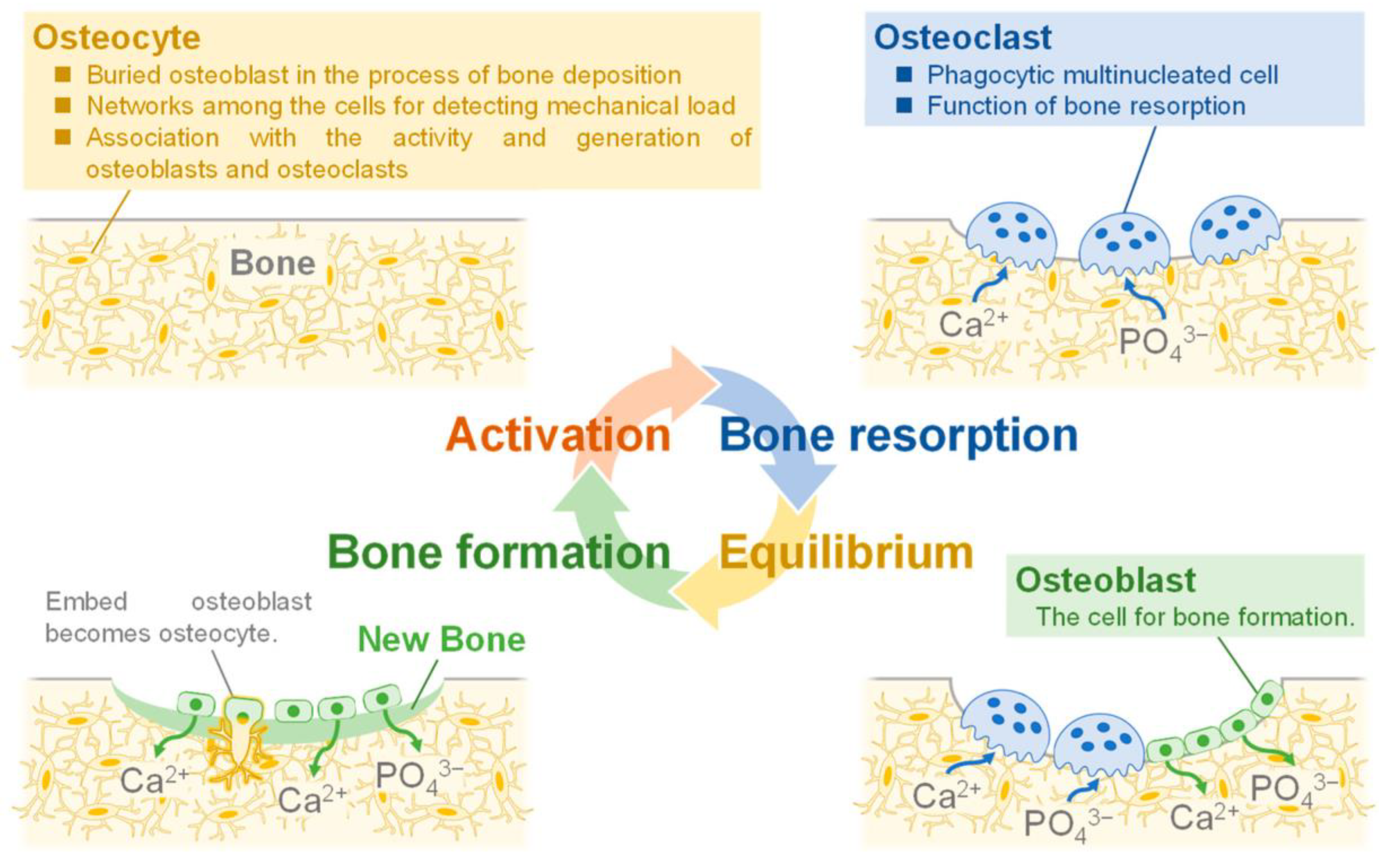
Figure 1. Representation of bone formation process [3].
Ijms 24 13157 g001
Figure 2. Representation of HA crystal structure and applications of doped nanoparticles [12].
Ijms 24 13157 g002
Figure 3. The crystal structure of HA [16].
Ijms 24 13157 g003
Scheme 1. Chronological representation of HA synthesis routes [40,41,42,43,44,45,46,47,48,49,50].
Ijms 24 13157 sch001
Figure 4. HA incorporation into organic/inorganic materials in order to improve bone tissue regeneration [89].
Ijms 24 13157 g004
Figure 5. Representation of doped and non-doped HA. Reproduced with permission from the Royal Society of Chemistry, 2012 [38].
Ijms 24 13157 g005
Figure 6. Schematic representation of stoichiometric HA structure (a) of Cu–HA (blue arrow), featuring the localization of the copper cation, either at the site of the calcium cation (b) or in the hydroxyl channel. (c) The arrow indicates the position of the copper atom [156].
Ijms 24 13157 g006
Figure 7. Natural sources used to obtain HA: (a) biowaste/animal bones; (b) eggshells; (c) marine organisms; (d) naturally derived biomolecules; (e) biomembranes. Reproduced with permission from Elsevier, 2013 [206].
Ijms 24 13157 g007
Table 1. The main synthesis methods and properties of synthesized HA.
Synthesis MethodMorphologySizeCharacteristics References
Sol–gel MethodSpherical, rods, needle, tube5–1000 nm Capacity to synthesize homogeneous
materials, mechanical properties enhanced due to nanocrystalline grain structures and high purity.
[52,53,54,55]
HydrothermalRod, needle, spherical, rollers, wire, spherulites, leaves, belts4–140 nmLow costs, good homogenicity and solubility of the precursors, tailorable nucleation, and growth of the crystal.[56,57,58]
Chemical PrecipitationNeedle, whisker, rods, spherical, platelets 5–130 nmLow costs, controlled environment, and enhanced porosity.[39,59,60]
BiosourcesIrregular, sphere, flakes, needles, rods6–180 nmCapacity to retain trace element, increase bioactivity, and be eco-friendly.[61,62]
MicroemulsionSpherical, rods, irregular, needle, flakes5–100 nmGood anti-aggregation property, outstanding stability, and self-controlled development of apertures.[63,64,65]
CombustionNeedle, rods, irregular, spherical40–200 nmSimplicity, high purity of the obtained products, low energy consumption, and high purity of synthesized material.[56,66,67]
Table 2. Doping material introduced into HA structures and their improved properties [3].
Doping IonBiologic Activity
CationNa+Enhanced osteoconductivity and improved cell proliferation
K+Enhanced thermal stability
Mg2+Improved crystal growth, crystallization, thermal stability, and dissolution rate
Sr2+Enhanced bone regeneration and formation, and inhibited bone resorption
Mn2+Enhanced cell adhesion
Zn2+Enhanced bone formation and regeneration, increased antimicrobial activity
Anion
CO32−High specific surface area, reduced crystallite size, enhanced osteoconductive properties, increased solubility
SiO44−Enhanced bioactivity, improved dissolution rate
FImproved stability, reduced solubility, and increased remineralization
ClEnhanced osteoconductive properties, and increased solubility
Disclaimer/Publisher’s Note: The statements, opinions and data contained in all publications are solely those of the individual author(s) and contributor(s) and not of MDPI and/or the editor(s). MDPI and/or the editor(s) disclaim responsibility for any injury to people or property resulting from any ideas, methods, instructions or products referred to in the content.