Perspective

Can Nitric Oxide-Based Therapy Be Improved for the Treatment of Cancers? A Perspective

by
Birandra K. Sinha
Mechanistic Toxicology Branch, Division of Translational Toxicology, National Institute of Environmental Health Sciences, NIH, Research Triangle Park, NC 27709, USA
Int. J. Mol. Sci. 2023, 24(17), 13611; https://doi.org/10.3390/ijms241713611
Submission received: 7 August 2023 / Revised: 30 August 2023 / Accepted: 31 August 2023 / Published: 2 September 2023
(This article belongs to the Section Molecular Oncology)

Abstract

:
Since the early observations that nitric oxide (NO) at high concentrations is cytotoxic to cancer cells and that it may play an important role in the treatment of human cancers, a significant number of compounds (NO-donors) have been prepared to deliver NO to tumors. NO also sensitizes various clinically active anticancer drugs and has been shown to induce the reversal of multi-drug resistance in tumor cells expressing ATP-binding cassette-transporter proteins. For the successful treatment of cancers, NO needs to be delivered precisely to tumors, and its adverse toxicity must be limited. Like other chemotherapeutics, the precise delivery of drugs has been a problem and various attempts have been made, such as the encapsulation of drugs in lipid polymers, to overcome this. This prospective study examines the use of various strategies for delivering NO (using NO-donors) for the treatment of cancers. Finding and utilizing such a delivery system is an important step in delivering cytotoxic concentrations of NO to tumors without adverse reactions, leading to a successful clinical outcome for patient management.

1. Introduction

The growth and survival of cancer cells in vivo is complex and involves many pathways. Tumors, in general, are heterogeneous, containing many different types of cells, including cancer stem cells, which are resistant to therapy. Furthermore, cancer cells develop resistance during therapy. Therefore, successful cancer treatment in the clinic has been difficult. Nitric oxide (NO) has emerged as a promising therapeutic for the treatment of many human cancers [1,2,3]. NO, a free radical produced endogenously by nitric oxide synthase (NOS), has a relatively short half-life (2.0–0.18 s) under physiological conditions [4]; thus, it is difficult to deliver it selectively to cellular targets. As a free radical, NO chemically reacts with cellular protein-SH and many other free radicals in biological environments, producing adverse reactions [5]. Additionally, different concentrations of NO have different physiological effects: at a low concentration, NO can be used to regulate vasodilation, nerve transmission, anti-inflammatory responses, and antioxidant responses, and it also promotes tumor growth [6,7,8]. In contrast, at high concentrations, NO inhibits tumor growth and enhances the apoptosis/ferroptosis of tumor cells via the formation of both reactive oxygen and reactive nitrogen species [8]. Because of this double-edged nature of NO in the regulation of physiological processes, NO must be delivered selectively to its target sites to limit off-target adverse effects.
Various molecular structures and chemical materials have been developed to exogenously deliver NO using synthetic NO donors (e.g., S-nitrosothiols (RSNOs), N-diazeniumdiolates, and metal–NO complexes); a number of reviews are available [9,10,11,12,13,14,15,16]. The breakdown and release of NO are dependent upon the molecular structure of the donor, which allows the rate of release and effective targeting of NO to be controlled in biological systems. This breakdown property of NO-donors has been utilized therapeutically by exploiting the microenvironment of cancers, as they exhibit lower pH values due to altered cellular metabolisms. Thousands of NO-donors have now been synthesized and tested in various tumor models. Many of these compounds show significant activity in terms of inhibiting tumor cell growth in both in vitro and in vivo tumor models. A number of tumor-specific NO-donors, e.g., J-SK (O2-(2,4-dinitrophenyl) 1-[(4-ethoxycarbonyl)piperazin-1-yl] diazen-1-ium-1,2-diolate), which are activated only in tumors and are effective in killing tumor cells without causing significant toxicity, have also been synthesized [17,18].
NO has been shown to sensitize drug-resistance tumor cells to chemotherapeutic drugs, e.g., adriamycin and cis-platin [19,20]. This would indicate that highly active NO- donors (including encapsulated) can be utilized in combination with other therapeutic modalities, including chemotherapy, radiation therapy, photothermal therapy, and photodynamic therapy. Studies from our laboratory have also shown the utility of NO generating compounds in reversing resistance mediated by the overexpression of ATP-binding cassettes.
(ABC)-transporters [21,22]: Research has shown that the overexpression of a family of efflux proteins is responsible for chemotherapy resistance [23,24]. These efflux proteins remove drugs from the cancer cells in an energy-dependent manner and belong to the ABC-transporter superfamily. We have shown that NO directly inhibits the functions of ATPase activities of both topoisomerase II [25] and ABC-transporters [26] following the nitrosylation of cysteines. This inhibition of ATPase functions in multi-drug-resistant tumor cells results in the enhanced cellular accumulation of drugs and the reversal of resistance [26]. Our findings are important, as they suggest that NO-based therapy can be utilized to overcome drug-resistance, which is one of the major challenges facing the treatment of cancers in clinics.
Some NO-generating compounds, such as nitroglycerine (GTN) and nitrates, are currently utilized in the clinic for the treatment of acute angina [27,28,29]. GTN has also been shown to exhibit a positive response in combination with vinorelbine and cis-platin [30]. Furthermore, a slow-release transdermal GTN patch has also been shown to be effective in men with prostate tumors whose disease was progressing following surgery or radiotherapy [31]. In addition, dietary arginine (which increases NO formation) has been utilized in combination with radiation therapy in some clinical trials [32]. However, while thousands of NO-donors have been synthesized and tested as anti-cancer drugs, and some have demonstrated significant anti-cancer activities in vivo, very few of these newer compounds have been evaluated in the clinic, and none of the NO-donors are currently utilized as anticancer drugs (or in combination) in the clinic.
Because high concentrations of NO are required for effective tumor cell killing, the need to deliver NO precisely to tumor targets is of paramount importance. Here, I examine the use of various delivery systems for NO-donors that may improve the selective delivery of cytotoxic concentrations of NO for the treatment of cancers. The major goal of this perspective is to examine the current and future delivery systems of NO-donors (NO) to tumors that may lead to therapeutic effects. It appears that the synthesis of additional NO-donors alone will not advance the treatment of cancers, as improvements in delivery systems are urgently needed for the future of NO-based therapy (as a single agent or in combination) in the clinic.

2. Prodrug Strategy for NO-Donors

Strategies using prodrugs have been effective in solving many drug delivery problems. In order to develop prodrugs, it is extremely important to have a balance between stability and the ability to rapidly convert prodrugs into their active forms under normal physiological conditions. It is believed that, in healthy tissue where pH homeostasis is maintained near pH 7.4, cancer tissues will promote a rapid NO release at the site due to lower pH (4–6), thereby mitigating off-target cytotoxicity. Various NO-donors (prodrugs) that are activated by cellular enzymes to release NO at tumor sites have also been designed and synthesized. These NO-releasing prodrugs are activated by various cellular enzymes, including esterase and glutathione transferase (GST) [18,33]. These NO prodrugs have been shown to have a significant advantage over direct non-specific NO-donors, as they reduce their side effects. Of importance for the treatment of cancers are NO-prodrugs that are activated by glutathione-dependent GST, as a number of tumors, including drug-resistant tumor cells, overexpress these enzymes.
Huang et al. [9] synthesized a number of O2-(Sulfonylethyl-derived) diazeniumdiolates which were active against GST-expressing tumor cells. Some of these compounds were shown to be significantly better than JS-K, an originally synthesized GST-activated NO-donor prodrug. We have also utilized JS-K in our studies and found this compound to be highly active against P-gp- and GST-expressing multi-drug-resistant ovarian tumors [21]. In addition, we found JS-K to be effective in reversing P-glycoprotein 170 (P-gp)-mediated drug resistance against multi-drug-resistant (MDR) tumor cells. Furthermore, we found esterase-mediated activation of the prodrug NCX4040 to be extremely cytotoxic to both P-gp-expressing ovarian and breast cancer resistance protein (BCRP)-expressing breast tumor cells. It also caused the reversal of both P-gp- and BCRP-mediated drug resistance in cells [22]. NCX4040, originally synthesized as an anti-carcinogenic compound, was found to be active against human colon cancer cells and did not display any significant toxicity in vivo.
It should be noted that these NO prodrugs have neither been fully evaluated in clinical trials nor are currently being utilized in the clinic for the treatment of cancers.

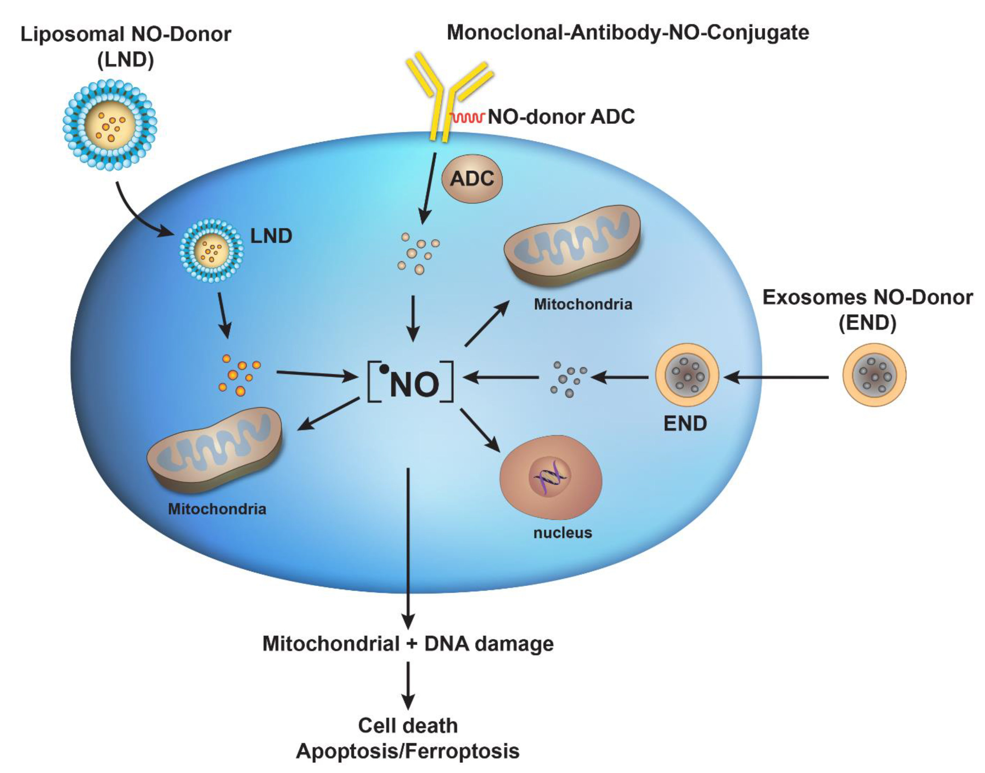
3. Antibody–Drug Conjugates for Targeted Delivery of NO in Tumors

Monoclonal antibody drugs with high target recognition and affinity have emerged as important treatment modalities for cancers. However, they lack sufficient therapeutic efficacy and have a narrow therapeutic window. Therefore, the strategy involving antibody/peptide–drug conjugates (ADC/PDC) has been utilized recently [34]. An antibody–drug conjugate (ADC) is a targeted drug delivery system composed of an antibody linked to an active cytotoxic drug via a lysine or -SH function. Since the antibody specifically recognizes tumor cells, it can deliver the drugs directly to the surfaces of tumor cells, thereby enhancing the anticancer effect and increasing the therapeutic efficacy. It is important that the target antigen be highly or preferentially expressed on the cancer cell surface (and minimally expressed in healthy tissue) to bind with the circulating ADC. It is also important that the target antigen have minimal secretion in circulation from the cancer cells, as antibodies can bind to these secreted receptors in the circulation, reducing the amount of antibodies available for target cell binding and internalization. Hence, selecting an appropriate target antigen is a critical step for the success of ADCs.
Recently, Sun et al. [35] prepared a conjugated humanized cluster of differentiation 24 (CD24) monoclonal antibody with a NO-donor which was connected to the antibody using an -SH function. Conjugates between the antibody and the NO-donor were stable in the blood circulatory system, with a low concentration of GSH. However, it was readily cleaved in tumor cells containing a high amount of GSH to generate two molecules of NO. The NO–antibody conjugate showed significant cytotoxicity, both in vitro and in vivo, against hepatic carcinoma cells, with fewer side effects on normal tissues.
More studies are needed to expand this concept of antibody–NO conjugates in order to provide a clinically active drug conjugate. It will also be useful to prepare antibody–drug conjugates of other highly active NO-delivery compounds, e.g., JS-K analogs and NCX4040, and to examine their effectiveness against a variety of tumor models.
Ruthenium–NO complexes have also been utilized for the targeted delivery of NO to tumors, resulting in the delivery of high concentrations of NO in tumor cells [36]. Studies using cis-[RuII(NO+)Cl(dcbpy)2]2 conjugated to a polyclonal antibody showed enhanced cytotoxicity against the liver hepatocellular carcinoma cell line HepG2. These metal complexes were stable in tumor cells and delivered cytotoxic concentrations of NO. In another study, [RuII(bpy)2L(NO+)]n+ was utilized as an NO-donor against the mitochondria of cancer cells [37]. Mitochondria are extremely important, as they provide cellular energy and play an important role in cell survival and death. The voltage-dependent anion channel (VDAC) plays a central role in mitochondria-mediated apoptosis and ferroptosis by generating reactive oxygen species (ROS). The formation of ROS is significantly increased in the presence of NO. Therefore, VDAC has been utilized as a potential target for ruthenium complex NO-donors, and [RuII(NO+)Cl(dcbpy)2]3−anti-VDAC antibody conjugates have been prepared and evaluated as anticancer drugs. The cytotoxicity of the cis-[RuII(NO+)Cl(dcbpy)2]3−-anti-VDAC antibody was significantly enhanced in comparison to the free cis-[RuII(NO+)Cl(dcbpy)2]3−complex. This cytotoxic effect was found to result from a site-specific interaction of the complex with VDAC and the release of NO.
While Ru–NO complexes may potentially be useful as anticancer drugs, the accumulation of NO–ruthenium complexes in tumor cells is a problem due to the lack of solubility at pH values higher than 4.0. This needs to be addressed before these complexes can be utilized in clinical settings.

4. Liposomes-Based Delivery of NO-Donors

The encapsulation of therapeutics within liposomes is an effective strategy for the controlled delivery of drugs to their targets. Liposomes are nanostructures composed of an inner aqueous core surrounded by a bilayer of phospholipids, and have been used to enhance drug delivery against many diseases, including cancer [38]. Liposomes, as drug delivery vehicles, provide many benefits, e.g., reduced immune response, increased cellular uptake, and protection of drug payload from premature actions or breakdown. Clinically active drugs, e.g., adriamycin, paclitaxel, and cis-platin, have serious side effects that limit their full potential in the clinic. For example, adriamycin causes cumulative dose-dependent cardiotoxicity that can be life-threatening, and is, therefore, dose-limiting [39]. Similarly, paclitaxel and cis-platin show peripheral neuropathy [40] and nephrotoxicity [41], respectively, in humans. As potent and effective cytotoxic drugs, these chemotherapeutic agents have benefited notably from encapsulation into nanoparticles, which significantly improves their specificity toward cancer cells, concomitantly decreasing their toxicity and thus allowing for the administration of higher doses to patients.
The preparation of NO-releasing liposomes utilizing alkaline interior aqueous phases enables the encapsulation of varying concentrations of NO with long shelf-lives (>3 months). The use of liposomal NO donors as anticancer agents has shown increased therapeutic potential over small-molecule NO-donors [42,43]. NO-releasing liposomes represent an important strategy for cancer treatment due to their pH-triggered release. It has been suggested that varying the lipid bilayer content (e.g., phospholipid composition, charge) may further enhance the stability of this NO-delivery system and influence NO-release payloads. In addition, utilizing a more thermally stable lipid (e.g., 1,2-distearoyl-sn-glycero-3-phosphocholine) could potentially prolong the NO-release duration even further due to its greater bilayer rigidity. Earlier works have focused on utilizing S-nitrosothiol or N-diazeniumdiolate NO-donors in liposomal delivery systems [44]. It would be very interesting to encapsulate recently developed, highly active analogs of anticancer NO-donors in FDA-approved liposomes. Such liposomal NO-donors may provide a much better opportunity for the successful treatment of cancers in the clinic.
Recent studies employing multilayered liposomes have shown increased stability of the core drug due to its unique fabrication methodology [45]. In this technique, the liposomal layers are layered above each other using a layer-by-layer technique, resembling onion-like layering. It is expected that encapsulated NO-donors will have enhanced protection from decay and will have increased sustained release to specific tumor targets. However, at this time, a multilayer liposome delivery system has not been tried with NO-donors. Thus, there is significant need to utilize this multi-layering approach to encapsulate newer and more active NO-releasing compounds for evaluation as improved anticancer drugs.

5. Exosomes as a Delivery System for NO-Donors

The use of exosomes as drug delivery systems is a recent and emerging field [46,47,48]. Exosomes are bilayer membrane-coated extracellular vesicles measuring between 40 and 100 nm in diameter, and are considered natural nanoparticles which can carry a cargo of a wide variety of compounds, e.g., drugs, lipids, proteins, RNAs, and DNAs. Exosomes have been proposed for drug-delivery purposes, as they can be loaded with both small molecules and macromolecules, which can be used as therapeutic tools to treat cancers. Exosomes play an important role in intercellular communications, and as natural carriers, exosomes have the advantages of low immunogenicity and direct delivery of drugs to cells. Exosomes also have many desirable features, such as long circulating half-lives, the intrinsic ability to target tissues, biocompatibility, and minimal or no inherent toxicity, overcoming the limitations observed with the majority of other delivery systems. Recent studies have shown the simplicity of isolation and the possibility of commercial scale-up and utilization for tumor-targeting. These features make exosomes ideal nanoparticles for drug delivery, with wide therapeutic applications. In addition, exosome-based delivery systems have been shown to bypass the effects of ABC-transporter proteins and to overcome the multi-drug resistance of tumor cells [49].
The key to using exosomes as drug carriers, however, is the effective loading of exogenous drugs (chemotherapeutics and NO-donors) into exosomes. Currently, sonication, electroporation, transfection, incubation, extrusion, saponin-assisted loading, transgenesis, freeze–thaw cycles, thermal shock, pH gradient methods, and hypotonic dialysis have been applied to load drugs into exosomes [50]. The clinical application of exosomes as drug carriers (and NO-donors) depends upon methods of importing drugs into exosomes efficiently. Furthermore, selecting the correct source of exosomes is very important, as certain cancer-cell-derived exosomes have been shown to have pro-cancer and pro-resistance effects, making them unsuitable as drug carriers [51].
At the moment, the delivery of NO-donors using exosomes has not been attempted or reported for the treatment of diseases, including cancers. We have recently initiated an exosome system for delivery with selective highly active NO-donors; however, it too early for any conclusions to be made.

6. Conclusions and Future Directions

Nanomedicine faces many challenges, especially in clinical trials. In clinical applications, NO, as a gas messenger that can act on the entire body, requires careful consideration due to its physical and chemical properties and its accumulation in non-target organs and tissues, which pose considerable toxicity. Furthermore, some NO nanomedicines may cause oxidative stress, immune responses, protein oxidation, and DNA damage. The existing NO-donors use the targeting properties of materials to release NO at a specific site, and certain NO-donors can be used for slow release over time, but they still lack the precise control needed to release appropriate concentrations of NO. As NO is a double-edged sword, proper concentrations of NO must be utilized to treat tumors; otherwise, NO could promote further aggravation of the disease or other toxic manifestations. Therefore, it is necessary to design and develop smarter NO nanomedicines that can release the appropriate concentrations of NO in vivo. It has been suggested that we need to design smart NO-donors that can be turned on or off to release appropriate amounts of NO according to the pathophysiological requirements. In this scenario, when they are turned on, they continuously release the desired concentration of NO at the tumor site, and when they are turned off, the release of NO stops. Furthermore, it is necessary that the NO nanomedicine remains localized within the tumor until the next time the smart NO-donors are turned on without breakdown. Although the characteristics of nanomaterials can be used to achieve the coordinated release of NO, various strategies have also been developed for the design of NO-releasing materials, including the incorporation of photo-activable, charge-switchable, or bacteria-targeting groups [52,53]. While these strategies have led to the development of some excellent NO-donors and nanomaterials, none of these have found utility in the treatment of cancers.
Because NO is different from most other anticancer drugs and it has some diverse effects, the delivery of specific concentrations of NO to tumors is essential to mitigate the adverse effects. However, unlike other chemotherapeutics, it is cytotoxic to all kind of tumors, and can sensitize various clinically active anticancer drugs. In addition, it can also inhibit the ATPase functions of ABC transporters [22,25], resulting in the reversal of multi-drug resistance. These properties of NO-generating compounds make them extremely desirable as anticancer drugs. Therefore, a significant number of NO-donors have been synthesized for the strict purpose of curing human cancers; many of these compounds are active in killing tumor cells in vitro at sub-micromolecular ranges and also show significant activity in vivo mouse tumor models. What is most interesting is that several nitric oxide-releasing drugs have been utilized for a long time to treat cardiovascular diseases, including isosorbide mononitrate, which is a long-term nitric oxide-releasing drug administered to patients with hypertension [28]. Furthermore, other nitric oxide-releasing drugs, e.g., nitroglycerin and sodium nitroprusside (SNP), are also currently used as short-acting anti-angina drugs to rapidly decrease blood pressure [27]. While these drugs are used in millions of patients every day, no nitric oxide-releasing drugs are currently utilized for treatment of cancers in the clinic.
Advances in drug delivery system(s) do offer exciting prospects in terms of packaging active NO-donors into the clinic and beyond. Significant improvements have been made in the field of encapsulation of chemotherapeutics [54,55]. The FDA has now approved various liposomes for therapy which should be tried with highly active anticancer NO-donors. Our recent studies indicate that certain NO-donors induce ferroptosis-mediated tumor cell death and that combinations of erastin or RSL3 (Ras-selected ligand3) with NO-donors enhances tumor cell killing [56]. Erastin and RSL3 are small molecules known to induce ferroptosis in tumor cells. These studies further suggest the utility of NO-donors and NO in killing tumor cells. Hence, the utilization of encapsulated NO-donors in combination with ferroptosis inducers may be of significance for the treatment of Ras-mutated tumors in the clinic. While approaches utilizing antibody–drug conjugate delivery systems are expensive and difficult in terms of obtaining a high NO-payload, these methods are highly specific for the precise delivery of cytotoxic concentrations of NO to tumors. It is my opinion that ADC may be the future of NO-donor (drug) delivery, and may be able to mitigate off-target toxicity. Furthermore, recent advances in the study of exosomes for the delivery of chemotherapeutics also suggest that NO-donors can be packaged into exosomes for the treatment of human diseases, including cancers. Figure 1 summarizes the possible roles of these strategies in the future of NO nanomedicine for the treatment of human cancers.
Finally, there is also a significant need to test NO-donors and NO-medicine in reliable models to predict the outcomes in patients. It is difficult and unethical to test and compare nanomedicines against free drugs in patients. Currently, much of the cytotoxicity testing is conducted in 2D-tissue culture systems, and is sometimes combined with humanized mouse models. Traditional 2D cell culture systems do not mimic the 3D culture systems, as they lack the complexity of the tumor microenvironments in patients and often fail to predicate patient outcomes [57,58,59,60]. Developing 3D model systems to test NO-donors and NO-nanomedicines would be a step forward for more accurately predicting the therapeutic outcomes. It would also be advantageous to develop 3D model systems from patient-derived tumor tissues and stem cells for predicting the outcomes of NO nanomedicine (and NO-donors) for both personalized medicine and for overcoming drug resistance in the clinic.

Funding

This research was supported by the intramural research program (grant numbers ZIAE505013922) of the National Institute of Environmental Health Sciences, NIH. The statements contained herein do not necessarily represent the statements, opinions, or conclusions of NIEHS, NIH, or the US Government.

Acknowledgments

The author wishes thank B. Alex Merrick, Erik J. Tokar, Greg Travlos, and Amar Reddy for their critical review of the manuscript and Constance Sinha for help with the editing of the manuscript.

Conflicts of Interest

The author declares no actual or potential conflict of interest.

References

  1. Hirst, D.; Robson, T. Nitric oxide in cancer therapeutics: Interaction with cytotoxic chemotherapy. Curr. Pharm. Des. 2010, 16, 411–420. [Google Scholar] [CrossRef] [PubMed]
  2. Choudhari, S.K.; Chaudhary, M.; Bagde, S.; Gadbail, A.R.; Joshi, V. Nitric oxide and cancer: A review. World J. Surg. Oncol. 2013, 11, 118. [Google Scholar] [CrossRef] [PubMed]
  3. Sinha, B.K. Role of Oxygen and Nitrogen Radicals in the Mechanism of Anticancer Drug Cytotoxicity. J. Cancer Sci. Ther. 2020, 12, 10–18. [Google Scholar]
  4. Thomas, D.D.; Liu, X.; Kantrow, S.P.; Lancaster, J.R., Jr. The biological lifetime of nitric oxide: Implications for the perivascular dynamics of NO and O2. Proc. Natl. Acad. Sci. USA 2001, 98, 355–360. [Google Scholar] [CrossRef]
  5. Stamler, J.S.; Lamas, S.; Fang, F.C. Nitrosylation. the prototypic redox-based signaling mechanism. Cell 2001, 106, 675–683. [Google Scholar] [CrossRef] [PubMed]
  6. Holotiuk, V.V.; Kryzhanivska, A.Y.; Churpiy, I.K.; Tataryn, B.B.; Ivasiutyn, D.Y. Role of nitric oxide in pathogenesis of tumor growth and its possible application in cancer treatment. Exp. Oncol. 2019, 41, 210–215. [Google Scholar] [CrossRef]
  7. Porrini, C.; Ramarao, N.; Tran, S.L. Dr. NO and Mr. Toxic—The versatile role of nitric oxide. Biol. Chem. 2020, 401, 547–572. [Google Scholar] [CrossRef]
  8. Burke, A.J.; Sullivan, F.J.; Giles, F.J.; Glynn, S.A. The yin and yang of nitric oxide in cancer progression. Carcinogenesis 2013, 34, 503–512. [Google Scholar] [CrossRef]
  9. Huang, Z.; Fu, J.; Zhang, Y. Nitric Oxide Donor-Based Cancer Therapy: Advances and Prospects. J. Med. Chem. 2017, 60, 7617–7635. [Google Scholar] [CrossRef]
  10. Huerta, S. Nitric oxide for cancer therapy. Future Sci. OA 2015, 1, FSO44. [Google Scholar] [CrossRef]
  11. Seabra, A.B.; de Lima, R.; Calderon, M. Nitric oxide releasing nanomaterials for cancer treatment: Current status and perspectives. Curr. Top. Med. Chem. 2015, 15, 298–308. [Google Scholar] [CrossRef] [PubMed]
  12. Alimoradi, H.; Greish, K.; Gamble, A.B.; Giles, G.I. Controlled Delivery of Nitric Oxide for Cancer Therapy. Pharm. Nanotechnol. 2019, 7, 279–303. [Google Scholar] [CrossRef] [PubMed]
  13. Li, Y.; Yoon, B.; Dey, A.; Nguyen, V.Q.; Park, J.H. Recent progress in nitric oxide-generating nanomedicine for cancer therapy. J. Control. Release Off. J. Control. Release Soc. 2022, 352, 179–198. [Google Scholar] [CrossRef]
  14. Li, C.Y.; Anuraga, G.; Chang, C.P.; Weng, T.Y.; Hsu, H.P.; Ta, H.D.K.; Su, P.F.; Chiu, P.H.; Yang, S.J.; Chen, F.W.; et al. Repurposing nitric oxide donating drugs in cancer therapy through immune modulation. J. Exp. Clin. Cancer Res. 2023, 42, 22. [Google Scholar] [CrossRef]
  15. Pieretti, J.C.; Pelegrino, M.T.; Nascimento, M.H.M.; Tortella, G.R.; Rubilar, O.; Seabra, A.B. Small molecules for great solutions: Can nitric oxide-releasing nanomaterials overcome drug resistance in chemotherapy? Biochem. Pharmacol. 2020, 176, 113740. [Google Scholar] [CrossRef] [PubMed]
  16. Mintz, J.; Vedenko, A.; Rosete, O.; Shah, K.; Goldstein, G.; Hare, J.M.; Ramasamy, R.; Arora, H. Current Advances of Nitric Oxide in Cancer and Anticancer Therapeutics. Vaccines 2021, 9, 94. [Google Scholar] [CrossRef] [PubMed]
  17. Shami, P.J.; Saavedra, J.E.; Wang, L.Y.; Bonifant, C.L.; Diwan, B.A.; Singh, S.V.; Gu, Y.; Fox, S.D.; Buzard, G.S.; Citro, M.L.; et al. JS-K, a glutathione/glutathione S-transferase-activated nitric oxide donor of the diazeniumdiolate class with potent antineoplastic activity. Mol. Cancer Ther. 2003, 2, 409–417. [Google Scholar]
  18. Maciag, A.E.; Saavedra, J.E.; Chakrapani, H. The nitric oxide prodrug JS-K and its structural analogues as cancer therapeutic agents. Anticancer Agents Med. Chem. 2009, 9, 798–803. [Google Scholar] [CrossRef]
  19. Liu, J.; Li, C.; Qu, W.; Leslie, E.; Bonifant, C.L.; Buzard, G.S.; Saavedra, J.E.; Keefer, L.K.; Waalkes, M.P. Nitric oxide prodrugs and metallochemotherapeutics: JS-K and CB-3-100 enhance arsenic and cisplatin cytolethality by increasing cellular accumulation. Mol. Cancer Ther. 2004, 3, 709–714. [Google Scholar] [CrossRef]
  20. Bonavida, B. Sensitizing activities of nitric oxide donors for cancer resistance to anticancer therapeutic drugs. Biochem. Pharmacol. 2020, 176, 113913. [Google Scholar] [CrossRef]
  21. Sinha, B.K.; Perera, L.; Cannon, R.E. Reversal of drug resistance by JS-K and nitric oxide in ABCB1- and ABCG2-expressing multi-drug resistant human tumor cells. Biomed. Pharmacother. Biomed. Pharmacother. 2019, 120, 109468. [Google Scholar] [CrossRef] [PubMed]
  22. Sinha, B.K.; Perera, L.; Cannon, R.E. NCX-4040, a Unique Nitric Oxide Donor, Induces Reversal of Drug-Resistance in Both ABCB1- and ABCG2-Expressing Multidrug Human Cancer Cells. Cancers 2021, 13, 1680. [Google Scholar] [CrossRef] [PubMed]
  23. Robey, R.W.; Pluchino, K.M.; Hall, M.D.; Fojo, A.T.; Bates, S.E.; Gottesman, M.M. Revisiting the role of ABC transporters in multidrug-resistant cancer. Nat. Rev. Cancer 2018, 18, 452–464. [Google Scholar] [CrossRef] [PubMed]
  24. Feyzizadeh, M.; Barfar, A.; Nouri, Z.; Sarfraz, M.; Zakeri-Milani, P.; Valizadeh, H. Overcoming multidrug resistance through targeting ABC transporters: Lessons for drug discovery. Expert Opin. Drug Discov. 2022, 17, 1013–1027. [Google Scholar] [CrossRef] [PubMed]
  25. Sinha, B.K.; Kumar, A.; Mason, R.P. Nitric oxide inhibits ATPase activity and induces resistance to topoisomerase II-poisons in human MCF-7 breast tumor cells. Biochem. Biophys. Rep. 2017, 10, 252–259. [Google Scholar] [CrossRef]
  26. Sinha, B.K.; Bortner, C.D.; Mason, R.P.; Cannon, R.E. Nitric oxide reverses drug resistance by inhibiting ATPase activity of p-glycoprotein in human multi-drug resistant cancer cells. Biochim. Biophys. Acta Gen. Subj. 2018, 1862, 2806–2814. [Google Scholar] [CrossRef]
  27. Watanabe, H.; Kakihana, M.; Ohtsuka, S.; Sugishita, Y. Preventive effects of angiotensin-converting enzyme inhibitors on nitrate tolerance during continuous transdermal application of nitroglycerin in patients with chronic heart failure. Jpn. Circ. J. 1998, 62, 353–358. [Google Scholar] [CrossRef]
  28. Kruzliak, P.; Pechanova, O.; Kara, T. New perspectives of nitric oxide donors in cardiac arrest and cardiopulmonary resuscitation treatment. Heart Fail. Rev. 2014, 19, 383–390. [Google Scholar] [CrossRef]
  29. He, M.; Wang, D.; Xu, Y.; Jiang, F.; Zheng, J.; Feng, Y.; Cao, J.; Zhou, X. Nitric Oxide-Releasing Platforms for Treating Cardiovascular Disease. Pharmaceutics 2022, 14, 1345. [Google Scholar] [CrossRef]
  30. Yasuda, H.; Yamaya, M.; Nakayama, K.; Sasaki, T.; Ebihara, S.; Kanda, A.; Asada, M.; Inoue, D.; Suzuki, T.; Okazaki, T.; et al. Randomized phase II trial comparing nitroglycerin plus vinorelbine and cisplatin with vinorelbine and cisplatin alone in previously untreated stage IIIB/IV non-small-cell lung cancer. J. Clin. Oncol. Off. J. Am. Soc. Clin. Oncol. 2006, 24, 688–694. [Google Scholar] [CrossRef]
  31. Siemens, D.R.; Heaton, J.P.; Adams, M.A.; Kawakami, J.; Graham, C.H. Phase II study of nitric oxide donor for men with increasing prostate-specific antigen level after surgery or radiotherapy for prostate cancer. Urology 2009, 74, 878–883. [Google Scholar] [CrossRef] [PubMed]
  32. Heys, S.D.; Ogston, K.; Miller, I.; Hutcheon, A.W.; Walker, L.G.; Sarker, T.K.; Dewar, J.; Ah-See, A.K.; Eremin, O. Potentiation of the response to chemotherapy in patients with breast cancer by dietary supplementation with L-arginine: Results of a randomised controlled trial. Int. J. Oncol. 1998, 12, 221–225. [Google Scholar] [CrossRef] [PubMed]
  33. Sharma, K.; Chakrapani, H. Site-directed delivery of nitric oxide to cancers. Nitric Oxide Biol. Chem./Off. J. Nitric Oxide Soc. 2014, 43, 8–16. [Google Scholar] [CrossRef] [PubMed]
  34. Narayan, R.; Blonquist, T.M.; Emadi, A.; Hasserjian, R.P.; Burke, M.; Lescinskas, C.; Neuberg, D.S.; Brunner, A.M.; Hobbs, G.; Hock, H.; et al. A phase 1 study of the antibody-drug conjugate brentuximab vedotin with re-induction chemotherapy in patients with CD30-expressing relapsed/refractory acute myeloid leukemia. Cancer 2020, 126, 1264–1273. [Google Scholar] [CrossRef]
  35. Sun, F.; Wang, Y.; Luo, X.; Ma, Z.; Xu, Y.; Zhang, X.; Lv, T.; Zhang, Y.; Wang, M.; Huang, Z.; et al. Anti-CD24 Antibody-Nitric Oxide Conjugate Selectively and Potently Suppresses Hepatic Carcinoma. Cancer Res. 2019, 79, 3395–3405. [Google Scholar] [CrossRef] [PubMed]
  36. Tfouni, E.; Truzzi, D.R.; Tavares, A.; Gomes, A.J.; Figueiredo, L.E.; Franco, D.W. Biological activity of ruthenium nitrosyl complexes. Nitric Oxide Biol. Chem./Off. J. Nitric Oxide Soc. 2012, 26, 38–53. [Google Scholar] [CrossRef]
  37. Ramos, L.C.B.; Rodrigues, F.P.; Biazzotto, J.C.; de Paula Machado, S.; Slep, L.D.; Hamblin, M.R.; da Silva, R.S. Targeting the mitochondrial VDAC in hepatocellular carcinoma using a polyclonal antibody-conjugated to a nitrosyl ruthenium complex. J. Biol. Inorg. Chem. 2018, 23, 903–916. [Google Scholar] [CrossRef]
  38. Olusanya, T.O.B.; Haj Ahmad, R.R.; Ibegbu, D.M.; Smith, J.R.; Elkordy, A.A. Liposomal Drug Delivery Systems and Anticancer Drugs. Molecules 2018, 23, 907. [Google Scholar] [CrossRef]
  39. Kong, C.Y.; Guo, Z.; Song, P.; Zhang, X.; Yuan, Y.P.; Teng, T.; Yan, L.; Tang, Q.Z. Underlying the Mechanisms of Doxorubicin-Induced Acute Cardiotoxicity: Oxidative Stress and Cell Death. Int. J. Biol. Sci. 2022, 18, 760–770. [Google Scholar] [CrossRef]
  40. Postma, T.J.; Vermorken, J.B.; Liefting, A.J.; Pinedo, H.M.; Heimans, J.J. Paclitaxel-induced neuropathy. Ann. Oncol. 1995, 6, 489–494. [Google Scholar] [CrossRef]
  41. Pabla, N.; Dong, Z. Cisplatin nephrotoxicity: Mechanisms and renoprotective strategies. Kidney Int. 2008, 73, 994–1007. [Google Scholar] [CrossRef] [PubMed]
  42. Suchyta, D.J.; Schoenfisch, M.H. Controlled release of nitric oxide from liposomes. ACS Biomater. Sci. Eng. 2017, 3, 2136–2143. [Google Scholar] [CrossRef] [PubMed]
  43. Suchyta, D.J.; Schoenfisch, M.H. Anticancer potency of nitric oxide-releasing liposomes. RSC Adv. 2017, 7, 53236–53246. [Google Scholar] [CrossRef] [PubMed]
  44. Suchyta, D.J.; Schoenfisch, M.H. Encapsulation of N-Diazeniumdiolates within Liposomes for Enhanced Nitric Oxide Donor Stability and Delivery. Mol. Pharm. 2015, 12, 3569–3574. [Google Scholar] [CrossRef]
  45. Nakhaei, P.; Margiana, R.; Bokov, D.O.; Abdelbasset, W.K.; Jadidi Kouhbanani, M.A.; Varma, R.S.; Marofi, F.; Jarahian, M.; Beheshtkhoo, N. Liposomes: Structure, Biomedical Applications, and Stability Parameters with Emphasis on Cholesterol. Front. Bioeng. Biotechnol. 2021, 9, 705886. [Google Scholar] [CrossRef]
  46. Kumar, D.N.; Chaudhuri, A.; Aqil, F.; Dehari, D.; Munagala, R.; Singh, S.; Gupta, R.C.; Agrawal, A.K. Exosomes as Emerging Drug Delivery and Diagnostic Modality for Breast Cancer: Recent Advances in Isolation and Application. Cancers 2022, 14, 1435. [Google Scholar] [CrossRef]
  47. Aqil, F.; Gupta, R.C. Exosomes in Cancer Therapy. Cancers 2022, 14, 500. [Google Scholar] [CrossRef]
  48. Chinnappan, M.; Srivastava, A.; Amreddy, N.; Razaq, M.; Pareek, V.; Ahmed, R.; Mehta, M.; Peterson, J.E.; Munshi, A.; Ramesh, R. Exosomes as drug delivery vehicle and contributor of resistance to anticancer drugs. Cancer Lett. 2020, 486, 18–28. [Google Scholar] [CrossRef]
  49. Gonzalez-Sarrias, A.; Iglesias-Aguirre, C.E.; Cortes-Martin, A.; Vallejo, F.; Cattivelli, A.; Del Pozo-Acebo, L.; Del Saz, A.; Lopez de Las Hazas, M.C.; Davalos, A.; Espin, J.C. Milk-Derived Exosomes as Nanocarriers to Deliver Curcumin and Resveratrol in Breast Tissue and Enhance Their Anticancer Activity. Int. J. Mol. Sci. 2022, 23, 2860. [Google Scholar] [CrossRef]
  50. Xi, X.M.; Xia, S.J.; Lu, R. Drug loading techniques for exosome-based drug delivery systems. Pharmazie 2021, 76, 61–67. [Google Scholar] [CrossRef]
  51. Chen, L.; Wang, L.; Zhu, L.; Xu, Z.; Liu, Y.; Li, Z.; Zhou, J.; Luo, F. Exosomes as Drug Carriers in Anti-Cancer Therapy. Front. Cell Dev. Biol. 2022, 10, 728616. [Google Scholar] [CrossRef]
  52. Eroy-Reveles, A.A.; Mascharak, P.K. Nitric oxide-donating materials and their potential in pharmacological applications for site-specific nitric oxide delivery. Future Med. Chem. 2009, 1, 1497–1507. [Google Scholar] [CrossRef] [PubMed]
  53. Poh, W.H.; Rice, S.A. Recent Developments in Nitric Oxide Donors and Delivery for Antimicrobial and Anti-Biofilm Applications. Molecules 2022, 27, 674. [Google Scholar] [CrossRef] [PubMed]
  54. Nirmala, M.J.; Kizhuveetil, U.; Johnson, A.; Balaji, G.; Nagarajan, R.; Muthuvijayan, V. Cancer nanomedicine: A review of nano-therapeutics and challenges ahead. RSC Adv. 2023, 13, 8606–8629. [Google Scholar] [CrossRef] [PubMed]
  55. Fulton, M.D.; Najahi-Missaoui, W. Liposomes in Cancer Therapy: How Did We Start and Where Are We Now. Int. J. Mol. Sci. 2023, 24, 6615. [Google Scholar] [CrossRef] [PubMed]
  56. Sinha, B.K.; Bortner, C.D.; Jarmusch, A.K.; Tokar, E.J.; Murphy, C.; Wu, X.; Winter, H.; Cannon, R.E. Ferroptosis-Mediated Cell Death Induced by NCX4040, The Non-Steroidal Nitric Oxide Donor, in Human Colorectal Cancer Cells: Implications in Therapy. Cells 2023, 12, 1626. [Google Scholar] [CrossRef]
  57. Benien, P.; Swami, A. 3D tumor models: History, advances and future perspectives. Future Oncol. 2014, 10, 1311–1327. [Google Scholar] [CrossRef]
  58. Drost, J.; Clevers, H. Organoids in cancer research. Nat. Rev. Cancer 2018, 18, 407–418. [Google Scholar] [CrossRef]
  59. Anderle, N.; Koch, A.; Gierke, B.; Keller, A.L.; Staebler, A.; Hartkopf, A.; Brucker, S.Y.; Pawlak, M.; Schenke-Layland, K.; Schmees, C. A Platform of Patient-Derived Microtumors Identifies Individual Treatment Responses and Therapeutic Vulnerabilities in Ovarian Cancer. Cancers 2022, 14, 2895. [Google Scholar] [CrossRef]
  60. Senkowski, W.; Gall-Mas, L.; Falco, M.M.; Li, Y.; Lavikka, K.; Kriegbaum, M.C.; Oikkonen, J.; Bulanova, D.; Pietras, E.J.; Vossgrone, K.; et al. A platform for efficient establishment and drug-response profiling of high-grade serous ovarian cancer organoids. Dev. Cell 2023, 58, 1106–1121. [Google Scholar] [CrossRef]
Figure 1. Various delivery systems for NO nanomedicine utilizing liposomal-encapsulated NO-donors (LNDs), antibody-NO-drug conjugates (ADCs), and exosome-encapsulated NO-donors (END) for the treatment of cancers in the clinic. Shown here is a tumor cell in which these nanomedicines containing NO-donors are taken up by passive transport (endocytosis) and release their NO-donor payloads. NO-donors then release cytotoxic •NO, which combines with ROS (O2−) to generate peroxynitrite. Both ROS and RNS induce damage to tumor mitochondria and nuclear DNA, causing cellular death.
Ijms 24 13611 g001
Disclaimer/Publisher’s Note: The statements, opinions and data contained in all publications are solely those of the individual author(s) and contributor(s) and not of MDPI and/or the editor(s). MDPI and/or the editor(s) disclaim responsibility for any injury to people or property resulting from any ideas, methods, instructions or products referred to in the content.

