
Role of Lnc-RNAs in the Pathogenesis and Development of Diabetic Retinopathy


Abstract
:1. Introduction
2. Diabetic Retinopathy at a Molecular Level
2.1. Oxidative Stress
2.2. Neurodegeneration
2.3. Inflammatory Process
2.4. Angiogenesis
3. Gene Regulation as a New Approach
3.1. Long Non-Coding RNAs
- Genic Lnc-RNAs: They are situated in exonic or intronic regions and are transcribed in distinct regions that pass across protein-coding sites; depending on their location, they can be defined as follows:
- ○
- Sense Lnc-RNAs: They overlap one or more exons of neighboring mRNAs on the same protein-coding strand.
- ○
- Antisense Lnc-RNAs: They overlap one or more exons of neighboring mRNAs on the opposite non-coding strand.
- ○
- Intronic Lnc-RNAs: They originate only from the intronic regions of a protein-coding gene in either direction.
- ○
- Bidirectional Lnc-RNAs: They have promoters in common with protein-encoding genes but are transcribed in the opposite direction.
- ○
- Promoter upstream Lnc-RNAs: They are located upstream of a promoter.
- ○
- 3’-UTR-associated Lnc-RNAs: They are transcribed from a protein-coding gene’s 3’-UTR region.
- Intergenic Lnc-RNAs: They are situated within the genomic interval between two genes [26], not intersecting with any protein-coding sites.
- -
- Pre-transcriptional mechanisms involve the modification of genes without changing the DNA sequence of the organism. These modifications may be chromatin remodeling, genomic imprinting and X chromosome inactivation. Lnc-RNAs can regulate gene transcription via histone modification, including histone methylation, acetylation and ubiquitination. In addition, Lnc-RNAs directly bind to DNA methyltransferase (DNMT), which can lead to promoter methylation and can also affect the expression of genes by controlling chromatin looping and recruiting chromatin-modifying enzymes to the DNA [29].
- -
- Transcriptional regulation occurs when Lnc-RNAs block the promoter region or interact with an RNA-binding protein to locate on the gene promoter region or regulate the activity of transcription factors [29].
- -
- Post-transcriptional regulation involves the complementary pairing of Lnc-RNA with target mRNA sequences to control the rate of translation or lead to RNA degradation [29]. Lnc-RNAs can act as a precursor of some miRNAs to regulate gene expression, or they can act as endogenous competitive RNAs, binding to miRNAs and thereby upregulating the translation of the corresponding mRNAs [26].
3.2. Main Involved Pathways
4. Discussion and Conclusions
Author Contributions
Funding
Conflicts of Interest
References
- Ortiz, G.; Lopez, E.S.; Salica, J.P.; Potilinski, C.; Fernández Acquier, M.; Chuluyan, E.; Gallo, J.E. Alpha-1-antitrypsin ameliorates inflammation and neurodegeneration in the diabetic mouse retina. Exp. Eye Res. 2018, 174, 29–39. [Google Scholar] [CrossRef] [PubMed]
- Potilinski, M.C.; Lorenc, V.; Perisset, S.; Gallo, J.E. Mechanisms behind Retinal Ganglion Cell Loss in Diabetes and Therapeutic Approach. Int. J. Mol. Sci. 2020, 21, 2351. [Google Scholar] [CrossRef] [PubMed]
- Wang, W.; Lo, A.C.Y. Diabetic Retinopathy: Pathophysiology and Treatments. Int. J. Mol. Sci. 2018, 19, 1816. [Google Scholar] [CrossRef] [PubMed]
- Stitt, A.W.; Curtis, T.M.; Chen, M.; Medina, R.J.; McKay, G.J.; Jenkins, A.; Gardiner, T.A.; Lyons, T.J.; Hammes, H.-P.; Simó, R.; et al. The progress in understanding and treatment of diabetic retinopathy. Prog. Retin. Eye Res. 2015, 51, 156–186. [Google Scholar] [CrossRef] [PubMed]
- Capitão, M.; Soares, R. Angiogenesis and Inflammation Crosstalk in Diabetic Retinopathy. J. Cell Biochem. 2016, 117, 2443–2453. [Google Scholar] [CrossRef]
- Stuart, K.; Jing, X.; Joshua, F.; Peter, V.W.; Hugh, R.T.; Mohamed, D. The Prevalence of Diabetic Retinopathy in Australian Adults with Self-Reported Diabetes: The National Eye Health Survey. Ophthalmology 2017, 124, 977–984. [Google Scholar] [CrossRef]
- Teo, Z.L.; Tham, Y.C.; Yu MChee, M.L.; Rim, T.H.; Chueng, N.; Bikbov, M.M.; Wang, Y.X.; Tang, Y.; Lu, Y.; Wong, I.Y.; et al. Global prevalence of diabetic retinopathy and projection of burden through 2045: Systematic review and Meta-Analysis. Ophthalmology 2021, 128, 1580–1591. [Google Scholar] [CrossRef]
- Rodríguez, M.L.; Pérez, S.; Mena-Mollá, S.; Desco, M.C.; Ortega, Á.L. Oxidative Stress and Microvascular Alterations in Diabetic Retinopathy: Future Therapies. Oxid. Med. Cell Longev. 2019, 2019, 4940825. [Google Scholar] [CrossRef]
- Cui, Y.; Xu, X.; Bi, H.; Zhu, Q.; Wu, J.; Xia, X.; Ren, Q.; Ho, P.C. Expression modification of uncoupling proteins and MnSOD in retinal endothelial cells and pericytes induced by high glucose: The role of reactive oxygen species in diabetic retinopathy. Exp. Eye Res. 2006, 83, 807–816. [Google Scholar] [CrossRef]
- Beltramo, E.; Porta, M. Pericyte loss in diabetic retinopathy: Mechanisms and consequences. Curr. Med. Chem. 2013, 20, 3218–3225. [Google Scholar] [CrossRef]
- Barber, A.J.; Gardner, T.W.; Abcouwer, S.F. The significance of vascular and neural apoptosis to the pathology of diabetic retinopathy. Investig. Ophthalmol. Vis. Sci. 2011, 52, 1156–1163. [Google Scholar] [CrossRef]
- Adamiec-Mroczek, J.; Zając-Pytrus, H.; Misiuk-Hojło, M. Caspase-Dependent Apoptosis of Retinal Ganglion Cells During the Development of Diabetic Retinopathy. Adv. Clin. Exp. Med. 2015, 24, 531–535. [Google Scholar] [CrossRef]
- Kang, Q.; Yang, C. Oxidative stress and diabetic retinopathy: Molecular mechanisms, pathogenetic role and therapeutic implications. Redox Biol. 2020, 37, 101799. [Google Scholar] [CrossRef]
- Potilinski, M.C.; Ortiz, G.A.; Salica, J.P.; Lopez, E.S.; Fernández Acquier, M.; Chuluyan Gallo, J.E. Elucidating the mechanism of action of alpha-1-antitrypsin using retinal pigment epithelium cells exposed to high glucose. Potential use in diabetic retinopathy. PLoS ONE 2020, 15, e0228895. [Google Scholar] [CrossRef]
- Ibrahim, A.S.; El-Remessy, A.B.; Matragoon, S.; Zhang, W.; Patel, Y.; Khan, S.; Al-Gayyar, M.M.; El-Shishtawy, M.M.; Liou, G.I. Retinal microglial activation and inflammation induced by amadori-glycated albumin in a rat model of diabetes. Diabetes 2011, 60, 1122–1133. [Google Scholar] [CrossRef]
- Koleva-Georgieva, D.N.; Sivkova, N.P.; Terzieva, D. Serum inflammatory cytokines IL-1beta, IL-6, TNF-alpha and VEGF have influence on the development of diabetic retinopathy. Folia Med. 2011, 53, 44–50. [Google Scholar]
- Huang, H.; Gandhi, J.K.; Zhong, X.; Wei, Y.; Gong, J.; Duh, E.J.; Vinores, S.A. TNFalpha is required for late BRB breakdown in diabetic retinopathy, and its inhibition prevents leukostasis and protects vessels and neurons from apoptosis. Investig. Ophthalmol. Vis. Sci. 2011, 52, 1336–1344. [Google Scholar] [CrossRef]
- Romeo, G.; Liu, W.H.; Asnaghi, V.; Kern, T.S.; Lorenzi, M. Activation of nuclear factor-kappaB induced by diabetes and high glucose regulates a proapoptotic program in retinal pericytes. Diabetes 2002, 51, 2241–2248. [Google Scholar] [CrossRef]
- Liou, G. Diabetic retinopathy: Role of inflammation and potential therapies for anti-inflammation. World J. Diabetes 2010, 1, 12–18. [Google Scholar] [CrossRef]
- Langmann, T. Microglia activation in retinal degen-eration. J. Leukoc. Biol. 2007, 81, 1345–1351. [Google Scholar] [CrossRef]
- Shelton, M.D.; Distier, A.M.; Kern, T.S.; Mieyal, J.J. Glutar- edoxin regulates autocrine and paracrine proinflammatory responses in retinal glial (muller) cells. J. Biol. Chem. 2009, 284, 4760–4766. [Google Scholar] [CrossRef]
- Taurone, S.; Ralli, M.; Nebbioso, M.; Greco, A.; Artico, M.; Attanasio, G.; Gharbiya, M.; Plateroti, A.M.; Zamai, L.; Micera, A. The role of inflammation in diabetic retinopathy: A review. Eur. Rev. Med. Pharmacol. Sci. 2020, 24, 10319–10329. [Google Scholar] [CrossRef]
- Cheng, L.; Bu, H.; Portillo, J.A.; Li, Y.; Subauste, C.S.; Huang, S.S.; Kern, T.S.; Lin, F. Modulation of retinal Müller cells by complement receptor C5aR. Investig. Ophthalmol. Vis. Sci. 2013, 54, 8191–8198. [Google Scholar] [CrossRef]
- Kaštelan, S.; Orešković, I.; Bišćan, F.; Kaštelan, H.; Gverović Antunica, A. Inflammatory and angiogenic biomarkers in diabetic retinopathy. Biochem. Med. 2020, 30, 385–399. [Google Scholar] [CrossRef]
- Boon, R.A.; Jaé, N.; Holdt, L.; Dimmeler, S. Long Noncoding RNAs: From Clinical Genetics to Therapeutic Targets? J. Am. Coll. Cardiol. 2016, 67, 1214–1226. [Google Scholar] [CrossRef]
- Zhang, L.; Dong, Y.; Wang, Y.; Gao, J.; Lv, J.; Sun, J.; Li, M.; Wang, M.; Zhao, Z.; Wang, J.; et al. Long non-coding RNAs in ocular diseases: New and potential therapeutic targets. FEBS J. 2019, 286, 2261–2272. [Google Scholar] [CrossRef]
- Biswas, S.; Sarabusky, M.; Chakrabarti, S. Diabetic Retinopathy, lncRNAs, and Inflammation: A Dynamic, Interconnected Network. J. Clin. Med. 2019, 8, 1033. [Google Scholar] [CrossRef]
- Leti, F.; DiStefano, J.K. Long Noncoding RNAs as Diagnostic and Therapeutic Targets in Type 2 Diabetes and Related Complications. Genes 2017, 8, 207. [Google Scholar] [CrossRef]
- Wawrzyniak, O.; Zarębska, Ż.; Rolle, K.; Gotz-Więckowska, A. Circular and long non-coding RNAs and their role in ophthalmologic diseases. Acta Biochim. Pol. 2018, 65, 497–508. [Google Scholar] [CrossRef]
- Liu, J.Y.; Yao, J.; Li, X.M.; Song, Y.C.; Wang, X.Q.; Li, Y.J.; Yan, B.; Jiang, Q. Pathogenic role of lncRNA-MALAT1 in endothelial cell dysfunction in diabetes mellitus. Cell Death Dis. 2014, 5, e1506. [Google Scholar] [CrossRef]
- Biswas, S.; Thomas, A.A.; Chen, S.; Aref-Eshghi, E.; Feng, B.; Gonder, J.; Sadikovic, B.; Chakrabarti, S. MALAT1: An Epigenetic Regulator of Inflammation in Diabetic Retinopathy. Sci. Rep. 2018, 8, 6526. [Google Scholar] [CrossRef]
- Puthanveetil, P.; Chen, S.; Feng, B.; Gautam, A.; Chakrabarti, S. Long non-coding RNA MALAT1 regulates hyperglycaemia induced inflammatory process in the endothelial cells. J. Cell Mol. Med. 2015, 19, 1418–1425. [Google Scholar] [CrossRef]
- Dong, N.; Xu, B.; Shi, H. Long noncoding RNA MALAT1 acts as a competing endogenous RNA to regulate Amadori-glycated albumin-induced MCP-1 expression in retinal microglia by a microRNA-124-dependent mechanism. Inflamm. Res. 2018, 67, 913–925. [Google Scholar] [CrossRef]
- Radhakrishnan, R.; Kowluru, R.A. Long Noncoding RNA MALAT1 and Regulation of the Antioxidant Defense System in Diabetic Retinopathy. Diabetes 2021, 70, 227–239. [Google Scholar] [CrossRef]
- Liu, P.; Jia, S.B.; Shi, J.M.; Li, W.J.; Tang, L.S.; Zhu, X.H.; Tong, P. LncRNA-MALAT1 promotes neovascularization in diabetic retinopathy through regulating miR-125b/VE-cadherin axis. Biosci. Rep. 2019, 39, BSR20181469. [Google Scholar] [CrossRef]
- Yu, L.; Fu, J.; Yu, N.; Wu, Y.; Han, N. Long noncoding RNA MALAT1 participates in the pathological angiogenesis of diabetic retinopathy in an oxygen-induced retinopathy mouse model by sponging miR-203a-3p. Can. J. Physiol. Pharmacol. 2020, 98, 219–227. [Google Scholar] [CrossRef]
- Han, N.; Tian, W.; Yu, N.; Yu, L. YAP1 is required for the angiogenesis in retinal microvascular endothelial cells via the inhibition of MALAT1-mediated miR-200b-3p in high glucose-induced diabetic retinopathy. J. Cell Physiol. 2020, 235, 1309–1320. [Google Scholar] [CrossRef]
- Li, Q.; Pang, L.; Yang, W.; Liu, X.; Su, G.; Dong, Y. Long Non-Coding RNA of Myocardial Infarction Associated Transcript (LncRNA-MIAT) Promotes Diabetic Retinopathy by Upregulating Transforming Growth Factor-β1 (TGF-β1) Signaling. Med. Sci. Monit. 2018, 24, 9497–9503. [Google Scholar] [CrossRef]
- Tu, Y.; Zhu, M.; Wang, Z.; Wang, K.; Chen, L.; Liu, W.; Shi, Q.; Zhao, Q.; Sun, Y.; Wang, X.; et al. Melatonin inhibits Müller cell activation and pro-inflammatory cytokine production via upregulating the MEG3/miR-204/Sirt1 axis in experimental diabetic retinopathy. J. Cell. Physiol. 2020, 235, 8724–8735. [Google Scholar] [CrossRef]
- Zhao, Y.; Chen, X.; Tong, X.L. Effect of lncRNA MEG3 on retinopathy in diabetic rats through regulating Fox01 expression. Eur. Rev. Med. Pharmacol. Sci. 2019, 23, 9163–9170. [Google Scholar] [CrossRef]
- Tong, P.; Peng, Q.-H.; Gu, L.-M.; Xie, W.-W.; Li, W.-J. LncRNA-MEG3 alleviates high glucose induced inflammation and apoptosis of retina epithelial cells via regulating miR-34a/SIRT1 axis. Exp. Mol. Pathol. 2018, 107, 102–109. [Google Scholar] [CrossRef] [PubMed]
- Xiao, F.; Li, L.; Fu, J.S.; Hu, Y.X.; Luo, R. Regulation of the miR-19b-mediated SOCS6-JAK2/STAT3 pathway by lncRNA MEG3 is involved in high glucose-induced apoptosis in hRMECs. Biosci. Rep. 2020, 40, BSR20194370. [Google Scholar] [CrossRef] [PubMed]
- Luo, R.; Jin, H.; Li, L.; Hu, Y.X.; Xiao, F. Long Noncoding RNA MEG3 Inhibits Apoptosis of Retinal Pigment Epithelium Cells Induced by High Glucose via the miR-93/Nrf2 Axis. Am. J. Pathol. 2020, 190, 1813–1822. [Google Scholar] [CrossRef] [PubMed]
- Qiu, G.-Z.; Tian, W.; Fu, H.-T.; Li, C.-P.; Liu, B. Long non-coding RNA-MEG3 is involved in diabetes mellitus-related microvascular dysfunction. Biochem. Biophys. Res. Commun. 2016, 471, 135–141. [Google Scholar] [CrossRef]
- He, Y.; Dan, Y.; Gao, X.; Huang, L.; Lv, H.; Chen, J. DNMT1-mediated lncRNA MEG3 methylation accelerates endothelial-mesenchymal transition in diabetic retinopathy through the PI3K/Akt/mTOR signaling pathway. Am. J. Physiol. Endocrinol. Metab. 2021, 320, E598–E608. [Google Scholar] [CrossRef]
- Thomas, A.A.; Biswas, S.; Feng, B.; Chen, S.; Gonder, J.; Chakrabarti, S. lncRNA H19 prevents endothelial-mesenchymal transition in diabetic retinopathy. Diabetologia 2019, 62, 517–530. [Google Scholar] [CrossRef]
- Fan, G.; Gu, Y.; Zhang, J.; Xin, Y.; Shao, J.; Giampieri, F.; Battino, M. Transthyretin Upregulates Long Non-Coding RNA MEG3 by Affecting PABPC1 in Diabetic Retinopathy. Int. J. Mol. Sci. 2019, 20, 6313. [Google Scholar] [CrossRef]
- Luo, R.; Li, L.; Hu, Y.X.; Xiao, F. LncRNA H19 inhibits high glucose-induced inflammatory responses of human retinal epithelial cells by targeting miR-19b to increase SIRT1 expression. Kaohsiung J. Med. Sci. 2021, 37, 101–110. [Google Scholar] [CrossRef]
- Shi, G.-J.; Shi, G.-R.; Zhou, J.-Y.; Zhang, W.-J.; Gao, C.-Y.; Jiang, Y.-P.; Zi, Z.-G.; Zhao, H.-H.; Yang, Y.; Yu, J.-Q. Involvement of growth factors in diabetes mellitus and its complications: A general review. Biomed. Pharmacother. 2018, 101, 510–527. [Google Scholar] [CrossRef]
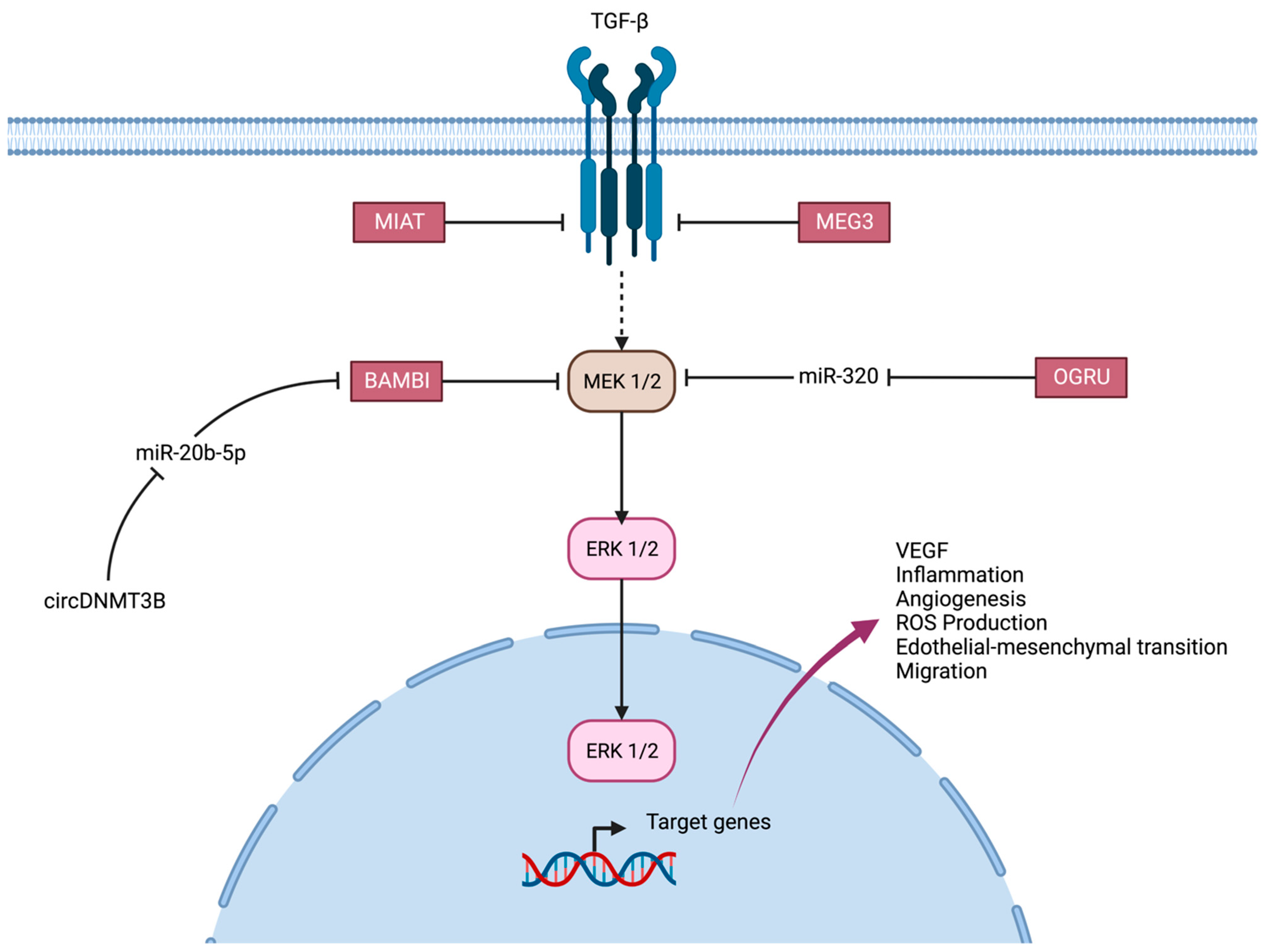
- Fu, S.; Zheng, Y.; Sun, Y.; Lai, M.; Qiu, J.; Gui, F.; Zeng, Q.; Liu, F. Suppressing long noncoding RNA OGRU ameliorates diabetic retinopathy by inhibition of oxidative stress and inflammation via miR-320/USP14 axis. Free Radic. Biol. Med. 2021, 169, 361–381. [Google Scholar] [CrossRef]
- Yu, C.; Yang, K.; Meng, X.; Cao, B.; Wang, F. Downregulation of Long Noncoding RNA MIAT in the Retina of Diabetic Rats with Tail-vein Injection of Human Umbilical-cord Mesenchymal Stem Cells. Int. J. Med. Sci. 2020, 17, 591–598. [Google Scholar] [CrossRef] [PubMed]
- Zhang, J.; Chen, C.; Wu, L.; Wang, Q.; Chen, J.; Zhang, S.; Chen, Z. C-myc contributes to the release of Müller cells-derived proinflammatory cytokines by regulating lncRNA MIAT/XNIP pathway. Int. J. Biochem. Cell Biol. 2019, 114, 105574, Erratum in Int. J. Biochem. Cell Biol. 2019, 116, 105611. [Google Scholar] [CrossRef] [PubMed]
- Yu, X.; Ma, X.; Lin, W.; Xu, Q.; Zhou, H.; Kuang, H. Long noncoding RNA MIAT regulates primary human retinal pericyte pyroptosis by modulating miR-342–3p targeting of CASP1 in diabetic retinopathy. Exp. Eye Res. 2020, 202, 108300. [Google Scholar] [CrossRef] [PubMed]
- Zhang, J.; Chen, M.; Chen, J.; Lin, S.; Cai, D.; Chen, C.; Chen, Z. Long non-coding RNA MIAT acts as a biomarker in diabetic retinopathy by absorbing miR-29b and regulating cell apoptosis. Biosci. Rep. 2017, 37, BSR20170036. [Google Scholar] [CrossRef] [PubMed]
- Yan, B.; Yao, J.; Liu, J.Y.; Li, X.M.; Wang, X.Q.; Li, Y.J.; Tao, Z.F.; Song, Y.C.; Chen, Q.; Jiang, Q. lncRNA-MIAT regulates microvascular dysfunction by functioning as a competing endogenous, RNA. Circ. Res. 2015, 116, 1143–1156. [Google Scholar] [CrossRef] [PubMed]
- Zhu, K.; Hu, X.; Chen, H.; Li, F.; Yin, N.; Liu, A.L.; Shan, K.; Qin, Y.W.; Huang, X.; Chang, Q.; et al. Downregulation of circRNA DMNT3B contributes to diabetic retinal vascular dysfunction through targeting miR-20b-5p and BAMBI. EBioMedicine 2019, 49, 341–353. [Google Scholar] [CrossRef]
- Zhang, S.J.; Chen, X.; Li, C.P.; Li, X.M.; Liu, C.; Liu, B.H.; Shan, K.; Jiang, Q.; Zhao, C.; Yan, B. Identification and Characterization of Circular RNAs as a New Class of Putative Biomarkers in Diabetes Retinopathy. Investig. Ophthalmol. Vis. Sci. 2017, 58, 6500–6509. [Google Scholar] [CrossRef]
- Luo, R.; Xiao, F.; Wang, P.; Hu, Y.X. lncRNA H19 sponging miR-93 to regulate inflammation in retinal epithelial cells under hyperglycemia via XBP1s. Inflamm. Res. 2020, 69, 255–265. [Google Scholar] [CrossRef]
- Hajjari, M.; Salavaty, A. HOTAIR: An oncogenic long non-coding RNA in different cancers. Cancer Biol. Med. 2015, 12, 1. [Google Scholar] [CrossRef]
- Zhao, D.; Zhao, Y.; Wang, J.; Wu, L.; Liu, Y.; Zhao, S.; Guo, F.; Ma, X.; Zhang, H.; Li, Z.; et al. Long noncoding RNA Hotair facilitates retinal endothelial cell dysfunction in diabetic retinopathy. Clin. Sci. 2020, 134, 2419–2434. [Google Scholar] [CrossRef]
- Biswas, S.; Feng, B.; Chen, S.; Liu, J.; Aref-Eshghi, E.; Gonder, J.; Ngo, V.; Sadikovic, B.; Chakrabarti, S. The Long Non-Coding RNA HOTAIR Is a Critical Epigenetic Mediator of Angiogenesis in Diabetic Retinopathy. Investig. Ophthalmol. Vis. Sci. 2021, 62, 20. [Google Scholar] [CrossRef] [PubMed]
- Thomas, A.A.; Feng, B.; Chakrabarti, S. ANRIL: A Regulator of VEGF in Diabetic Retinopathy. Investig. Ophthalmol. Vis. Sci. 2017, 58, 470–480. [Google Scholar] [CrossRef] [PubMed]
- Wei, J.-C.; Shi, Y.-L.; Wang, Q. LncRNA ANRIL knockdown ameliorates retinopathy in diabetic rats by inhibiting the NF-κB pathway. Eur. Rev. Med. Pharmacol. Sci. 2019, 23, 7732–7739. [Google Scholar] [CrossRef] [PubMed]
- Yin, L.; Sun, Z.; Ren, Q.; Su, X.; Zhang, D. Long Non-Coding RNA BANCR Is Overexpressed in Patients with Diabetic Retinopathy and Promotes Apoptosis of Retinal Pigment Epithelial Cells. Med. Sci. Monit. 2019, 25, 2845–2851. [Google Scholar] [CrossRef]
- Zhang, X.; Zou, X.; Li, Y.; Wang, Y. Downregulation of lncRNA BANCR participates in the development of retinopathy among diabetic patients. Exp. Ther. Med. 2019, 17, 4132–4138. [Google Scholar] [CrossRef]
- Zhang, R.; Ma, X.; Jiang, L.; Xia, W.; Li, H.; Zhao, N.; Cui, X.; Zhang, N.; Zhou, H.; Xu, S. Decreased lncRNA SNHG16 Accelerates Oxidative Stress Induced Pathological Angiogenesis in Human Retinal Microvascular Endothelial Cells by Regulating miR-195/mfn2 Axis. Curr. Pharm. Des. 2021, 27, 3047–3060. [Google Scholar] [CrossRef]
- Cai, F.; Jiang, H.; Li, Y.; Li, Q.; Yang, C. Upregulation of long non-coding RNA SNHG16 promotes diabetes-related RMEC dysfunction via activating NF-κB and PI3K/AKT pathways. Mol. Ther. Nucleic Acids 2021, 24, 512–527. [Google Scholar] [CrossRef]
- Sun, Y.; Liu, Y.X. LncRNA HOTTIP improves diabetic retinopathy by regulating the p38-MAPK pathway. Eur. Rev. Med. Pharmacol. Sci. 2018, 22, 2941–2948. [Google Scholar] [CrossRef]
- Li, X.J. Long non-coding RNA nuclear paraspeckle assembly transcript 1 inhibits the apoptosis of retina Müller cells after diabetic retinopathy through regulating miR-497/brain-derived neurotrophic factor axis. Diabetes Vasc. Dis. Res. 2018, 15, 204–213. [Google Scholar] [CrossRef]
- Shao, K.; Xi, L.; Cang, Z.; Chen, C.; Huang, S. Knockdown of NEAT1 exerts suppressive effects on diabetic retinopathy progression via inactivating TGF-β1 and VEGF signaling pathways. J. Cell Physiol. 2020, 235, 9361–9369. [Google Scholar] [CrossRef]
- Li, Y.; Xu, F.; Xiao, H.; Han, F. Long noncoding RNA BDNF-AS inversely regulated BDNF and modulated high-glucose induced apoptosis in human retinal pigment epithelial cells. J. Cell Biochem. 2018, 119, 817–823. [Google Scholar] [CrossRef] [PubMed]
- Zhao, C.; Fei, X.; Xu, B.; Lu, Y.; Zhang, Q. Long non-coding RNA HEIH contributes to diabetic retinopathy by regulating miR-939/VEGF axis. Int. J. Clin. Exp. Pathol. 2019, 12, 2022–2033. [Google Scholar]
- Yu, X.; Luo, Y.; Chen, G.; Liu, H.; Tian, N.; Zen, X.; Liu, Q. Long noncoding RNA IGF2AS regulates high-glucose induced apoptosis in human retinal pigment epithelial cells. IUBMB Life 2019, 71, 1611–1618. [Google Scholar] [CrossRef] [PubMed]
- Ke, N.; Pi, L.H.; Liu, Q.; Chen, L. Long noncoding RNA SNHG7 inhibits high glucose-induced human retinal endothelial cells angiogenesis by regulating miR-543/SIRT1 axis. Biochem. Biophys. Res. Commun. 2019, 514, 503–509. [Google Scholar] [CrossRef]
- Cao, X.; Xue, L.D.; Di, Y.; Li, T.; Tian, Y.J.; Song, Y. MSC-derived exosomal lncRNA SNHG7 suppresses endothelial-mesenchymal transition and tube formation in diabetic retinopathy via miR-34a-5p/XBP1 axis. Life Sci. 2021, 272, 119232. [Google Scholar] [CrossRef] [PubMed]
- Zhang, Y.; Zheng, L.; Xu, H.; Ling, L. Circ_0084043 Facilitates High Glucose-Induced Retinal Pigment Epithelial Cell Injury by Activating miR-128-3p/TXNIP-Mediated Wnt/β-Catenin Signaling Pathway. J. Cardiovasc. Pharmacol. 2021, 78, e112–e121. [Google Scholar] [CrossRef]
- Yu, J.; Qin, M.; Li, J.; Cui, S. LncRNA SNHG4 sponges miR-200b to inhibit cell apoptosis in diabetic retinopathy. Arch. Physiol. Biochem. 2021, ahead of print. [CrossRef]
- Niu, T.; An, Y.; Lv, T.; Liu, D. Long non-coding RNA RPSAP52 upregulates Timp3 by serving as the endogenous sponge of microRNA-365 in diabetic retinopathy. Exp. Ther. Med. 2020, 20, 246. [Google Scholar] [CrossRef]
- Shao, J.; Pan, X.; Yin, X.; Fan, G.; Tan, C.; Yao, Y.; Xin, Y.; Sun, C. KCNQ1OT1 affects the progression of diabetic retinopathy by regulating miR-1470 and epidermal growth factor receptor. J. Cell Physiol. 2019, 234, 17269–17279. [Google Scholar] [CrossRef]
- Shi, Y.; Chen, C.; Xu, Y.; Liu, Y.; Zhang, H.; Liu, Y. LncRNA FENDRR promotes high-glucose-induced proliferation and angiogenesis of human retinal endothelial cells. Biosci. Biotechnol. Biochem. 2019, 83, 869–875. [Google Scholar] [CrossRef]
- Gong, Q.; Dong, W.; Fan, Y.; Chen, F.; Bian, X.; Xu, X.; Qian, T.; Yu, P. LncRNA TDRG1-Mediated Overexpression of VEGF Aggravated Retinal Microvascular Endothelial Cell Dysfunction in Diabetic Retinopathy. Front. Pharmacol. 2020, 10, 1703. [Google Scholar] [CrossRef] [PubMed]
- Yan, H.; Yao, P.; Hu, K.; Li, X.; Li, H. Long non-coding ribonucleic acid urothelial carcinoma-associated 1 promotes high glucose-induced human retinal endothelial cells angiogenesis through regulating micro-ribonucleic acid-624-3p/vascular endothelial growth factor C. J. Diabetes Investig. 2021, 12, 1948–1957. [Google Scholar] [CrossRef] [PubMed]
- Wang, Y.; Wang, L.; Guo, H.; Peng, Y.; Nie, D.; Mo, J.; Ye, L. Knockdown of MALAT1 attenuates high-glucose-induced angiogenesis and inflammation via endoplasmic reticulum stress in human retinal vascular endothelial cells. Biomed. Pharmacother. 2020, 124, 109699. [Google Scholar] [CrossRef] [PubMed]
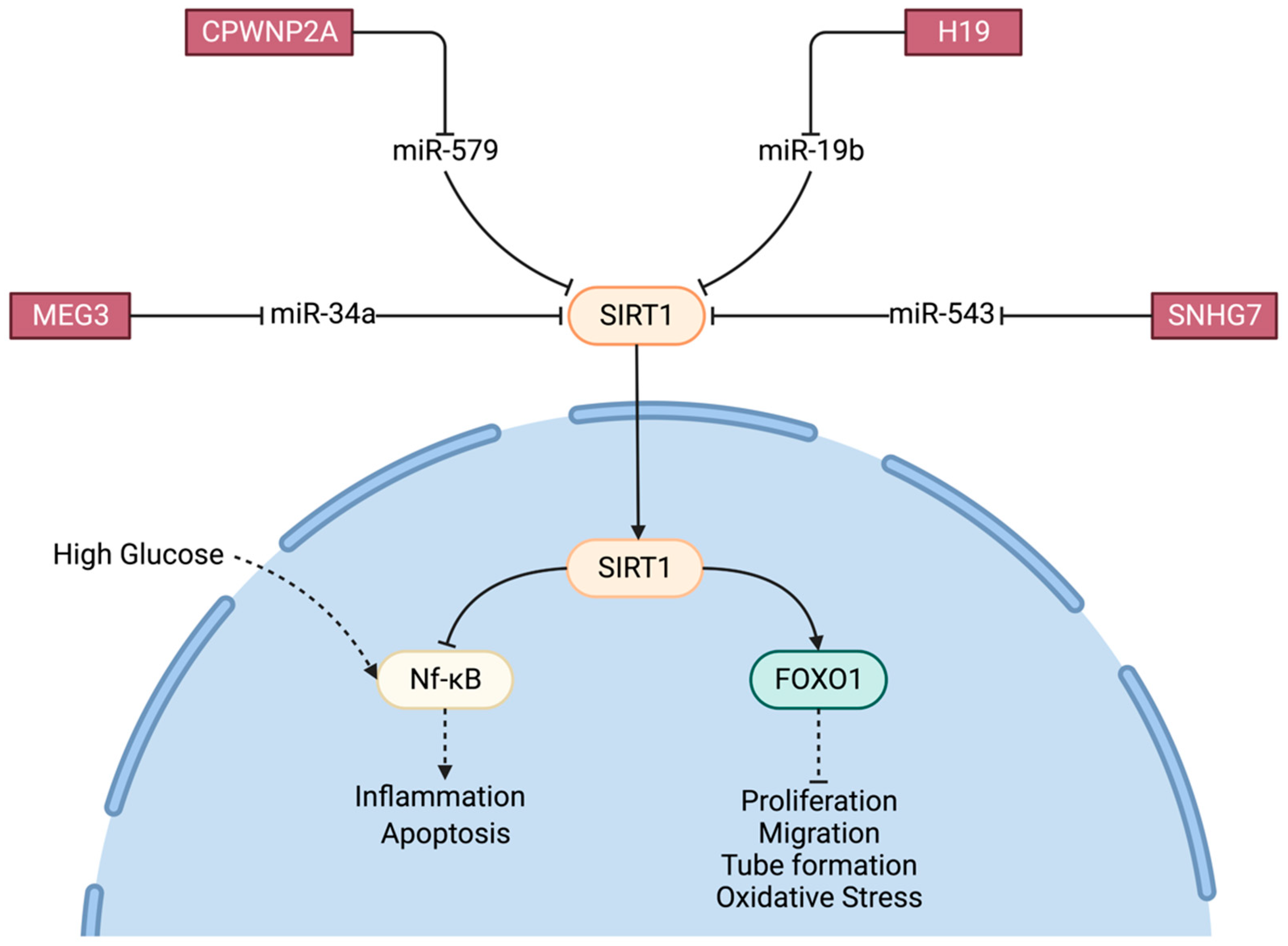
- Liu, C.; Ge, H.M.; Liu, B.H.; Dong, R.; Shan, K.; Chen, X.; Yao, M.D.; Li, X.M.; Yao, J.; Zhou, R.M.; et al. Targeting pericyte-endothelial cell crosstalk by circular RNA-cPWWP2A inhibition aggravates diabetes-induced microvascular dysfunction. Proc. Natl. Acad. Sci. USA 2019, 116, 7455–7464. [Google Scholar] [CrossRef]
- Gao, C.; Lin, X.; Fan, F.; Liu, X.; Wan, H.; Yuan, T.; Zhao, X.; Luo, Y. Status of higher TGF-β1 and TGF-β2 levels in the aqueous humour of patients with diabetes and cataracts. BMC Ophthalmol. 2022, 22, 156. [Google Scholar] [CrossRef]
- Sun, X.; Lu, Y.; Lei, T. TPTEP1 suppresses high glucose-induced dysfunction in retinal vascular endothelial cells by interacting with STAT3 and targeting VEGFA. Acta Diabetol. 2021, 58, 759–769. [Google Scholar] [CrossRef]
- Gysens, F.; Mestdagh, P.; de Lavergne, E.D.B.; Maes, T. Unlocking the secrets of long non-coding RNAs in asthma. Thorax 2022, 77, 514–522. [Google Scholar] [CrossRef]
- Qiu, Y.Y.; Wu, Y.; Lin, M.J.; Bian, T.; Xiao, Y.L.; Qin, C. LncRNA-MEG3 functions as a competing endogenous RNA to regulate Treg/Th17 balance in patients with asthma by targeting microRNA-17/ RORγt. Biomed. Pharmacother. 2019, 111, 386–394. [Google Scholar] [CrossRef]
- Li, Z.; Li, J.; Tang, N. Long noncoding RNA Malat1 is a potent autophagy inducer protecting brain microvascular endothelial cells against oxygen-glucose deprivation/reoxygenation-induced injury by sponging miR-26b and upregulating ULK2 expression. Neuroscience 2017, 354, 1–10. [Google Scholar] [CrossRef]
- Wang, J.; Cao, B.; Han, D.; Sun, M.; Feng, J. Long non-coding RNA H19 induces cerebral ischemia reperfusion injury via activation of autophagy. Aging Dis. 2017, 8, 71–84. [Google Scholar] [CrossRef]
- Zhu, M.; Li, N.; Luo, P.; Jing, W.; Wen, X.; Liang, C.; Tu, J. Peripheral blood leukocyte expression of lncRNA MIAT and its diagnostic and prognostic value in ischemic stroke. J. Stroke Cerebrovasc. Dis. 2018, 27, 326–337. [Google Scholar] [CrossRef] [PubMed]
- Yan, H.; Rao, J.; Yuan, J.; Gao, L.; Huang, W.; Zhao, L.; Ren, J. Long non-coding RNA MEG3 functions as a competing endogenous RNA to regulate ischemic neuronal death by targeting miR-21/PDCD4 signaling pathway. Cell Death Dis. 2017, 8, 3211. [Google Scholar] [CrossRef] [PubMed]
- Chen, S.; Wang, M.; Yang, H.; Mao, L.; He, Q.; Jin, H.; Ye, Z.; Luo, X.; Xia, Y.; Hu, B. LncRNA TUG1 sponges microRNA-9 to promote neurons apoptosis by up-regulated Bcl2l11 under ischemia. Biochem. Biophys. Res. Commun. 2017, 485, 167–173. [Google Scholar] [CrossRef] [PubMed]
- Liu, J.; Li, Q.; Zhang, K.S.; Hu, B.; Niu, X.; Zhou, S.M.; Zhou, S.-M.; Li, S.-G.; Luo, Y.-P.; Wang, Y.; et al. Downregulation of the long non-coding RNA Meg3 promotes angiogenesis after ischemic brain injury by activating notch signaling. Mol. Neurobiol. 2017, 54, 8179–8190. [Google Scholar] [CrossRef] [PubMed]
- Eissmann, M.; Gutschner, T.; Hammerle, M.; Gunther, S.; Caudron-Herger, M.; Gross, M.; Schirmacher, P.; Rippe, K.; Braun, T.; Zornig, M.; et al. Loss of the abundant nuclear non-coding RNA MALAT1 is compatible with life and development. RNA Biol. 2012, 9, 1076–1087. [Google Scholar] [CrossRef]
- Ji, P.; Diederichs, S.; Wang, W.; Boing, S.; Metzger, R.; Schneider, P.M.; Tidow, N.; Brandt, B.; Buerger, H.; Bulk, E.; et al. MALAT-1, a novel noncoding RNA, and thymosin beta4 predict metastasis and survival in early-stage non-small cell lung cancer. Oncogene 2003, 22, 8031–8041. [Google Scholar] [CrossRef]
- Schmidt, L.H.; Spieker, T.; Koschmieder, S.; Schaffers, S.; Humberg, J.; Jungen, D.; Bulk, E.; Hascher, A.; Wittmer, D.; Marra, A.; et al. The long noncoding MALAT-1 RNA indicates a poor prognosis in non-small cell lung cancer and induces migration and tumor growth. J. Thorac. Oncol. 2011, 6, 1984–1992. [Google Scholar] [CrossRef]
- Meseure, D.; Vacher, S.; Lallemand, F.; Alsibai, K.D.; Hatem, R.; Chemlali, W.; Nicolas, A.; De Koning, L.; Pasmant, E.; Callens, C.; et al. Prognostic value of a newly identified MALAT1 alternatively spliced transcript in breast cancer. Br. J. Cancer 2016, 114, 1395–1404. [Google Scholar] [CrossRef]
- Feng, T.; Shao, F.; Wu, Q.; Zhang, X.; Xu, D.; Qian, K.; Xie, Y.; Wang, S.; Xu, N.; Wang, Y.; et al. miR-124 downregulation leads to breast cancer progression via LncRNA-MALAT1 regulation and CDK4/E2F1 signal activation. Oncotarget 2016, 7, 16205–16216. [Google Scholar] [CrossRef]
- Yang, L.; Bai, H.S.; Deng, Y.; Fan, L. High MALAT1 expression predicts a poor prognosis of cervical cancer and promotes cancer cell growth and invasion. Eur. Rev. Med. Pharmacol. Sci. 2015, 19, 3187–3193. [Google Scholar]
- Luo, J.H.; Ren, B.; Keryanov, S.; Tseng, G.C.; Rao, U.N.; Monga, S.P.; Strom, S.; Demetris, A.J.; Nalesnik, M.; Yu, Y.P.; et al. Transcriptomic and genomic analysis of human hepatocellular carcinomas and hepatoblastomas. Hepatology 2006, 44, 1012–1024. [Google Scholar] [CrossRef]
- Luo, F.; Sun, B.; Li, H.; Xu, Y.; Liu, Y.; Liu, X.; Lu, L.; Li, J.; Wang, Q.; Wei, S.; et al. A MALAT1/HIF-2alpha feedback loop contributes to arsenite carcinogenesis. Oncotarget 2016, 7, 5769–5787. [Google Scholar] [CrossRef]
- Lai, M.C.; Yang, Z.; Zhou, L.; Zhu, Q.Q.; Xie, H.Y.; Zhang, F.; Wu, L.M.; Chen, L.M.; Zheng, S.S. Long non-coding RNA MALAT-1 overexpression predicts tumor recurrence of hepatocellular carcinoma after liver transplantation. Med. Oncol. 2012, 29, 1810–1816. [Google Scholar] [CrossRef] [PubMed]
- Li, Q.; Dai, Y.; Wang, F.; Hou, S. Differentially expressed long non-coding RNAs and the prognostic potential in colorectal cancer. Neoplasma 2016, 63, 977–983. [Google Scholar] [CrossRef] [PubMed]
- Amodio, N.; Raimondi, L.; Juli, G.; Stamato, M.A.; Caracciolo, D.; Tagliaferri, P.; Tassone, P. MALAT1: A druggable long non-coding RNA for targeted anti-cancer approaches. J. Hematol. Oncol. 2018, 11, 63. [Google Scholar] [CrossRef] [PubMed]


| Overproduction of Molecules | Mechanisms Involved | Site/Effect of Damage | References | |
|---|---|---|---|---|
| Oxidative Stress | ROS AGEs | PKC, NFkB NrF2, TNF-α | mitochondria capillary cells | [8,9] |
| Neurodegeneration | Caspases, Bax, Bak | NFkB, SIRT1 Akt, Cox2, TGF-β | RGCs Pericytes | [4,10,11,12,13,14] |
| Inflammatory Process | TNF-α, IL-6, IL-8, IL-1β, iNOS, ICAM1, Complement factor | PKC—NFkB Müller cells | capillary cell death, increase vascular permeability | [5,15,16,17,18,19,20,21,22,23] |
| Angiogenesis | VEGF | Ischemia, PEDF | hypoxia, vitreo-retinal neovascularization | [4,5,24] |
| Lnc-RNA | Full Name | How is Lnc-RNA Expression Affected in High-Glucose/Hyperglycemia/Diabetes Conditions? | Effects and Targets | References |
|---|---|---|---|---|
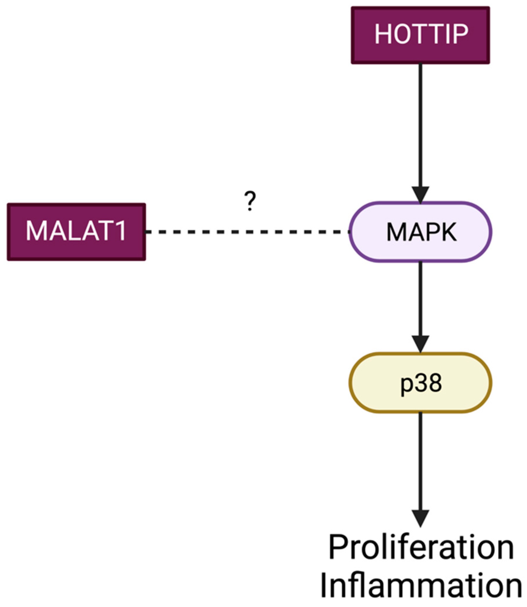
| Lnc MALAT-1 | Metastasis-Associated Adenocarcinoma Transcript 1 | Upregulation in retinal endothelial cells and diabetic retinas. Upregulation in the vitreous humor, aqueous humor samples and fibrovascular membranes of diabetic patients. | Inflammatory molecules (IL-6, Tnf-α, IL-1Beta, MCP-1). Neovascularization-related proteins (VEGF, MMP2 and MMP9). Related to GRP78 producing angiogenesis and inflammation in hRVECs. Regulate cell proliferation via p38 MAPK signaling pathways. Binds Sp1, avoiding Nrf2 nuclear movement, impeding the transcription of antioxidant response enzymes (HO-1 and Sod2). Binds to miR-125b, producing VE-cadherin activation. Binds to miR-203a-3p, elevating HIF-1alpha and VEGFA. Binds to miR-200b-3p, producing proliferation, migration and tube formation of hRMECs. | [30] (a), [31] (a, c) [32] (c) [33] (h) [34] (a, c) [35] (c) [36] (a, c), [37] (c), [30] (a), [30] (a), [38] (a, c) |
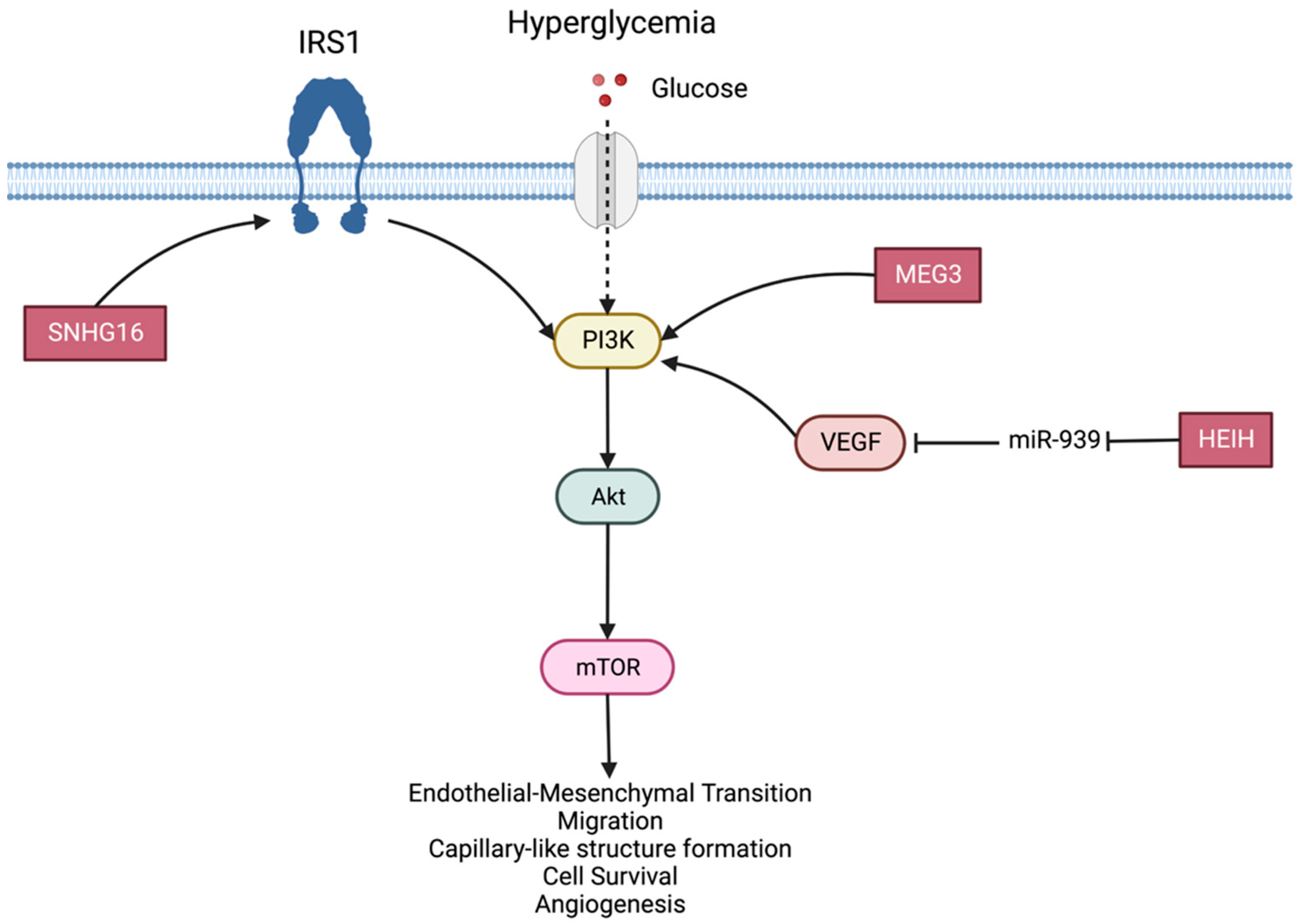
| Lnc MEG3 | Maternally expressed gene 3 | Decreased in serum of diabetic patients and HG-treated RPE cells. Reduced in retinal endothelial cells and diabetic retinas. | MEG3 overexpression reduces VEFG and TGF-Beta1. MEG3 knockdown increases proliferation, migration and tube formation of retinal endothelial cells. MEG3 reduction increases retinal angiogenesis and can aggravate vascular leakage and inflammation. Methylation of CpG islands of the MEG promoter by DNMT1 leads to PI3K/Akt/mTOR signaling pathway activation, promoting endothelial–mesenchymal transition. MEG3 activates PI3K/Akt signaling pathway and regulates retinal endothelial cell events related to angiogenesis. MEG3 decreases the level of miR-233-3p, partially repressing the progression of DR and suppressing proliferation of hRECs. Lnc MEG3 expression into the vitreous cavity reduced Fox01 (oxidative stress, proliferation, apoptosis, differentiation and autophagy regulator) and IL-1Beta. Regulates miR-19b, suppressing cell apoptosis and enhancing cell viability in hRMECs. Binds to miR-34a, promoting the expression of Sirt1 and inhibiting NF-kB pathway, inflammatory response (IL-1-Beta, IL-6 and TNF-α) and apoptosis (Bcl-2/Bax ratio) in Müller cells and ARPE-19 cell line. Possibly binds to miR-204, promoting Sirt1 pathway. Targets miR-93, increasing Nrf2 in ARPE-19 cells. Consequently, it inhibits apoptosis (inhibits cleaved caspase-3 and Bcl2 and increases Bax) and inflammation (decreases IL-6 and TNF-α). | [39] (a, c, h) [40] (a), [39] (a, c), [41] (c), [42] (c), [43] (c, h), [44] (a, c), [45] (a, c), [46] (a, c, h), [47] (a, c, h), [46] (a, c, h), [48] (c), [49] (a, c), [50] (c, h), [34] (c) |
| Lnc MIAT | Myocardial-infarction-associated transcript Retinal non-coding RNA 2 | Upregulated MIAT expression in plasma, retinal endothelial cells and Müller cells. Upregulated MIAT levels on fibrovascular membranes of diabetic patients. MIAT expression increased in hRPE cells cultured with HG. | Associated with cell proliferation, apoptosis and migration. Implicated in the regulation of vascular function, angiogenesis and vascular leakage. Upregulated MIAT produces microvascular dysfunctions (blood flow disruption, basement membrane thickening, pericyte loss and acellular capillaries). Regulator of retinal neurodegeneration. Activates TGF-Beta 1 pathway, reducing cell viability. Upregulates TGF-Beta expression in the aqueous humor in diabetic patients. Targets miR-29b, reversing Müller cell apoptosis. Binds to miR-342-3p to regulate caspase-1 expression and consequent pericyte pyroptosis. Binds to miR-150-5p, modulating VEGF expression at a transcriptional level in retinal endothelial cells. MIAT is regulated by C-myc, releasing IL-1Beta, TNF--α and IL-6 through TXNIP. | [51] (a, c), [38] (a, h), [52] (a, c), [53] (c, h), [54] (a, c), [55] (a, c), [56] (c, h) [57] (a, c) |
| Lnc H19 | Derives from paternally imprinted H19 gene | Downregulated in retinal epithelial cells with high glucose. Downregulated in vitreous humor samples from patients with PDR. | Overexpression of H19 inhibits inflammation in ARPE-19 cells. Binds XBP1-suppressor miR-93, increasing XBP1 which reduces inflammation (TNF--α and other inflammatory mediators) and apoptosis. Binds to miR-19b and upregulates SIRT1, reducing the expression of TNF-α, IL-1Beta and IL-6 in ARPE-19 cells. H19 overexpression blocks the MPK-ERK1/2 pathway, preventing glucose-induced endothelial–mesenchymal transition by suppressing TGF-Beta 1. | [58] (c), [48] (c), [46] (a, c, h), [59] (r), [52] (c) |
| Lnc HOTAIR | HOX Transcript Antisense RNA | Increased in diabetic retinas and high-glucose-stimulated RECs. | Related to proliferation, invasion, migration and permeability of HG-stimulated RECs. Related to acellular capillaries and vascular leakage in vivo. Contributes to glucose-induced mitochondrial and DNA damage. Facilitates the epigenetic activation of VEGF-A. VE-cadherin transcription inhibition. | [59] (c), [60] (a, c), [61] (a, c, h) |
| Lnc ANRIL | Lnc-antisense non-coding RNA in the INK4 locus | Upregulated in diabetic retinopathy and retinal tissues of DR rats. | Direct and indirect role as a recruiter of chromatin remodeling complexes, upregulating VEGF mRNA expression. Regulates NF-kB and IL-1, IL-6 and MCP-1. Related to apoptosis in retinal tissues. | [62] (a, c) [63] (a) |
| Lnc BANCR | B-Raf proto-oncogene, serine/threonine kinase-activated non-protein coding RNA | Plasmatic levels allow distinguishing between diabetic patients without obvious complications. | Possible biomarker for diabetic retinopathy. BANCR overexpression inhibited apoptosis in ARPE-19 cells under high glucose treatment. | [64] (c, h) [65] (c, h) |
| Lnc SNHG16 | Small nucleolar RNA host gene 16 | Upregulated after HG exposure in a dose- and time-dependent pattern. | Associated with hRMEC proliferation. Related to HIF-1alpha and VEGF expression. Binds to miR-146a-5p and miR-7-5p related to NF-kB and PI3K/AKT pathways. | [66] (c) [67] (c) |
| Lnc HOTTIP | HOXA transcript at the distal tip | Upregulated in retinal vascular cells and retinas of diabetic animals. | Promotes retinal inflammatory processes by activating p38-MAPK. Related to neovascularization and tube formation on vascular endothelial cells. | [68] (a, c) [68] (a, c) |
| Lnc NEAT1 | The nuclear paraspeckle assembly transcript 1 | Downregulated in Müller cells under diabetic conditions. Increased in hRECs under HG. | Related to BNDF expression, promoting cell differentiation, inhibiting inflammation and protecting photoreceptors and RGCs. Related to TGF-beta1 and VEGF signaling. | [69] (a, c), [70] (a, c, h) |
| Lnc BDNF-AS | Brain-derived neurotrophic factor antisense | Abundantly expressed in retina. Upregulated in RPE cells exposed to high glucose. | BDNF antisense. Related to ischemic injury in RGCs and apoptosis. | [71] (c) |
| Lnc HEIH | Hepatocellular Carcinoma Upregulated EZH2-Associated | Highly expressed in serum of DR patients. Increased expression on ARPE-19 cells exposed to HG. | Related to cell injury and apoptosis (releasing cytochrome C and enhancing the caspase-3 pathway). Binds to miR-939, increasing VEGF and consequently PI3K/AKT signaling pathway activation. | [72] (c, h) |
| Lnc IGF2-AS | Insulin-like growth factor 2 antisense transcript | Upregulated on ARPE-19 cells with HG in a concentration-dependent manner. | Related to apoptosis (caspase-9). May act through AKT signaling pathway. | [73] (c) |
| Lnc SNHG7 | Small nucleolar RNA host gene 7 | Downregulated under HG exposure. | Suppress cell proliferation, migration and angiogenesis. Inhibits EndMT. Acts through miR-543/SIRT1. Binds to miR-34a-5p/XBP1, avoiding EndMT and angiogenesis in HG-treated hRMECs and retinal inflammation and Müller glia activation in DR. | [74] (c), [75] (c), [74] (c), [76] (c) |
| Lnc SNHG4 | Small Nucleolar RNA Host Gene 4 | Downregulated in DR. Not downregulated in diabetic patients without obvious complications. | Related to protection against cytokines production, inflammation and apoptosis. Binds to miR-200b/Oxr1. | [77] (c) |
| Lnc RPSAP52 | Ribosomal Protein SA Pseudogene 52 | Lower plasma levels in diabetic patients’ RPE cells. Downregulated in RPE cells exposed to HG. | Reduces the apoptotic rate of RPE cells. Sponges miR-365 to upregulate Timp3 (decreasing apoptosis). | [78] (c, h) |
| Lnc KCNQ1OT1 | KCNQ1 overlapping transcript 1 | Higher in aqueous humor of DR patients. | Promotes cell proliferation and angiogenesis in hRECs. Binds to miR-1470, increasing EGFR and AKT pathway signaling. | [79] (c, h) |
| Lnc FENDRR | FOXF1 Adjacent Non-Coding Developmental Regulatory RNA | Increased in blood of DR patients. Increased in HG-exposed hRECs. | Increases the expression of FOXF1. Promotes proliferation, migration, capillary formation and VEGF expression. | [80] (c, h) |
| Lnc TDRG1 | Human testis development-related gene 1 | Overexpressed in fibrovascular membranes of patients with PDR. Highly expressed in hRECs exposed to HG. | Related to endothelial cell dysfunction and VEGF expression. | [81] (c, h) |
| Lnc UCA1 | Urothelial-cancer-associated 1 | Upregulated in endothelial cells in DM and in diabetic nephropathy in rats. Upregulated in fibrovascular membranes and in the blood of patients with DR. | Binds to miR-624-3p, leading to cell proliferation, migration and angiogenesis by promoting VEGF-C expression in endothelial cells. | [82] (c, h) |
Disclaimer/Publisher’s Note: The statements, opinions and data contained in all publications are solely those of the individual author(s) and contributor(s) and not of MDPI and/or the editor(s). MDPI and/or the editor(s) disclaim responsibility for any injury to people or property resulting from any ideas, methods, instructions or products referred to in the content. |
© 2023 by the authors. Licensee MDPI, Basel, Switzerland. This article is an open access article distributed under the terms and conditions of the Creative Commons Attribution (CC BY) license (https://creativecommons.org/licenses/by/4.0/).
Share and Cite
Perisset, S.; Potilinski, M.C.; Gallo, J.E. Role of Lnc-RNAs in the Pathogenesis and Development of Diabetic Retinopathy. Int. J. Mol. Sci. 2023, 24, 13947. https://doi.org/10.3390/ijms241813947
Perisset S, Potilinski MC, Gallo JE. Role of Lnc-RNAs in the Pathogenesis and Development of Diabetic Retinopathy. International Journal of Molecular Sciences. 2023; 24(18):13947. https://doi.org/10.3390/ijms241813947
Chicago/Turabian StylePerisset, Sofia, M. Constanza Potilinski, and Juan E. Gallo. 2023. "Role of Lnc-RNAs in the Pathogenesis and Development of Diabetic Retinopathy" International Journal of Molecular Sciences 24, no. 18: 13947. https://doi.org/10.3390/ijms241813947
APA StylePerisset, S., Potilinski, M. C., & Gallo, J. E. (2023). Role of Lnc-RNAs in the Pathogenesis and Development of Diabetic Retinopathy. International Journal of Molecular Sciences, 24(18), 13947. https://doi.org/10.3390/ijms241813947