Article

Phylogeny of PmCCD Gene Family and Expression Analysis of Flower Coloration and Stress Response in Prunus mume

1
Beijing Key Laboratory of Ornamental Plants Germplasm Innovation and Molecular Breeding, National Engineering Research Center for Floriculture, School of Landscape Architecture, Beijing Forestry University, Beijing 100083, China
2
Beijing Laboratory of Urban and Rural Ecological Environment, Engineering Research Center of Landscape Environment of Ministry of Education, School of Landscape Architecture, Beijing Forestry University, Beijing 100083, China
3
Key Laboratory of Genetics and Breeding in Forest Trees and Ornamental Plants of Ministry of Education, School of Landscape Architecture, Beijing Forestry University, Beijing 100083, China
*
Authors to whom correspondence should be addressed.
Int. J. Mol. Sci. 2023, 24(18), 13950; https://doi.org/10.3390/ijms241813950
Submission received: 28 July 2023 / Revised: 2 September 2023 / Accepted: 8 September 2023 / Published: 11 September 2023
(This article belongs to the Special Issue Advanced Research in Plant Responses to Environmental Stresses 2.0)

Abstract

:
The CCD gene family plays a crucial role in the cleavage of carotenoids, converting them into apocarotenoids. This process not only impacts the physiology and development of plants but also enhances their tolerance toward different stresses. However, the character of the PmCCD gene family and its role in ornamental woody Prunus mume remain unclear. Here, ten non-redundant PmCCD genes were identified from the P. mume genome, and their physicochemical characteristics were predicted. According to the phylogenetic tree, PmCCD proteins were classified into six subfamilies: CCD1, CCD4, CCD7, CCD8, NCED and CCD-like. The same subfamily possessed similar gene structural patterns and numbers of conserved motifs. Ten PmCCD genes were concentrated on three chromosomes. PmCCD genes exhibited interspecific collinearity with P. armeniaca and P. persica. Additionally, PmCCD genes had obvious specificity in different tissues and varieties. Compared with white-flowered ‘ZLE’, PmCCD1 and PmCCD4 genes were low-expressed in ‘HJH’ with yellow petals, which suggested PmCCD1 and PmCCD4 might be related to the formation of yellow flowers in P. mume. Nine PmCCD genes could respond to NaCl or PEG treatments. These genes might play a crucial role in salt and drought resistance in P. mume. Moreover, PmVAR3 and PmSAT3/5 interacted with PmCCD4 protein in yeast and tobacco leaf cells. This study laid a foundation for exploring the role of the PmCCD gene family in flower coloration and stress response in P. mume.

1. Introduction

Carotenoids are a class of C40 terpenoid compounds and their derivatives, mainly composed of eight isoprene units. In plants, carotenoids play a role in photosynthesis assistance and photoprotection and the biosynthesis of plant hormones abscisic acid and strigolactone [1,2,3]. Moreover, they enable the flowers, fruits and other organs of higher plants to present a variety of brilliant colors and aromas, thus attracting birds and insects to participate in plant pollination and seed dispersal [4].
Carotenoid cleavage dioxygenases (CCDs) can oxidatively cleave carotenoids at one or both ends of the molecule to produce different apocarotenoids, which participate in plant growth, development and stress response [5]. Tan et al. [6] identified the first carotenoid cleavage dioxygenases viviparous 14 (VP14) in maize mutants. Subsequently, CCD genes were identified in multiple species. In Arabidopsis thaliana, CCD genes were divided into two categories: carotenoid cleavage dioxygenases (CCD) and 9-cis-epoxycarotenoid cleavage dioxygenases (NCEDs), according to whether the substrate of its catalytic cleavage undergoes epoxidation. The CCD subfamily mainly included CCD1, CCD4, CCD7 and CCD8.The NCED subfamily included NCED2, NCED3, NCED5, NCED6 and NCED9 [7]. Since then, the homologs found in other plants have been named in the classification of Arabidopsis manner. In addition, a novel group known as CCD-like (CCDL) has been identified in Oryza sativa, Sorghum bicolor, Solanum lycopersicum and Malus pumila [8,9,10].
The essential physiological functions of carotenoid lysates in plants have attracted significant attention to the lyases and their products involved in carotenoid metabolism. The main functional principle of CCD genes is to specifically cleave the double bonds on different carotenoid substrates to generate a variety of cleavage products, thus playing different biological functions. The substrates of CCD1 genes have universality, which can preferentially and symmetrically cut the double bond at the 9,10 (9′, 10′) position of the carotenoid to produce flavor and aromatic substances as well as aldehydes or ketones. Simultaneously, it can also cleave double bonds at positions 5,6 (5′, 6′) and 7,8 (7′, 8′). In Arabidopsis, S. lycopersicum, Cucumis melo, Petunia hybrida, Laurus nobilis, Fragaria ananassa and Lycium chinense, the involvement of CCD1 genes has been reported in the biosynthesis of volatile aromas, such as β-ionone [11,12,13,14,15,16,17]. CCD4 genes play a crucial role in plant color formation and aroma production. In Chrysanthemum morifolium, inhibiting the expression of the CmCCD4a gene through RNAi could lead to a mutation from white to yellow flowers [18]. Suppressing the CCD4 gene in potato caused the carotenoid in the tubers to increase and appear yellow [19]. Differential expression of the BnCCD4 gene led to the emergence of yellow and white Brassica napus, and BnCCD4 could cleave α-carotene as a substrate to produce α- ionone [20]. In addition, a unique cleavage activity of CitCCD4 was found in citrus, which could cleave at the 7, 8 (7′, 8′) double-bond sites β-cryptoxanthin or zeaxanthin to produce orange-red apocarotenoid β-citraurin [21]. Studies have revealed that plant CCD1 and CCD4 also actively participate in responses to heat, drought, salt and other stresses [22]. For example, overexpression of CsCCD4b enhanced the stress tolerance to salt, dehydration and oxidation in Arabidopsis [23]. In higher plants, CCD7 and CCD8 produced the signaling substance strigolactone by cleaving β-carotene at the 9′, 10′ double-bond sites [24]. Strigolactone has been implicated in governing the development of lateral roots and branches in plants, as well as response to stress [25]. Ectopic expression of CpCCD7 and CpCCD8 restored the branching phenotype of Arabidopsis mutants max3-9 and max4-1, respectively [26]. Under phosphorus deficiency, ZmCCD7 upregulated the gene expression [27]. Similarly, in Poplar trees subjected to H2O2, drought and salt stress, the CCD8 gene actively responds to stress stimuli [28].
NCEDs can oxidatively cleave violaxanthin and neoxanthin with an epoxy structure at the 11,12 double-bond sites to generate xanthoxin, which represents the initial step of ABA biosynthesis in plants [29]. The NCED gene can directly affect plant growth, development and stress response by regulating ABA synthesis. Currently, NCED3 has been proven to be involved in abiotic stress responses in multiple species [30,31,32,33]. In rice, OsNCED1 and OsNCED2 regulated the ABA levels in response to drought conditions, thereby enhancing drought tolerance [34,35]. Overexpression of OsNCED5 enhanced the salt and water stress tolerance of rice leaves and accelerated leaf senescence. OsNCED5 also altered plant size and leaf morphology and delayed seed germination and flowering time in Arabidopsis [36]. In addition, overexpression of CrNCED1 in citrus improved tolerance to multiple abiotic stresses [37]. In Stylosanthes guianensis, drought stress induced the expression of SgNCED1 and the accumulation of ABA [38]. Additionally, the expression of the CstNCED gene played a vital role in regulating ABA levels during style senescence, corm dormancy and drought stress in Crocus sativus [39].
P. mume is a famous ornamental tree, rich in flower color, elegant flower fragrance and tree shape. CCD genes have a major effect on flower color and fragrance formation, the morphological construction of growth and development and stress response in plants [5]. Therefore, it is significant to identify CCD gene family members in P. mume. Here, we identified 10 PmCCD genes and performed bioinformatics analysis, including gene physicochemical characteristics, a phylogenetic tree, chromosome distribution, promoters, collinearity and protein interactions. The expression profiles of PmCCD genes in various tissues, varieties of yellow and white flowers and stresses (NaCl or PEG treatments) were analyzed by qRT-PCR. The interacting proteins of PmCCD4 were verified by using a yeast two-hybrid system and luciferase complementation experiment. In summary, this study laid the foundation for the functional research of the PmCCD genes and amplified candidate genes for the breeding of P. mume.

2. Results

2.1. Identification of CCD Gene Family Members in P. mume

To obtain HMM for the REP65 domain (PF03055), the Pfam database was utilized. HMMER3 software was applied to search for CCD genes in the P. mume genome with E-value ≤ 10−5. Finally, 10 PmCCD genes were identified and named following the Arabidopsis nomenclature.
The physicochemical properties and secondary structures of 10 PmCCD proteins were analyzed (Table S1). The PmCCD proteins exhibited a length ranging from 247 to 622 amino acids, with 80% of the proteins having 500–600 aa. The PmCCD1-like-c protein was the smallest, while PmNCED3 encoded the most amino acids. Additionally, three members of the NCED subfamily encoded more than 600 aa. The protein molecular weight varied between 27.71 kDa (PmCCD1-like-c) and 69.06 kDa (PmCCD1-like-a). For isoelectric points, PmCCD1-like-b and PmNCED6 were 7.06 and 7.92, implying neutrality. The isoelectric points of the other six proteins were less than 7, indicating that they were acidic. The instability index ranged from 28.96 (PmCCD1-like-a) to 46.09 (PmCCD7), with three unstable proteins and seven stable proteins. The grand average of hydropathicity was negative, showing that all PmCCD proteins were soluble. The aliphatic index was 64.7 to 88.24 and indicated that CCD proteins had good thermal stability. PmCCD1-like-c had the lowest total number of negatively charged residues (Asp + Glu) and total number of positively charged residues (Arg + Lys). The maximum number of (Asp + Glu) in PmCCD1-like-a was 76, while the maximum number of (Arg + Lys) in PmNCED6 was 67.
Except for PmCCD1 and PmCCD1-like-c proteins, chloroplast targeting peptides were present at the other eight PmCCDs sequences, indicating that eight PmCCDs might be localized in the chloroplast. Subcellular localization analysis of the PmCCD proteins revealed that the majority of these proteins were found to be localized within the chloroplasts, while PmCCD1 and PmCCD1-like-c were localized in the cytoplasm, which was consistent with the prediction of signal peptides.
Secondary structure prediction showed that all the family members consisted of an α-helix, extended strand, β-sheet and random coil. Out of them, the β-sheet was lower than 10%, followed by the α-helix. Except for PmCCD1-like-a and PmCCD1-like-b, all members were lower than 20%. However, the proportion of random coil was the highest, PmCCD1-like-b was 41.18%, and the remaining was more than 50%, indicating that the secondary structure of CCD family members was mainly random coil (Figure S1).

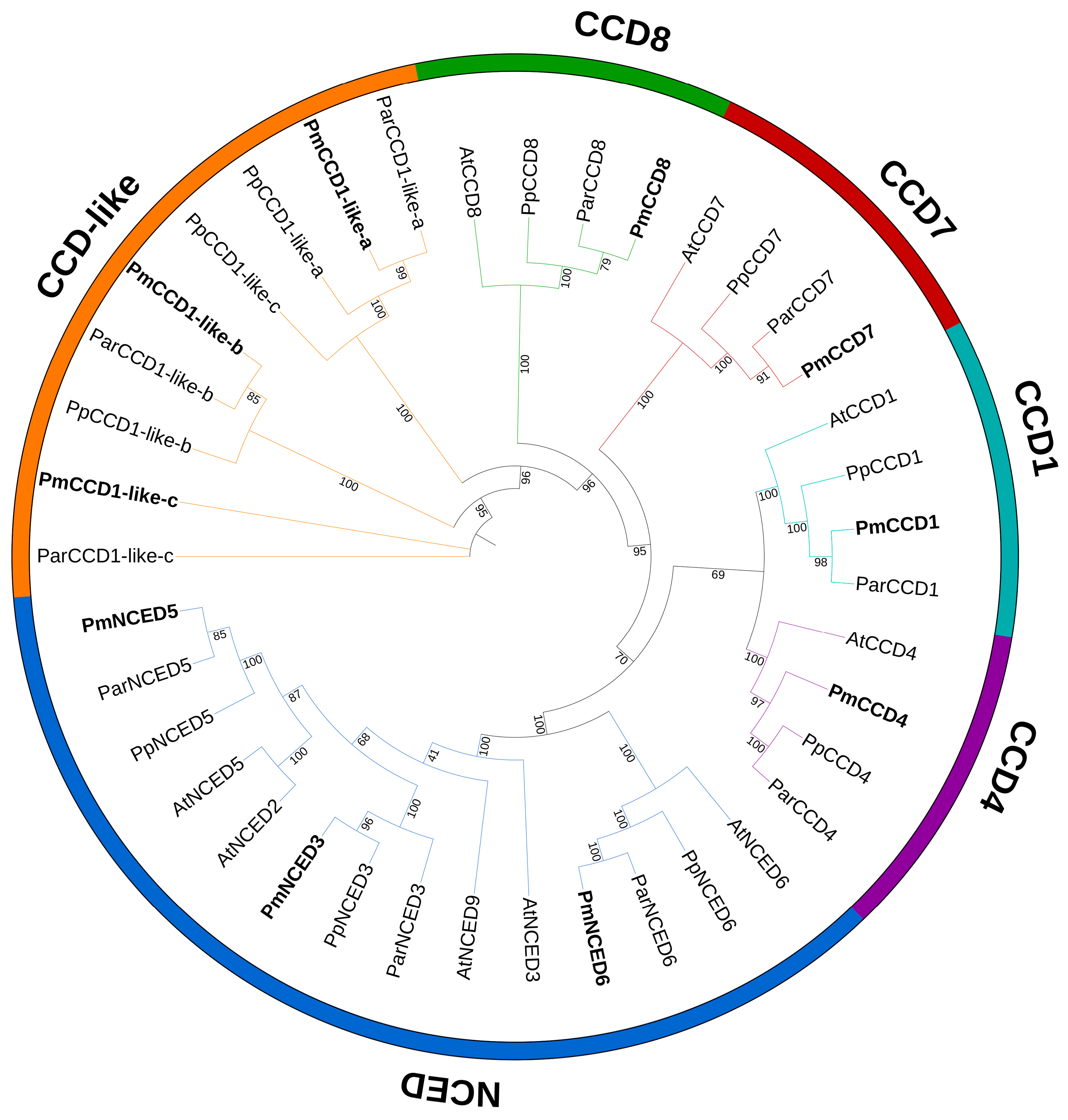
2.2. Phylogenetic Analysis and Protein Sequence Alignment

To investigate the systematic evolutionary relationships and potential functions of the PmCCDs, we generated a phylogenetic tree of CCD proteins from P. mume, Prunus armeniaca, Prunus persica and Arabidopsis using the maximum likelihood method (Figure 1). Based on the classification system of Arabidopsis, the PmCCD proteins were divided into six subfamilies: CCD1, CCD4, CCD7, CCD8, NCED and CCD-like. Obviously, there was no CCD-like subfamily in Arabidopsis, but the CCD-like subfamily has been confirmed in many species [10,40,41]. This suggested that CCD genes differed significantly in the evolutionary process of different species. The CCD members were unevenly distributed among the subfamilies. The CCD1, CCD4, CCD7 and CCD8 subfamilies each contained one member, while the NCED and CCD-like subfamilies had three members each. The number and distribution of CCD proteins in P. armeniaca and P. persica were consistent with P. mume. Additionally, we found PmCCD proteins shared a closer genetic distance with ParCCDs.
Next, protein multiple sequence alignment was performed on 10 identified CCD family members. Ten proteins possessed a conserved RPE65 domain (Figure 2C). Further analysis revealed that CCD proteins contained conserved histidine residues, essential for their enzymatic function (Figure S2). These findings indicated that PmCCD proteins had the potential to exhibit enzymatic activity.

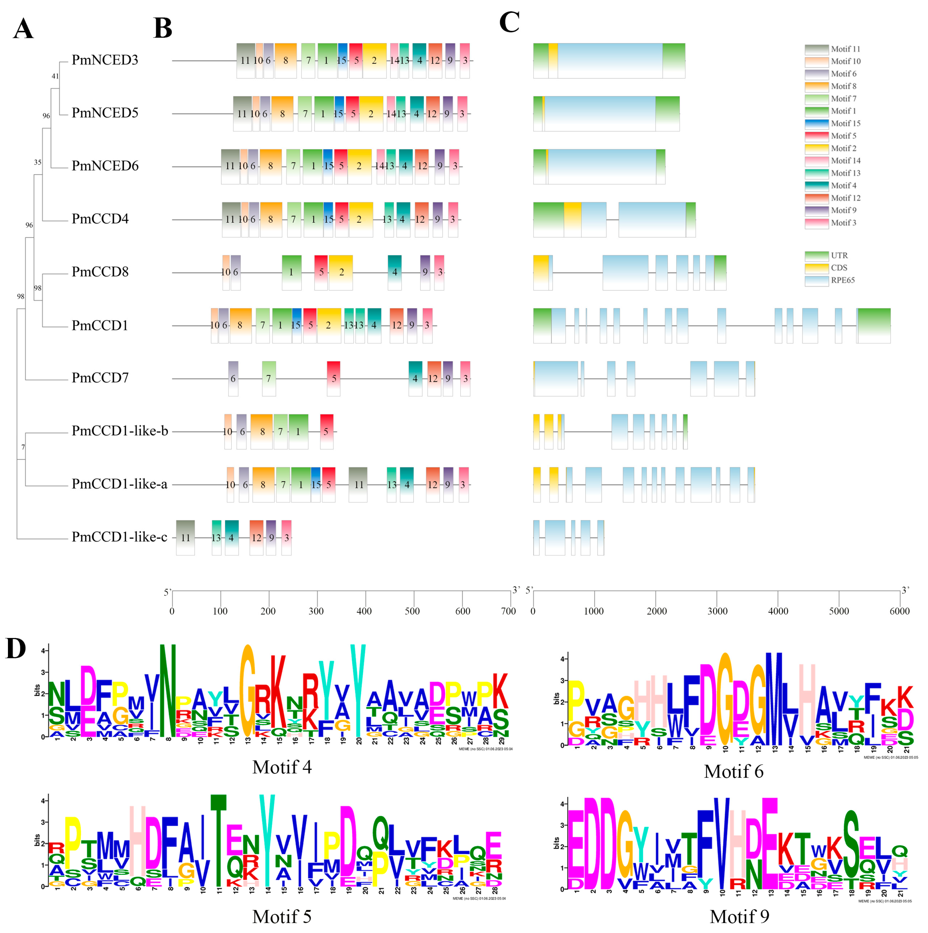
2.3. Gene Structure and Conserved Motif

To gain further search structural characteristics of PmCCD genes, the exon and intron patterns were analyzed by using the phylogenetic tree (Figure 2A,C). The PmCCD family showed considerable variation in gene structure, with exon numbers ranging from 1 to 14 and intron numbers ranging from 0 to 13. Among them, PmCCD1 contained the most exons and introns. On the other hand, the NCED subfamily lacked introns, while the PmCCD4 subfamily had a relatively brief structure with one intron and two exons. Further analysis revealed three members; specifically, PmCCD7, PmCCD1-like-a and PmCCD1-like-c were short of the untranslated region (UTR).
Then, using the MEME website to predict the motifs of PmCCD proteins, a total of 15 motifs were obtained. Figure 2B illustrates that the quantity of motifs in the PmCCD family changed from 6 to 15. Interestingly, although the NCED subfamily had a relatively simple gene structure, it contained all 15 motifs. The number of motifs differed greatly in the PmCCD-like subfamily members. Concretely, PmCCD1-like-b and PmCCD1-like-c had only 6 motifs, while PmCCD1-like-a contained 13 motifs. Except for PmCCD1-like-c, all PmCCD proteins contained 4, 5, 6 and 9 motifs, demonstrating significant conservation across these four motifs. The motif diagrams of the four motif elements illustrated that each motif comprised fully conserved sites, and these motifs might be related to their common function (Figure 2D). Meanwhile, the motif composition further supported the phylogenetic tree branch, indicating that the PmCCD4 protein was most closely related to PmNCED proteins, while PmCCD7 and PmCCD8 proteins had the most distant relationship with PmNCED.

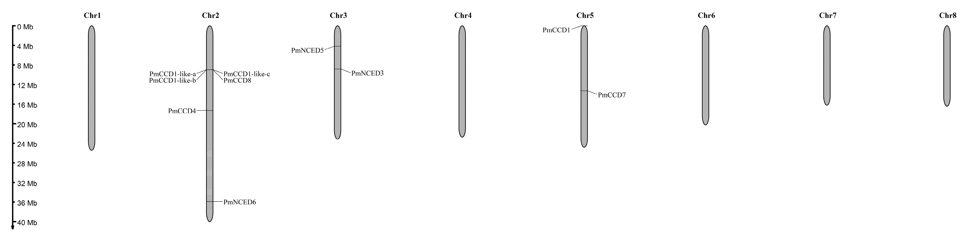
2.4. Chromosomal Distribution and Collinearity Analysis

According to the location information of the PmCCD family genes on the chromosome, a gene distribution map was generated with MG2C software (Figure 3). The results indicated that all 10 PmCCD genes were located on the chromosome and clustered on three chromosomes: chr2, chr3 and chr5. The chromosomal distribution of PmCCD genes was uneven, with six PmCCD genes (60%) located on chromosome 2. And chromosome 3 and chromosome 5 each included two PmCCD genes.
Intraspecific collinearity analysis was performed on the 10 PmCCD genes, and collinearity was absent. Neither tandem duplication nor segmental duplication made a contribution to the expansion of the PmCCD gene family. To explore the evolutionary relationship between the PmCCD genes and other Prunus plants, we constructed a collinearity map for P. mume, P. armeniaca and P. persica (Figure 4). As depicted in the map, PmCCD genes formed eight or nine collinear gene pairs with P. armeniaca or P. persica. Moreover, the Ka/Ks ratios between the collinear CCD gene pairs were performed to investigate the adaptive evolutionary relationships among three species (Table S2). The results revealed that the Ka/Ks values between P. mume and P. armeniaca varied from 0.1 to 0.8, while those between P. mume and P. persica varied from 0.1 to 0.7. All Ka/Ks ratios were <1, indicating that genes had undergone varying degrees of purifying selection. Next, to estimate the divergence time of orthologous gene pairs, Ks values were employed. The divergence time between P. mume and P. armeniaca started at 0.47, increased to 102.47 Mya and concentrated at 0.47–1.62 Mya. In P. mume and P. persica, it began at 0.86 and increased to 44.55 Mya, and 1.31–2.44 Mya occurred in most collinear genes. This suggested the CCD orthologous genes might diverge earlier in P. mume and P. armeniaca.

2.5. Promoter Cis-Acting Element Analysis

To predict the potential regulatory mechanisms of PmCCD genes, an examination of the cis-acting elements in the promoter region was performed (Figure 5). Based on their functional relevance, the promoter regions were categorized. The results displayed that the PmCCD gene promoter contained light-responsive, hormone-responsive, biotic and abiotic stress and plant growth and development elements. Environmental factors and hormonal signals might exert complex regulatory effects on the expression of the PmCCD genes. There were 11 types of light-responsive elements presented in the PmCCD genes, with PmCCD8 having the highest number (9 types). Furthermore, PmCCD1 harbored the highest number of light-responsive elements with 69, while PmNCED6 exhibited the lowest number with 42. The PmCCD genes contained 10 types of hormone-responsive elements, including abscisic acid, auxin, salicylic acid, jasmonic acid and gibberellin. The ABRE (ABA response element) was the most extensively distributed among these elements, as it was present in all genes. Among these, PmNCED6 held the most ABRE elements. Notably, the promoter of PmCCDs contained 12 responsive elements to biotic and abiotic stresses, including the MYB element, MYC element, W box (WRKY binding element), LTRE (low-temperature-responsive element), etc. This suggested that transcription factors such as MYB, MYC and WRKY might bind to PmCCD genes, exerting significant regulatory functions in, for example, low-temperature, drought, salt and trauma response. Furthermore, plant growth and development are primarily focused on circadian rhythm, endosperm development and meristem development processes.

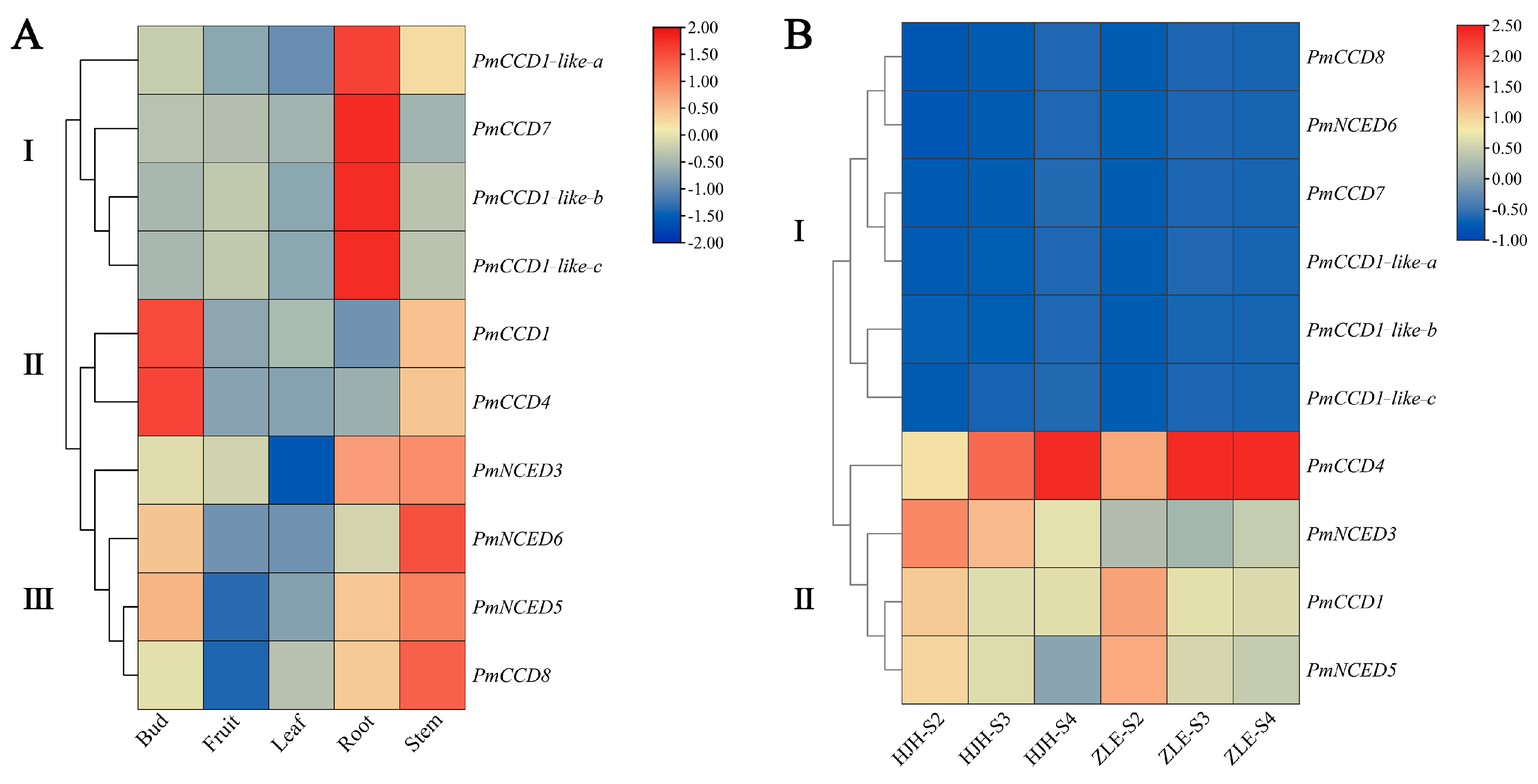
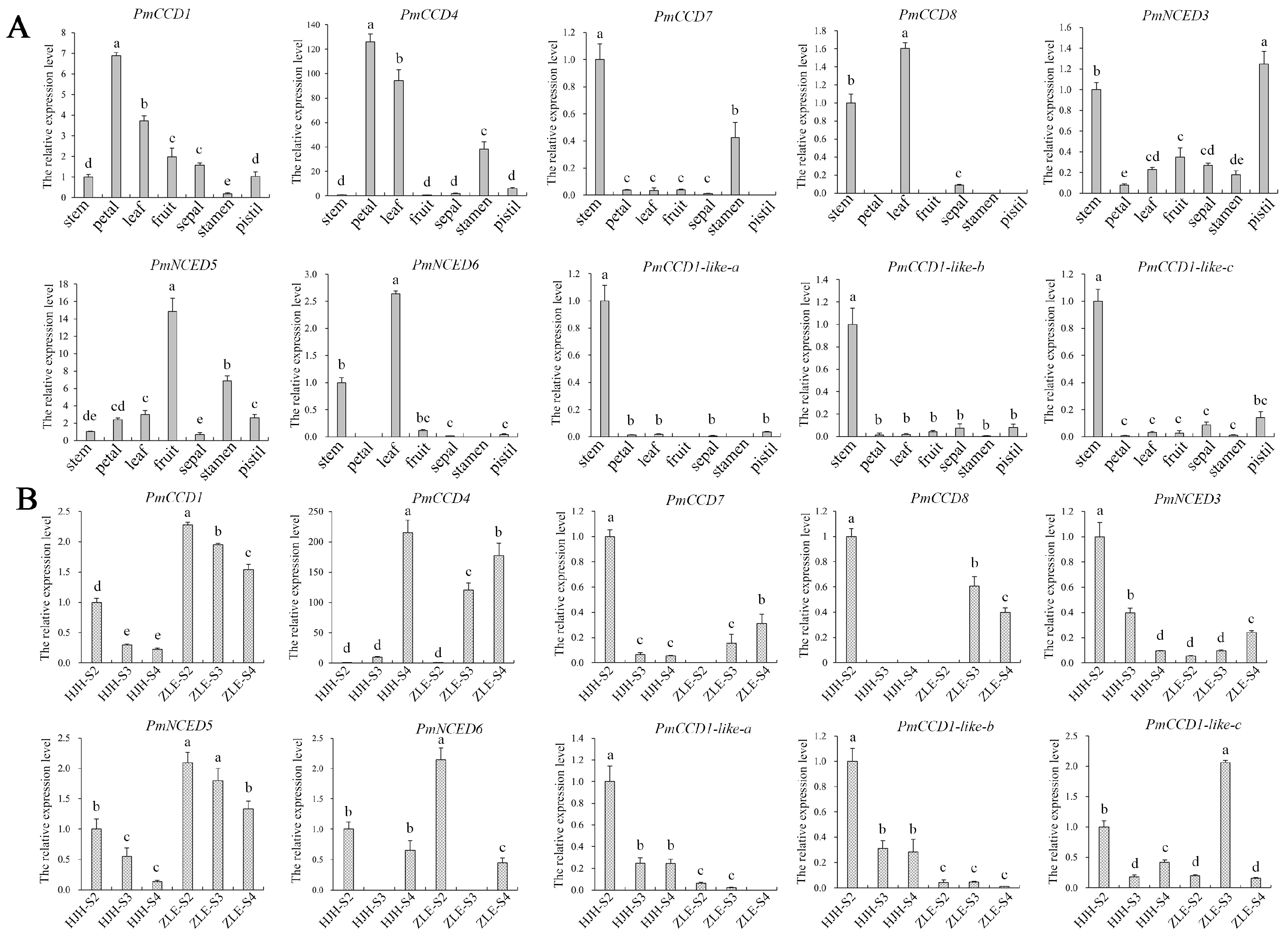
2.6. Expression Patterns of PmCCD Genes in Various Tissues and Different Varieties

To elucidate the biological function of the PmCCD genes, we analyzed the expression patterns in various tissues and varieties. The tissue-specific expression patterns of the PmCCD genes are presented in Figure 6A and Figure 7A. The heatmap shows that the tissue expression patterns of PmCCD genes were divided into three categories (Figure 6A and Table S4). The class I, II and III genes were mainly highly expressed in roots, flower buds or stems, respectively. Next, expression analysis of the stems, leaves, petals, fruits, sepals, stamens and pistils of ‘HJH’ were analyzed with qRT-PCR (Figure 7A). PmCCD1, PmCCD4, PmNCED3, PmNCED5, PmCCD1-like-b and PmCCD1-like-c were expressed in all tissues. We predicted the extensive involvement of these genes in plant growth and development. PmCCD1 and PmCCD4 shared a similar expression pattern, exhibiting high expression levels in petals, while PmCCD4 also displayed significantly high expression in leaves. PmCCD7 was highly expressed in stems and pistils, whereas PmCCD8 displayed high expression levels in stems and leaves. PmNCED3, PmNCED5 and PmNCED6 belonged to the NCED subfamily and were highly expressed in stamens, fruits and leaves, respectively. This indicated that genes within the same family gradually acquired different functions as genes evolved. Members of the PmCCD1-like subfamily exhibited stem-specific expression, with low expression levels in other tissues. PmCCD1-like genes probably had a specialized function in the stem.
The expression analysis of the PmCCD genes was carried out during the flowering process in P. mume ‘HJH’ with yellow flowers and P. mume ‘ZLE’ with white flowers using transcriptome sequencing (Figure 6B and Table S3). The results indicated significant variation in the expression of the PmCCD genes across different varieties and flowering stages. Two distinct groups were formed based on the expression patterns of the PmCCD genes. Group I comprised the genes PmNCED6, PmCCD7, PmCCD8, PmCCD1-like-a, PmCCD1-like-b and PmCCD1-like-c. They exhibited negligible expression levels during the flowering stages in two varieties, consistent with the tissue specificity. The finding implied that group I genes were not involved in forming yellow flowers in P. mume. In addition, we observed distinct expression patterns in the NCED subfamily. PmNCED3 and PmNCED5 belonged to group II. PmNCED3 was highly expressed in ‘HJH’-S2, while PmNCED5 was highest in ‘ZLE’-S2. In group II, PmCCD1 and PmCCD4 were expressed at a higher level in ‘ZLE’ compared to ‘HJH’. We speculated that the low expression of PmCCD1 and PmCCD4 in ‘HJH’ caused the accumulation of carotenoids in the flowers, resulting in the appearance of yellow-flowered P. mume. Furthermore, PmCCD4 showed a 2-fold and 22.5-fold higher expression in ‘ZLE’ than in ‘HJH’ during the S2 and S3 stages, respectively. To verify the expression levels of PmCCDs in different flowering stages of ‘HJH’ and ‘ZLE’, we carried out qRT-PCR analysis. The gene expression trends were consistent with the transcriptome data analysis (Figure 7B). Earlier studies have indicated that the variation in yellow flower color in chrysanthemums and petunias can be attributed to the cleavage of carotenoids facilitated by CCD genes [18,42]. We speculated that PmCCD1 and PmCCD4 might play a critical role, especially PmCCD4, in the formation of yellow flowers in ‘HJH’.

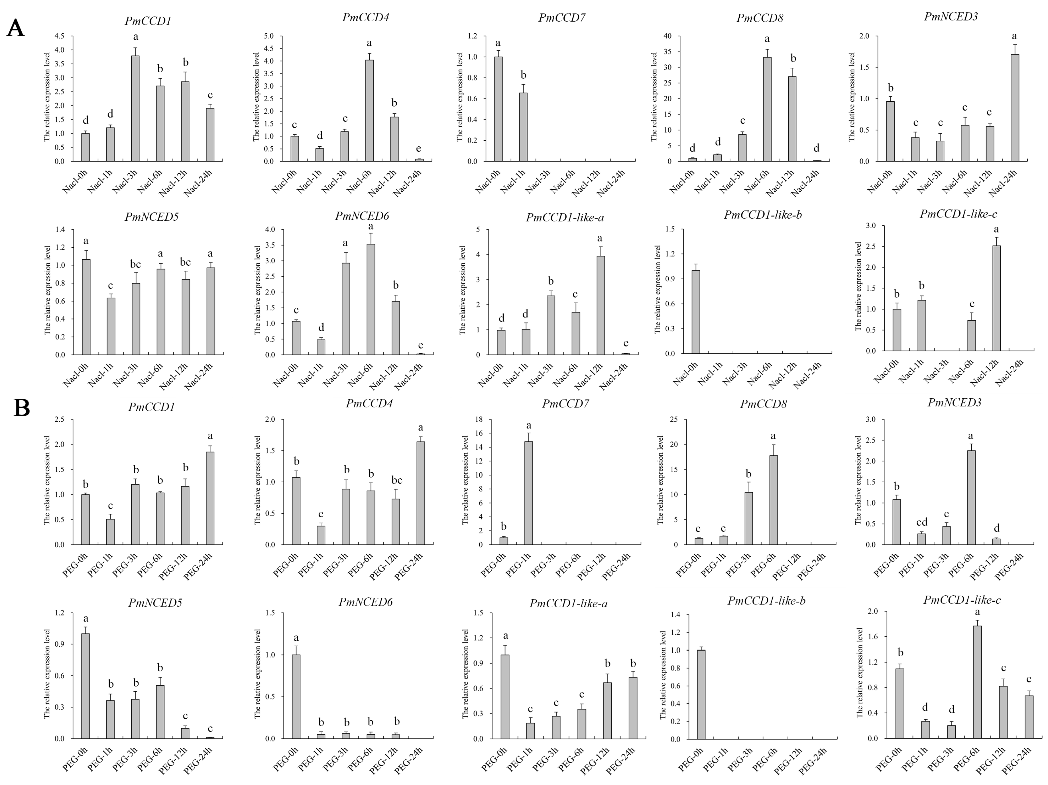
2.7. Expression Analysis of PmCCD Genes in Abiotic Stress Treatments

To investigate the ability of PmCCD genes in stress response, we conducted qRT-PCR analysis to assess the gene expression level under NaCl or PEG4000 treatments. Figure 8 displays that NaCl or PEG stress affected PmCCD gene expression levels. Under NaCl treatment, the expression of PmCCD1, PmCCD4, PmCCD8 and PmNCED6 increased and then decreased. In contrast, PmCCD1 peaked at 3 h, while PmCCD4, PmCCD8 and PmNCED6 were most abundant at 6 h. PmNCED3 and PmNCED5, belonging to the NCED subfamily, had down-expression in the early stage of NaCl treatment, and at 24 h, PmNCED3 rapidly increased, while the expression of PmNCED5 began to recover. The expression patterns of PmCCD1-like-a and PmCCD1-like-c in the CCD-like subfamily were similar, with the highest accumulation at 12 h, and then decreased rapidly. Only PmCCD7 expressed a downward trend, without expression at 3 h to 24 h (Figure 8A). Under PEG treatment, PmCCD genes showed a new expression pattern. PmCCD1 and PmCCD4 possessed high expression at 24 h, indicating that PmCCD1 and PmCCD4 might have the potential to resist PEG stress persistently. The expression accumulation of PmCCD8, PmNCED3 and PmCCD1-like-c was the highest at 6 h. However, the expression levels of PmNCED5 and PmNCED6 decreased during 1–24 h. PmCCD7 was only expressed at 1 h, which was 14.8 times higher than that at 0 h, and then the expression decreased rapidly at 3–24 h (Figure 8B). PmCCD1-like-b was expressed in neither NaCl nor PEG treatment. In conclusion, PmCCD genes might participate in NaCl and PEG stress responses, and each PmCCD gene had a unique expression pattern.

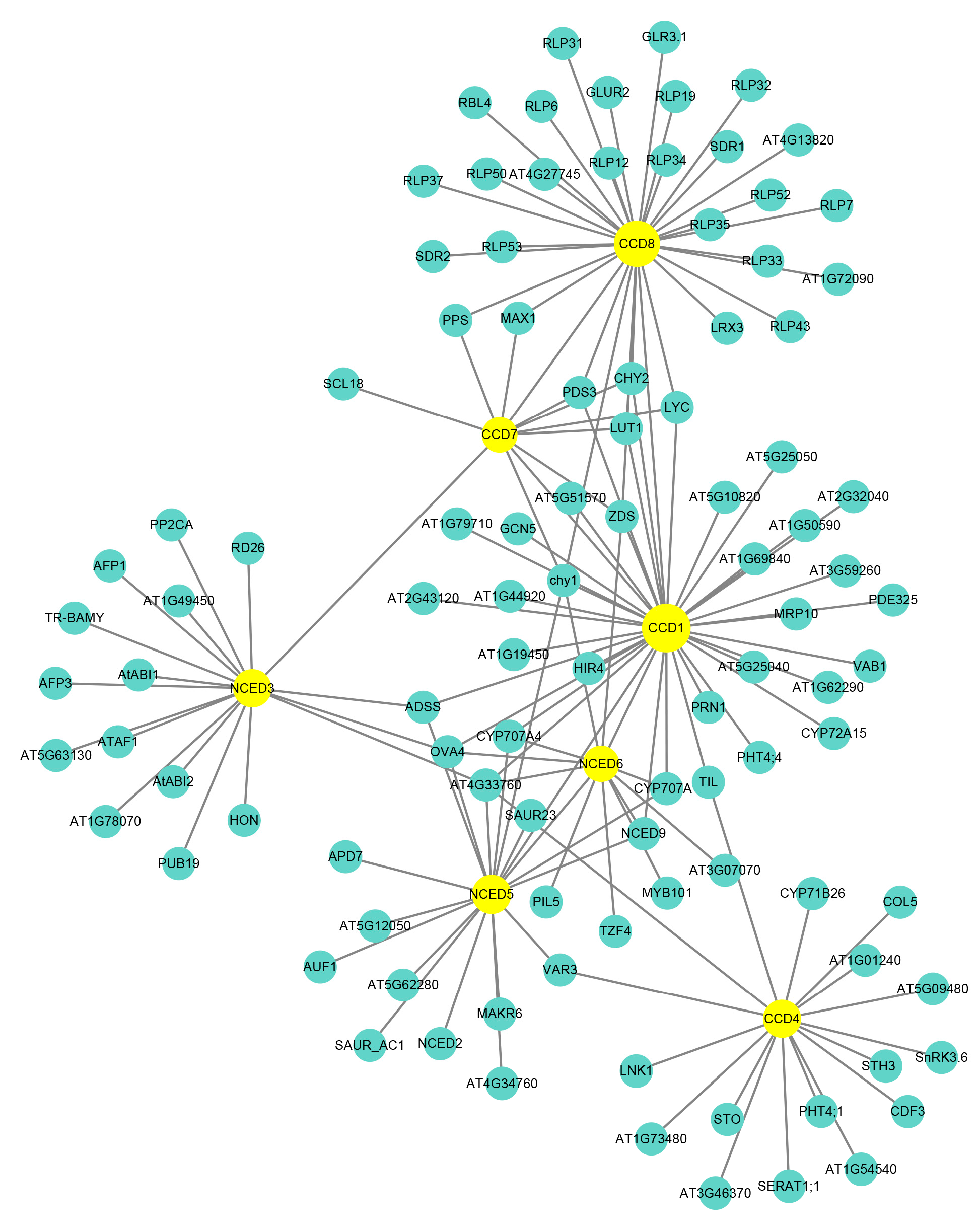
2.8. Protein Interaction Network Analysis

Interaction network analysis could uncover the relationship between proteins and predicted protein function. Using the AraNet V2 website, we constructed an interaction network with a homologous protein from Arabidopsis. Figure 9 displays seven PmCCD proteins with orthologous proteins in Arabidopsis, and CCD family members interacted with each other: CCD1 and CCD8, CCD7 and NCED3, CCD7 and CCD1, CCD7 and CCD8, NCED5 and NCED6, NCED5 and CCD1 and NCED6 and CCD1. This suggested CCD members might participate in the same signal transduction and biological processes by forming dimers or polymers through protein interactions.
The interaction network diagram showed that CCDs could interact with proteins, such as MYB101, RD26, ATAF1, CHY1, ZOS, SnRK3.6, VAR3 and SAT, suggesting CCDs might function through interactions with other proteins. Further analysis revealed that CCDs had 142 interacting proteins, of which CCD1 protein had the most interacting proteins with 33, while CCD7 protein had only 12 interacting proteins. The interaction network displayed that CCDs were involved in essential roles in the biosynthesis and signal transduction of hormones such as ABA, GA and auxin. CCDs functioned in various plant growth and development processes, such as embryo and stamen development. They also responded to biotic stresses like pathogen invasion and abiotic stresses, including drought, low temperature, high temperature, injury and high salinity. CCDs were involved in metabolic processes, such as carotenoid metabolism and chlorophyll biosynthesis. PmCCD family proteins might have similar functionality to AtCCD. Overall, the interaction network served as a valuable reference for investigating the potential functions of PmCCDs.

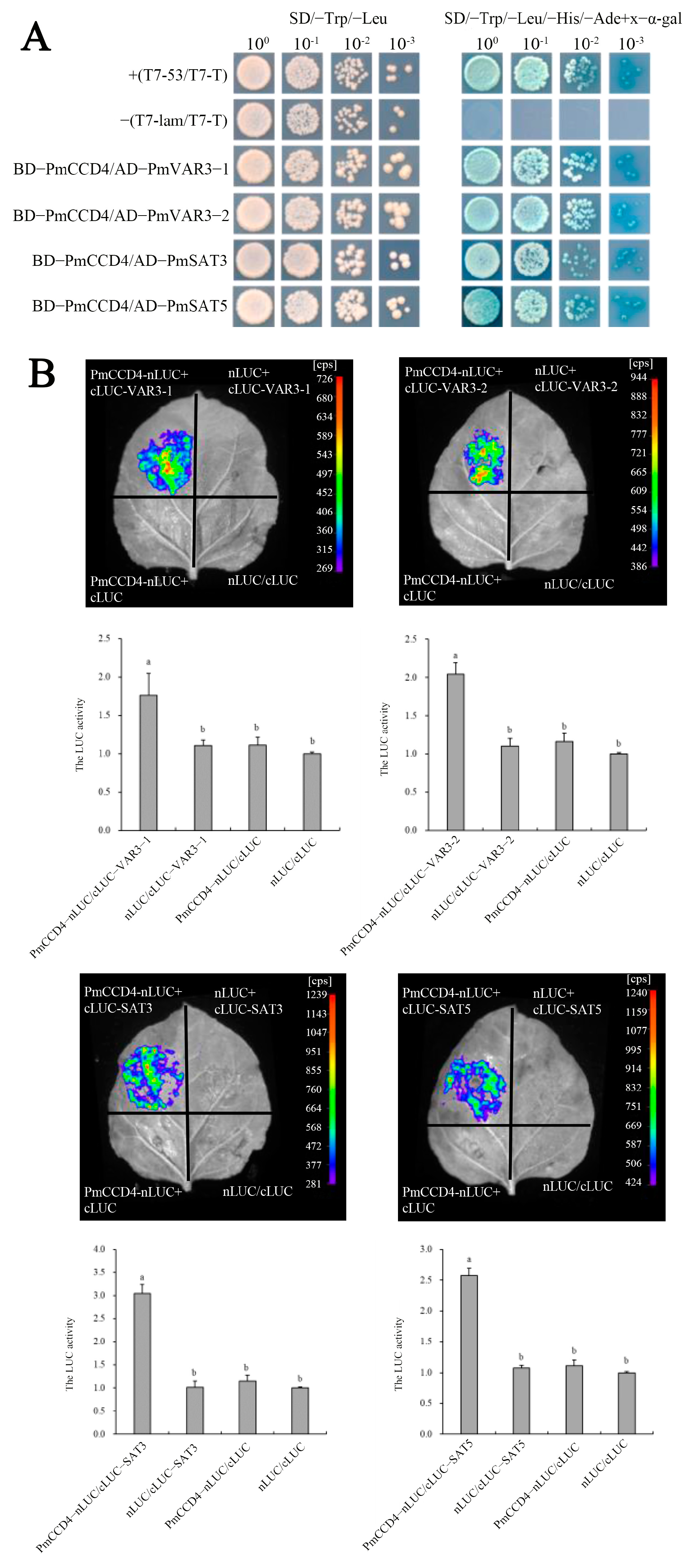
2.9. Interaction of PmCCD4 with Other Proeins

We utilized the Uniprot website to further predict the interaction between PmCCD4 and other proteins. The website showed that CCD4 could interact with VAR3 (variegated 3) and SAT (serine acetyltransferase) to perform its function in Arabidopsis. In Figure 9, the interaction network also predicted interactions between PmCCD4 and PmVAR3 or PmSAT. Therefore, the interaction between PmCCD4 and PmVAR3-1/2 or PmSAT3/5 was validated using yeast two-hybrid assays and luciferase complementation experiments. The yeast two-hybrid assays showed that the combinations of pGBKT7-PmCCD4 and pGADT7-PmVAR3-1/2, as well as pGBKT7-PmCCD4 and pGADT7- PmSAT3/5, were able to grow and turn blue on an SD/-Leu/-Trp/-His/-Ade/x-α-gal solid medium (Figure 10A). This indicated that PmCCD4 could interact with PmVAR3-1/2 and PmSAT3/5 in vitro. Next, the detection of a strong chemiluminescent signal in the luciferase complementation test group confirmed the interaction of PmCCD4 with Pm VAR3-1/2 or PmSAT3/5 separately. Moreover, the high luciferase activity also supported this conclusion (Figure 10B).

3. Discussion

CCD, a small gene family, was involved in the forming of aromatic compounds and plant hormones from catalytic carotenoids, contributing to the fragrance, color, abscisic acid and strigolactone formation in plants. Currently, the CCD family genes have been identified in various plants, such as 9 in Arabidopsis [43], 19 in tobacco [44], 7 in tomato [9], 21 in apple [10], 30 in rapeseed [45] and 12 in strawberry [46]. In this research, we identified 10 PmCCD genes through the P. mume genome. Multiple sequence alignment revealed that all the proteins exhibited the conserved RPE65 domain. Additionally, most of the PmCCD genes contained conserved active sites with His residues. The conserved His residues in CCD proteins determined their enzymatic activity [47]. We speculated that the majority of PmCCD proteins possessed catalytic activity. The 10 PmCCD genes were distributed unevenly among chromosomes 2, 3 and 5. The CCD gene family might have specific evolutionary patterns across different species. Subcellular localization predictions suggested PmCCD1 and PmCCD1-like-c were likely to function in the cytoplasm, indicating their potential exclusion from chlorophyll photosynthesis. CCD4 was located in plastids to cleave carotenoids. We hypothesized that PmCCD4 exhibited similar catalytic activity. The localizations of PmNCED3, PmNCED5 and PmNCED6 in the chloroplast were consistent with previous studies [48]. In Arabidopsis, the collaboration of AtCCD7 and AtCCD8 converted β-carotene into the caprolactone belonging to strigolactone precursor, subsequently enhancing plant growth and development [49]. The chloroplast localizations of PmCCD7 and PmCCD8 were intimately linked to the synthesis pathway of strigolactone, which was intricately involved in the cleavage of β-carotene in plastids.
The CCD gene family in the majority of plants was classified into two major subfamilies: CCD and NCED. In Arabidopsis, the CCD subfamily contained four members: CCD1, CCD4, CCD7 and CCD8. The NCED subfamily included five members, namely NCED2, NCED3, NCED5, NCED6 and NCED9. Furthermore, a novel group called CCD-like (CCDL) have been identified in many species [8,44]. Homologs of CCDL genes were absent in Arabidopsis. According to the phylogenetic tree analysis, we characterized and categorized the PmCCD gene family into CCD1, CCD4, CCD7, CCD8, NCED (NCED3, NCED5 and NCED6) and CCD-like subfamilies. The number and grouping of CCD family members in P. armeniaca and P. persica were consistent with PmCCD, and PmCCD proteins exhibited a closer genetic distance to ParCCD. Compared to Arabidopsis, the PmCCD gene family lacked NCED2 and NCED9, which suggested a gradual functional replacement during the evolutionary process of PmNCED genes. However, the PmCCD gene family possessed an additional CCDL subfamily. In addition, the NCED subfamily was most closely related to the CCD1 and CCD4 groups, while their relationship with the CCD7 and CCD8 groups was the furthest.
Genomic structure and motif analysis revealed considerable variations in exon/intron patterns and conserved motifs among PmCCD members across different subfamilies. Notably, within the same subfamily, a general similarity was observed in terms of the quantity and distribution of exons/introns and conserved motifs. All members of the PmNCED subfamily lacked introns, aligning with previous reports in plants [7,8]. Furthermore, PmNCED contained the highest number of conserved motifs, which were more conserved than other groups. This phenomenon was widespread in plants [50]. Based on motif analysis and sequence alignment, except for PmCCD1-like-b and PmCCD1-like-c, PmCCD proteins possessed the four conserved histidine residues required for enzymatic activity. Currently, it remains unclear whether the absence of conserved histidine residues in PmCCD1-like-b and PmCCD1-like-c would affect enzymatic activity and protein functionality. Therefore, elucidating the active sites will contribute to exploring the functionality of CCDL genes, potentially leading to new insights into the CCD gene family.
Gene duplication events have exerted a significant impact on the evolutionary trajectory of gene families [51]. These duplications not only expanded the gene family but also played a pivotal role in gene diversification, potentially leading to significant morphological changes in plants. Our intraspecific collinearity analysis of 10 PmCCD genes revealed the absence of intraspecific collinearity among PmCCD genes. This suggested that gene duplication events might not be the primary driver of PmCCD gene expansion. Interspecific collinearity analysis serves as a valuable approach for exploring the evolutionary dynamics of gene families across different species. We conducted interspecific collinearity analysis between P. mume and two other Prunus species (P. armeniaca and P. persica) and had homologous gene pairs. According to the Ka/Ks values, the CCD orthologous pairs showed an earlier divergence in P. mume and P. armeniaca. This observation provided additional evidence supporting a closer phylogenetic relationship between them.
The tissue-specific expression patterns of genes are closely associated with their functional characteristics. To investigate the expression patterns of the PmCCDs in various tissues of P. mume, we employed qRT-PCR technology in the seven tissues (stem, leaf, petal, fruit, sepal, stamen and pistil). The PmCCD genes exhibited distinct tissue specificity. Among them, PmCCD1 and PmCCD4 were expressed highly in petals. Research showed that CCD1 and CCD4 primarily participated in the form of volatile compounds and flower color through cleavage carotenoids [52,53,54,55]. We speculated that PmCCD1 and PmCCD4 might be involved in the formation of flower color and fragrance in the ‘HJH’. CCD7 and CCD8 were involved in the synthesis of strigolactone which was primarily synthesized in the roots [56]. PmCCD7 was highly expressed in the root, consistent with AtCCD7 [57]. This implied that PmCCD7 might have similar functionality to AtCCD7. In addition, PmCCD8 exhibited higher expression levels in stems and leaves, suggesting that PmCCD8 primarily functions in stems and leaves. PmNCED3, PmNCED5 and PmNCED6 were highly expressed in stamens, fruits and leaves, respectively. This indicated that genes within the same family gradually acquired different functions during the evolutionary process. Members of the PmCCD1-like subfamily had stem-specific expression while showing low expression in other tissues. The PmCCD1-like genes were likely to have unique functions in stems.
CCD1 and CCD4 participate in the degradation of carotenoids, providing unique colors, flavors and aromas to fruits and flowers. Previous studies have shown that CCD1 regulates the volatile aroma compound β-ionone production in petunia, tomato and L. chinense [14,17,58]. The variation in petal color among different chrysanthemum mutants was attributed to differential expression levels of the CmCCD4 gene, resulting in variations in carotenoid content [59]. Inhibiting the expression of the CmCCD4 gene through RNAi could transform white chrysanthemum flowers into yellow flowers [18]. In Brassica species, mutation of the CCD4 genes led to the formation of yellow flowers [20]. Similarly, in O. fragrans and Rhododendron japonicum, the expression level of the CCD4 gene determined the carotenoid content, thereby leading to differences in flower color [60,61]. The CCD4 gene changed Lilium brownie petals from yellow to white one day after anthesis [62]. Compared to other tissues, PmCCD1 and PmCCD4 were expressed highly in petals. Next, we analyzed the expression of PmCCD genes during the flowering process of the yellow-flowered ‘HJH’ and white-flowered ‘ZLE’. The results revealed that the expression levels of PmCCD1 and PmCCD4 in the yellow-flowered ‘HJH’-S2 and S3 were lower than those of the white-flowered ‘ZLE’. We speculated that the low expression of PmCCD1 and PmCCD4 in ‘HJH’ caused the accumulation of carotenoids in the flowers, resulting in the appearance of yellow-flowered P. mume. Whether PmCCD1 and PmCCD4 are related to the formation of floral fragrance requires further exploration.
In plants, CCD genes can participate in abiotic stress response, while the involvement of PmCCDs in abiotic stress has not been studied so far. The analysis of the PmCCDs promoter showed that the PmCCDs promoter region contained several cis-acting elements related to stress, such as W-box, MYB, LTR, MYC, etc. Therefore, the expression pattern of PmCCDs treated with NaCl and PEG4000 was investigated by qRT-PCR. We found that PmCCD1 and PmCCD4 were involved in response to NaCl and PEG treatments. PmCCD7 could respond to PEG stress within 1 h and was not persistent. Additionally, PmCCD8 displayed responsiveness to NaCl stress and PEG stress. Based on the available information, PmCCD8 might have a higher potential for stress resistance than PmCCD7. However, it is essential to conduct further experiments and investigations to fully understand and validate the comparative stress resistance potential between PmCCD7 and PmCCD8. The NCED subfamily was involved in plant resistance by regulating ABA synthesis [63,64]. The biosynthesis of ABA in Arabidopsis involved the participation of AtNCED3, AtNCED5, AtNCED6 and AtNCED9 [30,65]. The promoters of PmNCED genes were enriched with ABRE motifs, indicating that the PmNCED subfamily was involved in ABA-related biological processes of P. mume. Under NaCl treatment, the expression abundance of PmNCED3 and PmNCED6 was the highest at 24 h and 6 h, respectively. However, only PmNCED3 was expressed in PEG treatment. It is worth noting that PmNCED3 may play a more significant role in osmotic stress, which requires further validation. Regarding the CCD-like group, the functional characterization of these genes is yet to be investigated. PmCCD1-like-a and PmCCD1-like-c had the potential to resist stress. Exploring the functionality of CCD-like group genes will provide a new perspective for studying the CCD gene family.
Interacting proteins might regulate each other, be closely related in function or participate in the same signaling pathway or physiological process. According to yeast two-hybrid system and luciferase complementation experiments, PmCCD4 interacted with PmVAR3 protein or PmSAT3/5 proteins. In Arabidopsis, AtVAR3 interacted with AtCCD4, and VAR3 could regulate the function of the CCD4 enzyme as a signaling molecule, thereby regulating carotenoid catabolism [66]. In P. mume, whether the binding of PmVAR3 protein with PmCCD4 affects the function of PmCCD4 to cleavage carotenoid still needs further exploration. SAT genes were involved in plant development and response to various stresses [67,68]. We speculated that PmSAT3/5 might have stronger resistance to various stresses when interacting with PmCCD4 protein. This is also a topic worthy of further study.

4. Materials and Methods

4.1. Identification of PmCCDs

The genome of P. mume was available on NCBI database [69]. Using Pfam database, we downloaded the configuration file for the REP65 domain (PF03055). The HMMER3 software was used to obtain the CCD protein of P. mume with E-value ≤ 10−5 [41]. The REP65 domains of the PmCCD proteins were determined using NCBI-CD-search.
The ExPASy (https://web.expasy.org/protparam/, accessed on 15 June 2023) was employed to seek CDS length, theoretical isoelectric points (pI), amino acid sequences (aa), instability index, molecular weights (MWs) and aliphatic index grand averages of hydropathicity (GRAVYs) of PmCCDs. The N-terminal targeting signals and subcellular localization were predicted through iPSOR (http://ipsort.hgc.jp/, accessed on 15 June 2023) and WoLF PSORT (https://wolfpsort.hgc.jp/, accessed on 16 June 2023), respectively [70]. The SOPMA website (https://npsa-prabi.ibcp.fr/cgi-bin/npsa_automat.pl?page=npsa_sopma.html, accessed on 16 June 2023) was utilized for protein secondary structure prediction [71].

4.2. Phylogenetic Tree Analysis and Protein Sequence Alignment of PmCCD Proteins

To unravel the evolution of PmCCD proteins, we constructed an evolutionary tree using CCD gene families from P. armeniaca and P. persica, as well as Arabidopsis. The genomes of P. armeniaca and P. persica were acquired from NCBI, and the sequences of AtCCD were sourced from TAIR database (https://www.arabidopsis.org/, accessed on 17 June 2023). MEGA7.0 was utilized to construct the phylogenetic tree (bootstrap = 1000) with the maximum likelihood method. The phylogenetic tree was drawn by using iTOL (https://itol.embl.de/, accessed on 17 June 2023). Multiple sequence alignment of 10 PmCCD gene family members was performed using MEGA7.0 and visualization through Muscle (https://www.ebi.ac.uk/Tools/msa/muscle/, accessed on 17 June 2023).

4.3. Gene Structure and Conservation Motif Analysis of PmCCD Sequences

The structure and conserved domain of PmCCD genes were estimated and drawn by using TBtools [72]. The motif structure of PmCCD proteins was explored through the MEME program (the motif limit was set to 15), and other default parameters were used.

4.4. Chromosome Localization and Collinearity Analysis of PmCCDs

According to the chromosome location information of PmCCD genes provided by P. mume genome database, we mapped the gene distribution with MG2C tool (http://mg2c.iask.in/mg2c_v2.1/, accessed on 17 June 2023). Intraspecific and interspecific collinearity of CCD genes (P. mume, P. armeniaca and P. persica) were analyzed and visualized using MCScanX [73]. The mutation rates of nonsynonymous replacement rate (Ka) and synonymous replacement rate (Ks) of CCD collinear gene pairs were predicted by using TBtools. PmCCD gene divergence time (T) was calculated by the following equation: T = dS/2λ × 106 Mya, λ = 1.5 × 10⁻⁸ for dicots [74].

4.5. Analysis of PmCCDs Expression Pattern

Expression patterns of PmCCDs in different varieties and various tissues: The 5-year-old P. mume ‘Huangjinhe’ (‘HJH’) with yellow flowers and 5-year-old P. mume ‘Zaolve’ (‘ZLE’) with white flowers were planted in the natural environment of Chongqing, China. The ‘HJH’ is a rare yellow flower variety in P. mume. However, the yellow color of the ‘HJH’ is not stable. As the flowers blossom, the yellow color gradually fades away. During the big bud stage (Stage2, S2), the petals were yellow. In the half-bloom stage (Stage3, S3), the outer edge of the petals turned white. The petals were almost white during the full-bloom stage (Stage4, S4). To explore the role of PmCCDs in P. mume with yellow flowers, the petals of S2, S3 and S4 were taken from ‘HJH’ and ‘ZLE’ for transcriptome sequencing (NCBI accession no. PRJNA854285). Seven tissues, including stems, leaves, petals, fruits, sepals, stamens and pistils were collected from the ‘HJH’. Meanwhile, TBtools was used to plot expression heatmaps of 10 PmCCDs at flower buds, fruits, leaves, roots and stems of P. mume wild species (GEO No. GSE40162), as well as three flowering stages of ‘HJH’ and ‘ZLE’.
Expression patterns of PmCCDs under abiotic stress treatments: The annual stems (20 cm) of ‘ZLE’ with the same growth state were obtained in April. The annual stems were treated by inserting them into 160 mmol/L NaCl and 20%PEG4000 solution, separately. Samples were taken at 0 h, 1 h, 3 h, 6 h, 12 h and 24 h, respectively. Using the untreated annual stems (0 h) as a blank control, qRT-PCR was performed on the treated annual stems. The plant samples were subjected to identical air humidity (65–70%), light duration (16 h/8 h) and light intensity (150 μmol·m−2·s−1). The samples were stored at −80 °C to maintain their integrity and stability until RNA extraction, with 3 biological replicates.

4.6. RNA Extraction and qRT-PCR Analysis

Total RNA from various tissues and varieties of ‘HJH’ was isolated according to the user manual of the RNA extraction kit (Takara, Beijing, China). The synthesis of first strand cDNA by RNA (1 µg) was reversed with PrimeScriptTMRT Reagent Kit with gDNA Eraser reagent (Takara, Beijing, China). TB Green II Premix Ex Taq (Takara, Beijing, China) was employed to perform a qRT-PCR system (10 µL), including 2 µL template cDNA. The relative expression levels were calculated by using 2−ΔΔCt method, with the reference gene being the protein phosphatase 2A (PP2A) gene. Ten specific primers for PmCCDs are shown in Table S5. A minimum of three replicates were performed for each qRT-PCR assay.

4.7. Analysis of PmCCD Promoter Element and Protein Interaction Network

From the genome of P. mume, we searched for the PmCCD sequences and extracted the 2000 bp upstream of the TSS (Transcription Start Site) as the promoter. The promoter cis-acting regulatory elements were operated using PLACE (https://www.dna.affrc.go.jp/PLACE/?action=newplace, accessed on 17 June 2023) [75]. The AraNet V2 tool was used to construct protein interaction networks of PmCCD proteins [76]. The visualization of the protein interaction network was performed using Cytoscape and STRING software (http://string-db.org/, accessed on 17 June 2023) [77].

4.8. Yeast Two-Hybrid System

PmCCD4 was constructed into pGBKT7 vector, and PmVAR3-1/2 and PmSAT3/5 genes were constructed into pGADT7 vector. Plasmid pGBKT7-PmCCD4 and pGBKT7 were transferred into Y2HGold yeast strains, coated on SD-trp/X-α-gal defect screening plates and cultured at 30 °C for 3–5 days for yeast self-activation detection. Then, the pGBKT7-PmCCD4 and pGADT7-PmVAR3-1/2 or pGADT7-PmSAT3/5 genes were co-transferred to Y2HGold strain and coated on SD-trp-his and SD-trp-leu-his-ade/X-α-gal defect screening medium, respectively. The proteins were observed and determined for interactions. Specific primers are listed in Table S6.

4.9. Luciferase Complementation Experiment

PmCCD4 was linked to the nLUC vector, and the PmVAR3-1/2 and PmSAT3/5 genes were linked to the cLUC vector. The recombinant vectors were transformed into an Agrobacterium strain of GV3101 (pSoup-p19) and expressed in N. benthamiana. We applied the prepared 0.32 mg/mL D-luciferin potassium salt solution to the leaves. Then, we observed and took photos using the molecular imaging system LB983 NightOwl II (Berthold Technologies, Baden-Württemberg, Germany).

5. Conclusions

In this study, 10 non-redundant PmCCD genes were identified in P. mume. The characteristics of PmCCD genes showed diversity. All PmCCD proteins had the RPE65 conserved domain. In addition, most CCD proteins contained four conserved histidine sites. Based on the phylogenetic tree, PmCCD proteins were divided into six subfamilies: CCD1, CCD4, CCD7, CCD8, NCED and CCD-like. PmCCD genes exhibited interspecific collinearity with P. armeniaca and P. persica. The analysis of promoter and protein interaction indicated that environmental factors and hormone signals had complex regulation of PmCCD genes. The qRT-PCR results showed that PmCCD genes had obvious tissues and varieties. Compared with white-flowered ‘ZLE’, PmCCD1 and PmCCD4 genes were low-expressed in ‘HJH’ with yellow petals, which suggested PmCCD1 and PmCCD4 might be related to the formation of yellow flowers in P. mume. Nine PmCCD genes could respond to NaCl or PEG treatment. These genes might play a crucial role in salt and drought resistance in P. mume. Moreover, PmVAR3 and PmSAT3/5 were found to interact with PmCCD4 protein in yeast and tobacco leaf cells. These results will establish valuable bioinformatics foundations for further exploring the functions of PmCCD genes.

Supplementary Materials

The following supporting information can be downloaded at: https://www.mdpi.com/article/10.3390/ijms241813950/s1.

Author Contributions

Q.Z. conceived and designed the experiments. F.B. revised the manuscript. A.D. performed the data analysis and drafted the manuscript. W.C. and T.C. contributed reagents/materials/analysis tools. All authors have read and agreed to the published version of the manuscript.

Funding

This work was supported by the National Natural Science Foundation of China (31800596).

Acknowledgments

We greatly appreciate the editors and reviewers for handling our manuscript and providing critical suggestions.

Conflicts of Interest

The authors declare there is no conflict of interest.

References

  1. Frank, H.A.; Cogde, I.P.R.J. Carotenoids in photosynthesis. Photochem. Photobiol. 1996, 3, 257–264. [Google Scholar] [CrossRef]
  2. Qin, X.; Zeevaart, J.A. The 9-cis-epoxycarotenoid cleavage reaction is the key regulatory step of abscisic acid biosynthesis in water-stressed bean. Proc. Natl. Acad. Sci. USA 1999, 96, 15354–15361. [Google Scholar] [CrossRef] [PubMed]
  3. Jia, K.; Baz, L.; Al-Babili, S. From carotenoids to strigolactones. J. Exp. Bot. 2018, 69, 2189–2204. [Google Scholar] [CrossRef] [PubMed]
  4. Nisar, N.; Li, L.; Lu, S.; Khin, N.C.; Pogson, B.J. Carotenoid Metabolism in Plants. Mol. Plant 2015, 8, 68–82. [Google Scholar] [CrossRef]
  5. Walter, M.H.; Strack, D. ChemInform Abstract: Carotenoids and Their Cleavage Products: Biosynthesis and Functions. ChemInform 2011, 28, 663–692. [Google Scholar] [CrossRef]
  6. Tan, B.C.; Schwartz, S.H.; Zeevaart, J.A.; McCarty, D.R. Genetic Control of Abscisic Acid Biosynthesis in Maize. Proc. Natl. Acad. Sci. USA 1997, 94, 12235–12240. [Google Scholar] [CrossRef] [PubMed]
  7. Tan, B.; Joseph, L.M.; Deng, W.; Liu, L.; Li, Q.; Cline, K.; McCarty, D.R. Molecular characterization of the Arabidopsis 9-cis-epoxycarotenoid dioxygenase gene family. Plant J. 2003, 35, 44–56. [Google Scholar] [CrossRef]
  8. Vallabhaneni, R.; Bradbury, L.M.T.; Wurtzel, E.T. The carotenoid dioxygenase gene family in maize, sorghum, and rice. Arch. Biochem. Biophys. 2010, 504, 104–111. [Google Scholar] [CrossRef]
  9. Wei, Y.; Wan, H.; Wu, Z.; Wang, R.; Ruan, M.; Ye, Q.; Li, Z.; Zhou, G.; Yao, Z.; Yang, Y. A Comprehensive Analysis of Carotenoid Cleavage Dioxygenases Genes in Solanum L. Plant Mol. Biol. Rep. 2016, 34, 512–523. [Google Scholar] [CrossRef]
  10. Chen, H.; Zuo, X.; Shao, H.; Fan, S.; Ma, J.; Zhang, D.; Zhao, C.; Yan, X.; Liu, X.; Han, M. Genome-wide analysis of carotenoid cleavage oxygenase genes and their responses to various phytohormones and abiotic stresses in apple (Malus domestica). Plant Physiol. Biochem. 2018, 123, 81–93. [Google Scholar] [CrossRef]
  11. Schmidt, H.; Kurtzer, R.; Eisenreich, W.; Schwab, W. The Carotenase AtCCD1 from Arabidopsis thaliana Is a Dioxygenase. J. Biol. Chem. 2006, 281, 9845–9851. [Google Scholar] [CrossRef]
  12. Ilg, A.; Bruno, M.; Beyer, P.; Al-Babili, S. Tomato carotenoid cleavage dioxygenases 1A and 1B: Relaxed double bond specificity leads to a plenitude of dialdehydes, mono-apocarotenoids and isoprenoid volatiles. FEBS Open Bio 2014, 4, 584–593. [Google Scholar] [CrossRef]
  13. Ibdah, M.; Azulay, Y.; Portnoy, V.; Wasserman, B.; Bar, E.; Meir, A.; Burger, Y.; Hirschberg, J.; Schaffer, A.A.; Katzir, N. Functional characterization of CmCCD1, a carotenoid cleavage dioxygenase from melon. Phytochemistry 2006, 67, 1579–1589. [Google Scholar] [CrossRef]
  14. Simkin, A.J.; Underwood, B.A.; Auldridge, M.; Loucas, H.M.; Shibuya, K.; Schmelz, E.; Clark, D.G.; Klee, H.J. Circadian Regulation of the PhCCD1 Carotenoid Cleavage Dioxygenase Controls Emission of β-Ionone, a Fragrance Volatile of Petunia Flowers. Plant Physiol. 2004, 136, 3504–3514. [Google Scholar] [CrossRef] [PubMed]
  15. Yahyaa, M.; Berim, A.; Isaacson, T.; Marzouk, S.; Bar, E.; Davidovich-Rikanati, R.; Lewinsohn, E.; Ibdah, M. Isolation and Functional Characterization of Carotenoid Cleavage Dioxygenase-1 from Laurus nobilis L. (Bay Laurel) Fruits. J. Agric. Food Chem. 2015, 63, 8275–8282. [Google Scholar] [CrossRef]
  16. García-Limones, C.; Schnäbele, K.; Blanco-Portales, R.; Luz Bellido, M.; Caballero, J.L.; Schwab, W.; Muñoz-Blanco, J. Functional Characterization of FaCCD1: A Carotenoid Cleavage Dioxygenase from Strawberry Involved in Lutein Degradation during Fruit Ripening. J. Agric. Food Chem. 2008, 56, 9277–9285. [Google Scholar] [CrossRef] [PubMed]
  17. Tian, X.; Ji, J.; Wang, G.; Jin, C.; Guan, C.; Wu, G. Molecular cloning and characterization of a novel carotenoid cleavage dioxygenase 1 from Lycium chinense. Biotechnol. Appl. Biochem. 2015, 62, 772–779. [Google Scholar] [CrossRef] [PubMed]
  18. Ohmiya, A.; Kishimoto, S.; Aida, R.; Yoshioka, S.; Sumitomo, K. Carotenoid Cleavage Dioxygenase (CmCCD4a) Contributes to White Color Formation in Chrysanthemum Petals. Plant Physiol. 2006, 142, 1193–1201. [Google Scholar] [CrossRef]
  19. Bruno, M.; Beyer, P.; Al-Babili, S. The potato carotenoid cleavage dioxygenase 4 catalyzes a single cleavage of β-ionone ring-containing carotenes and non-epoxidated xanthophylls. Arch. Biochem. Biophys. 2015, 572, 126–133. [Google Scholar] [CrossRef]
  20. Zhang, B.; Liu, C.; Wang, Y.; Yao, X.; Wang, F.; Wu, J.; King, G.J.; Liu, K. Disruption of a CAROTENOID CLEAVAGE DIOXYGENASE 4 gene converts flower colour from white to yellow in Brassica species. New Phytol. 2015, 206, 1513–1526. [Google Scholar] [CrossRef]
  21. Ma, G.; Zhang, L.; Matsuta, A.; Matsutani, K.; Yamawaki, K.; Yahata, M.; Wahyudi, A.; Motohashi, R.; Kato, M. Enzymatic Formation of β-Citraurin from β-Cryptoxanthin and Zeaxanthin by Carotenoid Cleavage Dioxygenase 4 in the Flavedo of Citrus Fruit. Plant Physiol. 2013, 163, 682–695. [Google Scholar] [CrossRef] [PubMed]
  22. Xue, G.; Hu, L.; Zhu, L.; Chen, Y.; Qiu, C.; Fan, R.; Ma, X.; Cao, Z.; Chen, J.; Shi, J.; et al. Genome-Wide Identification and Expression Analysis of CCO Gene Family in Liriodendron chinense. Plants 2023, 12, 1975. [Google Scholar] [CrossRef]
  23. Baba, S.A.; Jain, D.; Abbas, N.; Ashraf, N. Overexpression of Crocus carotenoid cleavage dioxygenase, CsCCD4b, in Arabidopsis imparts tolerance to dehydration, salt and oxidative stresses by modulating ROS machinery. J. Plant Physiol. 2015, 189, 114–125. [Google Scholar] [CrossRef]
  24. Alder, A.; Jamil, M.; Marzorati, M.; Bruno, M.; Vermathen, M.; Bigler, P.; Ghisla, S.; Bouwmeester, H.; Beyer, P.; Al-Babili, S. The Path from β-Carotene to Carlactone, a Strigolactone-Like Plant Hormone. Science 2012, 335, 1348–1351. [Google Scholar] [CrossRef] [PubMed]
  25. Liu, J.; Novero, M.; Charnikhova, T.; Ferrandino, A.; Schubert, A.; Ruyter-Spira, C.; Bonfante, P.; Lovisolo, C.; Bouwmeester, H.J.; Cardinale, F. CAROTENOID CLEAVAGE DIOXYGENASE 7 modulates plant growth, reproduction, senescence, and determinate nodulation in the model legume Lotus japonicus. J. Exp. Bot. 2013, 64, 1967–1981. [Google Scholar] [CrossRef] [PubMed]
  26. Wang, X.; Liu, D.; Lin, J.; Zhu, T.; Liu, N.; Yang, X.; Ma, J.; Sui, S. Carotenoid Cleavage Dioxygenase Genes of Chimonanthus praecox, CpCCD7 and CpCCD8, Regulate Shoot Branching in Arabidopsis. Int. J. Mol. Sci. 2021, 22, 8750. [Google Scholar] [CrossRef]
  27. Pan, X.; Zheng, H.; Zhao, J.; Xu, Y.; Li, X. ZmCCD7/ZpCCD7 encodes a carotenoid cleavage dioxygenase mediating shoot branching. Planta 2016, 243, 1407–1418. [Google Scholar] [CrossRef]
  28. Wei, H.; Movahedi, A.; Liu, G.; Li, Y.; Liu, S.; Yu, C.; Chen, Y.; Zhong, F.; Zhang, J. Comprehensive Analysis of Carotenoid Cleavage Dioxygenases Gene Family and Its Expression in Response to Abiotic Stress in Poplar. Int. J. Mol. Sci. 2022, 23, 1418. [Google Scholar] [CrossRef]
  29. Qin, X.; Zeevaart, J.A.D. Overexpression of a 9-cis-Epoxycarotenoid Dioxygenase Gene in Nicotiana plumbaginifolia Increases Abscisic Acid and Phaseic Acid Levels and Enhances Drought Tolerance. Plant Physiol. 2002, 128, 544–551. [Google Scholar] [CrossRef]
  30. Iuchi, S.; Kobayashi, M.; Taji, T.; Naramoto, M.; Seki, M.; Kato, T.; Tabata, S.; Kakubari, Y.; Yamaguchi-Shinozaki, K.; Shinozaki, K. Regulation of drought tolerance by gene manipulation of 9-cis-epoxycarotenoid dioxygenase, a key enzyme in abscisic acid biosynthesis in Arabidopsis. Plant J. 2001, 27, 325–333. [Google Scholar] [CrossRef]
  31. Fujita, Y.; Fujita, M.; Shinozaki, K.; Yamaguchi-Shinozaki, K. ABA-mediated transcriptional regulation in response to osmotic stress in plants. J. Plant Res. 2011, 124, 509–525. [Google Scholar] [CrossRef] [PubMed]
  32. Huang, Y.; Guo, Y.; Liu, Y.; Zhang, F.; Wang, Z.; Wang, H.; Wang, F.; Li, D.; Mao, D.; Luan, S.; et al. 9-cis-Epoxycarotenoid Dioxygenase 3 Regulates Plant Growth and Enhances Multi-Abiotic Stress Tolerance in Rice. Front. Plant Sci. 2018, 9, 162–180. [Google Scholar] [CrossRef] [PubMed]
  33. Cai, X.; Jiang, Z.; Tang, L.; Zhang, S.; Li, X.; Wang, H.; Liu, C.; Chi, J.; Zhang, X.; Zhang, J. Genome-wide characterization of carotenoid oxygenase gene family in three cotton species and functional identification of GaNCED3 in drought and salt stress. J. Appl. Genet. 2021, 62, 527–543. [Google Scholar] [CrossRef] [PubMed]
  34. Changan, S.S.; Ali, K.; Kumar, V.; Garg, N.K.; Tyagi, A. Abscisic acid biosynthesis under water stress: Anomalous behavior of the 9-cis-epoxycarotenoid dioxygenase1 (NCED1) gene in rice. Biol. Plant. 2018, 62, 663–670. [Google Scholar] [CrossRef]
  35. Hu, F.; Huang, L.; Bao, Y.; Qin, S.; Min, N.; Lyu, J.; Zhang, S.; Huang, G.; Zhang, J.; Wang, W.; et al. An ABA Synthesis Enzyme Allele OsNCED2 Promotes the Aerobic Adaption in Upland Rice; Cold Spring Harbor Laboratory Press: New York, NY, USA; Cold Spring Harbor: Cold Spring Harbor, NY, USA, 2020. [Google Scholar] [CrossRef]
  36. Huang, Y.; Jiao, Y.; Xie, N.; Guo, Y.; Zhang, F.; Xiang, Z.; Wang, R.; Wang, F.; Gao, Q.; Tian, L.; et al. OsNCED5, a 9-cis-epoxycarotenoid dioxygenase gene, regulates salt and water stress tolerance and leaf senescence in rice. Plant Sci. 2019, 287, 110188. [Google Scholar] [CrossRef] [PubMed]
  37. Xian, L.; Sun, P.; Hu, S.; Wu, J.; Liu, J. Molecular cloning and characterization of CrNCED1, a gene encoding 9-cis-epoxycarotenoid dioxygenase in Citrus reshni, with functions in tolerance to multiple abiotic stresses. Planta 2014, 239, 61–77. [Google Scholar] [CrossRef]
  38. Yang, J.; Guo, Z. Cloning of a 9-cis-epoxycarotenoid dioxygenase gene (SgNCED1) from Stylosanthes guianensis and its expression in response to abiotic stresses. Plant Cell Rep. 2007, 26, 1383–1390. [Google Scholar] [CrossRef]
  39. Ahrazem, O.; Rubio-Moraga, A.; Trapero, A.; Gomez-Gomez, L. Developmental and stress regulation of gene expression for a 9-cis-epoxycarotenoid dioxygenase, CstNCED, isolated from Crocus sativus stigmas. J. Exp. Bot. 2012, 63, 681–694. [Google Scholar] [CrossRef]
  40. Yue, X.; Zhang, Y.; Yang, C.; Li, J.; Rui, X.; Ding, F.; Hu, F.; Wang, X.; Ma, W.; Zhou, K. Genome-wide identification and expression analysis of carotenoid cleavage oxygenase genes in Litchi (Litchi chinensis Sonn.). BMC Plant Biol. 2022, 22, 394–412. [Google Scholar] [CrossRef]
  41. Cheng, D.; Wang, Z.; Li, S.; Zhao, J.; Wei, C.; Zhang, Y. Genome-Wide Identification of CCD Gene Family in Six Cucurbitaceae Species and Its Expression Profiles in Melon. Genes 2022, 13, 262. [Google Scholar] [CrossRef]
  42. Phadungsawat, B.; Watanabe, K.; Mizuno, S.; Kanekatsu, M.; Suzuki, S. Expression of CCD4 gene involved in carotenoid degradation in yellow-flowered Petunia × hybrida. Sci. Hortic. 2020, 261, 108916. [Google Scholar] [CrossRef]
  43. Auldridge, M.E.; Block, A.; Vogel, J.T.; Dabney-Smith, C.; Mila, I.; Bouzayen, M.; Magallanes-Lundback, M.; DellaPenna, D.; McCarty, D.R.; Klee, H.J. Characterization of three members of the Arabidopsis carotenoid cleavage dioxygenase family demonstrates the divergent roles of this multifunctional enzyme family. Plant J. 2006, 45, 982–993. [Google Scholar] [CrossRef] [PubMed]
  44. Zhou, Q.; Li, Q.; Li, P.; Zhang, S.; Liu, C.; Jin, J.; Cao, P.; Yang, Y. Carotenoid Cleavage Dioxygenases: Identification, Expression, and Evolutionary Analysis of This Gene Family in Tobacco. Int. J. Mol. Sci. 2019, 20, 5796. [Google Scholar] [CrossRef] [PubMed]
  45. Zhou, X.; Jia, L.; Duan, M.; Chen, X.; Qiao, C.; Ma, J.; Zhang, C.; Jing, F.; Zhang, S.; Yang, B.; et al. Genome-wide identification and expression profiling of the carotenoid cleavage dioxygenase (CCD) gene family in Brassica napus L. PLoS ONE 2020, 15, e238179. [Google Scholar] [CrossRef] [PubMed]
  46. Wang, Y.; Ding, G.; Gu, T.; Ding, J.; Li, Y. Bioinformatic and expression analyses on carotenoid dioxygenase genes in fruit development and abiotic stress responses in Fragaria vesca. Mol. Genet. Genom. 2017, 292, 895–907. [Google Scholar] [CrossRef] [PubMed]
  47. Daruwalla, A.; Kiser, P.D. Structural and Mechanistic Aspects of Carotenoid Cleavage Dioxygenases (CCDs). Biochim. Biophys. Acta Mol. Cell Biol. Lipids 2020, 1865, 158590. [Google Scholar] [CrossRef]
  48. Pei, X.; Wang, X.; Fu, G.; Chen, B.; Nazir, M.F.; Pan, Z.; He, S.; Du, X. Identification and functional analysis of 9-cis-epoxy carotenoid dioxygenase (NCED) homologs in G. hirsutum. Int. J. Biol. Macromol. 2021, 182, 298–310. [Google Scholar] [CrossRef]
  49. Bruno, M.; Vermathen, M.; Alder, A.; Wüst, F.; Schaub, P.; Steen, R.; Beyer, P.; Ghisla, S.; Al Babili, S. Insights into the formation of carlactone from in-depth analysis of the CCD8-catalyzed reactions. FEBS Lett. 2017, 591, 792–800. [Google Scholar] [CrossRef]
  50. Liu, J.; Yuan, X.; Quan, S.; Zhang, M.; Kang, C.; Guo, C.; Zhang, Z.; Niu, J. Genome-Wide Identification and Expression Analysis of NCED Gene Family in Pear and Its Response to Exogenous Gibberellin and Paclobutrazol. Int. J. Mol. Sci. 2023, 24, 7566. [Google Scholar] [CrossRef]
  51. Paterson, A.H.; Bowers, J.E.; Chapman, B.A. Ancient polyploidization predating divergence of the cereals, and its consequences for comparative genomics. Proc. Natl. Acad. Sci. USA 2004, 101, 9903–9908. [Google Scholar] [CrossRef]
  52. Sun, Z.; Hans, J.; Walter, M.H.; Matusova, R.; Beekwilder, J.; Verstappen, F.W.A.; Ming, Z.; van Echtelt, E.; Strack, D.; Bisseling, T.; et al. Cloning and characterisation of a maize carotenoid cleavage dioxygenase (ZmCCD1) and its involvement in the biosynthesis of apocarotenoids with various roles in mutualistic and parasitic interactions. Planta 2008, 228, 789–801. [Google Scholar] [CrossRef] [PubMed]
  53. Ilg, A.; Beyer, P.; Al-Babili, S. Characterization of the rice carotenoid cleavage dioxygenase 1 reveals a novel route for geranial biosynthesis. FEBS J. 2009, 276, 736–747. [Google Scholar] [CrossRef] [PubMed]
  54. Watanabe, K.; Oda-Yamamizo, C.; Sage-Ono, K.; Ohmiya, A.; Ono, M. Alteration of flower colour in Ipomoea nil through CRISPR/Cas9-mediated mutagenesis of carotenoid cleavage dioxygenase 4. Transgenic Res. 2018, 27, 25–38. [Google Scholar] [CrossRef] [PubMed]
  55. Song, M.; Lim, S.; Kim, J.K.; Jung, E.S.; John, K.M.M.; You, M.; Ahn, S.; Lee, C.H.; Ha, S. In planta cleavage of carotenoids by Arabidopsis carotenoid cleavage dioxygenase 4 in transgenic rice plants. Plant Biotechnol. Rep. 2016, 10, 291–300. [Google Scholar] [CrossRef]
  56. Umehara, M.; Hanada, A.; Yoshida, S.; Akiyama, K.; Arite, T.; Takeda-Kamiya, N.; Magome, H.; Kamiya, Y.; Shirasu, K.; Yoneyama, K.; et al. Inhibition of shoot branching by new terpenoid plant hormones. Nature 2008, 455, 195–200. [Google Scholar] [CrossRef]
  57. Booker, J.; Auldridge, M.; Wills, S.; McCarty, D.; Klee, H.; Leyser, O. MAX3/CCD7 Is a Carotenoid Cleavage Dioxygenase Required for the Synthesis of a Novel Plant Signaling Molecule. Curr. Biol. 2004, 14, 1232–1238. [Google Scholar] [CrossRef]
  58. Simkin, A.J.; Schwartz, S.H.; Auldridge, M.; Taylor, M.G.; Klee, H.J. The tomato carotenoid cleavage dioxygenase 1 genes contribute to the formation of the flavor volatiles β-ionone, pseudoionone, and geranylacetone. Plant J. 2004, 40, 882–892. [Google Scholar] [CrossRef]
  59. Yoshioka, S.; Aida, R.; Yamamizo, C.; Shibata, M.; Ohmiya, A. The carotenoid cleavage dioxygenase 4 (CmCCD4a) gene family encodes a key regulator of petal color mutation in chrysanthemum. Euphytica 2012, 184, 377–387. [Google Scholar] [CrossRef]
  60. Zhang, Q.; Li, Y.; Zhang, R.; Shi, D.; Lin, N.; Guo, P.; Wang, Y.; Shang, F.; Liu, Y. Transcriptome and carotenoids profiling of flowers in different Osmanthus fragrans cultivars provide insight into transcriptional control network of carotenoid-related genes expression. Sci. Hortic. 2022, 303, 111201. [Google Scholar] [CrossRef]
  61. Ureshino, K.; Nakayama, M.; Miyajima, I. Contribution made by the carotenoid cleavage dioxygenase 4 gene to yellow colour fade in azalea petals. Euphytica 2016, 207, 401–417. [Google Scholar] [CrossRef]
  62. Hai, N.T.L.; Masuda, J.; Miyajima, I.; Thien, N.Q.; Mojtahedi, N.; Hiramatsu, M.; Kim, J.; Okubo, H. Involvement of Carotenoid Cleavage Dioxygenase 4 Gene in Tepal Color Change in Lilium brownii var. colchesteri. J. Jpn. Soc. Hortic. Sci. 2012, 81, 366–373. [Google Scholar] [CrossRef]
  63. Auldridge, M.E.; McCarty, D.R.; Klee, H.J. Plant carotenoid cleavage oxygenases and their apocarotenoid products. Curr. Opin. Plant Biol. 2006, 9, 315–321. [Google Scholar] [CrossRef] [PubMed]
  64. Chernys, J.T.; Zeevaart, J.A.D. Characterization of the 9-Cis-Epoxycarotenoid Dioxygenase Gene Family and the Regulation of Abscisic Acid Biosynthesis in Avocado1. Plant Physiol. 2000, 124, 343–354. [Google Scholar] [CrossRef] [PubMed]
  65. Lefebvre, V.; North, H.; Frey, A.; Sotta, B.; Seo, M.; Okamoto, M.; Nambara, E.; Marion-Poll, A. Functional analysis of Arabidopsis NCED6 and NCED9 genes indicates that ABA synthesized in the endosperm is involved in the induction of seed dormancy. Plant J. 2006, 45, 309–319. [Google Scholar] [CrossRef]
  66. Næsted, H.; Holm, A.; Jenkins, T.; Nielsen, H.B.; Harris, C.A.; Beale, M.H.; Andersen, M.; Mant, A.; Scheller, H.; Camara, B.; et al. Arabidopsis VARIEGATED 3 encodes a chloroplast-targeted, zinc-finger protein required for chloroplast and palisade cell development. J. Cell Sci. 2004, 117, 4807–4818. [Google Scholar] [CrossRef]
  67. Watanabe, M.; Mochida, K.; Kato, T.; Tabata, S.; Yoshimoto, N.; Noji, M.; Saito, K. Comparative Genomics and Reverse Genetics Analysis Reveal Indispensable Functions of the Serine Acetyltransferase Gene Family in Arabidopsis. Plant Cell 2008, 20, 2484–2496. [Google Scholar] [CrossRef]
  68. Freeman, J.L.; Salt, D.E. The metal tolerance profile of Thlaspi goesingense is mimicked in Arabidopsis thaliana heterologously expressing serine acetyl-transferase. BMC Plant Biol. 2007, 7, 63. [Google Scholar] [CrossRef]
  69. Zhang, Q.; Chen, W.; Sun, L.; Zhao, F.; Huang, B.; Yang, W.; Tao, Y.; Wang, J.; Yuan, Z.; Fan, G.; et al. The genome of Prunus mume. Nat. Commun. 2012, 3, 1318. [Google Scholar] [CrossRef]
  70. Horton, P.; Park, K.J.; Obayashi, T.; Fujita, N.; Harada, H.; Adams-Collier, C.J.; Nakai, K. WoLF PSORT: Protein localization predictor. Nucleic Acids Res. 2007, 35, W585–W587. [Google Scholar] [CrossRef]
  71. Geourjon, C.; Deleage, G. SOPMA: Significant improvements in protein secondary structure prediction by consensus prediction from multiple alignments. Comput. Appl. Biosci. 1995, 11, 681–684. [Google Scholar] [CrossRef]
  72. Chen, C.; Chen, H.; Zhang, Y.; Thomas, H.R.; Frank, M.H.; He, Y.; Xia, R. TBtools: An Integrative Toolkit Developed for Interactive Analyses of Big Biological Data. Mol. Plant 2020, 13, 1194–1202. [Google Scholar] [CrossRef] [PubMed]
  73. Wang, Y.; Tang, H.; DeBarry, J.D.; Tan, X.; Li, J.; Wang, X.; Lee, T.H.; Jin, H.; Marler, B.; Guo, H.; et al. MCScanX: A toolkit for detection and evolutionary analysis of gene synteny and collinearity. Nucleic Acids Res. 2012, 40, e49. [Google Scholar] [CrossRef] [PubMed]
  74. Yang, Q.; Yuan, C.; Cong, T.; Wang, J.; Zhang, Q. Genome-wide identification of three-amino-acid-loop-extension gene family and their expression profile under hormone and abiotic stress treatments during stem development of Prunus mume. Front. Plant Sci. 2022, 13, 1006360–1006381. [Google Scholar] [CrossRef] [PubMed]
  75. Higo, K.; Ugawa, Y.; Iwamoto, M.; Korenaga, T. Plant cis-acting regulatory DNA elements (PLACE) database: 1999. Nucleic Acids Res. 1999, 27, 297–300. [Google Scholar] [CrossRef] [PubMed]
  76. Lee, T.; Yang, S.; Kim, E.; Ko, Y.; Hwang, S.; Shin, J.; Shim, J.E.; Shim, H.; Kim, H.; Kim, C.; et al. AraNet v2: An improved database of co-functional gene networks for the study of Arabidopsis thaliana and 27 other nonmodel plant species. Nucleic Acids Res. 2015, 43, D996–D1002. [Google Scholar] [CrossRef] [PubMed]
  77. Shannon, P.; Markiel, A.; Ozier, O.; Baliga, N.S.; Wang, J.T.; Ramage, D.; Amin, N.; Schwikowski, B.; Ideker, T. Cytoscape: A Software Environment for Integrated Models of Biomolecular Interaction Networks. Genome Res. 2003, 13, 2498–2504. [Google Scholar] [CrossRef]
Figure 1. Phylogenetic tree of CCD proteins of P. mume and other plant species. A maximum likelihood (ML) tree with 1000 bootstrap replicates was constructed using MEGA7. Turquoise denotes CCD1 subfamily, purple denotes CCD4 subfamily, red denotes CCD7 subfamily, green denotes CCD8 subfamily, blue denotes NCED subfamily, and orange denotes CCD-like subfamily.
Ijms 24 13950 g001
Figure 2. Phylogenetic relationship, motif analysis and gene structure of CCD gene family in P. mume. (A) Phylogenetic tree of PmCCD proteins. (B) MEME was utilized to analyze the motif composition of PmCCD proteins. Different colored rectangles with numbers 1–15 represent distinct patterns. (C) Gene structure of PmCCDs. Introns are visualized using gray lines. Green boxes indicate UTR regions, yellow boxes represent the CDS regions, and blue boxes denote the RPE65 conserved domain of PmCCDs. (D) Protein sequences of motifs 4, 5, 6 and 9.
Ijms 24 13950 g002
Figure 3. Chromosome distribution of the PmCCD genes.
Ijms 24 13950 g003
Figure 4. Analysis of collinearity of CCD genes in P. mume, P. armeniaca and P. persica. The red lines represent the collinear gene pairs within the CCD gene family, while the gray lines represent other collinear gene pairs within the genome. Red indicates the chromosomes of P. mume, green means the chromosomes of P. armeniaca, and blue represents the chromosomes of P. persica.
Ijms 24 13950 g004
Figure 5. The cis-element analysis of PmCCD gene promoter regions. The heatmap shows different types of cis-elements in the PmCCD genes. Elements were divided into four categories. The colored scale refers to the number of each cis-element in the promoter of the PmCCD genes. Numbers represent the number of cis-elements in a gene.
Ijms 24 13950 g005
Figure 6. Hierarchical clustering heatmap of PmCCD gene expression profiles in various tissues and varieties. (A) Heatmap of PmCCD gene expression profiles in various tissues. (B) Heatmap of PmCCD gene expression profiles in ‘HJH’ and ‘ZLE’. Roman numerals mean classification. The colored scale represents the relative expression levels and is displayed at the right. Red means high expression, while blue means low expression.
Ijms 24 13950 g006
Figure 7. qRT-PCR analysis of the expression patterns of 10 PmCCD genes in various tissues and varieties. (A) qRT-PCR analysis of PmCCD genes in different tissues (stem, leaf, flower, fruit, sepal, stamen and pistil) of the ‘HJH’ variety. The x-axis represents the tissue types, while the y-axis indicates the relative expression levels. (B) qRT-PCR analysis of PmCCD gene expression in ‘HJH’ and ‘ZLE’. The x-axis represents the different flowering stages of ‘HJH’ and ‘ZLE’. The relative expression levels are depicted on the y-axis. Standard deviation error bars represent three independent replicates. Statistically significant differences (p < 0.05) are denoted by different letters (a–e).
Ijms 24 13950 g007
Figure 8. The qRT-PCR analysis of the expression patterns of PmCCD genes under stress treatment. (A) Expression patterns of PmCCD genes under NaCl treatment. (B) Expression patterns of PmCCD genes in PEG4000 treatment. The x-axis represents stress treatment time. The relative expression levels are depicted on the y-axis. Standard deviation error bars represent three independent replicates. Statistically significant differences (p < 0.05) are denoted by different letters (a–e).
Ijms 24 13950 g008
Figure 9. Analysis of PmCCD protein interaction network in P. mume. The yellow circle represents PmCCD proteins, and the blue circle represents interacting proteins.
Ijms 24 13950 g009
Figure 10. Interaction of PmCCD4 with PmVAR3 or PmSAT3/5 by using yeast two-hybrid system and luciferase complementation experiment. (A) Yeast two-hybrid assay for protein–protein interaction between PmCCD4 and PmVAR3 or PmSAT3/5. T7-53/T7-T means a positive control; T7-lam/T7-T means a negative control. BD and AD represent the pGBKT7 and pGADT7 vectors, respectively. The SD/-Trp-Leu medium lacks tryptophan and leucine. The SD/-Trp-Leu-His-Ade medium lacks tryptophan, leucine, histidine and adenine. X-α-gal was used to stain positive colonies. (B) The interaction between PmCCD4 and PmVAR3 or PmSAT3/5 was determined by using luciferase complementation experiment. Different regions of Nicotiana benthamiana leaves were injected with Agrobacterium GV3101 (pSoup-p19) strains carrying different fusion vectors. Fluorescence imaging was conducted 3 days after injection. Scale bar, 1 cm. cps, signal counts per second. Luminescence activity was measured. Standard deviation error bars represent three independent replicates. Statistically significant differences (p < 0.05) are denoted by different letters (a–b).
Ijms 24 13950 g010
Disclaimer/Publisher’s Note: The statements, opinions and data contained in all publications are solely those of the individual author(s) and contributor(s) and not of MDPI and/or the editor(s). MDPI and/or the editor(s) disclaim responsibility for any injury to people or property resulting from any ideas, methods, instructions or products referred to in the content.

