1. Introduction
Cancer is one of the leading causes of death worldwide. Despite efforts to overcome various types of cancer, cancer continues to increase in prevalence [
1]. Common anticancer therapies currently applied to patients include chemotherapy, radiotherapy, and surgical resection. Among them, photodynamic therapy (PDT) shows high potential as a local therapy since the therapy is performed in a specific region by external triggers. PDT is an anticancer therapy that can selectively kill cancer cells with reactive oxygen species (ROS) by activating photosensitizers using light at a specific wavelength. When the photosensitizer is activated by light, the excited photosensitizer falls to the ground state and releases energy to the molecules nearby. Then, oxygen molecules that absorbed the released energy convert into ROS, which shows high cell toxicity [
2]. However, PDT is limited to tumor cells near the skin due to the limitation of the depth of transmission of the light used [
3]. Sonodynamic therapy (SDT), a similar therapy technique to PDT, is expected to overcome the limitation of PDT since it uses ultrasound as an energy source [
4]. Though, to date, the mechanism of SDT is not clear, it is known that the sonosensitizer is activated by ultrasound and turns oxygen molecules into ROS [
5]. Additionally, SDT is considered a more advanced therapy than PDT in terms of the fact that it both induces apoptosis and improves anti-tumor immunity [
6]. Furthermore, combination therapy is emerging to enhance the therapeutic efficacy to overcome the insufficient anticancer effect of single therapy. Various studies have been conducted on combinations of different therapies, such as gene therapy, chemotherapy, and PDT [
7,
8]. Combination therapy is meaningful in that it can simultaneously exert the advantages of each treatment.
Doxorubicin, which intercalates into the DNA double-strand and inhibits the synthesis of DNA, is often selected as a chemotherapeutic drug [
9]. In addition, verteporfin is used as a sonosensitizer that generates ROS in response to ultrasound. Many studies have verified the efficacy of combination therapy using drugs, such as chemotherapeutic agents and photo or sonosensitizers. However, the drugs that are used in the combination therapy are themselves vulnerable to biological conditions due to enzymes and various elements in the blood, leading to drug degradation, which lowers the anticancer effect. Therefore, several nanocarriers that can protect and deliver drugs safely by encapsulating them have been investigated [
10]. For example, microbubbles [
11], albumin nanoparticles [
12], polymeric nanoparticles [
13], and nanoemulsions [
14] have been developed as nanocarriers for loading various therapeutic drugs. Among them, polymersomes are emerging as novel drug carriers. Polymersomes are liposome-like particles made by the self-assembly of amphiphilic copolymers. They are composed of hydrophobic double layers and a hydrophilic core part, enabling the simultaneous carrying of both hydrophobic and hydrophilic agents in the particles. Compared to liposomes that have a similar structure, polymersomes have strength in terms of relatively low leakage and high stability. They can also release drugs in a sustainable manner in response to external stimuli, such as pH, ultrasound, or temperature [
15]. Thus, polymersomes are being actively used in many studies [
16,
17,
18,
19]. However, there are not many studies on combination therapy using polymersomes; for those that have been reported, the drug loading efficiency is low.
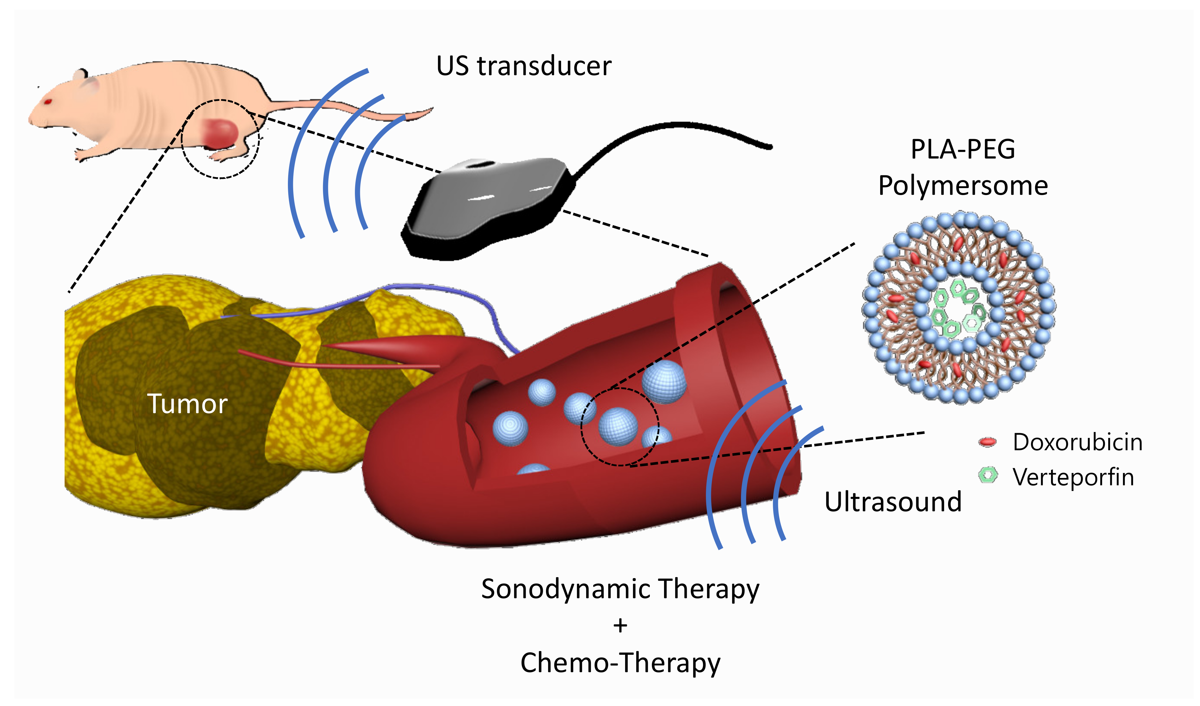
In this study, we developed verteporfin and doxorubicin co-loaded polymersomes (VP-DOX-PLs) composed of PLA-PEG (2k–1k) di-block copolymers to simultaneously deliver both hydrophobic and hydrophilic drugs with a single nanocarrier (
Figure 1). Verteporfin, which is a hydrophobic drug, was loaded into the hydrophobic shell, and doxorubicin, which is a hydrophilic drug, was loaded into the hydrophilic core. When the polymersomes were exposed to ultrasound, ROS were generated by the sonodynamic effect of verteporfin followed by the released doxorubicin. We demonstrated through in vitro and in vivo experiments that the therapeutic efficacy of this combination therapy is superior to that of single SDT or chemotherapy, respectively. Our results indicate that the VP-DOX-PLs have high potential as novel carriers showing effective anticancer therapy through combined SDT and chemotherapy so that they can overcome the unsatisfactory anticancer effect of conventional single therapy while delivering anticancer agents safely to target sites.
3. Discussion
In this study, we developed VP-DOX-PLs for the combination of chemotherapy and sonodynamic therapy, resulting in increased therapeutic efficacy compared to single therapies. Two drugs, doxorubicin and verteporfin, are loaded into polymersomes. Doxorubicin, which is commonly used as an anticancer drug, enters the nucleus of the cell and binds to DNA, arresting cell division. Verteporfin is one of the sonosensitizers for sonodynamic therapy. A sonosensitizer absorbs ultrasonic energy and releases energy to the molecules around it. In this process, nearby oxygens excite and become radicals, known collectively as ROS, which damage DNA or proteins in the cell, leading to the apoptosis of the cell. The developed VP-DOX-PLs drug delivery system could deliver drugs into tumors through the following steps: (1) development of doxorubicin and verteporfin co-delivering polymersomes; (2) enhanced tumor accumulation by EPR effect and the intracellular delivery of doxorubicin and verteporfin using polymersomes; (3) the local treatment of sonodynamic therapy due to ultrasound irradiation.
In the first step, chemotherapeutic agents (doxorubicin) and a sonosensitizer (verteporfin) were loaded in different parts of the polymersome. Polymersomes are self-assembled nanoparticles that are known to have superior stability to that of other particles and show relatively low leakage of loaded drugs [
18]. Polymersomes are capable of loading hydrophilic drugs within the aqueous core of the polymersomes and hydrophobic drugs loaded within the membrane bilayer. Therefore, polymersomes possess broad possibilities in drug delivery [
21,
22] and gene and protein delivery [
19] systems for therapeutic application. Since polymersomes are developed in a thermodynamically stable way, various particles, such as micelles and polymeric particles, can be formed, as well as polymersomes. The packing parameter,
, is commonly used to predict the morphology of the self-assembled formulations (where
is the length of the block copolymer,
is the area of the hydrophilic part, and
is the volume of the hydrophobic part). Other than this method, the morphology can be roughly predicted through the molecular weight ratio of the hydrophilic polymer (
[
18]. Polymersomes are known to be formed when
. From this point of view, PLA (2k)-PEG (1k) copolymer has a molecular weight that is sufficient for the self-assembly of polymersomes in solution. In general, the bilayer of polymersomes could be clearly observed by TEM imaging; however, due to the low molecular weight of the PLA-PEG (2k–1k) used, the boundary of the double membrane was ambiguous (
Figure 2A). To investigate whether the core part of VP-DOX-PLs is hydrophilic, hydrophilic 5 nm gold nanoparticles were loaded into polymersomes. TEM imaging of gold nanoparticle-loaded VP-DOX-PLs revealed that gold nanoparticles were located inside the core part of VP-DOX-PLs, indicating that the core part is hydrophilic (
Supplementary Figure S1). Therefore, in the case of VP-DOX-PLs, hydrophilic doxorubicin was in the core of the polymersomes, and hydrophobic verteporfin was located in the double membrane of the polymersomes. Doxorubicin was loaded into PLs using the pH gradient method, which is an active loading method of drugs according to the transmembrane pH outside and inside the liposome [
23]. Since doxorubicin enters the polymersome core and forms (DOX-NH
3)
2SO
4 crystallization, it showed 95.05% loading efficiency. Compared to that of passive loading, the pH gradient method showed approximately 1.8 times higher loading efficiency. The stability of VP-DOX-PLs was evaluated by measuring the size for 48 h (
Figure 2E,F). As the initial burst of both drugs is carried out during the first 24 h (
Figure 2C,D), the stability of VP-DOX-PL is sufficient to deliver the drug to the tumor.
In the second step, once VP-DOX-PLs enter the body through intravenous injection, they accumulate near tumor sites by the EPR effect [
24]. The dense PEG corona of the PEG copolymer vesicles was shown to deter membrane opsonization and significantly extend in vivo circulation times [
25]. Due to the extended circulation time by the PEG surface, VP-DOX-PLs were successfully delivered into the tumor region by passive targeting. Nanoparticles larger than 10 nm generally cannot penetrate the blood vessels and enter the cells from normal blood vessels. However, abnormal blood vessels are formed around cancer cells because cancer cells secrete a lot of blood vessel growth factors. These blood vessels have looser walls than normal blood vessels, allowing larger nanoparticles to penetrate the vessels and enter the cells. The in vivo biodistribution of VP-DOX-PLs reveals that VP-DOX-PLs were able to accumulate at the tumor site at 24 h post-injection (
Figure 5). The fluorescence of verteporfin of tumor tissue was highest compared to the other major organs, especially the liver and kidney. In this process, another function of ultrasound could be affected. When ultrasound is irradiated, the blood vessel oscillates, and the permeability of the vessel increases, which is called sonoporation. Next, VP-DOX-PLs located near tumor tissue need to enter the cell. The in vitro cellular uptake results showed that VP-DOX-PLs successfully delivered both verteporfin and doxorubicin into cells. As seen in
Figure 3, the fluorescence of doxorubicin was spread throughout the cell, while verteporfin’s fluorescence was found near the nucleus of the cell. To check whether VP-DOX-PLs directly penetrate the nucleus through the new mechanism, rather than by endocytosis or the fusion of nanoparticles, the cellular uptake of the particles was analyzed in 3D using confocal microscopy (
Supplementary Figure S2). The fluorescence of verteporfin did not penetrate the nucleus but was located near the nucleus. It is estimated that the particles entered the Golgi apparatus through clathrin-dependent endocytosis [
26]. Since doxorubicin is hydrophilic, it spread throughout the cell, but hydrophobic verteporfin stayed in the Golgi apparatus. However, there is no clear explanation of the exact cell uptake mechanism of VP-DOX-PLs, which needs to be further investigated.
In the third step, after the accumulation of VP-DOX-PLs on the tumor site by passive targeting, the ultrasound irradiation specific to the tumor site enables the local treatment of sonodynamic therapy. The results of combination therapy showed more effective cancer treatment than other single therapies in both the in vitro and in vivo experiments. To confirm the cytotoxic effect according to the uptake time of the particles, cytotoxicity experiments were performed at various time points of (3, 6, and 24) h (
Figure 4B, and
Supplementary Figure S3). The cell viability of the PLs group showed no significant difference from that of the control, meaning that the PL itself is non-toxic. VP-DOX-PLs without ultrasound and DOX-PLs showed similar anticancer effects by arresting DNA replication and the cell division of doxorubicin. Moreover, as the incubation time of particles increased, cell viability was decreased in groups loading doxorubicin. This result indicates chemotherapy does not show an instant response in viability, while sonodynamic therapy is instant. Consistent with the in vitro results, the therapeutic efficacy of both drugs using polymersomes with ultrasound was also determined by the in vivo mouse model. After the accumulation of VP-DOX-PLs, cell apoptosis of cancer cells occurs by DNA repair disruption due to the intercalation of doxorubicin. Furthermore, when ultrasound is irradiated to the local tumor region, the sonodynamic therapy effect occurs, and ROS is generated by the verteporfin [
27,
28]. Note that the VP-PLs and VP-DOX-PLs exposed to ultrasound showed higher cytotoxicity than those without ultrasound (
Figure 4B), indicating ultrasound is a trigger for sonodynamic therapy. Therefore, off-targeted VP-DOX-PLs did not damage other organs (
Supplementary Figure S4). In addition, verteporfin was used as a sonosensitizer, but it is also a photosensitizer. The difference between sonodynamic therapy and photodynamic therapy is the trigger of therapy: ultrasound for sonodynamic therapy and laser for photodynamic therapy. However, as it is easier for an ultrasound to non-invasively reach deep tissues and activate an accumulated sonosensitizer than a laser, ultrasound is advantageous for treating cancers that occur deep inside the body, such as pancreatic cancer [
29].
4. Materials and Methods
4.1. Materials
Poly-L-lactide-poly (ethylene glycol) was purchased from Creative PEG Works (Chapel Hill, NC, USA). Verteporfin, tetrahydrofuran, acetonitrile, and other reagents were purchased from Sigma–Aldrich (St. Louis, MO, USA). Doxorubicin was purchased from MedChemExpress (Monmouth Junction, NJ, USA). DMEM, fetal bovine serum (FBS), and antibiotic–antimycotic (AA) were purchased from Welgene (Gyeongsan, Korea). MIA PaCa-2 cancer cells were sourced from the Korean Cell Line Bank (Seoul, Korea). All other chemicals and solvents were analytical grade.
4.2. Synthesis of VP-DOX-PLs
Both verteporfin and doxorubicin-loading PLA-PEG polymersomes (VP-DOX-PLs) were formed by a solvent switch method. Briefly, 20 mg of PLA-PEG copolymers (PLA (2k)—PEG (1k)) and 0.02 mg of verteporfin were dissolved in 1 mL of tetrahydrofuran (THF). Then, the polymer solution was slowly dropped into 1 mL of water at a ratio of 1 mL/h to form only verteporfin-loading PLA-PEG polymersomes (VP-PLs). The polymer solution was slowly stirred at 25 °C overnight to remove the organic solvent THF. After, the free verteporfin was removed by centrifugation using a 3 kDa amicon tube (Millipore, Burlington, MA, USA) for 30 min at 14,000 g and redispersed in distilled water. Doxorubicin was loaded into VP-PLs by the ion gradient method. Doxorubicin hydrochloride (1 mg/mL) was added to 1 mL of 250 mM ammonium sulfate solution. Then, VP-PLs were reacted with doxorubicin solution at 400 rpm for 2 h at 25 °C. Unloaded doxorubicin was removed using 3 kDa amicon tubes again. Finally, VP-DOX-PLs were redispersed in distilled water.
4.3. Characterization of VP-DOX-PLs
The size distribution of the particles was analyzed using a dynamic light scattering device (Zetasizer Nano, Malvern, Malvern Instruments, Herrenberg, Germany). The morphological characteristics of VP-DOX-PLs were determined by transmission electron microscopy (TEM; JEM-2100F, JEOL, Tokyo, Japan). The encapsulated efficiency of doxorubicin was measured using reversed-phase high-performance liquid chromatography (Agilent HPLC 1200, Agilent, Santa Clara, CA, USA). Additionally, the encapsulation efficiency of verteporfin was measured using UV–Vis spectrophotometry (Evolution™ 60S, Thermo Scientific, East Grinstead, UK). The in vitro release rate of either doxorubicin or verteporfin from VP-DOX-PLs was measured by placing VP-DOX-PLs in a dialysis membrane (Spectra/Por® 7, MWCO 2 kD, SPECTRUMLABS, USA), and incubation with 10 mL phosphate-buffered saline (PBS) at 37 °C in shaking conditions. At the predetermined time points, incubated 10 mL of the PBS buffer was replaced with fresh PBS buffer, and the amount of the released drug was measured through HPLC or UV–Vis spectrophotometry.
4.4. In Vitro Stability of VP-DOX-PLs
The in vitro stability of VP-DOX-PLs depending on temperature was analyzed by the change in the size of VP-DOX-PLs through a dynamic light scattering device (Zetasizer Nano, Malvern, Malvern Instruments, Herrenberg, Germany). VP-DOX-PLs were incubated at (4 or 25) °C in PBS solution. Additionally, the stability of VP-DOX-PLs was determined in DW and 10% FBS solution (in DMEM) at 37 °C. After a predetermined time, the size of VP-DOX-PLs was measured through DLS until 48 h for both experiments.
4.5. Intracellular Uptake of VP-DOX-PLs
Human pancreatic cancer cells (MIA PaCa-2 cells, Korean Cell Line Bank, Seoul, Korea) were maintained in RPMI-1640 containing 10% fetal bovine serum and 1% penicillin/streptomycin at 37 °C in a humidified atmosphere of 5% CO2. To visualize the intracellular delivery efficiency of VP-DOX-PLs, MIA PaCa-2 cells (2 × 10⁵ cells per well) were seeded in confocal dishes. Doxorubicin and verteporfin were treated at the concentration of (200 and 100) ng/mL, respectively, and incubated for 24 h. The cells were washed twice using cold PBS and fixed using 4% paraformaldehyde for 15 min. Then, the cells were stained with DAPI solutions, and fluorescence images of DOX-PLs, VP-PLs, and VP-DOX-PLs were obtained using confocal laser scanning microscopy (LSM 880, Zeiss, Jena, Germany). For the quantitative analysis, the same experiment was conducted with Accuri C6 Plus (BD Accuri C6 Plus, BD, Piscataway, NJ, USA), and 10,000 cells were counted for each group. The fluorescence of doxorubicin was measured through the FITC filter, and the fluorescence of verteporfin was measured using an APC filter.
4.6. Confirmation of ROS Generation under the Exposure of Ultrasound
The generation of reactive oxygen species (ROS) was confirmed under the exposure of ultrasound to determine the sonodynamic therapy potential of VP-DOX-PLs. MIA PaCa-2 cells were seeded in 96-well plates (1 × 10⁴ cells per well) and incubated for a day. Empty PLs (no verteporfin and no doxorubicin), DOX-PLs, and VP-DOX-PLs were treated in the cells. Each group was treated with the same amount of verteporfin (a concentration of 1 μg/mL of verteporfin). Ultrasound was irradiated with 0.3 W/cm2 ultrasonic power and a 50% duty cycle for 10 s per well using Sonopuls 490 (Enraf Sonopuls, Harlev, Denmark). The amount of reactive oxygen species (ROS) generation of MIA PaCa-2 cells was measured using a DCFDA Cellular ROS Detection Assay Kit (Abcam, Cambridge, UK), according to the manufacturer’s protocol. The fluorescence intensity was measured using a microplate reader (EnSpire multimode plate reader, PerkinElmer, Waltham, MA, USA) at Ex/Em = 485/535 nm/nm.
4.7. In Vitro Cell Viability following Ultrasound Irradiation
Human pancreatic cancer cells (MIA PaCa-2 cells) were sourced from the Korean Cell Line Bank (Seoul, Korea). To verify the therapeutic effect of chemotherapy and sonodynamic therapy, an in vitro cell viability test was performed using the MTT assay. MIA PaCa-2 (2 × 10⁴ cells per well) was seeded in 96-well cell culture plates and incubated for 24 h (37 °C, 5% CO2). Ultrasound was irradiated to each group at 0.3 W/cm2, 50% duty cycle, 10 s per well, and further incubated for (3, 6, and 24) h. The concentration of doxorubicin and verteporfin was (200 and 100) ng/mL, respectively. After the incubation of each treated group, the cells were washed twice using cold PBS, exchanged into fresh medium, and further incubated for 24 h. Next, 30ul of MTT solution (0.5 mg/mL) were added to each well, and after 3 h incubation, DMSO (200 μL) was added. The absorbance was measured at a 595 nm wavelength by a microplate reader (Bio-Tek, Winooski, VT, USA).
4.8. In Vivo Studies
All in vivo studies conformed to the Guide for the Care and Use of Laboratory Animals published by the National Institutes of Health, USA (NIH publication no. 85-23, 1985, revised 1996). Mice were maintained under the guidelines of an approved protocol from the Institutional Animal Care and Use Committee (IACUC) of Sogang University (Republic of Korea, Approval Date: 01/20/2021 Approval Code: IACUCSGU2021_02).
4.9. In Vivo and Ex Vivo Imaging of VP-DOX-PLs in MIA PaCa-2 Tumor-Bearing Mice
To determine the accumulation of VP-DOX-PLs in the tumor, in vivo and ex vivo imaging of VP-DOX-PLs after intravenous injection was performed using the FOBI system. For the subcutaneous tumor model, 5-week-old female Balb/C nude mice were injected in the lower flank with MIA PaCa-2 cells (1 × 10⁶ cells per mouse) (Raonbio, Seoul, Korea). When the volume of tumor reached 150 mm3, VP-DOX-PLs were intravenously injected for a biodistribution study. The biodistribution of VP-DOX-PLs was monitored using the FOBI system through an NIR filter (Fluorescence labeled Organism Bioimaging, NeoScience, Suwon, Korea), and the intensity of the fluorescence signal was quantified by NEOimage software (NeoScience, Suwon, Korea). At 24 h post-injection, tumors and major organs, including the heart, lung, spleen, liver, and kidneys, were excised and imaged with an FOBI system.
4.10. Anti-Tumor Effects of VP-DOX-PLs with US Irradiation
To investigate the anti-tumor effects of VP-DOX-PLs, MIA PaCa-2 cells (1 × 10⁶) were injected subcutaneously into 5-week-old female Balb/C nude mice (n = 3, Raonbio, Seoul, Korea). When the tumor volume reached (30–50) mm3, each group of mice were intravenously injected at an equivalent dose of 5 mg/kg of doxorubicin and 1 mg/kg of verteporfin three times every two days. For ultrasound-positive groups, tumors were irradiated by the ultrasound (2.0 W/cm2, 50% duty cycle, 1 min per mouse) soon after i.v. injection. The volume of the tumor was measured by an external caliper based on the following formula: tumor volume = (length × width2)/2. The body weights were monitored during the experiment. At the end of the experiment, mice were sacrificed, and the tumors and major organs were examined for histological analysis. For histological analysis, a part of the excised tumors and major organs were fixed in 4% paraformaldehyde, embedded in paraffin, sectioned (5 μm), and stained with an H&E and TUNEL assay kit following the manufacturer’s protocols.
4.11. Statistical Analysis
All experimental data obtained from the cultured cells and animals are expressed as the mean ± standard error from at least 3 independent experiments. The statistical significance of the difference between experimental and control groups was determined by Student’s t-test. Statistical significance was established at p < 0.05, and significant differences are shown by asterisks in the figures.


