1. Introduction
Dental caries, which is the most prevalent worldwide disease, is ranked as the third largest non-infectious disease by the World Health Organization [
1]. In the past several decades, dental caries has received substantial interest in numerous studies and initiatives. Among these research studies, dental resin composite (DRC) is an effective method in repairing decayed teeth because of its excellent esthetics and convenient operation qualities [
2,
3,
4], which enable more precise polishing and exhibit better wear resistance. Note, however, that in long-term dental resin restoration, the growing bacteria will create oral biofilms on the resin’s surface, particularly the gap formatted by polymerization shrinkage, and continuously degrade the current resin composite and dental tissue nearby, causing secondary caries and treatment failure [
5,
6]. To satisfy current clinical requirements and lengthen the longevity of tooth restorations, innovative and multifunctional dental resin composites are urgently needed.
DRC is mainly composed of a resin matrix and fillers; thus many researchers have concentrated on the development of these components [
7,
8]. For instance, Lucas et al. designed a dental adhesive consisting of polyhexamethylene guanidine hydrochloride (PHMGH) with antibacterial activity [
9]. Zhou et al. developed a novel antibacterial resin containing a low concentration of dimethylaminohexadecyl methacrylate (DMAHDM), which could inhibit biofilms from saliva [
10]. Unfortunately, the physicochemical qualities may be impaired due to the long alkyl chain of quaternary ammonium compounds based on the above approaches of resin matrices [
11]. For the filler section, the functional fillers can greatly enhance the performances or impose new functions on DRC [
12], such as the strong antibacterial effects [
13,
14] of ZnO nanoparticles [
15]. It is worth noting that metallic and oxide particles can change the shade of materials, influencing the color and light transmission of the DRC accordingly [
16,
17]. The cumulative toxic effects of the long-term released ions also deserve further investigation [
18,
19].
In particular, as a photocatalytic antibacterial agent filler, titanium dioxide (TiO
2) has attracted much attention in the field of dental materials due to its excellent performance [
20]. TiO
2 can be activated by absorbing ultra-high bandgap energy radiation [
21]. Then reactive oxygen species (ROS) are generated, which have an antibacterial impact on bacteria as a result of the holes formed in the activated TiO
2 [
22]. Based on this superior efficacy of TiO
2, Akira et al. proposed a resin composite with a TiO
2 filler, which demonstrated enhanced antibacterial properties [
23]. However, the wide bandgap of TiO
2 requires UV radiation [
24,
25], which restricts its potential for additional clinical use. To address this weakness, several approaches, including doping with other species, have been thoroughly investigated to inspire TiO
2 under visible light irradiation. For example, TiO
2 has been doped with nitrogen (N) in earlier studies [
26,
27]. When exposed to visible light, N-doped TiO
2 displays a narrowing bandgap and rising absorption, and more ROS are released concurrently to increase the antibacterial activity [
28]. In addition, Costa et al. increased the photocatalytic activity by substituting strontium (Sr) for Ti in TiO
2 [
29], which can be explained in terms of the lattice deformation brought about by the higher radius of Sr [
30]. More recently, with further research studies on modified TiO
2, there has been a lot more interest in the co-doping of TiO
2, which may produce more significant photocatalytic activity compared to single-element doping. According to Zhao’s study, co-doping copper (Cu) and N in TiO
2 caused a narrower bandgap and more visible region of light absorption compared to TiO
2 which was only doped with N [
31]. This has rarely been reported for the application of Sr-N-co-doped TiO
2 (Sr-N-TiO
2) nanoparticles in dentistry.
However, the current modified methods of DRC have been mostly focused on a single function, which is far from optimal. Thus, there remains a need for more multifunctional DRCs. Hydroxyapatite (HA) has been noted as a suitable inorganic filler for bionic dental restoration materials. HA is a critical constituent of bone and dental tissues in the human body [
32], which can release calcium and phosphorus ions to promote remineralization and repair the demineralized areas [
33,
34]. Par et al. proved that DRC containing HA could form a precipitate layer on the surface after being immersed in artificial saliva (SBF) [
35]. Meanwhile, HA–TiO
2 composite was synthesized, which was a new type of photocatalyst and exhibited excellent degradation performance for decontamination [
36,
37]. These above studies motivated us to fuse TiO
2 and HA to synthesize a multifunctional DRC.
In this study, a novel and multifunctional dental resin composite composed of Sr-N-TiO2 and HA reinforcing fillers was proposed. Specifically, the anatase-phase Sr-N-TiO2 was synthesized by a sol-hydrothermal process. With the help of these distinctive elements, the TiO2 was capable of activation under visible light irradiation and improved its antibacterial properties. To demonstrate the resin’s performance, the resin was thoroughly characterized in terms of its antibacterial activity, bioactive mineralization capacity, biocompatibility, and the physiochemical properties necessary for its application in restoration. Based on the research and analysis of the experimental results, the proposed DRC offers a fresh approach for clinical restoration and has potential in practical clinical applications. By simultaneously eliminating cariogenic bacterial species and remineralizing new minerals at the restoration margin, the combined antibacterial and mineralization therapy seemed to be more ideal.
4. Materials and Methods
4.1. Synthesis and Characterization of the Modified Nanoparticles
Sol-hydrothermal method was used to create Sr-N co-doped TiO
2 nanoparticles [
70]. In order to carry out the hydrolysis, a mixed solution containing 5 mL of tetrabutyl titanate (Tianjin Guangfu Fine Chemical Research Institute, Tianjin, China) and 5 mL of anhydrous ethanol was first added dropwise to another hybrid solution made up of 20 mL of anhydrous ethanol, 5 mL of water, 1 mL of 70% nitric acid, 17.16 mL of ammonia solution, and 0.3109 g of strontium nitrate. The other products were purchased from Beijing Chemical Company, Beijing, China. By stirring constantly for 2 h, the yellowish translucent sol was created. In a stainless-steel jar, the as-prepared sol was maintained at 160 °C for 6 h. Then the production was immersed in anhydrous ethanol and sonicated for 30 min, centrifuged at high speed for 10 min. Finally, the production was dried at 60 °C for 24 h, and obtained by calcining at 450 °C for 2 h to produce Sr-N-TiO
2 nanoparticles. All reagents were analytical grade and used without further purification.
The field emission scanning electron microscope (FE-SEM, JEOL JSM-6700F, Tokyo, Japan) operated at 3.0 kV was used to measure the surface morphology of the sample. We redispersed Sr-N-TiO2 NPs in ethanol, dropped them on carbon-coated copper grids, and used a transmission electron microscope (TEM, JEOL, Tokyo, Japan) to observe them. X-ray photoelectron spectroscopy (XPS, Thermo Kalpha, Waltham, MA, USA) was used to determine the valence states and chemical makeup of elements. Through the use of X-ray diffraction (XRD, X’Pert PRO MPD, Almelo, The Netherlands), the crystal structure of the modified TiO2 sample was identified. A LabRAM ARAMIS system was used to obtain the Raman spectra. The He-Ne laser light at 633 nm was used as the excitation source. Ten-second accumulations led to data gathering. On a Shimadzu UV-3600 UV–Vis (Kyoto, Japan) spectrophotometer, the electronic absorption spectra were captured.
4.2. Preparation of DRCs
The resin matrix was synthesized, containing 49 wt% bisphenol-A-glycidyldimethacrylate (Bis-GMA) and 49 wt% triethylene glycol dimethacrylate (TEGDMA), with 1 wt% of camphorquinone (CQ) and 1 wt% of 2-(Dimethylamino)ethyl methacrylate (DMAEMA). All products were analytical grade without further purification from Sigma Aldrich Chemical Co., St. Louis, MO, USA. The Sr-N-TiO
2 nanoparticles were mixed up with nano-hydroxyapatite (n-HA, 20 nm, Emperor Nano, Nanjing, China) as reinforcing fillers, and silicon dioxide (SiO
2, 5 μm, Aladdin, Shanghai, China) was the traditional filler. Then they were mixed into the resin matrix with the SpeedMixer DAC 150 (FlackTek Inc., Shanghai, China). The compositions of the fillers in each group are presented in
Table 1, and the load was maintained at 60 wt%.
4.3. Physicochemical Properties
4.3.1. Degree of Conversion
With the help of a Fourier-transform infrared spectrometer (FT-IR, Bruker VERTEX 80v, Salbruken, Germany), the degree of conversion (DC) was ascertained. Firstly, we applied a small amount of the DRCs to the KBR slice and analyzed it. Following a 20 s light-curing unit exposure, it was examined. The process was repeated three times to measure separately the spectra at 20 s, 40 s, and 60 s.
Two absorption bands were examined in order to determine the DC. After exposure to radiation, the acrylate double bonds’ (C=C) absorbance intensity at 1636 cm
−1 decreased. The internal standard was the carbonyl (C=O) absorbance peak at 1720 cm
−1. Equation below was used to compute the DC:
where AC=C and AC=O represented, respectively, the absorbance peaks at 1636 cm
−1 of C=C and 1720 cm
−1 of C=O. The absorbances of the functional group before and after light-curing were, respectively, (AC=C/AC=O)
0 and (AC=C/AC=O)
t. Measurements were made in the dark and repeated three times.
4.3.2. Curing Depth
The method described in ISO 4049: 2009 was used to measure the curing depth (CD) of composites. The cylindrical specimens of each composite (d = 4.0 mm, h = 10.0 mm) were made. The light-curing equipment photopolymerized the composite for 20 s, starting from one side. The uncured material was removed after irradiation. A digital micrometer was used to measure the height of the cured composite. Each measurement was carried out five times.
4.3.3. Water Contact Angle
Sessile drop analysis was used to calculate the water contact angle (WCA) using the OCA 15EC goniometer from Data Physics in Filderstadt, Germany. We used fine sandpaper to smooth out the samples. The surface of the sample was then sprayed with 5 μL of deionized water. The process was repeated three times.
4.4. Antibacterial Properties and Antibacterial Mechanism
4.4.1. Bacterial Culture
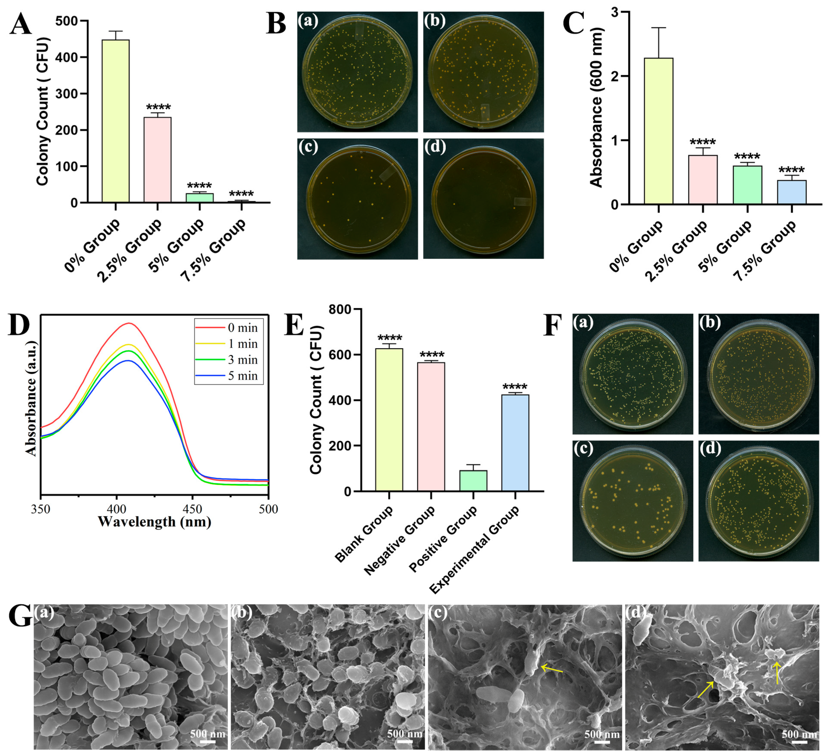
In a bacteria incubator (SLI-1200, SANYO, Tokyo, Japan), the frozen precultured Streptococcus mutans (S. mutans, UA159) were revived and injected on an agar plate with sterilized brain–heart infusion broth (BHI, Oxoid, Basingstoke, UK) culture media. Before the experiment, a single bacteria colony was added to a new sterilized BHI and cultured for 24 h. By altering the absorbance at 600 nm of the bacteria strain with a microplate reader (Synergy HT, Biotek, Winooski, VT, USA), the concentration of the bacteria was managed.
4.4.2. Colony-Forming Units (CFU) Counting
The 6 mm × 2 mm cylindrical specimens were co-cultured with 2 mL bacterial suspension (1 × 10⁷ CFU/L) for 24 h. To get rid of non-adherent bacteria, PBS buffer was used to rinse the co-cultured specimens gently. The adherent S. mutans were collected, diluted, and inoculated on BHI broth agar. The number of colonies that developed on each plate was then counted after a 24 h anaerobic incubation period at 37 °C.
The following equation was used to calculate the antibacterial rate (AR) for each group: AR = (CFU0 − CFU)/CFU0 × 100% (where CFU0 represents the average colony count of the control group and CFU represents the average colony count of the experimental group). The above experiment recurred three times.
4.4.3. Crystal Violet Staining Assay
The 24 h co-cultured specimens were dipped for 30 min into a glutaraldehyde solution (Solarbio, Beijing, China) and stained for 20 min by crystal violet solution. Then 95% alcohol was used to decolorize the samples and they were transferred to a new 96-well plate to record the OD values at 600 nm.
4.4.4. Detection of ROS Release
The release of ROS under different processing was analyzed with a DPBF probe (Macklin, Shanghai, China). The Sr-N-TiO2 nanoparticles were prepared in PBS solution with a concentration of 2.0 mg/mL. The solution was transferred to 96-well plates, and placed under LED light for 0, 1, 3, and 5 min, respectively. The reduction in the characteristic peak of DPBF at 410 nm by UV–Vis absorption spectroscopy demonstrated the generation of ROS.
4.4.5. CFU Counting with NAC
The growth of colonies with the addition of NAC (Aladdin Industrial Corporation, Shanghai, China), which was the ROS scavenger, was examined by the CFU method. The Sr-N-TiO2 nanoparticles were prepared into BHI solution with a concentration of 4 mg/mL. The groups in the 96-well plate were as follows: the blank control group (50 μL BHI), the negative control group (25 μL NAC and 25 μL BHI), the positive control group (25 μL Sr-N-TiO2 and 25 μL BHI), and the experimental group (25 μL NAC and 25 μL Sr-N-TiO2). Then 50 μL bacterial suspension (1 × 10⁷ CFU/L) was added per well. According to GB/T 21510-2008, the plate was incubated at 37 °C with constant shaking for 4 h and co-cultured for 24 h. The S. mutans were diluted and inoculated on BHI broth agar. The number of colonies was counted and analyzed.
4.4.6. SEM of the Bacteria Attached to the Surface
The preparation conditions were the same as 4.4.2. After removing the non-adhered bacteria, the bacteria were fixed for 12 h with 2.5% glutaraldehyde and then dehydrated for 15 min at each concentration using a gradient ethanol series of 30%, 50%, 70%, 80%, 90%, and 100% (v/v). Before SEM analysis, the samples were dried and gold-sprayed.
4.4.7. Bacterial Live/Dead Staining
The stain was mixed with SYTO 9 dye and propidium iodide (PI) dye (Bestbio, Shanghai, China) in a 1:1 ratio. The specimens were treated in the same way as 4.4.2, then examed using confocal laser scanning microscopy (CLSM) after staining for 30 min.
4.5. Remineralization Properties
The 6 mm × 2 mm cylindrical specimens were incubated at 37 °C with constant shaking in 5 mL synthetic saliva (SBF, Solarbio, Beijing, China). At predetermined intervals (14 d and 28 d), samples were taken out and dried at 50 °C. By using a FE-SEM with EDS, the morphological surface alterations and element quantification were carried out.
4.6. Cytotoxic Properties
Following the instructions of ISO 10993-5 standard, the samples were soaked in high-glucose medium (DMEM, HyClone, Logan, UT, USA) to set up the extraction solution for 24 h at 37 °C. The following five groups were marked: blank control group, 0% group, 2.5% group, 5% group, and 7.5% group. L929 cells (Cells Resource Center, Shanghai Institutes of Biological Science, Shanghai, China) in the logarithmic growth phase were placed in the 96-well plates with 2000 cells per well and incubated for 24 h in the cell incubator. After removing the supernatant, 200 μL extraction solution per well was added. On day 1, day 2, and day 3, cell counting kit-8 (CCK-8, Beyotime, Shanghai, China) solution was added (200 μL per well) and incubated for 1 h, 2 h, and 3 h. Later, to assess cell proliferation, the OD values were determined with a microreader (Bio-Rad 680) at a 450 nm wavelength.
The equation can be used to calculate: RGR = OD
0/OD
t × 100% (where OD
0 is the average OD value of the control group and OD
t is the average OD value of the experimental group). Based on ISO standards and the US Pharmacopoeial Convention [
71] in
Table 6, the RGR and toxicity grades were assessed.
4.7. Statistical Analysis
The software GraphPad Prism 8 (GraphPad, San Diego, CA, USA) was used to obtain the statistical analysis. All data were recorded as mean ± standard deviation (Mean ± SD). A one-way analysis of variance was used to perform multiple group comparisons. * p < 0.05, ** p < 0.01, *** p < 0.001, and **** p < 0.0001. Ns indicated not significant.


 ,
, 


