Article

Artemisia annua Extract Attenuate Doxorubicin-Induced Hepatic Injury via PI-3K/Akt/Nrf-2-Mediated Signaling Pathway in Rats

by
Karim Samy El-Said
1,
Ahmed S. Haidyrah
2,
Maysa A. Mobasher
3,
Arwa Ishaq A. Khayyat
4,
Afnan Shakoori
5,
Noorah Saleh Al-Sowayan
6,*,
Ibrahim Omar Barnawi
7 and
Reham A. Mariah
8
1
Biochemistry Division, Chemistry Department, Faculty of Science, Tanta University, Tanta 31527, Egypt
2
Digital & Smart Laboratories (DSL), King Abdulaziz City for Science & Technology (KACST), Riyadh 11442, Saudi Arabia
3
Department of Pathology, Biochemistry Division, College of Medicine, Jouf University, Sakaka 72388, Saudi Arabia
4
Biochemistry Department, Science College, King Saud University, Riyadh 11451, Saudi Arabia
5
Laboratory Medicine Department, Faculty of Applied Medical Sciences, Umm Al-Qura University, Makkah 21955, Saudi Arabia
6
Department of Biology, College of Science, Qassim University, Buraydah 52377, Saudi Arabia
7
Department of Biological Sciences, Faculty of Science, Taibah University, Al-Madinah Al-Munawwarah 41321, Saudi Arabia
8
Department of Medical Biochemistry, Faculty of Medicine, Tanta University, Tanta 31527, Egypt
*
Author to whom correspondence should be addressed.
Int. J. Mol. Sci. 2023, 24(21), 15525; https://doi.org/10.3390/ijms242115525
Submission received: 2 October 2023 / Revised: 19 October 2023 / Accepted: 20 October 2023 / Published: 24 October 2023
(This article belongs to the Section Biochemistry)

Abstract

:
Doxorubicin (DOX), which is used to treat cancer, has harmful effects that limit its therapeutic application. Finding preventative agents to thwart DOX-caused injuries is thus imperative. Artemisia annua has numerous biomedical uses. This study aims to investigate the attenuative effect of Artemisia annua leaf extract (AALE) treatment on DOX-induced hepatic toxicity in male rats. A phytochemical screening of AALE was evaluated. Forty male rats were used; G1 was a negative control group, G2 was injected with AALE (150 mg/kg) intraperitoneally (i.p) daily for a month, 4 mg/kg of DOX was given i.p to G3 once a week for a month, and G4 was injected with DOX as G3 and with AALE as G2. Body weight changes and biochemical, molecular, and histopathological investigations were assessed. The results showed that AALE contains promising phytochemical constituents that contribute to several potential biomedical applications. AALE mitigated the hepatotoxicity induced by DOX in rats as evidenced by restoring the alterations in the biochemical parameters, antioxidant gene expression, and hepatic histopathological alterations in rats. Importantly, the impact of AALE against the hepatic deterioration resulting from DOX treatment is through activation of the PI-3K/Akt/Nrf-2 signaling, which in turn induces the antioxidant agents.

1. Introduction

Chemotherapeutic medications are frequently used to treat cancer. Despite their effectiveness, there are related negative consequences on the vital organs, so it is necessary to find novel ways to lessen these side effects [1]. Doxorubicin (DOX) is an antineoplastic drug that is frequently used to treat hematological malignancies, breast cancer, and sarcomas [2]. Although DOX has an anticancer effect, its use is restricted due to its negative impacts on non-cancerous cells [3]. DOX works by damaging the DNA of cancer cells, which stops them from multiplying, while it causes oxidative stress in non-cancerous cells and reduces mitochondrial activity [4]. The metabolism of DOX occurs through liver microsomal enzymes, which in turn leads to the buildup of harmful intermediates that have been linked to the development of liver damage [5]. DOX could promote oxidative stress via suppressing Nrf-2, which mediates cellular redox homeostasis and antioxidant responses by inducing the antioxidant response elements (ARE) of antioxidant genes [6]. A protein kinase Akt is a crucial regulator for apoptosis and survival and is activated via phosphoinositide 3-kinase (PI3-K)-dependent events. Reactive oxygen species (ROS), which promote p38, deactivate pAKT signaling, and cause apoptosis, have been found to be closely related to chemotherapy [7]. In addition to reducing oxidative stress and cell injury, activating the PI3-K/Akt signaling pathway can also suppress autophagy and apoptosis [8]. Alternative medicine is thought to be one strategy that could reduce the harmful effects of chemotherapy. Natural constituents provide promising antioxidants that could reduce DOX-induced liver malfunctions. For instance, naringenin ameliorated DOX-induced hepatotoxicity by reducing the oxidative stress [9]. Furthermore, the treatment with diosmin showed good impacts against DOX-induced hepatotoxicity via restoration of hepatic antioxidant factors [10].
Artemisia annua L. (A. annua), a member of the Asteraceae family, is a well-known medicinal herb used as an anti-malaria, anthelmintics, anti-diabetes, antioxidation, anti-inflammation, and immunomodulation agent [11,12,13]. The bioactive ingredients contained in A. annua and their antitumor activities have been reported via increased cell percentages in the S and G2/M phases, and caspase 3 activation [14]. In addition, A. annua are rich in flavonoids, polysaccharides, terpenoids, sterols, coumarins, acetylenes, and other bioactive compositions [15,16,17]. Furthermore, a previous study reported that the biologically active constituents in A. annua could provide potential protective effects against DOX-induced cardiotoxicity in experimental animals [18]. The administration of A. judaica extract alone or in combination with cyclophosphamide presented a significant attenuation of hepatic carcinoma in male rats [19]. Due to the limited evidence about the effects of A. annua on DOX-induced hepatotoxicity, the current study investigated the possible mechanism of the treatment with Artemisia annua leaf extract (AALE) against liver injury that was induced by DOX treatment in rats.

2. Results

2.1. Phytochemical Content of Artemisia annua Leaves

Quantitative phytochemical analysis of Artemisia annua leaves (AAL) showed that the total phenolic and flavonoid contents in the AAL were 21.36 ± 0.85 mg GAE/g DW and 69.86 ± 2.37 mg QUE/g DW, respectively. The total antioxidant capacity (TAC) of the AAL was 287.65 ± 5.54 mg AE/g DW. Saponin and anthocyanin contents were 415.28 ± 3.95 mg/g DW 3.98 ± 0.45 mg ECG/g DW, respectively. The DPPH scavenging activity (%) of AAL was 81% ± 1.57, and the concentration of AAL able to inhibit 50% of DPPH was 4.55 ± 0.34 mg/mL (Table 1).

2.2. GC-MS Analysis of Artemisia annua Leaf Extract

The gas chromatography-mass spectrometry (GC-MS) technique was used to analyze the bioactive compounds present in AALE. The results showed that 2,4,6-trimethyl-1,3,6-heptatriene was the most abundant compound, followed by 3-butenoic acid, 2-oxo-4-phenyl, β-caryophyllenea, α-santonin, spathulenol, and 3,5-heptadien-2-ol, 2,6-dimethyl. These phytochemicals’ peak area percentages (P.A %) were 11.37, 9.81, 8.95, 7.79, 6.83, and 6.57, respectively. The retention times (RTs) were 13.47, 16.21, 15.63, 25.39, 22.37, and 13.19, respectively (Figure 1 and Table 2).

2.3. Effect of AALE Treatment on the Percentages of Body Weight Changes

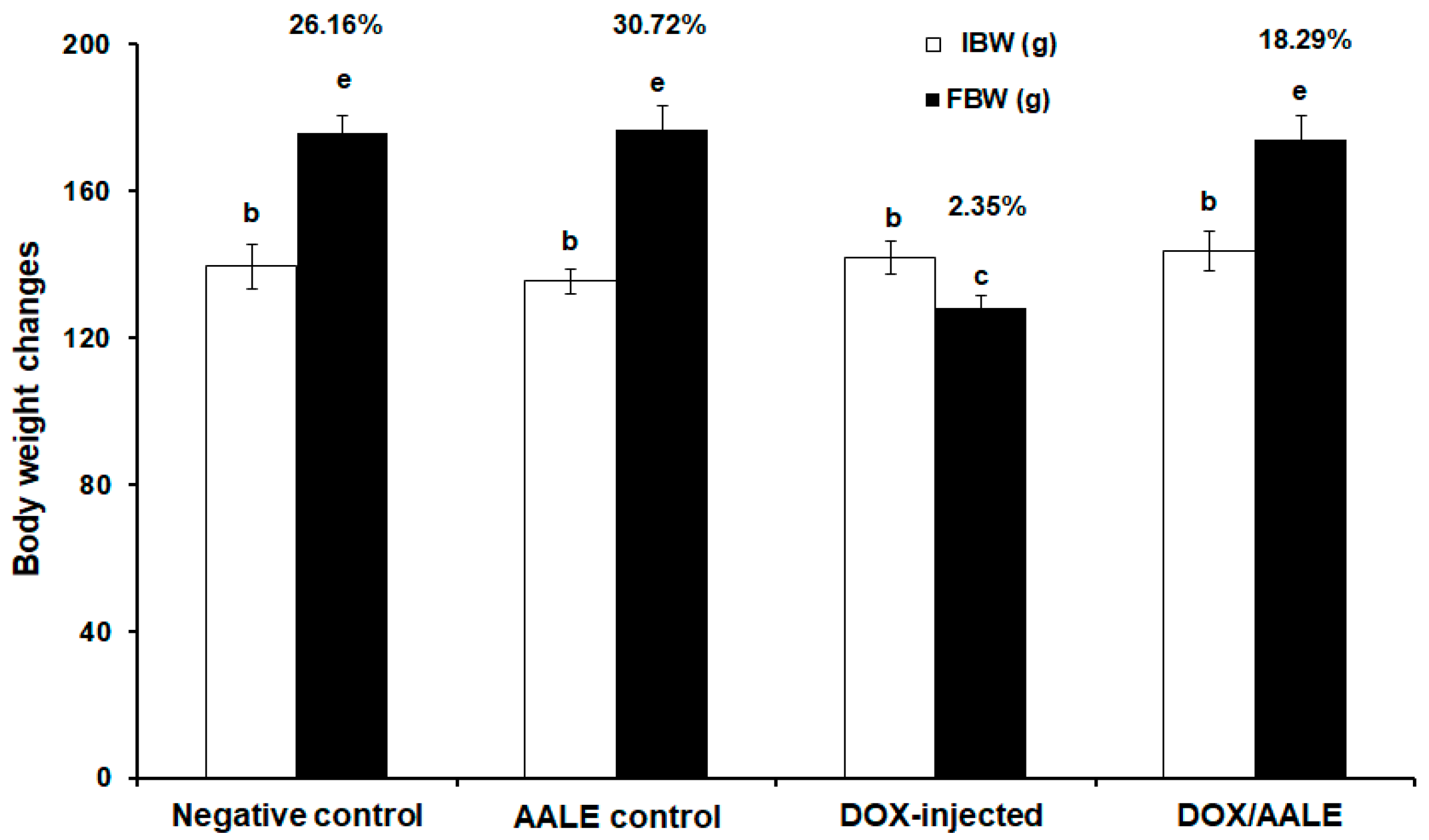
The results showed that there was significant decrease (p ≤ 0.05) in the percentage of body weight change in the DOX-injected group, up to 2.35%, when compared to the control groups. However, treatment of DOX/AALE led to significant improvement in the % BW, up to 18.29%, compared with the DOX-injected group (Figure 2).

2.4. Effect of AALE Treatment on the Alterations of Serum Biochemical Parameters Induced by DOX

Liver transaminases (AST and ALT) were significantly elevated (p ≤ 0.05) in the sera of the DOX-intoxicated group, reaching 97.58 ± 2.46 and 67.78 ± 1.44 U/L, respectively, when compared to the normal and AALE control groups. DOX/AALE-treated rats showed a considerable drop in the levels of AST and ALT, up to 56.39 ± 1.98 and 34.89 ± 1.16 U/L, respectively (Table 3). Rats that were injected with DOX showed a significant increase (p < 0.05) in alkaline phosphatase (ALP) serum level, at 511.8 ± 5.86 U/L, when compared to their control groups (287.6 ± 4.04 and 281.2 ± 2.95 U/L); however, the treatment with AALE caused a significant decrease in serum ALP level as compared to the DOX-intoxicated group (347.2 ± 6.49 U/L). Furthermore, the results showed that there were significant increases (p < 0.05) in the total and direct bilirubin of the DOX-injected group. Treating DOX-intoxicated rats with AALE improved the total and direct bilirubin serum levels (Table 3).

2.5. Treatment with AALE Modulated the Alterations of Hepatic Biochemical Parameters Induced by DOX

Hepatic MDA level was significantly increased (p < 0.05) in the DOX-injected group, by 2.5-fold compared to the control groups (Figure 3A). The GSH level was significantly decreased in the DOX-intoxicated group, by −2.7-fold compared to the control groups (Figure 3B). The group of rats that were treated with DOX/AALE showed a significant decrease in the MDA level compared to the DOX-treated group (0.305 ± 0.023 versus 0.579 ± 0.018 nmol/mg protein). However, treatment with DOX/AALE led to a significant increase in the GSH level as compared to the DOX-treated group (17.19 ± 0.94 versus 9.16 ± 0.54 mg/mg protein) (Figure 3).
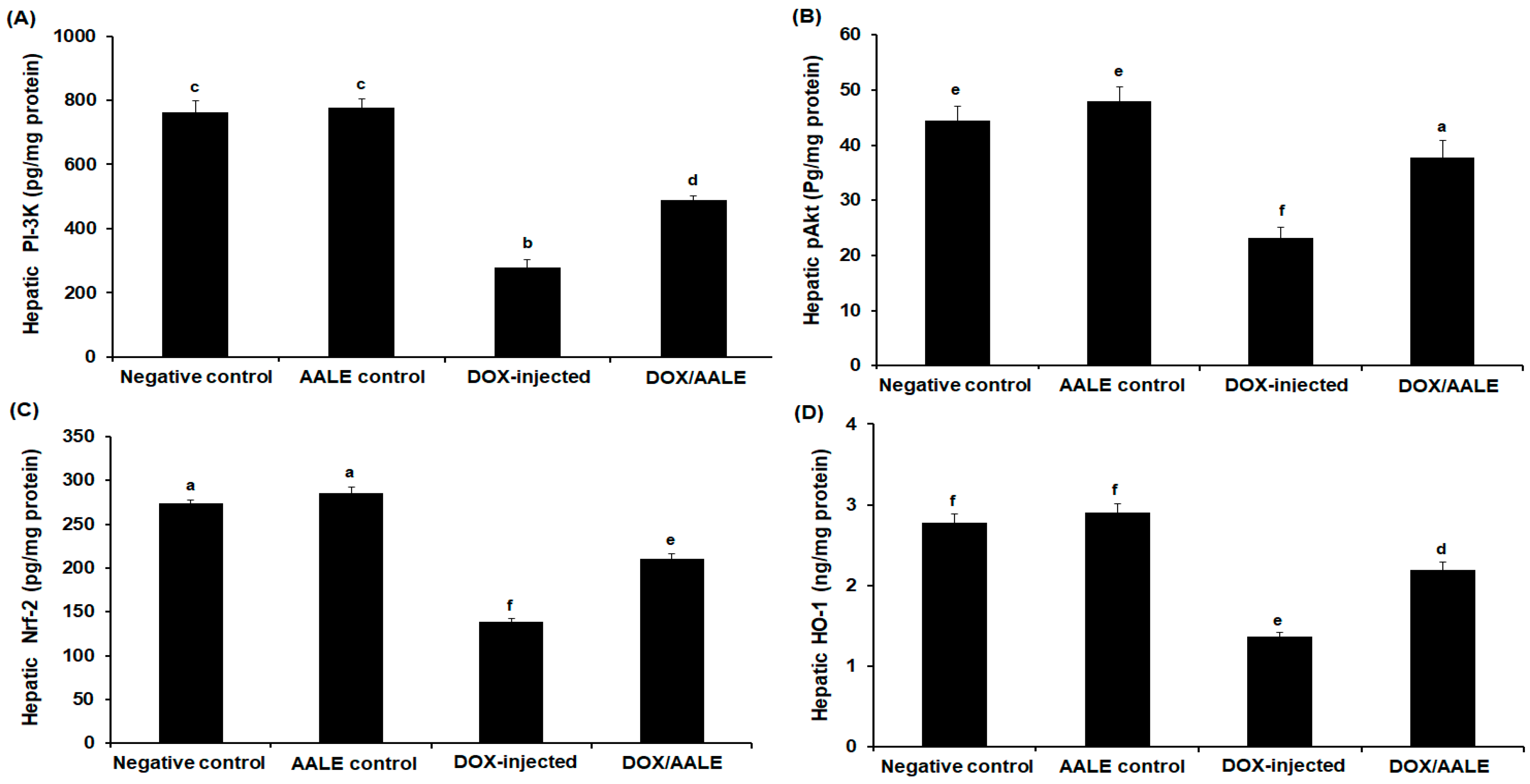
As shown in Figure 4A, the intraperitoneal injection of DOX in rats was accompanied by a remarkable reduction in hepatic PI-3K to 281 pg/mg protein versus control (760.52 ± 29.56 pg/mg protein) and AALE control groups (775.33 ± 25.78 pg/mg protein). Furthermore, the results reported a 2.1-fold decrease in the hepatic Akt of DOX-treated rats when compared to the control group (21.61 ± 1.89 versus 45.14 ± 2.96 pg/mg protein). This reduction has been improved by the treatment with AALE (37.79 ± 3.14 pg/mg protein) (Figure 4B). Similarly, hepatotoxicity induced by DOX injection demonstrated a 1.9-fold reduction in hepatic Nrf-2 concentration versus control (273.92 ± 3.91 versus 140.24 ± 3.27 pg/mg protein). In the DOX-injected group (139 ± 3.58 pg/mg protein), rats treated with AALE showed a significant increase (p < 0.05) in hepatic levels of Nrf-2, as their levels reached 211.82 ± 5.79 pg/mg protein (Figure 4C). Hepatic HO-1 levels were markedly increased in the DOX/AALE-administrated group, almost close to the normal levels (Figure 4D).

2.6. Effect of AALE Treatment on Antioxidant Gene Expression

The relative gene expression levels of SOD, CAT, GPX, GST, and GR were determined to investigate the impact of DOX and/or AALE treatments on the gene expression of the antioxidants in liver tissues of rats. The molecular analysis showed that there were significant downregulations (p < 0.05) in the hepatic SOD and CAT genes by around 0.5-fold compared to the control groups; however, the treatment of DOX-intoxicated rats with AALE led to significant upregulation of these genes, almost close to the control groups (Table 4). As compared to the control groups, DOX-injected rats showed significant downregulation in the GPX, GST, and GR genes. Interestingly, rats treated with DOX/AALE showed marked up-regulation in the hepatic GPX, GST, and GR genes (Table 4).

2.7. Treatment with AALE Restored Hepatic Histopathological Alterations Induced by DOX

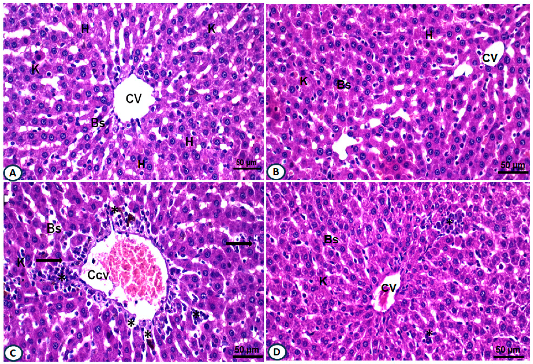
The histopathological investigations of H&E-stained liver sections from the negative control group showed normal hepatocyte lobulation with alternated blood sinusoids lined by an endothelial cell layer. Hepatocytes revealed centrally located nuclei (Figure 5A). The liver section of rats administered with AALE showed normal hepatic architecture, almost regular central veins with activated Kupffer cells were noticed (Figure 5B). However, the liver section of rats injected with DOX displayed severe congested and dilated central vein; hepatocytes were mostly degenerated with vacuolated cytoplasm, and nuclear changes such as pyknotic nuclei and disrupted blood sinusoids were observed, along with cellular infiltrations (Figure 5C). The liver section of the group treated with DOX/AALE exhibited an improvement in the hepatic architectures, represented by regular central vein, with fewer congestions, few numbers of binucleated hepatocytes, less cellular infiltrations, and almost normal Kupffer cells (Figure 5D).

3. Discussion

Although chemotherapy is helpful in treating cancer, side effects limit its usefulness in tumor therapy [20]. One of the most effective anthracycline-derivative anticancer medicines is DOX; however, its therapeutic use has been severely constrained due to toxicity [21]. Liver is a vulnerable organ to DOX damage. Therefore, it is important to find effective and safe agents against the hepatotoxicity of DOX. Antioxidant agents alleviate hepatotoxicity via adjustment of oxidative stress and apoptosis [22]. The A. annua is well-known for its medicinal properties in preventing many ailments. Tue bioactive constituents of A. annua are valuable sources for developing pharmaceuticals [23]. The defensive capacity of A. annua as a powerful antioxidant cure against free radical harm has been detailed [24]. The present study addressed the novel impact of AALE versus the hepatotoxicity induced by DOX in rats via the PI-3K/Akt/Nrf-2-mediated signaling pathway.
The phytochemistry and the valuable pharmacological activity of Artemisia were reviewed by a previous study by Bisht et al. (2021) [25]. This study showed that the AALE contains adequate promising contents of phenolics, flavonoids, saponins, and anthocyanins as well as DPPH scavenging activity. The antioxidant effect of AALE is related to its phytochemical compounds, which contribute to its biomedical potential [26]. These findings agree with previous studies that screened the phytochemical profiles of Artemisia [27,28,29]. In the current study, GC-MS analysis for AALE showed that about seventeen potent compounds were detected; these compounds belonged to terpene, aliphatic, and aromatic compounds. It has been reported that the AALE was found to contain steroids and terpenoids, with several phyto-constituents screened by the GC-MS method [30]. For instance, 2,4,6-trimethyl-1,3,6-heptatriene was detected as a bioactive agent in AALE, which agreed with Liu et al. (2022), who detected the same constituent as the main component in AALE [31]. Interestingly, in this study, three natural sesquiterpenes, β-caryophyllenea, α-Santonin, and spathulenol, were abundantly identified in AALE, which provide several biological activities, including antioxidant, antimicrobial, anticarcinogenic, and anti-inflammatory effects [32,33,34].
This study revealed that the DOX-injected group showed a significant decrease in their % BW; this could be due to the toxic effects of DOX on vital tissues and metabolic disorders, in agreement with previous studies that established the impact of DOX administration on the BW of experimental animals [35,36]. They attributed these adverse effects of DOX on the BW to decreased appetite, reduced feed intake, disruption of basal metabolism, and inhibition of protein production caused by DOX toxicity. Treatment with DOX/AALE led to significant improvement in the % BW; this finding agreed with previous reports demonstrating the effect of medicinal products on the improvement of body weight loss promoted by DOX injection [37,38]. Sera AST, ALT, and ALP activities as well as bilirubin levels were significantly elevated in the DOX-intoxicated rats due to hepatocellular damage, loss of hepatic functioning, and leakage to the bloodstream from necrotic hepatocytes, because of DOX-induced toxicity; however, treatment of DOX-injected rats with AALE caused a significant reduction in these parameters. This could be due to the improvement of hepatic functioning induced by AALE treatment. High levels of blood markers for both hepatocellular and hepatobiliary degenerations and the ameliorative effects of natural products were previously reported [39,40].
The DOX has been shown to cause an imbalance of redox status and attenuation of the antioxidants that led to the oxidization of macromolecules, leading to liver injuries [10,39,40,41]. Allied with previous results, the study showed a significant increase in the MDA levels with a reduction in GSH levels herein in DOX-injected rats; this could be assigned to the impaired hepatic tissue under the damaging effects of DOX. Jagetia and Lalrinengi (2017) reported that due to its ability to increase MDA and deplete GSH, DOX may increase liver damage [42]. Concomitant administration of AALE with DOX led to a reduction in the hepatic MDA and an increase in GSH concentration to relieve the oxidative stress and restore antioxidant/oxidant hemostasis, which is a key factor in suppressing liver injury induced by DOX. This is consistent with previous publications that reported the impact of natural constituents on oxidative stress induced by DOX [43,44,45]. The decreased hepatic HO-1 and Nrf-2 protein has been implicated in the process of DOX-induced liver injury [46]. In our work, rats administered with DOX showed significant decreases in the hepatic Nrf-2 and HO-1 levels; these reductions were reversed by AALE treatment, suggesting the impact of these herbs in mitigating oxidative stress caused by DOX. The modulation of the PI-3K/Akt signaling pathway regulates cell development; numerous downstream proteins are activated by phosphorylated Akt [47]. In the present study, DOX administration led to the suppression of Nrf-2, PI-3K, and Akt levels in liver tissues of rats. Treatment with DOX/AALE for 30 days restored hepatic Nrf-2, PI-3K, and Akt levels. These findings highlight the attenuative effect of AALE on the hepatotoxicity brought on by DOX via Nrf2/HO-1 activation and PI-3K/Akt axis modulation.
This study reported that administration of DOX to rats led to significant downregulations in the hepatic antioxidant genes. These results were in accordance with a previous report indicating that DOX triggers oxidative stress by building up ROS in animals’ liver and decreases antioxidant gene expression [48]. Interestingly, the supplementation of DOX-administered rats with AALE resulted in the upregulation of the antioxidant gene expression, which agreed with previous reports demonstrating the impact of natural products on gene expression of antioxidant genes upon the administration of DOX [9,49]. A previous report stated that DOX-treated rats can increase liver enzyme activity and histological alterations due to liver injury [49]. In the current study, the histopathological investigations of liver sections from DOX-injected rats showed severe congested central vein and degenerated hepatocytes; this finding was in accordance with Mete et al.’s (2016) results, which reported necrosis of biliary ducts in the DOX-treated group [50]. However, these histological alterations in the hepatic structures were dramatically restored in the rats that were concomitantly treated with DOX/AALE, which exhibited an improvement in the hepatic architectures, represented by regular central vein, fewer congestions, and less cellular infiltrations, implying protection from DOX-induced hepatic damage. These findings are in line with previous studies that reported the ameliorative effects of natural products against DOX-induced hepatotoxicity in rats [50,51,52].

4. Materials and Methods

4.1. Chemicals

Doxorubicin was purchased from Hikma Pharmacy Company (Cairo, Egypt). Aluminum chloride, potassium acetate, lead acetate, phenol, and sodium nitroprusside were purchased from Merck Company (Darmstadt, Germany).

4.2. Collection of Plant and Extract Preparation

The leaves of A. annua were obtained from the Crop Institute Agricultural Research Center, Giza, Egypt. The plant was authenticated and complied with relevant institutional guidelines. A specialist identified A. annua leaves, and a specimen was placed in the Tanta University Herbarium (code #TAN-147). The leaves were pulverized after being shade dried. A mixture of 500 mL 70% ethanol and 50 g of leaves powder was used; after filtering, the Artemisia annua leaf extract (AALE) was dried.

4.3. Determination of Phytochemical Content of A. annua Leaves

Contents of the total phenolic, flavonoid, and the total antioxidant capacities (TACs) in AALE were determined [53,54,55]. Saponin and anthocyanin were estimated according to Hiai et al. (1975) [56]. The DPPH radical scavenging capacity was spectrophotometrically evaluated [57].

4.4. Gas Chromatography and Mass Spectrum (GC-MS) Profiling of AALE

The phytoconstituents of AALE were determined using Trace GC 1310-ISQ mass spectrometer “GC-MS” (Thermo Scientific, Austin, TX, USA). By comparing the components’ retention times and mass spectra to those in the WILEY 09 and NIST 11 mass spectral databases, the components were identified.

4.5. Rats and Experimental Design

Forty male Sprague Dawley rats (130–150 g) were purchased from Alexandria University, Egypt. The animals were housed and maintained for a week’s acclimatization period. Our research was conducted in accordance with the standards endorsed by the Tanta University Faculty of Science Research Ethical Committee (IACUC-SCI-TU-0321). Rats were equally divided into four groups. G1 was injected i.p with saline in a proportional volume and in the same time scheme of other experimental groups; G2 was injected with AALE (150 mg/kg) i.p. daily for a month [58]; G3 was injected with 4 mg/kg of DOX i.p once a week for a month [59]; G4 was injected with DOX as G3 and administered with AALE as G2. The percentage of body weight changes (% b.wt) was calculated. All groups had been anesthetized by isoflurane, blood samples were collected by cardiac puncture, and then sera were separated for biochemical assays. Liver tissues were collected in ice-cold saline and homogenized for other biochemical and gene expression analysis. Furthermore, liver tissues were sectioned in buffered formalin for investigations into histopathology.

4.6. Biochemical Analysis

Serum levels of aspartate aminotransferase (AST) (catalog no. AS106145), alanine aminotransferase (ALT) (catalog no. AL103145), alkaline phosphatase (ALP) (catalog no. AP1020), total bilirubin (catalog no. BR1111), and direct bilirubin (catalog no. BR1112) were assessed using colorimetric kits (Spectrum Diagnostics, Egypt). Hepatic malondialdehyde (MDA) and reduced glutathione (GSH) levels (catalog no. MD2529 and catalog no. GR2511, respectively) were measured by using their kit (Biodiagnostic, Egypt). Liver homogenates were used for determination of phospho-PI-3 kinase by using their ELISA kit (catalog no. ab207485) (Abcam, Boston, MA, USA). Hepatic rat’s phospho-Akt concentrations were assessed by rat’s pAkt (Ser473) ELISA kit (catalog no. MBS775153). Furthermore, nuclear Nrf-2 levels in liver were evaluated using rat’s ELISA kits from MyBioSource, Inc., San Diego CA, USA (catalog no.: MBS752046). Hemoxygenase-1 (HO-1) levels in the liver were calculated using a rat-specific ELISA kit from Elabscience Biotechnology Inc., Houston TX, USA (catalogue no.: E-EL-R0864).

4.7. Molecular Analysis

Gene expression analysis was performed by using SYBR Green to assess mRNA expression of antioxidant genes in the tissues of the liver, using β-actin as a reference. The primers used are shown in Table 5, their sequence similarity was checked with BLAST (www.ncbi.nlm.nih.gov/blast/Blast.cgi accessed on April 2023).

4.8. Histopathological Investigations

Formalin-fixed liver sections were embedded in paraffin blocks after being processed in different alcohol and xylene grades. To investigate severe cellular damage, sections (5 μm) were produced, stained with hematoxylin and eosin, and examined under light microscope (Optika light microscope (B-350) [60].

4.9. Statistical Analysis

Graph Pad Prism software (San Diego, CA, USA) was used to analyze the one-way ANOVA results, followed by Tukey’s test for multiple comparisons, acceptable significance was recorded when p ≤ 0.05.

5. Conclusions

The AALE could be a potent beneficial agent against hepatotoxicity resulting from DOX treatment. This effect of AALE is through the activation of PI-3K/Akt/Nrf-2 signaling pathway, which in turn induces antioxidant enzymes. The combination of AALE and DOX may serve as a new strategy for efficient chemotherapy. Further studies should investigate the effects of AALE against other toxicities induced by DOX on the vital organs and their different mechanisms.

Author Contributions

Conceptualization, supervision, investigation, writing—original draft preparation, writing—review and editing K.S.E.-S.; methodology, software, and resources A.I.A.K. and N.S.A.-S.; validation, formal analysis, investigation, and resources, M.A.M. and A.S.; data curation, writing—original draft preparation, writing review and editing, I.O.B., A.S.H. and R.A.M.; All authors have read and agreed to the published version of the manuscript.

Funding

This research received no external funding.

Institutional Review Board Statement

The study was conducted according to the guidelines of the Declaration of Helsinki, and the study was approved by the animal care and use committee, Faculty of Science, Tanta University (IACUC-SCI-TU-0321), Egypt.

Informed Consent Statement

Not applicable.

Data Availability Statement

Data sharing not applicable.

Acknowledgments

Authors acknowledged Faculty of Science, Tanta University, Tanta, Egypt.

Conflicts of Interest

The authors declare no conflict of interest.

References

  1. El-Naggar, S.A.; El-Said, K.S. Antitumor efficacy of EDTA co-treatment with cisplatin in tumor-bearing mice. Braz. J. Pharm. Sci. 2020, 56, e18536. [Google Scholar] [CrossRef]
  2. Argenziano, M.; Gigliotti, C.L.; Clemente, N.; Boggio, E.; Ferrara, B.; Trotta, F.; Pizzimenti, S.; Barrera, G.; Boldorini, R.; Bessone, F.; et al. Improvement in the anti-tumor efficacy of doxorubicin nanosponges in in vitro and in mice bearing breast tumor models. Cancers 2020, 12, 162. [Google Scholar] [CrossRef] [PubMed]
  3. Li, S.; Yuan, S.; Zhao, Q.; Wang, B.O.; Wang, X.; Li, K. Quercetin enhances chemotherapeutic effect of doxorubicin against human breast cancer cells while reducing toxic side effects of it. Biomed. Pharmacother. 2018, 100, 441–447. [Google Scholar] [CrossRef]
  4. Tan, Q.; Yan, X.; Song, L.; Yi, H.; Li, P.; Sun, G.; Yu, D.; Li, L.; Zeng, Z.; Guo, Z. Induction of mitochondrial dysfunction and oxidative damage by antibiotic drug doxycycline enhances the responsiveness of glioblastoma to chemotherapy. Med. Sci. Monit. 2017, 23, 4117–4125. [Google Scholar] [CrossRef]
  5. Timm, K.N.; Ball, V.; Miller, J.J.; Savic, D.; West, J.A.; Griffin, J.L.; Tyler, D.J. Metabolic effects of doxorubicin on the rat liver assessed with hyperpolarized MRI and metabolomics. Front. Physiol. 2022, 12, 782745. [Google Scholar] [CrossRef]
  6. Shaw, P.; Chattopadhyay, A. Nrf2-ARE signaling in cellular protection: Mechanism of action and the regulatory mechanisms. J. Cell. Physiol. 2020, 235, 3119–3130. [Google Scholar] [CrossRef]
  7. Wang, R.; Deng, D.; Shao, N.; Xu, Y.; Xue, L.; Peng, Y.; Liu, Y.; Zhi, F. Evodiamine activates cellular apoptosis through suppressing PI3K/AKT and activating MAPK in glioma. OncoTargets Ther. 2018, 11, 1183–1192. [Google Scholar] [CrossRef]
  8. Sabnam, S.; Pal, A. Relevance of Erk1/2-PI3K/Akt signaling pathway in CEES-induced oxidative stress regulates inflammation and apoptosis in keratinocytes. Cell Biol. Toxicol. 2019, 35, 541–564. [Google Scholar] [CrossRef]
  9. Wali, A.F.; Rashid, S.; Rashid, S.M.; Ansari, M.A.; Khan, M.R.; Haq, N.; Alhareth, D.Y.; Ahmad, A.; Rehman, M.U. Naringenin regulates doxorubicin-induced liver dysfunction: Impact on oxidative stress and inflammation. Plants 2020, 9, 550. [Google Scholar] [CrossRef]
  10. Al Asmari, A.F.; Alharbi, M.; Alqahtani, F.; Alasmari, F.; AlSwayyed, M.; Alzarea, S.I.; Al-Alallah, I.A.; Alghamdi, A.; Hakami, H.M.; Alyousef, M.K.; et al. Diosmin alleviates doxorubicin-induced liver injury via modulation of oxidative stress-mediated hepatic inflammation and apoptosis via Nf-kB and MAPK pathway: A preclinical study. Antioxidants 2021, 10, 1998. [Google Scholar] [CrossRef]
  11. Xing, Y.Y.; Xu, Y.Q.; Jin, X.; Shi, L.L.; Guo, S.W.; Yan, S.M.; Shi, B.L. Optimization extraction and characterization of Artemisia ordosica polysaccharide and its beneficial effects on antioxidant function and gut microbiota in rats. RSC Adv. 2020, 10, 26151–26164. [Google Scholar] [CrossRef]
  12. Nurlybekova, A.; Kudaibergen, A.; Kazymbetova, A.; Amangeldi, M.; Baiseitova, A.; Ospanov, M. Traditional use, phytochemical profiles, and pharmacological properties of Artemisia genus from central Asia. Molecules 2022, 27, 5128. [Google Scholar] [CrossRef]
  13. Mohanty, B.; Puri, S.; Kesavan, V. A review on therapeutic potential of Artemisia nilagirica. J. Plant Biochem. Physiol. 2018, 6, 205. [Google Scholar] [CrossRef]
  14. Lang, S.J.; Schmiech, M.; Hafner, S.; Paetz, C.; Steinborn, C.; Huber, R.; El Gaafary, M.; Werner, K.; Schmidt, C.Q.; Syrovets, T.; et al. Antitumor activity of an Artemisia annua herbal preparation and identification of active ingredients. Phytomedicine 2019, 62, 152962. [Google Scholar] [CrossRef]
  15. Yang, X.L.; Deng, S.Q.; De Philippis, R.; Chen, L.Z.; Hu, C.Z.; Zhang, W.H. Chemical composition of volatile oil from Artemisia ordosica and its allelopathic effects on desert soil microalgae, Palmellococcus miniatus. Plant Physiol. Biochem. 2012, 51, 153–158. [Google Scholar] [CrossRef] [PubMed]
  16. Xing, Y.Y.; Zheng, Y.K.; Yang, S.; Zhang, L.H.; Guo, S.W.; Shi, L.L.; Xu, Y.Q.; Jin, X.; Yan, S.M.; Shi, B.L. Artemisia ordosica polysaccharide alleviated lipopolysaccharide-induced oxidative stress of broilers via Nrf2/Keap1 and TLR4/NF-κB pathway. Ecotoxicol. Environ. Saf. 2021, 223, 112566. [Google Scholar] [CrossRef]
  17. Wang, Q.H.; Pa, B.; Gong, J.H.; Bao, W.Q.; Hao, J.S.; Xu, Y.H. Phenylpropanoids, flavonoids, and terpenoids from Artemisia ordosica Krasch. Magn. Reason. Chem. 2019, 57, 326–330. [Google Scholar] [CrossRef] [PubMed]
  18. Aktaş, I.; Özmen, Ö.; Tutun, H.; Yalçın, A.; Türk, A. Artemisinin attenuates doxorubicin induced cardiotoxicity and hepatotoxicity in rats. Biotech. Histochem. 2020, 95, 121–128. [Google Scholar] [CrossRef]
  19. Mokhamer, E.M.; Zidan, A.A.; El.Ghayesh, N.K.; Abdel-Aziz, K.K. Attenuation of trichloroacetic acid-induced hepatocellular carcinoma by Artemisia judaica ethanolic extract in male rats. J. Basic Appl. Zool. 2022, 83, 2426. [Google Scholar] [CrossRef]
  20. Nurgali, K.; Jagoe, R.T.; Abalo, R. Editorial: Adverse effects of cancer chemotherapy: Anything new to improve tolerance and reduce sequelae? Front. Pharmacol. 2018, 9, 245. [Google Scholar] [CrossRef]
  21. Peter, S.; Alven, S.; Maseko, R.B.; Aderibigbe, B.A. Doxorubicin-based hybrid compounds as potential anticancer agents: A review. Molecules 2022, 27, 4478. [Google Scholar] [CrossRef] [PubMed]
  22. Wang, R.; Dong, Z.; Lan, X.; Liao, Z.; Chen, M. Sweroside alleviated LPS-induced inflammation via SIRT1 mediating NF-kappaB and FoxO1 signaling pathways in RAW264.7 Cells. Molecules 2019, 24, 872. [Google Scholar] [CrossRef] [PubMed]
  23. Septembre-Malaterre, A.; Lalarizo Rakoto, M.; Marodon, C.; Bedoui, Y.; Nakab, J.; Simon, E.; Hoarau, L.; Savriama, S.; Strasberg, D.; Guiraud, P.; et al. Artemisia annua, a Traditional plant brought to light. Int. J. Mol. Sci. 2020, 21, 4986. [Google Scholar] [CrossRef] [PubMed]
  24. Chukwurah, P.N.; Brisibe, E.A.; Osuagwu, A.N.; Okoko, T. Protective capacity of Artemisia annua as a potent antioxidant remedy against free radical damage. Asian Pac. J. Trop. Biomed. 2014, 4, 92–98. [Google Scholar] [CrossRef]
  25. Bisht, D.; Kumar, D.; Kumar, D.; Dua, K.; Chellappan, D.K. Phytochemistry and pharmacological activity of the genus artemisia. Arch. Pharm. Res. 2021, 44, 439. [Google Scholar] [CrossRef] [PubMed]
  26. Bordean, M.E.; Ungur, R.A.; Toc, D.A.; Borda, I.M.; Marțiș, G.S.; Pop, C.R.; Filip, M.; Vlassa, M.; Nasui, B.A.; Pop, A.; et al. Antibacterial and phytochemical screening of Artemisia species. Antioxidants 2023, 12, 596. [Google Scholar] [CrossRef]
  27. Trifan, A.; Zengin, G.; Sinan, K.I.; Sieniawska, E.; Sawicki, R.; Maciejewska-Turska, M.; Skalikca-Woźniak, K.; Luca, S.V. Unveiling the phytochemical profile and biological potential of five Artemisia species. Antioxidants 2022, 11, 1017. [Google Scholar] [CrossRef]
  28. Mazandarani, M.; Majidi, Z.; Zarghami, M.; Parastoo, M.; Hemati, H.; Fatemeh, F. Total phenol, flavonoid, anthocyanin, and antioxidant activities in different parts of Artemisia annua L. in two localities (North of Iran). J. Med. Plants Prod. 2012, 1, 13–21. [Google Scholar]
  29. Wan, X.L.; Niu, Y.; Zheng, X.C.; Huang, Q.; Su, W.P.; Zhang, J.F.; Zhang, L.L.; Wang, T. Antioxidant capacities of Artemisia annua L. leaves and enzymatically treated Artemisia annua L. in vitro and in broilers. Anim. Feed Sci. Technol. 2016, 221, 27–34. [Google Scholar] [CrossRef]
  30. Hameed, I.H.; Altameme, H.J.; Idan, S.A. Artemisia annua: Biochemical products analysis of methanolic aerial parts extract and anti-microbial capacity. Res. J. Pharmaceut. Biol. Chem. Sci. 2016, 7, 1843–1868. [Google Scholar]
  31. Liu, M.; Su, Y.; Guo, Y. Headspace-low water absorption trap technique: Analysis of low-abundance volatile compounds from fresh Artemisia Annua L. with GC–MS. J. Chromatogr. Sci. 2022, 60, 907–915. [Google Scholar] [CrossRef]
  32. Francomano, F.; Caruso, A.; Barbarossa, A.; Fazio, A.; La Torre, C.; Ceramella, J.; Mallamaci, R.; Saturnino, C.; Iacopetta, D.; Sinicropi, M.S. β-Caryophyllene: A sesquiterpene with countless biological properties. Appl. Sci. 2019, 9, 5420. [Google Scholar] [CrossRef]
  33. Dangroo, N.A.; Singh, J.; Gupta, N.; Singh, S.; Kaul, A.; Khuroo, M.A.; Sangwan, P.L. T- and B-cell immunosuppressive activity of novel α-santonin analogs with humoral and cellular immune response in Balb/c mice. MedChemComm 2016, 8, 211–219. [Google Scholar] [CrossRef] [PubMed]
  34. Ziaei, A.; Ramezani, M.; Wright, L.; Paetz, C.; Schneider, B.; Amirghofran, Z. Identification of spathulenol in Salvia mirzayanii and the immunomodulatory effects. Phytother. Res. 2011, 25, 557–562. [Google Scholar] [CrossRef]
  35. Dundar, H.A.; Kiray, M.; Kir, M.; Kolatan, E.; Bagriyanik, A.; Altun, Z.; Aktas, S.; Ellidokuz, H.; Yilmaz, O.; Mutafoglu, K.; et al. Protective effect of acetyl-L-carnitine against doxorubicin-induced cardiotoxicity in wistar albino rats. Arch. Med. Res. 2016, 47, 506–514. [Google Scholar] [CrossRef] [PubMed]
  36. Shaker, R.A.; Abboud, S.H.; Assad, H.C.; Hadi, N. Enoxaparin attenuates doxorubicin induced cardiotoxicity in rats via interfering with oxidative stress, inflammation, and apoptosis. BMC Pharmacol. Toxicol. 2018, 19, 3. [Google Scholar] [CrossRef]
  37. Koss-Mikołajczyk, I.; Todorovic, V.; Sobajic, S.; Mahajna, J.; Gerić, M.; Tur, J.A.; Bartoszek, A. Natural products counteracting cardiotoxicity during cancer chemotherapy: The special case of doxorubicin, a comprehensive review. Int. J. Mol. Sci. 2021, 22, 10037. [Google Scholar] [CrossRef]
  38. Yi, X.; Wang, F.; Feng, Y.; Zhu, J.; Wu, Y. Danhong injection attenuates doxorubicin-induced cardiotoxicity in rats via suppression of apoptosis: Network pharmacology analysis and experimental validation. Front. Pharmacol. 2022, 13, 929302. [Google Scholar] [CrossRef]
  39. Hozayen, W.G.; Abou Seif, H.S.; Amin, S. Protective effects of ruitn and/or hesperidin against doxorubicin induced hepatotoxicity. Int. J. Clin. Nutr. 2014, 2, 11–17. [Google Scholar] [CrossRef]
  40. Ahmed, O.M.; Elkomy, M.H.; Fahim, H.I.; Ashour, M.B.; Naguib, I.A.; Alghamdi, B.S.; Mahmoud, H.U.R.; Ahmed, N.A. Rutin and quercetin counter doxorubicin-induced liver toxicity in wistar rats via their modulatory effects on inflammation, oxidative stress, apoptosis, and Nrf2. Oxid. Med. Cell Longev. 2022, 2022, 2710607. [Google Scholar] [CrossRef]
  41. Bradic, J.; Andjic, M.; Novakovic, J.; Kocovic, A.; Tomovic, M.; Petrovic, A.; Nikolic, M.; Mitrovic, S.; Jakovljevic, V.; Pecarski, D. Lady’s Bedstraw as a powerful antioxidant for attenuation of doxorubicin-induced cardiotoxicity. Antioxidants 2023, 12, 1277. [Google Scholar] [CrossRef] [PubMed]
  42. Jagetia, G.C.; Lalrinengi, C. Treatment of mice with naringin alleviates the doxorubicin-induced oxidative stress in the liver of Swiss albino mice. MOJ Anat. Physiol. 2017, 4, 267–278. [Google Scholar] [CrossRef]
  43. Kebiechc, M.; Lakroun, Z.; Lahouel, M.; Bouayed, J.; Meraihi, A. Evaluation of epirubicin-induced acute oxidative stress toxicity in rat liver cells and mitochondria, and the prevention of toxicity through quercetin administration. Exp. Toxicol. Pathol. 2009, 61, 161–167. [Google Scholar] [CrossRef]
  44. El-Said, K.S.; Atta, A.; Mobasher, M.A.; Germoush, M.O.; Mohamed, T.M.; Salem, M.M. Quercetin mitigates rheumatoid arthritis by inhibiting adenosine deaminase in rats. Mol. Med. 2022, 28, 24. [Google Scholar] [CrossRef]
  45. El-Said, K.S.; Hussein, S.; Alrashdi, B.M.; Mahmoud, H.A.; Ibrahim, M.A.; Elbakry, M.; El-Tantawy, H.; Kabil, D.I.; El-Naggar, S.A. Musa sp. leaves extract ameliorates the hepato-renal toxicities induced by cadmium in mice. Molecules 2022, 27, 559. [Google Scholar] [CrossRef]
  46. Barakat, B.M.; Ahmed, H.I.; Bahr, H.I.; Elbahaie, A.M. Protective effect of boswellic acids against doxorubicin-induced hepatotoxicity: Impact on Nrf2/HO-1 defense pathway. Oxid. Med. Cell Longev. 2018, 2018, 8296451. [Google Scholar] [CrossRef]
  47. Liu, R.; Chen, Y.; Liu, G.; Li, C.; Song, Y.; Cao, Z.; Li, W.; Hu, J.; Lu, C.; Liu, Y. PI3K/AKT pathway as a key link modulates the multidrug resistance of cancers. Cell Death Dis. 2020, 11, 797. [Google Scholar] [CrossRef]
  48. Prasanna, P.L.; Renu, K.; Valsala Gopalakrishnan, A. New molecular and biochemical insights of doxorubicin-induced hepatotoxicity. Life Sci. 2020, 250, 117599. [Google Scholar] [CrossRef]
  49. Alshabanah, O.A.; Hafez, M.M.; Al-Harbi, M.M.; Hassan, Z.K.; Al Rejaie, S.S.; Asiri, Y.A.; Sayed-Ahmed, M.M. Doxorubicin toxicity can be ameliorated during antioxidant L-carnitine supplementation. Oxid. Med. Cell Longev. 2010, 3, 428–433. [Google Scholar] [CrossRef]
  50. Mete, R.; Oran, M.; Topcu, B.; Oznur, M.; Seber, E.S.; Gedikbasi, A.; Yetisyigit, T. Protective effects of onion (Allium cepa) extract against doxorubicin-induced hepatotoxicity in rats. Toxicol. Ind. Health 2016, 32, 551–557. [Google Scholar] [CrossRef]
  51. Guo, H.; Liu, Y.; Wang, L.; Zhang, G.; Su, S.; Zhang, R.; Zhang, J.; Li, A.; Shang, C.; Bi, B.; et al. Alleviation of doxorubicin–induced hepatorenal toxicities with sesamin via the suppression of oxidative stress. Hum. Exp. Toxicol. 2016, 35, 1183–1193. [Google Scholar] [CrossRef]
  52. Espírito Santo, S.G.; Monte, M.G.; Polegato, B.F.; Barbisan, L.F.; Romualdo, G.R. Protective effects of omega-3 supplementation against doxorubicin-induced deleterious effects on the liver and kidneys of rats. Molecules 2023, 28, 3004. [Google Scholar] [CrossRef]
  53. Singleton, V.L.; Orthofer, R.; Lamuela-Raventos, R.M. [14] Analysis of total phenols and other oxidation substrates and antioxidants by means of folin-ciocalteu reagent. Methods Enzymol. 1999, 299, 152–178. [Google Scholar] [CrossRef]
  54. Zhishen, J.; Mengcheng, T.; Jianming, W. The determination of ûavonoid contents in mulberry and their scavenging effects on superoxide radicals. Food Chem. 1999, 64, 555–559. [Google Scholar] [CrossRef]
  55. Prieto, P.; Pineda, M.; Aguilar, M. Spectrophotometric quantitation of antioxidant capacity through the formation of a phosphomolybdenum complex: Specific application to the determination of vitamin E. Anal. Biochem. 1999, 269, 337–341. [Google Scholar] [CrossRef] [PubMed]
  56. Hiai, S.; Oura, H.; Odaka, Y.; Nakajima, T. A colorimetric estimation of Ginseng saponins. Planta Med. 1975, 28, 363–369. [Google Scholar] [CrossRef]
  57. Blois, M.S. Antioxidant determinations by the use of a stable free radical. Nature 1958, 181, 1199–1200. [Google Scholar] [CrossRef]
  58. Ekiert, H.; Świątkowska, J.; Klin, P.; Rzepiela, A.; Szopa, A. Artemisia annua–importance in traditional medicine and current state of knowledge on chemistry, biological activity, and possible applications. Planta Med. 2021, 87, 584–599. [Google Scholar] [CrossRef]
  59. Warpe, V.S.; Mali, V.R.; Arulmozhi, S.; Bodhankar, S.L.; Mahadik, K.R. Cardioprotective effect of ellagic acid on doxorubicin induced cardiotoxicity in wistar rats. J. Acute Med. 2015, 5, 1–8. [Google Scholar] [CrossRef]
  60. Bancroft, J.D.; Gamble, M. Theory and Practice of Histological Techniques, 6th ed.; Churchill Livingstone: London, UK, 2008; pp. 352–360. [Google Scholar]
Figure 1. The phytochemical components that are most prevalent in AALE.
Ijms 24 15525 g001
Figure 2. The initial and final body weights and their percentages of change in the different groups. Means that do not share a letter were significantly different (p < 0.05).
Ijms 24 15525 g002
Figure 3. (A) Hepatic MDA and (B) GSH levels in the different groups. The values represented means ± S.D. Means that do not share a letter were significantly different (p < 0.05).
Ijms 24 15525 g003
Figure 4. (A) Hepatic phosphatidylinositol 3-kinase (PI-3K), (B) hepatic protein kinase B (Akt), (C) hepatic nuclear factor erythroid 2-related factor 2 (Nrf-2), and (D) hepatic hemoxygenase (HO-1) levels in the different groups. The values represent means ± S.D. Means that do not share a letter were significantly different (p < 0.05).
Ijms 24 15525 g004
Figure 5. (A) Photomicrograph of liver section of normal control group shows normal hepatic structure: regular central veins (CV), normal hepatocytes (H), with normal blood sinusoids (Bs) and Kupffer cells (K). (B) Liver section of AALE control group shows mostly noral hepatocytes, with normal Bs and K (C). Liver section of DOX-injected group exhibits disorganization of hepatic architecture, severe congestion and dilated CV, cellular infiltrations (*), vacuolated cytoplasm (V), pyknotic nuclei (arrows), with irregular blood sinusoids and distinct K. (D) Liver section of DOX/AALE-treated group shows improvement in the hepatic organization, represented by fewer congestions in the central vein, few binucleated hepatocytes, less cellular infiltrations, and almost normal Kupffer cells (X 400).
Ijms 24 15525 g005
Table 1. Quantitative phytochemical analysis of AAL.
Phytochemical AnalysisAAL
Total phenolic (mg GAE/g DW)21.36 ± 0.85
Total flavonoids (mg QE/g DW)69.86 ± 2.37
TAC (mg AAE/g DW)287.65 ± 5.54
Saponin (mg/g DW)415.28 ± 3.95
Anthocyanin (mg ECG/g DW)3.98 ± 0.45
DPPH scavenging %81% ± 1.57
IC50 of DPPH (mg/mL)4.55 ± 0.34
DW: Dry weight, GAE: Gallic acid equivalent, QE: Quercetin equivalent. TAC: Total antioxidant capacity, ECG: Epicatechin gallate, AAE: Ascorbic acid equivalent.
Table 2. GC-MS analyses of Artemisia annua leaf extract (AALE).
No.RT (min)NameM.F.P.A%
13.651,2-15,16-DiepoxyhexadecaneC16H30O23.65
24.253,5-Hexadien-2-ol2-methylC7H12O3.93
35.14Cholestan -3-ol,2 methylene, (3ß,5α)C28H48O1.78
48.753,5-Heptadienal,2- ethylidene-6-methylC10H14O3.87
59.27Exo-2,7,7-trimethylbicyclo[2.2.1] eptan-2-olC10H18O2.30
612.022-Cyclohexen-1-one, 3-methyl-6-(1-methylethyl)C10H16O2.65
713.193,5-Heptadien-2-ol, 2,6-dimethylC9H16O6.57
813.472,4,6-Trimethyl-1,3,6-heptatrieneC10H1611.37
915.63β-CaryophylleneaC10H248.95
1016.213-Butenoic acid, 2-oxo-4-phenylC10H8O39.81
1117.591,3,3-Trimethyl-2-oxabicyclo[2.2.2]octaneC10H18O2.78
1222.37SpathulenolC15H24O6.83
1325.00PhytolC20H40O3.54
1425.39α-SantoninC15H18O37.79
1526.04Naphtho[1,2-b]furan-2,6(3H,4H)-dione,3a,5,5a,9, 9a,9b-Hexahydro-9-hydroxy-3,5a,9-trimethylC15H20O43.25
1627.331-Naphthalenecarboxylic acid, 5,6,7,8-tetrahydroC11H12O21.32
1729.72Propanedioic acid, (phenylmethyl)-, diethyl esterC14H18O42.74
RT: Retention time, M.F.: Molecular formula, P.A%: Peak area percentage.
Table 3. Serum aspartate transaminase (AST), alanine transaminase (ALT), alkaline phosphatase (ALP), total bilirubin (T.B), and direct bilirubin (D.B) in different groups.
GroupsAST (U/L)ALT (U/L)ALP (U/L)T.B. (mg/dL)D.B. (mg/dL)
Normal control34.17 ± 1.15 f23.43 ± 0.59 a287.6 ± 4.04 e0.68 ± 0.014 c0.136 ± 0.005 a
AALE control30.64 ± 0.83 f20.21 ± 0.73 a281.2 ± 2.95 e0.60 ± 0.011 c0.129 ± 0.008 a
DOX-treated97.58 ± 1.46 b67.78 ± 1.44 e511.8 ± 5.86 b 1.57 ± 0.056 b0.417 ± 0.009 d
DOX/AALE56.39 ± 1.68 c34.89 ± 0.96 a,d347.2 ± 6.49 f0.92 ± 0.015 c,f0.251 ± 0.007 e
The values represent mean ± S.D. Number of rats was 10 per group. Means that do not share a letter in each column were significantly different (p < 0.05).
Table 4. Molecular analysis showed the fold change of superoxide dismutase (SOD), catalase (CAT), glutathione peroxidase (GPX), glutathione-S-transferase (GST), and glutathione reductase (GR) genes in different groups.
GroupsSODCATGPXGSTGR
Normal control1.01 ± 0.05 e1.00 ± 0.06 a1.02 ± 0.05 d1.00 ± 0.07 f1.00 ± 0.06 c
AALE control1.12 ± 0.03 e1.25 ± 0.04 a1.30 ± 0.06 d1.28 ± 0.05 f1.31 ± 0.04 c
DOX-treated0.46 ± 0.08 b0.53 ± 0.05 f0.37 ± 0.04 e 0.41 ± 0.06 b0.50 ± 0.009 a
DOX/AALE0.85 ± 0.06 e0.91 ± 0.06 a0.78 ± 0.09 d0.92 ± 0.05 f0.74 ± 0.007 a,c
The values represent mean ± S.D. Number of rats was 10 per group. Means that do not share a letter in each column were significantly different (p < 0.05).
Table 5. Forward and reverse primer sequences.
GeneAccession NumberForward Sequence (5′-3′)Reverse Sequence (5′-3′)
SODNM_017050CGAGCATGGGTTCCATGTCCTGGACCGCCATGTTTCTTAG
CATNM_012520.2ACAACTCCCAGAAGCCTAAGAATGGCTTTTCCCTTGGCAGCTATG
GPXNM_030826.4GGAGAATGGCAAGAATGAAGACCGCAGGAAGGTAAAGAG
GSTXM_343545.8GCTGGAGTGGAGTTTGAAGAAGTCCTGACCACGTCAACATAG
GRNM_053906.2TTCTGGAACTCGTCCACTAGGCCATGTGGTTACTGCACTACTTCC
β-actinNM_031144.3ATCGCTGACAGGATGCAGAAGAGAGCCACCAATCCACACAGA
Disclaimer/Publisher’s Note: The statements, opinions and data contained in all publications are solely those of the individual author(s) and contributor(s) and not of MDPI and/or the editor(s). MDPI and/or the editor(s) disclaim responsibility for any injury to people or property resulting from any ideas, methods, instructions or products referred to in the content.