Article

Total Flavonoids in Artemisia absinthium L. and Evaluation of Its Anticancer Activity

Xinjiang Key Laboratory of Biological Resources and Genetic Engineering, College of Life Science and Technology, Xinjiang University, Urumqi 830017, China
*
Authors to whom correspondence should be addressed.
Int. J. Mol. Sci. 2023, 24(22), 16348; https://doi.org/10.3390/ijms242216348
Submission received: 21 October 2023 / Revised: 7 November 2023 / Accepted: 10 November 2023 / Published: 15 November 2023
(This article belongs to the Section Bioactives and Nutraceuticals)

Abstract

:
To overcome the shortcomings of traditional extraction methods, such as long extraction time and low efficiency, and considering the low content and high complexity of total flavonoids in Artemisia absinthium L., in this experiment, we adopted ultrasound-assisted enzymatic hydrolysis to improve the yield of total flavonoids, and combined this with molecular docking and network pharmacology to predict its core constituent targets, so as to evaluate its antitumor activity. The content of total flavonoids in Artemisia absinthium L. reached 3.80 ± 0.13%, and the main components included Astragalin, Cynaroside, Ononin, Rutin, Kaempferol-3-O-rutinoside, Diosmetin, Isorhamnetin, and Luteolin. Cynaroside and Astragalin exert their cervical cancer inhibitory functions by regulating several signaling proteins (e.g., EGFR, STAT3, CCND1, IGFIR, ESR1). Gene Ontology (GO) and Kyoto Encyclopedia of Genes and Genomes (KEGG) pathway enrichment analysis showed that the anticancer activity of both compounds was associated with the ErbB signaling pathway and FoxO signaling pathway. MTT results showed that total flavonoids of Artemisia absinthium L. and its active components (Cynaroside and Astragalin) significantly inhibited the growth of HeLa cells in a concentration-dependent manner with IC50 of 396.0 ± 54.2 μg/mL and 449.0 ± 54.8 μg/mL, respectively. Furthermore, its active components can mediate apoptosis by inducing the accumulation of ROS.

1. Introduction

Cervical cancer is one of the most common gynecologic malignancies worldwide and is a serious health risk for women due to its extremely high morbidity and mortality [1]. In 2020, 604,000 new cases and 342,000 deaths occurred worldwide [2]. The persistence of high-risk human papillomavirus (HPV) infection is a major factor in the development of cervical cancer [3]. Currently, the main treatment options for cervical cancer include surgical resection, radiation therapy, chemotherapy, locally targeted therapy, and immunotherapy [4,5,6]. Although conventional treatments are effective for early-stage cervical cancer, they have limited efficacy for locally staged and metastatic cervical cancer [7]. Most importantly, long-term use of chemotherapeutic agents, such as cisplatin, adriamycin, paclitaxel, and bleomycin, is prone to toxic side effects and drug resistance [8,9]. Therefore, biomedical researchers are also constantly searching for an alternative drug with better safety and effectiveness as a new strategy for cancer treatment.
Artemisia is a genus of small herbs and shrubs found in the northern temperate regions [10]. The species is often reported as a valuable additive for spice plants and the alcohol industry. In addition, the Artemisia species is rich in essential nutrients such as fatty acids, carbohydrates, dietary fiber, proteins, essential amino acids, vitamins, and minerals and thus is widely used as traditional food, tea, and dietary supplements [11,12]. Notably, the herbs of this species are also considered raw materials for oil extraction and have a high food value, as many of them are used in cooking [13,14]. Wei et al. [15] found that the extract of Artemisia absinthium inhibited the growth of hepatoma cells through the induction of apoptosis that might be mediated by endoplasmic reticulum stress and the mitochondria-dependent pathway and that three different organic-phase extracts effectively inhibited the growth of H22 cells in vivo in a mouse model of H22 tumors and improved the survival rate of tumor-bearing mice without significant toxicity. Meanwhile, Nazeri et al. [16] found that the methanolic extract of Artemisia absinthium triggered the apoptotic process in human colorectal cancer HCT-116 cells via downregulation of Bcl-2 and activation of caspase-3 and BAX. Shafi et al. [17] reported that Artemisia absinthium inhibits proliferation of human breast cancer MDA-MB-231 and MCF-7 cells through the induction of apoptosis by regulating Bcl-2 family proteins and MEK/MAPK signaling. Therefore, we chose this herb, Artemisia absinthium L., for the study of its antitumor activity.
A. absinthium L. belongs to Campanulales, family Asteraceae, and is commonly known as Wormwood [18]. This medicinal perennial is mainly distributed in Xinjiang, Sichuan, and Yunnan in China [19], and is rich in secondary metabolites including flavonoids, alkaloids, phenols, terpenoids, and derivatives with various pharmacological activities, such as antibacterial, anti-inflammatory, antiviral, hepatoprotective, antioxidant and anticancer [12,20,21,22,23,24]. In addition to its traditional medical applications, it has a high food value. Also, one of the most popular spirits of the 19th and 20th centuries, absinthe, whose main component is A. absinthium L., is a highly pure alcoholic beverage with psychoactive properties due to the high content of α- and β-tulip ketones [25,26].
Craciunescu et al. [27] showed that the total phenolic acid and total flavonoid contents of Artemisia absinthium L. extracts were significantly higher compared to Arnica montana L. and that in recent years, Artemisia absinthium L. has attracted great attention in cancer therapy due to its total phenolic and total flavonoid contents [16]. Total phenols and total flavonoids, which are important natural constituents of Artemisia absinthium L., showed significant inhibitory effects on the Huh-7 cell line but were not toxic to the Vero cell line [28]. Thus, we wanted to explore the antitumor mechanism of flavonoids in Artemisia absinthium L. Bioflavonoids, also known as flavonoids, are natural plant constituents [29]. More than 8000 species have been identified and isolated from plants, whose (C6-C3-C6) structures are formed through two phenolic hydroxyl groups of benzene rings (A and B rings), interconnected by a three-carbon bond [30,31]. They can be classified into six groups based on structural diversity, including flavones, isoflavones, flavanones, flavonols, flavanols, and anthocyanins [32]. Many studies have reported that flavonoids have a significant role in treating and preventing tumors [33,34,35,36]. The extraction methods of flavonoids mainly include heating, reflux extraction, soaking, organic solvent extraction, microwave-assisted extraction, ultrasound-assisted extraction, and enzymatic hydrolysis methods [37,38,39]. In the enzymatic hydrolysis method, cellulase is usually used to destroy the plant cell wall to release the intracellular material [40]. Ultrasonic extraction can continuously shake the extract to accelerate the dissolution of intracellular substances and improve the extraction rate of total flavonoids and the utilization rate of raw materials [41,42]. Although the traditional extraction method equipment is relatively simple, the extraction is time-consuming and inefficient. It is easy to destroy the thermally unstable components at high temperatures [43,44]. To overcome the shortcomings of the traditional extraction method and consider the low content and complexity of total flavonoids in A. absinthium L., in this experiment, we adopted ultrasound-assisted enzymatic hydrolysis to improve the yield of total flavonoids, which has the characteristics of mildness and environmental protection [45,46,47].
The response surface optimization method (RSM) has been widely used in process optimization based on a rational experimental design [48]. A multiple quadratic regression equation is used to fit the functional relationship between the factors and the response values. Finally, the regression equation is analyzed and predicted to obtain the best process parameters. Box–Behnken design (BBD) is a statistical model of the response surface design method. BBD represents an independent quadratic design that does not contain embedded or fractional factorial designs and is easy to design and statistically analyze compared to other designs [49,50]. It can reduce the number, cost, and time of experiments [51,52]. The main components include Astragalin, Cynaroside, Ononin, Rutin, Kaempferol-3-O-rutinoside, Diosmetin, Isorhamnetin, and Luteolin.
Network pharmacology is a systematic approach based on analyzing networks of disease, gene, protein targets, and drug interactions [53]. The synergistic effects of components and multi-target drugs and their potential mechanisms are better elucidated by analyzing complex and multi-level interaction networks. It has been widely used to study the molecular mechanisms of herbal medicines, herbal drug pairs, and herbal complexes acting on diseases [54]. Molecular docking is a method to design drugs by simulating the interaction pattern between receptors and drugs. In recent years, the application of molecular docking to explain the relevant mechanism of action has become a trend in developing new drugs [55]. In this study, we used a network pharmacology approach to explore the major bioactive components of total flavonoids in A. absinthium L. and predict their effective molecular targets and potential mechanisms in treating cervical cancer.
In summary, ultrasound-assisted enzymatic hydrolysis was used to optimize the extraction process of total flavonoids from A. absinthium L. based on single-factor experiments combined with the BBD experimental design, and multiple linear regression and binomial fitting of the experimental data were used to perform predictive analysis of the best process. The antitumor activity of the total flavonoids and their active ingredients (Cynaroside and Astragalin) obtained under the optimal extraction conditions were evaluated, showing good anti-cervical cancer activity. Furthermore, its active components can mediate apoptosis by inducing the accumulation of ROS. This study provided a theoretical basis for the further development and comprehensive application of A. absinthium L.

2. Results

2.1. Standard Curve

Based on the measurement results, the standard curve was plotted with the absorbance at 415 nm as the vertical coordinate and the quercetin concentration as the horizontal coordinate. The linear regression equation of total flavonoids in A. absinthium L. was obtained as y = 9.7675x − 0.0164, and the linear correlation coefficient R2 = 0.9974, suggesting that the standard had a good linear relationship.

2.2. Single-Factor Experiments

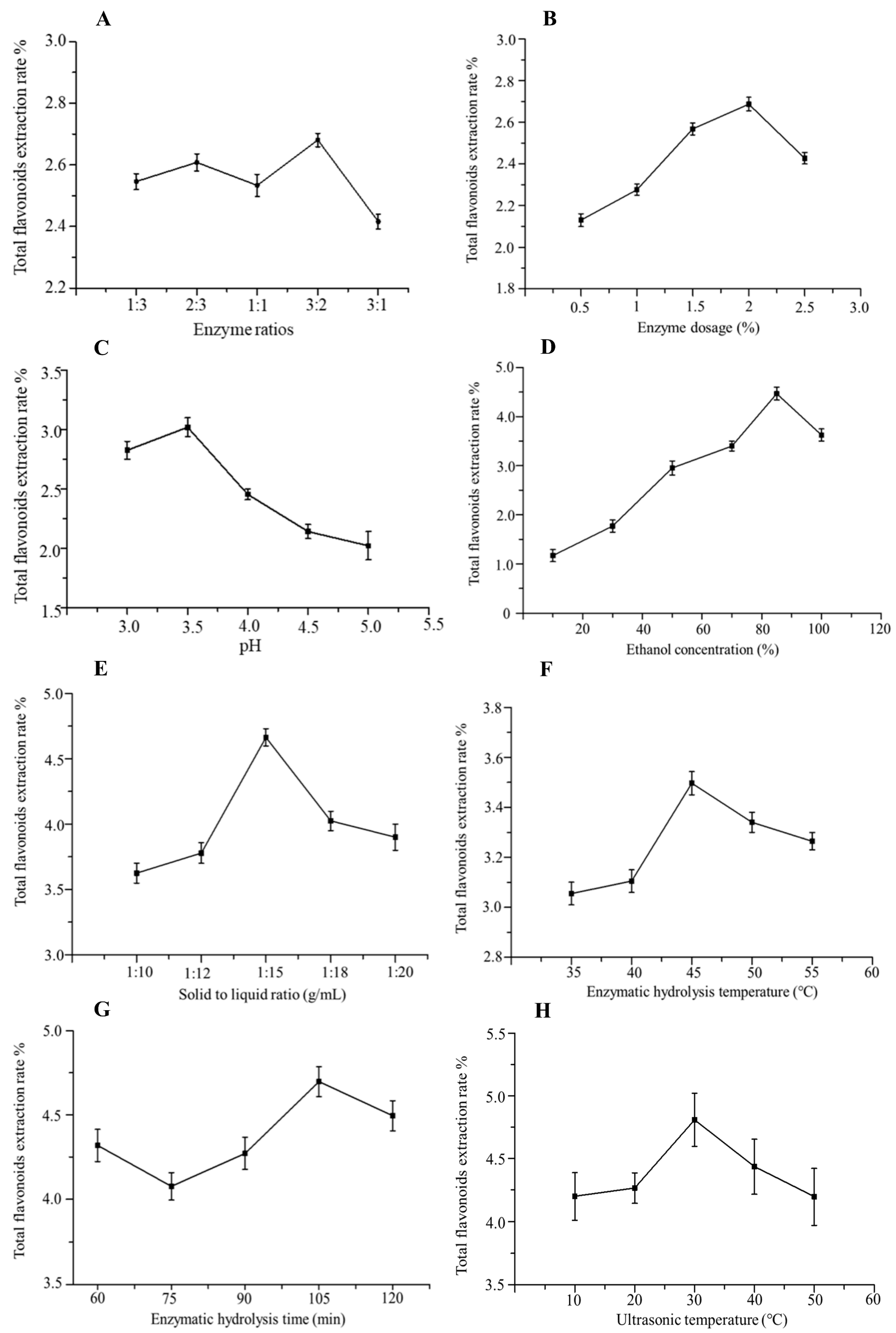
2.2.1. Effect of Compound Enzyme Ratio on the Extraction Rate of Total Flavonoids

The fixed solid–liquid ratio was 1:10, the amount of the enzyme was 1.0%, pH was 4.0, the enzymatic hydrolysis temperature was 50 °C, the enzymatic hydrolysis time was 90 min, and the sonication time was 20 min. The effects of cellulase and pectinase at different ratios (1:3, 2:3, 1:1, 3:2, 3:1 g/g) on extracting total flavonoids in A. absinthium L. were investigated. As shown in Figure 1A, the total flavonoid yield showed a trend of increasing and then decreasing with the increased cellulase ratio. When the ratio of cellulase to pectinase was 3:2, the total flavonoid yield reached 2.68%. It is possible that the increased cellulase ratio may have a stronger effect on the damage of the cell wall and intercellular matrix of A. absinthium L., which effectively leads to the release of intracellular components. However, when the cellulase ratio reached a certain degree, some fibers were attached to the surface of A. absinthium L., which prevented the release of total flavonoids, resulting in a decrease in the total flavonoid yield. Therefore, the ratio of cellulose to pectinase was chosen to be 3:2.

2.2.2. Effect of Enzyme Dosage on the Extraction Rate of Total Flavonoids

Based on the above-optimized compound enzyme ratio, the solid–liquid ratio was fixed at 1:10, pH was 4.0, the enzymatic hydrolysis temperature was 50 °C, the enzymatic hydrolysis time was 90 min and the sonication time was 20 min. The effects of the amount of compound enzyme (0.5%, 1.0%, 1.5%, 2.0%, 2.5%) on the total flavonoid yield of A. absinthium L. were investigated. As shown in Figure 1B, the total flavonoid yield significantly increased with the increase in the amount of the enzyme compound and then decreased. When the amount of enzyme was 2.0%, the total flavonoid yield reached the maximum value of 2.72%. Therefore, the optimal amount of the enzyme is 2.0%.

2.2.3. Effect of pH on the Extraction Rate of Total Flavonoids

The optimum pH of cellulase for the experiment was between 4.0 and 6.5, and the optimum pH of pectinase was between 3.0 and 6.0. Based on the above-optimized ratio and amount of the complex enzymes, the solid–liquid ratio was fixed at 1:10, the enzymatic hydrolysis temperature was 50 °C, the enzymatic hydrolysis time was 90 min, and the sonication time was 20 min. The effects of pH (3.0, 3.5, 4.0, 4.5, 5.0) on the total flavonoid yield of A. absinthium L. were investigated. As shown in Figure 1C, the total flavonoid yield showed a trend of increasing and then decreasing with the increase in pH from 3.0 to 5.0. When the pH was 3.5, the total flavonoid yield reached a maximum of 3.07%. Therefore, a pH of 3.5 was chosen for the enzymatic hydrolysis.

2.2.4. Effect of Ethanol Concentration on the Extraction Rate of Total Flavonoids

Based on the above-optimized ratio and dosage of complex enzymes and pH, the solid–liquid ratio was fixed at 1:10, enzymatic hydrolysis temperature was 50 °C, enzymatic hydrolysis time was 90 min, and sonication time was 20 min. The effects of ethanol concentrations (10%, 30%, 50%, 70%, 85%, and 100%) on the yield of total flavonoids in A. absinthium L. were investigated. As shown in Figure 1D, a lower ethanol concentration is generally suitable for extracting polar flavonoids, and a higher ethanol concentration is ideal for extracting non-polar flavonoids. When the concentration of ethanol increased from 10% to 85%, the extraction amount of total flavonoids was increased and reached the highest amount at 85% ethanol. Therefore, the optimum ethanol dosage was 85%.

2.2.5. Effect of Solid–Liquid Ratio on the Extraction Rate of Total Flavonoids

Based on the above-optimized ratio and amount of complex enzymes, pH, and ethanol concentration, the enzymatic hydrolysis temperature was 50 °C, the enzymatic hydrolysis time was 90 min, and the sonication time was 20 min. The effects of solid–liquid ratios (1:5, 1:8, 1:10, 1:12, 1:15, 1:20 g/mL) on the total flavonoid yield of A. absinthium L. were investigated (Figure 1E). When the solid–liquid ratio was 1:10 (g/mL), it was difficult for the material to be completely immersed in the solvent, resulting in incomplete extraction. When the solid–liquid ratio was 1:15 (g/mL), the maximum yield of total flavonoids reached 1.24%. Therefore, 1:15 (g/mL) was chosen as the best solid–liquid ratio.

2.2.6. Effect of Enzymatic Hydrolysis Temperature on the Extraction Rate of Total Flavonoids

Based on the above-optimized compound enzyme ratio and dosage, pH, ethanol concentration, and solid–liquid ratio, the enzymatic hydrolysis time was 90 min and the sonication time was 20 min. The effect of enzymatic hydrolysis temperature (30, 40, 50, 60, 70 °C) on the yield of total flavonoids in A. absinthium L. was investigated. As shown in Figure 1F, the total flavonoid yield showed a trend of increasing and then decreasing with the increase in enzymatic hydrolysis temperature. When the enzymatic hydrolysis temperature was 45 °C, the total flavonoid yield reached 3.51%. Therefore, the optimal temperature of enzymatic hydrolysis was 45 °C.

2.2.7. Effect of Enzymatic Hydrolysis Time on the Extraction Rate of Total Flavonoids

Based on the above-optimized compound enzyme ratio and amount, pH, ethanol concentration, solid–liquid ratio, and enzymatic hydrolysis temperature, the effect of enzymatic hydrolysis time (45, 60, 75, 90, 105, 120 min) on the yield of total flavonoids in A. absinthium L. was investigated by fixing the sonication time of 20 min. The compound enzyme failed to destroy the cell wall sufficiently under a shorter enzymatic hydrolysis time, which resulted in a low total flavonoid yield. When the enzymatic hydrolysis time was extended to 105 min, the total flavonoid yield reached 4.59% (Figure 1G). Therefore, the enzymatic hydrolysis time of 105 min was chosen.

2.2.8. Effect of Ultrasonic Temperature on the Extraction Rate of Total Flavonoids

Based on the above-optimized ratio and dosage of complex enzymes, pH, ethanol concentration, solid–liquid ratio, and enzymatic hydrolysis temperature and time, the effects of sonication temperature (20 °C, 40 °C, 60 °C, 80 °C, 100 °C, 120 °C) on the yield of total flavonoids in A. absinthium L. were investigated. The increase in sonication temperature has a certain promotional effect on the extraction of total flavonoids between 10 °C and 30 °C. The highest total flavonoid yield reached 4.75% at 30 °C (Figure 1H). Therefore, the optimal sonication temperature of 30 °C was selected.

2.3. Analysis of Extraction Parameters Using Response Surface Method

2.3.1. Response Surface Experimental Design

Response surface methodology is an important method in condition optimization, and only a few experiments are required to obtain statistically acceptable results. Based on the single-factor experiments, three factors (solid–liquid ratio (A), enzymatic hydrolysis temperature (B), and ethanol concentration (C)) with significant effects on the extraction rate of total flavonoids from A. absinthium L. were selected for the three-level response surface method experiments (Table 1). The 17 response values for different combinations are shown in Table 2. Applying a quadratic linear regression fitted to the data and the significance and variance analysis yielded the following binary, multinomial regression model. Y = 3.81 − 0.19A + 3.424E − 003B + 0.16C − 0.27AB + 0.11AC − 0.026BC − 0.68A2 − 0.47B2 − 0.45C2. As shown in Table 3, the regression model was highly significant (p < 0.01), and the out-of-fit term p = 0.1379 was not significant (confidence level of 95%), indicating a good fit of the model. The correlation coefficient of the regression model equation R2 = 0.9373 indicates that the correlation of the predicted and true values is high, and the error is small, which can well reflect the experimental data. The CV value indicates the accuracy of the test, and the model’s CV value is 7.14%, suggesting the test is reliable. One equation A < 0.05 is significant, B and C > 0.05 is not significant, binomial A2, B2, and C2 < 0.01 is highly significant, and interaction term AB < 0.05 is significant, while AC and BC > 0.05 is not significant. It indicates that the effect of each experimental factor on the response value is not a simple linear relationship but a non-linear relationship. Examining the magnitude of the p-value and F-value, the degree of influence of each factor on the total flavonoid yield can be obtained as A > C > B.

2.3.2. Response Surface Analysis
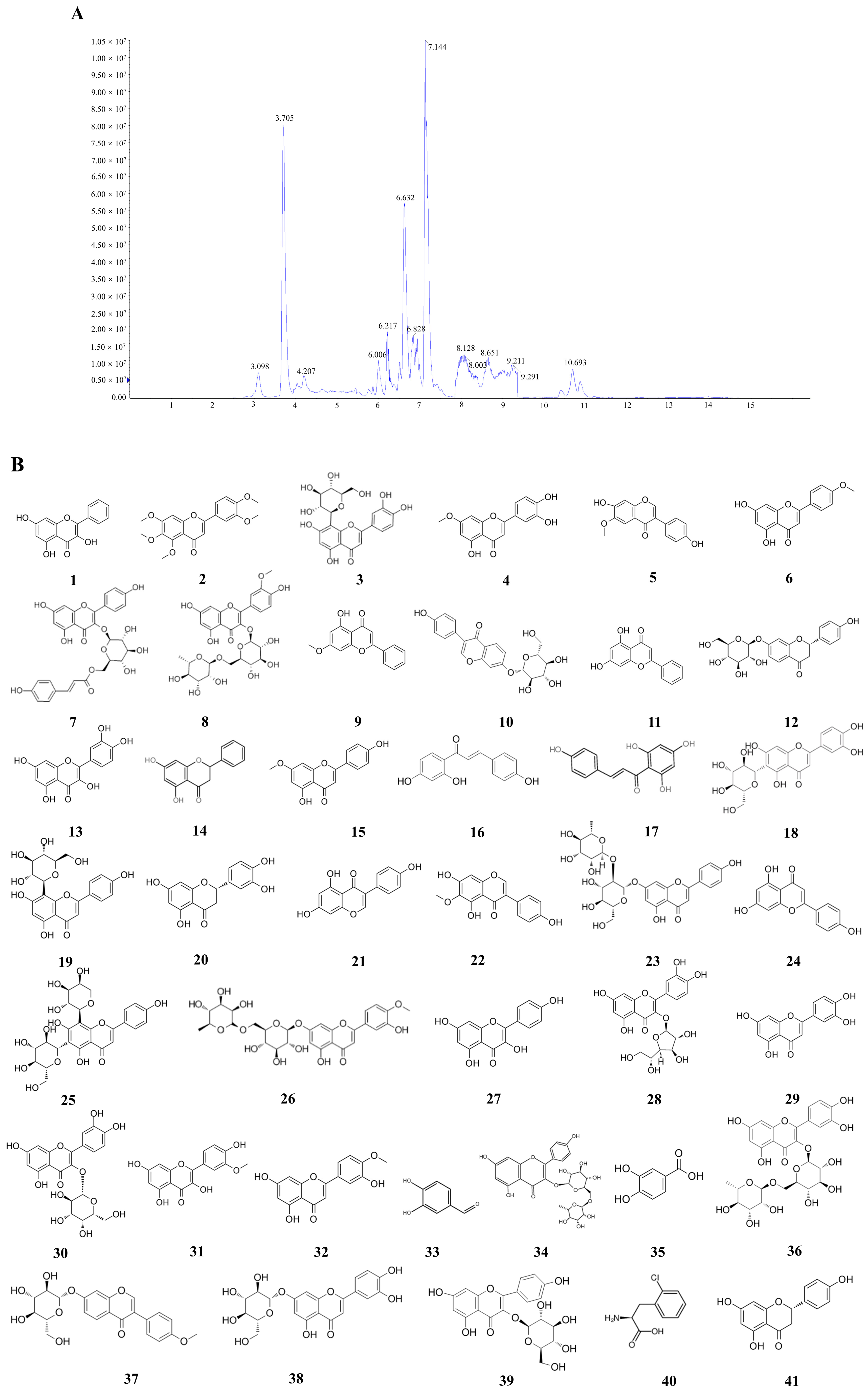
The steeper the response surface, the greater the effect of the factor on the total flavonoid yield and the more sensitive it is to the operating conditions. The contour lines can reflect the significance of the interaction between the factors. The oval shape indicates that the interaction between the two elements is significant, while the circle shows no significant change. As shown in Figure 2A, the response surface of the interaction term AB is steeper. The elliptical shape of the contour line shows that there is a significant interaction between the temperature of the enzymatic hydrolysis and the solid–liquid ratio that influences the rate of extraction of total flavonoids from A. absinthium L. Instead of increasing initially and then declining with an increase in enzymatic hydrolysis temperature, the extraction rate of total flavonoids increased progressively with the increase in solid–liquid ratio when the enzymatic hydrolysis temperature was fixed. The interaction terms AC (Figure 2B) and BC (Figure 2C) were relatively smooth, i.e., the interaction was weak, and the contours were approximately circular, indicating the interactions between the solid–liquid ratio and ethanol concentration and enzymatic hydrolysis temperature and ethanol concentration were not significant. The effect on the extraction rate of total flavonoids was not significant. The effect on the extraction rate of total flavonoids was negligible. The optimal conditions for extraction of total flavonoids from absinthe were solid–liquid ratio 1:14.6, ethanol concentration 87.45%, ultrasonic temperature 30 °C, enzyme ratio 3:2, enzyme dosage 2%, enzymatic hydrolysis temperature 45.19 °C, enzymatic hydrolysis time 105 min, and enzymatic hydrolysis pH 3.5. In order to facilitate the experimental operation, the ideal extraction conditions were established: solid–liquid ratio 1:15, ethanol concentration 85%, ultrasonic temperature 30 °C, enzyme ratio 3:2, enzyme dosage 2%, enzymatic hydrolysis temperature 45 °C, enzymatic hydrolysis time 105 min, enzymatic hydrolysis pH 3.5. The results showed that the yield of total flavonoid content was 3.8%. The results showed that the total flavonoid content of A can be extracted by the surface reaction method under optimum technical conditions.

2.4. LC-MS

LC-MS was used to qualitatively and quantitatively analyze the main components of total flavonoids in A. absinthium. The total ion flow chromatogram of total flavonoids showed that 41 major peaks were well separated (Figure 3). Based on the information of the excimer ion peaks given by the primary mass spectrometry and the structural fragmentation information of the secondary mass spectrometry, combined with the retention time of UPLC and the references, the possible structures of 41 chemical components were deduced, including 37 flavonoids, 2 phenolic compounds, 1 terpenoid and 1 amino acid and its derivatives (Figure 3B, Table 4).

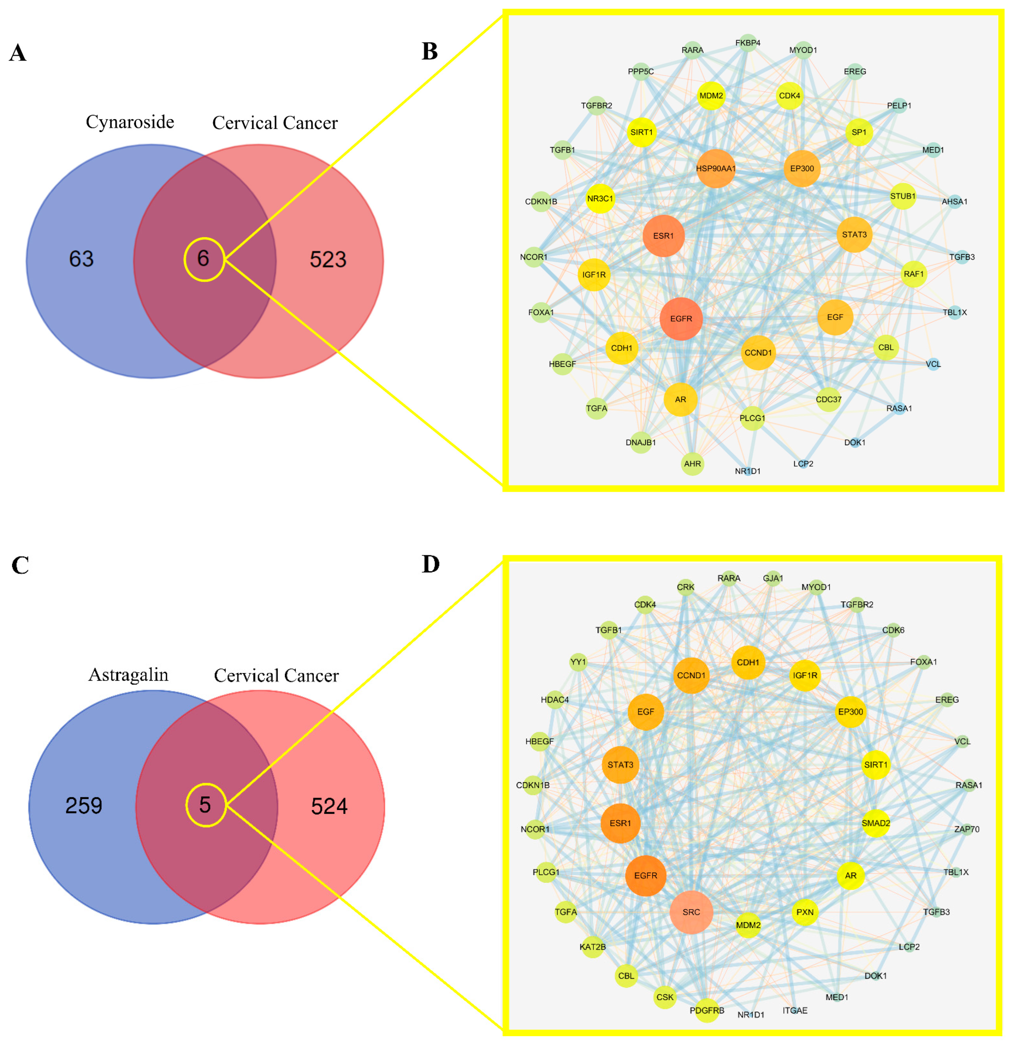
2.5. Screening Mutual Targets of Drugs and Diseases

The core active ingredient targets of total flavonoids were matched with cervical cancer targets, from which the two main active ingredient compounds (Cynaroside and Astragalin) were selected to have six and five compound targets, respectively (Figure 4A,C). The target interaction PPI network graph (Cynaroside) was obtained with the STRING online analysis platform with 46 nodes and 331 edges, and the average node degree value was 14.4. The PPI network showed (Figure 4B) that EGFR, ESR, HSP90AA1, EP300, STAT3, EGF, and CCND1 were the main targets for cervical cancer. The PPI network graph (Figure 4D, Astragalin) had a total of 45 nodes and 279 edges with an average node degree value of 12.4. The PPI network showed that SRC, EGFR, ESR1, STAT3, EGF, CCND1, CDH1, and IGF1R were the main targets of cervical cancer.

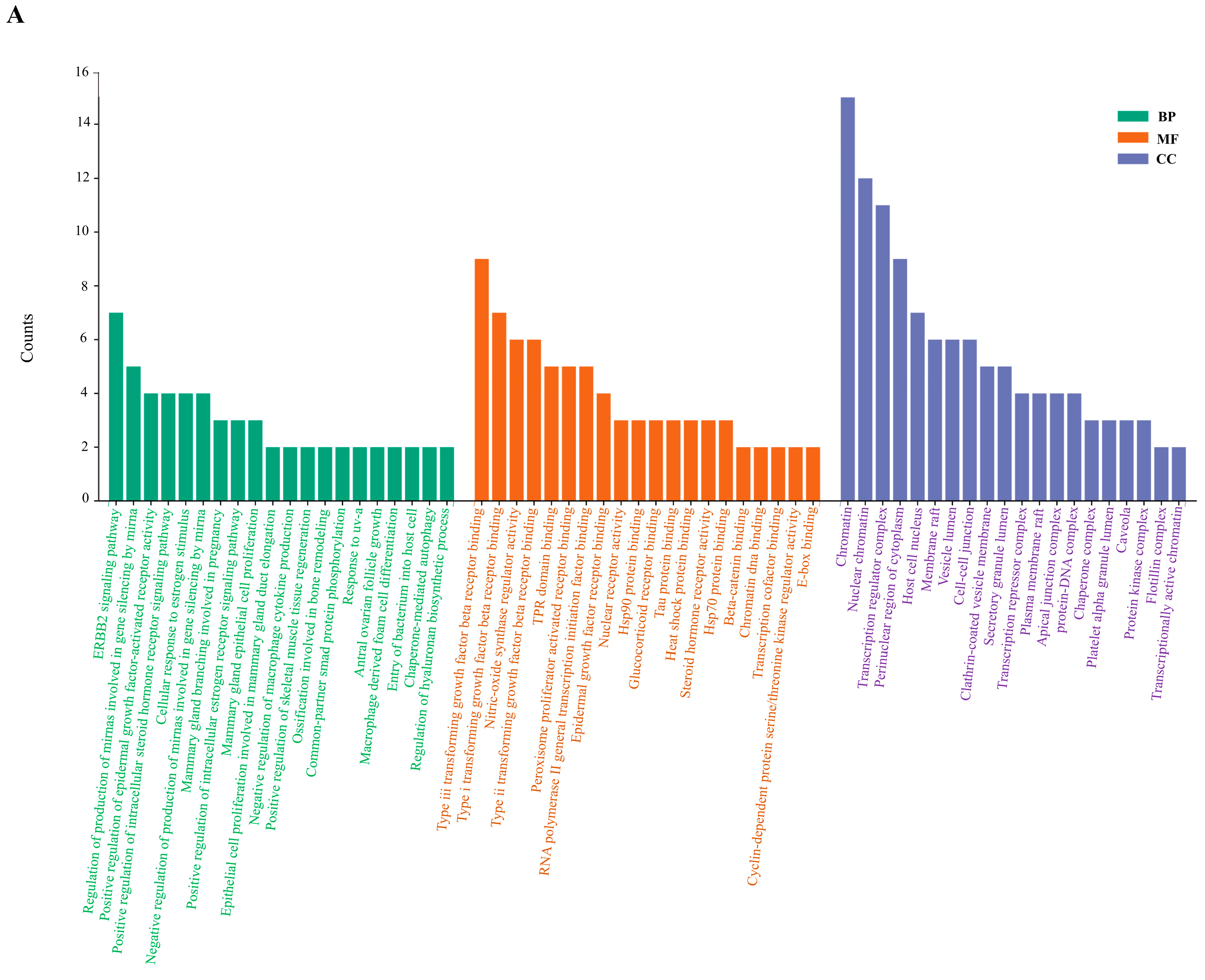
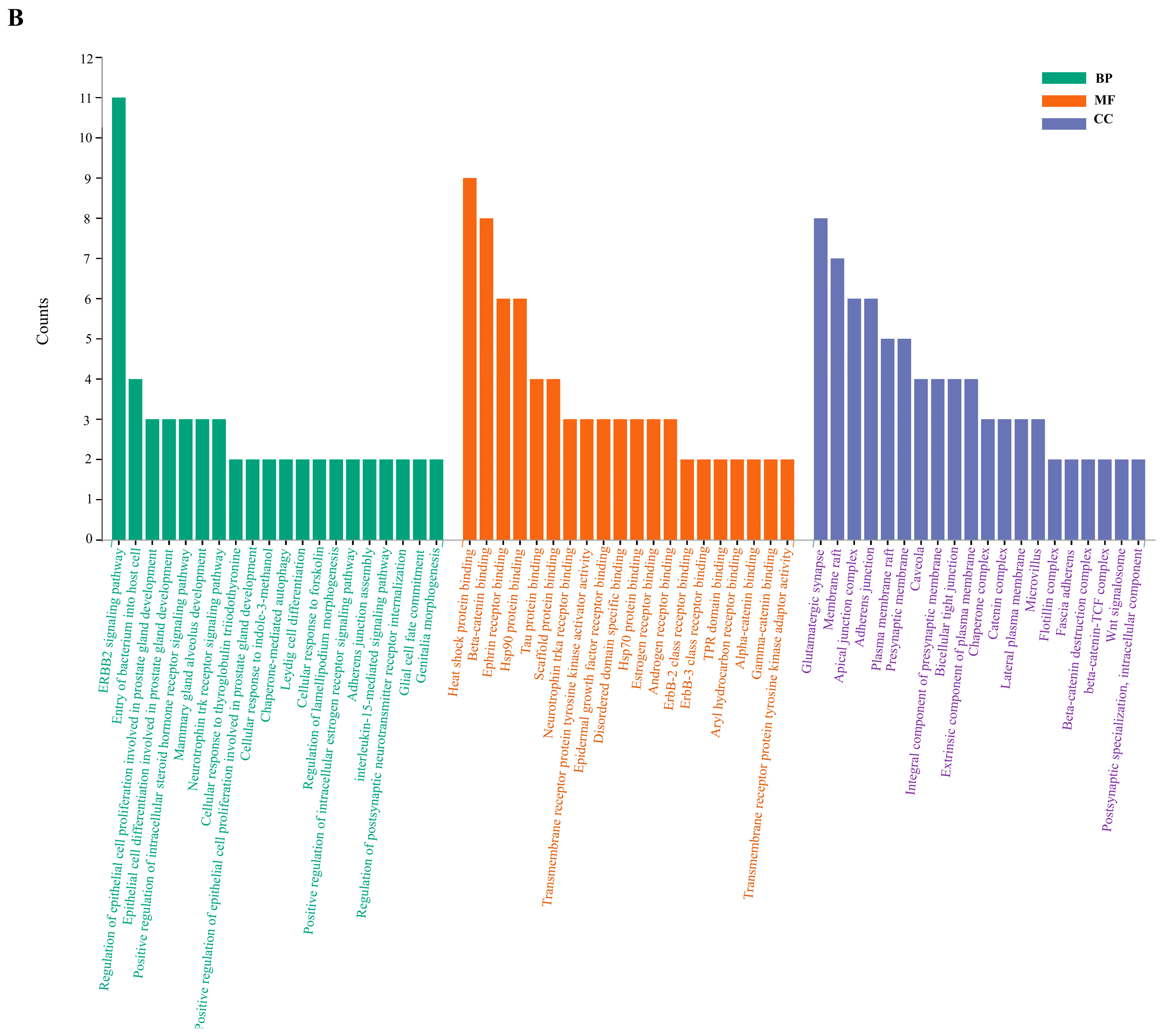
2.6. Enrichment Analysis of GO and KEGG Pathway

In the GO functional enrichment analysis of key targets, GO terms were screened according to p (p < 0.05, Q < 0.05) values. There were 590, 40, and 67 GO terms associated with biological processes, cellular components, and molecular functions (Cynaroside), respectively. As shown in Figure 5A, the first 20 biological processes were associated with the ERBB2 signaling pathway, epidermal growth factor, cellular response to estrogen stimulus, and mammary gland epithelial cell proliferation, among others. For cellular components, the targets are enriched in Chromatin and Nuclear chromatin. Molecular functional analysis revealed Type iii transforming growth factor beta receptor binding, Type i transforming growth factor beta receptor binding, Nitric-oxide synthase regulator activity, and Transcription regulator complex. Another active component had 418, 45, and 50 GO terms related to biological processes, cellular components, and molecular functions, respectively (Astragalin). As shown in Figure 5B, the first 20 biological processes related to the ERBB2 signaling pathway, entry of bacterium into host cell, regulation of epithelial cell proliferation involved in prostate gland development, epithelial cell differentiation involved in prostate gland development, and positive regulation of intracellular steroid hormone receptor signaling pathway. For cellular components, the targets are enriched in Flotillin complex, Fascia adherens, Beta-catenin destruction complex, beta-catenin-TCF complex, and Wnt signalosome. Molecular functional analyses revealed heat shock protein binding, Beta-catenin binding, Ephrin receptor binding, Hsp90 protein binding, and Tau protein binding. KEGG signaling pathway enrichment analysis contained 93 pathways (Cynaroside) and 83 pathways (Astragalin), respectively. Screening enrichment of the top 20 pathways included bladder cancer, pancreatic cancer, endocrine resistance, glioma, chronic myeloid leukemia, non-small cell lung cancer, prostate cancer, colorectal cancer, melanoma and FoxO signaling pathway (Figure 5C); and bladder cancer, endometrial cancer, prostate cancer, glioma, thyroid cancer, and ErbB signaling pathway (Figure 5D).

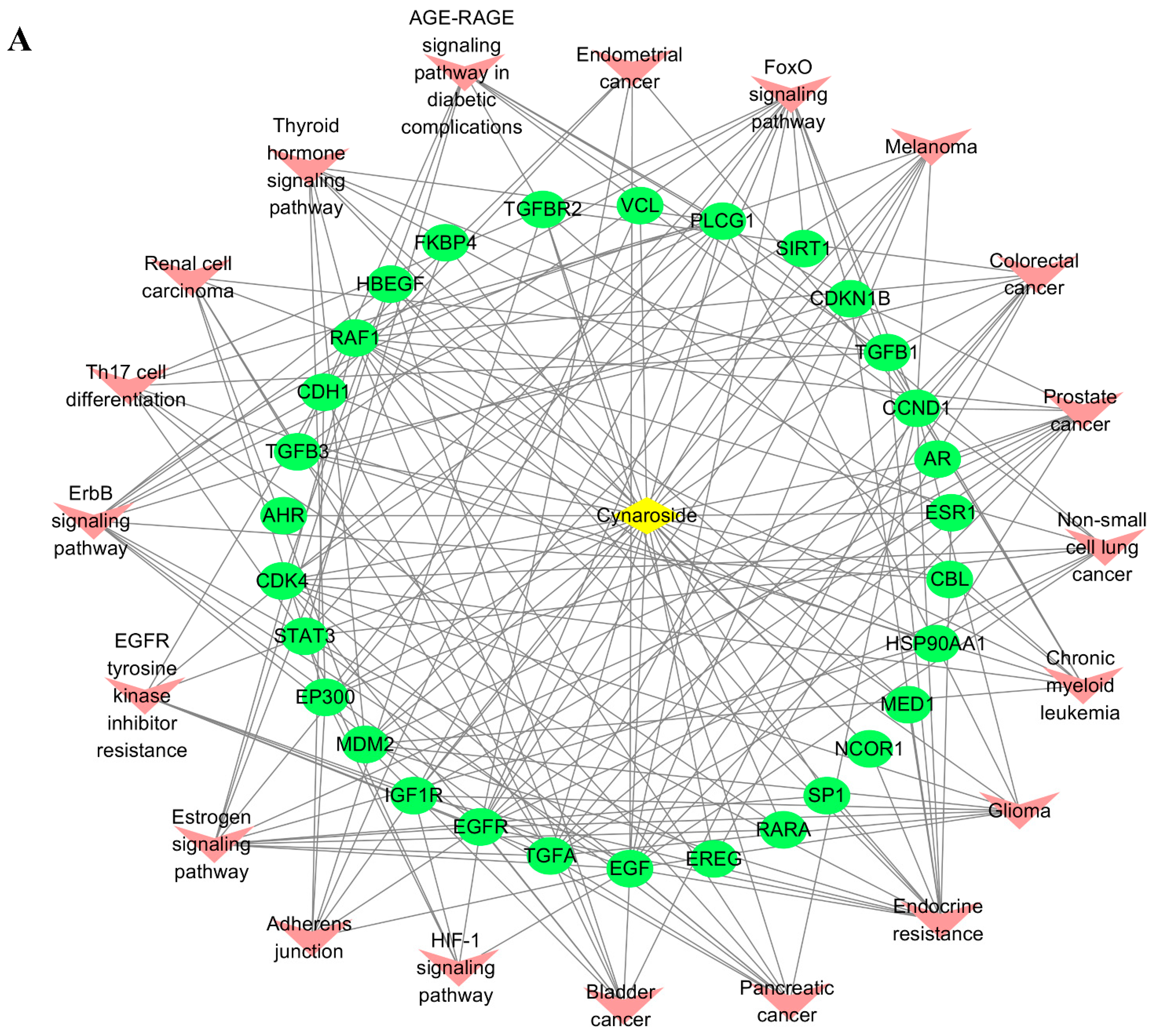
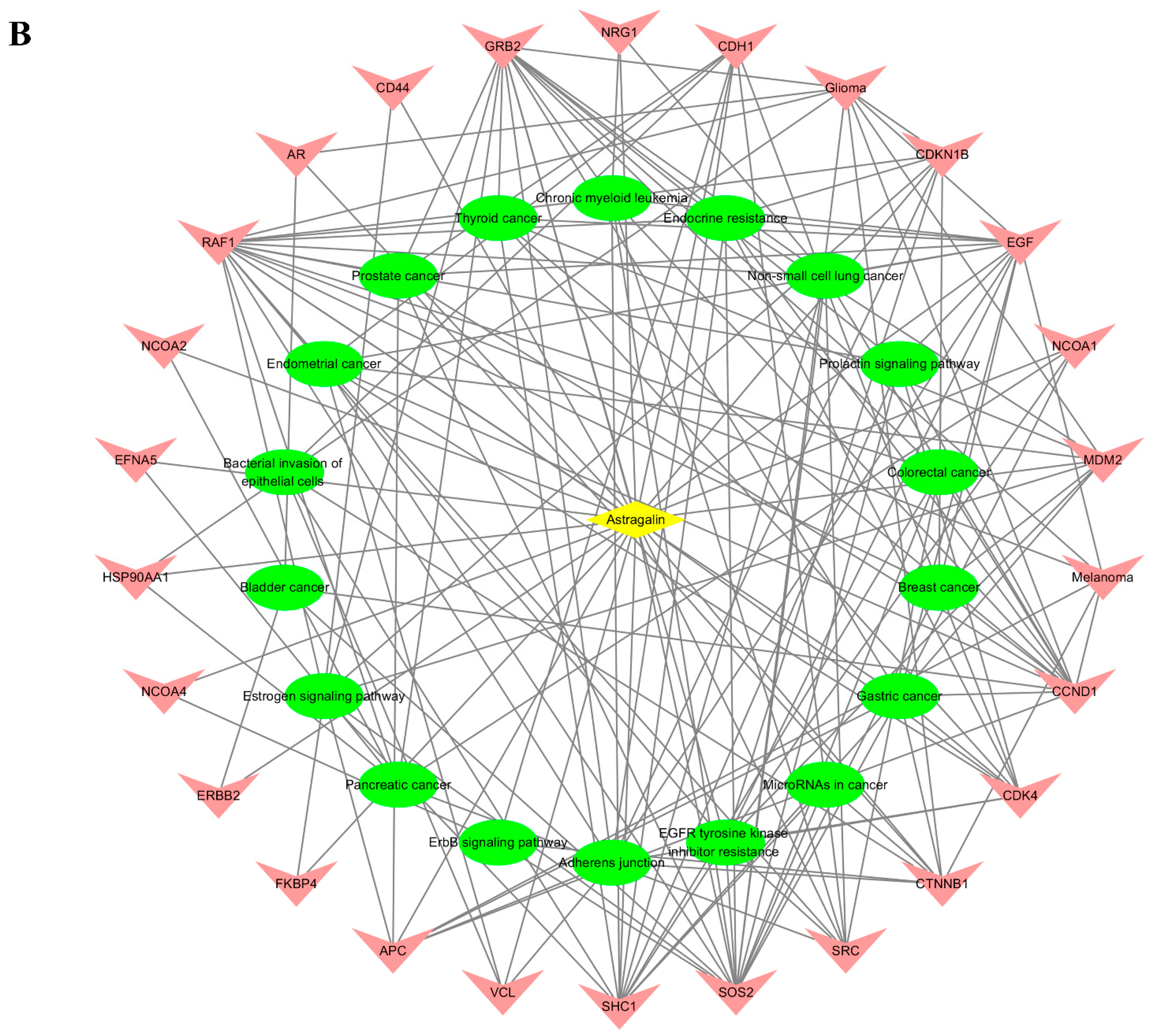
2.7. Active Ingredients–Shared Key Targets–Signal Pathway Network Diagram Construction

A component–target–channel network diagram was formed using Cytoscape 3.7.2 to comprehensively clarify the mechanism of active ingredients in cervical cancer (Figure 6). In the figure, the yellow diamond represents the component in the drug, the green oval represents the target of the component, and the pink V represents the pathway enriched in the target. It shows that each active compound can act on multiple targets, that each target can also respond to multiple active ingredients, and that each target can have an effect on multiple pathways.

2.8. Key Active Ingredients and Core Target Molecular Docking Results

Molecular docking results of key active ingredients and core targets. Molecular docking was performed for the core targets with the highest degree of PPI network and their corresponding active ingredients. The docked protein data were downloaded from the PDB database for EGFR (PDB ID 5wb7) and SRC (PDB ID 4hxj). The 3D structures were imported into AutoDock 1.5.7 software and docked with the active compounds. The core targets in the PPI network and their corresponding small molecule drug ligands were docked using AutoDock. The lower the binding energy, the better the ligand will bind to the protein. The molecules with the lowest binding energy were visualized using Pymol 2.2.0 software (Figure 7). The main form of interaction is hydrogen bonding. This result suggests that their combination may play an important role in the treatment of cervical cancer with total flavonoids.

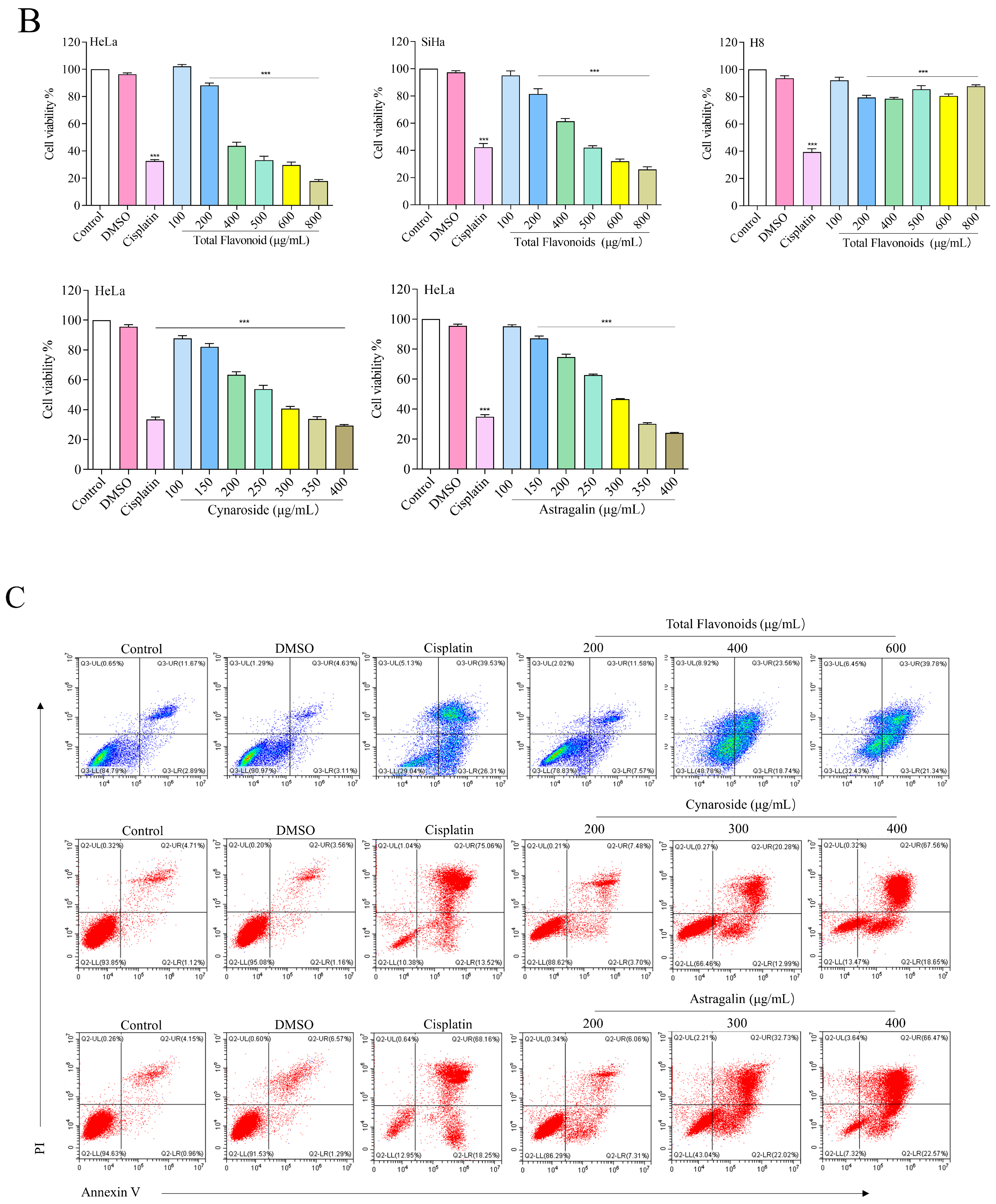
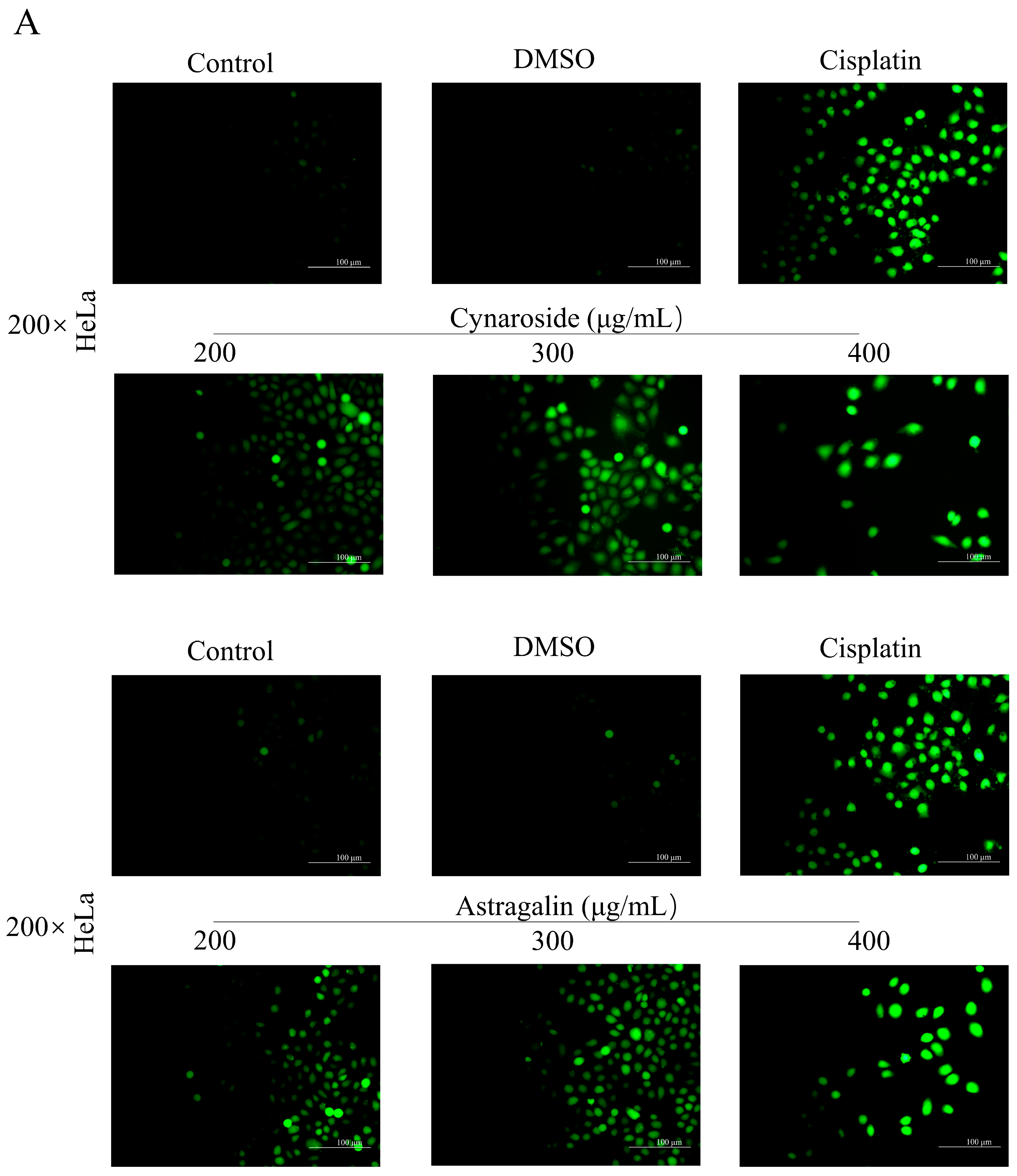
2.9. Total Flavonoids and Active Ingredients Can Inhibit Cell Proliferation and Induce Apoptosis

To evaluate the antitumor effect of total flavonoids from A. absinthium L., HeLa cells were treated with different concentrations of total flavonoids and their active ingredients for 24 h. The morphological changes of the cells were observed by inverted microscopy. HeLa cells showed a small and round morphology, with a significant decrease in cell number. The active ingredients Cynaroside and Astragalin also showed similar results (Figure 8A). In addition, the inhibitory effect of total flavonoids on the proliferation of HeLa, SiHa, and H8 cells was examined, and the activity of Cynaroside and Astragalin was also examined against HeLa, respectively. As shown in Figure 8B, total flavonoids inhibited the growth of human cervical cancer cells HeLa and SiHa in a dose-dependent manner, and the 24 h semi-inhibitory concentration (IC50) values were 396.0 ± 54.2 μg/mL and 449.0 ± 54.8 μg/mL, respectively. Interestingly, total flavonoids had a weak effect on the growth of human immortalized human cervical normal epithelial cells H8. Meanwhile, Cynaroside and Astragalin also inhibited the activity of HeLa cells in a dose-dependent manner with IC50 of 263.4 ± 3.7 μg/mL and 282.3 ± 5.0 μg/mL (Figure 8B). The effect of total flavonoids on HeLa cell apoptosis was further detected. After Annexin V-FITC/PI staining, the assay was performed by flow cytometry. The total flavonoids significantly induced HeLa cell apoptosis in a dose-dependent manner while causing a low level of necrosis. The results suggested that total flavonoids of A. absinthium L. had good anti-cervical cancer activity. Subsequently, we further tested whether the inhibitory effect of Cynaroside and Astragalin on the growth of HeLa cells was also associated with the induction of apoptosis. The results showed that low and high concentrations inhibited the proliferation of HeLa cells. Cynaroside and Astragalin significantly increased the percentage of apoptotic HeLa cells in a concentration-dependent manner compared to the control group (Figure 8C). Apoptosis of HeLa cells was further observed by Hoechst 33342 staining after 24 h with Cynaroside and Astragalin. The results revealed that most untreated cells showed round, intact, and uniformly stained nuclei. In contrast, the drug-treated group showed a bright blue color with increasing concentration, as the nucleus structure was altered and the chromatin of the nucleus underwent concentration-dependent fixation and breakage (Figure 8D).

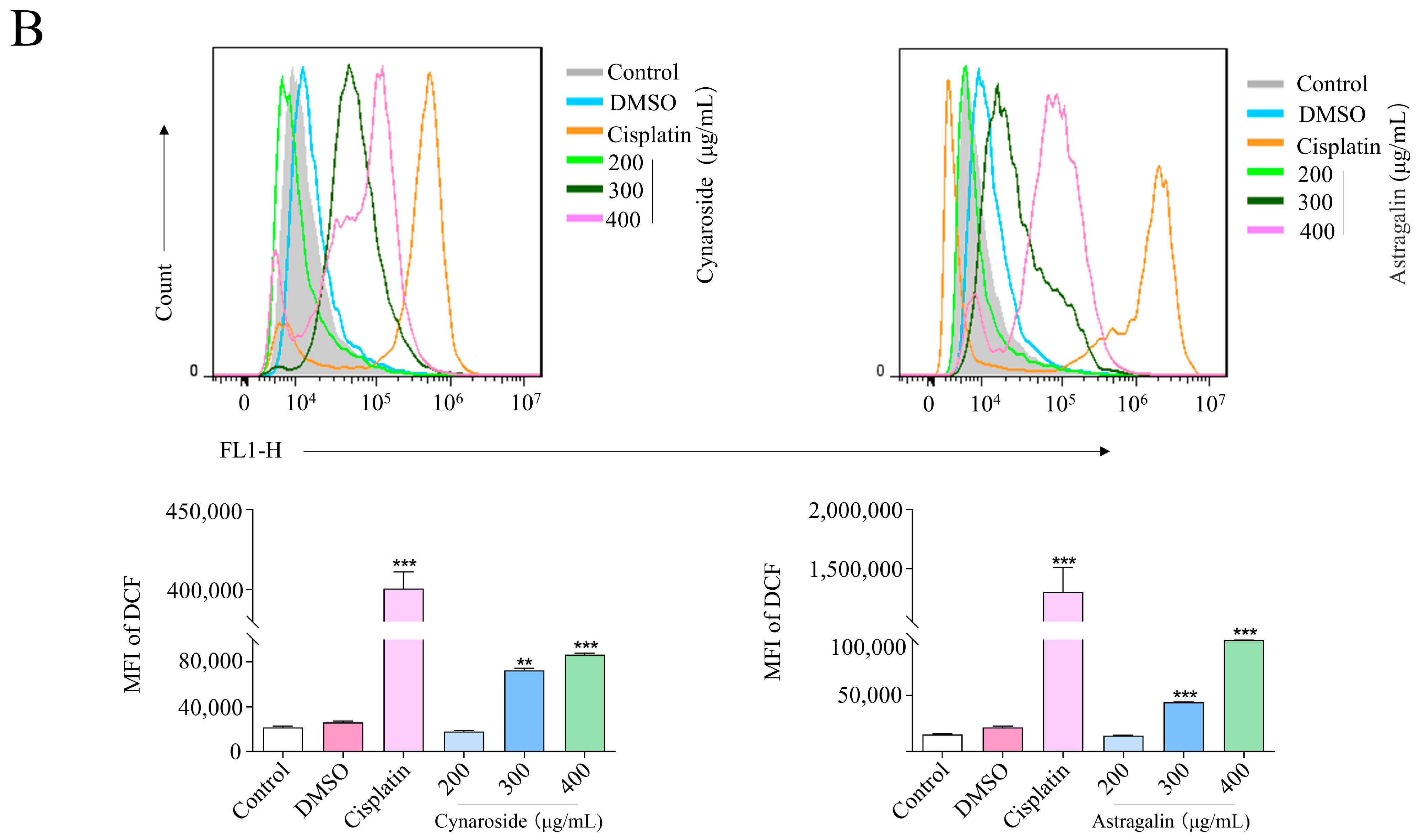
2.10. Induction of ROS Accumulation by Active Ingredients of Total Flavonoids

Reactive oxygen species (ROS), a signaling molecule in cell regulation, can induce apoptosis. ROS dose plays an important role not only in energy metabolism but also in cellular physiological processes. Therefore, DCF-DA, a specific fluorescent dye for ROS detection, was used as a fluorescent probe to assess the changes in intracellular ROS levels after Cynaroside and Astragalin treatment of HeLa cells. DCF-DA rapidly crosses the cell membrane and is converted to DCFH by intracellular lipases. After staining, Cynaroside and Astragalin-treated cells showed green fluorescence in a dose-dependent manner (Figure 9A). Flow cytometry results showed a dramatic increase in cell fluorescence after 24 h of treatment compared to the control group (Figure 9B). Together, these results suggest that ROS plays an essential role in Cynaroside and Astragalin-induced apoptosis in HeLa cells.

3. Discussion

To date, significant efforts have been made to develop antitumor drugs. Natural products have been given attention due to their considerable bioactivities and safety. The extraction methods of natural components mainly include heating, reflux, immersion, organic solvent extraction, microwave-assisted extraction, and enzymatic hydrolysis methods, etc. In order to improve the extraction rate of total flavonoids in A. absinthium L., the combination of the ultrasonic extraction method with the enzymatic hydrolysis method was performed in this study, which has been widely used in the extraction of active ingredients from plants due to advantages including convenience, efficiency, and environmental friendliness. Cellulase can decompose plant tissues under mild conditions and accelerate the release of active ingredients, and ultrasonics can accelerate the dissolution of intracellular substances. Therefore, the combination of the two methods greatly improves the extraction efficiency of plant active ingredients. To a certain extent, it also significantly reduces the extraction cost and is greener and healthier. In addition, we conducted the design test and data analysis of ultrasound-assisted enzymatic extraction of total flavonoids by a single-factor test based on response surface optimization. It overcomes the drawback that the orthogonal test design can only deal with discrete level values but cannot find the best combination of factors and the optimal response face value over the whole region. At the same time, it makes up for the lack of interaction effects and other mixed effects. It can also reduce the number of experiments and time consumption. Similarly, Saliha et al. used a response surface optimization ultrasound-assisted extraction method to extract total phenols from A. absinthium L. and determined the optimal conditions for maximum extraction of active ingredients [154]. Dai et al. [155] extracted flavonoids from Saussurea involucrate by ultrasonication, and Xu et al. [156] extracted total flavonoids from sea red fruits by enzymatic hydrolysis. Yun et al. [157] found that the extraction of flavonoids baicalein and wogonin from Scutellaria baicalensis roots by ultrasound-assisted enzymatic hydrolysis almost doubled the conversion rate and significantly increased the productivity compared to the enzymatic hydrolysis method. Cheng et al. [158] reported that the extraction rate of flavonoids by ultrasonic-assisted extraction could reach 2.980 mg/g. Ou et al. [159] found the highest total flavonoids content achieved was 1.99 ± 0.02 g RE/100 g by a combination of supercritical CO2 and ultrasound. Thus, we effectively extracted the total flavonoids from A. absinthium L. by ultrasound-assisted enzymatic digestion in a time-saving manner.
Furthermore, total flavonoids of A. absinthium L. were prepared using the optimal extraction conditions, and their antitumor activity was evaluated. The results showed that total flavonoids of A. absinthium L. had good anti-cervical cancer activity. Sultan et al. found that extracts of A. absinthium L. could promote apoptosis in colon cancer cells through activation of the mitochondrial pathway [16]. It was also reported that extracts of A. absinthium L. were able to mediate the growth and apoptosis of hepatocellular carcinoma cells through endoplasmic reticulum stress and mitochondria-dependent pathways [9]. In addition, extracts of A. absinthium L. have anticancer and antioxidant effects on various tumors by increasing the intracellular amount of free radicals in cancer cells, which leads to DNA damage and, consequently, apoptosis [23]. As such, the anticancer activity of the active components of total flavonoids was also evaluated. In previous studies, it was confirmed that apoptosis is a physiological process of cells. Cancer occurs when this balance is disrupted, either by an increase in cell proliferation or a decrease in cell death [160]. Moreover, apoptosis induction is considered the primary pathway by which biological agents inhibit the proliferation of tumor cells. Thus, our study tested whether the inhibition of the proliferation of cervical cancer HeLa cells by Cynaroside and Astragalin was achieved through apoptosis. As we expected, Cynaroside and Astragalin significantly increased the rate of apoptosis in HeLa cells in a dose-dependent manner. The results suggest that Cynaroside and Astragalin inhibit the proliferation of cervical cancer cells by inducing apoptosis. In addition, we found that Cynaroside and Astragalin apoptosis induction may be associated with intracellular ROS levels in HeLa cells, and the excessive production of ROS leads directly to apoptosis. Therefore, ROS accumulation is considered one of the main targets of cancer therapy. Yuan et al. [161] found that Isoliquiritigenin induced apoptosis by increasing intracellular ROS levels in HeLa cells. Lim et al. [162] found that indole-3-carbinol mediated apoptosis in lung cancer H1299 cells by increasing ROS production. These are consistent with our results, which showed that Cynaroside and Astragalin inhibit the development and progression of cervical cancer HeLa cells by increasing the accumulation of ROS.

4. Materials and Methods

4.1. Materials and Reagents

A. absinthium L. powder (Purchased from Xinjiang Urumqi Tianyi Feng Biotechnology Co., Ltd., Urumqi, China, provided by Xinjiang Baokang Pharmaceutical Co., Ltd., and crushed to 200 mesh by high-speed universal pulverizer). The source of the A. absinthium L. provided by Xinjiang Baokang Pharmaceutical Co., Ltd. is in line with the requirements of the Plant Protection Law of the People’s Republic of China and the Biosafety Law of the People’s Republic of China and other relevant regulations and standards: the quality of the product is in line with the Drug Administration Law, the Good Manufacturing Practices for Drugs, the Good Manufacturing Practice for Drugs, and the Good Manufacturing Practices for Drugs, and other relevant regulations and standards. Ethanol, quercetin control (Shanghai Maclean Biochemical Technology Co., Ltd., Shanghai, China), sodium nitrite, aluminum nitrate, sodium hydroxide, aluminum trichloride, potassium acetate, methanol, ethyl acetate, petroleum ether, isopropanol (Tianjin Xinbote Chemical Co., Ltd., Tianjin, China).

4.2. Methods

4.2.1. Quercetin Standard Curve Plotting

Quercetin 5.0 mg was weighed precisely and fixed to 25 mL with methanol to obtain a mass concentration of 0.2 mg/mL standard solution, which was stored in a refrigerator at 4 °C for use. Amounts of 0.1 mL, 0.2 mL, 0.3 mL, 0.4 mL, and 0.5 mL of quercetin control solution were added into 10 mL volumetric flasks, and 0.1 mol/mL AlCl3 solution was added for 6 min, then 1 mol/mL C2H3KO2 solution was added for 3 mL, and finally the volume with methanol solution was fixed to the scale. The corresponding solution without quercetin was used as a blank control, 100 μL was aspirated, and the absorbance at 415 nm was measured. The standard curve was plotted with absorbance (y) as the vertical coordinate and quercetin concentration (x) as the horizontal coordinate.

4.2.2. Extraction of Total Flavonoids

After weighing 2.0 g of A. absinthium L. powder in a 50 mL centrifuge tube, ethanol solution at a ratio of 1:10 was added, adding a certain amount of complex enzymes (cellulase and pectinase) according to the mass ratio of A. absinthium L., carrying out enzymatic hydrolysis in a water bath at a certain pH and temperature (mixed once at 30 min), and then inactivating the enzyme in a water bath at 100 °C for 5 min, adding 10 mL of ethanol solution at a concentration of 80%, and finally sonicating the above enzymatic solution at a certain power, sonication temperature, and sonication time. Then, 10 mL of ethanol solution was added, and the above enzymatic solution was sonicated at a certain ultrasonic power, ultrasonic temperature, and ultrasonic time. Finally, the supernatant was taken from the sonicated sample solution after centrifugation, and the absorbance of each solvent was measured at 415 nm with a blank as the control, and the total flavonoid extraction rate and antitumor activity were calculated. (The blank was divided into those with and without enzyme, and the effect of enzyme addition on the yield and activity was evaluated before and after the enzyme addition).

4.2.3. Determination of Total Flavonoid Content

Weigh accurately 2.0 g of the dried powder of A. absinthium L. crushed with a high-speed universal grinder, add a certain volume fraction of aqueous ethanol solution according to a certain solid–liquid ratio; extract with ultrasound for a certain time in an ultrasonic cleaner, filter, and concentrate the extract with a rotary evaporator under reduced pressure to remove the solvent. The concentrated solution was transferred to a 100 mL volumetric flask, and the volume was fixed with ethanol to the scale. Precisely measure 1.0 mL of A. absinthium L. flavonoids extract with aluminum trichloride-potassium acetate method to detect the absorbance value (A) of total flavonoid extract and bring to the standard. The curve regression equation calculated the total flavonoid concentration and then converted it into flavonoid content.
Total flavonoid extraction amount (mg/g) = C × V × N/M
C: concentration of total flavonoids measured (mg/mL);
V: volume of extraction solution (mL);
N: dilution times;
M: mass of raw material (g).

4.2.4. Single-Factor Experimental Design

Weighing 2.0 g of A. absinthium L., the factors that affected the total flavonoid extraction were selected by the controlled variable method for the one-way experiment. Some of the factors are ethanol concentration, enzyme complex ratio, enzyme complex amount, pH, material-liquid ratio, enzyme hydrolysis temperature, enzyme hydrolysis time, sonication temperature, and sonication time. The influence of other factors on the extraction rate of flavonoids from A. absinthium L. was investigated using a single-factor experimental design.

4.3. Optimization of Extraction Conditions of Total Flavonoids by Response Surface Methodology

The results of the single-factor experiments were combined. Three factors, including solid–liquid ratio, enzymatic hydrolysis temperature, and ethanol content, were selected to significantly affect the extraction rate of total flavonoids from A. absinthium L. Regression analysis was performed using DesignExpert 8.0 software to calculate the optimal extraction conditions using a regression equation, and validation test was performed three times in parallel to obtain the optimal extraction conditions by the response surface method and complete analysis of each factor.

4.4. LC-MS

4.4.1. Metabolites Extraction

The sample was thawed on ice. After 30 s vortex, the 200 μL sample was dried under gentle nitrogen flow. The sample was then reconstituted in 200 μL of 50% methanol containing 0.1% formic acid. After vortexing for 30 s, it was subjected to ultrasound in an ice bath for 15 min. The sample was then centrifuged at 12,000 rpm (RCF = 13,800× g, R = 8.6 cm) for 15 min at 4 °C. The resulting supernatant was passed through a 0.22 μm filter membrane. It was then transferred to 2 mL glass vials and stored at −80 °C until analysis by UHPLC-MS/MS.

4.4.2. MS Analysis

AB Sciex QTrap 6500+ mass spectrometer was applied for assay development. Typical ion source parameters were IonSpray Voltage: +5000/−4500 V, Curtain Gas: 35 psi, Temperature: 500 °C, Ion Source Gas 1: 55 psi, Ion Source Gas 2: 60 psi.

4.4.3. Screening of Core Active Ingredients and Targets

PharmMapper (http://lilab-ecust.cn/pharmmapper/, accessed on 6 April 2022) and SwissTargetPrediction databases (http://www.swisstargetprediction.ch/, accessed on 6 April 2022) were used to retrieve the gene targets of the active ingredients. The obtained targets were then mapped to the UniProt database (https://www.uniprot.org/, accessed on 6 April 2022) for normalization.

4.4.4. Disease-Related Gene Mining

Targets associated with cervical cancer were collected from the OMIM database (https://www.omim.org/, accessed on 6 April 2022) and DrugBank database (https://www.drugbank.ca/, accessed on 6 April 2022). Normalization of gene names and definition of species as “human” was performed using the UniProt database. Venn diagrams of active ingredients and cervical cancer targets were mapped using a bioinformatics platform.

4.4.5. Protein–Protein Interaction Network Construction and Analysis

A target PPI network was obtained by compiling potential targets from the STRING database (https://string-db.org/, accessed on 6 April 2022). The species selection parameter was set to “Homo sapiens”, and the minimum required confidence in the interaction score was set to 0.900. The PPI network was then visualized using Cytoscape 3.7.2 software.

4.4.6. Gene Ontology (GO) and the Kyoto Encyclopedia of Genes and Genomes (KEGG) Enrichment

Gene Ontology (GO) and Kyoto Encyclopedia of Genes and Genomes (KEGG) enrichments were performed using Bioconductor Cluster Profiler for core drug group ontology (GO) enrichment analysis of potential target genes and KEGG pathway enrichment analysis of tissues. Pathways were ranked according to the number of molecules in the pathway with a critical value (p < 0.05).

4.4.7. Network Construction

Compound target pathway networks were constructed using Cytoscape 3.7.2. Topological properties were analyzed using Cytoscape’s Network Analyzer plug-in to identify key components and targets.

4.4.8. Molecular Docking Ensures the Interaction between Targets and Key Compounds

Molecular docking ensures interactions between targets and key compounds. Target protein structures were obtained from the RCSB PDB database (http://www.rcsb.org/, accessed on 6 April 2022). Two-dimensional structures of ginseng leaf actives were sought from the PubChem database. ChemBio Draw 3D and Autodock Tool were applied to optimize the structures of key compounds and targets, including 3D chemical structure creation, energy minimization, and format conversion. PyMol was used to process proteins, including removal of ligands, correction of protein structures, and removal of water. Docking was carried out using R 4.1.3 software and Autodock Vina to select the molecule with the lowest binding energy in the docked conformation and observe binding effects by matching to the original ligand and intermolecular interactions.

4.5. Cell Culture

HeLa, SiHa, and H8 cells were obtained from the Xinjiang Key Laboratory of Biological Resources and Genetic Engineering, Xinjiang University (Urumqi, Xinjiang, China) and were cultured in RPMI1640 with DMEM (Gibco, Thermo Fisher Scientific, Waltham, MA, USA) medium having 10% fetal bovine serum (MRC, Changzhou, China), 50 U/mL each of penicillin and streptomycin (MRC, Changzhou, China), respectively, at 37 °C in a humidified environment with 5% CO2.

4.6. MTT

HeLa and SiHa cells at the logarithmic stage were inoculated with 5 × 10³ cells /mL in 96-well plates and treated with different concentrations of total flavonoids (100, 200, 400, 500, 600, and 800 μg/mL) and Cynaroside and Astragalin (100, 150, 200, 250, 300, 350, and 400 μg/mL). Control (blank), 0.3%DMSO (negative control), and 35 μg/mL cisplatin (positive control) were set, with 6 replicates in each group. After 24 h of incubation, the waste liquid was discarded by centrifugation, and 100 μL MTT (0.5 mg/mL) was added to each of them and incubated at 37 °C for 4 h under dark conditions. The supernatant was removed by centrifugation, dissolved with 150 μL DMSO, and the OD value of each well was detected at 490 nm. The following formula calculated cell survival rate: cell viability (%) = (ODtreated/ODcontrol) × 100%.

4.7. Apoptosis

HeLa cells were inoculated with 1 × 10⁵ cells/dish in 60 mm culture dishes, and the cells were treated with different concentrations of total flavonoids (200, 400, and 600 μg/mL), Cynaroside, and Astragalin (200, 300, and 400 μg/mL) for 24 h, respectively. The digested cells were collected from each group, centrifuged at 1200 r/min for 7 min, washed twice with PBS (4–5 mL), and the supernatant was discarded. An amount of 100 μL of binding buffer (1 × binding buffer) was added to each group to resuspend the cells. Cells were resuspended by adding 2.5 μL Annenxin V-FITC and 5 μL PI, mixed and shaken for 10 min in an ice bath, resuspended by 300 μL of 1 × binding buffer, and detected by FASC. The data were analyzed using FlowJo7.6 software.

4.8. Hochest

HeLa cells were inoculated 1 × 10⁵ cells/well in 6-well plates and incubated for 24 h. Cells were treated with different concentrations of flavonoids, kynaroside, and astragalin and incubated at 37 °C for 24 h. The supernatant was discarded and washed twice with precooled PBS buffer, followed by fixation with precooled 4% paraformaldehyde for 10 min at 4 °C. After washing with PBS buffer, the cells were stained with Hoechst 33258 dye for 20 min in the dark. After washing with PBS, the cells were stained with Hoechst 33258 for 20 min under low light. After dyeing for 20 min, the cells were slowly washed twice with PBS for 3 min each time and photographed with an inverted fluorescent microscope to observe the changes in the karyotype.

4.9. ROS

Logarithmic growth stage HeLa cells were inoculated at a density of 1 × 10⁵/mL in 60 mm dishes for 24 h for cell apposition. HeLa cells were treated with various concentrations of total flavonoids, Cynaroside, and Astragalin and incubated at 37 °C for 24 h. Cells were collected, and the supernatant was discarded. Cells were suspended in the DCFH-DA dilution solution and incubated for 20 min at 37 °C. Cells were shaken every 3–5 min. The cells were washed 3 times with serum-free medium to adequately remove the unbound DCFH-DA fluorescent probe, which was detected by flow cytometry and analyzed by FlowJo 7.6 software.

4.10. Statistics

The experimental results were processed using Graphpad Prism 8 software, and the Box–Behnken experimental design and data analysis were performed using DesignExpert 8.0 software. All data were expressed as the mean ± standard deviation (S.D.). p < 0.05 was considered statistically significant.

5. Conclusions

In summary, Artemisia L. is traditionally used in food, spices, condiments, and beverages. Its leaves are the most commonly used edible part, and the above-ground part known as a “herb” is also widely used. Thus, we have extracted and optimized the extract of A. absinthium L. The optimal extraction conditions of the process were as follows: enzyme ratio 3:2, enzyme dosage 2%, enzymatic hydrolysis temperature 45 °C, enzymatic hydrolysis time 105 min, pH 3.5, solid–liquid ratio 1:15, ethanol volume percentage 85%, and sonication temperature 30 °C. The actual extraction rate of total flavonoids under these conditions could reach 3.80 ± 0.13%, which was similar to the total flavonoid extraction predicted by the model (3.84%). The optimized extraction process was simple, stable, and feasible to provide a theoretical basis for the further development and comprehensive application of A. absinthium L. Moreover, results from network pharmacology and molecular docking indicate that EGFR and SRC are the key targets of the two core components of total flavonoids against cervical cancer, so the total flavonoids of A. absinthium L. might be used to treat cervical cancer. In addition, both Cynaroside and Astragalin inhibited the proliferation of HeLa cells and induced apoptosis by increasing the production of ROS.

Author Contributions

M.H., K.Y., L.X. and J.L. participated in every step of the trial, including the design of the study, acquisition of data, and analysis of data. S.Y. participated in the acquisition of data and material preparation. The first draft of the manuscript was co-written by M.H. and K.Y., and all authors commented on previous versions of the manuscript and provided critical feedback. All authors have read and agreed to the published version of the manuscript.

Funding

This research was supported by the Natural Science Foundation of Xinjiang Uyghur Autonomous Region, China (No. 2023D01C36) and the National Natural Science Foundation of China (No. 31860258, 32060229) by Lijie Xia and the Key research and development program in Xinjiang Uygur Autonomous Region (No. 2022B03002-2) by Jinyao Li.

Institutional Review Board Statement

Not applicable.

Informed Consent Statement

Not applicable.

Data Availability Statement

The datasets used and/or analyzed during the current study are available from the corresponding author on reasonable request.

Acknowledgments

We thank our laboratory members for their technical advice and helpful discussion.

Conflicts of Interest

The authors declare no conflict of interest.

References

  1. Marquina, G.; Manzano, A.; Casado, A. Targeted Agents in Cervical Cancer: Beyond Bevacizumab. Curr. Oncol. Rep. 2018, 20, 40. [Google Scholar] [CrossRef] [PubMed]
  2. Sung, H.; Ferlay, J.; Siegel, R.L.; Laversanne, M.; Soerjomataram, I.; Jemal, A.; Bray, F. Global Cancer Statistics 2020: GLOBOCAN Estimates of Incidence and Mortality Worldwide for 36 Cancers in 185 Countries. CA Cancer J. Clin. 2021, 71, 209–249. [Google Scholar] [CrossRef]
  3. Hu, Z.; Ma, D. The precision prevention and therapy of HPV-related cervical cancer: New concepts and clinical implications. Cancer Med. 2018, 7, 5217–5236. [Google Scholar] [CrossRef] [PubMed]
  4. Eifel, P.J. Chemoradiotherapy in the treatment of cervical cancer. Semin. Radiat. Oncol. 2006, 16, 177–185. [Google Scholar] [CrossRef]
  5. Kumar, L.; Harish, P.; Malik, P.S.; Khurana, S. Chemotherapy and targeted therapy in the management of cervical cancer. Curr. Probl. Cancer 2018, 42, 120–128. [Google Scholar] [CrossRef] [PubMed]
  6. Mauricio, D.; Zeybek, B.; Tymon-Rosario, J.; Harold, J.; Santin, A.D. Immunotherapy in Cervical Cancer. Curr. Oncol. Rep. 2021, 23, 61. [Google Scholar] [CrossRef] [PubMed]
  7. Liontos, M.; Kyriazoglou, A.; Dimitriadis, I.; Dimopoulos, M.A.; Bamias, A. Systemic therapy in cervical cancer: 30 years in review. Crit. Rev. Oncol. Hematol. 2019, 137, 9–17. [Google Scholar] [CrossRef] [PubMed]
  8. Ali, H.; Jabeen, A.; Maharjan, R.; Nadeem-Ul-Haque, M.; Aamra, H.; Nazir, S.; Khan, S.; Olleik, H.; Maresca, M.; Shaheen, F. Furan-Conjugated Tripeptides as Potent Antitumor Drugs. Biomolecules 2020, 10, 1684. [Google Scholar] [CrossRef]
  9. Takahashi, M.; Kataoka, S.; Kobayashi, E.; Udagawa, Y.; Aoki, D.; Oie, S.; Kozu, A.; Nozawa, S. Differences in apoptosis induced by anticancer drugs in sublines (SKG-3a, SKG-3b) from a human uterine cervical epidermoid carcinoma. Oncol. Res. 1999, 11, 71–75. [Google Scholar]
  10. Bora, K.S.; Sharma, A. The genus Artemisia: A comprehensive review. Pharm. Biol. 2011, 49, 101–109. [Google Scholar] [CrossRef]
  11. Trendafilova, A.; Moujir, L.M.; Sousa, P.M.; Seca, A.M. Research advances on health effects of edible Artemisia species and some sesquiterpene lactones constituents. Foods 2020, 10, 65. [Google Scholar] [CrossRef] [PubMed]
  12. Szopa, A.; Pajor, J.; Klin, P.; Rzepiela, A.; Elansary, H.O.; Al-Mana, F.A.; Mattar, M.A.; Ekiert, H. Artemisia absinthium L.—Importance in the history of medicine, the latest advances in phytochemistry and therapeutical, cosmetological and culinary uses. Plants 2020, 9, 1063. [Google Scholar] [CrossRef]
  13. Parada, M.; Carrió, E.; Vallès, J. Ethnobotany of food plants in the Alt Emporda region (Catalonia, Iberian Peninsula). J. Appl. Bot. Food Qual. 2011, 84, 11–25. [Google Scholar]
  14. Allen, G. The Herbalist in the Kitchen; University of Illinois Press: Champaign, IL, USA, 2010. [Google Scholar]
  15. Wei, X.; Xia, L.; Ziyayiding, D.; Chen, Q.; Liu, R.; Xu, X.; Li, J. The Extracts of Artemisia absinthium L. Suppress the Growth of Hepatocellular Carcinoma Cells through Induction of Apoptosis via Endoplasmic Reticulum Stress and Mitochondrial-Dependent Pathway. Molecules 2019, 24, 913. [Google Scholar] [CrossRef] [PubMed]
  16. Nazeri, M.; Mirzaie-Asl, A.; Saidijam, M.; Moradi, M. Methanolic extract of Artemisia absinthium prompts apoptosis, enhancing expression of Bax/Bcl-2 ratio, cell cycle arrest, caspase-3 activation and mitochondrial membrane potential destruction in human colorectal cancer HCT-116 cells. Mol. Biol. Rep. 2020, 47, 8831–8840. [Google Scholar] [CrossRef]
  17. Shafi, G.; Hasan, T.N.; Syed, N.A.; Al-Hazzani, A.A.; Alshatwi, A.A.; Jyothi, A.; Munshi, A. Artemisia absinthium (AA): A novel potential complementary and alternative medicine for breast cancer. Mol. Biol. Rep. 2012, 39, 7373–7379. [Google Scholar] [CrossRef]
  18. Moacă, E.-A.; Pavel, I.Z.; Danciu, C.; Crăiniceanu, Z.; Minda, D.; Ardelean, F.; Antal, D.S.; Ghiulai, R.; Cioca, A.; Derban, M. Romanian wormwood (Artemisia absinthium L.): Physicochemical and nutraceutical screening. Molecules 2019, 24, 3087. [Google Scholar] [CrossRef]
  19. Mohammadian, A.; Moradkhani, S.; Ataei, S.; Shayesteh, T.H.; Sedaghat, M.; Kheiripour, N.; Ranjbar, A. Antioxidative and hepatoprotective effects of hydroalcoholic extract of Artemisia absinthium L. in rat. J. HerbMed Pharmacol. 2016, 5, 29–32. [Google Scholar]
  20. Ahmad, F.; Khan, R.A.; Rasheed, S. Study of analgesic and anti-inflammatory activity from plant extracts of Lactuca scariola and Artemisia absinthium. J. Islam. Acad. Sci. 1992, 5, 111–114. [Google Scholar]
  21. Mishra, K.; Sharma, N.; Diwaker, D.; Ganju, L.; Singh, S. Plant derived antivirals: A potential source of drug development. J. Virol. Antivir. Res. 2013, 2, 2. [Google Scholar]
  22. Juteau, F.; Jerkovic, I.; Masotti, V.; Milos, M.; Mastelic, J.; Bessière, J.-M.; Viano, J. Composition and antimicrobial activity of the essential oil of Artemisia absinthium from Croatia and France. Planta Med. 2003, 69, 158–161. [Google Scholar] [CrossRef] [PubMed]
  23. Koyuncu, I. Evaluation of anticancer, antioxidant activity and phenolic compounds of Artemisia absinthium L. extract. Cell. Mol. Biol. 2018, 64, 25–34. [Google Scholar] [CrossRef] [PubMed]
  24. Anadanovic-Brunet, J.M.; Djilas, S.M.; Cetkovic, G.S.; Tumbas, V.T. Free-radical scavenging activity of wormwood (Artemisia absinthium L.) extracts. J. Sci. Food Agr. 2005, 85, 265–272. [Google Scholar] [CrossRef]
  25. Kshirsagar, S.G.; Rao, R.V. Antiviral and immunomodulation effects of Artemisia. Medicina 2021, 57, 217. [Google Scholar] [CrossRef] [PubMed]
  26. Mladenova, O.M. Grapes and Wine in the Balkans: An Ethno-Linguistic Study; Otto Harrassowitz Verlag: Wiesbaden, Germany, 1998; Volume 32. [Google Scholar]
  27. Craciunescu, O.; Constantin, D.; Gaspar, A.; Toma, L.; Utoiu, E.; Moldovan, L. Evaluation of antioxidant and cytoprotective activities of Arnica montana L. and Artemisia absinthium L. ethanolic extracts. Chem. Cent. J. 2012, 6, 97. [Google Scholar] [CrossRef]
  28. Ali, M.; Iqbal, R.; Safdar, M.; Murtaza, S.; Mustafa, G.; Sajjad, M.; Bukhari, S.A.; Huma, T. Antioxidant and antibacterial activities of Artemisia absinthium and Citrus paradisi extracts repress viability of aggressive liver cancer cell line. Mol. Biol. Rep. 2021, 48, 7703–7710. [Google Scholar] [CrossRef]
  29. Cheng, Y.; Xue, F.; Yu, S.; Du, S.; Yang, Y. Subcritical water extraction of natural products. Molecules 2021, 26, 4004. [Google Scholar] [CrossRef]
  30. Pietta, P.-G. Flavonoids as antioxidants. J. Nat. Prod. 2000, 63, 1035–1042. [Google Scholar] [CrossRef]
  31. Zhang, Q.; Yang, W.; Liu, J.; Liu, H.; Lv, Z.; Zhang, C.; Chen, D.; Jiao, Z. Identification of Six Flavonoids as Novel Cellular Antioxidants and Their Structure-Activity Relationship. Oxid. Med. Cell. Longev. 2020, 2020, 4150897. [Google Scholar] [CrossRef]
  32. Choy, K.W.; Murugan, D.; Leong, X.-F.; Abas, R.; Alias, A.; Mustafa, M.R. Flavonoids as natural anti-inflammatory agents targeting nuclear factor-kappa B (NFκB) signaling in cardiovascular diseases: A mini review. Front. Pharmacol. 2019, 10, 1295. [Google Scholar] [CrossRef]
  33. Iwashina, T. The structure and distribution of the flavonoids in plants. J. Plant Res. 2000, 113, 287. [Google Scholar] [CrossRef]
  34. Maleki, S.J.; Crespo, J.F.; Cabanillas, B. Anti-inflammatory effects of flavonoids. Food Chem. 2019, 299, 125124. [Google Scholar] [CrossRef] [PubMed]
  35. Chanput, W.; Krueyos, N.; Ritthiruangdej, P. Anti-oxidative assays as markers for anti-inflammatory activity of flavonoids. Int. Immunopharmacol. 2016, 40, 170–175. [Google Scholar] [CrossRef] [PubMed]
  36. Luan, F.; Peng, L.; Lei, Z.; Jia, X.; Zou, J.; Yang, Y.; He, X.; Zeng, N. Traditional Uses, Phytochemical Constituents and Pharmacological Properties of Averrhoa carambola L.: A Review. Front. Pharmacol. 2021, 1814, 699899. [Google Scholar] [CrossRef] [PubMed]
  37. He, J.; Wu, L.; Yang, L.; Zhao, B.; Li, C. Extraction of Phenolics and Flavonoids from Four Hosta Species Using Reflux and Ultrasound-Assisted Methods with Antioxidant and α-Glucosidase Inhibitory Activities. BioMed Res. Int. 2020, 2020, 6124153. [Google Scholar] [CrossRef] [PubMed]
  38. Liao, J.; Guo, Z.; Yu, G. Process intensification and kinetic studies of ultrasound-assisted extraction of flavonoids from peanut shells. Ultrason. Sonochem. 2021, 76, 105661. [Google Scholar] [CrossRef] [PubMed]
  39. Liu, W.; Zhou, C.-L.; Zhao, J.; Chen, D.; Li, Q.-H. Optimized microwave-assisted extraction of 6-gingerol from Zingiber officinale roscoeand evaluation of antioxidant activity in vitro. Acta Sci. Pol. Technol. Aliment. 2014, 13, 155–168. [Google Scholar] [CrossRef]
  40. Liu, L.; Wen, W.; Zhang, R.; Wei, Z.; Deng, Y.; Xiao, J.; Zhang, M. Complex enzyme hydrolysis releases antioxidative phenolics from rice bran. Food Chem. 2017, 214, 1–8. [Google Scholar] [CrossRef]
  41. Yu, M.; Wang, B.; Qi, Z.; Xin, G.; Li, W. Response surface method was used to optimize the ultrasonic assisted extraction of flavonoids from Crinum asiaticum. Saudi J. Biol. Sci. 2019, 26, 2079–2084. [Google Scholar] [CrossRef]
  42. Lee, L.-S.; Lee, N.; Kim, Y.H.; Lee, C.-H.; Hong, S.P.; Jeon, Y.-W.; Kim, Y.-E. Optimization of ultrasonic extraction of phenolic antioxidants from green tea using response surface methodology. Molecules 2013, 18, 13530–13545. [Google Scholar] [CrossRef]
  43. Chávez-González, M.L.; Sepúlveda, L.; Verma, D.K.; Luna-García, H.A.; Rodríguez-Durán, L.V.; Ilina, A.; Aguilar, C.N. Conventional and emerging extraction processes of flavonoids. Processes 2020, 8, 434. [Google Scholar] [CrossRef]
  44. Chaves, J.O.; De Souza, M.C.; Da Silva, L.C.; Lachos-Perez, D.; Torres-Mayanga, P.C.; Machado, A.P.d.F.; Forster-Carneiro, T.; Vázquez-Espinosa, M.; González-de-Peredo, A.V.; Barbero, G.F. Extraction of flavonoids from natural sources using modern techniques. Front. Chem. 2020, 864, 507887. [Google Scholar] [CrossRef] [PubMed]
  45. Wang, J.; Sun, B.; Cao, Y.; Tian, Y.; Li, X. Optimisation of ultrasound-assisted extraction of phenolic compounds from wheat bran. Food Chem. 2008, 106, 804–810. [Google Scholar] [CrossRef]
  46. Daoying, Z.; Xiangtin, S.; Liang, Z.; Haowen, Z.; Hao, H. Ultrasound-assisted Enzymatic Extraction of Total Flavonoids from Ploygonum perfoliatum L. J. Gannan Med. Univ. 2019, 39, 552–557. [Google Scholar]
  47. Du, L.; Gu, L.; Wei, Z.; Gao, H.; Guangjie, Z. Studyon Extraction of Flavonoids from Peanut Shells by Enzymatic Hydrolysis Method and Their Antioxidant Activity and Antibacterial Activity. China Condiment 2022, 47, 195–199. [Google Scholar]
  48. Zou, T.-B.; Wang, M.; Gan, R.-Y.; Ling, W.-H. Optimization of ultrasound-assisted extraction of anthocyanins from mulberry, using response surface methodology. Int. J. Mol. Sci. 2011, 12, 3006–3017. [Google Scholar] [CrossRef]
  49. Zhang, L.; Jiang, Y.; Pang, X.; Hua, P.; Gao, X.; Li, Q.; Li, Z. Simultaneous optimization of ultrasound-assisted extraction for flavonoids and antioxidant activity of Angelica keiskei using response surface methodology (RSM). Molecules 2019, 24, 3461. [Google Scholar] [CrossRef]
  50. Bezerra, M.A.; Santelli, R.E.; Oliveira, E.P.; Villar, L.S.; Escaleira, L.A. Response surface methodology (RSM) as a tool for optimization in analytical chemistry. Talanta 2008, 76, 965–977. [Google Scholar] [CrossRef]
  51. Arya, A.; Chahal, R.; Nanda, A.; Kaushik, D.; Bin-Jumah, M.; Rahman, M.H.; Abdel-Daim, M.M.; Mittal, V. Statistically Designed Extraction of Herbs Using Ultrasound Waves: A Review. Curr. Pharm. Des. 2021, 27, 3638–3655. [Google Scholar] [CrossRef]
  52. Jing, C.L.; Dong, X.F.; Tong, J.M. Optimization of Ultrasonic-Assisted Extraction of Flavonoid Compounds and Antioxidants from Alfalfa Using Response Surface Method. Molecules 2015, 20, 15550–15571. [Google Scholar] [CrossRef]
  53. Chen, L.; Cao, Y.; Zhang, H.; Lv, D.; Zhao, Y.; Liu, Y.; Ye, G.; Chai, Y. Network pharmacology-based strategy for predicting active ingredients and potential targets of Yangxinshi tablet for treating heart failure. J. Ethnopharmacol. 2018, 219, 359–368. [Google Scholar] [CrossRef] [PubMed]
  54. Yuan, C.; Wang, M.H.; Wang, F.; Chen, P.Y.; Ke, X.G.; Yu, B.; Yang, Y.F.; You, P.T.; Wu, H.Z. Network pharmacology and molecular docking reveal the mechanism of Scopoletin against non-small cell lung cancer. Life Sci. 2021, 270, 119105. [Google Scholar] [CrossRef] [PubMed]
  55. Saikia, S.; Bordoloi, M. Molecular Docking: Challenges, Advances and its Use in Drug Discovery Perspective. Curr. Drug Targets 2019, 20, 501–521. [Google Scholar] [CrossRef] [PubMed]
  56. Huang, H.; Chen, A.Y.; Rojanasakul, Y.; Ye, X.; Rankin, G.O.; Chen, Y.C. Dietary compounds galangin and myricetin suppress ovarian cancer cell angiogenesis. J. Funct. Foods 2015, 15, 464–475. [Google Scholar] [CrossRef] [PubMed]
  57. Ha, T.K.; Kim, M.E.; Yoon, J.H.; Bae, S.J.; Yeom, J.; Lee, J.S. Galangin induces human colon cancer cell death via the mitochondrial dysfunction and caspase-dependent pathway. Exp. Biol. Med. 2013, 238, 1047–1054. [Google Scholar] [CrossRef] [PubMed]
  58. Zhang, H.T.; Luo, H.; Wu, J.; Lan, L.B.; Fan, D.H.; Zhu, K.D.; Chen, X.Y.; Wen, M.; Liu, H.M. Galangin induces apoptosis of hepatocellular carcinoma cells via the mitochondrial pathway. World J. Gastroenterol. 2010, 16, 3377–3384. [Google Scholar] [CrossRef]
  59. Liang, X.; Wang, P.; Yang, C.; Huang, F.; Wu, H.; Shi, H.; Wu, X. Galangin Inhibits Gastric Cancer Growth Through Enhancing STAT3 Mediated ROS Production. Front. Pharmacol. 2021, 12, 646628. [Google Scholar] [CrossRef]
  60. Liu, D.; You, P.; Luo, Y.; Yang, M.; Liu, Y. Galangin Induces Apoptosis in MCF-7 Human Breast Cancer Cells Through Mitochondrial Pathway and Phosphatidylinositol 3-Kinase/Akt Inhibition. Pharmacology 2018, 102, 58–66. [Google Scholar] [CrossRef]
  61. Dong, Y.; Ji, G.; Cao, A.; Shi, J.; Shi, H.; Xie, J.; Wu, D. Effects of sinensetin on proliferation and apoptosis of human gastric cancer AGS cells. Zhongguo Zhong Yao Za Zhi = Zhongguo Zhongyao Zazhi = China J. Chin. Mater. Medica 2011, 36, 790–794. [Google Scholar]
  62. Kim, S.M.; Ha, S.E.; Lee, H.J.; Rampogu, S.; Vetrivel, P.; Kim, H.H.; Venkatarame Gowda Saralamma, V.; Lee, K.W.; Kim, G.S. Sinensetin Induces Autophagic Cell Death through p53-Related AMPK/mTOR Signaling in Hepatocellular Carcinoma HepG2 Cells. Nutrients 2020, 12, 2462. [Google Scholar] [CrossRef]
  63. Rezakhani, N.; Goliaei, B.; Parivar, K.; Nikoofar, A. Effects of X-irradiation and sinensetin on apoptosis induction in MDA-MB-231 human breast cancer cells. Int. J. Radiat. Res. 2020, 18, 75–82. [Google Scholar]
  64. Kim, S.J.; Pham, T.H.; Bak, Y.; Ryu, H.W.; Oh, S.R.; Yoon, D.Y. Orientin inhibits invasion by suppressing MMP-9 and IL-8 expression via the PKCα/ ERK/AP-1/STAT3-mediated signaling pathways in TPA-treated MCF-7 breast cancer cells. Phytomed. Int. J. Phytother. Phytopharm. 2018, 50, 35–42. [Google Scholar] [CrossRef] [PubMed]
  65. Thangaraj, K.; Balasubramanian, B.; Park, S.; Natesan, K.; Liu, W.; Manju, V. Orientin Induces G0/G1 Cell Cycle Arrest and Mitochondria Mediated Intrinsic Apoptosis in Human Colorectal Carcinoma HT29 Cells. Biomolecules 2019, 9, 418. [Google Scholar] [CrossRef] [PubMed]
  66. Tian, F.; Tong, M.; Li, Z.; Huang, W.; Jin, Y.; Cao, Q.; Zhou, X.; Tong, G. The Effects of Orientin on Proliferation and Apoptosis of T24 Human Bladder Carcinoma Cells Occurs Through the Inhibition of Nuclear Factor-kappaB and the Hedgehog Signaling Pathway. Med. Sci. Monit. Int. Med. J. Exp. Clin. Res. 2019, 25, 9547–9554. [Google Scholar] [CrossRef] [PubMed]
  67. Guo, Q.; Tian, X.; Yang, A.; Zhou, Y.; Wu, D.; Wang, Z. Orientin in Trollius chinensis Bunge inhibits proliferation of HeLa human cervical carcinoma cells by induction of apoptosis. Monatshefte Chem. Chem. Mon. 2014, 145, 229–233. [Google Scholar] [CrossRef]
  68. Chou, L.F.; Chen, C.Y.; Yang, W.H.; Chen, C.C.; Chang, J.L.; Leu, Y.L.; Liou, M.J.; Wang, T.H. Suppression of Hepatocellular Carcinoma Progression through FOXM1 and EMT Inhibition via Hydroxygenkwanin-Induced miR-320a Expression. Biomolecules 2019, 10, 20. [Google Scholar] [CrossRef]
  69. Ao, H.; Li, Y.; Li, H.; Wang, Y.; Han, M.; Guo, Y.; Shi, R.; Yue, F.; Wang, X. Preparation of hydroxy genkwanin nanosuspensions and their enhanced antitumor efficacy against breast cancer. Drug Deliv. 2020, 27, 816–824. [Google Scholar] [CrossRef]
  70. Leu, Y.L.; Wang, T.H.; Wu, C.C.; Huang, K.Y.; Jiang, Y.W.; Hsu, Y.C.; Chen, C.Y. Hydroxygenkwanin Suppresses Non-Small Cell Lung Cancer Progression by Enhancing EGFR Degradation. Molecules 2020, 25, 941. [Google Scholar] [CrossRef]
  71. Zang, Y.Q.; Feng, Y.Y.; Luo, Y.H.; Zhai, Y.Q.; Ju, X.Y.; Feng, Y.C.; Wang, J.R.; Yu, C.Q.; Jin, C.H. Glycitein induces reactive oxygen species-dependent apoptosis and G0/G1 cell cycle arrest through the MAPK/STAT3/NF-κB pathway in human gastric cancer cells. Drug Dev. Res. 2019, 80, 573–584. [Google Scholar] [CrossRef]
  72. Lee, E.J.; Kim, S.Y.; Hyun, J.W.; Min, S.W.; Kim, D.H.; Kim, H.S. Glycitein inhibits glioma cell invasion through down-regulation of MMP-3 and MMP-9 gene expression. Chem. Biol. Interact. 2010, 185, 18–24. [Google Scholar] [CrossRef]
  73. Zhang, B.; Su, J.P.; Bai, Y.; Li, J.; Liu, Y.H. Inhibitory effects of O-methylated isoflavone glycitein on human breast cancer SKBR-3 cells. Int. J. Clin. Exp. Pathol. 2015, 8, 7809–7817. [Google Scholar] [PubMed]
  74. Shim, H.Y.; Park, J.H.; Paik, H.D.; Nah, S.Y.; Kim, D.S.; Han, Y.S. Acacetin-induced apoptosis of human breast cancer MCF-7 cells involves caspase cascade, mitochondria-mediated death signaling and SAPK/JNK1/2-c-Jun activation. Mol. Cells 2007, 24, 95–104. [Google Scholar] [PubMed]
  75. Hsu, Y.L.; Kuo, P.L.; Lin, C.C. Acacetin inhibits the proliferation of Hep G2 by blocking cell cycle progression and inducing apoptosis. Biochem. Pharmacol. 2004, 67, 823–829. [Google Scholar] [CrossRef] [PubMed]
  76. Shen, K.H.; Hung, S.H.; Yin, L.T.; Huang, C.S.; Chao, C.H.; Liu, C.L.; Shih, Y.W. Acacetin, a flavonoid, inhibits the invasion and migration of human prostate cancer DU145 cells via inactivation of the p38 MAPK signaling pathway. Mol. Cell. Biochem. 2010, 333, 279–291. [Google Scholar] [CrossRef]
  77. Hsu, Y.L.; Kuo, P.L.; Liu, C.F.; Lin, C.C. Acacetin-induced cell cycle arrest and apoptosis in human non-small cell lung cancer A549 cells. Cancer Lett. 2004, 212, 53–60. [Google Scholar] [CrossRef]
  78. Han, R.; Yang, H.; Lu, L.; Lin, L. Tiliroside as a CAXII inhibitor suppresses liver cancer development and modulates E2Fs/Caspase-3 axis. Sci. Rep. 2021, 11, 8626. [Google Scholar] [CrossRef]
  79. Da’i, M.; Wikantyasning, E.R.; Wahyuni, A.S.; Kusumawati, I.T.D.; Saifudin, A.; Suhendi, A. Antiproliferative properties of tiliroside from Guazuma ulmifolia lamk on T47D and MCF7 cancer cell lines. Natl. J. Physiol. Pharm. Pharmacol. 2016, 6, 627. [Google Scholar] [CrossRef]
  80. Liu, T.; Cao, L.; Zhang, T.; Fu, H. Molecular docking studies, anti-Alzheimer’s disease, antidiabetic, and anti-acute myeloid leukemia potentials of narcissoside. Arch. Physiol. Biochem. 2020, 129, 405–415. [Google Scholar] [CrossRef]
  81. Park, M.H.; Hong, J.E.; Park, E.S.; Yoon, H.S.; Seo, D.W.; Hyun, B.K.; Han, S.-B.; Ham, Y.W.; Hwang, B.Y.; Hong, J.T. Anticancer effect of tectochrysin in colon cancer cell via suppression of NF-kappaB activity and enhancement of death receptor expression. Mol. Cancer 2015, 14, 124. [Google Scholar] [CrossRef]
  82. Oh, S.-B.; Hwang, C.J.; Song, S.-Y.; Jung, Y.Y.; Yun, H.-M.; Sok, C.H.; Sung, H.C.; Yi, J.-M.; Park, D.H.; Ham, Y.W. Anti-cancer effect of tectochrysin in NSCLC cells through overexpression of death receptor and inactivation of STAT3. Cancer Lett. 2014, 353, 95–103. [Google Scholar] [CrossRef]
  83. Wang, Y.; Ke, R.-J.; Jiang, P.-R.; Ying, J.-H.; Lou, E.-Z.; Chen, J.-Y. The effects of tectochrysin on prostate cancer cells apoptosis and its mechanism. Zhongguo Ying Yong Sheng Li Xue Za Zhi = Zhongguo Yingyong Shenglixue Zazhi = Chin. J. Appl. Physiol. 2019, 35, 283–288. [Google Scholar]
  84. Yao, Z.; Xu, X.; Huang, Y. Daidzin inhibits growth and induces apoptosis through the JAK2/STAT3 in human cervical cancer HeLa cells. Saudi J. Biol. Sci. 2021, 28, 7077–7081. [Google Scholar] [CrossRef] [PubMed]
  85. Ryu, S.; Lim, W.; Bazer, F.W.; Song, G. Chrysin induces death of prostate cancer cells by inducing ROS and ER stress. J. Cell. Physiol. 2017, 232, 3786–3797. [Google Scholar] [CrossRef] [PubMed]
  86. Zhong, X.; Liu, D.; Jiang, Z.; Li, C.; Chen, L.; Xia, Y.; Liu, D.; Yao, Q.; Wang, D. Chrysin induced cell apoptosis and inhibited invasion through regulation of TET1 expression in gastric cancer cells. OncoTargets Ther. 2020, 13, 3277. [Google Scholar] [CrossRef]
  87. Lima, A.P.B.; Almeida, T.C.; Barros, T.M.B.; Rocha, L.C.M.; Garcia, C.C.M.; da Silva, G.N. Toxicogenetic and antiproliferative effects of chrysin in urinary bladder cancer cells. Mutagenesis 2020, 35, 361–371. [Google Scholar] [CrossRef]
  88. Ronnekleiv-Kelly, S.M.; Nukaya, M.; Díaz-Díaz, C.J.; Megna, B.W.; Carney, P.R.; Geiger, P.G.; Kennedy, G.D. Aryl hydrocarbon receptor-dependent apoptotic cell death induced by the flavonoid chrysin in human colorectal cancer cells. Cancer Lett. 2016, 370, 91–99. [Google Scholar] [CrossRef]
  89. Zhou, Y.; Ho, W.S. Combination of liquiritin, isoliquiritin and isoliquirigenin induce apoptotic cell death through upregulating p53 and p21 in the A549 non-small cell lung cancer cells. Oncol. Rep. 2014, 31, 298–304. [Google Scholar] [CrossRef]
  90. He, S.-H.; Liu, H.-G.; Zhou, Y.-F.; Yue, Q.-F. Liquiritin (LT) exhibits suppressive effects against the growth of human cervical cancer cells through activating Caspase-3 in vitro and xenograft mice in vivo. Biomed. Pharmacother. 2017, 92, 215–228. [Google Scholar] [CrossRef]
  91. Xie, R.; Gao, C.-C.; Yang, X.-Z.; Wu, S.-N.; Wang, H.-G.; Zhang, J.-L.; Yan, W.; Ma, T.-H. Combining TRAIL and liquiritin exerts synergistic effects against human gastric cancer cells and xenograft in nude mice through potentiating apoptosis and ROS generation. Biomed. Pharmacother. 2017, 93, 948–960. [Google Scholar] [CrossRef]
  92. Wang, J.-R.; Li, T.-Z.; Wang, C.; Li, S.-M.; Luo, Y.-H.; Piao, X.-J.; Feng, Y.-C.; Zhang, Y.; Xu, W.-T.; Zhang, Y. Liquiritin inhibits proliferation and induces apoptosis in HepG2 hepatocellular carcinoma cells via the ROS-mediated MAPK/AKT/NF-κB signaling pathway. Naunyn-Schmiedeberg’s Arch. Pharmacol. 2020, 393, 1987–1999. [Google Scholar] [CrossRef]
  93. Yoshida, M.; Sakai, T.; Hosokawa, N.; Marui, N.; Matsumoto, K.; Fujioka, A.; Nishino, H.; Aoike, A. The effect of quercetin on cell cycle progression and growth of human gastric cancer cells. FEBS Lett. 1990, 260, 10–13. [Google Scholar] [CrossRef]
  94. Choi, J.-A.; Kim, J.-Y.; Lee, J.-Y.; Kang, C.-M.; Kwon, H.-J.; Yoo, Y.-D.; Kim, T.-W.; Lee, Y.-S.; Lee, S.-J. Induction of cell cycle arrest and apoptosis in human breast cancer cells by quercetin. Int. J. Oncol. 2001, 19, 837–844. [Google Scholar] [CrossRef] [PubMed]
  95. Shafabakhsh, R.; Asemi, Z. Quercetin: A natural compound for ovarian cancer treatment. J. Ovarian Res. 2019, 12, 55. [Google Scholar] [CrossRef] [PubMed]
  96. Ghafouri-Fard, S.; Shabestari, F.A.; Vaezi, S.; Abak, A.; Shoorei, H.; Karimi, A.; Taheri, M.; Basiri, A. Emerging impact of quercetin in the treatment of prostate cancer. Biomed. Pharmacother. 2021, 138, 111548. [Google Scholar] [CrossRef] [PubMed]
  97. Zhou, J.; Fang, L.; Liao, J.; Li, L.; Yao, W.; Xiong, Z.; Zhou, X. Investigation of the anti-cancer effect of quercetin on HepG2 cells in vivo. PLoS ONE 2017, 12, e0172838. [Google Scholar] [CrossRef] [PubMed]
  98. Kumar, M.S.; Nair, M.; Hema, P.; Mohan, J.; Santhoshkumar, T. Pinocembrin triggers Bax-dependent mitochondrial apoptosis in colon cancer cells. Mol. Carcinog. 2007, 46, 231–241. [Google Scholar] [CrossRef]
  99. Zheng, Y.; Wang, K.; Wu, Y.; Chen, Y.; Chen, X.; Hu, C.W.; Hu, F. Pinocembrin induces ER stress mediated apoptosis and suppresses autophagy in melanoma cells. Cancer Lett. 2018, 431, 31–42. [Google Scholar] [CrossRef]
  100. Gao, J.; Lin, S.; Gao, Y.; Zou, X.; Zhu, J.; Chen, M.; Wan, H.; Zhu, H. Pinocembrin inhibits the proliferation and migration and promotes the apoptosis of ovarian cancer cells through down-regulating the mRNA levels of N-cadherin and GABAB receptor. Biomed. Pharmacother. 2019, 120, 109505. [Google Scholar] [CrossRef]
  101. Gong, H. Pinocembrin suppresses proliferation and enhances apoptosis in lung cancer cells in vitro by restraining autophagy. Bioengineered 2021, 12, 6035–6044. [Google Scholar] [CrossRef]
  102. Wei, C.; Lu, J.; Zhang, Z.; Hua, F.; Chen, Y.; Shen, Z. Genkwanin attenuates lung cancer development by repressing proliferation and invasion via phosphatidylinositol 3-kinase/protein kinase B pathway. Mater. Express 2021, 11, 319–325. [Google Scholar]
  103. Kanazawa, M.; Satomi, Y.; Mizutani, Y.; Ukimura, O.; Kawauchi, A.; Sakai, T.; Baba, M.; Okuyama, T.; Nishino, H.; Miki, T. Isoliquiritigenin inhibits the growth of prostate cancer. Eur. Urol. 2003, 43, 580–586. [Google Scholar] [CrossRef] [PubMed]
  104. Maggiolini, M.; Statti, G.; Vivacqua, A.; Gabriele, S.; Rago, V.; Loizzo, M.; Menichini, F.; Amdò, S. Estrogenic and antiproliferative activities of isoliquiritigenin in MCF7 breast cancer cells. J. Steroid Biochem. Mol. Biol. 2002, 82, 315–322. [Google Scholar] [CrossRef]
  105. Ii, T.; Satomi, Y.; Katoh, D.; Shimada, J.; Baba, M.; Okuyama, T.; Nishino, H.; Kitamura, N. Induction of cell cycle arrest and p21CIP1/WAF1 expression in human lung cancer cells by isoliquiritigenin. Cancer Lett. 2004, 207, 27–35. [Google Scholar] [CrossRef] [PubMed]
  106. Hirchaud, F.; Hermetet, F.; Ablise, M.; Fauconnet, S.; Vuitton, D.A.; Prétet, J.-L.; Mougin, C. Isoliquiritigenin induces caspase-dependent apoptosis via downregulation of HPV16 E6 expression in cervical cancer Ca Ski cells. Planta Medica 2013, 79, 1628–1635. [Google Scholar] [CrossRef]
  107. Fernandes, I.; Leça, J.M.; Aguiar, R.; Fernandes, T.; Marques, J.C.; Cordeiro, N. Influence of crop system fruit quality, carotenoids, fatty acids and phenolic compounds in cherry tomatoes. Agric. Res. 2021, 10, 56–65. [Google Scholar] [CrossRef]
  108. Vadde, R.; Radhakrishnan, S.; Reddivari, L.; Vanamala, J.K. Triphala extract suppresses proliferation and induces apoptosis in human colon cancer stem cells via suppressing c-Myc/Cyclin D1 and elevation of Bax/Bcl-2 ratio. BioMed Res. Int. 2015, 2015, 649263. [Google Scholar] [CrossRef]
  109. Liu, X.; Jiang, Q.; Liu, H.; Luo, S. Vitexin induces apoptosis through mitochondrial pathway and PI3K/Akt/mTOR signaling in human non-small cell lung cancer A549 cells. Biol. Res. 2019, 52, 7. [Google Scholar] [CrossRef] [PubMed]
  110. Zhao, S.; Guan, X.; Hou, R.; Zhang, X.; Guo, F.; Zhang, Z.; Hua, C. Vitexin attenuates epithelial ovarian cancer cell viability and motility in vitro and carcinogenesis in vivo via p38 and ERK1/2 pathways related VEGFA. Ann. Transl. Med. 2020, 8, 1139. [Google Scholar] [CrossRef]
  111. Zhou, P.; Zheng, Z.-H.; Wan, T.; Wu, J.; Liao, C.-W.; Sun, X.-J. Vitexin Inhibits Gastric Cancer Growth and Metastasis through HMGB1-mediated Inactivation of the PI3K/AKT/mTOR/HIF-1α Signaling Pathway. J. Gastric Cancer 2021, 21, 439. [Google Scholar] [CrossRef]
  112. Zhang, Y.; Zhang, R.; Ni, H. Eriodictyol exerts potent anticancer activity against A549 human lung cancer cell line by inducing mitochondrial-mediated apoptosis, G2/M cell cycle arrest and inhibition of m-TOR/PI3K/Akt signalling pathway. Arch. Med. Sci. AMS 2020, 16, 446. [Google Scholar] [CrossRef]
  113. Yu, L.; Liu, X. Eriodictyol Suppresses Survival of Cervical Cancer Cells Through Mediation of PTEN/Akt Signaling Pathway. Curr. Top. Nutraceutical Res. 2020, 18, 196–201. [Google Scholar]
  114. Gossner, G.; Choi, M.; Tan, L.; Fogoros, S.; Griffith, K.A.; Kuenker, M.; Liu, J.R. Genistein-induced apoptosis and autophagocytosis in ovarian cancer cells. Gynecol. Oncol. 2007, 105, 23–30. [Google Scholar] [CrossRef] [PubMed]
  115. Pagliacci, M.; Smacchia, M.; Migliorati, G.; Grignani, F.; Riccardi, C.; Nicoletti, I. Growth-inhibitory effects of the natural phyto-oestrogen genistein in MCF-7 human breast cancer cells. Eur. J. Cancer 1994, 30, 1675–1682. [Google Scholar] [CrossRef] [PubMed]
  116. Kim, S.H.; Kim, S.H.; Kim, Y.B.; Jeon, Y.T.; Lee, S.C.; Song, Y.S. Genistein inhibits cell growth by modulating various mitogen-activated protein kinases and AKT in cervical cancer cells. Ann. N. Y. Acad. Sci. 2009, 1171, 495–500. [Google Scholar] [CrossRef] [PubMed]
  117. Yang, Y.-I.; Lee, K.-T.; Park, H.-J.; Kim, T.J.; Choi, Y.S.; Shih, I.-M.; Choi, J.-H. Tectorigenin sensitizes paclitaxel-resistant human ovarian cancer cells through downregulation of the Akt and NFκB pathway. Carcinogenesis 2012, 33, 2488–2498. [Google Scholar] [CrossRef]
  118. Zeng, L.; Yuan, S.; Shen, J.; Wu, M.; Pan, L.; Kong, X. Suppression of human breast cancer cells by tectorigenin through downregulation of matrix metalloproteinases and MAPK signaling in vitro. Mol. Med. Rep. 2018, 17, 3935–3943. [Google Scholar] [CrossRef]
  119. Jiang, C.-P.; Ding, H.; Shi, D.-H.; Wang, Y.-R.; Li, E.-G.; Wu, J.-H. Pro-apoptotic effects of tectorigenin on human hepatocellular carcinoma HepG2 cells. World J. Gastroenterol. WJG 2012, 18, 1753. [Google Scholar] [CrossRef]
  120. Xiong, L.; Lu, H.; Hu, Y.; Wang, W.; Liu, R.; Wan, X.; Fu, J. In vitro anti-motile effects of Rhoifolin, a flavonoid extracted from Callicarpa nudiflora on breast cancer cells via downregulating Podocalyxin-Ezrin interaction during Epithelial Mesenchymal Transition. Phytomed. Int. J. Phytother. Phytopharm. 2021, 93, 153486. [Google Scholar] [CrossRef]
  121. Ujiki, M.B.; Ding, X.-Z.; Salabat, M.R.; Bentrem, D.J.; Golkar, L.; Milam, B.; Talamonti, M.S.; Bell, R.H.; Iwamura, T.; Adrian, T.E. Apigenin inhibits pancreatic cancer cell proliferation through G2/M cell cycle arrest. Mol. Cancer 2006, 5, 76. [Google Scholar] [CrossRef]
  122. Liu, L.-Z.; Fang, J.; Zhou, Q.; Hu, X.; Shi, X.; Jiang, B.-H. Apigenin inhibits expression of vascular endothelial growth factor and angiogenesis in human lung cancer cells: Implication of chemoprevention of lung cancer. Mol. Pharmacol. 2005, 68, 635–643. [Google Scholar] [CrossRef]
  123. Oh, E.-K.; Kim, H.-J.; Bae, S.-M.; Park, M.-Y.; Kim, Y.-W.; Kim, T.-E.; Ahn, W.-S. Apigenin-induced apoptosis in cervical cancer cell lines. Korean J. Obstet. Gynecol. 2008, 874–881. [Google Scholar] [CrossRef]
  124. Kim, P.S.; Shin, J.H.; Jo, D.S.; Shin, D.W.; Choi, D.-H.; Kim, W.J.; Park, K.; Kim, J.K.; Joo, C.G.; Lee, J.S. Anti-melanogenic activity of schaftoside in Rhizoma Arisaematis by increasing autophagy in B16F1 cells. Biochem. Biophys. Res. Commun. 2018, 503, 309–315. [Google Scholar] [CrossRef] [PubMed]
  125. Lewinska, A.; Adamczyk-Grochala, J.; Kwasniewicz, E.; Deregowska, A.; Wnuk, M. Diosmin-induced senescence, apoptosis and autophagy in breast cancer cells of different p53 status and ERK activity. Toxicol. Lett. 2017, 265, 117–130. [Google Scholar] [CrossRef] [PubMed]
  126. Helmy, M.; Ghoneim, A.I.; Katary, M.A.; Elmahdy, R.K. The synergistic anti-proliferative effect of the combination of diosmin and BEZ-235 (dactolisib) on the HCT-116 colorectal cancer cell line occurs through inhibition of the PI3K/Akt/mTOR/NF-κB axis. Mol. Biol. Rep. 2020, 47, 2217–2230. [Google Scholar] [CrossRef]
  127. Luo, H.; Rankin, G.O.; Li, Z.; DePriest, L.; Chen, Y.C. Kaempferol induces apoptosis in ovarian cancer cells through activating p53 in the intrinsic pathway. Food Chem. 2011, 128, 513–519. [Google Scholar] [CrossRef]
  128. Yoshida, T.; Konishi, M.; Horinaka, M.; Yasuda, T.; Goda, A.E.; Taniguchi, H.; Yano, K.; Wakada, M.; Sakai, T. Kaempferol sensitizes colon cancer cells to TRAIL-induced apoptosis. Biochem. Biophys. Res. Commun. 2008, 375, 129–133. [Google Scholar] [CrossRef]
  129. Wang, X.; Yang, Y.; An, Y.; Fang, G. The mechanism of anticancer action and potential clinical use of kaempferol in the treatment of breast cancer. Biomed. Pharmacother. 2019, 117, 109086. [Google Scholar] [CrossRef]
  130. Amado, N.G.; Predes, D.; Fonseca, B.F.; Cerqueira, D.M.; Reis, A.H.; Dudenhoeffer, A.C.; Borges, H.L.; Mendes, F.A.; Abreu, J.G. Isoquercitrin suppresses colon cancer cell growth in vitro by targeting the Wnt/β-catenin signaling pathway. J. Biol. Chem. 2014, 289, 35456–35467. [Google Scholar] [CrossRef]
  131. Huang, G.; Tang, B.; Tang, K.; Dong, X.; Deng, J.; Liao, L.; Liao, Z.; Yang, H.; He, S. Isoquercitrin inhibits the progression of liver cancer in vivo and in vitro via the MAPK signalling pathway. Oncol. Rep. 2014, 31, 2377–2384. [Google Scholar] [CrossRef]
  132. Horinaka, M.; Yoshida, T.; Shiraishi, T.; Nakata, S.; Wakada, M.; Nakanishi, R.; Nishino, H.; Sakai, T. The combination of TRAIL and luteolin enhances apoptosis in human cervical cancer HeLa cells. Biochem. Biophys. Res. Commun. 2005, 333, 833–838. [Google Scholar] [CrossRef]
  133. Cook, M.T. Mechanism of metastasis suppression by luteolin in breast cancer. Breast Cancer Targets Ther. 2018, 10, 89. [Google Scholar] [CrossRef] [PubMed]
  134. Pandurangan, A.K.; Esa, N.M. Luteolin, a bioflavonoid inhibits colorectal cancer through modulation of multiple signaling pathways: A review. Asian Pac. J. Cancer Prev. 2014, 15, 5501–5508. [Google Scholar] [CrossRef] [PubMed]
  135. Fu, T.; Wang, L.; Jin, X.-N.; Sui, H.-J.; Liu, Z.; Jin, Y. Hyperoside induces both autophagy and apoptosis in non-small cell lung cancer cells in vitro. Acta Pharmacol. Sin. 2016, 37, 505–518. [Google Scholar] [CrossRef] [PubMed]
  136. Qiu, J.; Zhang, T.; Zhu, X.; Yang, C.; Wang, Y.; Zhou, N.; Ju, B.; Zhou, T.; Deng, G.; Qiu, C. Hyperoside Induces Breast Cancer Cells Apoptosis via ROS-Mediated NF-κB Signaling Pathway. Int. J. Mol. Sci. 2019, 21, 131. [Google Scholar] [CrossRef] [PubMed]
  137. Li, Q.; Ren, F.-Q.; Yang, C.-L.; Zhou, L.-M.; Liu, Y.-Y.; Xiao, J.; Zhu, L.; Wang, Z.-G. Anti-proliferation effects of isorhamnetin on lung cancer cells in vitro and in vivo. Asian Pac. J. Cancer Prev. 2015, 16, 3035–3042. [Google Scholar] [CrossRef] [PubMed]
  138. Saud, S.M.; Young, M.R.; Jones-Hall, Y.L.; Ileva, L.; Evbuomwan, M.O.; Wise, J.; Colburn, N.H.; Kim, Y.S.; Bobe, G. Chemopreventive activity of plant flavonoid isorhamnetin in colorectal cancer is mediated by oncogenic Src and β-catenin. Cancer Res. 2013, 73, 5473–5484. [Google Scholar] [CrossRef]
  139. Ma, A.; Zhang, R. Diosmetin inhibits cell proliferation, induces cell apoptosis and cell cycle arrest in liver cancer. Cancer Manag. Res. 2020, 12, 3537. [Google Scholar] [CrossRef]
  140. Choi, J.; Jiang, X.; Jeong, J.B.; Lee, S.-H. Anticancer activity of protocatechualdehyde in human breast cancer cells. J. Med. Food 2014, 17, 842–848. [Google Scholar] [CrossRef]
  141. Li, Y.; Yu, X.; Wang, Y.; Zheng, X.; Chu, Q. Kaempferol-3-O-rutinoside, a flavone derived from Tetrastigma hemsleyanum, suppresses lung adenocarcinoma via the calcium signaling pathway. Food Funct. 2021, 12, 8351–8365. [Google Scholar] [CrossRef]
  142. Yee Kuen, C.; Galen, T.; Fakurazi, S.; Othman, S.S.; Masarudin, M.J. Increased cytotoxic efficacy of protocatechuic acid in A549 human lung cancer delivered via hydrophobically modified-chitosan nanoparticles as an anticancer modality. Polymers 2020, 12, 1951. [Google Scholar] [CrossRef]
  143. Nafees, S.; Mehdi, S.H.; Zafaryab, M.; Zeya, B.; Sarwar, T.; Rizvi, M.A. Synergistic Interaction of Rutin and Silibinin on Human Colon Cancer Cell Line. Arch. Med. Res. 2018, 49, 226–234. [Google Scholar] [CrossRef] [PubMed]
  144. Paudel, K.R.; Wadhwa, R.; Tew, X.N.; Lau, N.J.X.; Madheswaran, T.; Panneerselvam, J.; Zeeshan, F.; Kumar, P.; Gupta, G.; Anand, K. Rutin loaded liquid crystalline nanoparticles inhibit non-small cell lung cancer proliferation and migration in vitro. Life Sci. 2021, 276, 119436. [Google Scholar] [CrossRef] [PubMed]
  145. Zuo, F. Anti-breast Cancer Effect of Ononin and Its Mechanism in Vitro. Chin. Pharm. J. 2020, 194–198. [Google Scholar] [CrossRef]
  146. Ji, J.; Wang, Z.; Sun, W.; Li, Z.; Cai, H.; Zhao, E.; Cui, H. Effects of Cynaroside on Cell Proliferation, Apoptosis, Migration and Invasion though the MET/AKT/mTOR Axis in Gastric Cancer. Int. J. Mol. Sci. 2021, 22, 12125. [Google Scholar] [CrossRef]
  147. Chen, M.; Cai, F.; Zha, D.; Wang, X.; Zhang, W.; He, Y.; Huang, Q.; Zhuang, H.; Hua, Z.-C. Astragalin-induced cell death is caspase-dependent and enhances the susceptibility of lung cancer cells to tumor necrosis factor by inhibiting the NF-κB pathway. Oncotarget 2017, 8, 26941. [Google Scholar] [CrossRef]
  148. Wang, Z.; Lv, J.; Li, X.; Lin, Q. The flavonoid Astragalin shows anti-tumor activity and inhibits PI3K/AKT signaling in gastric cancer. Chem. Biol. Drug Des. 2021, 98, 779–786. [Google Scholar] [CrossRef]
  149. Wang, C.; Lyu, H.; Guo, Z. Metabolomic and Pathway Changes in Large-Leaf, Middle-Leaf and Small-Leaf Cultivars of Camellia sinensis (L.) Kuntze var. niaowangensis. Chem. Biodivers. 2021, 18, e2100132. [Google Scholar] [CrossRef]
  150. Shi, X.; Luo, X.; Chen, T.; Guo, W.; Liang, C.; Tang, S.; Mo, J. Naringenin inhibits migration, invasion, induces apoptosis in human lung cancer cells and arrests tumour progression in vitro. J. Cell. Mol. Med. 2021, 25, 2563–2571. [Google Scholar] [CrossRef]
  151. Lim, W.; Park, S.; Bazer, F.W.; Song, G. Naringenin-induced apoptotic cell death in prostate cancer cells is mediated via the PI3K/AKT and MAPK signaling pathways. J. Cell. Biochem. 2017, 118, 1118–1131. [Google Scholar] [CrossRef]
  152. Zhang, F.; Dong, W.; Zeng, W.; Zhang, L.; Zhang, C.; Qiu, Y.; Wang, L.; Yin, X.; Zhang, C.; Liang, W. Naringenin prevents TGF-β1 secretion from breast cancer and suppresses pulmonary metastasis by inhibiting PKC activation. Breast Cancer Res. 2016, 18, 38. [Google Scholar] [CrossRef]
  153. Bodet, C.; La, V.; Epifano, F.; Grenier, D. Naringenin has anti-inflammatory properties in macrophage and ex vivo human whole-blood models. J. Periodontal Res. 2008, 43, 400–407. [Google Scholar] [CrossRef] [PubMed]
  154. Sahin, S.; Aybastıer, O.; Işık, E. Optimisation of ultrasonic-assisted extraction of antioxidant compounds from Artemisia absinthium using response surface methodology. Food Chem. 2013, 141, 1361–1368. [Google Scholar] [CrossRef] [PubMed]
  155. Dai, C.Y.; Liao, P.R.; Zhao, M.Z.; Gong, C.; Dang, Y.; Qu, Y.; Qiu, L.S. Optimization of Ultrasonic Flavonoid Extraction from Saussurea involucrate, and the Ability of Flavonoids to Block Melanin Deposition in Human Melanocytes. Molecules 2020, 25, 313. [Google Scholar] [CrossRef]
  156. Yuxia, X.; Huabin, W. Effects of extracting technology of total flavonoids in Malus micromalus Makino by enzymic treatment and its flavonoid crude extract on proliferation of Hela cells in vitro. J. China Agric. Univ. 2013, 18, 119–127. [Google Scholar]
  157. Yun, C.; Wang, S.; Gao, Y.; Zhao, Z.; Miao, N.; Shi, Y.; Ri, I.; Wang, W.; Wang, H. Optimization of ultrasound-assisted enzymatic pretreatment for enhanced extraction of baicalein and wogonin from Scutellaria baicalensis roots. J. Chromatogr. B Anal. Technol. Biomed. Life Sci. 2022, 1188, 123077. [Google Scholar] [CrossRef] [PubMed]
  158. Cheng, Y.; Zhao, H.; Cui, L.; Hussain, H.; Nadolnik, L.; Zhang, Z.; Zhao, Y.; Qin, X.; Li, J.; Park, J.H.; et al. Ultrasonic-assisted extraction of flavonoids from peanut leave and stem using deep eutectic solvents and its molecular mechanism. Food Chem. 2024, 434, 137497. [Google Scholar] [CrossRef]
  159. Ou, H.; Zuo, J.; Gregersen, H.; Liu, X.-Y. Combination of supercritical CO2 and ultrasound for flavonoids extraction from Cosmos sulphureus: Optimization, kinetics, characterization and antioxidant capacity. Food Chem. 2024, 435, 137598. [Google Scholar] [CrossRef]
  160. Gerl, R.; Vaux, D.L. Apoptosis in the development and treatment of cancer. Carcinogenesis 2005, 26, 263–270. [Google Scholar] [CrossRef]
  161. Yuan, X.; Zhang, B.; Chen, N.; Chen, X.Y.; Liu, L.L.; Zheng, Q.S.; Wang, Z.P. Isoliquiritigenin treatment induces apoptosis by increasing intracellular ROS levels in HeLa cells. J. Asian Nat. Prod. Res. 2012, 14, 789–798. [Google Scholar] [CrossRef]
  162. Lim, H.M.; Park, S.H.; Nam, M.J. Induction of apoptosis in indole-3-carbinol-treated lung cancer H1299 cells via ROS level elevation. Hum. Exp. Toxicol. 2021, 40, 812–825. [Google Scholar] [CrossRef]
Figure 1. Effect of single factors on the extraction of total flavonoids from A. absinthium L. (A) Enzyme ratio; (B) enzyme dosage; (C) pH; (D) ethanol concentration; (E) solid–liquid ratio; (F) enzymatic hydrolysis temperature; (G) enzymatic hydrolysis time; (H) ultrasonic temperature.
Ijms 24 16348 g001
Figure 2. Response surface plots (3D) show different extraction parameters’ effects on the total phenolic yield. (A) Solid–liquid ratio and enzymatic hydrolysis temperature; (B) solid-to-liquid ratio and ethanol concentration; (C) enzymatic hydrolysis temperature and volume fraction of ethanol.
Ijms 24 16348 g002
Figure 3. (A) Ion flow chromatogram of total flavonoid extract from A. absinthium L. (B) Structural formulae of compounds in the total flavonoid extract of A. absinthium L.
Ijms 24 16348 g003
Figure 4. (A) Venn diagram of the total flavonoid component Cynaroside and cervical cancer target genes. (C) Venn diagram of the total flavonoid component Astragalin and cervical cancer target genes. (B,D) Shared Target Interactive PPI Network.
Ijms 24 16348 g004
Figure 5. (A,B) The GO enrichment analysis of core nodes. Including cellular components, molecular functions, and biological processes. (C) The top 20 pathways for KEGG enrichment analysis of the targets of Cynaroside. (D) The top 20 pathways for KEGG enrichment analysis of the targets of Astragalin.
Ijms 24 16348 g005aIjms 24 16348 g005bIjms 24 16348 g005c
Figure 6. (A) Total Flavonoid Component Cynaroside–Target–Disease Network Diagram. (B) Total Flavonoid Component Astragalin–Target–Disease Network Diagram.
Ijms 24 16348 g006aIjms 24 16348 g006b
Figure 7. Detailed target–compound interactions. (A) EGFR–Cynaroside. (B) SRC–Astragalin.
Ijms 24 16348 g007
Figure 8. Total flavonoids and active ingredients can inhibit cell proliferation and induce apoptosis. Cells were treated with different concentrations of total flavonoids, Cynaroside, and Astragalin for 24 h, respectively. (A) The viability of HeLa cells was detected by MTT assay. (B) The morphology of HeLa cells was observed by inverted microscopy. (C) After staining with Annexin V and PI, HeLa cells were analyzed by flow cytometry. (D) The effect of Heochst 33342 staining on cell chromatin was observed under an inverted fluorescence microscope. ANOVA analyzed data. * p < 0.05, ** p < 0.01, *** p < 0.001, compared with control.
Ijms 24 16348 g008aIjms 24 16348 g008bIjms 24 16348 g008cIjms 24 16348 g008d
Figure 9. Induction of ROS accumulation by active ingredients of total flavonoids. Cells were treated with different concentrations of total flavonoids, Cynaroside, and Astragalin for 24 h, respectively. (A) Fluorescence intensity of ROS was observed by inverted fluorescence microscopy; (B) cellular ROS levels were detected by flow cytometry. ANOVA analyzed data. ** p < 0.01, *** p < 0.001, compared with control.
Ijms 24 16348 g009aIjms 24 16348 g009b
Table 1. Response surface experimental design for three factors: solid–liquid ratio, enzymatic hydrolysis temperature and ethanol concentration.
ABC
LevelSolid-to-Liquid Ratio (g/mL)Enzymatic Hydrolysis Temperature (°C)Ethanol Concentration (%)
−1124070
0154585
11850100
Table 2. Response surface design and response results.
TestsA-Solid-to-Liquid
Ratio (g/mL)
B-Enzymatic Hydrolysis Temperature (°C)C-Ethanol Concentration (%)Extraction Yield (%)
1−1−102.4968
21102.29127
3−10−12.72895
41012.84924
50003.62145
61−102.51907
7−1012.85513
80112.86716
90−1−12.8646
100003.99514
110003.69056
120003.91323
1310−12.27592
14−1103.36115
150003.83645
1601−12.61121
170−113.22293
Table 3. Significance test for each regression coefficient and variance analysis.
Sources of VariationSum of SquaresDegree of Freedom (DOF)Mean SquareF Valuep ValueSignificance
Model4.9990.5511.630.0019significant
A0.2810.285.950.0448
B9.38 × 10−519.38 × 10−51.97 × 10−30.9659
C0.2210.224.530.0709
AB0.310.36.260.0409
AC0.0510.051.050.3399
BC2.62 × 10−312.62 × 10−30.0550.8213
A21.9411.9440.760.0004
B20.9110.9119.110.0033
C20.8710.8718.270.0037
Residual0.3370.048
Lack of Fit0.2430.0793.330.1379not significant
Pure Error0.09540.024
Cor Total5.3216
R20.9373
CV%7.14
Note: p < 0.05 indicates a significant difference, and p < 0.01 indicates a highly significant difference.
Table 4. Compounds are contained in total flavonoids and MS parameters.
NumberCompound NameStructuredCompound ClassActiveTumor TypeMechanismReferences
1GalanginC15H10O5FlavonoidsAnticancerOvarian cancer, colon cancer, liver cancer, gastric cancer, breast cancer,Mitochondrial pathway induces apoptosis and cell cycle arrest and inhibits angiogenesis[56,57,58,59,60]
2SinensetinC20H20O7FlavonoidsAnticancerGastric cancer, liver cancer, breast cancerRegulation of AMPK/mTOR pathway[61,62,63]
3OrientinC21H20O11FlavonoidsAnticancerBreast cancer, colon cancer, bladder cancer, cervical cancerInhibition of cell cycle arrest, inhibition of migration, and invasion[64,65,66,67]
4HydroxygenkwaninC16H12O6FlavonoidsAnticancerLiver cancer, breast cancer, lung cancerInduces apoptosis and inhibits cell proliferation[68,69,70]
5GlyciteinC16H12O5FlavonoidsAnticancerGastric cancer, glioma, breast cancerNF-κB/AP-1-dependent and non-dependent pathways inhibit cell invasion and cell cycle arrest[71,72,73]
6AcacetinC16H12O5FlavonoidsAnticancerBreast cancer, liver cancer, prostate cancer, lung cancerInhibition of cell cycle arrest and cell migration[74,75,76,77]
7TilirosideC30H26O13FlavonoidsAnticancerLiver cancer, breast cancerInhibits cell migration and invasion[78,79]
8NarcissosideC28H32O16FlavonoidsAntidiabetic [80]
9TectochrysinC16H12O4FlavonoidsAnticancerColon cancer, lung cancer, prostate cancerThe death receptor pathway induces apoptosis[81,82,83]
10DaidzinC21H20O9FlavonoidsAnticancerCervical cancerJAK2/STAT3 inhibits cell growth and induces apoptosis[84]
11ChrysinC15H10O4FlavonoidsAnticancerProstate cancer, gastric cancer, bladder cancer, colorectal cancerDownregulation of PI3K-AKT and ERK inhibits cell proliferation, cell [85,86,87,88]
12LiquiritinC21H22O9FlavonoidsAnti-inflammatory, anticancercervical cancer, gastric cancer, liver cancerUpregulation of p53 and p21 induces apoptosis[88,89,90,91,92]
13QuercetinC15H10O7FlavonoidsAnticancerGastric cancer, breast cancer, ovarian cancer, prostate cancer, liver cancerInhibition of cell cycle arrest, induction of apoptosis[93,94,95,96,97]
14PinocembrinC15H12O4FlavonoidsAnticancerColon cancer, melanoma, ovarian cancer, lung cancerInhibition of cell autophagy, proliferation, and migration[98,99,100,101]
15GenkwaninC16H12O5FlavonoidsAnticancerLung cancerInhibition of cell cycle blockade[102]
16IsoliquiritigeninC15H12O4FlavonoidsAnticancerProstate cancer, breast cancer, lung cancer, cervical cancerInhibition of cell cycle blockade[103,104,105,106]
17ChalconaringeninC20H14O5TerpenoidsAntioxidant [107]
18HomoorientinC21H20O11FlavonoidsAnticancerColon cancerInhibition of cell proliferation and induction of apoptosis[108]
19VitexinC21H20O10FlavonoidsAnticancerLung cancer, ovarian cancer, gastric cancerDownregulation of p-p38/p38 and p-ERK1/2/ERK1/2; Bcl-2/Bax ratios[109,110,111]
20EriodictyolC15H12O6FlavonoidsAnticancerLung cancer, cervical cancerInhibition of mTOR/PI3K/AKT signaling pathway and cell cycle blockade[112,113]
21GenisteinC15H10O5FlavonoidsAnticancerOvarian cancer, breast cancer, cervical cancerInhibition of cell cycle arrest, induction of apoptosis[114,115,116]
22TectorigeninC16H12O6FlavonoidsAnticancerOvarian cancer, breast cancer, liver cancerInactivation of AKT/IKK/IκB/NF-κB signaling pathway induces apoptosis[117,118,119]
23RhoifolinC27H30O14FlavonoidsAnticancerBreast cancerSuppression of EMT[120]
24ApigeninC15H10O5FlavonoidsAnticancerPancreatic cancer, lung cancer, cervical cancerInduction of cell cycle arrest and apoptosis[121,122,123]
25SchaftosideC26H28O14FlavonoidsAnticancerMelanomaIncreased cellular autophagy[124]
26DiosminC28H32O15FlavonoidsAnticancerBreast cancer, colorectal cancerInhibition of cell cycle arrest, induction of apoptosis[125,126]
27KaempferolC15H10O6FlavonoidsAnticancerOvarian cancer, colon cancer, breast cancerDownregulation of PI3K/AKT and hTERT induces apoptosis and senescence[127,128,129]
28IsoquercitrinC21H20O12FlavonoidsAnticancerColon cancer, liver cancerDownregulation of the PI3K-AKT/mTOR signaling pathway inhibits cell proliferation[130,131]
29LuteolinC15H10O6FlavonoidsAnticancerCervical cancer, breast cancer, colorectal cancerCell cycle arrest, inhibition of cell growth, and invasion[132,133,134]
30HyperosideC21H20O12FlavonoidsAnticancerbreast cancer, Lung cancerAKT/mTOR-p70S6K signaling pathway induces autophagy[135,136]
31IsorhamnetinC16H12O7FlavonoidsAnticancerLung cancer, colorectal cancerInhibition of PI3K AKT/mTOR signaling pathway to sup-press cell prolifera-tion[137,138]
32DiosmetinC16H12O6FlavonoidsAnticancerLiver cancerInhibits cell cycle arrest and induces apoptosis[139]
33Protocatechualdeh-ydeC7H6O3PhenolsAnticancerBreast cancerCell cycle arrest[140]
34Kaempferol-3-O-rutinosideC27H30O15FlavonoidsAnti-inflammatoryLung cancerInhibition of cancer cell growth through calcium signaling pathway calcium signaling pathway[141]
35protocatechuic acidC7H6O4PhenolsAnticancerLung cancerInduction of apoptosis[142]
36RutinC27H30O16FlavonoidsAnticancerColon cancer, lung cancerInduction of apoptosis[143,144]
37OnoninC22H22O9FlavonoidsAnticancerBreast cancerPromotes cell apoptosis; decreases cell invasion and migration[145]
38CynarosideC21H20O11FlavonoidsAnticancerGastric cancer.Cell cycle arrest[146]
39AstragalinC21H20O11FlavonoidsAnticancerLung cancer, gastric cancerInhibits cell proliferation, cell migration, and invasion[147,148]
402-Chloro-L-phenylalanineC9H10ClNO2Amino acids and their derivativesAntioxidant [149]
41NaringeninC15H12O5FlavonoidsAnti-inflammatory, anticancerLung cancer, breast cancerInhibition of cell proliferation, cell cycle arrest[150,151,152,153]
Disclaimer/Publisher’s Note: The statements, opinions and data contained in all publications are solely those of the individual author(s) and contributor(s) and not of MDPI and/or the editor(s). MDPI and/or the editor(s) disclaim responsibility for any injury to people or property resulting from any ideas, methods, instructions or products referred to in the content.

