Article

Light-Driven Tetra- and Octa-β-substituted Cationic Zinc(II) Phthalocyanines for Eradicating Fusarium oxysporum Conidia

by
Leandro M. O. Lourenço
1,*,
Ângela Cunha
2 and
Isabel N. Sierra-Garcia
2
1
LAQV-REQUIMTE, Department of Chemistry, University of Aveiro, 3810-193 Aveiro, Portugal
2
CESAM, Department of Biology, University of Aveiro, 3810-193 Aveiro, Portugal
*
Author to whom correspondence should be addressed.
Int. J. Mol. Sci. 2023, 24(23), 16980; https://doi.org/10.3390/ijms242316980
Submission received: 8 November 2023 / Revised: 21 November 2023 / Accepted: 28 November 2023 / Published: 30 November 2023
(This article belongs to the Section Molecular Pathology, Diagnostics, and Therapeutics)

Abstract

:
Photodynamic inactivation (PDI) is an emerging therapeutic approach that can effectively inactivate diverse microbial forms, including vegetative forms and spores, while preserving host tissues and avoiding the development of resistance to the photosensitization procedure. This study evaluates the antifungal and sporicidal photodynamic activity of two water-soluble amphiphilic tetra- and octa-β-substituted zinc(II) phthalocyanine (ZnPc) dyes with dimethylaminopyridinium groups at the periphery (ZnPcs 1, 2) and their quaternized derivatives (ZnPcs 1a, 2a). Tetra(1, 1a)- and octa(2, 2a)-β-substituted zinc(II) phthalocyanines were prepared and assessed as photosensitizers (PSs) for their effects on Fusarium oxysporum conidia. Antimicrobial photoinactivation experiments were performed with each PS at 0.1, 1, 10, and 20 µM under white light irradiation at an irradiance of 135 mW·cm–2, for 60 min (light dose of 486 J·cm−2). High PDI efficiency was observed for PSs 1a, 2, and 2a (10 µM), corresponding to inactivation until the method’s detection limit. PS 1 (20 µM) also achieved a considerable reduction of >5 log10 in the concentration of viable conidia. The quaternized PSs (1a, 2a) showed better PDI performance than the non-quaternized ones (1, 2), even at the low concentration of 1 µM, and a light dose of 486 J·cm−2. These cationic phthalocyanines are potent photodynamic drugs for antifungal applications due to their ability to effectively inactivate resistant forms, like conidia, with low concentrations and reasonable energy doses.

Graphical Abstract

1. Introduction

Fusarium is a genus of filamentous fungi that are commonly found in soil and plants. Within this genus, some species cause diseases in plants, animals, and humans [1,2,3,4,5]. Fusarium-related diseases have a substantial economic impact on a wide range of crops, including fruits, vegetables, cereals, and cellulose production [3,6,7]. In humans, Fusarium oxysporum is an opportunistic pathogen, responsible for conditions such as keratitis, onychomycosis, and invasive infections in both immunocompromised and immunocompetent individuals [4,8,9].
The asexual reproductive cycle of Fusarium oxysporum involves the formation of chlamydospores, macroconidia, and microconidia, which facilitate effective dispersion in the environment [10]. Plant infections primarily take place via the roots [11]. Conidia are dispersed by wind or rain and germinate in the rhizosphere soil. The advancing hyphae infiltrate root tissues and initiate the infection process [10]. Some strains of F. oxysporum are capable of producing mycotoxins, including fumonisins, enniatins, and beauvericin [12]. These mycotoxins can have harmful effects on both plants and human and animal health if contaminated plant material is ingested [13]. However, not all isolates of this fungus can produce mycotoxins. The mycotoxin profile varies among strains and may determine host specificity [14]. Typically, conventional fungicides are designed to address conidial germination and the early stages of development [15].
In managing Fusarium diseases in crops, the use of plant varieties that possess inherent resistance is the primary tool. This is due to the growing resistance of Fusarium to common chemical biocides. However, the level of resistance may differ based on the specific environmental conditions in which these plants are cultivated [16,17,18]. In certain regions, even when resistant plant varieties are employed, the threat of Fusarium spp. colonization can intensify under elevated temperatures [19]. Other commonly used strategies include pre-treating the seeds and seedlings with fungicides before planting, and applying fungicides throughout the crop development process [19,20]. However, these strategies are becoming less reliable due to the emergence of more tolerant fungal strains. Furthermore, the extensive use of current fungicides is viewed as a potential risk to both human health and the environment [6,7,9]. To mitigate the possibility of fungal contamination in postharvest crops, it is customary to subject them to treatments with chlorine or organic acids. However, these treatments can also have adverse effects on the environment [11,21].
The rising resistance of fungal pathogens to commonly used fungicides, coupled with the gradual prohibition of popular pesticides in the European Union, presents a significant constraint to chemical controls of fungal diseases in crops [22,23,24]. Biocontrol agents show promise for managing fungal plant diseases sustainably. However, their effectiveness is highly dependent on environmental conditions [25]. Consequently, there is a growing interest in the development of more effective technologies for managing pathogenic fungi. Photodynamic inactivation (PDI) has gained increasing attention as an alternative method for inactivating microorganisms within environmental contexts [26,27]. It has been proven to be successful in eradicating bacteria and fungi in animal hosts and their surrounding environments [28]. PDI relies on three harmless components: a photosensitizer (PS), visible light, and oxygen (3O2). The combination of these elements generates reactive oxygen species (ROS), such as singlet oxygen (1O2) and free radicals. These ROS cause lethal oxidative damage to microbial targets, including lipids, proteins, and nucleic acids, leading to the death of the target cells without significantly harming the host cells [29].
The utilization of PDI to combat plant pathogens represents a recent advancement in this technique, offering a promising alternative to toxic agrochemicals. PDI has shown to be effective against bacterial diseases, such as kiwifruit cancer [30] and citrus cancer [31], as well as phytopathogenic fungi such as Lasiodiplodia theobromae (the causative agent of vine trunk disease), Botrytis cinerea (associated with plant necrosis), and Colletotrichum sp. (responsible for anthracnose in various fruit trees) [32,33,34]. Fungal spores play a critical role in spreading fungal diseases, with fungal conidia being a significant target for photosensitization [35].
Investigating the structure–activity relationship plays is essential for developing potent photosensitizers (PSs) capable of inflicting lethal damage on plant pathogens within brief exposure to irradiation without harming host plant tissues. Porphyrin (Por) [33], chlorin (Chl) [36,37], and phthalocyanine (Pc) [38,39] dyes have been extensively employed in the PDI method. Pcs exhibit distinctive UV-visible spectra, typically characterized by a Soret band with a maximum wavelength at 350 nm and intense Q bands within the red/near-infrared range (600–800 nm) [38,39,40]. The physicochemical properties and biological activities of Pcs are significantly influenced by their structural features and specific functionalities. Modifying the Pc macrocycle by introducing different quaternized peripheral groups or incorporating metallic ions (e.g., Zn(II)), which can enhance the triplet excited state and singlet oxygen quantum yield, are reliable strategies for fine-tuning the physicochemical attributes of Pcs for improving efficacy against microbial targets [29,40].
The interaction of a PS with fungal spores is influenced by the overall hydrophobicity of the spore coating and the presence of charged groups within the PS structure [36,41]. Consequently, a PS can either remain adsorbed to the outer layers of spores (or vegetative hyphae) or penetrate the intracellular compartment, which expands the range of physiological and biochemical targets [33,42]. Conidia are a type of asexual spore produced by fungi. They are typically formed as a protective structure offering higher resistance to oxidative stress compared to prokaryotic cells [43], but also providing a broader array of subcellular targets for photosensitization [44]. Therefore, a photosensitizer with multi-target capability can overcome the intrinsic resistance of fungal spores to photosensitization [45].
This study investigates the antifungal photodynamic efficacy of four cationic zinc(II) Pc derivatives [46] against Fusarium oxysporum conidia and determines the relationship between the number of cationic peripheral substituents and the amount of positive charges on the tetra(1, 1a)- and octa(2, 2a)-substituted zinc(II) phthalocyanines.

2. Results

2.1. Synthesis and Photophysical Analysis of Phthalocyanine Dyes

Tetra(1, 1a)- and octa(2, 2a)-β-substituted zinc(II) phthalocyanines (Figure 1) were successfully synthesized and characterized using NMR techniques, following previously established protocols [46]. Absorption and emission spectra for 1, 1a and 2, 2a were acquired in dimethyl sulfoxide (DMSO) at low concentrations (10−6 M). The absorption spectra (Figure 2a) displayed the characteristic features of zinc(II) phthalocyanines, with a prominent Soret band spanning the 350–450 nm range and robust Q bands within the 600–800 nm range. When excited at different wavelengths, both ZnPcs exhibited emission bands with maxima between 684 and 696 nm (Figure 2b). Notably, the fluorescence quantum yields (ΦF) of PSs 1, 1a and 2, 2a in DMSO were between 0.11 and 0.24 [46], compared to ZnPc as standard reference (ΦF = 0.20 in DMSO) [47].
Considering the potential utilization of PSs 1, 1a and 2, 2a as agents targeting Fusarium oxysporum conidia, it was imperative to evaluate their capability to generate singlet oxygen species. A previous assessment of the 1O2 production capacity of these Pc derivatives [46], determined through the indirect method of measuring the absorption decay of 1,3-Diphenylisobenzofuran (DPBF), revealed that quaternized ZnPcs 1a and 2a had higher photosensitizing efficacy (65 and 89%, respectively) than non-quaternized ZnPcs 1 and 2 (35 and 40%, respectively).

2.2. Photodynamic Inactivation of Fusarium oxysporum Conidia

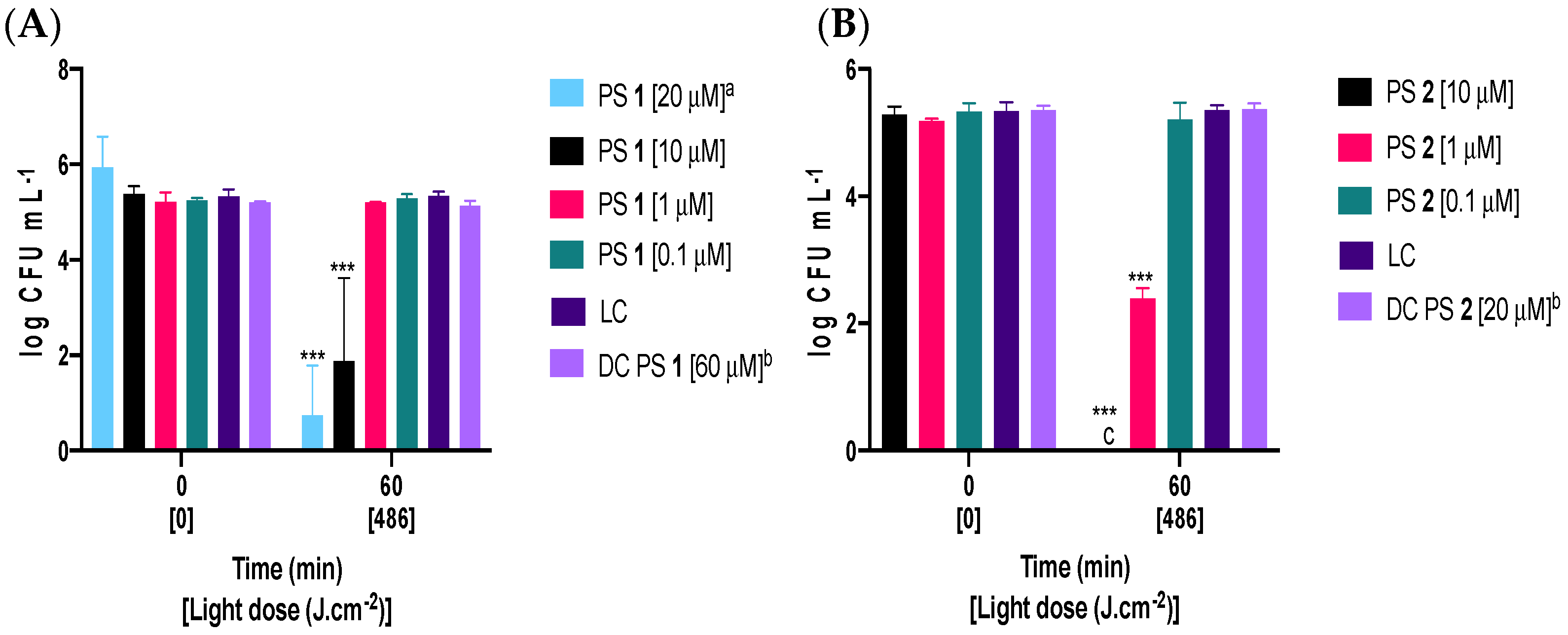
Figure 3 and Figure 4 display the logarithmic decrease in the number of viable Fusarium oxysporum conidia after 60 min of exposure to artificial white light with a fluence rate of 135 mW·cm−2 (light dose of 486 J·cm−2). Experiments were conducted in the presence of tetra-substituted PSs 1, 1a and octa-substituted PSs 2, 2a at concentrations of 0.1, 1, 10, and 20 μM. It is noteworthy that all PSs were stable and photostable during the experiments, and all were soluble in aqueous media (verified by the Beer-Lambert law) [46]. After 60 min of irradiation, the tetra-substituted PS 1 with a concentration of 20 μM led to a significant reduction (~5 log10) in the concentration of viable conidia. When the concentration was halved to 10 μM, it still caused a 3 log10 inactivation. Only octa-substituted PS 2, at a concentration of 10 μM, achieved complete inactivation of conidia, down to the detection limit. At the lowest tested concentrations of 1 and 0.1 µM, only octa-substituted PS 2 (1 µM) exhibited a significant effect on conidia viability, resulting in approximately 3 log10 reduction. None of the PSs caused lethal damage at the highest tested concentration in the absence of light (dark controls: DC PS 1 and DC PS 2, Figure 3A,B).
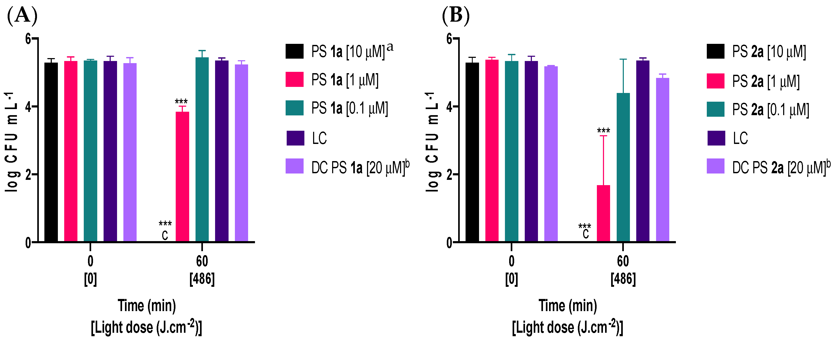
To determine the effectiveness of quaternized tetra-substituted PS 1a and octa-substituted PS 2a in inactivating Fusarium oxysporum, the concentration of viable conidia was evaluated before and after 60 min of irradiation with white light (Figure 4). Both PSs 1a and 2a, at a concentration of 10 μM, were able to completely inactivate conidia, reaching the detection limit of the method. Within 60 min of exposure to light, PS 2a was found to be more effective than PS 1a at a concentration of 1 μM, causing a reduction of approximately 3 log10 in comparison to only 1.5 log10 in conidia viability, respectively. However, at the lowest tested concentration (0.1 μM), PS 2a did not show any significant inactivation, with only a slight decrease of approximately 1 log10 in conidia viability. At the highest tested concentrations of PSs 1a and 2a, neither compound induced lethal damage in the absence of light, as shown in dark control conditions (DC PS 1a and DC PS 2a, Figure 4).

3. Discussion

The rise in fungal resistance to traditional antifungal treatments, specifically in conidia which are responsible for pathogen spread [48], has resulted in a significant amount of research being conducted to find new, effective, and environmentally friendly methods to control them. One such method is PDI. The efficiency of photosensitization heavily relies on the structure of PS molecules [49,50,51,52]. In particular, the presence of positive charges is crucial to enhancing the water solubility of PSs and achieving effective photosensitization of fungal targets [49,53]. In this regard, cationic tetra- and octa-substituted ZnPcs 1, 1a and 2, 2a (shown in Figure 1), were synthesized [46] and tested against conidia of Fusarium oxysporum, a model fungal pathogen. The effectiveness of PDI was quantified by assessing the logarithmic reduction in viable conidia for different PS concentrations (Figure 3 and Figure 4) under irradiation (60 min, 135 mW·cm−2, 486 J·cm−2).
With irradiation in the absence of PS, light alone did not induce a significant inactivation of conidia, and reversely no inactivation was observed in the absence of light and the presence of the highest PS concentration (LC, DC PSs 1, 1a and DC PSs 2, 2a, Figure 3 and Figure 4). Light does not affect the germination and growth of Fusarium conidia, although the light regime is known to modulate conidiation and tolerance of conidia to UV [54] and to induce the expression of light-protective metabolites, like carotenoids [55].
After 60 min under light exposure (light dose of 486 J·cm−2), the lowest tested concentration (0.1 µM) failed to cause any significant conidia inactivation. In order to attain lethal damage, higher concentrations of each PS (1, 10, or 20 μM) were assessed.
With a concentration of 1 µM of PS, and similar irradiation conditions (light dose of 486 J·cm−2), a significant inactivation could be observed with all tested PSs, except for PS 1, which represents the first evidence of the lower efficiency of this PS, in comparison with the other molecules. An estimate of the minimum inhibitory concentration (MIC) of thiopyridinium or ammonium phthalocyanines against conidia, pointed to values of 5 to 60 µM [38,39]. Therefore, assays conducted with a concentration of 1 µM may be interpreted as representing sub-lethal conditions, and allow the comparative assessment of the different PSs. A greater reduction in the concentration of viable conidia (~3.5 log10) was observed with PS 2a (1 µM, Figure 4B), ranking this PS as the most effective against Fusarium oxysporum conidia.
A further 10-fold increase in concentration of PS (10 µM), led to the complete inactivation of conidia with PSs 2, 1a, and 2a as illustrated in Figure 3B and Figure 4A,B, respectively. With 10 μM of PS 1, the inactivation corresponded to a ~4 log10 reduction in the concentration of viable conidia (Figure 3A). The moderate performance of PS 1 (10 µM, Figure 3A) in comparison to the high performance of PS 1a (10 µM, Figure 4A) may be interpreted as an indication that quaternization improves photosensitization capacity against conidia. An even higher concentration of PS (20 µM) was tested only for PS 1. Although a significant reduction in the concentration of viable conidia was observed (~5 log10), complete inactivation was still not attained. In the case of these PSs (1, 1a and 2, 2a), an even higher concentration may be required, since the increase in concentrations, with a light dose of 486 J·cm−2, confirmed that the cationic PSs efficiently inactivate Fusarium oxysporum conidia.
Comparing the obtained results with non-quaternized PSs 1, 2 (Figure 3A,B) and quaternized PSs 1a, 2a (Figure 4A,B) at a sub-lethal concentration (1 μM) shows that quaternized PSs 1a caused >1 log10 reductions, whereas non-quaternized Ps 2 caused a <2.5 log10 decrease in the concentration of viable conidia and PS 1 caused no reduction at all. An investigation of inactivation of a Gram-negative bacterial model, Escherichia coli, in both planktonic and biofilm forms, also confirmed that quaternized PSs 1a, 2a were more efficient than the non-quaternized ones when a concentration of 20 µM was used (PSs 1, 2) [46]. This fact is most probably associated with the difference in the 1O2 production (non-quaternized PSs 1, 2 < quaternized PSs 1a, 2a) and positive double-charge of each substituent that maximizes the electrostatic interactions of the quaternized derivatives (ZnPcs 1a, 2a) with the fungal spores.
The effect of the increase in the number of charges on the photosensitization capacity can be inferred from the comparison of the obtained results with tetra-substituted and octa-substituted PSs. In the octa-substituted PS 2 (Figure 3B), despite being considered an antimicrobial agent, the introduction of more cationic substituents to the β-position seemed to increase the antifungal activity when compared with tetra-substituted PS 1 (Figure 3A), which was well-observed at 1 and 10 μM and could be correlated with the previously mentioned difference in the 1O2 production (ZnPc 1 < ZnPc 2) and electrostatic interactions (four versus eight positive charges) with the target conidia.
Both PSs 1a and 2a led to the complete inactivation of conidia with a concentration of 10 μM (Figure 4A,B). In order to compare them in terms of inactivation efficiency, results obtained with lower concentrations need to be considered. At the lowest tested concentration (0.1 μM), PS 1a did not show any effect (Figure 4A), and PS 2a exhibited a slight inactivation (Figure 4B), displaying only a marginal reduction of approximately ~1 log10 in conidia viability. This suggests that PS 2a is slightly more effective than PS 1a. This trend is confirmed by the obtained results with a higher, but still sub-lethal, concentration (1 µM), indicating a higher photosensitization capacity of PS 2a (~3.5 log10 reduction) in comparison to PS 1a (~1.5 log10 reduction). Overall, this suggests that the increased number of positive charges on PS 2a possibly leads to an increased 1O2 production and stronger electrostatic interactions, which enhances the photoinactivation of conidia.
Regardless of differences in the photosensitization efficiency, which can be related to 1O2 generation capacity and the effect of the number of charges on solubility and affinity towards the spore material, all PSs have been shown to be effective against Fusarium oxysporum conidia at concentrations above 10 µM. Other studies of the photoinactivation of Fusarium oxysporum conidia with the use of ammonium phthalocyanines as photosensitizers needed higher concentrations of PS such as 40 and 60 μM at the same light doses [38]. The study found that quaternized derivatives were highly effective as photosensitizers and required only a concentration of 1 μM to significantly inactivate Fusarium oxysporum conidia. Although typically higher concentrations are required to photosensitize fungal structures, both non-quaternized PSs 1, 2 and quaternized PSs 1a, 2a probably target a diverse range of components of conidia, leading to high antifungal activity. These results suggest that a wide-range, multi-organism phytosanitary strategy, based on cationic zinc(II) phthalocyanines, may be possible.
It is noteworthy that fungal spores and vegetative forms (hyphae) have varying levels of susceptibility to PDI. Hyphae may indeed be more resistant to PDI than spores, depending on the nature and affinity of the PS to the target structures [56]. In the case of Fusarium, conidia are infectious forms, and the macroconidia are responsible for spreading the infection over long distances [57]. Infection begins when spores germinate and penetrate plant tissues through the roots without symptoms [58]. Therefore, the inactivation of conidia is considered the most straightforward and long-lasting way to control fungal infections in crops [34,59,60]. In future research, it will be necessary to evaluate the affinity of cationic phthalocyanines for both conidia and mycelium and to determine the susceptibilities of these structures to photosensitization. This information can be used to improve the design of phytosanitary protocols.

4. Materials and Methods

4.1. Synthesis and Photophysical Characterization of the Photosensitizers

The molecular structures of the cationic PSs bearing cationic groups, designated as 1, 1a and 2, 2a, are presented in Figure 1. The synthesis 1, 1a and 2, 2a was conducted following previously established experimental procedures [46], using reagents of high purity, procured from Merck, Steinheim, Germany. Analytical thin-layer chromatography (TLC) was performed on pre-coated silica gel sheets with a thickness of 0.2 mm (Merck, Darmstadt, Germany). As per the literature, solvents were either used in their as-received state or subjected to distillation and dehydration procedures [61]. 1H and 19F NMR spectra were recorded on a Bruker Avance-300 spectrometer based in Wissembourg, France, operating at 300.13 and 282.38 MHz, with tetramethylsilane (TMS) serving as the internal reference. Absorption and steady-state fluorescence spectra were acquired using a Shimadzu UV-2501PC spectrophotometer (Shimadzu, Kyoto, Japan) and a Horiba Jobin-Yvon FluoroMax Plus spectrofluorometer (Horiba Ltd., Kisshoint, Japan), respectively. The absorption and emission spectra of 1, 1a and 2, 2a were measured in DMSO within quartz optical cells with dimensions of 1 × 1 cm, at a temperature of 298.15 K. ΦF for 1, 1a and 2, 2a was determined in DMSO by comparing the area beneath the corrected emission spectra to that of ZnPc, which served as the standard (ΦF = 0.20 in DMSO) [47].

4.2. Photosenstizer Stock Solutions

Stock solutions of the PSs at a concentration of 500 µM were prepared in either DMF for photophysical analyses or DMSO for photodynamic inactivation experiments. These solutions were stored in a light-protected environment and were pre-treated by ultrasonic sonication for 30 min before each assay.

4.3. Light Source

All photodynamic inactivation experiments were conducted by subjecting the samples and light controls to white light within the range of 400–800 nm. The light was delivered through a fiber-optic probe connected to a 150 W quartz/halogen lamp (model LC122, LumaCare™ MBG Technologies Inc., New Port Beach, CA, USA). The irradiance, measured at 135 mW·cm–2, was determined using a Coherent FieldMaxII-Top energy meter in conjunction with a Coherent PowerSensPS19Q energy sensor.

4.4. Preparation of Stock Suspensions of Fusarium oxysporum Conidia

Fusarium oxysporum was cultivated in potato dextrose agar (PDA, Merck, KGaA, Darmstadt, Germany) for 7 days at 25 °C, following a previously described procedure [36]. To ensure the absence of hyphae in the conidia suspensions, a microscopic examination was performed using a Leitz Laborlux K microscope from Ernst Leitz GmbH, Wetzlar, Germany. The concentration of viable conidia was determined by subjecting an aliquot to serial dilutions in PBS (pH 7.4), and spread-plating on Rose Bengal chloramphenicol agar (Merck, KGaA, Darmstadt, Germany). After a 2-day incubation at 25 °C, colonies were counted, and the concentration of conidia was expressed as colony-forming units per milliliter (CFU·mL−1) of the suspension.
The fungal strain was provided by the Fungi and Plant Biology Laboratory—FunPlantLab of the Department of Biology, University of Aveiro. The strain was isolated from pine trees in Portugal. The spore suspensions were prepared following the procedure previously described in the literature [36]. To extract the conidia from PDA (Merck) cultures, 5 mL of phosphate-buffered saline (PBS) was added to 10 cm plates. Then, the mycelium was gently scraped using a sterilized glass spreader. The suspension was passed through sterilized cotton gauze to filter out hyphae and other debris, and concentrated by centrifugation (4000× g). The absence of hyphal material and a negligible proportion of microconidia was verified under light microscopy. The concentration of conidia in the suspension was determined by colony counting after serial dilution and spread-plating on Rose Bengal chloramphenicol agar (VWR, Leuven, Belgium). After an incubation period of 2 days at 25 °C, the colonies were counted, and the concentration of conidia was expressed as CFU·mL−1. The spore suspensions were stored at −20 °C until the experiment and then diluted with sterile PBS to achieve a suitable volume, with a concentration of spores of approximately 105 CFU·mL−1.
The microscopy inspection of the suspensions revealed that the presence of microconidia was negligible, as expected from the methods used to obtain conidia from stock cultures. Microconidia are smaller, unicellular, and often produced directly on the hyphae, and are more tightly attached to it compared to macroconidia. The latter are larger, multicellular (septated), and produced in specialized structures called macroconidiophores, which are separate from the mycelium. These structures aid in the long-distance dispersal of the loosely attached macroconidia [59,62]. The procedure of gently scraping the mycelium will more efficiently detach macroconidia, which can be easily washed with the buffer. Furthermore, the microconidia attached to the hyphae will be retained in the gauze upon filtration, and the low-speed centrifugation will further contribute to the selective precipitation of the large macroconidia.

4.5. Photodynamic Inactivation Assays

The photoinactivation assays were performed on PBS suspensions containing approximately 3 × 10⁵ CFU·mL−1 with a range of concentrations of 0.1, 1, 10, or 20 µM for PSs 1, 1a and 2, 2a. The tests were carried out in 24-well plates in a final volume of 1.5 mL of suspension. Conidia suspensions were preincubated with the PS, in the dark, for 30 min at room temperature, with magnetic gentle stirring. After the preincubation, light exposure was conducted for 1 h of constant irradiation. During irradiation, the suspension was kept under stirring on melting ice, to prevent heating. Aliquots of 100 μL were collected at the beginning (t = 0 min) and at the end of the irradiation (t = 60 min), serially diluted in PBS and spread-plated on Rose Bengal Chloramphenicol Agar, in triplicate, for the determination of the concentration of viable spores. Colonies were counted in the most convenient dilution after 48 h incubation at 25 °C. The average of the colonies in the replicates was used to estimate the concentration of viable conidia in the suspension expressed as CFU·mL−1. Two controls were included in each experiment: a light control (LC) submitted to the same irradiation conditions as the samples but without PS, and a dark control (DC) containing the highest PS concentration, but kept in the dark. Three independent assays were conducted for each PS. The inactivation efficiency was determined as the logarithmic (log10) reduction in the concentration of viable Fusarium oxysporum conidia during the corresponding irradiation period for each independent assay.

4.6. Statistical Assessment

The significance of inactivation (difference between the initial and final concentrations of viable conidia) was evaluated using a two-way univariate analysis of variance (ANOVA) model, followed by Tukey’s multiple-comparisons post hoc test. Significance was established at a threshold of p < 0.05.

5. Conclusions

The relations between structural features and the efficiency of photosensitization of Fusarium oxysporum conidia indicated that cationic PSs 1a, 2, and 2a (10 µM) were more efficient (reduction in the concentration of viable conidia down to the detection limit of the method) than the PS 1 (20 µM, achieving a reduction of ~5 log10 in the conidia viability). The different photodynamic activity against fungi was related to the ability to generate 1O2 species (following the increasing order of 1 (35%) < 2 (40%) < 1a (65%) < 2a (89%)) and with the electrostatic interactions (attending to the number of charges and their charge position on the peripheral substituent on the Pc structure). In this study, doubling the number of charges in the same peripheral substituent (PSs 1, 2 vs. PSs 1a, 2a) improved the photoinactivation process. The obtained findings provide a solid foundation for considering these positively charged PSs as promising candidates for novel phytosanitary agents, relying on the photodynamic management of fungal spores.

Author Contributions

L.M.O.L., PS synthesis, result analysis and interpretation of biological results, manuscript preparation, funding acquisition, and resources. I.N.S.-G., antimicrobial photodynamic evaluation assays, and analysis of biological results. Â.C., supervision and the design of antimicrobial photodynamic experiments, analysis, and interpretation of biological results, manuscript preparation, funding acquisition, and resources. All authors have read and agreed to the published version of the manuscript.

Funding

This work received financial support from PT national funds (FCT/MCTES, Fundação para a Ciência e a Tecnologia and Ministério da Ciência, Tecnologia e Ensino Superior) through projects LAQV-REQUIMTE (UIDB/50006/2020 and UIDP/50006/2020), CESAM (UIDP/50017/2020 + UIDB/50017/2020 + LA/P/0094/2020), P2020-PTDC/QUI-QOR/31770/2017 and PTDC/BIA-MIC/3157/2020.

Institutional Review Board Statement

Not applicable.

Informed Consent Statement

Not applicable.

Data Availability Statement

Data is contained within the article.

Acknowledgments

Thanks to the University of Aveiro and FCT/MCTES (Fundação para a Ciência e a Tecnologia and Ministério da Ciência, Tecnologia e Ensino Superior) for the financial support to LAQV-REQUIMTE (UIDB/50006/2020 and UIDP/50006/2020) and CESAM (UIDP/50017/2020, UIDB/50017/2020, and LA/P/0094/2020), funded by FCT/MCTES through national funds. The authors also acknowledge FCT/MCTES for the national funds received through projects P2020-PTDC/QUI-QOR/31770/2017 and PTDC/BIA-MIC/3157/2020. We thank Artur Alves, coordinator of the FunPlantLab, for providing the fungal strain used in this work.

Conflicts of Interest

The authors declare no conflict of interest.

References

  1. Van Diepeningen, A.D.; Feng, P.; Ahmed, S.; Sudhadham, M.; Bunyaratavej, S.; de Hoog, G.S. Spectrum of Fusarium Infections in Tropical Dermatology Evidenced by Multilocus Sequencing Typing Diagnostics. Mycoses 2015, 58, 48–57. [Google Scholar] [CrossRef]
  2. Ortoneda, M.; Guarro, J.; Madrid, M.P.; Caracuel, Z.; Roncero, M.I.G.; Mayayo, E.; Di Pietro, A. Fusarium oxysporum as a Multihost Model for the Genetic Dissection of Fungal Virulence in Plants and Mammals. Infect. Immun. 2004, 72, 1760–1766. [Google Scholar] [CrossRef]
  3. Gordon, T.R.; Swett, C.L.; Wingfield, M.J. Management of Fusarium Diseases Affecting Conifers. Crop Prot. 2015, 73, 28–39. [Google Scholar] [CrossRef]
  4. Wu, C.Y.; Chen, G.S.; Lan, C.C.E. Onychomycosis Caused by Fusarium solani in a Woman with Diabetes. Clin. Exp. Dermatol. 2009, 34, e772–e774. [Google Scholar] [CrossRef]
  5. Guilhermetti, E.; Takahachi, G.; Shinobu, C.S.; Svidzinski, T.I.E. Fusarium spp. as Agents of Onychomycosis in Immunocompetent Hosts. Int. J. Dermatol. 2007, 46, 822–826. [Google Scholar] [CrossRef]
  6. Edel-Hermann, V.; Lecomte, C. Current Status of Fusarium oxysporum Formae Speciales and Races. Phytopathology 2019, 109, 512–530. [Google Scholar] [CrossRef]
  7. Nag, P.; Paul, S.; Shriti, S.; Das, S. Defence Response in Plants and Animals against a Common Fungal Pathogen, Fusarium oxysporum. Curr. Res. Microb. Sci. 2022, 3, 100135. [Google Scholar] [CrossRef]
  8. João, A.L.; Lencastre, A.; Dutra, E.; Pessoa e Costa, T.; Formiga, A.; Neves, J. Fusarium spp.—An Emerging Pathogen in Chronic Diabetic Ulcer: Case Report and Review of the Literature. Int. J. Low. Extrem. Wounds 2021, 20, 67–72. [Google Scholar] [CrossRef]
  9. Nucci, M.; Anaissie, E. Fusarium Infections in Immunocompromised Patients. Clin. Microbiol. Rev. 2007, 20, 695–704. [Google Scholar] [CrossRef]
  10. Nelson, P.E.; Dignani, M.C.; Anaissie, E.J. Taxonomy, Biology, and Clinical Aspects of Fusarium Species. Clin. Microbiol. Rev. 1994, 7, 479–504. [Google Scholar] [CrossRef]
  11. Palmieri, D.; Ianiri, G.; Del Grosso, C.; Barone, G.; De Curtis, F.; Castoria, R.; Lima, G. Advances and Perspectives in the Use of Biocontrol Agents against Fungal Plant Diseases. Horticulturae 2022, 8, 577. [Google Scholar] [CrossRef]
  12. Jestoi, M. Emerging Fusarium-Mycotoxins Fusaproliferin, Beauvericin, Enniatins, And Moniliformin—A Review. Crit. Rev. Food Sci. Nutr. 2008, 48, 21–49. [Google Scholar] [CrossRef] [PubMed]
  13. Gurdaswani, V.; Ghag, S.B. Toxins from Fusarium Species and Their Role in Animal and Plant Diseases. In New and Future Developments in Microbial Biotechnology and Bioengineering; Elsevier: Amsterdam, The Netherlands, 2020; pp. 7–27. [Google Scholar]
  14. Achari, S.R.; Kaur, J.K.; Mann, R.C.; Sawbridge, T.; Summerell, B.A.; Edwards, J. Investigating the Effector Suite Profile of Australian Fusarium oxysporum Isolates from Agricultural and Natural Ecosystems. Plant Pathol. 2021, 70, 387–396. [Google Scholar] [CrossRef]
  15. de Menezes, H.D.; Massola, N.S.; Flint, S.D.; Silva, G.J.; Bachmann, L.; Rangel, D.E.N.; Braga, G.U.L. Growth under Visible Light Increases Conidia and Mucilage Production and Tolerance to UV-B Radiation in the Plant Pathogenic Fungus Colletotrichum acutatum. Photochem. Photobiol. 2015, 91, 397–402. [Google Scholar] [CrossRef]
  16. Zhao, B.; He, D.; Wang, L. Advances in Fusarium Drug Resistance Research. J. Glob. Antimicrob. Resist. 2021, 24, 215–219. [Google Scholar] [CrossRef]
  17. Chitwood-Brown, J.; Vallad, G.E.; Lee, T.G.; Hutton, S.F. Breeding for Resistance to Fusarium Wilt of Tomato: A Review. Genes 2021, 12, 1673. [Google Scholar] [CrossRef]
  18. Sampaio, A.M.; de Araújo, S.S.; Rubiales, D.; Vaz Patto, M.C. Fusarium Wilt Management in Legume Crops. Agronomy 2020, 10, 1073. [Google Scholar] [CrossRef]
  19. Koike, S.T.; Gordon, T.R. Management of Fusarium Wilt of Strawberry. Crop Prot. 2015, 73, 67–72. [Google Scholar] [CrossRef]
  20. Amini, J.; Sidovich, D. The Effects of Fungicides on Fusarium oxysporum f. sp. Lycopersici Associated with Fusarium Wilt of Tomato. J. Plant Prot. Res. 2010, 50, 172–178. [Google Scholar] [CrossRef]
  21. Ölmez, H.; Kretzschmar, U. Potential Alternative Disinfection Methods for Organic Fresh-Cut Industry for Minimizing Water Consumption and Environmental Impact. LWT Food Sci. Technol. 2009, 42, 686–693. [Google Scholar] [CrossRef]
  22. Lykogianni, M.; Bempelou, E.; Karamaouna, F.; Aliferis, K.A. Do Pesticides Promote or Hinder Sustainability in Agriculture? The Challenge of Sustainable Use of Pesticides in Modern Agriculture. Sci. Total Environ. 2021, 795, 148625. [Google Scholar] [CrossRef]
  23. Ran, X.; Hadiatullah, H.; Yuchi, Z.; Yang, X.; Zhu, X. Sustainable Use of Pesticides. Agriculture 2023, 13, 1393. [Google Scholar] [CrossRef]
  24. Zafeiriou, E.; Karelakis, C.; Martínez-Zarzoso, I.; Galanopoulos, K.; Gkika, D. Economic Development and Pesticide Use in EU Agriculture: A Nonlinear Panel Data Autoregressive Distributed Lag Approach. Agriculture 2023, 13, 1693. [Google Scholar] [CrossRef]
  25. Ons, L.; Bylemans, D.; Thevissen, K.; Cammue, B.P.A. Combining Biocontrol Agents with Chemical Fungicides for Integrated Plant Fungal Disease Control. Microorganisms 2020, 8, 1930. [Google Scholar] [CrossRef] [PubMed]
  26. do Prado-Silva, L.; Brancini, G.T.P.; Braga, G.Ú.L.; Liao, X.; Ding, T.; Sant’Ana, A.S. Antimicrobial Photodynamic Treatment (APDT) as an Innovative Technology to Control Spoilage and Pathogenic Microorganisms in Agri-Food Products: An Updated Review. Food Control 2022, 132, 108527. [Google Scholar] [CrossRef]
  27. Piksa, M.; Lian, C.; Samuel, I.C.; Pawlik, K.J.; Samuel, I.D.W.; Matczyszyn, K. The Role of the Light Source in Antimicrobial Photodynamic Therapy. Chem. Soc. Rev. 2023, 52, 1697–1722. [Google Scholar] [CrossRef] [PubMed]
  28. Aroso, R.T.; Schaberle, F.A.; Arnaut, L.G.; Pereira, M.M. Photodynamic Disinfection and Its Role in Controlling Infectious Diseases. Photochem. Photobiol. Sci. 2021, 20, 1497–1545. [Google Scholar] [CrossRef] [PubMed]
  29. Ruiz-González, R.; Setaro, F.; Gulías, Ò.; Agut, M.; Hahn, U.; Torres, T.; Nonell, S. Cationic Phthalocyanine Dendrimers as Potential Antimicrobial Photosensitisers. Org. Biomol. Chem. 2017, 15, 9008–9017. [Google Scholar] [CrossRef]
  30. Lopes, M.M.; Bartolomeu, M.; Gomes, A.T.P.C.; Figueira, E.; Pinto, R.; Reis, L.; Balcão, V.M.; Faustino, M.A.F.; Neves, M.G.P.M.S.; Almeida, A. Antimicrobial Photodynamic Therapy in the Control of Pseudomonas syringae pv. actinidiae Transmission by Kiwifruit Pollen. Microorganisms 2020, 8, 1022. [Google Scholar] [CrossRef] [PubMed]
  31. Ndemueda, A.; Pereira, I.; Faustino, M.A.F.; Cunha, Â. Photodynamic Inactivation of the Phytopathogenic Bacterium Xanthomonas citri subsp. Citri. Lett. Appl. Microbiol. 2020, 71, 420–427. [Google Scholar] [CrossRef]
  32. Garcia, M.; David, B.; Sierra-Garcia, I.N.; Faustino, M.A.F.; Alves, A.; Esteves, A.C.; Cunha, A. Photodynamic Inactivation of Lasiodiplodia theobromae: Lighting the Way towards an Environmentally Friendly Phytosanitary Treatment. Biol. Lett. 2021, 17, 20200820. [Google Scholar] [CrossRef] [PubMed]
  33. Ambrosini, V.; Issawi, M.; Sol, V.; Riou, C. Photodynamic Inactivation of Botrytis Cinerea by an Anionic Porphyrin: An Alternative Pest Management of Grapevine. Sci. Rep. 2020, 10, 17438. [Google Scholar] [CrossRef]
  34. Gonzales, J.C.; Brancini, G.T.P.; Rodrigues, G.B.; Silva-Junior, G.J.; Bachmann, L.; Wainwright, M.; Braga, G.Ú.L. Photodynamic Inactivation of Conidia of the Fungus Colletotrichum abscissum on Citrus Sinensis Plants with Methylene Blue under Solar Radiation. J. Photochem. Photobiol. B 2017, 176, 54–61. [Google Scholar] [CrossRef] [PubMed]
  35. Sharma, V.K.; Shah, M.P.; Parmar, S.; Kumar, A. Fungi Bio-Prospects in Sustainable Agriculture, Environment and Nano-Technology; Elsevier: Amsterdam, The Netherlands, 2021; ISBN 9780128217344. [Google Scholar]
  36. Sierra-Garcia, I.N.; Cunha, Â.; Lourenço, L.M.O. In Vitro Photodynamic Treatment of Fusarium oxysporum Conidia through the Action of Thiopyridinium and Methoxypyridinium Chlorins. J. Photochem. Photobiol. A Chem. 2022, 432, 114081. [Google Scholar] [CrossRef]
  37. Ryu, A.-R.; Han, C.-S.; Oh, H.-K.; Lee, M.-Y. Chlorin E6-Mediated Photodynamic Inactivation with Halogen Light against Microbes and Fungus. Toxicol. Environ. Health Sci. 2015, 7, 231–238. [Google Scholar] [CrossRef]
  38. Gamelas, S.R.D.; Sierra-Garcia, I.N.; Tomé, A.C.; Cunha, Â.; Lourenço, L.M.O. In Vitro Photoinactivation of Fusarium oxysporum Conidia with Light-Activated Ammonium Phthalocyanines. Int. J. Mol. Sci. 2023, 24, 3922. [Google Scholar] [CrossRef] [PubMed]
  39. Prandini, J.A.; Castro, K.A.D.F.; Biazzotto, J.C.; Brancini, G.T.P.; Tomé, J.P.C.; Lourenço, L.M.O.; Braga, G.Ú.L.; da Silva, R.S. Thiopyridinium Phthalocyanine for Improved Photodynamic Efficiency against Pathogenic Fungi. J. Photochem. Photobiol. B 2022, 231, 112459. [Google Scholar] [CrossRef]
  40. Ribeiro, C.P.S.; Lourenço, L.M.O. Overview of Cationic Phthalocyanines for Effective Photoinactivation of Pathogenic Microorganisms. J. Photochem. Photobiol. C Photochem. Rev. 2021, 48, 100422. [Google Scholar] [CrossRef]
  41. van Leeuwen, M.; Beeby, A.; Fernandes, I.; Ashworth, S.H. The Photochemistry and Photophysics of a Series of Alpha Octa(Alkyl-Substituted) Silicon, Zinc and Palladium Phthalocyanines. Photochem. Photobiol. Sci. 2014, 13, 62–69. [Google Scholar] [CrossRef]
  42. Al-Asmari, F.; Mereddy, R.; Sultanbawa, Y. A Novel Photosensitization Treatment for the Inactivation of Fungal Spores and Cells Mediated by Curcumin. J. Photochem. Photobiol. B 2017, 173, 301–306. [Google Scholar] [CrossRef]
  43. Yaakoub, H.; Mina, S.; Calenda, A.; Bouchara, J.-P.; Papon, N. Oxidative Stress Response Pathways in Fungi. Cell. Mol. Life Sci. 2022, 79, 333. [Google Scholar] [CrossRef]
  44. Ziental, D.; Mlynarczyk, D.T.; Czarczynska-Goslinska, B.; Lewandowski, K.; Sobotta, L. Photosensitizers Mediated Photodynamic Inactivation against Fungi. Nanomaterials 2021, 11, 2883. [Google Scholar] [CrossRef]
  45. Calzavara-Pinton, P.G.; Venturini, M.; Sala, R. A Comprehensive Overview of Photodynamic Therapy in the Treatment of Superficial Fungal Infections of the Skin. J. Photochem. Photobiol. B 2005, 78, 1–6. [Google Scholar] [CrossRef]
  46. Lourenço, L.M.O.; Rocha, D.M.G.C.; Ramos, C.I.V.; Gomes, M.C.; Almeida, A.; Faustino, M.A.F.; Almeida Paz, F.A.; Neves, M.G.P.M.S.; Cunha, Â.; Tomé, J.P.C. Photoinactivation of Planktonic and Biofilm Forms of Escherichia coli through the Action of Cationic Zinc(II) Phthalocyanines. ChemPhotoChem 2019, 3, 251–260. [Google Scholar] [CrossRef]
  47. Beveridge, A.C.; Bench, B.A.; Gorun, S.M.; Diebold, G.J. Evaluation of Photodynamic Therapy Agents through Transient Grating Measurements. J. Phys. Chem. A 2003, 107, 5138–5143. [Google Scholar] [CrossRef]
  48. Blango, M.G.; Kniemeyer, O.; Brakhage, A.A. Conidial Surface Proteins at the Interface of Fungal Infections. PLoS Pathog. 2019, 15, e1007939. [Google Scholar] [CrossRef] [PubMed]
  49. Ogunsipe, A.; Nyokong, T. Photophysical and Photochemical Studies of Sulphonated Non-Transition Metal Phthalocyanines in Aqueous and Non-Aqueous Media. J. Photochem. Photobiol. A Chem. 2005, 173, 211–220. [Google Scholar] [CrossRef]
  50. Sharma, M.; Visai, L.; Bragheri, F.; Cristiani, I.; Gupta, P.K.; Speziale, P. Toluidine Blue-Mediated Photodynamic Effects on Staphylococcal Biofilms. Antimicrob. Agents Chemother. 2008, 52, 299–305. [Google Scholar] [CrossRef]
  51. Beer, D.D.E.; Srinivasan, R.; Stewart, P.S. Direct Measurement of Chlorine Penetration into Biofilms during Disinfection. Appl. Environ. Microbiol. 1994, 60, 4339–4344. [Google Scholar] [CrossRef]
  52. Billings, N.; Millan, M.; Caldara, M.; Rusconi, R.; Tarasova, Y.; Stocker, R.; Ribbeck, K. The Extracellular Matrix Component Psl Provides Fast-Acting Antibiotic Defense in Pseudomonas Aeruginosa Biofilms. PLoS Pathog. 2013, 9, e1003526. [Google Scholar] [CrossRef] [PubMed]
  53. Cidlina, A.; Novakova, V.; Miletin, M.; Zimcik, P. Peripheral Substitution as a Tool for Tuning Electron-Accepting Properties of Phthalocyanine Analogs in Intramolecular Charge Transfer. Dalton Trans. 2015, 44, 6961–6971. [Google Scholar] [CrossRef]
  54. Costa, T.P.C.; Rodrigues, E.M.; Dias, L.P.; Pupin, B.; Ferreira, P.C.; Rangel, D.E.N. Different Wavelengths of Visible Light Influence the Conidial Production and Tolerance to Ultra-Violet Radiation of the Plant Pathogens Colletotrichum Acutatum and Fusarium fujikuroi. Eur. J. Plant Pathol. 2021, 159, 105–115. [Google Scholar] [CrossRef]
  55. Avalos, J.; Estrada, A.F. Regulation by Light in Fusarium. Fungal Genet. Biol. 2010, 47, 930–938. [Google Scholar] [CrossRef]
  56. Li, C.; Jia, X.; Bian, Y.; Qi, D.; Wu, J. Different Susceptibility of Spores and Hyphae of Trichophyton rubrum to Methylene Blue Mediated Photodynamic Treatment in Vitro. Mycoses 2021, 64, 48–54. [Google Scholar] [CrossRef]
  57. Magyar, D.; Vass, M.; Li, D.-W. Dispersal Strategies of Microfungi. In Biology of Microfungi; Springer: Berlin/Heidelberg, Germany, 2016; pp. 315–371. [Google Scholar]
  58. Husaini, A.M.; Sakina, A.; Cambay, S.R. Host–Pathogen Interaction in Fusarium oxysporum Infections: Where Do We Stand? Mol. Plant-Microbe Interact. 2018, 31, 889–898. [Google Scholar] [CrossRef]
  59. de Menezes, H.D.; Rodrigues, G.B.; de Teixeira, S.P.; Massola, N.S.; Bachmann, L.; Wainwright, M.; Braga, G.U.L. In Vitro Photodynamic Inactivation of Plant-Pathogenic Fungi Colletotrichum acutatum and Colletotrichum gloeosporioides with Novel Phenothiazinium Photosensitizers. Appl. Environ. Microbiol. 2014, 80, 1623–1632. [Google Scholar] [CrossRef]
  60. Hamminger, C.; Glueck, M.; Fefer, M.; Ckurshumova, W.; Liu, J.; Tenhaken, R.; Plaetzer, K. Photodynamic Inactivation of Plant Pathogens Part II: Fungi. Photochem. Photobiol. Sci. 2022, 21, 195–207. [Google Scholar] [CrossRef]
  61. Armarego, W.L.F. (Ed.) Purification of Laboratory Chemicals; Elsevier: Oxford, UK, 2017; ISBN 9780128054574. [Google Scholar]
  62. Gordon, T.R. Fusarium oxysporum and the Fusarium Wilt Syndrome. Annu. Rev. Phytopathol. 2017, 55, 23–39. [Google Scholar] [CrossRef]
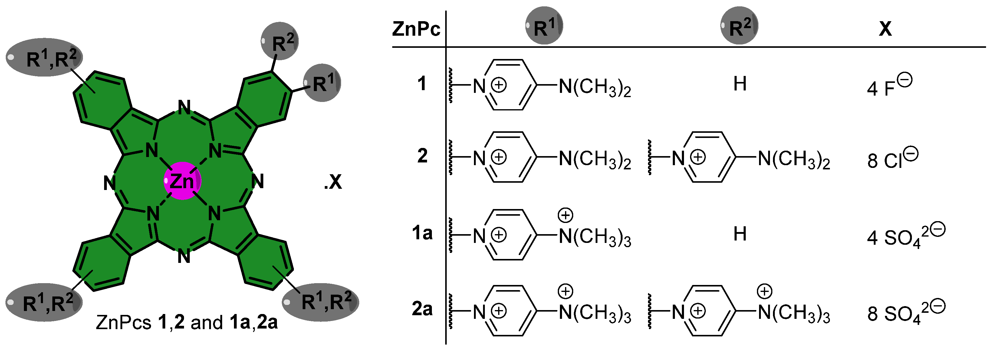
Figure 1. Structures of cationic tetra- and octa-substituted ZnPcs 1, 1a and 2, 2a, respectively.
Ijms 24 16980 g001
Figure 2. Normalized electronic spectra of (a) absorption and (b) emission spectra for compounds 1, 1a and 2, 2a in DMSO at 298 K (λexc = 630 nm).
Ijms 24 16980 g002
Figure 3. Variation in the concentration of viable conidia of Fusarium oxysporum after 60 min of irradiation with artificial white light, at a fluence rate of 135 mW·cm−2 (light dose of 486 J·cm−2) in the presence of (A) 0.1, 1, 10, and 20 μM of PS 1 or (B) 0.1, 1, and 10 μM of PS 2. LC, light control; DC, dark control, respectively. Values correspond to the average of three independent experiments with replicates. Error bars represent the standard deviation. a assay performed in duplicate; b one assay with three analytic replicates; c no colonies observed; *** significance < 0.0001.
Ijms 24 16980 g003
Figure 4. Variation in the concentration of viable conidia of Fusarium oxysporum after 60 min of irradiation with artificial white light, at a fluence rate of 135 mW·cm−2 (light dose of 486 J·cm−2) in the presence of 0.1, 1, and 10 μM for PSs (A) 1a or (B) 2a. LC, light control; DC, dark control. Values correspond to the average of three independent experiments with replicates. Error bars represent the standard deviation. a assay performed in duplicate; b one assay with three analytic replicates; c no colonies observed; *** significance < 0.0001.
Ijms 24 16980 g004
Disclaimer/Publisher’s Note: The statements, opinions and data contained in all publications are solely those of the individual author(s) and contributor(s) and not of MDPI and/or the editor(s). MDPI and/or the editor(s) disclaim responsibility for any injury to people or property resulting from any ideas, methods, instructions or products referred to in the content.