Article

Clonality and Diversity in the Soft Rot Dickeya solani Phytopathogen

by
Frédérique Van Gijsegem
1,*,
Perrine Portier
2,
Géraldine Taghouti
2 and
Jacques Pédron
1
1
Institute of Ecology and Environmental Sciences-Paris, Sorbonne Université, INRAE, 4 Place Jussieu, F-75252 Paris, France
2
Univ Angers, Institut Agro, INRAE, IRHS, SFR QUASAV, CIRM-CFBP, F-49000 Angers, France
*
Author to whom correspondence should be addressed.
Int. J. Mol. Sci. 2023, 24(24), 17553; https://doi.org/10.3390/ijms242417553
Submission received: 18 October 2023 / Revised: 5 December 2023 / Accepted: 6 December 2023 / Published: 16 December 2023
(This article belongs to the Collection Feature Papers in Molecular Microbiology)

Abstract

:
Bacterial diversity analyses often suffer from a bias due to sampling only from a limited number of hosts or narrow geographic locations. This was the case for the phytopathogenic species Dickeya solani, whose members were mainly isolated from a few hosts–potato and ornamentals–and from the same geographical area–Europe and Israel, which are connected by seed trade. Most D. solani members were clonal with the notable exception of the potato isolate RNS05.1.2A and two related strains that are clearly distinct from other D. solani genomes. To investigate if D. solani genomic diversity might be broadened by analysis of strains isolated from other environments, we analysed new strains isolated from ornamentals and from river water as well as strain CFBP 5647 isolated from tomato in the Caribbean island Guadeloupe. While water strains were clonal to RNS05.1.2A, the Caribbean tomato strain formed a third clade. The genomes of the three clades are highly syntenic; they shared almost 3900 protein families, and clade-specific genes were mainly included in genomic islands of extrachromosomal origin. Our study thus revealed both broader D. solani diversity with the characterisation of a third clade isolated in Latin America and a very high genomic conservation between clade members.

1. Introduction

Different forces including spreading via worldwide trade routes, dissemination to different hosts or survival in various environments drive structures of plant pathogen populations. This results in various degrees of genomic diversity from one bacterial species to another. The genus Dickeya is one example of such variations in diversity depending on the species [1,2].
Dickeya members are collectively responsible for soft rot, blackleg, stem rots, stunting, cankers and wilting on a large number of plant hosts, both monocots and dicots, including mostly vegetables and ornamentals but also trees [3]. Their most important virulence factor is the production and secretion of a battery of plant cell-wall-degrading enzymes that provoke the maceration of plant tissues, leading to cell lysis and release of nutrients. However, virulence also relies on several other factors that allow these bacteria to adapt to environmental changes encountered in planta and to face the stresses produced by plant defence responses [4].
The Dickeya genus consists of 13 species presenting differences in environmental habitats, plant host range and genomic diversity [1,3,5,6,7]. Among the species with a sufficient number of genomes available for diversity studies, several like D. fanghzongdai, D. dadantii, D. chrysanthemi and the D. zeae complex harbour a high genomic diversity with some strains being at the limit of species definition [1,2,8]. The previously described D. zeae species was even recently split into three species–D. zeae, D. oryzae and D. parazeae [2,6,7], but some strains of the two first species are still highly divergent from the type strains [2]. By contrast, D. dianthicola and D. solani members have been reported as genomically closely related [1,9,10,11,12,13,14]. Both species were responsible for important outbreaks in the same host potato, and consequently, most available genomes belong to isolates from this host plant. This introduced a bias due to the analysis mainly of strains isolated from only one host and only from Europe and the US. A recent study analysing D. dianthicola genomic diversity using a larger strain panel encompassing more strains isolated from potato over a wider time scale and more strains isolated from hosts other than potato revealed a higher diversity [15].
The host range of D. solani is quite narrow. This species was first described following an important outbreak ravaging potato in Europe in the 2000s [11]. Besides potato, it has been isolated only from two ornamentals, hyacinth and muscari [3]. Even if these strains have been isolated over a long time period [16], most of them are clonal, differing only from a few dozen to a few hundred SNPs [12,13,14,15,16,17]. The potato isolate RNS05.1.2A, however, is clearly distinct from other D. solani genomes, harbouring more than 30,000 SNPs with them [16]. Recently, two other strains isolated either from potato or from river water were shown to belong to this RNS05.1.2A clade [14]. A few genomes harbour medium diversity due to replacing horizontal gene transfers from D. dianthicola or RNS05.1.2A in the main core strain genome [16]. Concerning the accessory genomes, genomic variations in D. solani consist mainly in the presence of genes related to phages or even complete prophages [14,18]. Despite their very high genetic homogeneity, D. solani strains may vary considerably in their virulence levels (aggressiveness) as well as in their production of virulence factors and motility [18]. Analysis of D. solani biodiversity at the proteome level using matrix-assisted methods has unveiled intraspecies variations among 20 D. solani strains differing in their virulence-associated features in addition to the country of origin and year of isolation. These strains were grouped into four delineated clades and differed also in growth rate, plant tissue macerating potential and protease activities, in contrast to the high uniformity among the metabolic profiles, which diverged only in terms of gelatinase activity. However, a definitive link between pathogenicity and the recorded MALDI-TOF MS spectra could not be established [19].
In this work, we investigated if D. solani genomic diversity might be broadened through analysis of strains isolated from other environments: ornamentals, river water and a new host.

2. Results

2.1. Collection of New D. solani Strains Isolated in Different Environments

To broaden the sources of D. solani strains analysed for genomic diversity, we searched for strains isolated in environments other than potato (Table 1).
As the genomes of only two D. solani strains isolated from ornamentals have been analysed so far, we analysed two other strains isolated from hyacinth in the Netherlands: PO2019 and PO3796 (courtesy of Jan van der Wolf). We also took advantage of the two-year survey of soft rot Pectobacteriaceae we recently conducted in the Durance catchment, a river from the south of France [20] to chase D. solani isolates from river water. Four isolates were retrieved: FVG2-MFV017-A9 isolated from the Canal de Crillon, an irrigation canal fed by the river Durance, FVG9-S3-A17-E1 isolated from the Durance River itself and FVG13-S21A17-D9 and FVG14-S21A17-C8, both from a tributary of the Durance, the Grand Anguillon river (see Material and Methods for isolation procedure). New D. solani isolates genomically divergent from the type strain were also searched in the French Collection for Plant-associated Bacteria, CIRM-CFBP (https://cirm-cfbp.fr/; accesses on 12 December 2023), by using multilocus sequence analysis (MLSA) performed on the basis of recA, leuS and dnaX housekeeping genes. This allowed us to identify strain CFBP 5647 (isolated in Guadeloupe from tomato) that was mapped in a branch of the MLSA tree distinct both from the type strain and from RNS05.1.2A (Figure S1). The genomic characteristics of these seven strains are presented in Table 1. Furthermore, the complete sequence of CFBP 5647 was obtained using a hybrid assembly of Illumina short reads and ONT long reads. It consists of a single circular chromosome totalling 5,248,586 bp with a GC content of 56.2%. Annotation of this genome using the Rapid Annotation using Subsystems Technology (RAST) generated a total of 4978 predicted genes, including 4883 protein coding sequences, 74 tRNAs and 21 rRNAs organised in seven operons.
The accession numbers of the genome sequences presented here that were deposited at NCBI are presented in Table 1.

2.2. D. solani Genomic Diversity

2.2.1. The Relatedness between D. solani Members

To analyse the D. solani diversity, we compared our seven new genomes with the genomes of the three D. solani strains of the minor subgroup exemplified by strain RNS05.1.2A [14,16] and seven members of the previously defined main subgroup here defined as the “core” clade. Out of the 40 “core” genomes present in the NCBI database, we chose the genomes of the type strain IPO2222, the two strains isolated from ornamentals PPO9019 and PPO9134, the strain RNS07.7.3B that harbours horizontal genetic transfers from strain RNS05.1.2A [16], the well-studied strains RNS08.23.3.1.A and Ds0432-1 [16,21,22,23,24] and the strain D12 from Russia, far from the Western European seed trade network.
A whole genome multilocus sequence analysis (MLSA) phylogenomic tree built up from concatenated sequences of 3403 core proteins (Figure 1) showed that all strains isolated from French watersheds belong to the minor clade related to RNS05.1.2A, while the two additional strains isolated from ornamentals belong to the core clade. Strains within each of these clades appeared highly related. The Caribbean strain CFBP 5647 isolated from tomato forms a third “clade”.
Calculation of average nucleotide identity (ANI) values between all genomes of our strain panel confirmed the high relatedness between strains of the same clade. Indeed, the genomes of both minor and core clades shared 99.9–100% ANI between members of the same clade. The tomato strain CFBP 5647 exhibited 98.5–98.7% ANI with the other strains of our panel, a value that is in the range of ANI values between each member of the core clade compared to members of the minor RNS05.1.2A clade (98.7–98.8%) (Supplementary Table S1).
The relationships between the different D. solani strains were further addressed by comparing the protein-coding sequences of the 17 genomes using the SiLix gene family clustering tool. Proteins were classified as homologous to others in a given family if the amino acid identities were above 70%, with 80% minimal overlap. All D. solani strains shared 3873 protein families representing 79% to 86% of the genome content. For the core and RNS05.1.2A clades, as many as 4216 and 4456 protein families are common, representing 91–94% and 92–95% of the genome content, respectively. Reciprocally, members of the RNS05.1.2A clade only exhibited 13 to 38 specific protein families. Members of the core clade displayed 5 to 125 specific protein families, with the highest content displayed by the strains isolated from ornamentals (25 to 125 specific protein families). For its part, the CFBP 5647 strain displayed 456 specific protein families (see below).

2.2.2. Relatedness of D. solani Strains by SNP Analysis

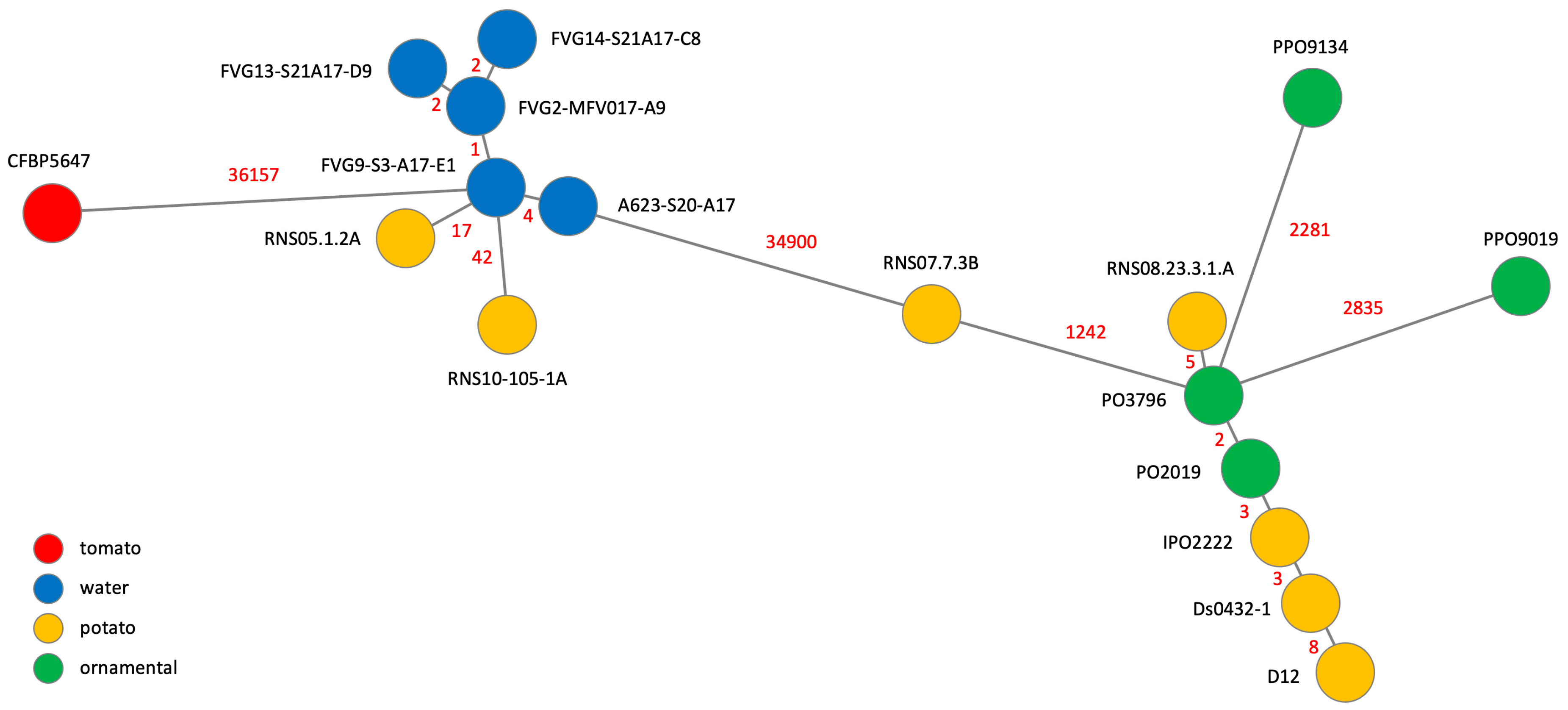
To gain further insight into D. solani relatedness, we constructed a Minimum Spanning Tree (MST) based on the whole set of single nucleotide polymorphisms (SNPs) present in the core genome identified in our strain panel (Figure 2).
As previously reported [12,13,14,16,17], members of the core clade only differ from each other by a few SNPs, except the potato strain RNS07.7.3B and the two ornamental strains PP09019 and PPO9134 that suffered gene replacement by horizontal gene transfer and homologous recombination with RNS05.1.2A and D. dianthicola, respectively. Notably, the two new genomes of strains isolated from ornamentals analysed in this study do not present such replacement and differ from the type strain IPO222 only by very few SNPs (Figure 2). Such clonality was also observed between members of the RNS05.1.2A clade. The five strains isolated from the Durance catchment only harbour 2 to 4 SNPS between each other. The two strains isolated from potato were a bit more distinct, harbouring 17 and 42 SNPs with the closest strain isolated from water.

2.3. Genomic Comparison of the Three D. solani Clades

2.3.1. Genomic Synteny

We compared the genomic architecture of the tomato strain CFBP 5647 with the type strain IPO2222 and the reference strain RNS05.1.2A using the MAUVE whole-genome alignment (Figure 3). The three genomes are highly syntenic with only one large inversion in CFBP 5647 as compared to the two other genomes. The alignment also highlighted specific regions that contain several genes related to phages and mobile genetic elements. This prompted us to predict integrated prophages based on PHASTER classification. CFBP 5647 harbours three complete prophages from which one is highly related to the Mutator phage Mu (P3, Table 2). RNS05.1.2A carries five complete prophages from which two are related to the CFBP 5647 prophage 1 (P2 and P3). RNS05.1.2A prophage P1 is included in a roughly 200,000 Kb complex repeat region (marked by a star in Figure 3) that gathers several genes linked to phages, mobile elements and methyl-accepting chemotaxix proteins (MCPs). No complete prophage was detected in IPO2222. As there were still other uncharacterised specific regions in the MAUVE alignment, we used another approach to search for clade-specific genes by using the SiLix gene family clustering tool.

2.3.2. Analysis of Clade-Specific Genes

The SiLix analysis revealed that 456, 137 and 221 protein families are present only in CFBP 5647, the core clade and the RNS05.1.2A clade, respectively. More than half of the CFBP 5647-specific protein families are clustered in four genomic regions (Table 3). Two of them showed similarities with ICEs (Integrative Conjugative Element). GR3 groups 65 protein families from which ten are related to plasmid replication, recombination and segregation functions of the Pseudomona fluorescens mobile genomic island 01 (PFGI-1) [25]. Even if a homolog of VirD4, an ATPase involved in T-DNA transfer, is present, GR3 does not carry a Pil or T4SS-like operon necessary for the biogenesis of a pilus allowing conjugative transfer. In addition, GR3 carries mainly genes encoding hypothetical proteins and some proteins involved in metabolism. GR5 presents characteristics of a Tn4371-related ICE that is partially present in the model D. dadantii strain 3937 [23,26]. This ICE regroups a conjugative Trb transfer system as well as VirD2 and VirD4 associated homologs, a plasmid-replication machinery, a DNA restriction system, an efflux transport system and some proteins putatively involved in metabolism including NRPS and PKS. GR7 regroups protein families involved in a second T4SS protein secretion system and an efflux system, while GR8 regroups a complete Mu prophage. Notably, CFBP 5647 possesses a CRISPR cluster (GR6), while no such systems have been described previously in D. solani. All CFBP 5647 genomic regions present signs of extrachromosomal origin (Table 3).
Members of the core clade only harbour five short genomic regions (5 to 15 protein families) consisting of extrachromosomal elements, hypothetical proteins or proteins involved in metabolism. This clade is the only one whose members possess the cluster of genes encoding endotoxins of the Bacillus thurigiensis-related Cyt family active on insects that have been characterised in D. dadantii (GR3) [27]. RNS05.1.2A prophages 3 and 4 are missing in RNS10-105-1A and A623-S20-A17, respectively, and thus do not appear in clade-specific genes.

2.4. Virulence of New Isolates

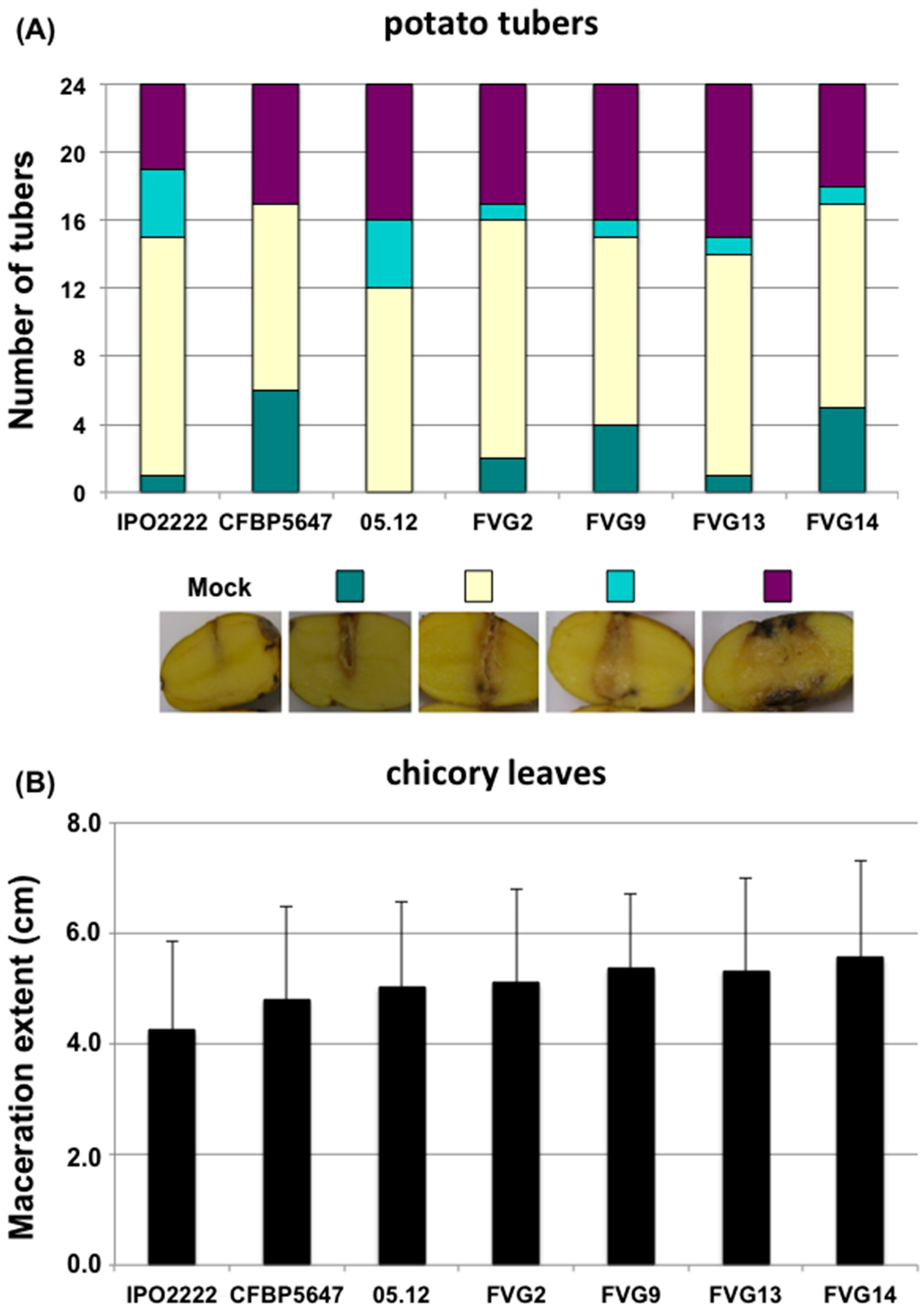
As even highly closely related D. solani strains might vary greatly in their aggressiveness (here defined as the quantitative production of symptoms in contrast to virulence defined as the qualitative ability of producing symptoms) [18], strains isolated from water in this study and the Caribbean tomato strain CFBP 5647 were tested for aggressiveness both on potato tubers and on detached chicory leaves (Figure 4). These strains were able to cause symptoms on both plants. Like the potato strain RNS05.1.2A, these strains were even more aggressive than the type strain IPO222 on chicory leaves (student test p < 0.05) (Figure 4B).

3. Discussion

Previous studies of D. solani genomic diversity [1,12,13,14,16,17] revealed the clonality of this bacterial species with the notable exception of strain RNS05.1.2A and two recently described related strains [14]. The analysed strains were, however, mainly isolated from the same host–potato–and from the same geographical area–Europe and Israel–via seed trade [28]. This points to the spread of a seminal clone that invaded the continent by seed trade dissemination. That is why in this work we extended D. solani genomic analysis to strains isolated from more diverse sources. The newly studied strains belong to three genomic clades exhibiting around 98% ANI values between each other: the core clade including most potato strains and two strains isolated from ornamentals, the minor clade with as reference strain RNS05.1.2A and a third “clade” defined by the strain CFBP 5647 isolated from tomato in Guadeloupe.
The two additional strains isolated from ornamentals belong to the core clade. They do not harbour signs of replacing horizontal gene transfer as described in the two previous strains analysed [16] and carry only a few SNPs as compared to other members of this clade. This reinforced the commonly accepted hypothesis that D. solani emerged in potato following transfer from ornamentals [29,30]. The four strains isolated from surface waters all belong to the clade exemplified by strain RNS05.1.2A. They harbour extremely few SNPs between each other and the other already analysed strain also isolated from the river Durance watershed [31]. As three of these strains have been isolated from the river Durance or canals fed by this river, we cannot exclude spreading of a single clone via the river. However, strains FVG13-S21A17-D9 and FVG14-S21A17-C8 have been isolated from a tributary of the Durance, implying at least two introduction sources in river waters. The two other members of this clade–RNS05.1.2A and RNS10-105-1A–both isolated from potato in the north of France, exhibit a few dozen SNPs with water strains and between each other. We may question if related strains are present in waterways in the north of France and might be a source of contamination explaining the occurrence of this rare clade in potato. To tackle this, it would be interesting to analyse the D. solani strains present in waterways in this region.
So, our study revealed both broader D. solani diversity with the characterisation of a third “clade” isolated in Latin America and a very high genomic conservation between clade members. In the Dickeya genus, diversity varies from one species to the other, with species like D. zeae and D. fanghzongdai encompassing strains so diverse that they are at the limit of species appurtenance, while other species are much more related [2]. In particular, the species D. dianthicola, another strain particularly studied in relation to potato outbreaks, shows both a high diversity and a high relatedness of strains isolated over a large scale of time and geographical origin [15]. As for D. solani, this highlights the high genomic stability for some members of this species. Similarly, in Pectobacterium brasiliense, Jonkheer et al. [32] reported such a high relatedness of several strains within a clade of strains isolated from different locations, besides a clade of more diverse strains. In this organism, clonality was related to a high aggressiveness. We have no indications of such a relationship in D. solani. Indeed, Golanovska et al. [18] showed that members of the core clade may vary greatly in their aggressiveness and, at least on detached plant organs, we did not observe differences in virulence between the three D. solani clades (Figure 4).
The previously reported D. solani core genome is very large, encompassing almost 4000 genes [1]. Adjunction of the more diverse Caribbean tomato strain CFBP 5647 did not reduce significantly this core genome, since out of the 3956 protein families present both in the core and RNS05.1.2A clades analysed in this study, only 81 are missing in CFBP 5647. As in other Dickeya species, the repertoire of virulence-associated genes is conserved in D. solani with only the presence of a split pelN gene in CFBP 5647.
Such a large core genome is characteristic of many Dickeya species in which the members share around 3500 protein families [1,2]. Notable exceptions are D. chrysanthemi [1], D. oryzae [7] and D. dianthicola, for which analysis of more diverse strains [15] revealed a core genome of only around 3000 protein families. This is, however, still very large if compared to, for example, E. coli, for which, using less stringent conditions (50% identity on 50% of the length of the proteins), the core genome was estimated to comprise only about 1500 orthologous genes [33].
Conversely, each of the three D. solani clades only harbouwred one to a few hundred specific genes. Many of them are clustered in genomic regions that often present signs of horizontal gene transfers or are associated with extrachromosomal elements as already reported for the core clade [12].
In conclusion, our analysis of D. solani showed that, besides the clonal spreading of ornamental and potato strains through Europe and Israel, another clade of clonal strains is observed, only in France until now, both in potato and in surface water. Further diversity was observed in a strain isolated from tomato in a Caribbean island. As already noticed for the related species D. dianthicola [15], it is thus critical in bacteria diversity studies to broaden the sources of analysed strains by varying both geographic locations and environments/hosts. The recent reports of D. solani occurrences in Latin America on diverse hosts [28,34,35] will clearly help to further characterise the diversity of this species.

4. Materials and Methods

4.1. dnaX-leuS-recA Phylogeny of Dickeya Solani Strains from CIRM-CFBP

Portions of genes dnaX, leuS and recA were sequenced following the protocol described for Pectobacterium isolates [36]. The sequences obtained for this study were manually checked, aligned and concatenated to reconstruct a phylogenetic tree (Figure S1). This allowed the identification of the distantly related strain CFBP 5647.

4.2. DNA Extraction, Genome Sequencing and Assembly

The bacterial genomes used in this study are presented in Table 1. Strains originating from water were isolated by filtering of 500 mL of surface water using 0.22 μm filters. The bacteria retained on the filters were suspended in water and serially diluted onto crystal violet pectate (CVP) medium, a semi-selective medium containing pectin that is widely used for the isolation of pectinolytic bacteria of the genera Pectobacterium and Dickeya [37]. Colonies forming pits on CVP plates were grown overnight in liquid medium (LB without NaCl), and qPCR amplifications were performed out of bacterial cell lysates with primers pelD1118d-F (VRC BTA CAA ACC SAC TCT G) and pelD1200d1-R (TGC GTT GYT RTT GAT GCT G), derived from the sequence of the gene pelD that is specific to the genus Dickeya. The Dickeya candidates were further purified on CVP plates and then on LB- plates (LB medium without added NaCl).
Total bacterial DNA was extracted from pure bacterial cultures using the Wizard genomic DNA purification kit (Promega) following the manufacturer’s protocol.
DNA quality checks (using the Qubit HS kit (ThermoFisher, Waltham, MA, USA) and visual quality checks on agarose gels) and genome sequencing were performed at the next-generation sequencing core facilities of the Institute for Integrative Biology of the Cell (Avenue de la Terrasse 91190 Gif-sur-Yvette France). Nextera DNA libraries were prepared from 50 ng of high-quality genomic DNA. Paired-end 2 × 75 pb sequencing was performed on an Illumina NextSeq500 apparatus with a High Output 150 cycle kit. CLC Genomics Workbench (Version 9.5.2, Qiagen Bioinformatics, Venlo, The Netherlands) was used to assemble reads. Final sequencing coverage was between 66 and 197. Genomic data for the new genome assemblies are presented in Table S2. Coding sequences were predicted and annotated using the Rapid Annotation using Subsystems Technology (RAST) server [38] with the Glimmer 3 prediction tool [39]. For the sake of comparison, genomes extracted from NCBI were imported in the RAST pipeline and re-annotated using Glimmer 3.
The complete genome of the CFBP 5647 strain was obtained using a hybrid assembly of Illumina short reads and ONT long reads. Long-read sequencing was performed on the genotoul platform (https://www.genotoul.fr; accessed 12 December 2023) with Oxford Nanopore GridION technology. The hybrid assembly of reads from the two sequencing strategies was performed using Unicycler v0.4.9 with default parameters [40].
All assemblies were deposited on the NCBI platform (https://www.ncbi.nlm.nih.gov/assembly; accessed 12 December 2023).

4.3. Genome Analysis

For all programs described in the following sections, default parameters were used.
Pairwise comparison of the genomes was computed using the average nucleotide identity (ANI) in the Pyani python module v0.2.10 (https://github.com/widdowquinn/pyani; accessed 12 December 2023) [41] with the BLAST algorithm (ANIb). The species threshold was set at 96%.
Orthologous sequences were clustered into homologous families using the SiLix software package v1.2.9 [42] with a 70% identity threshold and at least 80% overlap. Strain-specific and clade-specific gene families and gene families absent in only one of the analysed genomes were extracted from the SiLix output. For the construction of MLSA trees, common genes defined as genes present in all strains meeting the criteria of 70% identity threshold and at least 80% overlap were aligned using MUSCLE [43] software v5.1 and were filtered using the GBLOCK tool [44]. The alignments were used for building a phylogenetic tree with the BioNJ algorithm with SeaView software v5.0.5 [45], with 200 bootstrap replications.

4.4. Minimum Spanning Tree Analysis

SNPs were extracted from the aligned coding sequences of the core genome. To represent the possible evolutionary relationships between strains (minimum spanning tree), we used the online version of the software PHYLOViZ v2.0 [46].

4.5. MAUVE

Multiple alignments of the conserved genomic sequence were performed to analyse synteny and rearrangement events using the MAUVE software v2015-02-25 (https://darlinglab.org/mauve/mauve.html; accessed 12 December 2023) [47]. Genomes were manually realigned to the D. solani CFBP 5647 sequence to highlight synteny. Prophages were detected with the PHASTER search tool v4.3 [48,49].

4.6. Virulence Assays

Bacterial strains were plated on LB (consisting of LB medium without added NaCl (10 g/L peptone, 5 g/L yeast extract)) plates, incubated for 16 h at 28 °C and re-suspended in KPO4 50 mM pH 7.0 buffer. After wounding with a yellow tip, eight potato tubers and ten detached chicory leaves for each bacterial strain were inoculated with 2 × 10⁶ bacteria and incubated at 26 °C in closed boxes to allow high humidity. After five days of incubation, symptoms on potato tubers were categorised according to the following scale: 1: maceration zone < 2 mm; 2: maceration zone < 5 mm; 3: maceration zone < 10 mm; and 4: maceration zone > 10 mm. For chicory leaves, the length of rotted tissue after 24 h incubation was measured to assess disease severity. Both assays were carried out in triplicate, and the three sets of data were pooled.

Supplementary Materials

The following supporting information can be downloaded at: https://www.mdpi.com/article/10.3390/ijms242417553/s1. Reference [50] is cited in the supplementary materials.

Author Contributions

Conceptualization, F.V.G.; methodology, F.V.G. and J.P.; validation, F.V.G., J.P. and P.P.; formal analysis, F.V.G. and J.P.; investigation, F.V.G., P.P. and G.T.; resources, P.P. and G.T.; writing—original draft preparation, F.V.G.; writing—review and editing, F.V.G., J.P. and P.P.; funding acquisition, F.V.G. All authors have read and agreed to the published version of the manuscript.

Funding

This research was supported by the project SPREE (ANR-17-CE32-0004-04) financed by the French National Agency for Research.

Institutional Review Board Statement

Not applicable.

Informed Consent Statement

Not applicable.

Data Availability Statement

Data is contained within the article and supplementary material.

Acknowledgments

We thank Jan van der Wolf for sharing the PO 2019 and PO3796 strains, Ariane Toussaint for her help in prophage analysis, Cécile Dutrieux and Audrey Lathus for strain preservation at CIRM-CFBP.

Conflicts of Interest

The authors declare no conflict of interest.

References

  1. Pédron, J.; Van Gijsegem, F. Diversity in the Bacterial Genus Dickeya Grouping Plant Pathogens and Waterways Isolates. OBM Genet. 2019, 3, 098. [Google Scholar] [CrossRef]
  2. Hugouvieux-Cotte-Pattat, N.; Pédron, J.; Van Gijsegem, F. Insight into biodiversity of the recently rearranged genus Dickeya. Front. Plant Sci. 2023, 14, 1168480. [Google Scholar] [CrossRef]
  3. Toth, I.K.; Barny, M.; Brurberg, M.B.; Condemine, G.; Czajkowski, R.; Elphinstone, J.G.; Helias, V.; Johnson, S.B.; Moleleki, L.N.; Pirhonen, M.; et al. Pectobacterium and Dickeya: Environment to Disease Development. In Plant Diseases Caused by Pectobacterium and Dickeya Species; Van Gijsegem, F., van der Wolf, J.M., Toth, I.K., Eds.; Springer Nature: Cham, Switzerland, 2021; pp. 39–84. [Google Scholar]
  4. Van Gijsegem, F.; Hugouvieux-Cotte-Pattat, N.; Kraepiel, Y.; Lojkowska, E.; Moleleki, L.; Gorshkov, V.; Yedidia, I. Molecular interactions of Pectobacterium and Dickeya with plants. In Plant Diseases Caused by Pectobacterium and Dickeya Species; Van Gijsegem, F., van der Wolf, J.M., Toth, I.K., Eds.; Springer Nature: Cham, Switzerland, 2021; pp. 85–148. [Google Scholar]
  5. Toth, I.K.; Barny, M.-A.; Czajkowski, R.; Elphinstone, J.G.; LI, X.S.; Pédron, J.; Pirhonen, M.; Van Gijsegem, F. Pectobacterium and Dickeya: Taxonomy and evolution. In Plant Diseases Caused by Pectobacterium and Dickeya Species; Van Gijsegem, F., van der Wolf, J.M., Toth, I.K., Eds.; Springer Nature: Cham, Switzerland, 2021; pp. 13–38. [Google Scholar]
  6. Wang, X.; He, S.W.; Guo, H.B.; Han, J.G.; Thin, K.K.; Gao, J.S.; Wang, Y.; Zhang, X.X. Dickeya oryzae sp. nov., isolated from the roots of rice. Int. J. Syst. Evol. Microbiol. 2020, 70, 4171–4178. [Google Scholar] [CrossRef]
  7. Hugouvieux-Cotte-Pattat, N.; Van Gijsegem, F. Diversity within the Dickeya zeae complex, identification of Dickeya zeae and Dickeya oryzae members, proposal of the novel species Dickeya parazeae sp. nov. Int. J. Syst. Evol. Microbiol. 2021, 71, 005059. [Google Scholar] [CrossRef]
  8. Alic, S.; Pédron, J.; Dreo, T.; Van Gijsegem, F. Genomic characterization of the new Dickeya fangzhongdai species regrouping plant pathogens and environmental isolates. BMC Genom. 2019, 20, 34. [Google Scholar] [CrossRef]
  9. Oulghazi, S.; Khayi, S.; Lafkih, N.; Massaoudi, Y.; El Karkouri, A.; El Hassouni, M.; Faure, D.; Moumni, M. First report of Dickeya dianthicola causing blackleg on potato in Morocco. Plant Dis. 2017, 101, 1671–1672. [Google Scholar] [CrossRef]
  10. Ge, T.; Jiang, H.; Tan, E.H.; Johnson, S.B.; Larkin, R.P.; Charkowski, A.O.; Secor, G.; Hao, J. Pangenomic analysis of Dickeya dianthicola strains related to the outbreak of blackleg and soft rot of potato in USA. Plant Dis. 2021, 105, 3946–3955. [Google Scholar] [CrossRef]
  11. Van der Wolf, J.M.; Nijhuis, E.H.; Kowalewska, M.J.; Saddler, G.S.; Parkinson, N.; Elphinstone, J.G.; Pritchard, L.; Toth, I.K.; Lojkowska, E.; Potrykus, M.; et al. Dickeya solani Sp. Nov., a Pectinolytic Plant-Pathogenic Bacterium Isolated from Potato (Solanum tuberosum). Int. J. Syst. Evol. Microbiol. 2014, 64, 768–774. [Google Scholar] [CrossRef] [PubMed]
  12. Motyka-Pomagruk, A.; Zoledowska, S.; Misztak, A.E.; Sledz, W.; Mengoni, A.; Lojkowska, E. Comparative Genomics and Pangenome-Oriented Studies Reveal High Homogeneity of the Agronomically Relevant Enterobacterial Plant Pathogen Dickeya solani. BMC Genom. 2020, 21, 449. [Google Scholar] [CrossRef] [PubMed]
  13. Blin, P.; Robic, K.; Khayi, S.; Cigna, J.; Munier, E.; Dewaegeneire, P.; Laurent, A.; Jaszczyszyn, Y.; Hong, K.W.; Chan, K.G.; et al. Pattern and Causes of the Establishment of the Invasive Bacterial Potato Pathogen Dickeya solani and of the Maintenance of the Resident Pathogen D. dianthicola. Mol. Ecol. 2021, 30, 608–624. [Google Scholar] [CrossRef] [PubMed]
  14. Khayi, S.; Chan, K.-G.; Faure, D. Patterns of Genomic Variations in the Plant Pathogen Dickeya solani. Microorganisms 2022, 10, 2254. [Google Scholar] [CrossRef]
  15. Pédron, J.; van der Wolf, J.M.; Portier, P.; Caullireau, E.; Van Gijsegem, F. The broad host range plant pathogen Dickeya dianthicola shows a high genetic diversity. Microorganisms 2022, 10, 1024. [Google Scholar] [CrossRef]
  16. Khayi, S.; Blin, P.; Pédron, J.; Chong, T.-M.; Chan, K.-G.; Moumni, M.; Hélias, V.; Van Gijsegem, F.; Faure, D. Population Genomics Reveals Additive and Replacing Horizontal Gene Transfers in the Emerging Pathogen Dickeya solani. BMC Genom. 2015, 16, 788. [Google Scholar] [CrossRef]
  17. Pédron, J.; Schaerer, S.; Kellenberger, I.; Van Gijsegem, F. Early Emergence of Dickeya solani Revealed by Analysis of Dickeya Diversity of Potato Blackleg and Soft Rot Causing Pathogens in Switzerland. Microorganisms 2021, 9, 1187. [Google Scholar] [CrossRef]
  18. Golanowska, M.; Potrykus, M.; Motyka-Pomagruk, A.; Kabza, M.; Bacci, G.; Galardini, M.; Bazzicalupo, M.; Makalowska, I.; Smalla, K.; Lojkowska, E.; et al. Comparison of highly and weakly virulent Dickeya solani strains, with a view on the pangenome and panregulon of this species. Front. Microbiol. 2018, 9, 1940. [Google Scholar] [CrossRef]
  19. Motyka-Pomagruk, A.; Babinska-Wensierska, W.; Sledz, W.; Kaczorowska, A.K.; Lojkowska, E. Phyloproteomic study by MALDI-TOF MS in view of intraspecies variation in a significant homogenous phytopathogen Dickeya solani. Sci. Rep. 2023, 13, 18863. [Google Scholar] [CrossRef]
  20. Morris, C.E.; Lacroix, C.; Chandeysson, C.; Guilbaud, C.; Monteil, C.; Piry, S.; Rochelle Newall, E.; Fiorini, S.; Van Gijsegem, F.; Barny, M.A.; et al. Comparative abundance and diversity of populations of the Pseudomonas syringae and Soft Rot Pectobacteriaceae species complexes throughout the Durance River catchment from its French Alps sources to its delta. Peer Community J. 2023, 3, e88. [Google Scholar] [CrossRef]
  21. Garlant, L.; Koskinen, P.; Rouhiainen, L.; Laine, P.; Paulin, L.; Auvinen, P.; Holm, L.; Pirhonen, M. Genome sequence of Dickeya solani, a new soft rot pathogen of potato, suggests its emergence may be related to a novel combination of non-ribosomal peptide/polyketide synthetase clusters. Diversity 2013, 5, 824–842. [Google Scholar] [CrossRef]
  22. Brual, T.; Effantin, G.; Baltenneck, J.; Attaiech, L.; Grosbois, C.; Royer, M.; Cigna, J.; Faure, D.; Hugouvieux-Cotte-Pattat, N.; Gueguen, E. A natural single nucleotide mutation in the small regulatory RNA ArcZ of Dickeya solani switches off the antimicrobial activities against yeast and bacteria. PLoS Genet. 2023, 19, e1010725. [Google Scholar] [CrossRef]
  23. Pédron, J.; Mondy, S.; Raoul des Essarts, Y.; Van Gijsegem, F.; Faure, D. Genomic and metabolic comparison with Dickeya dadantii 3937 reveals the emerging Dickeya solani potato pathogen to display distinctive metabolic activities and T5SS/T6SS-related toxin repertoire. BMC Genom. 2014, 15, 283. [Google Scholar] [CrossRef] [PubMed]
  24. Raoul des Essarts, Y.; Pédron, J.; Blin, P.; Van Dijk, E.; Faure, D.; Van Gijsegem, F. Common and distinctive adaptive traits expressed in Dickeya dianthicola and Dickeya solani pathogens when exploiting potato plant host. Environ. Microbiol. 2019, 21, 1004–1018. [Google Scholar] [CrossRef] [PubMed]
  25. Mavrodi, D.V.; Loper, J.E.; Paulsen, I.T.; Thomashow, L.S. Mobile genetic elements in the genome of the beneficial rhizobacterium Pseudomonas fluorescens Pf-5. BMC Microbiol. 2009, 9, 8. [Google Scholar] [CrossRef] [PubMed]
  26. Van Houdt, R.; Toussaint, A.; Ryan, M.P.; Pembroke, J.T.; Mergeay, M.; Adley, C.C. Tn4731 ICE family of bacterial mobile genetic elements. In Bacterial Integrative Mobile Genetic Elements; Roberts, A.P., Mullany., P., Eds.; Landes Biosciences: Austin, TX, USA, 2011. [Google Scholar]
  27. Costechareyre, D.; Balmand, S.; Condemine, G.; Rahbe, Y. Dickeya dadantii, a plant pathogenic bacterium producing Cyt-Like entomotoxins, causes cepticemia in the pea aphid Acyrthosiphon pisum. PLoS ONE 2012, 7, e30702. [Google Scholar] [CrossRef] [PubMed]
  28. Van der Wolf, J.M.; Acuña, I.; De Boer, S.H.; Brurberg, M.B.; Cahill, G.; Charkowski, A.O.; Coutinho, T.; Davey, T.; Dees, M.W.; Degefu, Y.; et al. Diseases Caused by Pectobacterium and Dickeya Species Around the World. In Plant Diseases Caused by Pectobacterium and Dickeya Species; Van Gijsegem, F., van der Wolf, J.M., Toth, I.K., Eds.; Springer Nature: Cham, Switzerland, 2021; pp. 215–261. ISBN 978-3-030-61458-4. [Google Scholar]
  29. Slawiak, M.; van Beckhoven, J.R.C.M.; Speksnijde, A.G.C.L.; Czajkowski, R.L.; Grabe, G.; van der Wolf, J.M. Biochemical and genetical analysis reveal a new clade of biovar 3 Dickeya spp. strains isolated from potato in Europe. Eur. J. Plant Pathol. 2009, 125, 245–261. [Google Scholar] [CrossRef]
  30. Toth, I.K.; van der Wolf, J.M.; Saddler, G.; Lojkowska, E.; Hélias, V.; Pirhonen, M.; Tsror (Lahkim), L.; Elphinstone, J.G. Dickeya species: An emerging problem for potato production in Europe. Plant Pathol. 2011, 60, 385–399. [Google Scholar] [CrossRef]
  31. Ben Moussa, H.; Bertrand, C.; Rochelle-Newall, E.; Fiorini, S.; Pedron, J.; Barny, M.A. The diversity of soft rot Pectobacteriaceae along the Durance river stream in the south-east of France revealed by multiple seasonal surveys. Phytopathology 2022, 112, 1676–1685. [Google Scholar] [CrossRef] [PubMed]
  32. Jonkheer, E.M.; Brankovics, B.; Houwers, I.M.; van der Wolf, J.M.; Bonants, P.; Vreeburg, R.; Bollema, R.; de Haan, J.R.; Berke, L.; Smit, S.; et al. The Pectobacterium pangenome, with a focus on Pectobacterium brasiliense, shows a robust core and extensive exchange of genes from a shared gene pool. BMC Genom. 2021, 22, 265. [Google Scholar] [CrossRef] [PubMed]
  33. Lukjancenko, O.; Wassenaar, T.M.; Ussery, D.W. Comparison of 61 sequenced Escherichia coli genomes. Microb Ecol. 2010, 60, 708–720. [Google Scholar] [CrossRef]
  34. Leal Sanabria, G.; Plasencia-Márquez, O.; Martínez Zubiaur, Y.; Silvestre-Vañó, M.; Pérez-López, E. First Report of Potato (Solanum tuberosum) Blackleg Disease Caused by Dickeya solani in Mayabeque, Cuba. Plant Dis. 2023, 34. [Google Scholar] [CrossRef]
  35. Rodríguez-Parra, J.A.; Moreno-López, J.A.; González-Almario, A. Dickeya solani, Pectobacterium atrosepticum and Pseudomonas asplenii: Causal agents of bacterial soft rot in cyclamen plants (Cyclamen persicum Mill.) in Colombia. Can. J. Plant Pathol. 2022, 44, 485–503. [Google Scholar] [CrossRef]
  36. Portier, P.; Pédron, J.; Taghouti, G.; Fischer-Le Saux, M.; Caullireau, E.; Bertrand, C.; Laurent, A.; Chawki, K.; Oulgazi, S.; Moumni, M.; et al. Elevation of Pectobacterium carotovorum subsp. odoriferum to species level as Pectobacterium odoriferum sp. nov., proposal of Pectobacterium brasiliense sp. nov. and Pectobacterium actinidiae sp. nov., emended description of Pectobacterium carotovorum and description of Pectobacterium versatile sp. nov., isolated from streams and symptoms on diverse plants. Int. J. Syst. Evol. Microbiol. 2019, 69, 3207–3216. [Google Scholar] [PubMed]
  37. Hélias, V.; Hamon, P.; Huchet, E.; Wolf, J.V.D.; Andrivon, D. Two new effective semiselective crystal violet pectate media for isolation of Pectobacterium and Dickeya. Plant Pathol. 2012, 61, 339–345. [Google Scholar] [CrossRef]
  38. Aziz, R.K.; Bartels, D.; Best, A.A.; DeJongh, M.; Disz, T.; Edwards, R.A.; Formsma, K.; Gerdes, S.; Glass, E.M.; Kubal, M.; et al. The RAST Server: Rapid annotations using subsystems technology. BMC Genom. 2008, 9, 75. [Google Scholar] [CrossRef] [PubMed]
  39. Delcher, A.L.; Harmon, D.; Kasif, S.; White, O.; Salzberg, S.L. Improved microbial gene identification with GLIMMER. Nucleic Acids Res. 1999, 27, 4636–4641. [Google Scholar] [CrossRef] [PubMed]
  40. Koren, S.; Walenz, B.P.; Berlin, K.; Miller, J.R.; Phillippy, A.M. Canu. Scalable and accurate long-read assembly via adaptive k-mer weighting and repeat separation. Genome Res. 2017, 27, 722–736. [Google Scholar] [CrossRef] [PubMed]
  41. Pritchard, L.; Glover, R.H.; Humphris, S.; Elphinstone, J.G.; Toth, I.K. Genomics and taxonomy in diagnostics for food security: Soft-rotting enterobacterial plant pathogens. Anal. Methods 2016, 8, 12–24. [Google Scholar] [CrossRef]
  42. Miele, V.; Penel, S.; Duret, L. Ultra-fast sequence clustering from similarity networks with SiLiX. BMC Bioinform. 2011, 12, 116. [Google Scholar] [CrossRef]
  43. Edgar, R.C. MUSCLE: Multiple sequence alignment with high accuracy and high throughput. Nucleic Acids Res. 2004, 32, 1792–1797. [Google Scholar] [CrossRef]
  44. Castresana, J. Selection of Conserved Blocks from Multiple Alignments for Their Use in Phylogenetic Analysis. Mol. Biol. Evol. 2000, 17, 540–552. [Google Scholar] [CrossRef]
  45. Gouy, M.; Guindon, S.; Gascuel, O. SeaView Version 4: A Multiplatform Graphical User Interface for Sequence Alignment and Phylogenetic Tree Building. In Molecular Biology and Evolution; Oxford University Press: Oxford, UK, 2010; Volume 27, pp. 221–224. [Google Scholar]
  46. Ribeiro-Gonçalves, B.; Francisco, A.P.; Vaz, C.; Ramirez, M.; Carriço, J.A. PHYLOViZ Online: Web-based tool for visualization, phylogenetic inference, analysis and sharing of minimum spanning trees. Nucleic Acids Res. 2016, 44, W246–W251. [Google Scholar] [CrossRef]
  47. Darling, A.C.; Mau, B.; Blattner, F.R.; Perna, N.T. Mauve: Multiple alignment of conserved genomic sequence with rearrangements. Genome Res. 2004, 14, 1394–1403. [Google Scholar] [CrossRef] [PubMed]
  48. Arndt, D.; Grant, J.R.; Marcu, A.; Sajed, T.; Pon, A.; Liang, Y.; Wishart, D.S. PHASTER: A Better, Faster Version of the PHAST Phage Search Tool. Nucleic Acids Res. 2016, 44, W16–W21. [Google Scholar] [CrossRef] [PubMed]
  49. Zhou, Y.; Liang, Y.; Lynch, K.H.; Dennis, J.J.; Wishart, D.S. PHAST: A Fast Phage Search Tool. Nucleic Acids Res. 2011, 39, W347–W352. [Google Scholar] [CrossRef] [PubMed]
  50. Kumar, S.; Stecher, G.; Tamura, K. MEGA7: Molecular evolutionary genetics analysis version 7.0 for bigger datasets. Mol. Biol. Evol. 2016, 33, 1870–1874. [Google Scholar] [CrossRef] [PubMed]
Figure 1. Whole genome MLSA phylogenic tree. Phylogenic tree built up from the concatenated sequences of 3403 homologous protein sequences (197,918 variable sites). One hundred bootstrap replicates were performed to assess the statistical support of each node.
Ijms 24 17553 g001
Figure 2. Minimum spanning tree-based genome SNP analysis. The tree is based upon 60,195 SNPs. The length of each branch (log scale) expressed in SNP numbers is indicated.
Ijms 24 17553 g002
Figure 3. Synteny between the genomes of the three strains CFBP 5647, IPO2222 and RNS05.1.2A belonging to the three D. solani clades. Genomes were manually realigned to D. solani CFBP 5647 sequence to highlight synteny. Synteny analysis was performed using the MAUVE software v2015-02-25. P numbers indicate complete prophages as presented in Table 2. The genomic regions detected by the SiLix analysis in CFBP 5647 are indicated.
Ijms 24 17553 g003
Figure 4. Aggressiveness of D. solani strains isolated from water or tomato on potato tubers and chicory leaves. For potato (A), symptoms were assigned to four classes according to the extent of maceration five days post-inoculation (see Material and Methods). Pictures present examples of the typology of each class. For chicory leaves (B), disease severity was assessed by measuring the length of macerated tissue 24 h post-inoculation. Bars: standard deviation. Both assays were performed in triplicate, and the results were pooled.
Ijms 24 17553 g004
Table 1. Strains used in this study.
GenomesHost/HabitatCountry of
Isolation
Year of
Isolation
# Contigs# CDS *Accession
Number
CFBP 5647tomatoGuadeloupe, France-14883GCA_032681045.1
PO3796hyacinthNetherlands-1314596GCA_033099915.1
FVG2-MFV017-A9waterFrance2017764815GCA_033100015.1
FVG9-S3-A17-E1waterFrance2017834803GCA_033099995.1
FVG13-S21A17-D9waterFrance2017734829GCA_033100035.1
FVG14-S21A17-C8waterFrance2017774811GCA_033099975.1
IPO2222TpotatoNetherlands200714530GCA_001644705.1
D12potatoRussia2010304532GCA_014751545.1
Ds0432-1potatoFinland200414498GCA_002846975.1
RNS08.23.3.1.ApotatoFrance200814536GCA_000511285.2
RNS07.7.3BpotatoFrance2007294504GCA_001401695.1
PPO9019muscariNetherlands200624641GCA_002846995.1
PPO9134hyacinthNetherlands-224546GCA_001417915.1
RNS05.1.2ApotatoFrance200514868GCA_001401705.2
RNS10-105-1ApotatoFrance2010654712GCA_026891515.1
A623-S20-A17waterFrance201734733GCA_020406975.1
PO2019hyacinthNetherlands200914600GCA_017161585.1
* As defined by the RAST server (see Materials and Methods, Section 4).
Table 2. Complete * prophages detected in reference strains of the three D. solani clades.
Phage #Length# of ProteinsCoordinatesClosest Prophage
CFBP 5647
P142.9 Kb542038734-2081644Salmonella phage SEN5
P262.8 Kb593531751-3594647Staphylococcus phage StauST398-4
P336.4 Kb505003447-5038072Haemophilus phage SuMu
RNS05.1.2A
P127.4 Kb3542866-70288Peduovirus P24B2
P245.9 Kb572129925-2175882Salmonella phage SEN5
P343.9 Kb442520131-2564078Salmonella phage SEN5
P438.1 Kb524215256-4253442Bacteriophage SfI
P547.6 Kb714736805-4784434Edwardsiella phage GF-2
* Prophages are considered complete if they obtained a PHASTER phage completeness score between 100 and 150.
Table 3. Genomic regions specific of each D. solani clade.
Genomic RegionsGenes IDCoordinates# GenesPresence of HGT FeaturesPredicted Functions
CFBP 5647 (456 specific protein families)
GR1181-194178626-18843913yesextrachromosomal
GR21260-12661382164-13864757yesextrachromosomal
GR32855-29203090652-315802665yesT4SS, T6SS,
metabolism,
PFG1-like cluster
GR43381-33863679697-36865736yesPhage-related
GR54075-41534410800-450641378yesIce element
GR64313-43214669749-46823968yesCRISPR
GR74396-44454760648-480539449yesEfflux system,
T4SS
GR84628-46785003447-503808250yesMu prophage
Core clade (IPO2222) (137 specific protein families)
GR1519-527594202-6047998noHypothetical
GR22362-23772609670-262270815yesHypothetical,
Phage-related
GR32410-24142655792-26588175noCyt endotoxin
GR42765-27773011280-302386212yesExtrachromosomal
GR53717-37224059817-40652246noMetabolism, regulation
RNS05.1.2A clade (221 specific protein families)
GR1898-913929496-95187415noHypothetical
GR22051-20582171774-21769817yesPhage-related
GR32237-22412348346-23528535noMetabolism
GR42258-22642370494-23742106yesExtrachromosomal
GR53791-38104049489-406965819yesPlasmid-related
GR64187-41974443017-445826410noTransport,
hypothetical
GR74425-44864736909-477821261yesProphage P5
Disclaimer/Publisher’s Note: The statements, opinions and data contained in all publications are solely those of the individual author(s) and contributor(s) and not of MDPI and/or the editor(s). MDPI and/or the editor(s) disclaim responsibility for any injury to people or property resulting from any ideas, methods, instructions or products referred to in the content.