Specific Cationic Antimicrobial Peptides Enhance the Recovery of Low-Load Quiescent Mycobacterium tuberculosis in Routine Diagnostics
Abstract
:1. Introduction
2. Results
2.1. The Exposure of Mycobacteria to Some Cationic Antimicrobial Peptides Perturbs Normal Growth Patterns
2.2. The Peptide T14D Exposure Associated with the Disruption of the Membrane-Bound Mycobacteria Transcriptional Sensors Controlling Growing Phenotypes
2.3. Cationic Antimicrobial Peptides Stimulating Mycobacterial Growth Permeate and Disrupt Artificial Prokaryotic but Not Eukaryotic Cell Membrane Models
2.4. Cationic Antimicrobial Peptides Stimulating Mycobacterial Growth Cause the Leakage of Prokaryotic Bilayers While an Inhibitory Peptide Does Not
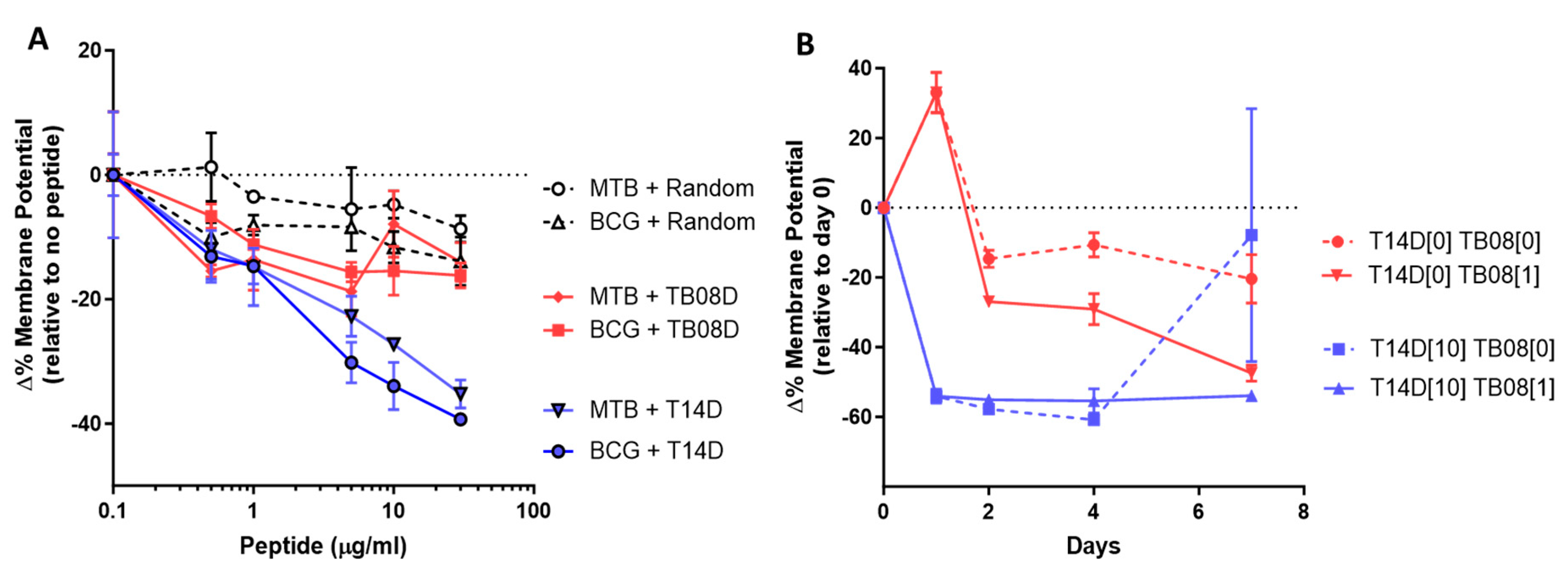
2.5. Cationic Antimicrobial Peptides Stimulating Mycobacterial Growth Reduce the Mycobacterial Membrane Potential, Which Can Then Be Maintained by Continued Low-Peptide Dosing
2.6. The Incorporation of Cationic Antimicrobial Peptides into Routine Sample Processing Protocols Enables a Greater Recovery of MTB from Tuberculosis Samples
3. Discussion
4. Materials and Methods
4.1. Peptides
4.2. Membrane Potential Studies
4.3. Leakage and DSC Studies
4.4. Validation of MTB Recovery Efficacy from Clinical Samples
Supplementary Materials
Author Contributions
Funding
Institutional Review Board Statement
Informed Consent Statement
Data Availability Statement
Acknowledgments
Conflicts of Interest
References
- Cox, R.A. Quantitative relationships for specific growth rates and macromolecular compositions of Mycobacterium tuberculosis, Streptomyces coelicolor A3(2) and Escherichia coli B/r: An integrative theoretical approach. Microbiology 2004, 150, 1413–1426. [Google Scholar] [CrossRef] [PubMed]
- Global Tuberculosis Report. 2022. Available online: www.who.int/teams/global-tuberculosis-programme/tb-reports/global-tuberculosis-report-2022 (accessed on 1 September 2023).
- Dijksteel, G.S.; Ulrich, M.M.W.; Middelkoop, E.; Boekema, B. Review: Lessons Learned from Clinical Trials Using Antimicrobial Peptides (AMPs). Front. Microbiol. 2021, 12, 616979. [Google Scholar] [CrossRef] [PubMed]
- Pirtskhalava, M.; Amstrong, A.A.; Grigolava, M.; Chubinidze, M.; Alimbarashvili, E.; Vishnepolsky, B.; Gabrielian, A.; Rosenthal, A.; Hurt, D.E.; Tartakovsky, M. DBAASP v3: Database of antimicrobial/cytotoxic activity and structure of peptides as a resource for development of new therapeutics. Nucleic Acids Res. 2021, 49, D288–D297. [Google Scholar] [CrossRef] [PubMed]
- Brogden, K.A. Antimicrobial peptides: Pore formers or metabolic inhibitors in bacteria? Nat. Rev. Microbiol. 2005, 3, 238–250. [Google Scholar] [CrossRef] [PubMed]
- De Leeuw, E.; Li, C.; Zeng, P.; Li, C.; Diepeveen-de Buin, M.; Lu, W.Y.; Breukink, E.; Lu, W. Functional interaction of human neutrophil peptide-1 with the cell wall precursor lipid II. FEBS Lett. 2010, 584, 1543–1548. [Google Scholar] [CrossRef] [PubMed]
- Schmitt, P.; Wilmes, M.; Pugnière, M.; Aumelas, A.; Bachère, E.; Sahl, H.G.; Schneider, T.; Destoumieux-Garzón, D. Insight into invertebrate defensin mechanism of action: Oyster defensins inhibit peptidoglycan biosynthesis by binding to lipid II. J. Biol. Chem. 2010, 285, 29208–29216. [Google Scholar] [CrossRef] [PubMed]
- Carlsson, A.; Engstrom, P.; Palva, E.T.; Bennich, H. Attacin, an antibacterial protein from Hyalophora cecropia, inhibits synthesis of outer membrane proteins in Escherichia coli by interfering with omp gene transcription. Infect. Immun. 1991, 59, 3040–3045. [Google Scholar] [CrossRef]
- Cho, J.H.; Sung, B.H.; Kim, S.C. Buforins: Histone H2A-derived antimicrobial peptides from toad stomach. Biochim. Biophys. Acta 2009, 1788, 1564–1569. [Google Scholar] [CrossRef]
- Kobayashi, S.; Takeshima, K.; Park, C.B.; Kim, S.C.; Matsuzaki, K. Interactions of the novel antimicrobial peptide buforin 2 with lipid bilayers: Proline as a translocation promoting factor. Biochemistry 2000, 39, 8648–8654. [Google Scholar] [CrossRef]
- Hilpert, K.; McLeod, B.; Yu, J.; Elliott, M.R.; Rautenbach, M.; Ruden, S.; Burck, J.; Muhle-Goll, C.; Ulrich, A.S.; Keller, S.; et al. Short cationic antimicrobial peptides interact with ATP. Antimicrob. Agents Chemother. 2010, 54, 4480–4483. [Google Scholar] [CrossRef]
- Marchand, C.; Krajewski, K.; Lee, H.-F.; Antony, S.; Johnson, A.A.; Amin, R.; Roller, P.; Kvaratskhelia, M.; Pommier, Y. Covalent binding of the natural antimicrobial peptide indolicidin to DNA abasic sites. Nucleic Acids Res. 2006, 34, 5157–5165. [Google Scholar] [CrossRef] [PubMed]
- Mardirossian, M.; Pérébaskine, N.; Benincasa, M.; Gambato, S.; Hofmann, S.; Huter, P.; Müller, C.; Hilpert, K.; Innis, C.A.; Tossi, A.; et al. The Dolphin Proline-Rich Antimicrobial Peptide Tur1A Inhibits Protein Synthesis by Targeting the Bacterial Ribosome. Cell Chem. Biol. 2018, 25, 530–539.e537. [Google Scholar] [CrossRef] [PubMed]
- Krizsan, A.; Volke, D.; Weinert, S.; Sträter, N.; Knappe, D.; Hoffmann, R. Insect-derived proline-rich antimicrobial peptides kill bacteria by inhibiting bacterial protein translation at the 70S ribosome. Angew. Chem. Int. Ed. Engl. 2014, 53, 12236–12239. [Google Scholar] [CrossRef] [PubMed]
- Greber, K.E.; Dawgul, M. Antimicrobial Peptides Under Clinical Trials. Curr. Top. Med. Chem. 2017, 17, 620–628. [Google Scholar] [CrossRef] [PubMed]
- Li, M.; Lai, Y.; Villaruz, A.E.; Cha, D.J.; Sturdevant, D.E.; Otto, M. Gram-positive three-component antimicrobial peptide-sensing system. Proc. Natl. Acad. Sci. USA 2007, 104, 9469–9474. [Google Scholar] [CrossRef] [PubMed]
- Kindrachuk, J.; Paur, N.; Reiman, C.; Scruten, E.; Napper, S. The PhoQ-activating potential of antimicrobial peptides contributes to antimicrobial efficacy and is predictive of the induction of bacterial resistance. Antimicrob. Agents Chemother. 2007, 51, 4374–4381. [Google Scholar] [CrossRef]
- Iavicoli, I.; Fontana, L.; Agathokleous, E.; Santocono, C.; Russo, F.; Vetrani, I.; Fedele, M.; Calabrese, E.J. Hormetic dose responses induced by antibiotics in bacteria: A phantom menace to be thoroughly evaluated to address the environmental risk and tackle the antibiotic resistance phenomenon. Sci. Total Environ. 2021, 798, 149255. [Google Scholar] [CrossRef]
- Gorr, S.U.; Brigman, H.V.; Anderson, J.C.; Hirsch, E.B. The antimicrobial peptide DGL13K is active against drug-resistant gram-negative bacteria and sub-inhibitory concentrations stimulate bacterial growth without causing resistance. PLoS ONE 2022, 17, e0273504. [Google Scholar] [CrossRef]
- Grimsey, E.; Collis, D.W.P.; Mikut, R.; Hilpert, K. The effect of lipidation and glycosylation on short cationic antimicrobial peptides. Biochim. Biophys. Acta Biomembr. 2020, 1862, 183195. [Google Scholar] [CrossRef]
- Hilpert, K.; Munshi, T.; López-Pérez, P.M.; Sequeira-Garcia, J.; Hofmann, S.; Bull, T.J. Discovery of Antimicrobial Peptides That Can Accelerate Culture Diagnostics of Slow-Growing Mycobacteria Including Mycobacterium tuberculosis. Microorganisms 2023, 11, 2225. [Google Scholar] [CrossRef]
- Bull, T.J.; Munshi, T.; Mikkelsen, H.; Hartmann, S.B.; Sørensen, M.R.; Garcia, J.S.; Lopez-Perez, P.M.; Hofmann, S.; Hilpert, K.; Jungersen, G. Improved Culture Medium (TiKa) for Mycobacterium avium Subspecies Paratuberculosis (MAP) Matches qPCR Sensitivity and Reveals Significant Proportions of Non-viable MAP in Lymphoid Tissue of Vaccinated MAP Challenged Animals. Front. Microbiol. 2016, 7, 2112. [Google Scholar] [CrossRef] [PubMed]
- Waddell, S.J. Reprogramming the Mycobacterium tuberculosis transcriptome during pathogenesis. Drug Discov. Today Dis. Mech. 2010, 7, e67–e73. [Google Scholar] [CrossRef]
- Salina, E.G.; Waddell, S.J.; Hoffmann, N.; Rosenkrands, I.; Butcher, P.D.; Kaprelyants, A.S. Potassium availability triggers Mycobacterium tuberculosis transition to, and resuscitation from, non-culturable (dormant) states. Open Biol. 2014, 4, 140106. [Google Scholar] [CrossRef]
- Stewart, G.R.; Wernisch, L.; Stabler, R.; Mangan, J.A.; Hinds, J.; Laing, K.G.; Butcher, P.D.; Young, D.B. The heat shock response of Mycobacterium tuberculosis: Linking gene expression, immunology and pathogenesis. Comp. Funct. Genom. 2002, 3, 348–351. [Google Scholar] [CrossRef] [PubMed]
- Tükenmez, H.; Sarkar, S.; Anoosheh, S.; Kruchanova, A.; Edström, I.; Harrison, G.A.; Stallings, C.L.; Almqvist, F.; Larsson, C. Mycobacterium tuberculosis Rv3160c is a TetR-like transcriptional repressor that regulates expression of the putative oxygenase Rv3161c. Sci. Rep. 2021, 11, 1523. [Google Scholar] [CrossRef] [PubMed]
- Rustad, T.R.; Harrell, M.I.; Liao, R.; Sherman, D.R. The enduring hypoxic response of Mycobacterium tuberculosis. PLoS ONE 2008, 3, e1502. [Google Scholar] [CrossRef] [PubMed]
- Betts, J.C.; Lukey, P.T.; Robb, L.C.; McAdam, R.A.; Duncan, K. Evaluation of a nutrient starvation model of Mycobacterium tuberculosis persistence by gene and protein expression profiling. Mol. Microbiol. 2002, 43, 717–731. [Google Scholar] [CrossRef]
- Keren, I.; Minami, S.; Rubin, E.; Lewis, K. Characterization and transcriptome analysis of Mycobacterium tuberculosis persisters. mBio 2011, 2, e00100–e00111. [Google Scholar] [CrossRef]
- Santangelo, M.d.l.P.; Klepp, L.; Nuñez-García, J.; Blanco, F.C.; Soria, M.; García-Pelayo, M.d.C.; Bianco, M.V.; Cataldi, A.A.; Golby, P.; Jackson, M.; et al. Mce3R, a TetR-type transcriptional repressor, controls the expression of a regulon involved in lipid metabolism in Mycobacterium tuberculosis. Microbiology 2009, 155, 2245–2255. [Google Scholar] [CrossRef]
- Park, H.; Guinn, K.M.; Harrell, M.I.; Liao, R.; Voskuil, M.I.; Tompa, M.; Schoolnik, G.K.; Sherman, D.R. Rv3133c/dosR is a transcription factor that mediates the hypoxic response of Mycobacterium tuberculosis. Mol. Microbiol. 2003, 48, 833–843. [Google Scholar] [CrossRef]
- Garcia-Fernandez, E.; Medrano, F.J.; Galan, B.; Garcia, J.L. Deciphering the transcriptional regulation of cholesterol catabolic pathway in mycobacteria: Identification of the inducer of KstR repressor. J. Biol. Chem. 2014, 289, 17576–17588. [Google Scholar] [CrossRef] [PubMed]
- Sarva, K.; Satsangi, A.T.; Plocinska, R.; Madiraju, M.; Rajagopalan, M. Two-component kinase TrcS complements Mycobacterium smegmatis mtrB kinase mutant. Tuberculosis 2019, 116S, S107–S113. [Google Scholar] [CrossRef] [PubMed]
- Gonzalo-Asensio, J.; Mostowy, S.; Harders-Westerveen, J.; Huygen, K.; Hernández-Pando, R.; Thole, J.; Behr, M.; Gicquel, B.; Martín, C. PhoP: A missing piece in the intricate puzzle of Mycobacterium tuberculosis virulence. PLoS ONE 2008, 3, e3496. [Google Scholar] [CrossRef] [PubMed]
- Shprung, T.; Peleg, A.; Rosenfeld, Y.; Trieu-Cuot, P.; Shai, Y. Effect of PhoP-PhoQ activation by broad repertoire of antimicrobial peptides on bacterial resistance. J. Biol. Chem. 2012, 287, 4544–4551. [Google Scholar] [CrossRef] [PubMed]
- Praporski, S.; Mechler, A.; Separovic, F.; Martin, L.L. Subtle Differences in Initial Membrane Interactions Underpin the Selectivity of Small Antimicrobial Peptides. ChemPlusChem 2015, 80, 91–96. [Google Scholar] [CrossRef]
- McCubbin, G.A.; Praporski, S.; Piantavigna, S.; Knappe, D.; Hoffmann, R.; Bowie, J.H.; Separovic, F.; Martin, L.L. QCM-D fingerprinting of membrane-active peptides. Eur. Biophys. J. Biophys. Lett. 2011, 40, 437–446. [Google Scholar] [CrossRef] [PubMed]
- Mechler, A.; Praporski, S.; Piantavigna, S.; Heaton, S.M.; Hall, K.N.; Aguilar, M.-I.; Martin, L. Structure and homogeneity of pseudo-physiological phospholipid bilayers and their deposition characteristics on carboxylic acid terminated self-assembled monolayers. Biomaterials 2009, 30, 682–689. [Google Scholar] [CrossRef]
- Mechler, A.; Praporski, S.; Atmuri, K.; Boland, M.; Separovic, F.; Martin, L.L. Specific and selective peptide-membrane interactions revealed using quartz crystal microbalance. Biophys. J. 2007, 93, 3907–3916. [Google Scholar] [CrossRef]
- Piantavigna, S.; Abdelhamid, M.E.; Zhao, C.; Qu, X.; McCubbin, G.A.; Graham, B.; Spiccia, L.; O’Mullane, A.P.; Martin, L.L. Mechanistic Details of the Membrane Perforation and Passive Translocation of TAT Peptides. Chem. Plus Chem. 2015, 80, 83–90. [Google Scholar] [CrossRef]
- Ön, A.; Vejzovic, D.; Jennings, J.; Parigger, L.; Cordfunke, R.A.; Drijfhout, J.W.; Lohner, K.; Malanovic, N. Bactericidal Activity to Escherichia coli: Different Modes of Action of Two 24-Mer Peptides SAAP-148 and OP-145, Both Derived from Human Cathelicidine LL-37. Antibiotics 2023, 12, 1163. [Google Scholar] [CrossRef]
- Hess, A.S.; Shardell, M.; Johnson, J.K.; Thom, K.A.; Strassle, P.; Netzer, G.; Harris, A.D. Methods and recommendations for evaluating and reporting a new diagnostic test. Eur. J. Clin. Microbiol. Infect. Dis. 2012, 31, 2111–2116. [Google Scholar] [CrossRef] [PubMed]
- Ramón-García, S.; Mikut, R.; Ng, C.; Ruden, S.; Volkmer, R.; Reischl, M.; Hilpert, K.; Thompson, C.J. Targeting Mycobacterium tuberculosis and other microbial pathogens using improved synthetic antibacterial peptides. Antimicrob. Agents Chemother. 2013, 57, 2295–2303. [Google Scholar] [CrossRef] [PubMed]
- Von Gundlach, A.; Ashby, M.P.; Gani, J.; Lopez-Perez, P.M.; Cookson, A.R.; Huws, S.A.; Rumancev, C.; Garamus, V.M.; Mikut, R.; Rosenhahn, A.; et al. BioSAXS Measurements Reveal That Two Antimicrobial Peptides Induce Similar Molecular Changes in Gram-Negative and Gram-Positive Bacteria. Front. Pharmacol. 2019, 10, 1127. [Google Scholar] [CrossRef]
- Kurbatova, E.V.; Cegielski, J.P.; Lienhardt, C.; Akksilp, R.; Bayona, J.; Becerra, M.C.; Caoili, J.; Contreras, C.; Dalton, T.; Danilovits, M.; et al. Sputum culture conversion as a prognostic marker for end-of-treatment outcome in patients with multidrug-resistant tuberculosis: A secondary analysis of data from two observational cohort studies. Lancet Respir. Med. 2015, 3, 201–209. [Google Scholar] [CrossRef]
- Azadi, D.; Motallebirad, T.; Ghaffari, K.; Shojaei, H. Mycobacteriosis and Tuberculosis: Laboratory Diagnosis. Open Microbiol. J. 2018, 12, 41–58. [Google Scholar] [CrossRef] [PubMed]
- Welch, D.F.; Guruswamy, A.P.; Sides, S.J.; Shaw, C.H.; Gilchrist, M.J. Timely culture for mycobacteria which utilizes a microcolony method. J. Clin. Microbiol. 1993, 31, 2178–2184. [Google Scholar] [CrossRef] [PubMed]
- Burdz, T.V.; Wolfe, J.; Kabani, A. Evaluation of sputum decontamination methods for Mycobacterium tuberculosis using viable colony counts and flow cytometry. Diagn. Microbiol. Infect. Dis. 2003, 47, 503–509. [Google Scholar] [CrossRef]
- Lipworth, S.; Hammond, R.; Baron, V.; Hu, Y.; Coates, A.; Gillespie, S. Defining dormancy in mycobacterial disease. Tuberculosis 2016, 99, 131–142. [Google Scholar] [CrossRef]
- Tuberculosis in England, 2022 Report (Data Up to End of 2021). Available online: www.gov.uk/government/publications/tuberculosis-in-england-2022-report-data-up-to-end-of-2021 (accessed on 1 November 2023).
- Tostmann, A.; Kik, S.V.; Kalisvaart, N.A.; Sebek, M.M.; Verver, S.; Boeree, M.J.; van Soolingen, D. Tuberculosis transmission by patients with smear-negative pulmonary tuberculosis in a large cohort in the Netherlands. Clin. Infect. Dis. 2008, 47, 1135–1142. [Google Scholar] [CrossRef]
- Mitchison, D.A.; Allen, B.W.; Carrol, L.; Dickinson, J.M.; Aber, V.R. A selective oleic acid albumin agar medium for tubercle bacilli. J. Med. Microbiol. 1972, 5, 165–175. [Google Scholar] [CrossRef]
- Piller, P.; Wolinski, H.; Cordfunke, R.A.; Drijfhout, J.W.; Keller, S.; Lohner, K.; Malanovic, N. Membrane Activity of LL-37 Derived Antimicrobial Peptides against Enterococcus hirae: Superiority of SAAP-148 over OP-145. Biomolecules 2022, 12, 523. [Google Scholar] [CrossRef] [PubMed]
- Malanovic, N.; Lohner, K. Antimicrobial Peptides Targeting Gram-Positive Bacteria. Pharmaceuticals 2016, 9, 59. [Google Scholar] [CrossRef] [PubMed]
- Queiroz, A.; Riley, L.W. Bacterial immunostat: Mycobacterium tuberculosis lipids and their role in the host immune response. Rev. Soc. Bras. Med. Trop. 2017, 50, 9–18. [Google Scholar] [CrossRef] [PubMed]

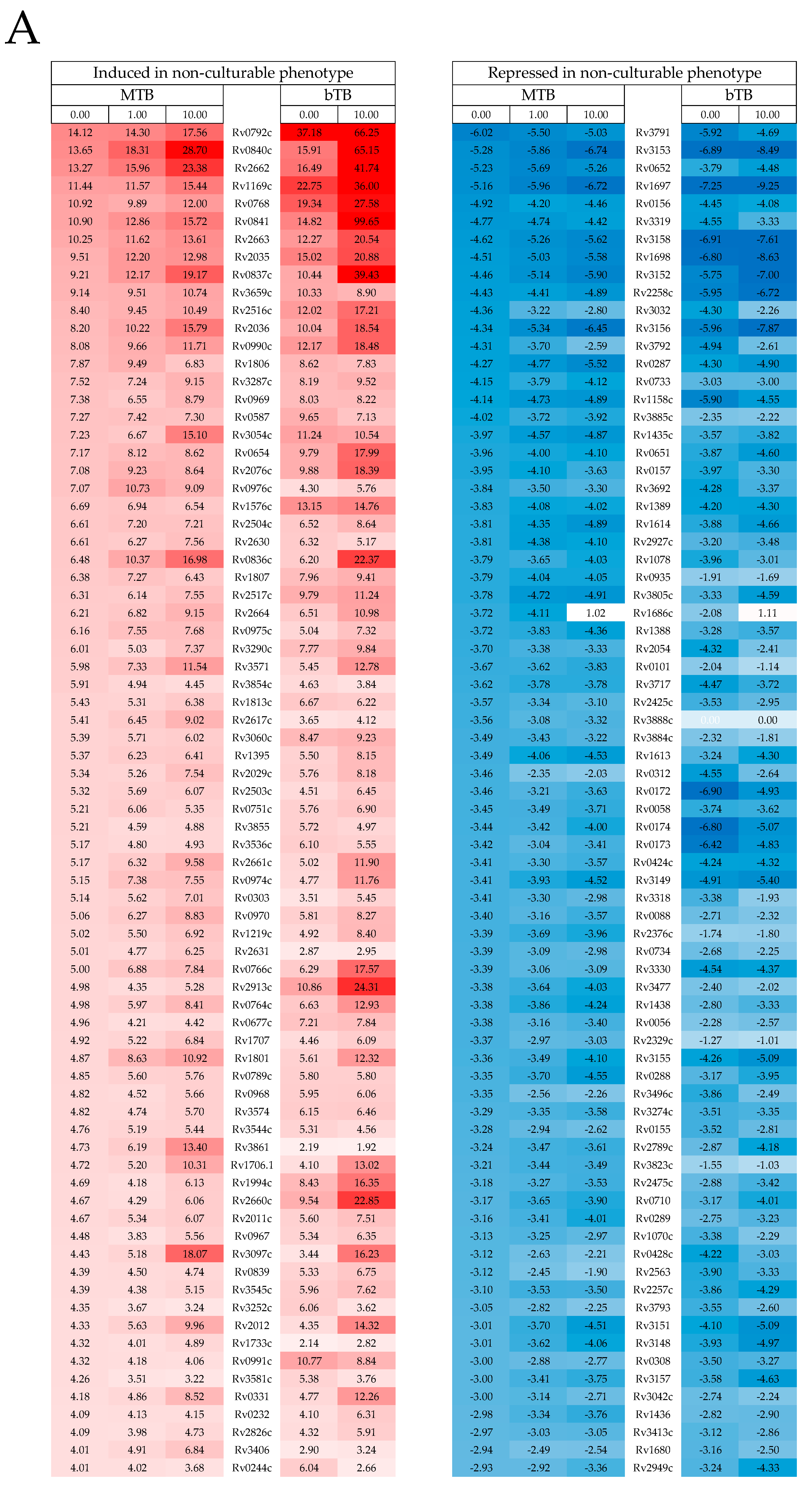
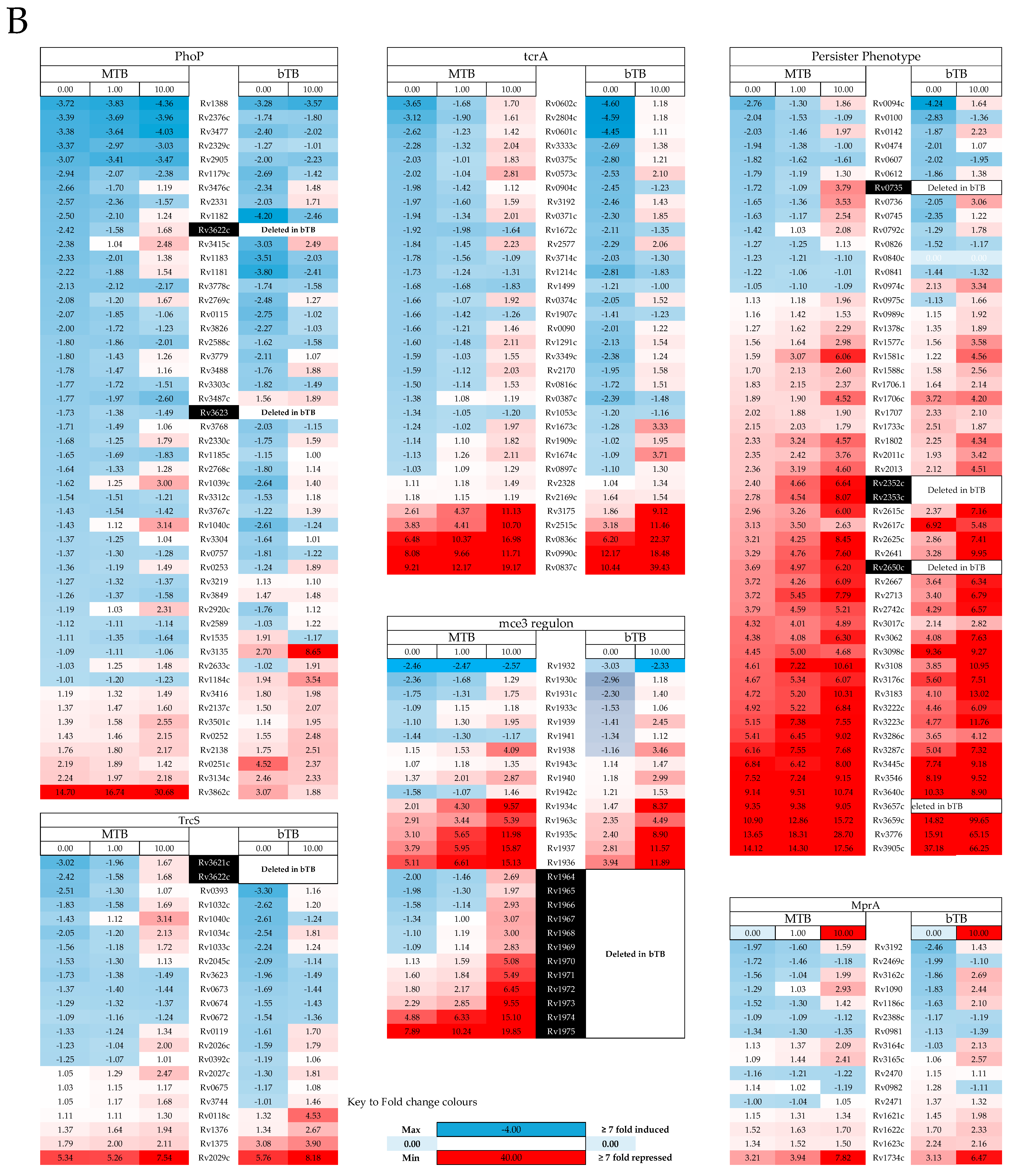

| Transcriptomic Profiles | MTB 0 h vs. 48 h | bTB 0 h vs. 48 h | MTB 48 h vs. 48 h | bTB 48 h vs. 48 h | ||||
|---|---|---|---|---|---|---|---|---|
| 0 μg/mL | 1 μg/mL | 10 μg/mL | 0 μg/mL | 10 μg/mL | 1 μg/mL | 10 μg/mL | 10 μg/mL | |
| Repressed after 48 h | Repressed Relative to Control | |||||||
| Geneset 1r | Geneset 2r | Geneset 3r | Geneset 4r | Geneset 5r | Geneset 6r | Geneset 7r | Geneset 8r | |
| Cholesterol use and cell wall synthesis (TcrA: Rv0602c) | ns | ns | 1.43 × 10⁻² | ns | ns | 4.09 × 10 −3 | 4.34 × 10 −6 | 9.58 × 10 −5 |
| Infection persistence (MprA: Rv0981) | ns | ns | ns | ns | 2.09 × 10⁻³ | ns | 3.91 × 10 −2 | 2.09 × 10 −2 |
| Growth and cord formation (PhoP: Rv0757) | ns | ns | ns | ns | ns | 2.16 × 10 −3 | 9.15 × 10 −3 | 4.75 × 10 −2 |
| Controls growth (TcrS: Rv1032c) | ns | ns | ns | ns | ns | ns | 7.95 × 10 −3 | 1.51 × 10 −2 |
| Controls dormancy (dosR: Rv3133c) | 2.16 × 10⁻¹⁵ | 3.81 × 10⁻¹³ | 8.48 × 10⁻¹⁵ | 3.95 × 10⁻¹⁴ | 8.48 × 10⁻¹³ | ns | ns | ns |
| Lipid metabolism and transport (mce3: Rv1963c) | 2.96 × 10⁻³ | 1.88 × 10⁻⁴ | 3.55 × 10⁻¹⁰ | ns | 1.26 × 10⁻³ | ns | 2.13 × 10 −15 | 4.59 × 10⁻² |
| Late stationary and dormancy (sigI: Rv1189) | 3.52 × 10⁻³ | 4.62 ×10⁻³ | 2.46 × 10⁻⁴ | ns | ns | ns | 5.38 × 10 −3 | ns |
| Non-culturable MTB profile (Induced geneset) | 3.34 × 10−38 | 6.63 × 10−38 | 1.11 × 10⁻³⁴ | 2.80 × 10 −38 | 4.21 × 10 −34 | ns | 3.16 × 10 −5 | 7.58 × 10 −6 |
| Non-culturable MTB profile (Repessed geneset) | 8.07 × 10⁻¹⁹ | 1.34 × 10⁻¹⁹ | 1.13 × 10−41 | 3.96 × 10 −14 | 5.04 × 10 −24 | 8.71 × 10 −3 | 2.37 × 10 −36 | 3.10 × 10 −31 |
| MTB Persiter profile (Induced geneset) | 4.82 × 10⁻³² | 1.21 × 10⁻³⁴ | 2.00 × 10⁻³² | 4.55 × 10 −32 | 1.16 × 10 −33 | ns | 1.46 × 10 −2 | 4.02 × 10 −4 |
| Enduring Hypoxic Response (Induced geneset) | 1.86 × 10−47 | 1.21 × 10−45 | 1.80 × 10−45 | 5.54 × 10 −47 | 1.02 × 10 −39 | ns | ns | ns |
| Cholesterol uptake and metabolism: (KstR: Rv3574) | 1.20 × 10⁻¹³ | 7.57 × 10⁻¹⁵ | 1.74 × 10⁻¹² | 1.74 × 10 −12 | 1.46 × 10 −13 | ns | ns | ns |
| Cell division and cell wall synthesis (MtrA: Rv3246c) | 7.28 × 10 −4 | 1.10 × 10 −2 | 1.82 × 10 −4 | 2.14 × 10 −2 | 4.28 × 10 −4 | ns | ns | ns |
| Induced after 48 h | Induced Relative to Control | |||||||
| Geneset-1i | Geneset-2i | Geneset 3i | Geneset-4i | Geneset-5i | Geneset-6i | Geneset-7i | Geneset-8i | |
| Non-culturable MTB profile (Induced geneset) | 3.08 × 10 −13 | 2.79 × 10 −18 | 6.14 × 10 −21 | 3.80 × 10 −15 | 7.91 × 10 −19 | ns | ns | ns |
| Non-culturable MTB profile (Repessed geneset) | 3.42 × 10 −14 | 7.60 × 10 −33 | 2.62 × 10 −42 | 2.74 × 10 −9 | 6.84 × 10 −37 | ns | ns | ns |
| Starvation response | 2.21 × 10 −3 | 1.73 × 10 −8 | 2.55 × 10 −24 | 4.27 × 10 −2 | 1.38 × 10 −24 | ns | ns | 5.10 × 10⁻⁴ |
| Aerobic Metabolism | 2.93 × 10 −14 | 2.38 × 10 −31 | 2.10 × 10 −42 | 2.74 × 10 −9 | 1.90 × 10 −37 | ns | ns | ns |
| Reactivation from chronic infection (whiB5 regulon) | ns | ns | ns | ns | ns | ns | 7.59 × 10 −3 | 2.88 × 10 −2 |
| Triclosan MTB growth inhibitor | ns | ns | ns | ns | ns | ns | ns | ns |
| Heat Shock Response | ns | ns | ns | ns | ns | ns | ns | ns |
| Isolate | Sample | Total Positive by Index Only | Positive by Index and Reference | Total Positive by Reference Only | ||
|---|---|---|---|---|---|---|
| Total Index Improved | Total Equal | Total Reference Improved | ||||
| MTB | Sputum Smear +ve | 2 (13) | 17 {3} | 3 | 2 {5} | 0 |
| Sputum Smear −ve | 8 (26) | 1 {46} | 0 | 2 {3} | 0 | |
| Faeces/Urine | 14 (29) | 1 {33} | 0 | 0 | 0 | |
| FNA/Bx./Pl.fl. | 2 (33) | 2 {8} | 0 | 2 {18} | 0 | |
| NTM | Sputum Smear +ve | 0 | 0 | 0 | 0 | 0 |
| Sputum Smear −ve | 4 (8) | 3 {12} | 0 | 0 | 0 | |
| Faeces/Urine | 3 (46) | 0 | 0 | 0 | 0 | |
| BAL | 1 (12) | 0 | 0 | 0 | 0 | |
Disclaimer/Publisher’s Note: The statements, opinions and data contained in all publications are solely those of the individual author(s) and contributor(s) and not of MDPI and/or the editor(s). MDPI and/or the editor(s) disclaim responsibility for any injury to people or property resulting from any ideas, methods, instructions or products referred to in the content. |
© 2023 by the authors. Licensee MDPI, Basel, Switzerland. This article is an open access article distributed under the terms and conditions of the Creative Commons Attribution (CC BY) license (https://creativecommons.org/licenses/by/4.0/).
Share and Cite
Bull, T.J.; Munshi, T.; Lopez-Perez, P.M.; Tran, A.C.; Cosgrove, C.; Bartolf, A.; Menichini, M.; Rindi, L.; Parigger, L.; Malanovic, N.; et al. Specific Cationic Antimicrobial Peptides Enhance the Recovery of Low-Load Quiescent Mycobacterium tuberculosis in Routine Diagnostics. Int. J. Mol. Sci. 2023, 24, 17555. https://doi.org/10.3390/ijms242417555
Bull TJ, Munshi T, Lopez-Perez PM, Tran AC, Cosgrove C, Bartolf A, Menichini M, Rindi L, Parigger L, Malanovic N, et al. Specific Cationic Antimicrobial Peptides Enhance the Recovery of Low-Load Quiescent Mycobacterium tuberculosis in Routine Diagnostics. International Journal of Molecular Sciences. 2023; 24(24):17555. https://doi.org/10.3390/ijms242417555
Chicago/Turabian StyleBull, Tim J., Tulika Munshi, Paula M. Lopez-Perez, Andy C. Tran, Catherine Cosgrove, Angela Bartolf, Melissa Menichini, Laura Rindi, Lena Parigger, Nermina Malanovic, and et al. 2023. "Specific Cationic Antimicrobial Peptides Enhance the Recovery of Low-Load Quiescent Mycobacterium tuberculosis in Routine Diagnostics" International Journal of Molecular Sciences 24, no. 24: 17555. https://doi.org/10.3390/ijms242417555
APA StyleBull, T. J., Munshi, T., Lopez-Perez, P. M., Tran, A. C., Cosgrove, C., Bartolf, A., Menichini, M., Rindi, L., Parigger, L., Malanovic, N., Lohner, K., Wang, C. J. H., Fatima, A., Martin, L. L., Esin, S., Batoni, G., & Hilpert, K. (2023). Specific Cationic Antimicrobial Peptides Enhance the Recovery of Low-Load Quiescent Mycobacterium tuberculosis in Routine Diagnostics. International Journal of Molecular Sciences, 24(24), 17555. https://doi.org/10.3390/ijms242417555


