Review

Role of Melatonin in Cancer: Effect on Clock Genes

by
César Rodríguez-Santana
1,
Javier Florido
1,
Laura Martínez-Ruiz
1,
Alba López-Rodríguez
1,
Darío Acuña-Castroviejo
1,2,3 and
Germaine Escames
1,2,3,*
1
Biomedical Research Center, Health Sciences Technology Park, University of Granada, 18016 Granada, Spain
2
Department of Physiology, Faculty of Medicine, University of Granada, 18071 Granada, Spain
3
CIBER de Fragilidad y Envejecimiento Saludable (CIBERFES), Instituto de Salud Carlos III, Instituto de Investigación Biosanitaria Ibs., 18016 Granada, Spain
*
Author to whom correspondence should be addressed.
Int. J. Mol. Sci. 2023, 24(3), 1919; https://doi.org/10.3390/ijms24031919
Submission received: 29 December 2022 / Revised: 15 January 2023 / Accepted: 17 January 2023 / Published: 18 January 2023
(This article belongs to the Special Issue The Role of the Circadian Clock in Cancer and Inflammatory Diseases)

Abstract

:
The circadian clock is a regulatory system, with a periodicity of approximately 24 h, that generates rhythmic changes in many physiological processes. Increasing evidence links chronodisruption with aberrant functionality in clock gene expression, resulting in multiple diseases, including cancer. In this context, tumor cells have an altered circadian machinery compared to normal cells, which deregulates the cell cycle, repair mechanisms, energy metabolism and other processes. Melatonin is the main hormone produced by the pineal gland, whose production and secretion oscillates in accordance with the light:dark cycle. In addition, melatonin regulates the expression of clock genes, including those in cancer cells, which could play a key role in the numerous oncostatic effects of this hormone. This review aims to describe and clarify the role of clock genes in cancer, as well as the possible mechanisms of the action of melatonin through which it regulates the expression of the tumor’s circadian machinery, in order to propose future anti-neoplastic clinical treatments.

1. Introduction

Disruptions in circadian rhythms have been linked to mammalian tumorigenesis in numerous studies [1]. At the molecular level, circadian rhythms are generated by a system of interlocking autoregulatory transcriptional/translational feedback loops, while melatonin (N-acetyl-5-methoxytryptamine, aMT) is the principal hormone that regulates circadian clock genes.
Although melatonin is produced by the pineal gland, higher concentrations are also produced in other tissues [2]. The synthesis of pineal melatonin is associated with light/dark photoperiods, with increasing levels produced at night [3]. This facilitates the chronobiotic regulation of numerous activities, such as immunomodulatory, anti-inflammatory, antioxidant and vasoregulatory activities [4,5,6,7].
Numerous studies have found a strong correlation between reduced melatonin levels and increased risk of tumor development [8,9,10,11,12]. The oncostatic properties of melatonin have been extensively studied. Although this hormone’s mechanisms of action in cancer remain unclear, its connection with the circadian clock may be key to a better understanding of these mechanisms.
This review therefore emphasizes the connection between the disruption of circadian rhythms, cancer and the role of melatonin in regulating clock genes.

2. General Mechanism of the Circadian Clock

In mammals, the circadian clock acts as a regulatory system, with a periodicity of approximately 24 h. This process is modulated by endogenous factors such as genetic or endocrine secretions, behavioral factors such as feeding/fasting, as well as external oscillating signals such as light/darkness and temperature cycles. All these inputs generate rhythmic changes in many physiological processes, including the endocrine system, the cell cycle, DNA damage repair, metabolism and the sleep/wake cycle [13].
The circadian clock is controlled by a central circadian pacemaker located in the suprachiasmatic nucleus (SCN) of the hypothalamus, which, in turn, controls the peripheral oscillators, located in practically all cells in the body [14]. The synchronization of the cells in different tissues requires the precise coordination of the circadian clock. However, although circadian rhythms are fundamental to the correct physiological functioning of the organism, these mechanisms can be lost or weakened due to different factors, such as aging or various pathologies [15].
At the molecular level, the central circadian clock is composed of a small number of genes whose expression forms a time-delayed transcription–translation feedback loop. The activating arm of the loop is composed of circadian locomotor output cycle kaput (CLOCK)/neuronal PAS domain protein 2 (NPAS2) and brain and muscle aryl hydrocarbon receptor nuclear translocator-like protein 1 (BMAL1), which form a heterodimer and connect with its promoter at CACGTG E-box sequences. The binding to the E-box by this heterodimer induces clock-controlled gene (CCG) expression. This facilitates the transcription of numerous genes, including the Cryptochrome (cry1 and cry2) and Period (per1, per2 and per3) genes. The CRY and PER proteins form a repressor complex that is translocated into the nucleus during the evening and physically interacts with the CLOCK/NPAS2:BMAL1 heterodimer in order to repress their own transcription. Subsequently, these proteins, which increase and accumulate in the cytoplasm, are phosphorylated by cysteine kinase 1 ε/δ (CK1 ε/δ) during the night for its degradation. They are targeted for ubiquitination by specific E3 ligases and are eventually degraded by the proteasome, thus further increasing their synthesis at the beginning of the day. The waxing and waning of this transcriptional feedback loop, which takes ~24 h to complete, represents the core mechanism of the circadian clock in mammals [16,17] (Figure 1).
In turn, Bmal1 is regulated by a second loop composed of the activator orphan retinoic acid receptor-related alpha (RORα), and the repressor, nuclear reactors reverse strand of protein ERB alpha (REV-ERBα) (Figure 1). At the same time, clock genes can be further modulated by epigenetic modifications and miRNA, as well as by post-transcriptional and post-translational modifications [17,18,19].

3. Clock Genes and Cancer

Alterations in clock genes are associated with different types of human cancer [20,21,22,23]. However, it is not understood how circadian disruption is associated with serious adverse health outcomes, including carcinogenesis.
The World Health Organization currently recognizes night shift work, which involves circadian disruption, as a probable carcinogen, classified as type 2A by the Agency for Research on Cancer (IARC) [24]. Since the 1980s, numerous epidemiological studies have linked an increased risk of different cancers, such as breast and prostate cancer, with night shift work by employees such as nurses and flight attendants on transatlantic flights [1,10,12,25]. It has been suggested that sleep deprivation, the light-induced suppression of melatonin and lifestyle changes are important mechanisms that could explain the possible link between shift work and cancer risk [24]. Moreover, studies of genetic variations in the circadian pathway in human clock genes have reported that the disruption of circadian rhythms is related to an increased risk of cancer [26]. In support of these findings, a significant relationship has been established between different types of cancer, such as breast, lung and prostate cancer, and variations in Bmal1, Clock, RORα and RORβ. This is due to the fact that clock genes regulate between approximately 50% and more than 80% of mammalian genome genes, including the tumor suppressor gene p53 and the oncogene c-Myc [27,28]. The disruption in the circadian clock is therefore involved in tumor development by altering the expression of genes involved in fundamental functions, such as the cell cycle, apoptosis, metabolism and energy, DNA repair, tumor immunity and metastasis [29]. In addition, the polymorphisms of other clock genes specific to each tumor type have been identified, suggesting that certain circadian genes might be more important than others in terms of predisposition to different types of cancer [26].
Intriguingly, given that cancer prognosis and survival have been associated with the level of circadian disruption in patient tumor tissues [30], clock genes, such as Bmal1, Per1 and npas2, could be considered potential prognostic biomarkers in certain cancers [31,32].
Nevertheless, other studies found no evidence of a relationship between interruptions in the circadian rhythm in night shift workers and carcinogenesis [33]. Recently, in a meta-analysis of 57 observational studies, no evidence of a relationship was reported between night shift work and an increased risk of cancer, especially in breast, prostate, ovarian, pancreatic, colorectal, non-Hodgkin’s lymph and stomach cancers [34]. However, these differences in results can be explained by the methodological differences of the different epidemiological studies, as well as in the statistical tools used [35]. Therefore, common criteria to clarify the relationship between an alteration in circadian rhythms and carcinogenesis are clearly required.

3.1. Clock Genes, Proliferation and Apoptosis

Malignant tumors are characterized by uncontrolled cell proliferation, partly due to a loss of control of cell cycle events caused by clock gene dysregulation [36]. Clock genes affect many biological pathways, including those involved in cell proliferation and apoptosis, by controlling the expression of cell cycle genes [29]. In addition, substantial evidence shows that progression through the cell cycle occurs at specific times of the day/night cycle, suggesting that one function of the circadian clock system is to control this fundamental process [37]. It is remarkable that circadian rhythms share some common features with the cell cycle, while disruptions in circadian clocks have been found to be related to carcinogenesis. For example, the BMAL1-CLOCK/NPAS2 heterodimer has been reported to repress c-Myc, an oncogene that contributes to the genesis of many human cancers, and whose protein expression is closely correlated with cell proliferation rates [30,38]. REV-ERBα and RORα have also been reported to regulate the expression of p21, a CCG that negatively regulates cell cycle progression [37]. Overexpressed PER1 also induces c-Myc and suppresses p21, while other studies have reported that PER1 inhibits Cyclin B1, Cdc2 and Wee1 expression, leading to a decrease in cancer cell proliferation [39]. This discrepancy may be due to differences in the methods used or in the characteristics of the cell lines used in the experiments [40]. Nevertheless, these data indicate that many genes crucial to the cell cycle are under the control of clock genes that are aberrantly expressed in many tumor tissues.
It has also been reported that many crucial genes involved in cell proliferation and apoptosis have periodic patterns of expression. These genes, which oscillate during a 24-h cycle, include the proliferation gene Ki-67, the tumor suppressor gene p53, the proto-oncogene Mdm2 and the apoptotic-related proteins BAX and BCL-2 [29]. PER1 and PER2 have been reported to be mainly associated with the upregulation of Ki-67, Mdm2 and Bax, as well as with the downregulation of Bcl-2, c-Myc and p53, in lung, pancreatic, hepatocellular and oral carcinoma cell lines [41,42,43]. In addition, there is a strong connection between Bmal1 and Per2 regulation and the PI3K/mTOR signaling pathway, one of the most frequently activated signaling pathways in tumorigenesis and the progression of cancer. For example, BMAL1 depletion leads to cell cycle disruption, which results in a substantial increase in the apoptotic cell population, and to the acceleration of cell invasion [44]. The PI3K/mTOR pathway has a major effect on the regulation of processes such as autophagy, proliferation and apoptosis, which sequentially affects the occurrence and development of cancer [44,45,46].
Many signaling pathways cooperate with clock genes involved in tumorigenesis; the altered expression of these genes can modify a range of downstream CCGs and tumor-related genes, which impacts tumor cell proliferation, apoptosis, migration and invasion.
Collectively, these data suggest that the circadian clock can control cell proliferation and apoptosis at multiple levels and that disruption of the circadian system is linked to tumor cell growth.

3.2. Clock Genes and Metastasis

The relationship between metastatic disease progression and circadian rhythms has been poorly characterized [47,48,49]. However, a large number of studies have demonstrated the participation of clock genes in metastasis. For example, low PER1/PER2 expression in different types of cancer, such as breast [50], glioma [51], gastric [52] and non-small cell lung cancer [53], is closely related to the development and metastasis of tumors. Moreover, much evidence has identified BMAL1 as a key element in metastasis in breast cancer and glioblastoma. BMAL1 regulates the expression and activity of matrix metalloproteinase 9 (MMP9), which controls cell migration and invasion [47,48]. MMP9, which is involved in the degradation of the tumor extracellular matrix, is a mediating factor with regard to the local invasion and distant metastasis of tumor cells. In human colorectal cancer, BMAL1 has been shown to induce metastasis by stimulating exosome secretion [54]. Exosomes derived from primary tumors have been shown to alter the microenvironment of secondary organs in order to facilitate the colonization and growth of metastatic tumors, whose quantity is dependent on circadian rhythms [54].

3.3. Clock Genes and Tumor Immunity

Circadian rhythms are also involved in the mammalian immune system, which involves various populations of immune cells, such as monocytes, natural killer cells, dendritic cells (DCs) and T and B lymphocytes, as well as responses to signals and their defensive functions, including cytokine levels [55,56]. Thus, when circadian homeostasis is disrupted, deregulation of the immune system produces immune suppression and the accelerated development of tumors. BMAL1 is the principal mediator of the circadian control of the immune system and also promotes anti-inflammatory states. Downregulation of BMAL1 has been found in hematologic malignancies such as diffuse large B-cell lymphoma, as well as acute lymphocytic and myeloid leukemias [57]. Deletion of BMAL1 affects the development of B lymphocytes [58]. In addition, a recent investigation by Wang, C. et al. [56] showed that, in murine models with the specific inhibition of Bmal1 in T and dendritic cells, regardless of the time at which melanoma cells were inoculated, the tumor volume was similar after 14 days of the experiment. However, in wild-type mice, significant differences in tumor growth were observed depending on the time of engraftment. Therefore, BMAL1 and cell-autonomous circadian oscillations in both DCs and T cells are critical for time-of-day differences in tumor volume.
BMAL1, CLOCK, REV-ERBα and RORα have also been reported to regulate immune functions and inflammation by modulating CCGs that encode a variety of proteins, including cytokines, chemokines and receptors [29]. However, the central pacemaker is also modulated by immune factors such as proinflammatory cytokines and IL-1/6, as well as by anti-inflammatory drugs, at the molecular and cellular levels, which results in the subsequent alteration of clock genes. In conclusion, the circadian clock and immune system can be said to exert bidirectional control [59].

3.4. Clock Genes and Chronotherapy

The toxicity, efficacy and even the pharmacokinetics or metabolism of a drug can vary with the time of day, depending on its mechanism of action [60]. In recent years, special attention has been paid to the administration of anticancer drugs depending on circadian rhythmicity in order to maximize efficacy and to reduce side effects. For example, there is evidence that, in synchronized esophageal cancer cells, DNA damage induced by cisplatin is greater when coinciding with lower levels of PER2 [61]. In in vivo melanoma models, mice treated at night showed a higher rate of cisplatin–DNA adduct removal and less toxicity than those treated in the morning, which coincides with maximal global and gene-specific repairs. Interestingly, differences in the effects of this treatment were not observed in Per1/2 knockout mice [62].
Chronotherapy has improved the implantation of medication for other pathologies, such as asthma [60,63], osteoarthritis and rheumatoid arthritis [64]. However, to date, studies carried out to evaluate chronochemotherapy for some types of cancer are contradictory. For example, chronotherapy in the case of ovarian cancer has shown no beneficial effect [65], while male patients with colorectal cancer lived significantly longer [66].
The main problem concerning the clinical application of chronotherapy is the need for studies of the circadian gene expression profiles of specific cancers. In addition, non-invasive methods to assess the circadian parameters of cancers need to be developed. Thus, although the use of chronotherapy in cancer treatment is an interesting avenue of study, more in-depth research is necessary [67].
Therefore, given all these data, the circadian clock clearly plays a fundamental role in tumor pathogenesis. Alterations in clock gene expression correlate with alterations in DNA replication, DNA repair and responses to DNA damage in the metabolism and in senescence. The control of cell proliferation and apoptosis is also lost, metastasis spreads, the immune system is altered and even drug resistance increases. Thus, the “repair” of circadian rhythms has been shown to be a possible therapeutic target against this disease. However, due to the great complexity of the pathways involved, further research to clarify the role of circadian clocks in cancer is required.

4. Melatonin and Circadian Rhythms

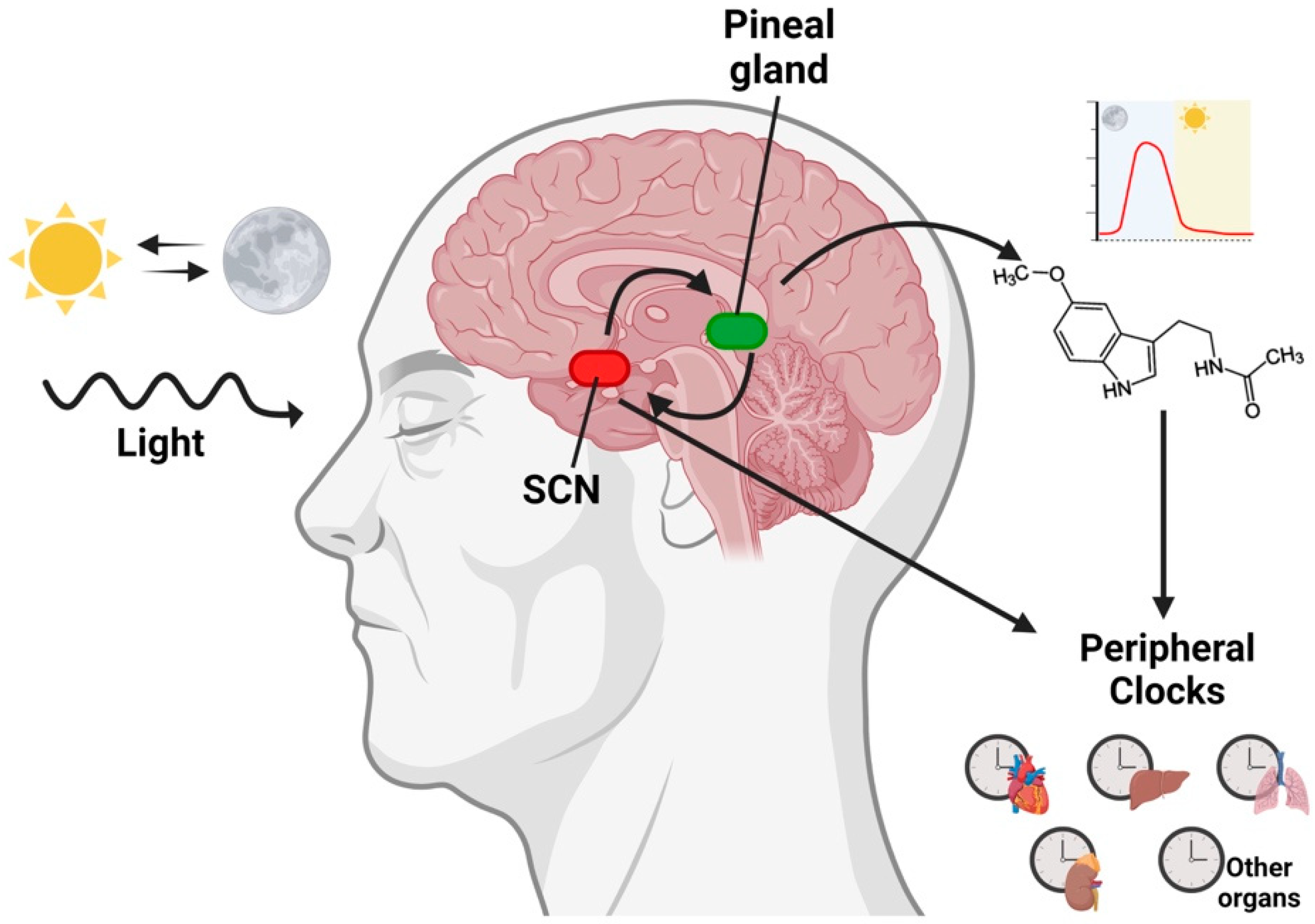
Melatonin, which is the principal hormone produced in the pineal gland following a circadian rhythm, is also produced in other tissues [2]. However, unlike pineal melatonin, extrapineal melatonin does not follow a circadian rhythm, and, to date, it is not known whether its synthesis is controlled by clock genes [68,69]. Pineal melatonin production is controlled by the suprachiasmatic nucleus (SCN), which transforms the photoperiodic signal into an endocrine signal (Figure 2) [69]. During the night, the polysynaptic pathway that establishes the SCN in the pineal gland gives rise to the stimulation of pinealocytes, which culminates in the expression of arylalkylamine N-acetyltransferase (AANAT), one of the key enzymes in the production of melatonin [70]. Once melatonin is synthesized in the pineal gland, it is rapidly released to the cerebrospinal fluid and blood due to its high diffusibility. As a result, the melatonin levels in both the fluid and blood have a circadian rhythm, which typically peaks at night [71]. All these rhythmic variations, including endocrine and non-endocrine rhythms, during approximately 24 h have a chronobiotic impact on the organism [3].
Melatonin, in turn, affects the SCN and, therefore, regulates the circadian phase to maintain rhythmic stability, although the way in which this is done is unclear. Melatonin membrane receptors have been identified in the SCN of vertebrates, while signal transduction pathways are involved in both MT1 and MT2 in order to induce an increase in the expression of the two clock genes, Per1 and Per2 [71,72]. Melatonin also has an acute inhibitory effect on neuronal firing through the inhibition of glutamatergic activity, independently of MT1 and MT2 [73]. In addition, melatonin also binds other proteins, such as calmodulin, calreticuline and quinone reductase II (previously called MT3), leading to various regulatory effects [7,74].
This hormone is also able to change the phase in the expression of Bmal1 and Rev-erbα [75,76]. It has been hypothesized that melatonin maintains the regulation of the circadian machinery through the ubiquitin–proteasome system (explained below). Thus, the effect of melatonin on the central pacemaker could act as a regulator of the circadian clock by regulating the correct time and rhythmic amplitude [69,77].
Therefore, pineal melatonin is a chronobiotic molecule that plays a major role in the coordination of circadian rhythmicity. Functionally, pineal melatonin produces a host of effects that can be controlled by the SCN and may also have a direct impact on numerous peripheral organs. Thus, melatonin plays multiple roles in the complex circadian system by influencing the phases of the central pacemaker, as well as of peripheral oscillators. In particular, pineal melatonin is involved in sleep promotion [78]. At the molecular level, increased melatonin levels are correlated with BMAL1 and CLOCK expression at night, which generates circadian rhythms through the transcriptional/translational feedback loop described above [79]. Additionally, melatonin has been shown to modulate, either positively or negatively, the expression of most central oscillator genes. Nevertheless, the mechanisms by which melatonin participates in the expression of circadian clock components are unclear.
Alterations in pineal melatonin levels, caused mainly by light exposure at night, can disrupt the circadian system and have, consequently, been related to numerous pathologies, including carcinogenesis [12].

5. Melatonin, Clock Genes and Cancer

Numerous studies have demonstrated the relationship between disrupted clock genes, cancer and decreased levels of melatonin. Moreover, the suppression of melatonin production is associated with an increased incidence of cancer [12,80], which could be correlated with a loss in the regulation of the circadian machinery. Additionally, numerous studies have described a large number of oncostatic effects of this hormone [74,81,82], some of which could be mediated through its effects on circadian rhythms.
Xiang, S. et al. [83] have described how the use of melatonin in MCF7 breast cancer cells (1 nM) in vitro represses RORα transactivation through the MT1 receptor activation pathway, leading to the suppression of Bmal1 mRNA expression. In addition, an increase in Per2 expression was observed following treatment with melatonin after 20 h. Per2 is an important tumor suppressor that is capable of inducing p53 expression. Thus, these findings provide additional data on the proapoptotic mechanism of melatonin in cancer cells. Interestingly, after the xenografting of MCF7 cells in rats with an intact circadian rhythm, clock gene circadian expression in MCF7 tumor cells was reestablished [83] (Table 1). Another type of tumor whose risk, progression and aggressiveness are closely related to circadian alterations is prostate cancer. In 2010, Jung-Hynes [84] studied the effect of melatonin (0.1–2 mM) on the expression of different clock genes in prostate cancer cell lines. The study demonstrated that melatonin caused a significant upregulation in Clock and Per2 mRNA and protein levels, as well as a significant downregulation of Bmal1 mRNA after 24 h of treatment. In addition, melatonin induced a rhythmic oscillation, suggesting that it is able to “resynchronize” the central clock genes in prostate cancer cells [84].

5.1. Melatonin and the Ubiquitin–Proteasome System

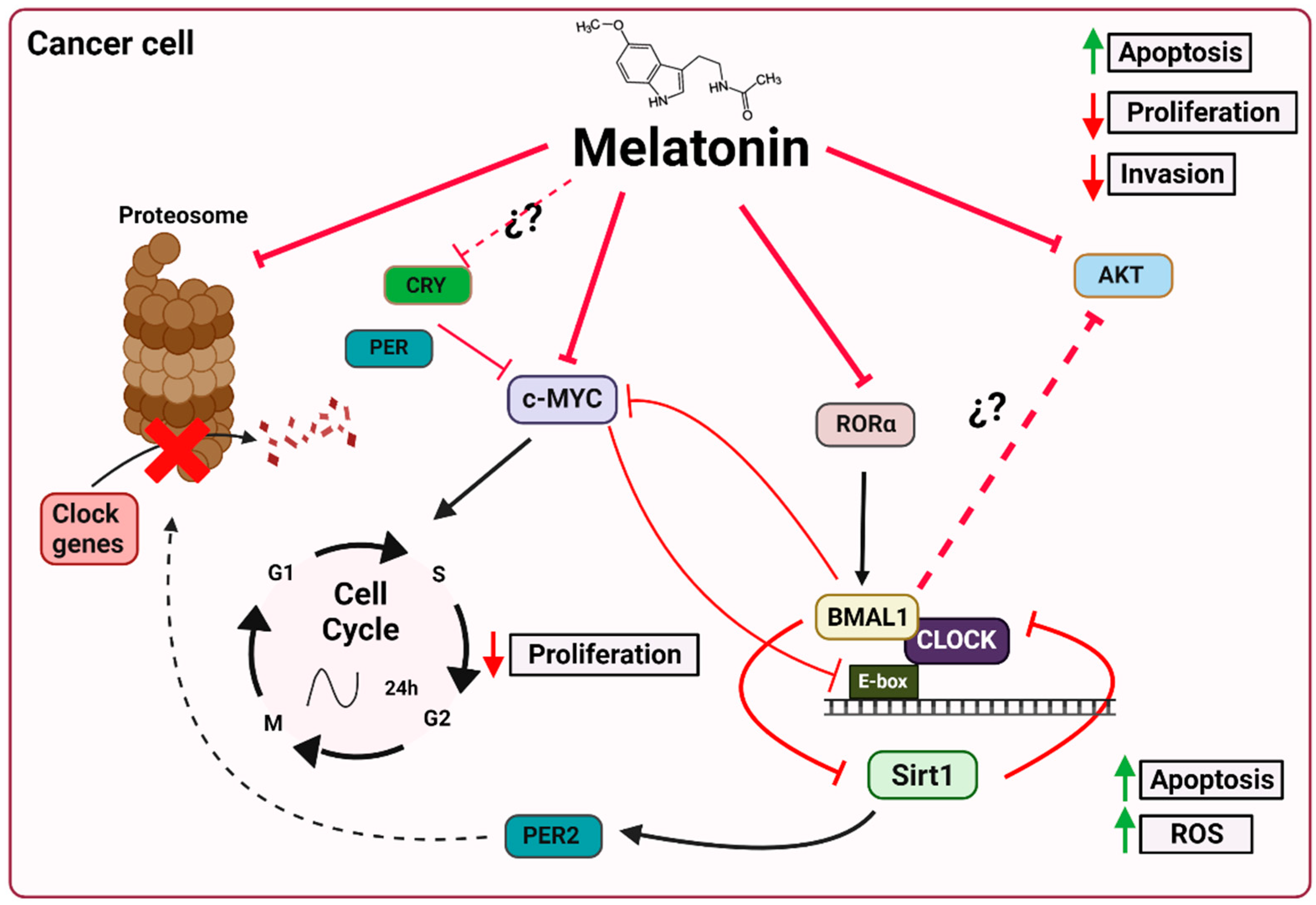
Several mechanisms have been described to explain the oncostatic effects of melatonin through its effect on clock genes. One of these mechanisms, mentioned above, is the ubiquitin–proteasome system, which enables the degradation of proteins such as BMAL1, REV-ERVα, CRY and PER to be regulated. However, while there is no direct evidence to show that melatonin directly inhibits proteasomes in the SCN, melatonin clearly may interfere with the activity of the ubiquitin–proteasome system in a variety of tissues, including the SCN [88,89]. Vriend and Reiter [76] have suggested that melatonin inhibits the proteasome, which provides selective stability to proteins during periods of elevated melatonin levels. This effect of melatonin on the proteasome results in increased levels of CRY, PER and REV-ERBα, which, in turn, regulate the transcription of Bmal1. However, the authors highlight the fact that BMAL1 is closely associated with the inhibition of melatonin in the proteasome, whose expression coincides with the nocturnal increase in this hormone [76]. Moreover, there is even experimental evidence that shows that melatonin inhibits the proteasomes of renal cancer cells (Figure 3) [88].
Currently, it is known that clock genes are closely linked to cell cycle-related genes, whose levels are controlled by the proteasome [90]. Thus, given that melatonin acts as a proteasome inhibitor, its effects on the inhibition of cyclin D1 and E, CDK2, CDK4 and c-Myc expression in brain cancer stem cells [91] or on the reduction of cyclin D1 and E expression in breast cancer cells [92,93] could be closely linked. In addition, in different cancer cells, melatonin and the proteasome inhibitor bortezomib have been shown to have similar proapoptotic effects, indirectly interacting with the levels of p53, Bcl-2, BAX, p21 and NF-kB, through the ubiquitin–proteasome system. Furthermore, other studies have reported that melatonin increases apoptosis due to an increase in the Bcl-2-interacting mediator (BIM) of cell death via the inhibition of proteasome activity [88].

5.2. Melatonin and c-Myc

Another important connection between the oncostatic effects of melatonin and circadian machinery is its ability to reduce the levels of c-Myc, which leads to antitumor and antiproliferative effects [91,94]. c-Myc, which is involved in oncogenesis processes, is overexpressed in many cancers. This is explained by the abnormal c-Myc levels, which enable cells with damaged DNA to pass through cell cycle checkpoints, thus contributing to the genesis of many cancers in humans [27,95]. In recent years, this oncogene has been found to control and, in turn, to be controlled by the circadian machinery [95]. Although the circadian connection is currently not entirely clear, MYC is now known to heterodimerize with its partner MAX in order to bind to E-box sequences (CACGTG) at cognate promoters for the purposes of gene regulation, such as the CLOCK-BMAL1 heterodimer [96]. MYC has also been shown to be regulated by CRY and PER proteins [41,97]. Furthermore, downregulation of BMAL1 is associated with high MYC expression levels in a variety of tumors, a phenomenon associated with poor clinical outcomes [98]. Given the multi-faceted involvement, such as increased cell cycle proliferation and increased glycolysis, of c-Myc in oncogenesis, as well as the oncostatic effects of melatonin by reducing c-Myc expression, we suggest that there is a clear link between this hormone and c-Myc, which needs to be studied in more depth (Figure 3).

5.3. Melatonin and Sirt1

Melatonin also modulates clock genes through sirtuin 1 (SIRT1). SIRTs are a type III histone/protein deacetylase whose activity depends on oscillating levels of nicotinamide adenine dinucleotide (NAD+). In turn, the biosynthesis or salvaging of NAD+ depends on the enzyme nicotinamide phosphoribosyl transferase (NAMPT), whose expression is characterized by a circadian rhythm [99,100]. Already in 2009, Jung-Hynes and Ahmad [101] proposed that the inhibition of SIRT1 could be the key to the regulation of the circadian machinery. This regulation occurs by controlling the acetylation levels of both BMAL1 and the circadian repressor PER2, thus boosting PER2 degradation and circadian cycle rhythmicity (Figure 3). SIRT1, which is associated with CLOCK, is recruited to the CLOCK:BMAL1 chromatin complex at circadian promoters, thus facilitating the circadian expression of numerous genes, including nampt. Therefore, a decrease in NAMPT levels and, chronically, an increase in NAD+ are related to abnormal circadian behavior and metabolism [102,103].
Sirt1, which is overexpressed in tumor cells, is correlated with the silencing of tumor suppressor genes and with cancer resistance to chemotherapy. Interestingly, melatonin has a dual effect on SIRT1 expression. In normal cells, melatonin levels increase, leading to a decrease in reactive oxygen species (ROS) production and regulation of cell homeostasis. However, in tumor cells, melatonin downregulates SIRT1, which results in proapoptotic and prooxidant activity [7]. Specifically, in human osteosarcoma, melatonin has been shown to inhibit SIRT1, resulting in increased prooxidant and antitumor activity [104]. The upregulation of SIRT1 by SRT1720 (a known SIRT1 activator) attenuates melatonin’s antioxidant and antitumor activity, indicating that its induction of ROS production in tumor cells is activated by SIRT1 [105]. In addition, in MCF7, Proietti et al. [105] have described how the inhibition of SIRT1 levels by melatonin produces the downregulation of the MDM2 pathway, a ubiquitin protein ligase, which enhances p53 acetylation and, therefore, p53 activity. Although the mechanisms by which melatonin affects SIRT1 are still not fully understood, in 2009, Hill et al. [85] were the first to suggest that melatonin, through the MT1 receptor, blocks the transcription of RORα, thus blocking the activation of Bmal1 expression and, therefore, of Sirt1 [85].

5.4. Melatonin and AKT

The Akt signaling pathway is involved in the inhibition of cell apoptosis and in stimulating cell proliferation following the activation of Akt/PKB, a serine/threonine kinase. In xenografts of breast cancer, the tumor growth rate and elevated levels of AKT and 3-phosphoinositide-dependent kinase-1 (PDK-1), an AKT stimulator, correlated with night-time light exposure and reduced levels of melatonin [106]. In addition, numerous studies have reported the oncostatic effects of exogenous melatonin due to AKT downregulation [107,108,109]. There is consistent evidence that shows that melatonin may affect the phosphorylation of AKT, PI3K and GSK3β due to the regulation of Bmal1. In 2013, Jung, C. H. et al. [110] published a study that showed that the activation of the PI3K-Akt-MMP-2 pathway depends on BMAL1. Specifically, they demonstrated how the inhibition of BMAL1 increased the levels of PI3K activity, AKT phosphorylation, MMP-2 protein and, consequently, cell invasion. An interesting connection has thus been established between the inhibitory effect of melatonin on the AKT pathway and its ability to regulate Bmal1 (Figure 3) [110].

6. Conclusions

Cancer is a complex, heterogeneous disease whose origin is closely related to chronodisruption. Clock genes are directly or indirectly involved in tumor development and in fundamental functions. Despite advances in cancer research, further investigation is required to understand its complexity and to find new therapeutic targets, develop new treatments and/or enhance and improve existing therapies. For many years, the scientific community has reported the numerous oncostatic effects of melatonin, which influences many physiological processes, including the “repair” of the circadian clock. This review describes the possible links between melatonin and the circadian clock in cancer. The mechanisms involved include the ubiquitin–proteasome system and its impact on SIRT1 and c-Myc. Therefore, a more exhaustive study of the effects of melatonin on clock genes could be key to understanding all these mechanisms.

Author Contributions

Writing—original draft preparation, C.R.-S.; writing—review and editing, G.E.; formal analysis, G.E.; resources, C.R.-S., L.M.-R., J.F. and A.L.-R.; supervision, G.E. and D.A.-C.; project administration, G.E. and D.A.-C.; funding acquisition, G.E. All authors have read and agreed to the published version of the manuscript.

Funding

This review was funded by grants from the MCIN/AEI/10.13039/501100011033, in Spain, and the ERDF (SAF2017-85903-P, ID2020-115112RB-I00), from the Consejería de Innovación, Ciencia y Empresa, Junta de Andalucía (P07-CTS-03135, P10-CTS-5784, and CTS-101). J.F. and L.M. are recipients of FPU fellowships from the Ministerio de Educación Cultura y Deporte, Spain.

Institutional Review Board Statement

Not applicable.

Informed Consent Statement

Not applicable.

Data Availability Statement

Not applicable.

Acknowledgments

We wish to thank Michael O’Shea for proofreading the paper.

Conflicts of Interest

The authors declare no conflict of interest.

Abbreviations

AANATarylalkylamine N-acetyltransferase
aMTmelatonin
BIMBcl-2-interacting mediator of cell death
Bmal1 brain and muscle aryl hydrocarbon receptor nuclear translocator-like protein 1
CCGsclock-controlled genes
CK1 ε/δ cysteine kinase 1 ε/δ
Clockcircadian locomotor output cycles kaput
CryChryptochrome
DCdendritic cell
IARCInternational Agency for Research on Cancer
MMP9matrix metalloproteinase 9
NAD+ nicotinamide adenine dinucleotide
NAMPTnicotinamide phosphoribosyl transferase
NPAS2neuronal PAS domain protein 2
PerPeriod
Rev-erbαreverse strand of protein ERB alpha
RORαorphan retinoic acid receptor-related alpha
ROSreactive oxygen species
SCNsuprachiasmatic nucleus
SIRT1sirtuin 1

References

  1. Cadenas, C.; Van De Sandt, L.; Edlund, K.; Lohr, M.; Hellwig, B.; Marchan, R.; Schmidt, M.; Rahnenführer, J.; Oster, H.; Hengstler, J.G. Loss of Circadian Clock Gene Expression Is Associated with Tumor Progression in Breast Cancer. Cell Cycle 2014, 13, 3282–3291. [Google Scholar] [CrossRef] [PubMed] [Green Version]
  2. Acuña-Castroviejo, D.; Escames, G.; Venegas, C.; Díaz-Casado, M.E.; Lima-Cabello, E.; López, L.C.; Rosales-Corral, S.; Tan, D.X.; Reiter, R.J. Extrapineal Melatonin: Sources, Regulation, and Potential Functions. Cell. Mol. Life Sci. 2014, 71, 2997–3025. [Google Scholar] [CrossRef] [PubMed]
  3. Reiter, R.J. Pineal Melatonin: Cell Biology of Its Synthesis and of Its Physiological Interactions. Endocr. Rev. 1991, 12, 151–180. [Google Scholar] [CrossRef] [PubMed] [Green Version]
  4. Cardinali, D.P.; Hardeland, R. Inflammaging, Metabolic Syndrome and Melatonin: A Call for Treatment Studies. Neuroendocrinology 2017, 104, 382–397. [Google Scholar] [CrossRef] [Green Version]
  5. Rahbarghazi, A.; Siahkouhian, M.; Rahbarghazi, R.; Ahmadi, M.; Bolboli, L.; Keyhanmanesh, R.; Mahdipour, M.; Rajabi, H. Role of Melatonin in the Angiogenesis Potential; Highlights on the Cardiovascular Disease. J. Inflamm. 2021, 18, 4. [Google Scholar] [CrossRef]
  6. Moslehi, M.; Moazamiyanfar, R.; Dakkali, M.S.; Rezaei, S.; Rastegar-Pouyani, N.; Jafarzadeh, E.; Mouludi, K.; Khodamoradi, E.; Taeb, S.; Najafi, M. Modulation of the Immune System by Melatonin; Implications for Cancer Therapy. Int. Immunopharmacol. 2022, 108, 108890. [Google Scholar] [CrossRef]
  7. Florido, J.; Rodriguez-Santana, C.; Martinez-Ruiz, L.; López-Rodríguez, A.; Acuña-Castroviejo, D.; Rusanova, I.; Escames, G. Understanding the Mechanism of Action of Melatonin, Which Induces ROS Production in Cancer Cells. Antioxidants 2022, 11, 1621. [Google Scholar] [CrossRef]
  8. Schernhammer, E.S.; Kroenke, C.H.; Laden, F.; Hankinson, S.E. Night Work and Risk of Breast Cancer. Epidemiology 2006, 17, 108–111. [Google Scholar] [CrossRef]
  9. LeMasters, G.K.; Genaidy, A.M.; Succop, P.; Deddens, J.; Sobeih, T.; Barriera-Viruet, H.; Dunning, K.; Lockey, J. Cancer Risk among Firefighters: A Review and Meta-Analysis of 32 Studies. J. Occup. Environ. Med. 2006, 48, 1189–1202. [Google Scholar] [CrossRef]
  10. Conlon, M.; Lightfoot, N.; Kreiger, N. Rotating Shift Work and Risk of Prostate Cancer [1]. Epidemiology 2007, 18, 182–183. [Google Scholar] [CrossRef]
  11. Viswanathan, A.N.; Schernhammer, E.S. Circulating Melatonin and the Risk of Breast and Endometrial Cancer in Women. Cancer Lett. 2009, 281, 1–7. [Google Scholar] [CrossRef] [Green Version]
  12. Touitou, Y.; Reinberg, A.; Touitou, D. Association between Light at Night, Melatonin Secretion, Sleep Deprivation, and the Internal Clock: Health Impacts and Mechanisms of Circadian Disruption. Life Sci. 2017, 173, 94–106. [Google Scholar] [CrossRef] [PubMed]
  13. Lowrey, P.L.; Takahashi, J.S. Mammalian Circadian Biology: Elucidating Genome-Wide Levels of Temporal Organization. Annu. Rev. Genom. Hum. Genet. 2004, 5, 407–441. [Google Scholar] [CrossRef] [PubMed] [Green Version]
  14. Stratmann, M.; Schibler, U. Properties, Entrainment, and Physiological Functions of Mammalian Peripheral Oscillators. J. Biol. Rhythms 2006, 21, 494–506. [Google Scholar] [CrossRef] [PubMed]
  15. Fishbein, A.B.; Knutson, K.L.; Zee, P.C. Circadian Disruption and Human Health. J. Clin. Investig. 2021, 131, 1–12. [Google Scholar] [CrossRef]
  16. Partch, C.L.; Green, C.B.; Takahashi, J.S. Molecular Architecture of the Mammalian Circadian Clock. Trends Cell Biol 2014, 24, 90–99. [Google Scholar] [CrossRef] [Green Version]
  17. Patke, A.; Young, M.W.; Axelrod, S. Molecular Mechanisms and Physiological Importance of Circadian Rhythms. Nat. Rev. Mol. Cell Biol. 2020, 21, 67–84. [Google Scholar] [CrossRef]
  18. Cho, H.; Zhao, X.; Hatori, M.; Yu, R.T.; Barish, G.D.; Lam, M.T.; Chong, L.; Ditacchio, L.; Atkins, A.R.; Glass, C.K.; et al. Regulation of Circadian Behavior and Metabolism by Rev-Erbα and Rev-Erbβ. Nature 2012, 485, 123–127. [Google Scholar] [CrossRef] [Green Version]
  19. Koike, N.; Yoo, S.-H.; Huang, H.-C.; Kumar, V.; Lee, C.; Kim, T.-K.; Takahashi, J.S. Transcriptional Architecture and Chromatin Landscape of the Core Circadian Clock in Mammals. Science 2012, 338, 349–354. [Google Scholar] [CrossRef] [Green Version]
  20. Tokunaga, H.; Takebayashi, Y.; Utsunomiya, H.; Akahira, J.I.; Higashimoto, M.; Mashiko, M.; Ito, K.; Niikura, H.; Takenoshita, S.I.; Yaegashi, N. Clinicopathological Significance of Circadian Rhythm-Related Gene Expression Levels in Patients with Epithelial Ovarian Cancer. Acta Obstet. Gynecol. Scand. 2008, 87, 1060–1070. [Google Scholar] [CrossRef]
  21. Relles, D.; Sendecki, J.; Chipitsyna, G.; Hyslop, T.; Yeo, C.J.; Arafat, H.A. Circadian Gene Expression and Clinicopathologic Correlates in Pancreatic Cancer. J. Gastrointest. Surg. 2013, 17, 443–450. [Google Scholar] [CrossRef] [PubMed]
  22. Gu, F.; Zhang, H.; Hyland, P.L.; Berndt, S.; Gapstur, S.M.; Wheeler, W.; The ELLIPSE Consortium; Amos, C.I.; Bezieau, S.; Bickeböller, H.; et al. Inherited Variation in Circadian Rhythm Genes and Risks of Prostate Cancer and Three Other Cancer Sites in Combined Cancer Consortia. Int. J. Cancer 2017, 141, 1794–1802. [Google Scholar] [CrossRef] [PubMed]
  23. Rahman, S.; Kraljevi, S. Circadian (De) Regulation in Head and Neck Squamous Cell Carcinoma. Int. J. Mol. Sci 2019, 20, 2662. [Google Scholar] [CrossRef] [PubMed] [Green Version]
  24. World Health Organization. IARC Monographs on the Evaluation of Carcinogenic Risks to Humans. Volume 98: Painting, Firefighting and Shiftwork; International Agency for Research on Cancer: Lyon, France, 2010; Volume 98.
  25. Fagundo-Rivera, J.; Gómez-Salgado, J.; García-Iglesias, J.J.; Gómez-Salgado, C.; Camacho-Martín, S.; Ruiz-Frutos, C. Relationship between Night Shifts and Risk of Breast Cancer among Nurses: A Systematic Review. Medicina 2020, 56, 680. [Google Scholar] [CrossRef]
  26. Mocellin, S.; Tropea, S.; Benna, C.; Rossi, C.R. Circadian Pathway Genetic Variation and Cancer Risk: Evidence from Genome-Wide Association Studies. BMC Med. 2018, 16, 1–8. [Google Scholar] [CrossRef] [Green Version]
  27. Dang, C.V. MYC on the Path to Cancer. Cell 2012, 149, 22–35. [Google Scholar] [CrossRef] [Green Version]
  28. Gotoh, T.; Vila-Caballer, M.; Liu, J.; Schiffhauer, S.; Finkielstein, C.V. Association of the Circadian Factor Period 2 to P53 Influences P53’s Function in DNA-Damage Signaling. Mol. Biol. Cell 2015, 26, 359–372. [Google Scholar] [CrossRef]
  29. Li, H.X. The Role of Circadian Clock Genes in Tumors. Onco. Targets. Ther. 2019, 12, 3645–3660. [Google Scholar] [CrossRef] [Green Version]
  30. Kiessling, S.; Beaulieu-Laroche, L.; Blum, I.D.; Landgraf, D.; Welsh, D.K.; Storch, K.F.; Labrecque, N.; Cermakian, N. Enhancing Circadian Clock Function in Cancer Cells Inhibits Tumor Growth. BMC Biol. 2017, 15, 1–18. [Google Scholar] [CrossRef] [Green Version]
  31. Mazzoccoli, G.; Panza, A.; Valvano, M.R.; Palumbo, O.; Carella, M.; Pazienza, V.; Biscaglia, G.; Tavano, F.; Di Sebastiano, P.; Andriulli, A.; et al. Clock Gene Expression Levels and Relationship with Clinical and Pathological Features in Colorectal Cancer Patients. Chronobiol. Int. 2011, 28, 841–851. [Google Scholar] [CrossRef]
  32. Yi, C.; Mu, L.; de la Longrais, I.A.R.; Sochirca, O.; Arisio, R.; Yu, H.; Hoffman, A.E.; Zhu, Y.; Katsaro, D. The Circadian Gene NPAS2 Is a Novel Prognostic Biomarker for Breast Cancer. Breast Cancer Res. Treat. 2010, 120, 663–669. [Google Scholar] [CrossRef] [PubMed] [Green Version]
  33. Barul, C.; Richard, H.; Parent, M.E. Night-Shift Work and Risk of Prostate Cancer: Results From a Canadian Case- Control Study, the Prostate Cancer and Environment Study. Am. J. Epidemiol. 2019, 188, 1801–1811. [Google Scholar] [CrossRef] [PubMed]
  34. Dun, A.; Zhao, X.; Jin, X.; Wei, T.; Gao, X.; Wang, Y.; Hou, H. Association between Night-Shift Work and Cancer Risk: Updated Systematic Review and Meta-Analysis. Front. Oncol. 2020, 10, 1–15. [Google Scholar] [CrossRef]
  35. Erren, T.C.; Lewis, P. Can Yesterday’s Smoking Research Inform Today’s Shiftwork Research? Epistemological Consequences for Exposures and Doses Due to Circadian Disruption at and off Work. J. Occup. Med. Toxicol. 2017, 12, 1–8. [Google Scholar] [CrossRef] [Green Version]
  36. Soták, M.; Sumová, A.; Pácha, J. Cross-Talk between the Circadian Clock and the Cell Cycle in Cancer. Ann. Med. 2014, 46, 221–232. [Google Scholar] [CrossRef]
  37. Gréchez-Cassiau, A.; Rayet, B.; Guillaumond, F.; Teboul, M.; Delaunay, F. The Circadian Clock Component BMAL1 Is a Critical Regulator of P21 WAF1/CIP1 Expression and Hepatocyte Proliferation. J. Biol. Chem. 2008, 283, 4535–4542. [Google Scholar] [CrossRef] [Green Version]
  38. Bretones, G.; Delgado, M.D.; León, J. Myc and Cell Cycle Control. Biochim. Biophys. Acta-Gene Regul. Mech. 2015, 1849, 506–516. [Google Scholar] [CrossRef]
  39. Gery, S.; Komatsu, N.; Baldjyan, L.; Yu, A.; Koo, D.; Koeffler, H.P. The Circadian Gene Per1 Plays an Important Role in Cell Growth and DNA Damage Control in Human Cancer Cells. Mol. Cell 2006, 22, 375–382. [Google Scholar] [CrossRef]
  40. Sato, F.; Wu, Y.; Kumar, U.; Liu, Y.; Morohashi, S.; Kato, Y.; Kijima, H. Has Pro-Apoptotic Effects during Cisplatin (CDDP) Treatment in Human Gingival Cancer CA9-22 Cells. Eur. J. Cancer 2011, 47, 1747–1758. [Google Scholar] [CrossRef]
  41. Hua, H.; Wang, Y.; Wan, C.; Liu, Y.; Zhu, B.; Yang, C.; Wang, X.; Wang, Z.; Cornelissen-Guillaume, G.; Halberg, F. Circadian Gene MPer2 Overexpression Induces Cancer Cell Apoptosis. Cancer Sci. 2006, 97, 589–596. [Google Scholar] [CrossRef]
  42. Sato, F.; Nagata, C.; Liu, Y.; Suzuki, T.; Kondo, J.; Morohashi, S.; Imaizumi, T.; Kato, Y.; Kijima, H. PERIOD1 Is an Anti-Apoptotic Factor in Human Pancreatic and Hepatic Cancer Cells. J. Biochem. 2009, 146, 833–838. [Google Scholar] [CrossRef] [PubMed]
  43. Li, H.X.; Fu, X.J.; Yang, K.; Chen, D.; Tang, H.; Zhao, Q. The Clock Gene PER1 Suppresses Expression of Tumor-Related Genes in Human Oral Squamous Cell Carcinoma. Oncotarget 2016, 7, 20574–20583. [Google Scholar] [CrossRef] [PubMed] [Green Version]
  44. Matsumoto, C.S.; Almeida, L.O.; Guimarães, D.M.; Martins, M.D.; Papagerakis, P.; Papagerakis, S.; Leopoldino, A.M.; Castilho, R.M.; Squarize, C.H. PI3K-PTEN Dysregulation Leads to MTOR-Driven Upregulation of the Core Clock Gene BMAL1 in Normal and Malignant Epithelial Cells. Oncotarget 2016, 7, 42393–42407. [Google Scholar] [CrossRef] [PubMed] [Green Version]
  45. Wang, Z.; Li, F.; Wei, M.; Zhang, S.; Wang, T. Circadian Clock Protein Period2 Suppresses the Pi3k/Akt Pathway and Promotes Cisplatin Sensitivity in Ovarian Cancer. Cancer Manag. Res. 2020, 12, 11897–11908. [Google Scholar] [CrossRef] [PubMed]
  46. Liu, H.; Gong, X.; Yang, K. Overexpression of the Clock Gene Per2 Suppresses Oral Squamous Cell Carcinoma Progression by Activating Autophagy via the PI3K/AKT/MTOR Pathway. J. Cancer 2020, 11, 3655–3666. [Google Scholar] [CrossRef] [Green Version]
  47. Wang, J.; Li, S.; Li, X.; Li, B.; Li, Y.; Xia, K.; Yang, Y.; Aman, S.; Wang, M.; Wu, H. Circadian Protein BMAL1 Promotes Breast Cancer Cell Invasion and Metastasis by Up-Regulating Matrix Metalloproteinase9 Expression. Cancer Cell Int. 2019, 19, 1–12. [Google Scholar] [CrossRef]
  48. Gwon, D.H.; Lee, W.Y.; Shin, N.; Kim, S.I.; Jeong, K.; Lee, W.H.; Kim, D.W.; Hong, J.; Lee, S.Y. BMAL1 Suppresses Proliferation, Migration, and Invasion of U87MG Cells by Downregulating Cyclin B1, Phospho-AKT, and Metalloproteinase-9. Int. J. Mol. Sci. 2020, 21, 2352. [Google Scholar] [CrossRef] [Green Version]
  49. Diamantopoulou, Z.; Castro-Giner, F.; Schwab, F.D.; Foerster, C.; Saini, M.; Budinjas, S.; Strittmatter, K.; Krol, I.; Seifert, B.; Heinzelmann-Schwarz, V.; et al. The Metastatic Spread of Breast Cancer Accelerates during Sleep. Nature 2022, 607, 156–162. [Google Scholar] [CrossRef] [PubMed]
  50. Broadberry, E.; Mcconnell, J.; Williams, J.; Yang, N.; Zindy, E.; Leek, A.; Waddington, R.; Joseph, L.; Howe, M.; Meng, Q.; et al. Disrupted Circadian Clocks and Altered Tissue Mechanics in Primary Human Breast Tumours. Breast Cancer Res. 2018, 20, 125. [Google Scholar] [CrossRef] [Green Version]
  51. Xia, H.; Niu, Z.; Ma, H.; Cao, S.; Hao, S.; Liu, Z.; Wang, F. Deregulated Expression of the Per1 and Per2 in Human Gliomas. Can J. Neurol. Sci. 2010, 37, 365–370. [Google Scholar] [CrossRef]
  52. Zhao, H.; Zeng, Z.; Yang, J.; Jin, Y.; Qiu, M.; Hu, X.; Han, J.; Liu, K. Prognostic Relevance of Period1 (Per1) and Period2 (Per2) Expression in Human Gastric Cancer. Int. J. Clin. Exp. Pathol. 2014, 7, 619–630. [Google Scholar]
  53. Xiang, R.U.N.; Cui, Y.U.E.; Wang, Y.; Xie, T.; Yang, X.; Wang, Z.H.U.; Li, J.; Li, Q. Circadian Clock Gene Per2 Downregulation in Non-Small Cell Lung Cancer Is Associated with Tumour Progression and Metastasis. Oncol. Rep. 2018, 40, 3040–3048. [Google Scholar] [CrossRef] [PubMed] [Green Version]
  54. Dong, P.; Wang, Y.; Liu, Y.; Zhu, C.; Lin, J.; Qian, R.; Hua, L.; Lu, C. BMAL1 Induces Colorectal Cancer Metastasis by Stimulating Exosome Secretion. Mol. Biol. Rep. 2022, 49, 373–384. [Google Scholar] [CrossRef] [PubMed]
  55. Scheiermann, C.; Gibbs, J.; Ince, L.; Loudon, A. Clocking in to Immunity. Nat. Rev. Immunol. 2018, 18, 423–437. [Google Scholar] [CrossRef]
  56. Wang, C.; Barnoud, C.; Cenerenti, M.; Sun, M.; Caffa, I.; Kizil, B.; Bill, R.; Liu, Y.; Pick, R.; Garnier, L.; et al. Dendritic Cells Direct Circadian Anti-Tumor Immune Response. Nature 2022. [Google Scholar] [CrossRef]
  57. Taniguchi, H.; Fernández, A.F.; Setién, F.; Ropero, S.; Ballestar, E.; Villanueva, A.; Yamamoto, H.; Imai, K.; Shinomura, Y.; Esteller, M. Epigenetic Inactivation of the Circadian Clock Gene BMAL1 in Hematologic Malignancies. Cancer Res. 2009, 69, 8447–8454. [Google Scholar] [CrossRef] [PubMed] [Green Version]
  58. Sun, Y.; Yang, Z.; Niu, Z.; Peng, J.; Li, Q.; Xiong, W.; Langnas, A.N.; Ma, M.Y.; Zhao, Y. MOP3, a Component of the Molecular Clock, Regulates the Development of B Cells. Immunology 2006, 119, 451–460. [Google Scholar] [CrossRef] [PubMed]
  59. Cermakian, N.; Lange, T.; Golombek, D.; Sarkar, D.; Nakao, A.; Shibata, S.; Mazzoccoli, G.; Cronobiologia, D.; Aires, B.; Brunswick, N.; et al. Crosstalk between the circadian clock circuitry and the immune system. Chronobiol. Int. J. Biol. Med. Rhythm. Res. 2020, 30, 870–888. [Google Scholar] [CrossRef]
  60. Cardinali, D.P.; Brown, G.M.; Pandi-Perumal, S.R. Chronotherapy. Handb. Clin. Neurol. 2021, 179, 357–370. [Google Scholar] [CrossRef]
  61. Redondo, J.A.; Bibes, R.; Drubbel, A.V.; Dassy, B.; Bisteau, X.; Maury, E.; Beck, B. Per2 Circadian Oscillation Sensitizes Esophageal Cancer Cells to Chemotherapy. Biology 2021, 10, 266. [Google Scholar] [CrossRef]
  62. Dakup, P.P.; Porter, K.I.; Little, A.A.; Gajula, R.P.; Zhang, H.; Skornyakov, E.; Kemp, M.G.; Van Dongen, H.P.A.; Gaddameedhi, S. The Circadian Clock Regulates Cisplatin-Induced Toxicity and Tumor Regression in Melanoma Mouse and Human Models. Oncotarget 2018, 9, 14524–14538. [Google Scholar] [CrossRef]
  63. Thakur, S.; Singh, B.; Mishra, V.; Yadav, N.; Giri, N.; Sharma, P.; Saini, A.; Garg, L.K. Bilayer Tablet Based Chronotherapeutics in the Management of Nocturnal Asthma: An Overview. Recent Pat. Drug Deliv. Formul. 2019, 13, 74–82. [Google Scholar] [CrossRef]
  64. Whibley, D.; Braley, T.J.; Kratz, A.L.; Murphy, S.L. Transient Effects of Sleep on Next-Day Pain and Fatigue in Older Adults with Symptomatic Osteoarthritis. J. Pain 2019, 20, 1373–1382. [Google Scholar] [CrossRef] [Green Version]
  65. Levi, F.; Benavides, M.; Chevelle, C.; Le Saunier, F.; Bailleul, F.; Missetc Regensberg, J.L.; Regensberg, C.; Vannetzel, J.M.; Reinberg, A.; Mathe, G. Chemotherapy of Advanced Ovarian Cancer with 4’-0-Tetrahydropyranyl Doxorubicin and Cisplatin: A Randomized Phase II Trial with an Evaluation of Circadian Timing and Dose-Intensity. J. Clin. Oncol. 1990, 8, 705–714. [Google Scholar] [CrossRef] [PubMed]
  66. Giacchetti, S.; Dugué, P.A.; Innominato, P.F.; Bjarnason, G.A.; Focan, C.; Garufi, C.; Tumolo, S.; Coudert, B.; Iacobelli, S.; Smaaland, R.; et al. Sex Moderates Circadian Chemotherapy Effects on Survival of Patients with Metastatic Colorectal Cancer: A Meta-Analysis. Ann. Oncol. 2012, 23, 3110–3116. [Google Scholar] [CrossRef] [PubMed]
  67. Sancar, A.; Van Gelder, R.N. Clocks, Cancer, and Chronochemotherapy. Science 2021, 371, 42. [Google Scholar] [CrossRef] [PubMed]
  68. Venegas, C.; García, J.A.; Escames, G.; Ortiz, F.; López, A.; Doerrier, C.; Garcıía-Corzo, L.; López, L.C.; Reiter, R.J.; Acuna-Castroviejo, D. Extrapineal Melatonin: Analysis of Its Subcellular Distribution and Daily Fluctuations. J. Pineal Res. 2012, 52, 217–227. [Google Scholar] [CrossRef] [PubMed]
  69. Acuña-Castroviejo, D.; Rahim, I.; Acuña-Fernández, C.; Fernández-Ortiz, M.; Solera-Marín, J.; Sayed, R.K.A.; Díaz-Casado, M.E.; Rusanova, I.; López, L.C.; Escames, G. Melatonin, Clock Genes and Mitochondria in Sepsis. Cell. Mol. Life Sci. 2017, 74, 3965–3987. [Google Scholar] [CrossRef]
  70. Klein, D.C. Arylalkylamine N-Acetyltransferase: “The Timezyme”. J. Biol. Chem. 2007, 282, 4233–4237. [Google Scholar] [CrossRef] [Green Version]
  71. Tan, D.X.; Xu, B.; Zhou, X.; Reiter, R.J. Associated Health Consequences and Rejuvenation of the Pineal Gland. Molecules 2018, 23, 301. [Google Scholar] [CrossRef] [Green Version]
  72. Kandalepas, P.C.; Mitchell, J.W.; Gillette, M.U.G. Melatonin Signal Transduction Pathways Require E-Box-Mediated Transcription of Per1 and Per2 to Reset the SCN Clock at Dusk. PLoS ONE 2016, 11, e0157824. [Google Scholar] [CrossRef]
  73. Escames, G.; León, J.; López, L.C.; Acuña-Castroviejo, D. Mechanisms of N-Methyl-D-Aspartate Receptor Inhibition by Melatonin in the Rat Striatum. J. Neuroendocrinol. 2004, 16, 929–935. [Google Scholar] [CrossRef] [PubMed]
  74. Targhazeh, N.; Reiter, R.J.; Rahimi, M.; Qujeq, D.; Yousefi, T.; Shahavi, M.H.; Mir, S.M. Oncostatic Activities of Melatonin: Roles in Cell Cycle, Apoptosis, and Autophagy. Biochimie 2022, 202, 34–48. [Google Scholar] [CrossRef]
  75. Liu, C.; Weaver, D.R.; Jin, X.; Shearman, L.P.; Pieschl, R.L.; Gribkoff, V.K.; Reppert, S.M. Molecular Dissection of Two Distinct Actions of Melatonin on the Suprachiasmatic Circadian Clock. Neuron 1997, 19, 91–102. [Google Scholar] [CrossRef] [PubMed] [Green Version]
  76. Vriend, J.; Reiter, R.J. Melatonin Feedback on Clock Genes: A Theory Involving the Proteasome. J. Pineal Res. 2015, 58, 1–11. [Google Scholar] [CrossRef] [PubMed]
  77. Reiter, R.J.; Xian, D.; Seok, T.; Kim, J.; Cruz, M.H.C. Delivery of Pineal Melatonin to the Brain and SCN: Role of Canaliculi, Cerebrospinal Fluid, Tanycytes and Virchow—Robin Perivascular Spaces. Brain Struct. Funct. 2014, 219, 1873–1887. [Google Scholar] [CrossRef]
  78. Hardeland, R.; Madrid, J.A.; Tan, D.-X.; Reiter, R.J. Melatonin, the Circadian Multioscillator System and Health: The Need for Detailed Analyses of Peripheral Melatonin Signaling. J. Pineal Res. 2012, 52, 139–166. [Google Scholar] [CrossRef] [PubMed]
  79. Reppert, S.M.; Weaver, D.R. Coordination of Circadian Clocks in Mammals. Nature 2002, 418, 935–941. [Google Scholar] [CrossRef]
  80. Schernhammer, E.; Chen, Æ.H.; Ritz, Æ.B. Circulating Melatonin Levels: Possible Link between Parkinson’ s Disease and Cancer Risk? Cancer Causes Control 2006, 17, 577–582. [Google Scholar] [CrossRef]
  81. Li, Y.; Li, S.; Zhou, Y.; Meng, X.; Zhang, J.J.; Xu, D.P.; Li, H. Bin Melatonin for the Prevention and Treatment of Cancer. Oncotarget 2017, 8, 39896–39921. [Google Scholar] [CrossRef] [Green Version]
  82. Bhattacharya, S.; Patel, K.K.; Dehari, D.; Agrawal, A.K.; Singh, S. Melatonin and Its Ubiquitous Anticancer Effects. Mol. Cell. Biochem. 2019, 462, 133–155. [Google Scholar] [CrossRef]
  83. Xiang, S.; Mao, L.; Duplessis, T.; Yuan, L.; Dauchy, R.; Dauchy, E.; Blask, D.; Frasch, T.; Hhill, S.M. Oscillation of Clock and Clock Controlled Genes Induced by Serum Shock in Human Breast Epithelial and Breast Cancer Cells: Regulation by Melatonin. Breast Cancer Basic Clin. Res. 2012, 6, 137–150. [Google Scholar] [CrossRef]
  84. Jung-Hynes, B.; Huang, W.; Reiter, R.J.; Ahmad, N. Melatonin Resynchronizes Dysregulated Circadian Rhythm Circuitry in Human Prostate Cancer Cells. J. Pineal Res. 2010, 49, 60–68. [Google Scholar] [CrossRef] [Green Version]
  85. Hill, S.M.; Frasch, T.; Xiang, S.; Yuan, L.; Duplessis, T.; Mao, L. Molecular Mechanisms of Melatonin Anticancer Effects. Integr. Cancer Ther. 2009, 8, 337–346. [Google Scholar] [CrossRef]
  86. Sánchez, D.I.; González-Fernández, B.; Crespo, I.; San-Miguel, B.; Álvarez, M.; González-Gallego, J.; Tuñón, M.J. Melatonin Modulates Dysregulated Circadian Clocks in Mice with Diethylnitrosamine-Induced Hepatocellular Carcinoma. J. Pineal Res. 2018, 65, 1–14. [Google Scholar] [CrossRef] [PubMed]
  87. Kwon, Y.J.; Seo, E.B.; Kwon, S.H.; Lee, S.H.; Kim, S.K.; Park, S.K.; Kim, K.; Park, S.G.; Park, I.C.; Park, J.W.; et al. Extracellular Acidosis Promotes Metastatic Potency via Decrease of the BMAL1 Circadian Clock Gene in Breast Cancer. Cells 2020, 9, 989. [Google Scholar] [CrossRef] [PubMed] [Green Version]
  88. Park, E.J.; Woo, S.M.; Min, K.J.; Kwon, T.K. Transcriptional and Post-Translational Regulation of Bim Controls Apoptosis in Melatonin-Treated Human Renal Cancer Caki Cells. J. Pineal Res. 2014, 56, 97–106. [Google Scholar] [CrossRef]
  89. Vriend, J.; Reiter, R.J. Melatonin as a Proteasome Inhibitor. Is There Any Clinical Evidence? Life Sci. 2014, 115, 8–14. [Google Scholar] [CrossRef]
  90. Smith, A.; Simanski, S.; Fallahi, M.; Ayad, N.G.; Smith, A.; Simanski, S.; Fallahi, M.; Ayad, N.G. Wee1 Degradation and Mitotic Entry Redundant Ubiquitin Ligase Activities Regulate Wee1 Degradation and Mitotic Entry. Cell Cycle 2007, 6, 2795–2799. [Google Scholar] [CrossRef]
  91. Lee, H.; Lee, H.J.; Jung, J.H.; Shin, E.A.; Kim, S.H. Melatonin Disturbs SUMOylation-Mediated Crosstalk between c-Myc and Nestin via MT1 Activation and Promotes the Sensitivity of Paclitaxel in Brain Cancer Stem Cells. J. Pineal Res. 2018, 65, 1–20. [Google Scholar] [CrossRef]
  92. Cini, G.; Neri, B.; Pacini, A.; Cesati, V.; Sassoli, C.; Quattrone, S.; D’Apolito, M.; Fazio, A.; Scapagnini, G.; Provenzani, A.; et al. Antiproliferative Activity of Melatonin by Transcriptional Inhibition of Cyclin D1 Expression: A Molecular Basis for Melatonin-Induced Oncostatic Effects. J. Pineal Res. 2005, 39, 12–20. [Google Scholar] [CrossRef]
  93. Rögelsperger, O.; Wlcek, K.; Ekmekcioglu, C.; Humpeler, S.; Svoboda, M.; Königsberg, R.; Klimpfinger, M.; Jäger, W.; Thalhammer, T. Melatonin Receptors, Melatonin Metabolizing Enzymes and Cyclin D1 in Human Breast Cancer. J. Recept. Signal Transduct. 2011, 31, 180–187. [Google Scholar] [CrossRef]
  94. Ferreira, L.C.; Orso, F.; Dettori, D.; Lacerda, J.Z.; Borin, T.F.; Taverna, D.; Zuccari, D.A.P.C. The Role of Melatonin on MiRNAs Modulation in Triple-Negative Breast Cancer Cells. PLoS ONE 2020, 15, e0228062. [Google Scholar] [CrossRef]
  95. Liu, Z.; Selby, C.P.; Yang, Y.; Lindsey-Boltz, L.A.; Cao, X.; Eynullazada, K.; Sancar, A. Circadian Regulation of C-MYC in Mice. Proc. Natl. Acad. Sci. USA 2020, 117, 21609–21617. [Google Scholar] [CrossRef]
  96. Wolf, E.; Lin, C.Y.; Eilers, M.; Levens, D.L. Taming of the Beast: Shaping Myc-Dependent Amplification. Trends Cell Biol. 2015, 25, 241–248. [Google Scholar] [CrossRef] [Green Version]
  97. Fu, L.; Pelicano, H.; Liu, J.; Huang, P.; Lee, C.C. The Circadian Gene Period2 Plays an Important Role in Tumor Suppression and DNA Damage Response In Vivo Lates Bmal1 Transcription through PAS-Mediated Reac-Tions with Other Transcription Factors. In the Best-Known Negative Feedback Loop, Cytoplasmic CRY1. Cell 2002, 111, 41–50. [Google Scholar] [CrossRef] [PubMed] [Green Version]
  98. Altman, B.J.; Hsieh, A.L.; Sengupta, A.; Krishnanaiah, S.Y.; Stine, Z.E.; Walton, Z.E.; Gouw, A.M.; Venkataraman, A.; Li, B.; Goraksha-Hicks; et al. MYC Disrupts the Circadian Clock and Metabolism in Cancer Cells. Cell Metab. 2015, 22, 1009–1019. [Google Scholar] [CrossRef] [PubMed] [Green Version]
  99. Nakahata, Y.; Sahar, S.; Astarita, G.; Kaluzova, M.; Sassone-Corsi, P. Circadian Control of the Nad+ Salvage Pathway by Clock-Sirt1. Science 2009, 324, 654–657. [Google Scholar] [CrossRef] [PubMed]
  100. Ramsey, K.M.; Yoshino, J.; Brace, C.S.; Abrassart, D.; Kobayashi, Y.; Marcheva, B.; Hong, H.-K.; Chong, J.L.; Buhr, E.D.; Lee, C.; et al. Circadian Clock Feedback Cycle through NAMPT-Mediated NAD + Biosynthesis. Science 2009, 324, 651–654. [Google Scholar] [CrossRef] [Green Version]
  101. Jung-Hynes, B.; Ahmad, N. SIRT1 Controls Circadian Clock Circuitry and Promotes Cell Survival: A Connection with Age-Related Neoplasms. FASEB J. 2009, 23, 2803–2809. [Google Scholar] [CrossRef] [Green Version]
  102. Imai, S.-i.; Guarente, L. NAD+ and Sirtuins in Aging and Disease. Trends Cell Biol. 2014, 24, 464–471. [Google Scholar] [CrossRef] [PubMed]
  103. Xie, N.; Zhang, L.; Gao, W.; Huang, C.; Huber, P.E.; Zhou, X.; Li, C.; Shen, G.; Zou, B. NAD+ Metabolism: Pathophysiologic Mechanisms and Therapeutic Potential. Signal Transduct. Target. Ther. 2020, 5, 227. [Google Scholar] [CrossRef]
  104. Cheng, Y.; Cai, L.; Jiang, P.; Wang, J.; Gao, C.; Feng, H.; Wang, C.; Pan, H.; Yang, Y. SIRT1 Inhibition by Melatonin Exerts Antitumor Activity in Human Osteosarcoma Cells. Eur. J. Pharmacol. 2013, 715, 219–229. [Google Scholar] [CrossRef] [PubMed]
  105. Proietti, S.; Cucina, A.; Dobrowolny, G.; D’Anselmi, F.; Dinicola, S.; Masiello, M.G.; Pasqualato, A.; Palombo, A.; Morini, V.; Reiter, R.J.; et al. Melatonin Down-Regulates MDM2 Gene Expression and Enhances P53 Acetylation in MCF-7 Cells. J. Pineal Res. 2014, 57, 120–129. [Google Scholar] [CrossRef]
  106. Dauchy, R.T.; Xiang, S.; Mao, L.; Brimer, S.; Wren, M.A.; Yuan, L.; Anbalagan, M.; Hauch, A.; Frasch, T.; Rowan, B.G.; et al. Circadian and Melatonin Disruption by Exposure to Light at Night Drives Intrinsic Resistance to Tamoxifen Therapy in Breast Cancer. Cancer Res. 2014, 74, 4099–4110. [Google Scholar] [CrossRef] [Green Version]
  107. Shen, Y.-Q.; Guerra-Librero, A.; Fernandez-Gil, B.I.; Florido, J.; García-López, S.; Martinez-Ruiz, L.; Mendivil-Perez, M.; Soto-Mercado, V.; Acuña-Castroviejo, D.; Ortega-Arellano, H.; et al. Combination of Melatonin and Rapamycin for Head and Neck Cancer Therapy: Suppression of AKT/MTOR Pathway Activation, and Activation of Mitophagy and Apoptosis via Mitochondrial Function Regulation. J. Pineal Res. 2018, 64, e12461. [Google Scholar] [CrossRef]
  108. Chen, K.; Zhu, P.; Chen, W.; Luo, K.; Shi, X.-J.; Zhai, W. Melatonin Inhibits Proliferation, Migration, and Invasion by Inducing ROS-Mediated Apoptosis via Suppression of the PI3K/Akt/MTOR Signaling Pathway in Gallbladder Cancer Cells. Aging 2021, 13, 22502–22515. [Google Scholar] [CrossRef]
  109. Phiboonchaiyanan, P.P.; Puthongking, P.; Chawjarean, V.; Harikarnpakdee, S.; Sukprasansap, M.; Chanvorachote, P.; Priprem, A.; Govitrapong, P. Melatonin and Its Derivative Disrupt Cancer Stem-like Phenotypes of Lung Cancer Cells via AKT Downregulation. Clin. Exp. Pharmacol. Physiol. 2021, 48, 1712–1723. [Google Scholar] [CrossRef]
  110. Jung, C.H.; Kim, E.M.; Park, J.K.; Hwang, S.G.; Moon, S.K.; Kim, W.J.; Um, H.D. Bmal1 Suppresses Cancer Cell Invasion by Blocking the Phosphoinositide 3-Kinase-Akt-MMP-2 Signaling Pathway. Oncol. Rep. 2013, 29, 2109–2113. [Google Scholar] [CrossRef]
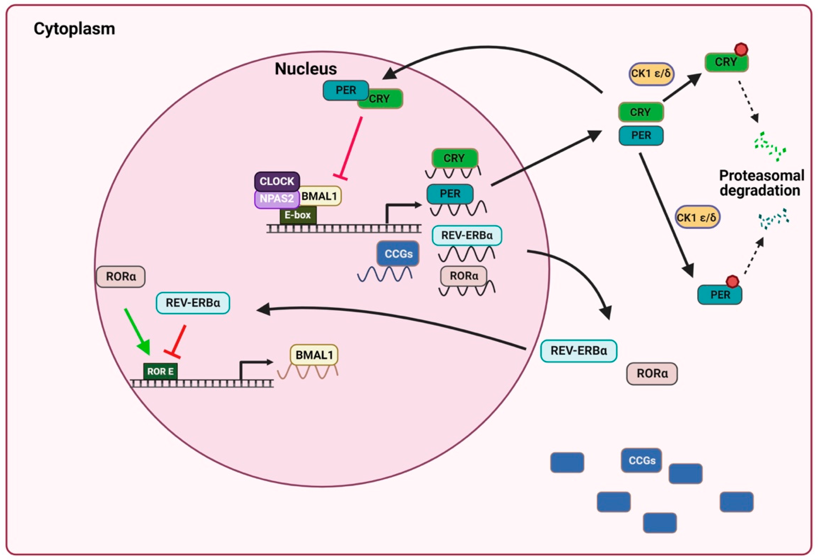
Figure 1. Schematic model of mammalian circadian clock mechanism. The molecular clock is composed of interconnected transcription feedback loops: the transcription factors CLOCK/NPAS2 and BMAL1 produce a heterodimer that binds to the E-box in the promoter and activates the transcription of Per, Cry, Rev-erbα, Rorα and CCGs. CRY and PER dimerize and enter the nucleus, where CLOCK-BMAL1-activated transcription is inhibited, thus generating an oscillatory pattern of gene expression. In the cytoplasm, PER and CRY are phosphorylated by CK1 ε/δ for its degradation. The REV-ERBα receptor inhibits Bmal1 expression, while RORα positively regulates Bmal1 expression. Brain and muscle aryl hydrocarbon receptor nuclear translocator-like protein 1 (Bmal1); cysteine kinase 1 ε/δ (CK1 ε/δ); circadian locomotor output cycles kaput (Clock); cryptochrome (Cry); neuronal PAS domain protein 2 (NPAS2); Period (Per); reverse strand of protein ERB alpha (Rev-erbα); orphan retinoic acid receptor-related alpha (RORα). Image created using BioRender.com (accessed on 22 November 2022).
Ijms 24 01919 g001
Figure 2. Schematic representation of the role of melatonin in the circadian system. In mammals, circadian rhythms are regulated by circadian clocks. The central clock is located in the SCN of the hypothalamus and controls pineal melatonin secretion in the absence of light, while melatonin also affects the SCN, which regulates chronobiotic activities. In peripheral tissues, clock genes are synchronized by the SCN and are also influenced by melatonin. Suprachiasmatic nucleus (SCN). Image created using BioRender.com (accessed on 22 November 2022).
Ijms 24 01919 g002
Figure 3. Proposed mechanisms by which melatonin affects the circadian machinery of cancer cells. By inhibiting the proteasome, melatonin can act post-translationally to regulate the circadian clock by stabilizing proteins, depending on the time of day and the levels of this hormone. Melatonin, together with BMAL1, PER and CRY, represses c-Myc, which arrests the cell cycle, thus decreasing cell proliferation. In addition, c-MYC binds to the E-box region and regulates the expression of certain clock genes. Through the MT1 receptor, melatonin blocks the transcription of RORα and also the activation of BMAL1 expression, as well as of Sirt1, with consequent proapoptotic and prooxidant effects. In addition, SIRT1 regulates BMAL1 by controlling acetylation levels and PER2 promoting its degradation. Melatonin inhibits the AKT pathway, which may occur via BMAL1. The inhibition of this pathway causes an increase in apoptosis and a decrease in cell proliferation and invasion. Brain and muscle aryl hydrocarbon receptor nuclear translocator-like protein 1 (Bmal1); circadian locomotor output cycles kaput (Clock); Chryptochrome (Cry); Period (Per); orphan retinoic acid receptor-related alpha (RORα); reactive oxygen species (ROS); sirtuin 1 (Sirt1). Image created using BioRender.com (accessed on 28 December 2022).
Ijms 24 01919 g003
Table 1. The effect of exogenous melatonin on clock genes in different cancers.
Tumor TypeStudy DesignDoseResultsAuthor
Breast cancer In vitro10 nM-↓ SIRT1 protein levels[85]
Prostate cancerIn vitro100 μM, 1 mM or 2 mM-↑ Per2 and Clock mRNA and protein levels
-↓ Bmal1 mRNA levels
-Rhythmic oscillation of Per2 and Dbp
[84]
Breast cancerIn vitro1 nM-↓ Bmal1 and RORα mRNA levels
-↑ Rev-erbα and Per2 mRNA levels at 20 h after treatment
[83]
Hepatocellular carcinomaIn vivo
Mice
5, 10 mg/kg-↓ Bmal1, Clock and RORα mRNA and protein levels
-↑ Per1, Per2, Per3, Cry1, CK1ε mRNA levels
-↓ Sirt1 mRNA levels
-↑ Rev-Erbα and Rev-Erbβ mRNA and protein levels
[86]
Breast cancerIn vitro1, 2 mMInhibition of LDH-A prevents ↓ Bmal1 under hypoxia conditions[87]
↓: decrease; ↑: increase.
Disclaimer/Publisher’s Note: The statements, opinions and data contained in all publications are solely those of the individual author(s) and contributor(s) and not of MDPI and/or the editor(s). MDPI and/or the editor(s) disclaim responsibility for any injury to people or property resulting from any ideas, methods, instructions or products referred to in the content.