Article

Cembrane Diterpenes Possessing Nonaromatic Oxacycles from the Hainan Soft Coral Sarcophyton mililatensis

1
College of Materials Science and Engineering, Central South University of Forestry and Technology, Changsha 410004, China
2
State Key Laboratory of Drug Research, Shanghai Institute of Materia Medica, Chinese Academy of Sciences, 555 Zu Chong Zhi Road, Zhangjiang Hi-Tech Park, Shanghai 201203, China
3
Shandong Laboratory of Yantai Drug Discovery, Bohai Rim Advanced Research Institute for Drug Discovery, Yantai 264117, China
4
Open Studio for Druggability Research of Marine Natural Products, Pilot National Laboratory for Marine Science and Technology (Qingdao), 1 Wenhai Road, Aoshanwei, Jimo, Qingdao 266237, China
5
Collaborative Innovation Center of Yangtze River Delta Region Green Pharmaceuticals and College of Pharmaceutical Science, Zhejiang University of Technology, Hangzhou 310014, China
*
Authors to whom correspondence should be addressed.
These authors contributed equally to this work.
Int. J. Mol. Sci. 2023, 24(3), 1979; https://doi.org/10.3390/ijms24031979
Submission received: 18 December 2022 / Revised: 16 January 2023 / Accepted: 17 January 2023 / Published: 19 January 2023
(This article belongs to the Section Macromolecules)

Abstract

:
Documents on the chemical composition of the soft coral Sarcophyton mililatensis are sparse. The present investigation of the Hainan soft coral S. mililatensis resulted in the discovery of six new cembrane diterpenes, sarcoxacyclols A–F (16) and four known analogs (710). Their structures were elucidated by extensive spectroscopic analysis along with a comparison with the data in current literature. The nonaromatic oxacycles in their structures were the rarely found tetrahydrofuran ether across C-1 and C-12 and tetrahydropyran ether across C-1 and C-11, respectively. Moreover, the absolute configuration of compound 4 was established unambiguously by X-ray diffraction analysis using Ga Kα radiation (λ = 1.34139 Å). Based on the biogenetical consideration, the absolute configurations of other five new compounds were tentatively assumed. Assessment of the bioactivity for these secondary metabolites revealed that compound 1 exhibited significant tumor necrosis factor (TNF)-α inhibitory activity (IC50 = 9.5 μmol/L), similar to the positive control dexamethasone (IC50 = 8.7 μmol/L), but no obvious cytotoxicity towards RAW264.7 cells (CC50 > 50 μmol/L). The preliminary molecular docking suggested the crucial roles of the hydroxyl and acetoxyl groups in the computational prediction of the binding mode between the diterpene and the protein.

1. Introduction

Nonaromatic oxacycles such as dihydrofuran and oxepane are frequently encountered structural units in a number of pharmacologically active marine natural products [1]. For instance, the FDA-approved anti-cancer drug Yondelis is a eunicellane diterpene that possesses a dihydrofuran motif [2]. Cembrane diterpenes, which could be regarded as the precursors of eunicellane diterpenes [3], also possess nonaromatic oxacycles. The majority of nonaromatic oxacycles in cembranes usually appear as five- to seven-membered lactones, while the minority of remainder are tetrahydrofuran and tetrahydropyran ethers [4]. Different structural features of tetrahydrofuran and tetrahydropyran ethers could be attributed to the various linkages in the cembrane diterpenes [5], including bridging carbons C-1/C-11 [6,7,8], C-1/C-12 [7,8,9], C-2/C-12 [6], C-3/C-7 [10], C-3/C-14 [11], C-4/C-8 [12], C-4/C-15 [13,14,15], C-8/C-11 [14], C-10/C-14 [6], and C-12/C-15 [7]. Among them, the number of cembrane diterpenes possessing a tetrahydrofuran ether across C-1 and C-12 and a tetrahydropyran ether across C-1 and C-11 were ca. 20 [6,7,8,9], which were extremely rare as compared to the large quantity of cembrane family members (over 600) [4]. The PTP1B inhibitory [7], antitumor [6,7], antibacterial [7], and antifouling [6] potencies of the above-mentioned two groups of cembranes were assessed. As a result, compounds such as sinulariol Z and (2E,7E)-4,11-dihydroxy-1,12-oxidocembra-2,7-diene could be developed as potential candidates for nontoxic antifoulants [6].
The assignment of absolute configuration to highly flexible cembrane diterpenes containing diverse functional groups remain challenging. Usually, X-ray crystallography techniques [16] and chemical derivatization approaches using chiral agents [17] are the primary choices [4]. However, the scarcity of isolates hampered the use of the two methods mentioned above in some cases. Instead, chiroptical properties of cembrane diterpenes, including electronic circular dichroism (ECD) aided by quantum chemical calculations, provided alternative approaches to establish the absolute configurations of this type of natural macrocyclic compounds [18]. Among the various calculation methods, the time-dependent density functional theory (TDDFT) ECD calculation is a powerful tool that has been frequently used. Till now, the efficiency of TDDFT ECD calculation has been demonstrated in the numerous stereochemical studies of conformationally flexible cembrane diterpenes [19,20,21] and their dimers [22,23,24].
In the course of our ongoing research aiming for structurally novel and pharmacologically active secondary metabolites from South China Sea marine fauna [25], we encountered the soft coral Sarcophyton mililatensis, which was collected from Xigu Island, Hainan Province, China. As far as we know, there are only six reports focused on the chemical constituents of S. mililatensis [26,27,28,29,30,31], indicating documents on chemical composition of this species are sparse. Our previous chemical investigations on the title animals resulted in the discovery of an array of diterpenes with four carbon frameworks [26,27,28,29], involving an unprecedented tricyclo[11.3.0.02,16]hexadecane carbon skeleton [28]. These studies disclosed the productivity of this species and inspired us to conduct further research on the title species, especially for structurally intriguing diterpenes. In the present study, ten cembrane diterpenes possessed different types of nonaromatic oxacycles. Among them, sarcoxacyclols A–F (16) were new compounds (Figure 1). As follows are the descriptions of the isolation, structural elucidation, and bioactivity screening of these secondary metabolites.

2. Results and Discussion

Freshly collected animals of S. mililatensis were immediately placed at –20 °C and kept frozen prior to extraction. The frozen samples were then extracted three times and the combined extract was separated by multiple rounds of chromatography to yield six new diterpenes, sarcoxacyclols A–F (16), along with four known related analogs 710 (Figure 1). These known compounds were readily identified as sarcophytrols M (7), P (8), Q (9) [7], and (2E,7E)-4,11-dihydroxy-l,12-oxidocembra-2,7,15,-triene (10) [9], respectively, by comparison of the NMR data and optical rotation with those reported in the literature.
In our previous work, the absolute configuration of sarcophytrol M (7), a cembrane diterpene possessing a furan ether isolated from the South China Sea soft coral Sarcophyton trocheliophorum, was determined as 1S,4S,11S,12R by the modified Mosher’s method [7]. Based on the C-4 epimeric relationship between sarcophytrols M (7) and P (8), the absolute configuration of compound 8 could be assigned as 1S,4R,11S,12R, which was further confirmed by the same Cotton effects at ca. 208 nm in their experimental ECD spectra (Figure 2) in the present study.
Sarcoxacyclol A (1), which was obtained as a colorless oil, possessed the molecular formula C22H36O5 on the basis of its HRESIMS ion peak at m/z 403.2448 ([M+Na]+, calcd. for C22H36O4Na, 403.2455), requiring five degrees of unsaturation. As revealed by the 1H and 13C NMR data (Table 1), there were one disubstituted double bond [δH 5.55 (1H, d, J = 15.8 Hz, H-2) and 6.11 (1H, d, J = 15.8 Hz, H-3); δC 129.8 (CH, C-2), 137.4 (CH, C-3)], one trisubstituted double bond [δH 5.42 (1H, dd, J = 4.3, 10.5 Hz, H-7); δC 130.1 (CH, C-7), 132.1 (qC, C-8)], and one acetyl group [δH 2.00 (3H, s, H3-OAc); δC 21.4 (CH3, CH3-OAc), 171.2 (qC, CO-OAc)] in the molecule of compound 1. These functionalities accounted for three degrees of unsaturation. Thus, the remaining two degrees of unsaturation suggested the bicyclic framework of this compound. In fact, the NMR data of compound 1 closely resembled those of the co-occurring sarcophytrol P (8) [7], which was a cembrane diterpene that possessed a tetrahydrofuran ether across C-1 and C-12 from the South China Sea soft coral S. trocheliophorum, revealing that they were structural analogs. Careful analysis of their NMR data revealed the only difference was the presence of an acetoxyl group at C-11 in compound 1 instead of the hydroxyl in compound 8. This replacement was supported by the diagnostic HMBC correlations of H-11 (δH 5.07), CO-OAc (δC 171.2), C-10 (δC 27.3), and C-12 (δC 84.5) (Figure 3). The observed NOE cross-peaks of H3-18 (δH 1.34)/H-2 (δH 5.55), H-2/H3-16 (δH 1.12), H3-16/H-14b (δH 2.31) and H-14b/H3-20 (δH 1.16) suggested the same orientation of H3-16, H3-18, and H3-20 (Figure 4). Whereas the lack of the NOE correlation of H-11 (δH 5.07)/H3-20 and the presence of H-3 (δH 6.11)/H-11 placed H-11 and H3-20 on the opposite faces of this molecule. Therefore, compound 1 was the 11-acetylation derivative of compound 8, which was consistent with their 42 mass unit difference. Considering their structural relationship, the absolute configuration of compound 1 was tentatively supposed to be the same as that of compound 8 (Figure 1).
Sarcoxacyclol B (2) was isolated as a colorless oil and had a molecular formula of C22H34O4 as established by the pseudo-molecular ion peak at m/z 385.2361 ([M+Na]+, calcd. for C22H34O4Na, 385.2349) in its HRESIMS spectrum, requiring six degrees of unsaturation. The 1H and 13C NMR spectra (Table 1) of compound 2 showed resonances attributed to one disubstituted double bond [δH 5.68 (1H, d, J = 15.5 Hz, H-2), 5.92 (1H, d, J = 15.5 Hz, H-3); δC 130.4 (CH, C-2), 136.4 (CH, C-3)], one trisubstituted double bond [δH 5.37 (1H, dd, J = 6.2, 8.8 Hz, H-7); δC 128.3 (CH, C-7), 132.6 (qC, C-8)], one terminal double bond [δH 4.71 (1H, s, H-16a), 4.96 (1H, s, 16b); δC 109.4 (CH2, C-16), 149.5 (qC, C-15)], one acetyl [δH 2.05 (3H, s, H3-OAc); δC 21.4 (CH3, CH3-OAc), 171.2 (qC, CO-OAc)]. All the above-mentioned functionalities accounted for four degrees of unsaturation; the left two indicated that compound 2 was a bicyclic diterpene. Indeed, the NMR data of compound 2 were almost superimposed on those of compound 1, suggesting the structural similarity of both compounds. Further comparison of their NMR data revealed that their gross structures differed at the substituent attached to C-1, which was a propen-2-yl group in compound 2 whereas a peculiar isopropyl alcohol moiety in compound 1. This propen-2-yl group was assigned to C-1 based on the HMBC correlations from H3-17 (δH 1.71) to C-1 (δC 88.1), C-15 (δC 149.5) and C-16 (δC 109.4) (Figure 3). Thus, the planar structure of compound 2 was determined as the dehydration derivative of compound 1. The cis-orientation of the propen-2-yl group and CH3-20 was deduced from the characteristic NOE cross-peak of H-16b (δH 4.96)/H3-20 (δH 1.18). The strong NOE correlations between H-2 (δH 5.68) and H3-17 (δH 1.71) and between H-3 (δH 5.92) H3-18 (δH 1.28) implied that H3-17 and H3-18 were trans-oriented. These NOE correlations (Figure 4) indicated that compound 2 and the co-isolated diterpene sarcophytrol M (7) [7] shared the same configurations for the four chiral centers C-1, C-4, C-11, and C-12. Tentatively, the absolute configuration of compound 2 was assumed to be 1S,4S,11S,12R.
Sarcoxacyclol C (3) was purified as a colorless oil with a molecular formula of C22H34O3, which was inferred from the HREIMS data (m/z 346.2507, M+, calcd. for C22H34O3, 346.2508). Its 1H NMR data (Table 1) revealed the presence of three vinyl methyls at δH 1.63 (3H, s, H3-19), 1.56 (3H, s, H3-18), and 1.75 (3H, s, H3-17), one methyl linked to a tertiary carbon at δH 1.18 (3H, s, H3-20), one methyl of an acetyl group at δH 2.06 (3H, s, H3-OAc), one proton linked to an oxygenated carbon at δH 4.88 (1H, d, J = 10.2 Hz, H-11), and four olefinic protons at δH 4.80 (1H, s, 16a), 5.10 (1H, s, H-16b), 5.12 (1H, t, J = 6.0 Hz, H-3), and 5.18 (1H, t, J = 6.0 Hz, H-7), which were indicative of two trisubstituted double bonds and one terminal double bond. The 13C NMR spectrum displayed 22 signals consisting of five methyls (δC 16.8, 17.6, 20.1, 21.5, and 21.8), eight methylenes (including seven aliphatic δC 24.9, 27.5, 32.0, 33.8, 35.3, 35.7, and 38.0, and one olefinic δC 110.1), three methines (including one oxygenated δC 75.7, two olefinic δC 119.9, and 126.2), and six quaternary carbons (including two oxygenated δC 83.5, 89.6, three olefinic δC 132.9, 136.6, 150.2, and one carbonyl δC 171.3) (Table 1). These abovementioned structural features of compound 3 were reminiscent of the co-isolated compound 2. According to the interpretation of the 2D NMR spectra (Figure 3 and Figure 4), compounds 3 and 2 differed by the double bond at C-2/C-3 in compound 2 shifting to C-3/C-4 in compound 3 accompanied with the disappearance of the hydroxyl at C-4. This alteration was supported by the consecutive spinning proton system from H2-2 (δH 2.14 m, 2.35 m) to H-3 (δH 5.12) along with the key HMBC correlations of H2-2/C-1 (δC 89.6), H3-18 (δH 1.56)/C-3 (δC 119.9), H3-18/C-4 (δC 136.6), and H3-18/C-5 (δC 38.0) (Figure 3). The NOESY spectra of diterpenes 2 and 3 (Figure 4) showed similar distributions of NOE cross-peaks, implying they shared the same configurations for the three chiral carbons C-1, C-11, and C-12. Based on the biogenetical consideration, the absolute configuration of compound 3 was tentatively supposed to be 1S,11S,12R, and its structure was shown as depicted in Figure 1.
Sarcoxacyclol D (4), colorless crystals, displayed a pseudo-molecular ion peak in the HRESIMS spectrum at m/z 361.2348 ([M+Na]+, calculated for C20H34O4Na, 361.2349), indicating that compound 4 possessed the same molecular formula C20H34O4 as that of the co-isolate sarcophytrol Q (9) [7]. Thus, four degrees of unsaturation were determined for this diterpene. The 1H and 13C NMR data (Table 2) revealed the presence of one trisubstituted double bond [δH 5.23 (1H, dd, J = 5.1, 10.2 Hz, H-7); δC 131.0 (CH, C-11), 132.6 (qC, C-8)], one disubstituted double bond [δH 5.20 (1H, d, J = 16.4 Hz, H-2), 6.11 (1H, d, J = 16.4 Hz, H-3); δC 125.1 (CH, C-2), 141.5 (CH, C-3)] and four oxygenated quaternary carbons [δC 70.9 (qC, C-12), 72.7 (qC, C-4), 75.1 (qC, C-15), 81.1 (qC, C-1)], which accounted for two degrees of unsaturation. The remaining two degrees of unsaturation strongly indicated one bicyclic structure incorporating a nonaromatic oxacycle for 4. Indeed, compound 4 was identical in all respects with the co-occurring sarcophytrol Q (9), which was a diterpene possessing a tetrahydropyran ether across C-1 and C-11 previously isolated from the South China Sea soft coral S. trocheliophorum [7]. Their structural difference was in the reversed configuration of C-4, which was deduced from the chemical shift of C-4 upfield, shifting from δC 74.3 in compound 9 to δC 72.7 in compound 4. This hypothesis was supported by the NOE correlations between H-2 (δH 5.20) and both H3-16 (δH 1.16) and H3-18 (δH 1.37) implying that H3-16 and H3-18 were co-facial (Figure 4). After many attempts, suitable single crystals of compound 4 were successfully obtained from the diethyl ether-acetone solvent system to elucidate its absolute configuration. The X-ray diffraction experiment with Ga Kα radiation (λ = 1.34139 Å) unambiguously established that the absolute configuration of compound 4 was 1S,4R,11R,12S (Flack parameter: −0.06 (9), Figure 5). Based on their epimeric relationship, the absolute configuration of compound 9 could be tentatively assumed to be 1S,4S,11R,12S, suggesting the revision of configurations of the C-11 and C-12 of compound 9 reported in the previous work [7].
Sarcoxacyclol E (5) had a molecular formula of C20H34O5 deduced from a pseudo-molecular ion peak at m/z 377.2294 ([M+Na]+, calcd. for C20H34O5Na, 377.2298) in the HRESIMS spectrum, requiring four degrees of unsaturation. The 1H and 13C NMR data of 5 (Table 2) displayed the signals attributed to one disubstituted double bond [δH 5.44 (1H, d, J = 15.9 Hz, H-2), 5.75 (1H, d, J = 15.9 Hz, H-3); δC 124.1 (CH, C-2), 140.7 (CH, C-3)] and one epoxide group [δH 2.89 (1H, d, J = 9.7 Hz, H-7); δC 66.8 (CH, C-7), 61.9 (qC, C-8)]. These two functionalities accounted for two degrees of unsaturation; the left two indicated that compound 5 was a bicyclic diterpene. The interpretation of its 1H–1H COSY spectrum (Figure 3) disclosed four segments: a. H-2–H-3, b. H2-5–H2-6–H-7, c. H2-9–H2-10–H-11, and d. H2-13–H2-14. The HMBC correlations (Figure 3) from H3-18 (δH 1.28) to C-3 (δC 140.7), C-4 (δC 74.0), and C-5 (δC 42.4) led to the connection of partial structures a and b via quaternary carbon C-4 bearing a hydroxyl group. The observed HMBC cross-peaks of H3-19 (δH 1.38)/C-7 (δC 66.8), H3-19/C-8 (δC 61.9) and H3-19/C-9 (δC 35.5) revealed the linkage of partial fragments b and c through C-8, along with the fixation of the epoxide group at C-7/C-8. The subunit d was found to be linked to subunits c and a by the oxygenated carbons C-12 and C-1, as deduced from the significant HMBC correlations of H3-20 (δH 1.17) with C-11 (δC 75.0), C-12 (δC 71.2), and C-13 (δC 37.3), and of both H-2 (δH 5.44) and H-14 (δH 2.22) with C-1 (δC 81.8), respectively. A peculiar isopropyl alcohol group was recognized by the mutual HMBC correlations from H3-16 (δH 1.16) and H3-17 (δH 1.11) to C-15 (δC 74.6). This group was attached to C-1, as indicated by the key HMBC cross-peaks of H3-16/C-1 and H3-17/C-1. Thus, the carbon framework of compound 5 was established as a cembrane skeleton. Indeed, the 13C chemical shifts of the C-11–C-12–C-13–C-14–C-1 segment of compound 5 were nearly identical to those of the co-isolate 4, suggesting the hydroxyl group bonded to C-12 and a tetrahydropyran ring bridged C-1 and C-11. Thus, the planar structure of compound 5 was established as an epoxidation derivative of compound 4. The above-mentioned almost identical chemical shifts of the C-11–C-12–C-13–C-14–C-1 segment suggested the same configurations for the three chiral centers C-1, C-11, and C-12 in compounds 4 and 5. The NOE correlations (Figure 4) between H-2 and H3-17 and between H-3 and H3-18, together with the trans configuration of double bond Δ2 (J = 15.9 Hz), suggested H3-17 and H3-18 were trans-oriented. This observation indicated the opposite configuration of C-4 in 5 compared with that of compound 4, which was further confirmed by the chemical shift of C-4 downshifted from δC 72.7 in compound 4 to δC 74.0 in compound 5. The diagnostic NOE cross-peak of H-7 (δH 2.89)/H-11 (δH 3.51) implied the co-facial orientation of H-7 and H-11. The trans geometry of the epoxide ring was deduced from the NOE cross-peaks of H-7/H-6a (δH 2.10) and H3-19/H-6b (δH 1.46). Hereto, the structure of compound 5 was depicted as shown in Figure 1, and the absolute configuration was tentatively assumed to be 1S,4S,7S,8S,11R,12S.
Sarcoxacyclol F (6) possessed the same molecular formula (C20H34O5) as that of sarcoxacyclol E (5), as deduced from the pseudo-molecular ion peak at m/z 377.2295 ([M+Na]+, calcd. for C20H34O5Na, 377.2298) in the HRESIMS spectrum. Inspection of the NMR data of compound 6 (Table 2) revealed that the planar structure of compound 6 should be the same as that of compound 5, which was further supported by the extensive analysis of its 2D NMR spectra (Figure 3). The clear NOE cross-peak of H3-19 (δH 1.27)/H-11 (δH 3.49) in the NOESY spectrum of compound 6 (Figure 4) suggested the same orientation for CH3-19 and H-11 in compound 6, which was different from that of compound 5. The trans geometry of the epoxide ring in compound 6 was indicated by the key NOE cross-peaks of H-7 (δH 2.95)/H-9a (δH 1.41) and H3-19/H-9b (δH 2.03). Consequently, compound 6 was established as the C-7/C-8 stereoisomer of compound 5, namely another epoxidation derivative of compound 4, and the absolute configuration was tentatively supposed as 1S,4S,7R,8R,11R,12S.
The origin of compounds 2, 5, and 6 is a matter needing discussion. To determine if these three diterpenes were natural products or derived from their co-occurring precursors during the isolation process, we re-checked the crude extract of the soft coral S. mililatensis by comparisons of the Rf values of secondary metabolites in the crude extract with that of pure samples 2, 5, and 6 on co-plate TLC as well as comparisons of their retention times on HPLC. We detected the compounds 2, 5, and 6 present in the Et2O-soluble portion of the acetone extract of the specimen. Evidence from these experiments excluded the possibility that the dehydration and epoxidation derivatives 2, 5, and 6 were artifacts obtained during the work-up.
Indeed, some reports have disclosed the coexistence of dehydration derivatives and their related alcohol precursors in one sample, and both of them have been found to be natural products (Figure S49). For instance, Bruce F. Bowden et al. found that the cembrane diterpene 2-[(E,E,E)-7’,8’-epoxy-4’,8’,12’-trimethylcyclotetradeca-1’,3’,11’-trieny1]propan-2-ol (S1) coexisted with its dehydration derivative named (E,E,E)-7,8-epoxy-1-isopropenyl-4,8,12-trimethylcyclotetradeca-1,3,11-triene (S2) as two natural products in the Australian soft coral Sarcophyton crassocaule [32]. Their chemical investigation of another Australian soft coral Nephthea brassica led to the discovery of a pair of co-isolates in the sample, namely the dehydration derivative cembrene A (S3) and its corresponding alcohol precursor nephthenol (S4) [33].
Moreover, several epimeric epoxy analogs were reported as co-occurring isolates in the soft corals (Figure S50). For instance, epimeric epoxy diterpenes sarcocrassolides A (S5) and B (S6) with close structural similarity coexisted in the South China Sea soft coral S. crassocaule, as shown in Xu et al.’s work [34]. It might be worthy to point out that the relative configuration of sarcocrassolide B (S6) was confirmed by the X-ray diffraction experiment. Sheu et al.’s study on the Formosan soft coral S. crassocaule also displayed the co-occurrence of epimeric epoxy diterpenes named crassocolides K–M (S7S9) [35]. These five cembrane diterpenes are representatives of two groups of diterpenes with epimeric epoxides at C-3/C-4. Seifert et al. have reported the discovery of diterpenes with epimeric epoxides at C-7/C-8, such as dihydrocembrene C (S10) and sarcophytoxide (S11) from the Indonesian soft coral Sarcophyton ehrenbergi [36]. These findings of co-occurring natural epimeric epoxy analogs indicate that there is possibly more than one enzyme to catalyze the epoxidation reaction in the metabolic process in the soft corals, resulting in the seemingly non-stereospecific natural products.
In general, the discovery of the dehydration and epoxidation derivatives in this study inspired us to explore the terpene biosynthetic gene clusters of this species in future, which may be in high demand in addition to the chemo- and bio-investigations.
There have been no reports assessing the tumor necrosis factor (TNF)-α inhibitory bioactivity of cembrane diterpenes possessing the tetrahydrofuran and tetrahydropyran ethers. Therefore, all the cembrane diterpenes 110 were subjected to this bioassay using lipopolysaccharide (LPS)-induced TNF-α protein release in RAW264.7 macrophages. As a result, they exhibited different levels of inhibition ratios, ranging from 5.5% to 52.4% at a concentration of 20 μmol/L. Among them, only the new secondary metabolite 1 was judged an active compound (inhibition ratio 52.4%). Further evaluation showed an IC50 value of 9.5 μmol/L for compound 1, which was similar to the positive control dexamethasone (IC50 = 8.7 μmol/L). Notably, diterpene 1 displayed no obvious cytotoxic activity towards RAW264.7 cells (CC50 > 50 μmol/L). Given the structural differences between compound 1 and its analog compound 2, it appeared that the isopropyl alcohol moiety at C-1 in the bicyclic cembrane structure played an important role in TNF-α inhibitory activity.
Then, a preliminary molecular docking experiment was performed using the highly resolved TNF-α crystal structure (PDB: 2AZ5 with a resolution of 2.10 Å) [37]. As shown in Figure 6, the preliminary computation clearly revealed the hydrogen bonds formed between the hydroxyl group at C-15 and the residue Leu120, between the hydroxyl group at C-4 and the residue Ile58, and between the acetoxyl group at C-11 and the residues Gly121 and Tyr151, which were lying in the active site. Moreover, compound 1 occupied the hydrophobic pocket, where methyl groups at C-7 and C-12 generate π-alkyl stacked interactions with the residues Leu57 and Tyr119. The low binding affinity of compound 1 (−8.02 kcal/mol), which was slightly lower than that of the previously reported ligand 6,7-dimethyl-3-{[methyl-(2-{methyl-[1-(3-trifluoromethyl-phenyl)-1H-indol-3-ylmethyl]-amino}-ethyl)-amino]-methyl}-chromen-4-one [37] in the TNF-α crystal structure in this study (−5.31 kcal/mol), indicated that compound 1 was a potential TNF-α inhibitor.
The study for the elaborated mechanism is in the plan.

3. Materials and Methods

3.1. General Experimental Procedures

Optical rotations were recorded on a Perkin-Elmer 241MC polarimeter. IR spectra were obtained on a Nicolet 6700 spectrometer (Thermo Scientific, Waltham, MA, USA). CD spectra were measured on a JASCO J-810 instrument. NMR spectra were measured on a Bruker DRX-500 or Bruker DRX-600 spectrometer (Bruker Biospin AG, Fällanden, Germany). Chemical shifts (δ) were reported in ppm with reference to the solvent signals, and coupling constants (J) were in Hz. ESIMS spectra were obtained on a Finngan-MAT-95 mass spectrometer. HRESIMS spectra were measured on an Agilent 1290-6545 UHPLC-QTOF mass spectrometer. Commercial silica gel (Qingdao Haiyang Chemical Group Co., Ltd., Qingdao, China, 200–300 and 400–600 mesh), Sephadex LH-20 gel (Amersham Biosciences, Piscataway, NJ, USA) were used for column chromatography, and precoated silica gel plates (Yan Tai Zi Fu Chemical Group Co., Yantai, China, G60 F-254) were used for analytical TLC. Reversed-phase (RP) HPLC was performed on an Agilent 1260 series liquid chromatography system equipped with a DAD G1315D detector at 210 and 254 nm. A semi-preparative ODS-HG-5 column [5 µm, 250 × 9.4 mm] was employed for the purifications. All solvents used for column chromatography and HPLC were of analytical grade (Shanghai Chemical Reagents Co., Ltd., Shanghai, China) and chromatographic grade (Dikma Technologies Inc., Foothill Ranch, CA, USA), respectively.

3.2. Biological Material

The soft corals of Sarcophyton mililatensis were collected at a depth of –20 m by SCUBA diving from the coast of Xigu Island, Hainan Province, China, in May 2014. They were frozen immediately at –20 °C after collection and identified by Prof. X.-B. Li from Hainan University. A voucher specimen (No. 14S-80) is available for inspection at the Shanghai Institute of Materia Medica, Chinese Academy of Sciences.

3.3. Extraction and Isolation

The frozen animals (400 g dry weight) were cut into pieces and thoroughly extracted with acetone at room temperature (3 × 1.5 L). The organic extract was evaporated to give a dark brown residue that was partitioned between Et2O and H2O. The upper layer was concentrated under reduced pressure to give an Et2O portion (13.5 g). The Et2O extract was separated into twenty-one fractions (A–U) by gradient silica gel column chromatography [0 → 100% Et2O (EE) in petroleum ether (PE)]. Fraction G was further purified by Sephadex LH-20 [PE/CH2Cl2/MeOH (2:1:1)], followed by silica gel column chromatography [PE/EE (4:1)] to give seven subfractions G1–G7. The sixth subfraction G6 was further purified by RP-HPLC [MeCN/H2O (85:15), 3.0 mL/min] to give compound 3 (1.8 mg, tR = 36.7 min). Similarly, fraction J was further purified by Sephadex LH-20 [PE/CH2Cl2/MeOH (2:1:1)], followed by silica gel column chromatography [PE/EE (2:1)] to yield seven subfractions J1–J7. Compound 2 (3.6 mg) was isolated from the seventh subfraction J7 by silica gel column chromatography [PE/EE (2:1)]. Through two-step purification, including Sephadex LH-20 [PE/CH2Cl2/MeOH (2:1:1)] and silica gel column chromatography [PE/ acetone (3:1)], fraction Q was divided into four subfractions, Q1–Q4. The first subfraction Q1 gave compounds 8 (3.1 mg) and 10 (4.3 mg) by silica gel column chromatography [PE/ acetone (7:3)] along with five mixtures Q1A–Q1E. Four mixtures Q1A, Q1B, Q1D, and Q1E yielded compounds 5 (2.5 mg, tR = 17.4 min), 6 (2.0 mg, tR = 16.9 min), 7 (12.0 mg, tR = 10.0 min), and 9 (3.4 mg, tR = 9.7 min) by RP-HPLC [MeCN/H2O (20:80), 3.0 mL/min; MeCN/H2O (40:60), 3.0 mL/min; MeCN/H2O (20:80), 3.0 mL/min; MeCN/H2O (38:62), 3.0 mL/min], respectively. The second subfraction Q2 yielded compound 1 (8.0 mg, tR = 12.5 min) by silica gel column chromatography [PE/acetone (7:3)] followed by RP-HPLC [MeCN/H2O (65:35), 3.0 mL/min]. Compound 4 (3.6 mg, tR = 4.9 min) was obtained from the fraction R by the two-step purification, including Sephadex LH-20 [PE/CH2Cl2/MeOH (2:1:1)] and the subsequent RP-HPLC [MeCN/H2O (50:50), 3.0 mL/min].

3.4. Spectroscopic Data of Compounds

Sarcoxacyclol A (1): colorless oil; [ α ] D 25 −49.6 (c 0.35, CHCl3); IR (KBr): νmax 3430, 2971, 2925, 2853, 1732, 1456, 1373, 1241, 1169, 1080, 1028, 983 cm−1; For 1H NMR (CDCl3, 500 MHz) and 13C NMR (CDCl3, 125 MHz) spectral data, see Table 1; HRESIMS m/z 403.2448 ([M+Na]+; calcd. for C22H36NaO5, 403.2455).
Sarcoxacyclol B (2): colorless oil; [ α ] D 19 −36.8 (c 0.20, CHCl3); IR (KBr): νmax 3445, 2969, 2923, 2851, 1733, 1449, 1372, 1238, 1113, 1076, 1027, 964, 899 cm−1; For 1H NMR (CDCl3, 500 MHz) and 13C NMR (CDCl3, 125 MHz) spectral data, see Table 1; HRESIMS m/z 385.2361 ([M+Na]+; calcd. for C22H34NaO4, 385.2349).
Sarcoxacyclol C (3): colorless oil; [ α ] D 25 +4.0 (c 0.35, CHCl3); IR (KBr): νmax 2955, 2923, 2853, 1739, 1457, 1375, 1236, 1132, 1076 cm−1; For 1H NMR (CDCl3, 600 MHz) and 13C NMR (CDCl3, 125 MHz) spectral data, see Table 1; HREIMS m/z 346.2507 (M+; calcd. for C22H34O3, 346.2502).
Sarcoxacyclol D (4): colorless crystals; mp 183.1-184.0°C; [ α ] D 25 +70.1 (c 0.35, CHCl3); IR (KBr): νmax 3440, 2920, 2850, 1463, 1383, 1078, 1047, 947 cm−1; For 1H NMR (CDCl3, 500 MHz) and 13C NMR (CDCl3, 125 MHz) spectral data, see Table 2; HRESIMS m/z 361.2348 (M+; calcd. for C20H34NaO4, 361.2349).
Sarcoxacyclol E (5): colorless oil; [ α ] D 25 +45.8 (c 0.35, CHCl3); IR (KBr): νmax 3440, 2923, 2853, 1462, 1377, 1132 cm−1; For 1H NMR (CDCl3, 600 MHz) and 13C NMR (CDCl3, 125 MHz) spectral data, see Table 2; HRESIMS m/z 377.2294 ([M+Na]+; calcd. for C20H34NaO5, 377.2298).
Sarcoxacyclol F (6): colorless oil; [ α ] D 25 +4.8 (c 0.20, CHCl3); IR (KBr): νmax 3443, 2917, 2849, 1574, 1538, 1457, 1384, 1131, 1049 cm−1; For 1H NMR (CDCl3, 600 MHz) and 13C NMR (CDCl3, 125 MHz) spectral data, see Table 2; HRESIMS m/z 377.2295 ([M+Na]+; calcd. for C20H34NaO5, 377.2298).

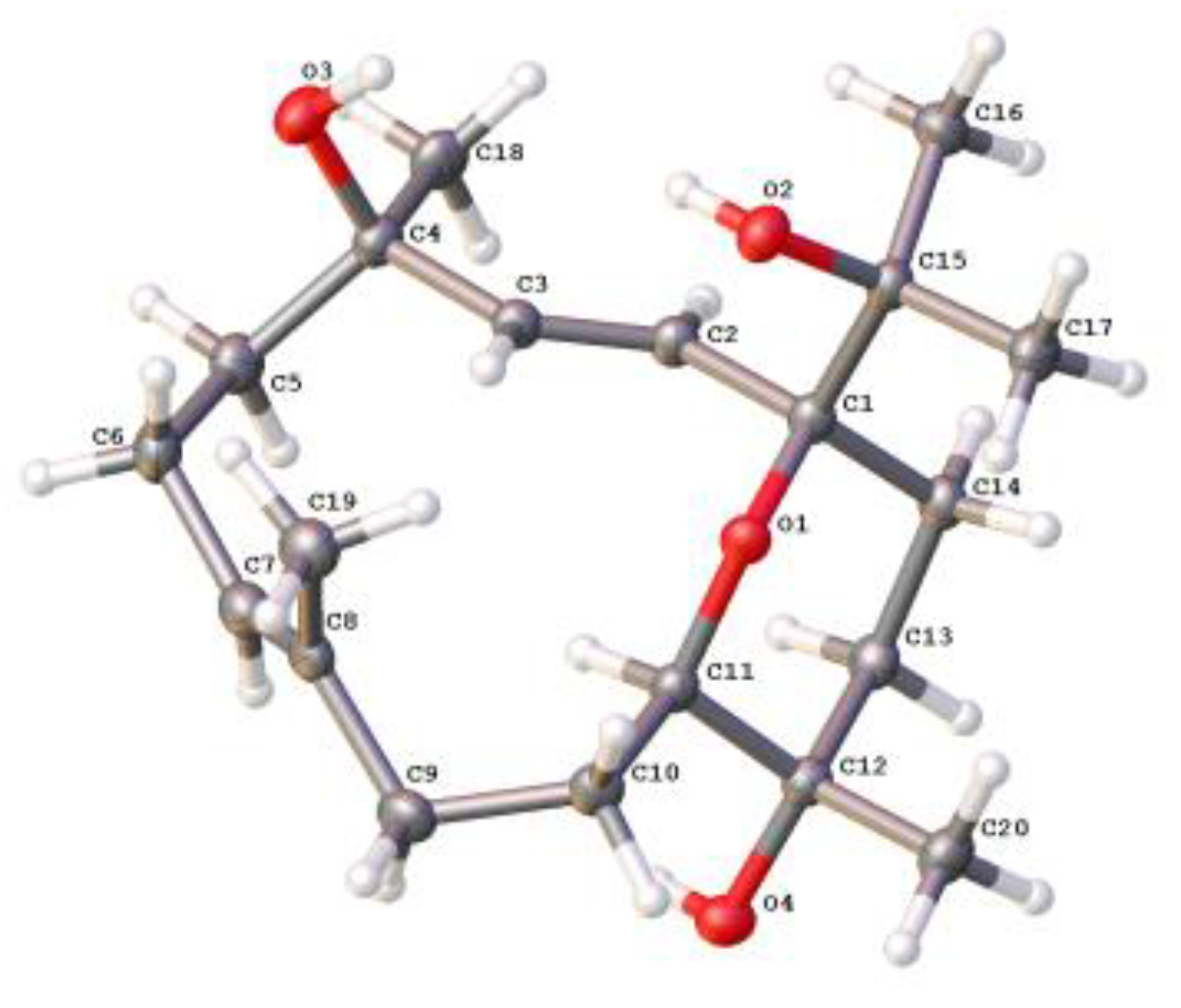
3.5. Crystal Structure Analysis of 4

Block crystals of compound 4 were obtained from diethyl ether-acetone solvent systems at 4 °C. Single-crystal X-ray diffraction of compound 4 (0.16 × 0.11 × 0.10 mm3) was performed on a Bruker D8 Venture diffractometer using Ga Kα radiation (λ = 1.34139 Å) at 170 K. The collected data integration and reduction were processed with SAINT software (V8.37A, 2015, Bruker AXS Inc., Karlsruhe, Germany), and multi-scan absorption corrections were performed using the SADABS program (V2016/2, 2016, Bruker AXS Inc., Karlsruhe, Germany). The structures were solved by direct methods using SHELXL (V2016/6, 2016, George M. Sheldrick) and refined on F2 by the full-matrix least-squares technique using the SHELXT-2015 program package. Crystallographic data for compound 4 in this article (Table S1) have been deposited at the Cambridge Crystallographic Data Centre (deposition numbers CCDC 2225113). Copies of these data can be obtained free of charge via www.ccdc.cam.ac.uk/conts/retrieving.html (accessed on 8 December 2022) or from the Cambridge Crystallographic Data Centre (12 Union Road, Cambridge CB21EZ, UK. Fax: (+44) 1223-336-033. E-mail: [email protected]).

3.6. TNF-α Inhibitory Activity Bioassay

A murine macrophage cell line, RAW264.7 cells, was obtained from the American Type Culture Collection (ATCC, Manassas, VA, USA). In the bioassay for anti-inflammation, cells were cultured in DMEM containing 10% FBS, 2 mmol/L L-glutamine, 100 μg/mL streptomycin, and 100 U/mL penicillin in a humidified incubator of 5% CO2 at 37 °C. For the cytotoxicity part, RAW264.7 cells were incubated with the compounds or the media (0.125% DMSO in DMEM containing 10% FBS) for 24h, respectively. CCK-8 reagents (20 μL per well) were added, and the OD values were collected after 1h incubation at 450 nm (650 nm calibration) by a microplate reader (Molecular Devices, Sunnyvale, CA, USA). For the anti-inflammatory activity assay, RAW264.7 cells were incubated with compounds or the media (0.125% DMSO in DMEM containing 10% FBS), and then cells were primed with LPS (1 μg/mL) for 24 h. The supernatants were centrifuged and then measured using the mouse TNF-α ELISA kit. The CC50 and IC50 were estimated using the log (inhibitor) vs. normalized response non-linear fit (Graph Pad Prism 6.0, GraphPad Software, San Diego, CA, USA). Dexamethasone was used as a positive control.

3.7. ECD Computational Protocol

Torsional sampling (MCMM) conformational searches using the OPLS_2005 force field were carried out by means of the conformational search module in the Macromodel 9.9.223 software [38], applying an energy window of 21 kJ/mol. Conformers above 1% Boltzmann populations were reoptimized with Gaussian 09 [39] at the B3LYP/6-311G(d,p) level with the IEFPCM solvent model for chloroform. Frequency analysis was also carried out to confirm that the reoptimized geometries were at the energy minima. Finally, the SpecDis1.62 software [40] was applied to obtain the Boltzmann-averaged ECD spectra and visualize the results.

3.8. Molecular Docking

The docking study was performed using the Operation Environment (ADT 1.5.7) software (https://autodock.scripps.edu/, accessed on 20 August 2022) between the compound and TNF-α (PDB: 2AZ5). The structure of the natural product was optimized by energy minimization using the MM2 method and converted to a readable format at the ADT interface. Replication of the experimental binding posed by molecular docking confirmed the suitability of the docking algorithm (RMSD <2 Å). The outcomes of the results were analyzed using the Discovery Studio Visualizer software, which reveals close contact, hydrogen bond, and hydrophobic interactions. The binding affinity of the known ligand 6,7-dimethyl-3-{[methyl-(2-{methyl-[1-(3-trifluoromethyl-phenyl)-1H-indol-3-ylmethyl]-amino}-ethyl)-amino]-methyl}-chromen-4-one [37] in the TNF-α crystal structure was −5.31 kcal/mol.

4. Conclusions

The present study provided the intriguing discovery of ten cembrane diterpenes from the Hainan soft coral S. mililatensis, which was rarely documented in literature. Among these secondary metabolites, six were new compounds. Their structures were elucidated by extensive spectroscopic analysis along with a comparison with existing data in the literature. Different types of nonaromatic oxacycles were found in their structures, which were identified as tetrahydrofuran and tetrahydropyran ethers that rarely occurred between C-1 and C-12 and between C-1 and C-11, respectively. Additionally, the absolute configuration of compound 4 was established unambiguously by an X-Ray diffraction experiment. The absolute configuration of 7 was assigned by the modified Mosher’s method in our previous work [7]. Based on the comparison of ECD spectra, the absolute configuration of compound 8 was revealed in the present study. Subsequently, the absolute configurations of the other five new compounds were tentatively assumed using the diterpenes 4, 7 and 8 as model compounds. In the bioassay, the new secondary metabolite, compound 1, exhibited significant TNF-α inhibitory activity with an IC50 value of 9.5 μmol/L and was not cytotoxic towards RAW264.7 cells (CC50 > 50 μmol/L). A molecular docking experiment gave insight into the key binding actions between the active compound 1 and TNF-α, which indicated the important roles of the hydroxyl groups at C-4 and C-15 and the acetoxyl group at C-11. Although the elaborated mechanism is still under investigation, the present preliminary pharmacological result revealed that the natural product, compound 1, could be developed as a potential lead compound or drug candidate of a new chemotype of nontoxic TNF-α inhibitors. This study not only extends the chemical and biological diversities of cembrane diterpenes possessing nonaromatic oxacycles but also establishes the productivity of the title species.

Supplementary Materials

The following supporting information can be downloaded at: https://www.mdpi.com/article/10.3390/ijms24031979/s1, Figures S1–S48: MS, NMR, and IR data of compounds 16; Figure S49: Examples of the coexistence of dehydration derivatives and their related alcohol precursors from different soft corals; Figure S50: Examples of the coexistence of epimeric epoxy analogs from different soft corals; Table S1: X-ray crystallographic data for compound 4.

Author Contributions

Conceptualization, L.-F.L. and Y.-W.G.; methodology, L.-F.L. and Y.-W.G.; software, Z.-H.C., Z.-Y.G., and S.-W.L.; validation, L.Z. and M.Y.; formal analysis, L.Z. and M.Y.; investigation, L.Z., M.Y., Z.-H.C., S.-W.L., and X.-Y.Y.; resources, L.-G.Y.; data curation, M.Y.; writing—original draft preparation, L.Z. and M.Y.; writing—review and editing, L.-F.L. and Y.-W.G.; visualization, L.-F.L. and Y.-W.G.; supervision, L.-F.L. and Y.-W.G.; project administration, L.-F.L. and Y.-W.G.; funding acquisition, L.-F.L. and Y.-W.G. All authors have read and agreed to the published version of the manuscript.

Funding

This research work was financially supported by the National Natural Science Foundation of China (NSFC) (Nos. 41876194, 81991521), the National Key Research and Development Program of China (No. 2022YFC2804100), and the SKLDR/SIMM Project (No. SIMM2103ZZ-06).

Institutional Review Board Statement

Not applicable.

Informed Consent Statement

Not applicable.

Data Availability Statement

The Data are contained within the article or Supplementary Materials.

Acknowledgments

We thank X.-B. Li from Hainan University for the taxonomic identification of the soft coral material.

Conflicts of Interest

The authors declare no conflict of interest.

References

  1. Haque, N.; Parveen, S.; Tang, T.; Wei, J.; Huang, Z. Marine natural products in clinical use. Mar. Drugs 2022, 20, 528. [Google Scholar] [CrossRef] [PubMed]
  2. Jiménez, C. Marine natural products in medicinal chemistry. ACS Med. Chem. Lett. 2018, 9, 959–961. [Google Scholar] [CrossRef] [PubMed] [Green Version]
  3. Li, G.; Dickschat, J.S.; Guo, Y.-W. Diving into the world of marine 2,11-cyclized cembranoids: A summary of new compounds and their biological activities. Nat. Prod. Rep. 2020, 37, 1367–1383. [Google Scholar] [CrossRef] [PubMed]
  4. Yang, B.; Zhou, X.-F.; Lin, X.-P.; Liu, J.; Peng, Y.; Yang, X.-W.; Liu, Y. Cembrane diterpenes chemistry and biological properties. Curr. Org. Chem. 2012, 16, 1512–1539. [Google Scholar] [CrossRef] [Green Version]
  5. Carroll, A.R.; Copp, B.R.; Davis, R.A.; Keyzers, R.A.; Prinsep, M.R. Marine natural products. Nat. Prod. Rep. 2022, 39, 1122–1171. [Google Scholar] [CrossRef]
  6. Lai, D.; Geng, Z.; Deng, Z.; van Ofwegen, L.; Proksch, P.; Lin, W. Cembranoids from the soft coral Sinularia rigida with antifouling activities. J. Agric. Food Chem. 2013, 61, 4585–4592. [Google Scholar] [CrossRef]
  7. Liang, L.-F.; Chen, W.-T.; Li, X.-W.; Wang, H.-Y.; Guo, Y.-W. New bicyclic cembranoids from the South China Sea soft coral Sarcophyton trocheliophorum. Sci. Rep. 2017, 7, 46584. [Google Scholar] [CrossRef] [Green Version]
  8. Li, J.-F.; Zeng, Y.-B.; Li, W.-S.; Luo, H.; Zhang, H.-Y.; Guo, Y.-W. Xishaglaucumins A–J, new cembranoids with anti-inflammatory activities from the South China Sea soft coral Sarcophyton glaucum. Chin. J. Chem. 2022, 40, 79–90. [Google Scholar] [CrossRef]
  9. Rao, C.B.; Satyanarayana, C.; Rao, D.S.; Rao, D.V.; Fahy, E.; Faulkner, D.J. Metabolites of the soft coral Sinularia ovispiculata from the Indian Ocean. J. Nat. Prod. 1993, 56, 2003–2007. [Google Scholar] [CrossRef]
  10. Yin, F.-Z.; Yao, L.-G.; Zhang, Z.-Y.; Wang, J.-R.; Wang, H.; Guo, Y.-W. Polyoxygenated cembranoids from the Hainan soft coral Lobophytum crassum. Tetrahedron 2021, 90, 132204. [Google Scholar] [CrossRef]
  11. Hegazy, M.E.F.; Su, J.-H.; Sung, P.-J.; Sheu, J.-H. Cembranoids with 3,14-ether linkage and a secocembrane with bistetrahydrofuran from the Dongsha Atoll soft coral Lobophytum sp. Mar. Drugs 2011, 9, 1243–1253. [Google Scholar] [CrossRef] [PubMed] [Green Version]
  12. Ahmed, A.; Chen, Y.-W.; Huang, C.-Y.; Tseng, Y.-J.; Lin, C.-C.; Dai, C.-F.; Wu, Y.-C.; Sheu, J.-H. Isolation and structure elucidation of cembranoids from a Dongsha Atoll soft coral Sarcophyton stellatum. Mar. Drugs 2018, 16, 210. [Google Scholar] [CrossRef] [PubMed] [Green Version]
  13. Fattorusso, E.; Romano, A.; Taglialatela-Scafati, O.; Irace, C.; Maffettone, C.; Bavestrello, G.; Cerrano, C. Oxygenated cembranoids of the decaryiol type from the Indonesian soft coral Lobophytum sp. Tetrahedron 2009, 65, 2898–2904. [Google Scholar] [CrossRef]
  14. Li, S.-W.; Cuadrado, C.; Huan, X.-J.; Yao, L.-G.; Miao, Z.-H.; Daranas, A.H.; Guo, Y.-W. Rare new bicyclic cembranoid ethers and a novel trihydroxy prenylated guaiane from the Xisha soft coral Lobophytum sp. Bioorg. Chem. 2020, 103, 104223. [Google Scholar] [CrossRef]
  15. Li, S.-W.; Cuadrado, C.; Yao, L.-G.; Daranas, A.H.; Guo, Y.-W. Quantum mechanical–NMR-aided configuration and conformation of two unreported macrocycles isolated from the soft coral Lobophytum sp.: Energy calculations versus coupling constants. Org. Lett. 2020, 22, 4093–4096. [Google Scholar] [CrossRef]
  16. Flack, H.D.; Bernardinelli, G. The use of X-ray crystallography to determine absolute configuration. Chirality 2008, 20, 681–690. [Google Scholar] [CrossRef]
  17. Seco, J.M.; Quiñoá, E.; Riguera, R. Assignment of the absolute configuration of polyfunctional compounds by NMR using chiral derivatizing agents. Chem. Rev. 2012, 112, 4603–4641. [Google Scholar] [CrossRef]
  18. Mándi, A.; Kurtán, T. Applications of OR/ECD/VCD to the structure elucidation of natural products. Nat. Prod. Rep. 2019, 36, 889–918. [Google Scholar] [CrossRef]
  19. Liang, L.-F.; Kurtán, T.; Mándi, A.; Yao, L.-G.; Li, J.; Lan, L.-F.; Guo, Y.-W. Structural, stereochemical, and bioactive studies of cembranoids from Chinese soft coral Sarcophyton trocheliophorum. Tetrahedron 2018, 74, 1933–1941. [Google Scholar] [CrossRef] [Green Version]
  20. Wang, C.-L.; Jin, T.-Y.; Liu, X.-H.; Zhang, J.-R.; Shi, X.; Wang, M.-F.; Huang, R.; Zhang, Y.; Liu, K.-C.; Li, G.-Q. Sinudenoids A–E, C19-norcembranoid diterpenes with unusual scaffolds from the soft coral Sinularia densa. Org. Lett. 2022, 24, 9007–9011. [Google Scholar] [CrossRef]
  21. Chao, C.-H.; Lin, K.-H.; Huang, C.-Y.; Hwang, T.-L.; Dai, C.-F.; Huang, H.-C.; Sheu, J.-H. Computationally assisted structural elucidation of cembranoids from the soft coral Sarcophyton tortuosum. Mar. Drugs 2022, 20, 297. [Google Scholar] [CrossRef] [PubMed]
  22. Jia, R.; Kurtán, T.; Mándi, A.; Yan, X.-H.; Zhang, W.; Guo, Y.-W. Biscembranoids formed from an α,β-unsaturated γ-lactone ring as a dienophile: Structure revision and establishment of their absolute configurations using theoretical calculations of electronic circulardichroism spectra. J. Org. Chem. 2013, 78, 3113–3119. [Google Scholar] [CrossRef] [PubMed]
  23. Sun, P.; Cai, F.-Y.; Lauro, G.; Tang, H.; Su, L.; Wang, H.-L.; Li, H.H.; Mándi, A.; Kurtán, T.; Riccio, R.; et al. Immunomodulatory biscembranoids and assignment of their relative and absolute configurations: Data set modulation in the density functional theory/nuclear magnetic resonance approach. J. Nat. Prod. 2019, 82, 1264–1273. [Google Scholar] [CrossRef] [PubMed]
  24. Yan, X.; Liu, J.; Huang, J.; Wang, Y.; Leng, X.; Li, T.; Ouyang, H.; Yan, X.; He, S. Bistrochelides H–L: Biscembranoids from the South China Sea soft coral Sarcophyton serenei. Phytochemistry 2022, 204, 113438. [Google Scholar] [CrossRef]
  25. Liu, J.; Gu, Y.-C.; Su, M.-Z.; Guo, Y.-W. Chemistry and bioactivity of secondary metabolites from South China Sea marine fauna and flora: Recent research advances and perspective. Acta Pharmacol. Sin. 2022, 43, 3062–3079. [Google Scholar] [CrossRef]
  26. Bu, Q.; Yang, M.; Yan, X.-Y.; Yao, L.-G.; Guo, Y.-W.; Liang, L.-F. New flexible cembrane-type macrocyclic diterpenes as TNF-α inhibitors from the South China Sea soft coral Sarcophyton mililatensis. Int. J. Biol. Macromol. 2022, 222, 880–886. [Google Scholar] [CrossRef]
  27. Bu, Q.; Yang, M.; Yan, X.-Y.; Li, S.-W.; Ge, Z.-Y.; Zhang, L.; Yao, L.-G.; Guo, Y.-W.; Liang, L.-F. Mililatensols A–C, new records of sarsolenane and capnosane diterpenes from soft coral Sarcophyton mililatensis. Mar. Drugs 2022, 20, 566. [Google Scholar] [CrossRef]
  28. Yang, M.; Li, X.-L.; Wang, J.-R.; Lei, X.; Tang, W.; Li, X.-W.; Sun, H.; Guo, Y.-W. Sarcomililate A, an unusual diterpenoid with tricyclo[11.3.0.02,16]hexadecane carbon skeleton, and its potential biogenetic precursors from the Hainan soft coral Sarcophyton mililatensis. J. Org. Chem. 2019, 84, 2568–2576. [Google Scholar] [CrossRef]
  29. Li, S.; Ye, F.; Zhu, Z.; Huang, H.; Mao, S.; Guo, Y. Cembrane-type diterpenoids from the South China Sea soft coral Sarcophyton mililatensis. Acta Pharm. Sin. B 2018, 8, 944–955. [Google Scholar] [CrossRef]
  30. Cuong, N.X.; Tuan, T.A.; Kiem, P.V.; Minh, C.V.; Choi, E.M.; Kim, Y.H. New cembranoid diterpenes from the Vietnamese soft coral Sarcophyton mililatensis stimulate osteoblastic differentiation in MC3T3-E1 cells. Chem. Pharm. Bull. 2008, 56, 988–992. [Google Scholar] [CrossRef]
  31. Minh, C.V.; Cuong, N.X.; Tuan, T.A.; Choi, E.M.; Kim, Y.H.; Van Kiem, P. A new 9,11-secosterol from the Vietnamese sea soft coral, Sarcophyton mililatensis, increases the function of osteoblastic MC3T3-E1 cells. Nat. Prod. Commun. 2007, 2, 1095–1100. [Google Scholar] [CrossRef]
  32. Bowden, B.F.; Coll, J.C.; Mitchell, S.J. Studies of Australian soft corals. XVIII. Further cembranoid diterpenes from soft corals of the genus Sarcophyton. Aust. J. Chem. 1980, 33, 879–884. [Google Scholar] [CrossRef]
  33. Blackman, A.J.; Bowden, B.F.; Coll, J.C.; Frick, B.; Mahendran, M.; Mitchell, S.J. Studies of Australian soft corals. XXIX. Several new cembranoid diterpenes from Nephthea brassica and related diterpenes from a Sarcophyton species. Aust. J. Chem. 1982, 35, 1873–1880. [Google Scholar] [CrossRef]
  34. Xu, X.-H.; Kong, C.-H.; Lin, C.-J.; Wang, X.; Lu, J.-H. Isolation and identification of a novel cembrane-type diterpenoid from the soft coral Sarcophyton crassocaule. Chem. J. Chin. Univ. 2003, 24, 1023–1025. [Google Scholar]
  35. Huang, H.-C.; Chao, C.-H.; Kuo, Y.-H.; Sheu, J.-H. Crassocolides G-M, cembranoids from the Formosan soft coral Sarcophyton crassocaule. Chem. Biodivers. 2009, 6, 1232–1242. [Google Scholar] [CrossRef]
  36. Shaker, K.H.; Müller, M.; Ghani, M.A.; Dahse, H.-M.; Seifert, K. Terpenes from the soft corals Litophyton arboreum and Sarcophyton ehrenbergi. Chem. Biodivers. 2010, 7, 2007–2015. [Google Scholar] [CrossRef]
  37. He, M.M.; Smith, A.S.; Oslob, J.D.; Flanagan, W.M.; Braisted, A.C.; Whitty, A.; Cancilla, M.T.; Wang, J.; Lugovskoy, A.A.; Yoburn, J.C.; et al. Small-molecule inhibition of TNF-α. Science 2005, 310, 1022–1025. [Google Scholar] [CrossRef]
  38. MacroModel, Schrödinger LLC: Portland, OR, USA, 2012.
  39. Gaussian 09, Revision B.01, Gaussian, Inc.: Wallingford, CT, USA, 2010.
  40. Bruhn, T.; Schaumlöffel, A.; Hemberger, Y.; Bringmann, G. SpecDis: Quantifying the comparison of calculated and experimental electronic circular dichroism spectra. Chirality 2013, 25, 243–249. [Google Scholar] [CrossRef]
Figure 1. Structures of compounds 110.
Ijms 24 01979 g001
Figure 2. Experimental ECD spectra of compounds 7 (red) and 8 (blue) in MeCN.
Ijms 24 01979 g002
Figure 3. Selected key COSY and HMBC correlations for compounds 16.
Ijms 24 01979 g003
Figure 4. Selected key ROESY correlations for compounds 16.
Ijms 24 01979 g004
Figure 5. Perspective ORTEP drawings of X-ray structures of compound 4.
Ijms 24 01979 g005
Figure 6. The binding modes of compound 1 at TNF-α crystal structure 2AZ5: (A) the clear combination of hydrogen bonds and other interactions within the target pocket; (B) two-dimensional ligand interaction diagrams of compound 1 at the crystal structure of the TNF-α protein domain; and (C) surfaces of 2AZ5 with the combined compound.
Ijms 24 01979 g006
Table 1. 1H NMR and 13C NMR data for compounds 1–3.
No.1 a2 a3 b
δH mult. (J, Hz)δC, typeδH mult. (J, Hz)δC, TypeδH mult. (J, Hz)δC, Type
1 91.4, qC 88.1, qC 89.6, qC
25.55 d (15.8)129.8, CH5.68 d (15.5)130.4, CH2.14 m; 2.35 m35.7, CH2
36.11 d (15.8)137.4, CH5.92 d (15.5)136.4, CH5.12 t (6.0)119.9, CH
4 72.4, qC 74.1, qC 136.6, qC
51.69 m; 1.86 m41.6, CH21.77 m; 1.82 m43.5, CH22.21 m38.0, CH2
62.01 m; 2.63 m22.9, CH22.20 m24.3, CH22.22 m24.9, CH2
75.42 dd (4.3, 10.5)130.1, CH5.37 dd (6.2, 8.8)128.3, CH5.18 t (6.0)126.2, CH
8 132.1, qC 132.6, qC 132.9, qC
91.86 m; 2.00 m34.8, CH21.76 m; 2.00 m34.2, CH21.67 m; 2.02 m33.8, CH2
101.54 m; 1.89 m27.3, CH21.56 m; 1.96 m27.3, CH21.53 m; 1.85 m27.5, CH2
115.07 d (9.4)77.1, CH5.01 d (9.7)76.5, CH4.88 d (10.2)75.7, CH
12 84.5, qC 84.2, qC 83.5, qC
131.58 m; 1.74 m35.8, CH21.59 m; 1.78 m35.3, CH21.55 m; 1.77 m35.3, CH2
141.68 m; 2.31 m30.8, CH22.03 m35.3, CH21.78 m; 1.93 m32.0, CH2
15 72.9, qC 149.5, qC 150.2, qC
161.12 s24.6, CH34.71 s; 4.96 s109.4, CH24.80 s; 5.10 s110.1, CH2
171.10 s26.0, CH31.71 s19.2, CH31.75 s20.1, CH3
181.34 s29.0, CH31.28 s28.6, CH31.56 s16.8, CH3
191.70 s16.3, CH31.66 s17.1, CH31.63 s17.6, CH3
201.16 s20.7, CH31.18 s21.0, CH31.18 s21.8, CH3
OAc 171.2, qC 171.2, qC 171.3, qC
2.00 s21.4, CH32.05 s21.4, CH32.06 s21.5, CH3
a Recorded at 500 and 125 MHz for 1H and 13C in CDCl3, respectively. b Recorded at 600 and 125 MHz for 1H and 13C in CDCl3, respectively. Assignments were deduced by analyzing 1D and 2D NMR spectra.
Table 2. 1H NMR and 13C NMR data for compounds 4–6.
No.4 a5 b6 b
δH mult. (J, Hz)δC, TypeδH mult. (J, Hz)δC, typeδH mult. (J, Hz)δC, Type
1 81.1, qC 81.8, qC 80.3, qC
25.20 d (16.4)125.1, CH5.44 d (15.9)124.1, CH5.53 d (16.0)124.4, CH
36.11 d (16.4)141.5, CH5.75 d (15.9)140.7, CH6.26 d (16.0)142.8, CH
4 72.7, qC 74.0, qC 73.6, qC
51.55 m; 1.93 m43.3, CH21.76 m; 2.03 m42.4, CH21.86 m38.0, CH2
62.06 m; 2.58 m22.9, CH22.08 m; 2.30 m24.2, CH21.59 m; 2. 02 m25.0, CH2
75.23 dd (5.1, 10.2)131.0, CH2.89 d (9.7)66.8, CH2.95 dd (4.2, 9.0)67.0, CH
8 132.6, qC 61.9, qC 60.3, qC
91.51 m; 1.70 m39.0, CH21.41 m; 2.15 m35.5, CH21.41 m; 2.03 m33.3, CH2
101.42 m; 1.88 m26.6, CH21.42 m; 1.87 m25.0, CH21.46 m; 1.90 m24.4, CH2
113.35 t (4.1)75.6, CH3.51 t (5.1)75.0, CH3.49 t (10.8)74.1, CH
12 70.9, qC 71.2, qC 70.3, qC
132.01 m; 2.21 m37.3, CH21.53 m; 1.70 m37.3, CH21.71 m36.7, CH2
141.54 m; 2.16 m28.5, CH21.60 m; 2.22 m28.2, CH21.61 m; 2.12 m28.2, CH2
15 75.1, qC 74.6, qC 75.0, qC
161.16 s24.7, CH31.16 s24.8, CH31.17 s24.7, CH3
171.09 s24.1, CH31.11 s24.0, CH31.14 s24.4, CH3
181.37 s28.0, CH31.28 s27.7, CH31.39 s27.2, CH3
191.70 s15.2, CH31.38 s17.8, CH31.27 s15.5, CH3
201.18 s19.6, CH31.17 s19.2, CH31.16 s19.7, CH3
a Recorded at 500 and 125 MHz for 1H and 13C in CDCl3, respectively. b Recorded at 600 and 125 MHz for 1H and 13C in CDCl3, respectively. Assignments were deduced by analyzing 1D and 2D NMR spectra.
Disclaimer/Publisher’s Note: The statements, opinions and data contained in all publications are solely those of the individual author(s) and contributor(s) and not of MDPI and/or the editor(s). MDPI and/or the editor(s) disclaim responsibility for any injury to people or property resulting from any ideas, methods, instructions or products referred to in the content.

