Article

Prunus Knotted-like Genes: Genome-Wide Analysis, Transcriptional Response to Cytokinin in Micropropagation, and Rootstock Transformation

by
Giulio Testone
1,
Emilia Caboni
2,
Simone D’Angeli
3,
Maria Maddalena Altamura
3 and
Donato Giannino
1,*
1
Institute for Biological Systems, National Research Council (CNR), Monterotondo, 00015 Rome, Italy
2
Council for Agricultural Research and Analysis of Agricultural Economics, Research Centre of Olive, Fruit and Citrus Crops, 00134 Rome, Italy
3
Department of Environmental Biology, “Sapienza” University of Rome, 00185 Rome, Italy
*
Author to whom correspondence should be addressed.
Int. J. Mol. Sci. 2023, 24(3), 3046; https://doi.org/10.3390/ijms24033046
Submission received: 28 December 2022 / Revised: 30 January 2023 / Accepted: 1 February 2023 / Published: 3 February 2023
(This article belongs to the Special Issue State-of-the-Art Molecular Plant Sciences in Italy)

Abstract

:
Knotted1-like homeobox (KNOX) transcription factors are involved in plant development, playing complex roles in aerial organs. As Prunus species include important fruit tree crops of Italy, an exhaustive investigation of KNOX genes was performed using genomic and RNA-seq meta-analyses. Micropropagation is an essential technology for rootstock multiplication; hence, we investigated KNOX transcriptional behavior upon increasing 6-benzylaminopurine (BA) doses and the effects on GF677 propagules. Moreover, gene function in Prunus spp. was assessed by Gisela 6 rootstock transformation using fluorescence and peach KNOX transgenes. Based on ten Prunus spp., KNOX proteins fit into I-II-M classes named after Arabidopsis. Gene number, class member distribution, and chromosome positions were maintained, and exceptions supported the diversification of Prunus from Cerasus subgenera, and that of Armeniaca from the other sections within Prunus. Cytokinin (CK) cis-elements occurred in peach and almond KNOX promoters, suggesting a BA regulatory role in GF677 shoot multiplication as confirmed by KNOX expression variation dependent on dose, time, and interaction. The tripled BA concentration exacerbated stress, altered CK perception genes, and modified KNOX transcriptions, which are proposed to concur in in vitro anomalies. Finally, Gisela 6 transformation efficiency varied (2.6–0.6%) with the genetic construct, with 35S:GFP being more stable than 35S:KNOPE1 lines, which showed leaf modification typical of KNOX overexpression.

1. Introduction

In all eukaryote genomes, TALE transcription factors are typified by a homeodomain (HD) with a three amino acid loop extension between helices I and II. Plant TALEs encompass the KNOTTED-like (KNOX) and BELL-like (BELL) factors that form heterodimers and cooperate in organ development [1]. The Arabidopsis KNOX (KNAT) classification was framed on structural and expression features into class I (HD identity >73% vs. maize Kn1 and meristem expression; STM, BP/KNAT1, KNAT2, and KNAT6), class II (one intron within the ELK domain and widespread transcription: KNAT3, KNAT4, KNAT5, and KNAT7), and class M (devoid of HD, KNATM), conventionally named KNOXI, KNOXII, and KNOXM. This grouping has been suitable to cluster KNOX from several plant species [2]. Synoptically, class I genes work to maintain meristematic identity and are associated with cell proliferation. Referring to epigeous organs, they have several functions, such as shoot apical meristem formation and development (STM) and meristem maintenance (KNAT6), leaf shape diversity [3], carpel identity (KNAT2), stem inflorescence architecture, and pedicel elongation (BP/KNAT1), including events of function redundancy [4] and hierarchy (e.g., BP can regulate KNAT2/6 [5]). Some of the class II gene functions have been unveiled in aerial organs; e.g., KNAT3 and KNAT7 act together to promote secondary cell wall synthesis in xylem vessels, but antagonistically in that of inter-fascicular fibers [6,7]. KNAT3/4/5 double or triple loss of function phenotypes recall those overexpressing KNOX1 in leaves; hence, class II members are likely to antagonize KNOXI functions, while epistasis and reciprocal control have had scarce evidence so far [8]. Recently, class II KNOXs were shown to coordinate fruit maturation of arabidopsis and tomato [9]. KNOXs cooperate with TFs and hormones. Among the former, BELLs recur as selective partners in regulatory networks; moreover, diverse TFs (AS1/AS2, YAB, SAW, CUC, PRC, JLO) act as KNOX upstream regulators [3]. As for hormones, KNOXs cope with auxins (AUX), gibberellins (GAs), cytokinins (CKs) [3], brassinosteroids [10], and abscisic acid (ABA) [11], including uni- and bilateral controls. For instance, BP mediates GA and CK balance in the shoot apical meristem (SAM) by regulating biosynthesis and catabolic genes, and increased CK levels trigger BP transcription.
Computational genome-wide studies on dicot fruit tree KNOX have proliferated [12,13], including Prunus mume TALEs [14]; gene function in aerial organs has mainly regarded class I KNOX of peach, plum, apple, pear, citrus [13,15,16,17,18]. Overall, class I genes were assessed to take part in the development of SAM, leaf shape, stem, axillary buds, and branching [19,20]. Focusing on KNAT1/BP orthologs, those of peach and pear (KNOPE1, PbKNOX1) share the repressive control of cell expansion and lignin synthesis [13,21]. Less is known about Prunus class II genes, though roles in secondary cell wall formation were assessed in forest trees [7]. Some mechanisms of KNOX protein interaction and control are conserved in dicot fruit trees, similarly to Arabidopsis. Briefly, there was evidence for KNOX/BELL interactions in Prunus [17]. Moreover, in apple, MdLBD11 (lateral bound domain) regulated plant development and growth by modulating two class I KNOXs (highly identical to STM and KNAT1) by mechanisms analogous to the Arabidopsis counterparts, AtAS2 and AtASL1. Moreover, KNOX–KNOX interactions were proven in citrus shoot development [22]. Referring to hormones, transcriptome-wide surveys showed that KNOX transcription is responsive to AUX, methyl jasmonate (MeJA), ethylene, and CK treatments in various organs, highlighting member-specific regulatory mechanisms. In-depth studies in apple evidenced that GA triggers MdKNOX19 (similar to KNAT1) transcription able to regulate ABI5 expression, responsible for ABA synthesis, forming a regulatory module [23,24]. Finally, the MdBLH/MdKNOX15 interaction (AtBLH1/KNAT2 homologs) represses MdGA2ox7 that deactivates bioactive GAs in apple tree dwarfism [16].
Micropropagation (MP) is an in vitro technology used by tree nursery industries to trade virus-free, genetically homogeneous rootstocks and cultivars. MP is carried out in an artificial and confined environment that per se is stress-generative (e.g., high nutritive contents, exogenous hormone supply, poor gas exchange, low light intensity), first affecting the plant at the morpho-physiological and epigenetic levels [25] and then affecting its functionality during outdoor adaptation. The main MP phases include explant selection/pretreatment, in vitro culture settlement, shoot multiplication (SM), rooting, and acclimatization of clones [26]. A wide body of literature deals with technical aspects of Prunus MP; briefly, start explants are usually shoot tips (apices and leaflets) and stem cuttings that bear leaves and axillary buds [27]. During SM, propagules (syn.: microshoots) become rich in offshoots that are further excised for shoot subculturing. Biologically, shoots develop from leaf axillary buds and from adventitious buds formed ex novo in stems’ inner layers or basal callus (in direct contact with medium). Axillary shoots are expected to be genetically identical to the mother plant, whilst genetic anomalies may occur in adventitious shoots, especially if they derive from callus. Technically, SM media are supplied with hormones, and CK type, concentration, and ratio with other hormones are empirically set to guarantee efficient shooting from axillary buds and preserve propagule health necessary for rooting and acclimation. In Prunus, BA, a synthetic CK, is widely used, and excess can cause anomalies in propagules [28,29]. Monitoring responses to stress at the molecular level can reveal the clone’s health status and provide information for solutions to mitigate and/or avoid aberrations. Contextually, the KNOXs are known to play roles in aerial organ development by cross talking with hormones and CKs (see above), and the study of their response to CK variation is expected to give insight into causes affecting propagule performance and subsequent acclimatization capacity. Relatedly, the expanding technologies of genome modification in Prunus are tightly dependent on MP [30,31]. Specifically, peach (the reckoned Prunus model species) is among the most recalcitrant to transformation, mainly due to inefficient regeneration, and thus is difficult to subject to gene function surveys and biotech-based breeding [32], while genetic engineering of several Prunus rootstocks has been feasible [33]. Finally, the fine control of TF to improve crop traits has been applied in trees [34] and desired in Prunus spp. [35].
This work aimed at providing a genome-wide computational and updated characterization of KNOX in Prunus fruit trees (PRUNOX), focusing on CK regulatory elements, followed by a survey on the PRUNOX transcription variation in response to CK during rootstock shoot multiplication. Gene transfer (GFP) was first assayed using the Gisela 6 rootstock system technology. Subsequently, the overexpression of peach KNOPE1, which is known to affect leaf shape in trees, was used to explore the gene’s effects in aerial organs and its usefulness as a phenotypic marker of transformation.

2. Results

2.1. Genome-Wide Characterization of KNOX from Prunus Diploid Fruit Trees

2.1.1. Protein Features

A phylogenetic tree was built (Figure 1A) using KNOX proteins from primary transcripts of ten Prunus spp. diploid fruit trees (PRUNOX), and the class I, II, and M members of Arabidopsis thaliana were used as an outgroup. Manual curation was mandatory to achieve highly confident coding sequences (Table S1), and the resulting phyletic groups were named referring to Arabidopsis classes. Among class I, a PRUNOX group, here named extra group, fell between the BP-like and KNAT2/6-like clades without having specific Arabidopsis counterparts. Overall, PRUNOXI and II maintained distinctive HD traits (identity > 70 and 55–58% vs. that of maize Kn1 HD; Table S1), class-specific motifs of MEINOX (KNOX1 and KNOX2) and class II-specific residues between the MEINOX and ELK [21,36]. The MEINOX of class M proteins, devoid of HD, was more similar to that of class I than II (30 and 26%). Residue conservation grade was investigated (Figure 1B) in all PRUNOXs, using peach KNOX proteins (KNOPEs) as reference. Briefly, MEINOX, ELK, and HD maintained high identity levels (90–100%) within each type of PRUNOX of the examined species, while a grade of variability mostly occurred in the N- and C-termini (Table S1). Overall, molecular weights and isoelectric points showed very modest variations among the proteins of a fixed group, except for KNOPE2.1 and KNOPEM2 (Figure 1B, Tables S1 and S2). Further analysis was focused on regions within variable stretches (conservation score ≤ 0.75) and bearing amino acid replacements (missense substitution at the DNA level), which turned out to be similar at the physical and chemical levels (Table S2). Moreover, divergent substitutions were classified and predicted to be tolerated (SIFT score ≥ 0.05), except for one observed within the extra group members.

2.1.2. Genomic Features

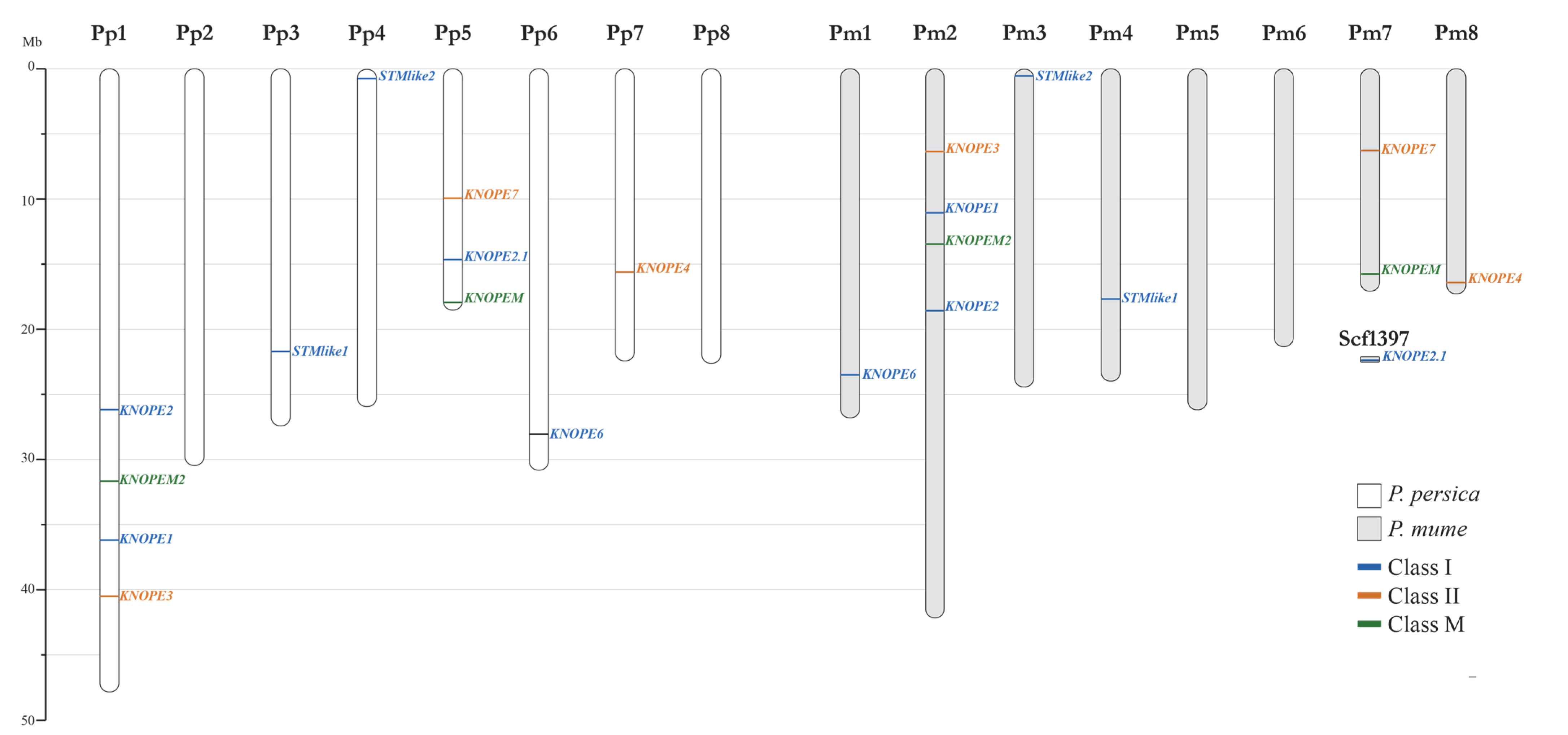
As for Prunus diploid genomes, 11 PRUNOX members were found in all species, except for P. avium which had 12 (Table S1). Moreover, PRUNOXIs were more numerous than PRUNOXIIs and PRUNOXMs and organized in a 6-3-2 module, except for P. avium which had a 6-4-2 one. The PRUNOXs were scattered on scaffolds/linkage groups (Table 1, Figure 2); hereafter, we provide details for the species (P. armeniaca, P. avium, P. dulcis, P. mira, P. mume, P. persica, P. salicina) that were fully assembled into eight chromosomes (Chrs).
As for PRUNOXI, the two-copy STM-like genes recurred on Chrs 3 and 4; the one-copy BP-like members were on Chrs 2 (Armeniaca section) and 1 (all the other); the two-copy KNAT2/6-like genes were on Chrs 2 and 7 (Armeniaca sect.) and Chrs 1 and 5 (all the others). The one-copy genes of the class I extra group resided on Chr 6, except that of P. mume (Chr 1). As for PRUNOXII, the 1–3-copy KNAT3/4/5-like genes were on Chrs 2 and 8 (Armeniaca sect.) and on Chrs 1 and 7 (all the others); the one-copy KNAT7-like members were on Chr 7 (Armeniaca sect.) and Chr 5 (all the others). Finally, the 1–2-copy KNATM genes lay on Chr 2 (Armeniaca sect.) or on Chrs 1 and 5 (all the others).
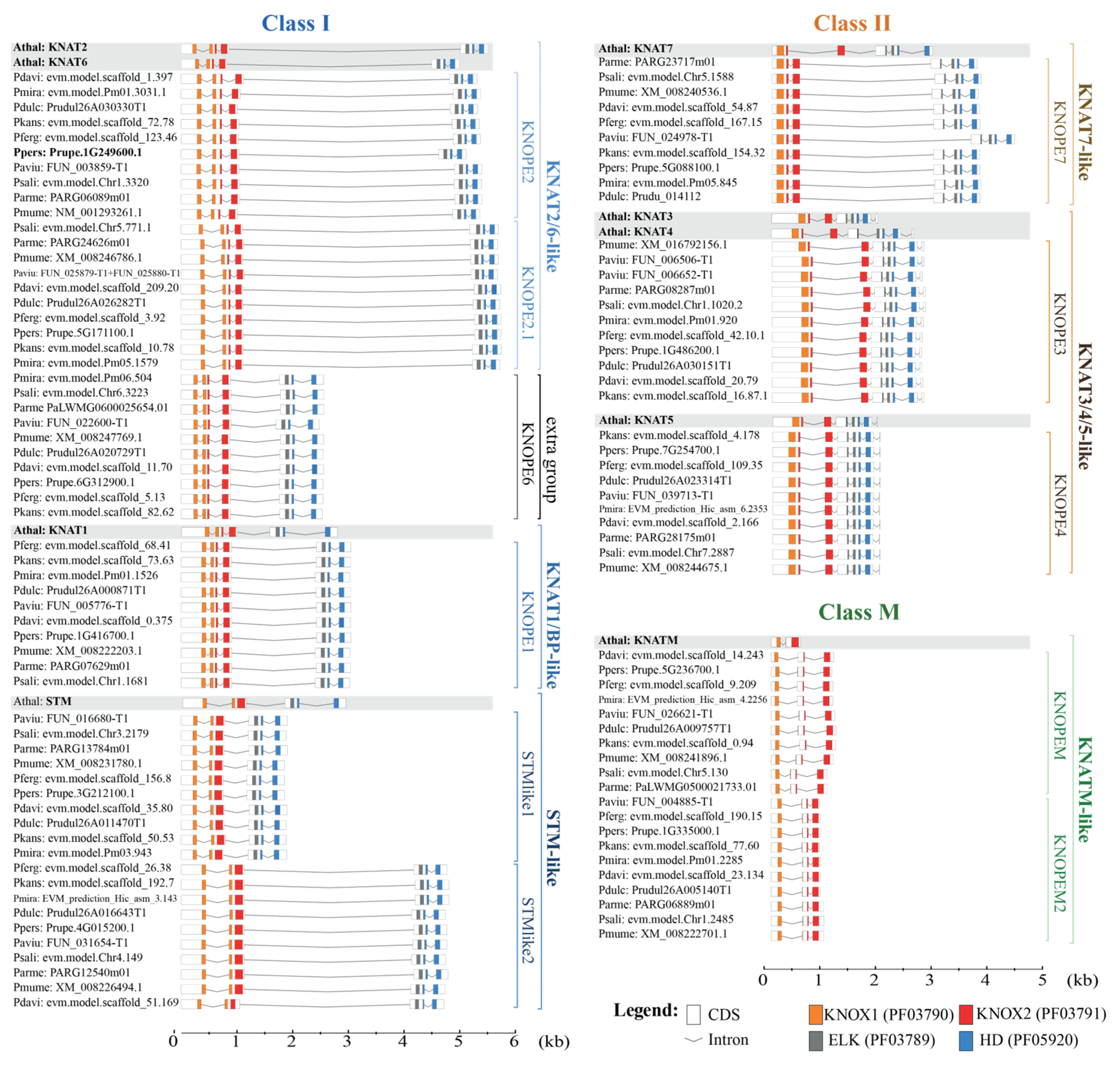
Synoptically, the PRUNOX intron–exon (IN-EX) organization was mostly conserved as in Arabidopsis, except for the extra group and the class M members (Figure 3), structurally different from the orthologs. Briefly, the PRUNOXI consisted of five EXs and four INs, except for STM-like genes with four EXs and three INs; moreover, the first IN position recurred in the KNOX1 domain. The PRUNOXII mainly harbored six EXs and five INs, except for the KNAT7-like group with the five-EX and four-IN organization. The PRUNOXII maintained one EN in the ELK, a class II peculiarity. The class M genes had three EXs and two INs, the latter residing respectively in KNOX1, similar to the PRUNOXI, and in KNOX2, as in PRUNOXII.
PRUNOX genomic organization was further analyzed for colinearity, duplication events, nonsynonymous/synonymous substitutions of colinear KNOX, and evolution time analysis using six Prunus spp. genomes (Table S1). Hereafter, we only report major outcomes that classify the Prunus multicopy orthologs to Arabidopsis STM, KNAT2/6, KNAT3/4, and KNATM genes as segmental duplications, that is, long stretches of duplicated sequences with high identity. Further manual alignments highlighted significant differences between introns (length/identity) in STM-like 1 and STM-like 2 gene groups (not shown); hence, transposon-mediated duplication—that is, gene fragments embedded into DNA transposons—might have taken place. Similarly, KNATM gene shuffling recalled transduplication. Retroduplication events (retrocopied intronless genes bearing a poly-A tail) were not addressed.
We looked for hormone-related cis elements sited in the KNOX promoters (1500 bp upstream the ATG) of peach and almond considering that they are the parents of GF677, a rootstock used for CK assays in this work. KNOX genes included a variable number of motifs for ABA, AUX, GAs, and ethylene (Table S3). Here, manual and bioinformatic tools deepened the search for CK motifs, which recurred in all PRUNOX members with similar abundance and positions in both species (Figure 4, Table S4). The PRUNOXI harbored 6–17 binding sites of 5–8 bp. The PRUNOXII hosted 7–18 motifs, mostly 5 bp long (>80%), while the PRUNOXM contained more than 14 elements.

2.1.3. Transcriptomic Features

We further addressed Prunus genome-wide transcription in aerial organs (Figure 5), using publicly available RNA-seq data (Table S5), and only fruit and leaves recurred in all species. The PRUNOXIs (vertical blue bar), regardless of developmental stage, shared low expression (blue z-score values) in fruit and leaves; however, their high transcription levels (orange values) characterized meristem-rich organs such as buds and stems (hosting vegetative/floral meristems and cambium). They were all also active in phloem tissues in tested species. Most of the PRUNOXIIs (vertical orange bar) showed opposite patterns to PRUNOXIs in leaves, though exceptions recurred for KNOPE4 orthologs, while behaviors in fruits, buds, and phloem varied among members and with species. The trends of class M genes often recalled those of class I.

2.2. PRUKNOX Responses to Cytokinin in GF677 Rootstock Microcuttings

2.2.1. Pheno-Histological Features

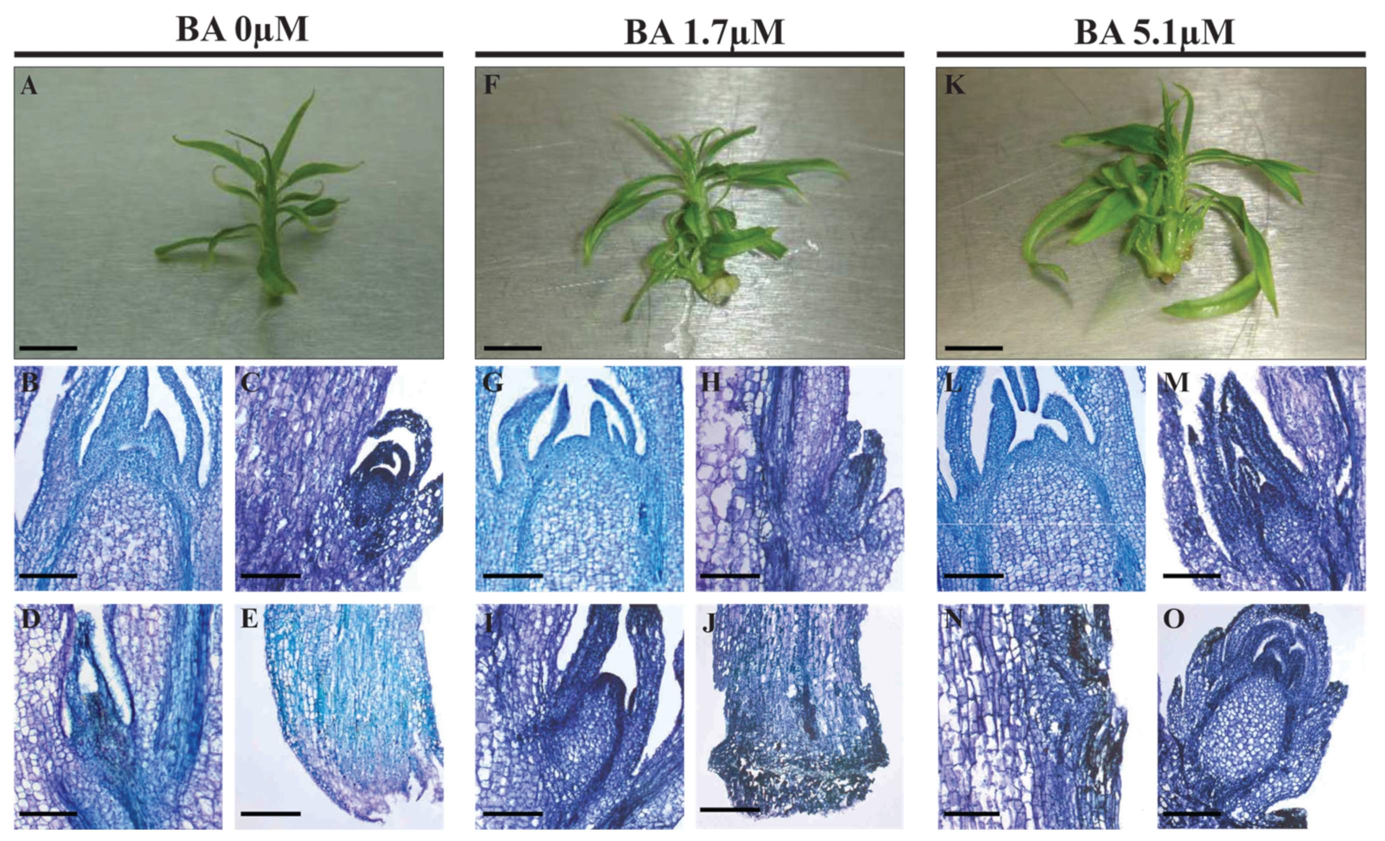
GF677 rootstock microshoots were grown on media containing 1.7 and 5.1 µM BA. Effects of CK treatment were evidenced by the formation of higher numbers of leaflets and side shoots than controls 10 days post-treatment (dpt), while stem length was unvaried (Figure 6A,F,K, and Table 2). In precocious histological analyses (3 dpt), the shoot apical meristem (SAM) and leaf axillary buds (LABs) of controls did not show anomalies (Figure 6B–D), and stems adjacent to the medium did not produce callus (Figure 6E). In microshoots treated with 1.7 µM BA, SAM and LABs (Figure 6G,H) were similar to controls; some LABs started elongation (Figure 6I), and callus occurred at the stem basis (Figure 6J). At 5.1 µM BA, SAM was unaffected (Figure 6L), though several microshoots bore LABs with normal (Figure 6M) or swollen (Figure 6O) morphology, or buds bulging from stem inner layers (Figure 6N), and callus at the stem basis (not shown).

2.2.2. PRUNOX Expression Patterns

Transcription of peach catalase (PpCAT) and CK-responsive (CKR) genes was monitored to mark events of oxidative stress and CK perception after BA treatment. CKRs were selected among Arabidopsis orthologs with ascertained behavior among the CK oxidases (PpCKX), responsive regulators (PpARR), and histidine kinases (PpHK). A further choice was based on genes acting in aerial organs of Rosaceae spp. and harboring CK-responsive motifs in promoters (Figure S1). A statistical analysis of the effects of BA dose (D), time (T), and interaction effect (DxT) was carried out to assess the significance of the influence on transcriptional responses of markers and PRUNOX and BELL genes (Table 3). As for PRUNOXI, BA concentration had significant effects at different levels on several genes tested. Similarly, time affected the expression of most class I KNOX genes (except for KNOPE6). DxT occurred for all class I genes except for STMlike1.
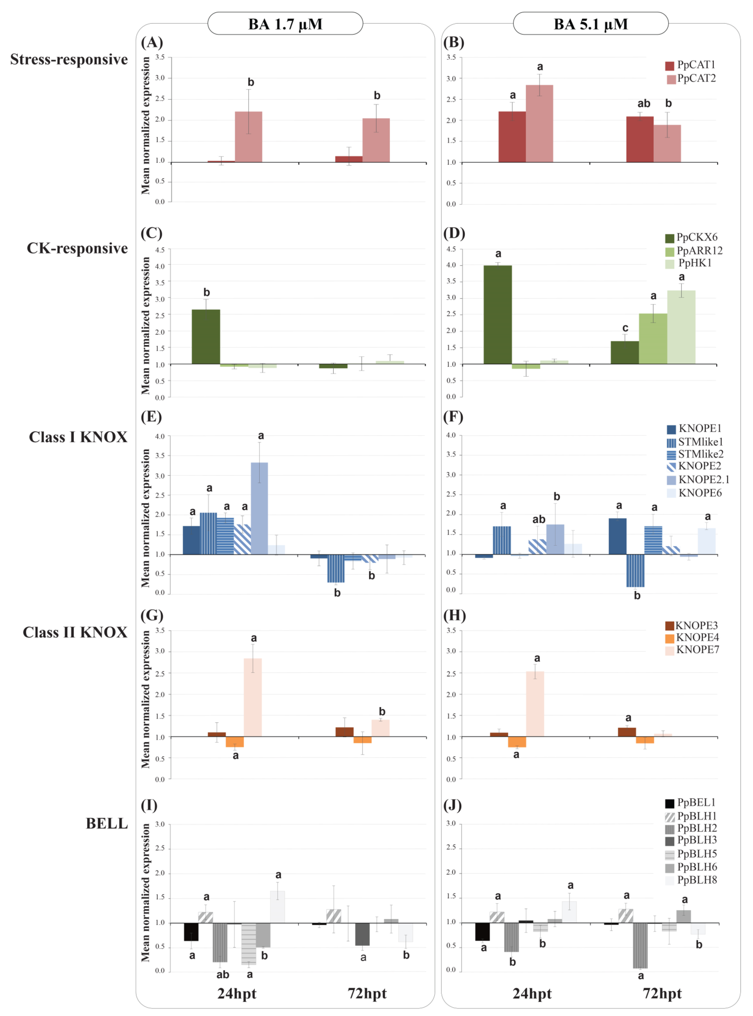
Hereafter, we briefly describe transcription trends (Figure 7) in time at fixed BA dose followed by some highlights of KNOX members’ peculiarities. As for stress markers (Figure 7A,B), PpCAT1 but not PpCAT2 was upregulated with time with 1.7 μM BA. At higher BA, both PpCAT genes were at least 2-fold upregulated in time course, at higher levels at 24 than 72 h post-treatment (hpt). As for CKR markers (Figure 7C,D), PpCKX6 expression was 2-fold higher at 24 hpt due to 1.7 μM BA, followed by restoration to control levels; PpARR12 and PpHK1 did not show significant response. Increasing BA to 5.1 μM caused a 4-fold increase in PpCKX6 transcription at 24 hpt followed by a level drop later on; the PpARR12 and PpHK1 genes were over 2-fold upregulated only at 72 hpt. All markers shared expression increase upon higher BA dose at 72 hpt, depicting a state of intense oxidative stress and modification of CK endogenous metabolism.
Regarding PRUNOXI after treatment with 1.7 μM BA (Figure 7E), all members showed higher transcription than controls (except for the unvaried KNOPE6). Onwards, all PRUNOX1 members decreased in expression to control levels, except for STMlike2, which was repressed. As for PRUNOXII (Figure 7G), KNOPE3 showed comparable expression to controls with time, KNOPE4 was only slightly repressed at 24 hpt, and KNOPE7 maintained upregulation that decreased in time course. Looking at the effects of higher BA concentration on PRUNOXI (Figure 7F), transcript upregulation was significant only for STMlike1, KNOPE2, and KNOPE2.1 at 24 hpt; onwards, the latter two had mRNA levels similar to control, while STMlike1 was strongly repressed; the expression of KNOPE1, KNOPE6, and STMlike1 increased significantly from 24 to 72 hpt. The PRUNOXII (Figure 7H) patterns were quite similar to those at low BA. Synoptically, at low BA, PRUNOXI shared the “up and down” regulation pattern with time, while member-specific diversified responses occurred at high BA from 24 to 72 hpt, suggesting that CK concentration alters regulatory mechanisms of PRUNOXI more intensely than those of PRUNOXII, these latter having a time-unvaried pattern under both BA dosages.
As for BELL responses to 1.7 μM BA (Figure 7I), BEL1, BLH2, BLH5, and BLH6 were repressed at 24 hpt, BLH1 and BLH8 were triggered, and BLH3 was similar to control; afterward, BLH3 and BLH8 were significantly repressed while the other members’ statuses were the same as those of controls. In media with higher BA (Figure 7J), BEL1, BLH2, and BLH5 were downregulated, while BLH1 and BLH8 were upregulated and BLH3 and BLH6 levels were the same as those of controls at 24 hpt. Subsequently, the BEL1, BLH3, and BLH5 mRNA levels were the same as those of controls, while those of BLH1 and BLH6 were higher and those of BLH2 and BLH8 were lower. Comparing the patterns at different BA dosages, six out of seven members (BLH6) shared similar patterns at 24 hpt, while discordant trends occurred for BLH2, BLH3, and BLH6 at 72 hpt, suggesting that a CK increase alters BELL transcription in a complex way.

2.2.3. Gene Coexpression Analysis

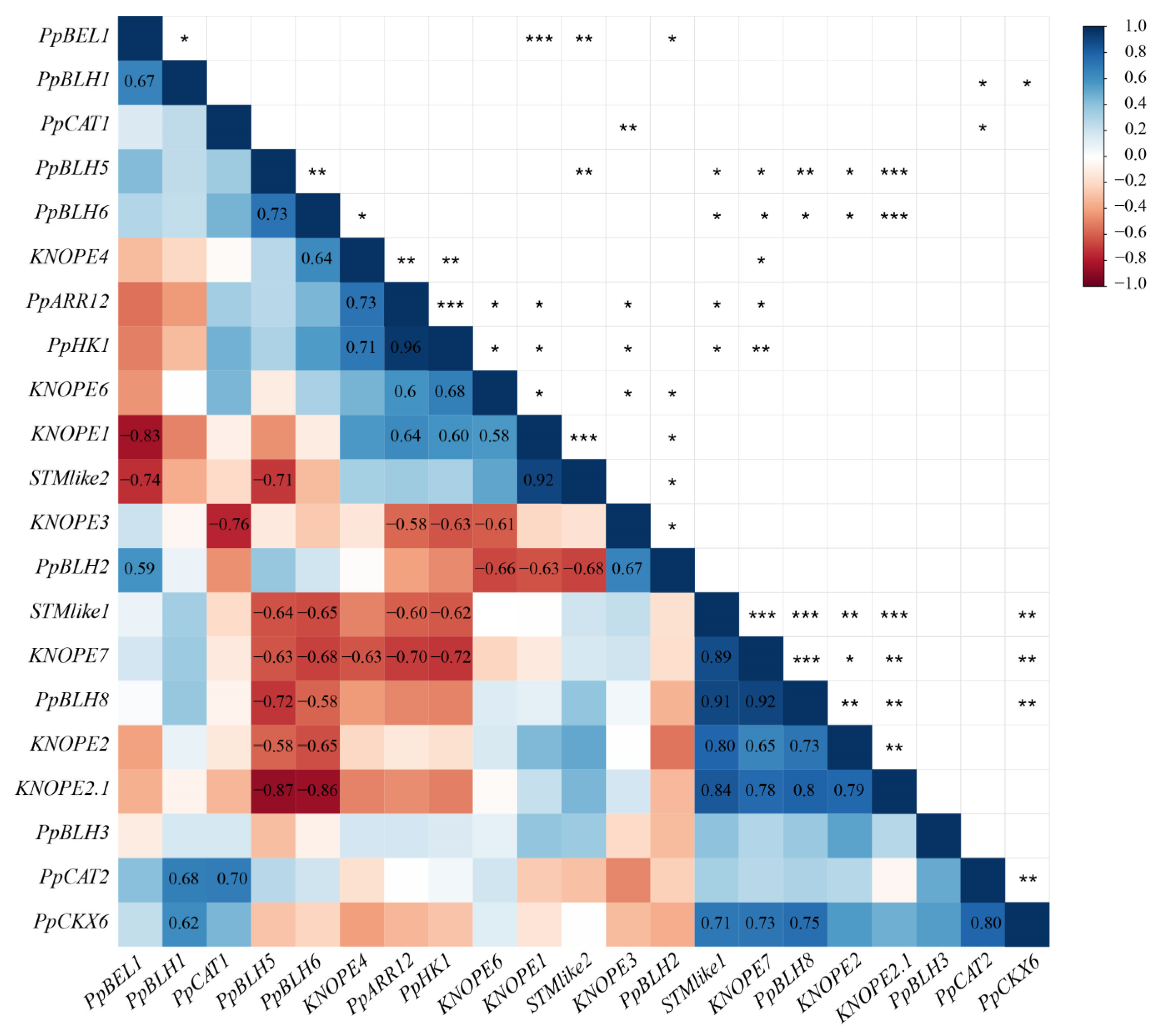
Correlation analysis among gene transcript abundance in response to BA dosage was carried out to assess gene coexpression (Figure 8); hereafter, we only report positive (0.7 ≤ r ≤ 1) or negative (−1 ≤ r ≤ −0.7) correlations with high significance (p < 0.01). Overall, class I STMlike1 and class II KNOPE4 and -7 positively correlated with CKR genes. As for class I genes, the couples KNOPE1/STMlike2, STMlike1/KNOPE2, STMlike1/KNOPE2.1, and KNOPE2/KNOPE2.1 showed high coexpression levels, whereas no significant correlation among class II members occurred (for the mentioned thresholds). Class I and II genes only showed positive correlations for the following duets: STMlike1/KNOPE7 and KNOPE2.1/KNOPE7. Synoptically, most class I KNOPEs (KNOPE1, STMlike1 and KNOPE2, KNOPE2 and KNOPE2.1) correlated with various BELLs, and each member showed positive or negative values depending on counterpart (e.g., KNOPE2.1 vs. PpBLH5 and -6 was negative and positive vs. PpBLH8). Finally, after scoring for KNOX/BELL correlations vs. CKR, the STMlike1/KNOPE7/PpBLH8 module was positively correlated with PpCKX6.

2.3. KNOPE1 Overexpression in Gisela 6 Rootstock

Regeneration of leaf explants followed a two-step procedure (details in Section 4) consisting of dark–liquid/light–solid on media RM1 and RM2 (Table S6); the latter is recommended for Gisela 6. After 6 weeks, regeneration frequency (percentage of explants forming novel shoots) was higher in the RM1/RM1 than in the RM2/RM2 (12.0 vs. 4.0 %), while the average shoot number per regenerating explant was similar (4.3 ± 0.5 vs. 4.5 ± 0.7). Consequently, we opted for RM1/RM1 to proceed with agro-infection (Figure S2); shoots originating from selection media (HYG or PTT) underwent a second selection, and three 35S:GFP and three 35S:KNOPE1 putative transgenic lines were rescued and named primary transgenic clones (PTCs). Southern blot analysis pointed at independent events of T-DNA multiple insertion in Gisela 6 genomes (Figure S3). Transformation frequency was 2.6% and 0.6%, respectively, for 35S:GFP and 35S:KNOPE1 events (Table S7). We subcultured the PTC and recovered six 35S:KNOPE1 plants. All 35S:GFP clones maintained stable expression over time (Figure 9A–I, Table S7). Differently (Figure 9S), the molecular analyses showed that the clones harbored the 35S:KNOPE1 transgene in the genome (genomic DNA PCR) but its message was undetected (RT-PCR), suggesting malfunctions (silencing), which we did not further address (e.g., detection of small interfering RNAs). Two plants showed phenotypes with altered margins in leaves (Figure 9K–P and Figure S2O), a distinctive trait of plants overexpressing KNOPE1/BP. Finally, one of the two also showed phenotype reversion later (Figure 9Q).

3. Discussion

3.1. PRUNOX

The complete KNOX catalog was retrieved from the genomes of 11 Prunus spp., and fine curation of gene structure was carried out for 35 out of 111 sequences. The phylogenetic tree of deduced proteins confirmed the separation into classes I, II, and M together with respective Arabidopsis orthologs [14]. Here, all 10 Prunus spp. had the KNATM branch, as already reported for Prunus spp. [14,21], which is a typical class of eudicots [2] and unfound in orchids [40]. A subgroup without Arabidopsis counterparts emerged in PRUNOXI, here named extra group (with peach KNOPE6 as reference), which is not unexpected considering the ample variation of KNOX number in dicot fruit trees. Specifically, the main diversity source of KNOPE6-like proteins resided in the N- and C-termini (not shown) as compared to the nearest members (e.g., KNAT2/6 and KNAT1). Moreover, KNOPE6-like gene structure differs from all the other PRUNOXIs in length and sequence (not position) of introns. Finally, all species maintained one KNOPE6-like gene (P. mume on Chr 1 and the other nine on Chr 6). These data suggest that KNOPE6-like may have specific roles in Prunus trees; one may regard fruit development as supported by KNOPE6 association with a QTL regulating several drupe characteristics [21]. As for protein variability, RNA-seq analyses indicate that coding sequences produce isoforms; for instance, both P. persica and P. mume harbor 11 KNOX genes encoding mRNAs for 17 and 12 proteins, implying splicing events. This was in agreement with a bioinformatic survey on P. mume TALEs [14]. For a given PRUNOX, amino acid variations (missense substitutions) among the Prunus spp. mainly occurred in N- and C-termini, but they were computed as similar and tolerated in most cases, hence supporting the conserved functions within the kind; however, substitutions potentially affecting functions were scored in KNOPE6 members.
The 10 Prunus spp. maintained the PRUNOX genomic organization in terms of the total number, class composition, and chromosome positions, with modest exceptions. This supports that the diversification of KNOX subfamilies took place [2] before the Prunus lineage diversification [41]. Referring to the Prunus subgenus, the shared PRUNOX colinearity brought out one group inclusive of Armeniaca section species (P. armeniaca and P. mume) and the other consisting of Persicae (P. persica and P. mira), Amigdalus (P. dulcis), and Prunus (P. salicina) sections. The separation was consistent with the genus evolutionary history and the high vicinity of P. mume and P. armeniaca compared to other Prunus species [42]. As for the Cerasus subgenus, P. avium hosted an additional copy of the class II KNOPE3-like gene, suggesting a specific role in the species of the deciduous corymbose group [42].
Several works report on KNOX responsiveness to CK in fruit tree organs [12,13] despite avoiding a computational search for CK-related motifs. Here, the investigation focused on peach and almond KNOX and showed that cis-elements of 5–8 bp occurred in all PRUNOX members abundantly and with conserved positions. Several other hormone-responsive cis-elements were scored (ABA, AUX, GAs, and ethylene) consistently with other fruit tree works [12,13,14]. Finally, giving speculation on RNA-seq meta-analysis related to CK, the abundant PRUNOXI expression in organs characterized by meristem activity (buds and stems) is consistent with the class I roles in participating in maintaining cells at undifferentiated stages in a context of CK–AUX equilibria necessary for meristem development [43].

3.2. PRUNOXI and -II Transcription Response to BA in Micropropagation

BA is routinely applied in MP; at 1.7 μM, increased numbers of leaves and lateral shoots, due to axillary bud activation, were expected [29]. The tripled dosage only raised the side shoot number, but histological sections showed the occurrence of budding from subcortical layers of the stem, of bud morphological abnormalities, and of abundant callus at the surface adjacent to the culture medium. At the molecular level, catalase genes (CAT) were upregulated by BA increase, while CK catabolism genes were affected by both concentration and time. These markers highlight a stress status associated with CK degradation as a response to hormone uptake. Indeed, at 72 hpt of BA high dose, the collective induction of CAT, CK oxidases, and CK receptors supported the stress exacerbation. These data are consistent with the concept that BA concentration variation is a multitype stress factor [44] and point at these markers as useful for the rapid monitoring of the physiological and health status of GF677 rootstock propagules.
CK responsive elements in promoters and transcriptional variation at 24 hpt support a rapid BA effect on PRUNOX, consistently with PRUNOXI triggering 3 h post-BA vascular uptake in stems [14]. Several experiments based on BA administration reveal KNOX message variation in long periods post-treatment [16,45]. These data pinpoint that responsiveness depends on supply methods, targeted tissue, and plant species. It is thought that KNOX genes respond to endogenous CK variations following BA applications since the BA root uptake in Arabidopsis seedlings did not cause KNOXI triggering, which instead occurred after endogenous CK levels were increased [46], and Arabidopsis KNOXs do not appear to be genes quickly responsive to BA [47]. In support, work on BA-induced caulogenesis from pine leaf showed that BA affected endogenous CK variation in the long term during which key KNOXI genes were responsive [48]. Relatedly, BA in the media can alter the inner CKs balance in GF677 [49]. In our system, CKR patterns after 72 hpt of high BA dosage may reflect endogenous CK variation (and hormone ratios) leading to altered KNOX profiles. Moreover, two gene regulatory aspects emerged from propagule responses: (a) PRUNOXIs are more sensitive to BA dose changes than PRUNOXIIs (Table 3), in particular STMlike2, KNOPE1, and KNOPE2.1; (b) all PRUNOXIs (except for KNOPE6) modulate expression in response to BA over time, and only KNOPE7 does so among PRUNOXIIs. This would suggest a kind of time-coordinated regulation between classes I and II over time [8] in response to CKs.
The propagule is a multiorgan system and KNOXs are multifunctional; therefore, establishing KNOX-specific (and/or undesired) effects on developmental changes due to BA increase requires additional experiments, here out of scope. However, considering the class I KNOX roles in maintaining cells in an undefined state and/or preventing cell expansion or lignification, the altered regulation could contribute to (a) the miniaturized state, typical of in vitro organs, or (b) tissue disorders that subtend hyperhydric traits [44]. Finally, it is known that KNOX and CK generate feedback-loop mechanisms of reciprocal control associated with cellular disorders [17], and these may contribute to the wide number of anomalies described for MP [50].
The tripled BA concentration did not affect stem height but did affect leaf and shoot numbers. This condition led to changes in PRUNOXI and BELL expression levels (and their correlations) at 72 hpt. However, in some cases, KNOX/BELL coexpression was preserved. Specifically, the STMlike1/KNOPE7/PpBLH8 coexpressed module included genes with orthologs that are involved in caulis development [6,51,52]. Hence, the unvaried gene module behavior may account for the unaffected stem trait. At 72 hpt of 5.1 µM, the increased PRUNOXI levels may also reflect the presence of more axillary meristems per propagule or de novo bud formation and shooting. Relatedly, it is known that almond STM-like transcription preceded the organization of adventitious meristems [53].

3.3. Transformation of Gisela 6 and KNOPE1 Phenotypes

In peach, stable [54] and/or transient [55] gene transfer has improved but remains a laborious, low-efficiency, and cultivar-specific process [54]. Alternatively, to assay technology efficiency and study KNOPE1 function, we employed the Gisela 6 transformation as used in different labs [56,57,58]. Regeneration efficiency is crucial for transformation; here we found that RM1 medium (regeneration frequency 12%, shoots per explant ca. 4%), based on QL and previously shown to be acceptable for the Montmorency cultivar [57], was also satisfactory for Gisela 6, despite the same authors recommending a specific one based on WPM. We may speculate that material origin and status may subtend the RM1 effect (e.g., different subclones of Gisela 6, hormonal treatments on material supplied by the company, and different in vitro growth conditions regarding light intensity). Finally, indirect organogenesis via callus prevailed, confirming that organogenesis-competent cells lay in wounded leaf mid-ribs [56]. The transformation frequency of Gisela 6 was reported to range from 0.5 to 3% [56,57,58], and here it consistently varied from 0.6 to 2.6%, depending on the vector type and transgene cassette. Two rounds of growth on the selection medium, molecular analyses, and phenotype selection (GFP functionality or leaf margin alteration for KNOPE1) were necessary to avoid technical escapes. However, malfunction of 35S:KNOPE1 was detected in clonal lines, together with phenotype reversion. Silencing events associated with transgenesis have been known for a long time [59], and here they were not further investigated (e.g., hypermethylation of transgene promoters, siRNA production due interference caused by the transgene vs. endogenous genes). The 35S:KNOPE1 plants bore leaf margin alteration similarly to KNOPE1 overexpression in Arabidopsis and several other simple leafed species overexpressing BP-like genes [17]. However, considering that KNOX overexpression causes pleiotropic and dramatic effects, the fine modulation of KNOXs by traditional approaches, such as the guide of time/tissue-specific or inducible promoters, or by novel genome editing strategies is envisaged [60].

4. Materials and Methods

4.1. Bioinformatic Surveys

The published genomes of 10 diploid Prunus species were used (Tables S1 and S3). They were P. persica, P. kansuensis, P. mira, P. ferganesis, and P. davidiana (peaches); P. armeniaca; P. mume (apricots); P. avium (sweet cherry); P. dulcis (almond); and P. salicina (plum). This study did not include ornamental, hybrid, and polyploid Prunus species.
As for genomic analyses, the reference assemblies and gene annotations were derived from the GDR database (www.rosaceae.org (accessed on 27 December 2022)), except for P. mume data from the NCBI repository. The keyword search of functional annotations used to identify PRUNOX genes was based on the term “KNOTTED” and terms related to KNOX domains (KNOX1, KNOX2, ELK, and HD). A similarity search was achieved using P. persica sequences as queries in each genome and using BLASTn (e-value cut-off ≤ 1 × 10⁻¹⁵). Gene models and annotations were manually curated (Table S1). The Gene Structure Display Server 2.0 (http://gsds.gao-lab.org (accessed on 27 December 2022)) was used to draw gene structures and protein domain organization. The MCScanX [61] program was used to perform the colinearity analyses of KNOX proteins within each Prunus spp. genome at the chromosome assembly level, and duplication types were classified into segmental, tandem, proximal, and others. Subsequently, colinear KNOXs were assessed for nonsynonymous and synonymous substitutions (Ka and Ks values) using PAL2NAL web service (http://www.bork.embl.de/pal2nal (accessed on 27 December 2022)). The formula to calculate the duplication time of colinear KNOX was T = Ks/2λ (with λ = 6.56 × 10⁻⁹ for dicots) [62]. The KNOX colinearity among Prunus spp. was determined by comparing chromosome map positions and graphed accordingly. The 1500 bp genomic sequences upstream the start codons of PRUNOX were submitted to the PLACE database for plant cis-acting regulatory element analysis. The program did not host CK binding motifs but was useful to detect other hormone-related motifs, which are listed in Table S3. Additionally, the Regulatory Sequence Analysis Tool 2022 (https://rsat.sb-roscoff.fr (accessed on 27 December 2022)) was run by using the same genomic sequences as above and CK motifs retrieved in [38,39] and reported in Figure 4 and Table S4.
As for protein analyses, deduced protein sequences were first validated by HMMSCAN (www.ebi.ac.uk/Tools/hmmer/search/hmmscan (accessed on 27 December 2022)) and then used to build a phylogenetic tree in MEGA7 ver. 7 (neighbor-joining method; 1000 bootstrap repetitions). The Scorecons server (www.ebi.ac.uk/thornton-srv/databases/cgi-bin/valdar/scorecons_server.pl (accessed on 27 December 2022)) and SIFT tool (sift.bii.a-star.edu.sg/www/SIFT_related_seqs_submit.html (accessed on 27 December 2022)) produced conservation scores and tolerance substitution scores, respectively. The PRUNOX sequences are in Table S1.
As for RNA-seq meta-analyses, publicly available transcriptomic data of Prunus spp. were mined in the NCBI Sequence Read Archive (SRA; www.ncbi.nlm.nih.gov/sra (accessed on 27 December 2022)) using Prunus [orgn:txid = 3754] as query and “RNA”, “Illumina”, “paired”, and “fastq” keywords. The results were visualized in the “SRA Run Selector” tool, and runs from “aerial organs” were selected, giving priority to those with replicates (if any). Overall, 63 paired-end runs were retrieved (Table S5) using the SRA Toolkit (http://github.com/ncbi/sra-tools (accessed on 27 December 2022)) and used in PRUNOX expression meta-analyses. Raw reads were quality-checked and filtered using Trimmomatic v0.39 [63]. High-quality clean reads were aligned to the respective genomes using HISAT2 v2.2.1 [64]; StringTie v2.1.5 [65] was used to assemble transcripts and estimate gene abundance in each sample. KNOX expression averages (for all replicates) were converted into z-scores and visualized using gplots package v. 3.1.3 (https://cran.r-project.org/web/packages/gplots/index.html (accessed on 27 December 2022)).

4.2. Cytokinin Assays

4.2.1. Micropropagation, Treatments, Sampling, and Histological Analyses

Micropropagated clones of GF677 rootstock (P. persica × P. amygdalus) were produced by CREA-OFA [29]. Briefly, single buds were grown in vials (Wheaton, IL, USA) on basal medium (BM, 10 mL) made of half-strength MS medium [66], microsalts, vitamins, sucrose (30 g/L), and agar (4 g/L; B&V, Parma, Italy), supplemented with hormones (0.4 mg/L BA, 0.1 mg/L GA3, 0.05 mg/L IAA). After two subcultures (10 days each), shoots were transferred into vessels (Magenta-Sigma, Bologna, Italy) with basal medium (50 mL) supplied with increasing doses of BA (0.0, 1.7, and 5.1 μM), generating shooting media SM 0, 1X, and 3X. The pH was adjusted (5.6) before agar addition, and sterilization followed at 121 °C for 20 min. All chemicals were by Sigma-Aldrich, Milan, Italy unless differently reported. Growth conditions were as follows: 24 ± 2 °C, light/dark—16/8 h, light intensity 37.5 μmol m−2 s−1 (Osram fluora L58 W/77 lamps, Monaco, Germany). The experimental matrix system consisted of propagules (n > 30; 5 p/jar) grown on SM0 (control), 1X, and 3X and replicated to allow sampling after 24 and 72 h and to avoid “open and close” repetitions. Materials for gene expression and histologic analyses derived from the same jars. As for RNA, propagules (n ≥ 12) for each treatment and timing were subdivided into three bulks (4 propagules each, named biological replicates), collected in tubes, flash-frozen in liquid nitrogen, and stored at −80 °C.

4.2.2. Morpho-Histological Analyses

Phenotyping was carried out at 10 days post-treatment by photographing 20 propagules per treatment and timing (5 p/jar) and measuring stem height, number of leaves on the main axis, and number of side shoots using ImageJ software (https://imagej.nih (accessed on 27 December 2022)). Student’s t-test was used to assess significant differences between treated and untreated samples at a given concentration.
Propagules (n ≥ 8) for each treatment and timing were immersed in ethanol (70% v/v) and stored at 4 °C. The dehydrated samples were embedded in Technovit 7100 (Heraeus Kulzer, Hanau, Germany), longitudinally sectioned at 8 μm with a Microm HM 350 SV microtome (Microm, Neuss, Germany), dried overnight at 40 °C, stained with 1% toluidine blue (w/v), dried and permanently mounted in Canada Balsam, and observed under light microscopy. Sections were digitally photographed using a Leica DMRB optical microscope equipped with a Leica DC 500 camera.

4.2.3. Gene Expression Analyses

Total RNA was isolated [67], DNAse-treated (RQ1, Promega, Madison, WI, USA), reverse-transcribed (2 µg) by Superscript III (Life Technologies, Carlsbad, CA, USA) at 55 °C using oligo (dT)20/random hexamer primer mixture (1:1). The cDNA (100 ng) was amplified in a volume (10 mL) containing Titan HotTaq EvaGreen qPCR Mix 1X (BIOATLAS, San Diego, CA, USA) and 0.3 mM of each primer. Triplicate reactions were performed at the following conditions: 15 min at 95 °C followed by 40 cycles of three-step amplification (30 s at 95 °C, 1 min at 59 °C, and 40 s at 72 °C). Primers are listed in Table S8. The threshold cycle (Ct) value of each gene was normalized with that of PpRPII [68,69] and compared with the Ct of untreated controls using the 2−ΔΔCt method [70].

4.3. Gene Transfer into Gisela 6

4.3.1. Plant Materials and In Vitro Culture

We purchased (Vivai Battistini, Cesena, Italy, www.battistinivivai.com (accessed on 27 December 2022)) in vitro clones of Gisela 6 plants. The rootstock is a hybrid derived from P. cerasus L. cv. Schattenmorelle (4n = 32) × P. canescens Bois (2n = 16), ploidy 2n = 3X = 24. Apices from these clones were further subcultured on several media, and the best one—ensuring the culture of vital clones for 40 days—was QBLI (3.3 g/L), which consisted of QL medium [71], vitamins and sucrose (20 g/L), BA (0.5 mg/L), NAA (0.05 mg/L), and agar (6 g/L) at pH 5.2. The clones underwent a 4-week recurrent transfer onto fresh QBLI (habituation); 3-week-old propagules bearing stem and leaves were moved onto McCown’s woody plant medium (WPM, Sigma-Aldrich, Milan, Italy) supplied with NAA (1 mg/L) until rooting was complete (2–3 weeks). Procedures were completed using sterilized (121 °C, 25 min) and film-sealed glass jars placed in growth chambers (16/8 h light/dark, 100 µmol s−1 m−2 PAR, 25 °C, and 60% HR). In vivo adaptation included clones (5 leaves and well-formed roots) that were transferred from jars to pots (5 × 5 cm, 100% sterilized peat, plastic film coverage) and kept for 2 days in baby rooms (20 °C, RH 80%; 16/8 h light/dark, 80 µmols-1 m-2 PAR). Two weeks later, plantlets (hardened stem, 10 leaves) were moved to larger pots (10 × 10 cm; peat/soil 1:1) in a glasshouse (RH 65–70%, 22–25 °C/spring–summer, 16–18 °C/autumn–winter) under natural light photoperiod and intensity.

4.3.2. Shoot Regeneration

Regeneration tests were as follows: Leaf explants from healthy in vitro clones underwent a 24 h dark pretreatment in liquid medium by gentle shaking and then were transferred to agarized regeneration media (RM) under light. The combinations dark–liquid/light–solid were on RM1/RM1 and RM2/RM2 media; RM2 was recommended for Gisela 6 [57]. RM1 (QL 3.38 g/L+ BA 3 mg/L + NAA 0.5 mg/L + sucrose 4% v/w) and RM2 (WPM + BA 2 mg/L + IBA 1 mg/L + sucrose 3% v/w) were at pH 4.8. Explants derived from shoots (length > 1 cm) from clones propagated on QLBI for 3–4 weeks. Leaves (length = 1.5–2 cm) were devoid of petioles and cut with three incisions perpendicular to the main vein (base, middle, and end of lamina). To test regeneration frequency, 50 explants (ca. 5 per Petri dish) were used for each treatment. The RM1/RM1 combination was satisfactory and used for transformation experiments.

4.3.3. Genetic Constructs and Transformation

The 35S:hptII/35S:GFP cassettes of the pCAMBIA1302 vector, conferring resistance to hygromycin (HYG) and synthetizing the GFP, were used in transformation tests. The 35S:pat/35S:KNOPE1 cassettes of the binary vector pBA002, conferring resistance to phosphinothricin (PPT), were previously described [17]. All recombinant plasmids were transferred into the A. tumefaciens strain EHA105 by freeze–thaw method.
Single colonies of recombinant A. tumefaciens (At) were grown (28 °C, 48 h dark) in LB (10 mL) plus antibiotics (nalidixic acid 30 mg/L, spectinomycin or kanamycin 50 mg/L); then, they were collected (2500× g, 2 min), suspended in RM1 (25 mL) supplied with acetosyringone (100 µM), and brought to 0.5 value of OD600. The explants were incubated in bacterial suspension (25 mL, 28 °C) for 3 days, and then At was removed by washing in RM1 (40 mL, 5 min) plus cefotaxime (500 mg/L) 6 times, followed by 5 washes with RM1 to remove the antibiotic. The explants were dried on sterile filter paper and were laid on solid RM1 plus cefotaxime (250 mg/L) in Petri dishes (6 explants each, 100 × 20 mm dish) for a fortnight at 25 °C. Afterward, the explants were moved onto solid RM1 without cefotaxime and with PPT (20 mg/L) or HYG (10 mg/L). PPT and HYG concentrations were established by previous dosage assays. Putative transgenic clones were screened based on PPT or HYG resistance (absence of chlorosis/necrosis) combined with a phenotypic trait (fringed leaves due to KNOPE1 or GFP fluorescence), moved to fresh media containing PPT or HYG for further selection for ca. 2 weeks, and finally transferred to rooting medium (without herbicide or antibiotic).

4.3.4. Transgene Analyses

Genome insertion was checked by Southern blot (Figure S3) and PCR analyses. The former was previously detailed for peach [17]; leaf gDNA (30 μg) was EcoRI-digested, fractionated in 0.8% agarose gel, blotted onto a membrane (Hybond N+, Amersham, UK), and hybridized with a probe (25 ng) that was radio-labeled with 5 μL of [32P]-dCTP (45 µL of TE buffer: 10 mM Tris·HCl pH 8.0, 1 mM EDTA) according to Rediprime II kit (Amersham). Overnight hybridization was at 60 °C (buffer NaPi 0.5 M pH 7.2, SDS 5%, EDTA 10 mM), followed by 2 washes (2X and 1X SSC/0.1% SDS, 60 °C, 10 min each) and membrane exposure (12 h, −80 °C) to films (Kodak Biomax, Boston, MA, USA). The HPTII probe (ca. 1100 bp) was excised (XhoI, Invitrogen, Waltham, MA, USA) from pCAMBIA1302; the BAR probe (ca. 676 bp) was excised (NheI/EcoRI, Buffer M Invitrogen) from pBA-KNOPE1 plasmid [17]. Fragments were purified (QIAquick gel extraction kit, Qiagen, Valencia, CA, USA) and did not harbor the EcoRI site.
As for PCR, the reaction mixture final conditions in 30 µL were as follows: gDNA (300 ng), primers (0.3 μM), dNTPs (0.2 mM), MgCl2 (2.5 mM), 1.25 U Taq polymerase, and 1X Buffer (Dream Taq Fermentas). Cycle conditions were as follows: denaturation start (95 °C/5 min), 35 amplification cycles (95 °C/30 s, 57 °C/30 s, 72 °C/120 s), and final extension (72 °C/5 min). Primers are listed in Table S9.
In Gisela 6 nontransformed plants, the KNOPE1-like messages were not detected or strongly downregulated in leaf blades (devoid of petioles); the use of specific primers allowed the detection of peach KNOPE1 expression from the transgene 35S:KNOPE1 by RT-PCR in clones previously checked for the transgene in the genome. The cDNA synthesis was described above; the PCR conditions (in 50 µL) were as follows: 200 ng cDNA, 1 µM of each primer, 0.5 mM dNTPs, 2.5U DreamTaq (Fermentas, Waltham, MA, USA), 1X buffer; start cycle 95 °C/5 min, 35 cycles: 95 °C/1 min, 62 °C/30 s, and 72 °C/90 s, final extension: 72 °C/5 min; the products (15 µL) were electrophoresed (0.8% agarose gels). Primers are listed in Table S9. The fluorescence in 35S:GFP Gisela 6 was detected using an Eclipse 80i microscope (Nikon, Tokyo, Japan) equipped with FITC filters (B-1E) and NIS-Element BR 2.20 software (Nikon) by directly observing organ tissues of transgenic and control plants.

4.4. Statistical Analyses

Data were analyzed by different statistical tests including Student’s t-test and two-way ANOVA. Pearson correlations were calculated using the “rcorr” function in the R environment v3.4.3 (Core Team, Vienna, Austria, https://www.R-project.org (accessed on 27 December 2022)).

Supplementary Materials

The following supporting information can be downloaded at: https://www.mdpi.com/article/10.3390/ijms24033046/s1.

Author Contributions

D.G. and G.T. were responsible for conceptualization, all work on Gisela 6, data curation, and original draft writing. G.T. performed bioinformatics, gene expression, and statistical analyses. E.C.: GF677 in vitro material and phenotyping. S.D.: histological analysis on GF677 clones. D.G., M.M.A. and E.C. supervised and reviewed writing. All authors have read and agreed to the published version of the manuscript.

Funding

The research was funded by the “Eumorfo” project (D.M. 299/7303/05) of the Italian Ministry of Agricultural, Food and Forestry Policies and further supported for all bioinformatic analyses by regular funds of National Research Council of Italy.

Institutional Review Board Statement

Not applicable.

Informed Consent Statement

Not applicable.

Data Availability Statement

Not applicable.

Acknowledgments

We are indebted to Chiara Nicolodi, who retired in 2020, and grateful for the Gisela 6 in vitro and transformation work. Emiliano Condello is thanked for helping in in vitro analyses of GF677 clones. Carla Ticconi is thanked for English proofreading, criticism, and suggestions.

Conflicts of Interest

The authors declare no conflict of interest. The funders had no role in the design of the study or in the analyses of the data.

References

  1. Burglin, T.R.; Affolter, M. Homeodomain proteins: An update. Chromosoma 2016, 125, 497–521. [Google Scholar] [CrossRef] [PubMed]
  2. Gao, J.; Yang, X.; Zhao, W.; Lang, T.; Samuelsson, T. Evolution, diversification, and expression of KNOX proteins in plants. Front. Plant Sci. 2015, 6, 882. [Google Scholar] [CrossRef] [PubMed]
  3. Hay, A.; Tsiantis, M. KNOX genes: Versatile regulators of plant development and diversity. Development 2010, 137, 3153–3165. [Google Scholar] [CrossRef] [PubMed]
  4. Scofield, S.; Dewitte, W.; Murray, J.A. A model for Arabidopsis class-1 KNOX gene function. Plant Signal Behav. 2008, 3, 257–259. [Google Scholar] [CrossRef]
  5. Zhao, M.; Yang, S.; Chen, C.Y.; Li, C.; Shan, W.; Lu, W.; Cui, Y.; Liu, X.; Wu, K. Arabidopsis BREVIPEDICELLUS interacts with the SWI2/SNF2 chromatin remodeling ATPase BRAHMA to regulate KNAT2 and KNAT6 expression in control of inflorescence architecture. PLoS Genet. 2015, 11, e1005125. [Google Scholar] [CrossRef]
  6. Wang, S.; Yamaguchi, M.; Grienenberger, E.; Martone, P.T.; Samuels, A.L.; Mansfield, S.D. The Class II KNOX genes KNAT3 and KNAT7 work cooperatively to influence deposition of secondary cell walls that provide mechanical support to Arabidopsis stems. Plant J. 2020, 101, 293–309. [Google Scholar] [CrossRef]
  7. Nookaraju, A.; Pandey, S.K.; Ahlawat, Y.K.; Joshi, C.P. Understanding the Modus Operandi of Class II KNOX Transcription Factors in Secondary Cell Wall Biosynthesis. Plants 2022, 11, 493. [Google Scholar] [CrossRef]
  8. Furumizu, C.; Alvarez, J.P.; Sakakibara, K.; Bowman, J.L. Antagonistic roles for KNOX1 and KNOX2 genes in patterning the land plant body plan following an ancient gene duplication. PLoS Genet. 2015, 11, e1004980. [Google Scholar] [CrossRef]
  9. Keren-Keiserman, A.; Shtern, A.; Levy, M.; Chalupowicz, D.; Furumizu, C.; Alvarez, J.P.; Amsalem, Z.; Arazi, T.; Alkalai-Tuvia, S.; Efroni, I.; et al. CLASS-II KNOX genes coordinate spatial and temporal ripening in tomato. Plant Physiol. 2022, 190, 657–668. [Google Scholar] [CrossRef]
  10. Hepworth, S.R.; Pautot, V.A. Beyond the Divide: Boundaries for Patterning and Stem Cell Regulation in Plants. Front. Plant Sci. 2015, 6, 1052. [Google Scholar] [CrossRef] [Green Version]
  11. Kim, D.; Cho, Y.H.; Ryu, H.; Kim, Y.; Kim, T.H.; Hwang, I. BLH1 and KNAT3 modulate ABA responses during germination and early seedling development in Arabidopsis. Plant J. 2013, 75, 755–766. [Google Scholar] [CrossRef]
  12. Jia, P.; Zhang, C.; Xing, L.; Li, Y.; Shah, K.; Zuo, X.; Zhang, D.; An, N.; Han, M.; Ren, X. Genome-Wide Identification of the MdKNOX Gene Family and Characterization of Its Transcriptional Regulation in Malus domestica. Front. Plant Sci. 2020, 11, 128. [Google Scholar] [CrossRef]
  13. Cheng, X.; Li, M.; Abdullah, M.; Li, G.; Zhang, J.; Manzoor, M.A.; Wang, H.; Jin, Q.; Jiang, T.; Cai, Y.; et al. In Silico Genome-Wide Analysis of the Pear (Pyrus bretschneideri) KNOX Family and the Functional Characterization of PbKNOX1, an Arabidopsis BREVIPEDICELLUS Orthologue Gene, Involved in Cell Wall and Lignin Biosynthesis. Front. Genet. 2019, 10, 632. [Google Scholar] [CrossRef]
  14. Yang, Q.; Yuan, C.; Cong, T.; Wang, J.; Zhang, Q. Genome-wide identification of three-amino-acid-loop-extension gene family and their expression profile under hormone and abiotic stress treatments during stem development of Prunus mume. Front. Plant Sci. 2022, 13, 1006360. [Google Scholar] [CrossRef]
  15. Srinivasan, C.; Liu, Z.; Scorza, R. Ectopic expression of class 1 KNOX genes induce adventitious shoot regeneration and alter growth and development of tobacco (Nicotiana tabacum L.) and European plum (Prunus domestica L.). Plant Cell Rep. 2011, 30, 655–664. [Google Scholar] [CrossRef]
  16. Jia, P.; Xing, L.; Zhang, C.; Chen, H.; Li, Y.; Zhang, D.; Ma, J.; Zhao, C.; Han, M.; Ren, X.; et al. MdKNOX15, a class I knotted-like transcription factor of apple, controls flowering and plant height by regulating GA levels through promoting the MdGA2ox7 transcription. Environ. Exp. Bot. 2021, 185, 104411. [Google Scholar] [CrossRef]
  17. Testone, G.; Bruno, L.; Condello, E.; Chiappetta, A.; Bruno, A.; Mele, G.; Tartarini, A.; Spano, L.; Innocenti, A.M.; Mariotti, D.; et al. Peach [Prunus persica (L.) Batsch] KNOPE1, a class 1 KNOX orthologue to Arabidopsis BREVIPEDICELLUS/KNAT1, is misexpressed during hyperplasia of leaf curl disease. J. Exp. Bot. 2008, 59, 389–402. [Google Scholar] [CrossRef]
  18. Zeng, R.F.; Fu, L.M.; Deng, L.; Liu, M.F.; Gan, Z.M.; Zhou, H.; Hu, S.F.; Hu, C.G.; Zhang, J.Z. CiKN1 and CiKN6 are involved in leaf development in citrus by regulating CimiR164. Plant J. 2022, 110, 828–848. [Google Scholar] [CrossRef]
  19. Scofield, S.; Murray, J.A. KNOX gene function in plant stem cell niches. Plant Mol. Biol. 2006, 60, 929–946. [Google Scholar] [CrossRef]
  20. Tsuda, K.; Hake, S. Diverse functions of KNOX transcription factors in the diploid body plan of plants. Curr. Opin. Plant Biol. 2015, 27, 91–96. [Google Scholar] [CrossRef] [Green Version]
  21. Testone, G.; Condello, E.; Verde, I.; Nicolodi, C.; Caboni, E.; Dettori, M.T.; Vendramin, E.; Bruno, L.; Bitonti, M.B.; Mele, G.; et al. The peach (Prunus persica L. Batsch) genome harbours 10 KNOX genes, which are differentially expressed in stem development, and the class 1 KNOPE1 regulates elongation and lignification during primary growth. J. Exp. Bot. 2012, 63, 5417–5435. [Google Scholar] [CrossRef] [PubMed]
  22. Zeng, R.F.; Zhou, H.; Fu, L.M.; Yan, Z.; Ye, L.X.; Hu, S.F.; Gan, Z.M.; Ai, X.Y.; Hu, C.G.; Zhang, J.Z. Two citrus KNAT-like genes, CsKN1 and CsKN2, are involved in the regulation of spring shoot development in sweet orange. J. Exp. Bot. 2021, 72, 7002–7019. [Google Scholar] [CrossRef] [PubMed]
  23. Jia, P.; Xing, L.; Zhang, C.; Zhang, D.; Ma, J.; Zhao, C.; Han, M.; Ren, X.; An, N. MdKNOX19, a class II knotted-like transcription factor of apple, plays roles in ABA signalling/sensitivity by targeting ABI5 during organ development. Plant Sci. 2021, 302, 110701. [Google Scholar] [CrossRef] [PubMed]
  24. Zhang, S.; Gottschalk, C.; van Nocker, S. Genetic mechanisms in the repression of flowering by gibberellins in apple (Malus × domestica Borkh.). BMC Genom. 2019, 20, 747. [Google Scholar] [CrossRef] [PubMed]
  25. Bednarek, P.T.; Orłowska, R. Plant tissue culture environment as a switch-key of (epi)genetic changes. Plant Cell Tissue Organ. Cult. 2020, 140, 245–257. [Google Scholar] [CrossRef]
  26. Hassan, S.A.M.; Zayed, N.S. Factor Controlling Micropropagation of Fruit Trees: A Review. Sci. Int. 2018, 6, 1–10. [Google Scholar] [CrossRef]
  27. George, E.F.; Hall, M.A.; Klerk, G.J.D. Plant Tissue Culture Procedure—Background. In Plant Propagation by Tissue Culture: Volume 1. The Background; George, E.F., Hall, M.A., Klerk, G.-J.D., Eds.; Springer: Dordrecht, The Netherlands, 2008; pp. 1–28. [Google Scholar]
  28. Cheong, E.J. Biotechnological approaches for improvement and conservation of Prunus species. Plant Biotechnol. Rep. 2012, 6, 17–28. [Google Scholar] [CrossRef]
  29. Gentile, A.; Jàquez Gutiérrez, M.; Martinez, J.; Frattarelli, A.; Nota, P.; Caboni, E. Effect of meta-Topolin on micropropagation and adventitious shoot regeneration in Prunus rootstocks. Plant Cell Tissue Org. Cult. 2014, 118, 373–381. [Google Scholar] [CrossRef]
  30. Van Nocker, S.; Gardiner, S.E. Breeding better cultivars, faster: Applications of new technologies for the rapid deployment of superior horticultural tree crops. Hortic. Res. 2014, 1, 14022. [Google Scholar] [CrossRef]
  31. Zong, X.; Denler, B.J.; Danial, G.H.; Chang, Y.; Song, G. Adventitious shoot regeneration and agrobacterium tumefaciens-mediated transient transformation of almond × peach hybrid rootstock ‘Hansen 536’. HortScience 2019, 54, 936–940. [Google Scholar] [CrossRef] [Green Version]
  32. Ricci, A.; Sabbadini, S.; Prieto, H.; Padilla, I.M.; Dardick, C.; Li, Z.; Scorza, R.; Limera, C.; Mezzetti, B.; Perez-Jimenez, M.; et al. Genetic Transformation in Peach (Prunus persica L.): Challenges and Ways Forward. Plants 2020, 9, 971. [Google Scholar] [CrossRef]
  33. Petri, C.; Alburquerque, N.; Faize, M.; Scorza, R.; Dardick, C. Current achievements and future directions in genetic engineering of European plum (Prunus domestica L.). Transgen. Res. 2018, 27, 225–240. [Google Scholar] [CrossRef]
  34. Zhang, L.; Fang, W.; Chen, F.; Song, A. The Role of Transcription Factors in the Regulation of Plant Shoot Branching. Plants 2022, 11, 1997. [Google Scholar] [CrossRef]
  35. Bianchi, V.J.; Rubio, M.; Trainotti, L.; Verde, I.; Bonghi, C.; Martinez-Gomez, P. Prunus transcription factors: Breeding perspectives. Front. Plant Sci. 2015, 6, 443. [Google Scholar] [CrossRef]
  36. Mukherjee, K.; Brocchieri, L.; Burglin, T.R. A comprehensive classification and evolutionary analysis of plant homeobox genes. Mol. Biol. Evol. 2009, 26, 2775–2794. [Google Scholar] [CrossRef]
  37. Shi, S.; Li, J.; Sun, J.; Yu, J.; Zhou, S. Phylogeny and classification of Prunus sensu lato (Rosaceae). J. Integr. Plant Biol. 2013, 55, 1069–1079. [Google Scholar] [CrossRef]
  38. Ramireddy, E.; Brenner, W.G.; Pfeifer, A.; Heyl, A.; Schmulling, T. In planta analysis of a cis-regulatory cytokinin response motif in Arabidopsis and identification of a novel enhancer sequence. Plant Cell Physiol. 2013, 54, 1079–1092. [Google Scholar] [CrossRef]
  39. Brenner, W.G.; Schmulling, T. Summarizing and exploring data of a decade of cytokinin-related transcriptomics. Front. Plant Sci. 2015, 6, 29. [Google Scholar] [CrossRef]
  40. Zhang, D.; Lan, S.; Yin, W.L.; Liu, Z.J. Genome-Wide Identification and Expression Pattern Analysis of KNOX Gene Family in Orchidaceae. Front. Plant Sci. 2022, 13, 901089. [Google Scholar] [CrossRef]
  41. Chin, S.W.; Shaw, J.; Haberle, R.; Wen, J.; Potter, D. Diversification of almonds, peaches, plums and cherries—Molecular systematics and biogeographic history of Prunus (Rosaceae). Mol. Phylogenet. Evol. 2014, 76, 34–48. [Google Scholar] [CrossRef]
  42. Hodel, R.G.; Zimmer, E.; Wen, J. A phylogenomic approach resolves the backbone of Prunus (Rosaceae) and identifies signals of hybridization and allopolyploidy. Mol. Phylogenet. Evol. 2021, 160, 107118. [Google Scholar] [CrossRef] [PubMed]
  43. Su, Y.H.; Liu, Y.B.; Zhang, X.S. Auxin-cytokinin interaction regulates meristem development. Mol. Plant 2011, 4, 616–625. [Google Scholar] [CrossRef] [PubMed]
  44. Polivanova, O.B.; Bedarev, V.A. Hyperhydricity in Plant Tissue Culture. Plants 2022, 11, 3313. [Google Scholar] [CrossRef] [PubMed]
  45. Zhang, S.; Pan, Y.; Zhi, C.; Zheng, Y.; Wang, X.; Li, X.; Cheng, Z. Genome-Wide Identification and Characterization of KNOTTED-Like Homeobox (KNOX) Homologs in Garlic (Allium sativum L.) and Their Expression Profilings Responding to Exogenous Cytokinin and Gibberellin. Int. J. Mol. Sci. 2021, 22, 9237. [Google Scholar] [CrossRef]
  46. Soucek, P.; Klima, P.; Rekova, A.; Brzobohaty, B. Involvement of hormones and KNOXI genes in early Arabidopsis seedling development. J. Exp. Bot. 2007, 58, 3797–3810. [Google Scholar] [CrossRef]
  47. Brenner, W.G.; Romanov, G.A.; Kollmer, I.; Burkle, L.; Schmulling, T. Immediate-early and delayed cytokinin response genes of Arabidopsis thaliana identified by genome-wide expression profiling reveal novel cytokinin-sensitive processes and suggest cytokinin action through transcriptional cascades. Plant J. 2005, 44, 314–333. [Google Scholar] [CrossRef]
  48. Alvarez, J.M.; Bueno, N.; Cuesta, C.; Feito, I.; Ordas, R.J. Hormonal and gene dynamics in de novo shoot meristem formation during adventitious caulogenesis in cotyledons of Pinus pinea. Plant Cell Rep. 2020, 39, 527–541. [Google Scholar] [CrossRef]
  49. Perez-Jimenez, M.; Cantero-Navarro, E.; Perez-Alfocea, F.; Cos-Terrer, J. Endogenous hormones response to cytokinins with regard to organogenesis in explants of peach (Prunus persica L. Batsch) cultivars and rootstocks (P. persica × Prunus dulcis). Plant Physiol. Biochem. 2014, 84, 197–202. [Google Scholar] [CrossRef]
  50. Ziv, M. Quality of micropropagated plants—Vitrification. In Vitro Cell Dev. Biol.—Plant 1991, 27, 64–69. [Google Scholar] [CrossRef]
  51. Groover, A.T.; Mansfield, S.D.; DiFazio, S.P.; Dupper, G.; Fontana, J.R.; Millar, R.; Wang, Y. The Populus homeobox gene ARBORKNOX1 reveals overlapping mechanisms regulating the shoot apical meristem and the vascular cambium. Plant Mol. Biol. 2006, 61, 917–932. [Google Scholar] [CrossRef]
  52. Kim, M.H.; Cho, J.S.; Jeon, H.W.; Sangsawang, K.; Shim, D.; Choi, Y.I.; Park, E.J.; Lee, H.; Ko, J.H. Wood Transcriptome Profiling Identifies Critical Pathway Genes of Secondary Wall Biosynthesis and Novel Regulators for Vascular Cambium Development in Populus. Genes 2019, 10, 690. [Google Scholar] [CrossRef]
  53. Santos, A.M.; Oliver, M.J.; Sánchez, A.M.; Oliveira, M.M. Expression of almond Knotted1 homologue (PdKn1) anticipates adventitious shoot initiation. In Vitro Cell Dev. Biol.—Plant 2012, 48, 40–49. [Google Scholar] [CrossRef]
  54. Prieto, H. Genetic Transformation Strategies in Fruit Crops. In Genetic Transformation; Alvarez, M., Ed.; IntechOpen: Rijeka, Croatia, 2011; pp. 81–99. [Google Scholar]
  55. Jia, H.F.; Guo, J.X.; Qin, L.; Shen, Y.Y. Virus-induced PpCHLH gene silencing in peach leaves (Prunus persica). J. Hortic. Sci. Biotechnol. 2010, 85, 528–532. [Google Scholar] [CrossRef]
  56. Song, G.Q.; Sink, K.C. Transformation of Montmorency sour cherry (Prunus cerasus L.) and Gisela 6 (P. cerasus × P. canescens) cherry rootstock mediated by Agrobacterium tumefaciens. Plant Cell Rep. 2006, 25, 117–123. [Google Scholar] [CrossRef]
  57. Song, G.Q.; Sink, K.C. Transformation of cherry: Prunus cerasus l. ‘Montmorency’ and Prunus cerasus × P. Canescen ‘Gisela 6’ mediated by Agrobacterium tumefaciens and a two-step selection system. Acta Hortic. 2007, 738, 683–690. [Google Scholar] [CrossRef]
  58. Zong, X.; Xu, L.; Tan, Y.; Wei, H. Development of genetically modified sweet cherry rootstock ‘Gisela 6’ with overexpression of PcMPK3-HA gene by Agrobacterium-mediated genetic transformation. Plant Cell Tissue Org. Cult. 2022, 151, 375–384. [Google Scholar] [CrossRef]
  59. Rajeevkumar, S.; Anunanthini, P.; Sathishkumar, R. Epigenetic silencing in transgenic plants. Front. Plant Sci. 2015, 6, 693. [Google Scholar] [CrossRef]
  60. Lowder, L.G.; Malzahn, A.; Qi, Y. Plant Gene Regulation Using Multiplex CRISPR-dCas9 Artificial Transcription Factors. Methods Mol. Biol. 2018, 1676, 197–214. [Google Scholar] [CrossRef]
  61. Wang, Y.; Tang, H.; Debarry, J.D.; Tan, X.; Li, J.; Wang, X.; Lee, T.H.; Jin, H.; Marler, B.; Guo, H.; et al. MCScanX: A toolkit for detection and evolutionary analysis of gene synteny and collinearity. Nucleic Acids Res. 2012, 40, e49. [Google Scholar] [CrossRef]
  62. He, Y.; Liu, X.; Ye, L.; Pan, C.; Chen, L.; Zou, T.; Lu, G. Genome-Wide Identification and Expression Analysis of Two-Component System Genes in Tomato. Int. J. Mol. Sci. 2016, 17, 1204. [Google Scholar] [CrossRef] [Green Version]
  63. Bolger, A.M.; Lohse, M.; Usadel, B. Trimmomatic: A flexible trimmer for Illumina sequence data. Bioinformatics 2014, 30, 2114–2120. [Google Scholar] [CrossRef] [PubMed]
  64. Kim, D.; Paggi, J.M.; Park, C.; Bennett, C.; Salzberg, S.L. Graph-based genome alignment and genotyping with HISAT2 and HISAT-genotype. Nat. Biotechnol. 2019, 37, 907–915. [Google Scholar] [CrossRef] [PubMed]
  65. Pertea, M.; Pertea, G.M.; Antonescu, C.M.; Chang, T.C.; Mendell, J.T.; Salzberg, S.L. StringTie enables improved reconstruction of a transcriptome from RNA-seq reads. Nat. Biotechnol. 2015, 33, 290–295. [Google Scholar] [CrossRef] [PubMed]
  66. Murashige, T.; Skoog, F. A Revised Medium for Rapid Growth and Bio Assays with Tobacco Tissue Cultures. Physiol. Plant. 1962, 15, 473–497. [Google Scholar] [CrossRef]
  67. Giannino, D.; Frugis, G.; Ticconi, C.; Florio, S.; Mele, G.; Santini, L.; Cozza, R.; Bitonti, M.B.; Innocenti, A.; Mariotti, D. Isolation and molecular characterisation of the gene encoding the cytoplasmic ribosomal protein S28 in Prunus persica [L.] Batsch. Mol. Gen. Genet. 2000, 263, 201–212. [Google Scholar] [CrossRef]
  68. Tong, Z.; Gao, Z.; Wang, F.; Zhou, J.; Zhang, Z. Selection of reliable reference genes for gene expression studies in peach using real-time PCR. BMC Mol. Biol. 2009, 10, 1. [Google Scholar] [CrossRef]
  69. Testone, G.; Condello, E.; Di Giacomo, E.; Nicolodi, C.; Caboni, E.; Rasori, A.; Bonghi, C.; Bruno, L.; Bitonti, M.B.; Giannino, D. The KNOTTED-like genes of peach (Prunus persica L. Batsch) are differentially expressed during drupe growth and the class 1 KNOPE1 contributes to mesocarp development. Plant Sci. 2015, 237, 69–79. [Google Scholar] [CrossRef]
  70. Livak, K.J.; Schmittgen, T.D. Analysis of relative gene expression data using real-time quantitative PCR and the 2(-Delta Delta C(T)) Method. Methods 2001, 25, 402–408. [Google Scholar] [CrossRef]
  71. Quoirin, M.; Lepoivre, P. Improved media for in vitro culture of Prunus sp. In Symposium on Tissue Culture for Horticultural Purposes 78; International Society for Horticultural Science: Leuven, Belgium, 1977; pp. 437–442. [Google Scholar]
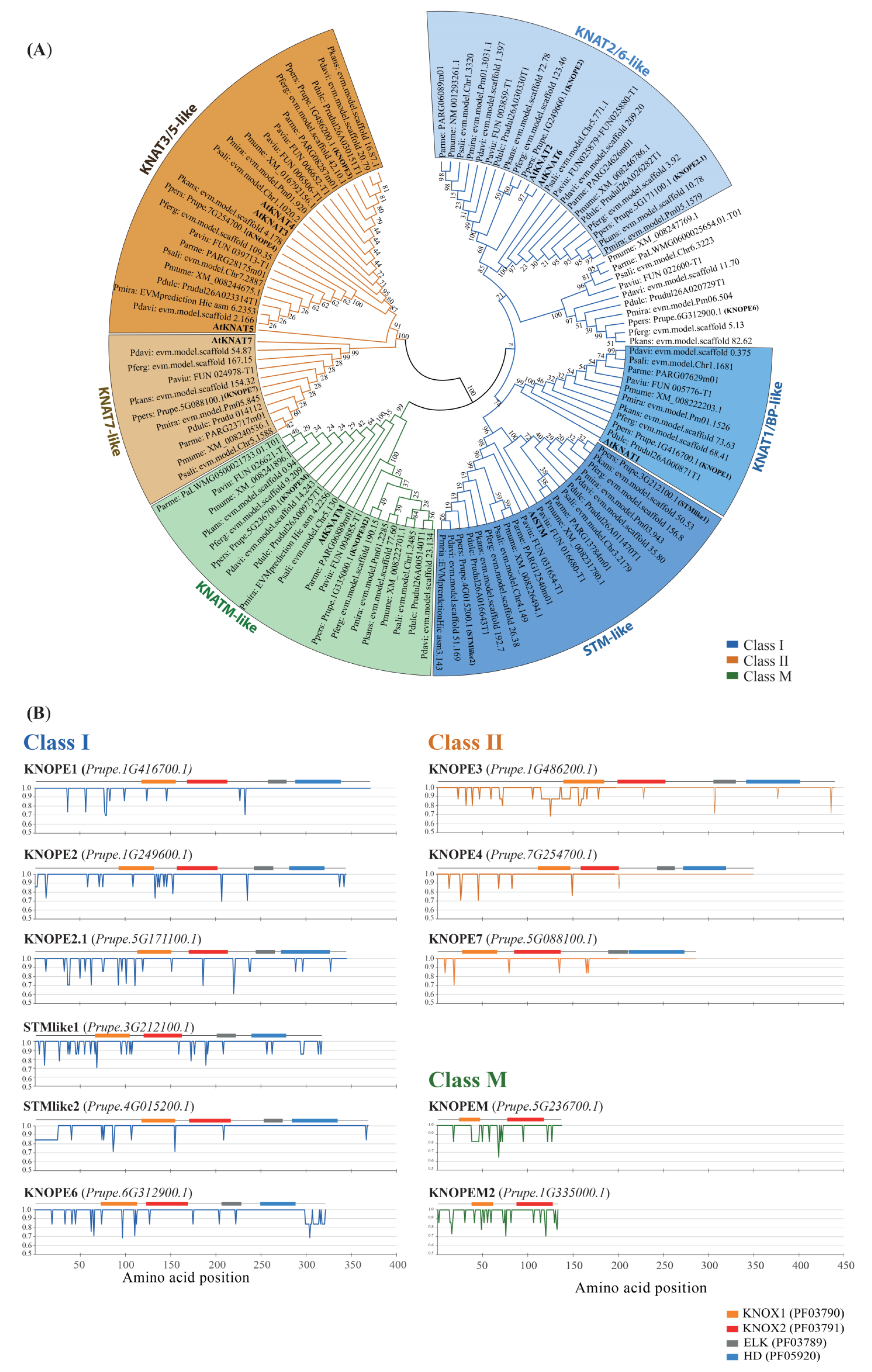
Figure 1. (A) Phylogenetic tree of PRUNOX deduced proteins, clustered into classes I, II, and M (blue, orange, and green shades) and named based on the presence of A. thaliana (bold) members in the clade. (B) Plots of amino acid variability within KNOX subgroups. The peach proteins were schemed (top of each panel) together with key domains (colored boxes) and used as reference for KNOX of other species. Identity variation (percent) of amino acids within Prunus spp. proteins (Y-axis) refers to amino acid positions (X-axis). Orange, KNOX1 (PF03790); red, KNOX2 (PF03791); grey, ELK (PF03789); blue, homeodomain (PF05920).
Ijms 24 03046 g001
Figure 2. Comparative schematic maps of KNOX genome distribution occurring in species of the Armeniaca section (e.g., P. mume, Pm) vs. that in other Prunus spp. (e.g., P. persica, Pp). The gene KNOPE2.1 is located in the unplaced scaffold1397 (Scf1397) in P. mume but in Chr 7 in P. armeniaca. The scale on the left represents chromosome lengths in megabases (Mb).
Ijms 24 03046 g002
Figure 3. Exon–intron structure and conserved motifs in PRUNOX genes. The organization of each class was compared to A. thaliana counterparts (grey-shaded). Boxes, coding sequences; gray lines, introns. KNOX conserved motifs are colored.
Ijms 24 03046 g003
Figure 4. Cytokinin binding motifs residing on PRUNOX genes’ promoters of P. persica and P. dulcis (left and right panels). The score accounts (covering 1.5 kb region before the start codon in Table S4) refer to the experimentally determined extended (ECRM; blue) and core (CRM; light blue) motifs [38], as well as octameric sequences (yellow/orange/brown) enriched in CK-responsive promoters [39].
Ijms 24 03046 g004
Figure 5. Prunus genome-wide transcription analysis of PRUNOX genes active in aerial organs. The expression profiles refer to 10 species through heat maps of the z-scores, where orange and blue indicate higher and lower expression, respectively. fl., flower.
Ijms 24 03046 g005
Figure 6. Effects of BA supply on GF677 microcuttings (A,F,K). Explants of the GF677 rootstock were grown on media without BA (AE) or containing BA at concentrations of 1.7 (FJ) and 5.1 µM (KO). Ten days post-treatment, the effects of BA dosage in increasing the numbers of side shoots and leaves (compare (F,K) vs. (A)) were visible. (BE,GJ,LO) Histological analyses of longitudinal sections at early stages (3 days after treatment). Median section through the dome of control (B) and BA-treated apices (G,L). Leaf axillary buds showed a plethora of phenotypes, including normal (C,D,H,M), elongating (I), and swollen (O). Section of stems portions adjacent to medium (E,J). Bar sizes: 0.5 cm in (A,F,K); 150 µm in (D,I,LN); 160 µm in (B,G,H,J,L,O); 170 µm in (C).
Ijms 24 03046 g006
Figure 7. Gene expression analyses of KNOPE and marker genes under different BA dosages. Expression levels of stress- (A,B) and CK-responsive (C,D) markers and of KNOPE (E,H) and BELL (I,J) genes in response to 1.7 µM (A,C,E,G,I) and 5.1 µM (B,D,F,H,J) of BA at 24 and 72 h post-treatment (hpt). Each value represents the mean ± standard error of three replicates. For each gene, different letters mean significant differences (p ≤ 0.05).
Ijms 24 03046 g007
Figure 8. Correlation plot between TALE and marker gene expression levels. Correlogram representing Pearson’s correlation coefficients (r) between TALE (PRUNOXI: STMlike1, STMlike2, KNOPE1, KNOPE2, KNOPE2.1, KNOPE6; PRUNOXII: KNOPE3, KNOPE4, KNOPE7; BELL: PpBEL1, PpBLH1, PpBLH2, PpBLH3, PpBLH5, PpBLH6, PpBLH8) and marker (stress-responsive: PpCAT1, PpCAT2; CK-responsive: PpCKX6, PpARR12, PpHK1) expression levels. Heat map is used to indicate the strength of correlation between the variables with ordering determined by hierarchical clustering. Red and blue indicate negative and positive correlations, respectively. Only significant (p ≤ 0.05) Pearson’s coefficients were reported in the colored squares. *, **, and *** = significant at p ≤ 0.05, 0.01, and 0.001, respectively.
Ijms 24 03046 g008
Figure 9. Genetic transformation of Gisela 6. (AJ) Examples of GFP fluorescence analysis in leaves and roots of Gisela 6 transgenic clones. (AD) P1 clone. (EH) P3 clone. Analysis of leaf margins (A,C,E,G) and lateral roots (B,D,F,H) under visible microscopy (A,B,E,F) and fluorescence (C,D,G,H). (KQ) 35S:KNOPE1 phenotypes in Gisela 6. Regenerated clones (K,N) and details of leaf margins (LO) and lamina vasculature (M,P) in nontransformed (KM) and 35S:KNOPE1 (NP) lines. (Q) 35S:KNOPE1 clone that reverted phenotype with time. (R) Vector schemes of pCAMBIA1302 (above) and pBA002 + KNOPE1 (below); dark grey bars, probes used in Southern blots which are reported in Figure S3; arrowheads, primers used to check for transgene integrity (black) and expression (red). (S) Upper panel, a check for p35S:KNOPE1:NOSt cassette integrity by PCR with gDNA. Six clones were rescued and analyzed; on the left, size of bands of DNA ladder in base pairs (bp); on the right, the amplicon size is specified. Mid panel, a check for KNOPE1 transgene expression in the six clones by RT-PCR using leaf blade RNA. Peach (Chiripa) KNOPE1-specific primers fell between the 5’UTR and the first exon (R). The constitutive RPII expression was assayed to check for correct retrotranscription and usage of equal cDNA amounts. The amplicon sizes are reported.
Ijms 24 03046 g009
Table 1. Summary of KNOX gene members across ten diploid Prunus spp. 1.
GenusPrunusArabidopsis
SubgenusPrunusCerasus-
SectionPersicaeArmeniacaAmygdalusPrunus--
Species 2:P. davP. fergP. kanP. mirP. perP. armP. mumP. dulP. salP. aviA. thaliana
Size (Mb):237237238243265240280240308338135
Class I
STM-like22222222221
KNAT1-like11111111111
KNAT2/6-like22222222222
extra group11111111110
subtotal66666666664
Class II
KNAT3/4/5-like22222222233
KNAT7-like11111111111
subtotal33333333344
Class M
KNATM-like01111111011
KNATM2-like21111111210
subtotal22222222221
Total KNOX111111111111111111129
1 Prunus phylogeny and classification as proposed by Zhang et al. [37]. 2 Species: Prunus davidiana; P. ferganensis; P. kansuensis; P. mira; P. persica; P. armeniaca; P. mume; P. dulcis; P. salicina; P. avium; Arabidopsis thaliana.
Table 2. Phenotypical traits of GF677 rootstock microcuttings grown on media supplied with different BA dosages.
BA (µM)Stem Height (cm)Leaves on Main AxisLateral Shoots
0.00.9 ± 0.212.8 ± 1.5 b0.0 ± 0.0 c
1.71.2 ± 0.317.2 ± 3.4 a2.3 ± 0.1 b
5.11.1 ± 0.220.3 ± 2.8 a2.9 ± 0.2 a
Significancen.s.*****
Different letters mean significant differences; ** p ≤ 0.01; *** p ≤ 0.001; n.s., not significant.
Table 3. Significance overview from ANOVA results relative to the gene expression levels in GF677 clones as affected by cytokinin dosage, time, and interaction.
GroupGeneDoseTimeDxT
Stress-responsivePpCAT1**n.s.n.s.
PpCAT2***n.s.*
CK-responsivePpCKX6******n.s.
PpARR12n.s.n.s.***
PpHK1n.s.n.s.***
Class I KNOXSTMlike1n.s.**n.s.
STMlike2*********
KNOPE1*********
KNOPE2n.s.***
KNOPE2.1******
KNOPE6n.s.n.s.*
Class II KNOXKNOPE3n.s.n.s.n.s.
KNOPE4n.s.n.s.n.s.
KNOPE7n.s.***n.s.
BELLPpBEL1*******
PpBLH1**n.s.*
PpBLH2n.s.****
PpBLH3n.s.n.s.n.s.
PpBLH5*******
PpBLH6****n.s.
PpBLH8n.s.***n.s.
n.s., not significant; * p ≤ 0.05; ** p ≤ 0.01; *** p ≤ 0.001.
Disclaimer/Publisher’s Note: The statements, opinions and data contained in all publications are solely those of the individual author(s) and contributor(s) and not of MDPI and/or the editor(s). MDPI and/or the editor(s) disclaim responsibility for any injury to people or property resulting from any ideas, methods, instructions or products referred to in the content.