Article

Novel Variants of SOX4 in Patients with Intellectual Disability

1
Institute of Human Genetics, University Hospital Essen, University of Duisburg-Essen, 47057 Duisburg, Germany
2
Northern Ireland Regional Genetics Service, Belfast City Hospital, Belfast BT9 7AB, UK
3
Limbach Genetics, 28209 Bremen, Germany
4
Medical Genetics and Applied Genomics, University of Tuebingen, 72076 Tuebingen, Germany
5
Department of Pediatrics, Faculty of Medicine, Ruhr University of Bochum, 44791 Bochum, Germany
6
Center for Rare Disease Essen (Essener Zentrum für Seltene Erkrankungen—EZSE), Universitätsmedizin Essen, 45147 Essen, Germany
*
Author to whom correspondence should be addressed.
Int. J. Mol. Sci. 2023, 24(4), 3519; https://doi.org/10.3390/ijms24043519
Submission received: 17 January 2023 / Revised: 2 February 2023 / Accepted: 3 February 2023 / Published: 9 February 2023
(This article belongs to the Special Issue Rare Diseases—Molecular Mechanisms and Therapeutic Strategies (IV))

Abstract

:
SOX4 is a transcription factor with pleiotropic functions required for different developmental processes, such as corticogenesis. As with all SOX proteins, it contains a conserved high mobility group (HMG) and exerts its function via interaction with other transcription factors, such as POU3F2. Recently, pathogenic SOX4 variants have been identified in several patients who had clinical features overlapping with Coffin–Siris syndrome. In this study, we identified three novel variants in unrelated patients with intellectual disability, two of which were de novo (c.79G>T, p.Glu27*; c.182G>A p.Arg61Gln) and one inherited (c.355C>T, p.His119Tyr). All three variants affected the HMG box and were suspected to influence SOX4 function. We investigated the effects of these variants on transcriptional activation by co-expressing either wildtype (wt) or mutant SOX4 with its co-activator POU3F2 and measuring their activity in reporter assays. All variants abolished SOX4 activity. While our experiments provide further support for the pathogenicity of SOX4 loss-of-function (LOF) variants as a cause of syndromic intellectual disability (ID), our results also indicate incomplete penetrance associated with one variant. These findings will improve classification of novel, putatively pathogenic SOX4 variants.

1. Introduction

The SOX family comprises 20 members clustered in eight groups named A to H [1]. SOX proteins have important roles in regulating the development of progenitor cells but also in determining genetic programs in differentiated tissues [2]. SOX proteins mainly function through interactions with a binding partner or by forming heterodimers with other SOX proteins or other transcription factors, or they act as homodimers [2]. For SOX4, syntenin and different POU proteins have been described as possible partners, depending on the target region [3,4,5]. Genetic alterations in SOX genes lead to severe congenital disorders covering a broad spectrum of developmental diseases. So far, half of the SOX genes have been associated with genetic disorders [6]. SOX4 (OMIM: *184430) is a transcription factor that, together with SOX11 and SOX12, constitutes the SOXC group. They share a common domain called the HMG box that shows nearly 100% identity within this subgroup and about 50% identity with the rest of the SOX family. SOXC proteins have pleiotropic functions and act in concert with each other, as well as in redundancy [5]. SOX4 is mainly expressed in T and pre-B lymphocytes and in the developing brain. Among other developmental processes, it regulates the inside-out pattern of cortical layer formation via the regulation of RELN expression in a redundant manner with SOX11 [7,8,9]. Pathogenic missense variants of SOX genes are mainly clustered within the HMG box, while truncating variants are scattered throughout the gene. Only a few variants “outside” of the HMG domain have been consistently associated with disease so far [6]. Regarding the SOXC group, pathogenic variants of SOX12 have not been reported yet, while variants of SOX11 cause a syndrome clinically overlapping with Coffin–Siris syndrome [10]. In addition, de novo variants of SOX4 have been described as causative for Coffin–Siris syndrome 10 (OMIM: #618506) and, very recently, even more pathogenic variants have been described in 17 patients [8,11]. In addition to that, a homozygous in-frame deletion of eight amino acids within the interdomain region of SOX4 was reported to be associated with features overlapping with these of previously reported patients [12]. Here, we report the identification and functional investigation of three novel SOX4 variants in patients with developmental delay and intellectual disability.

2. Results

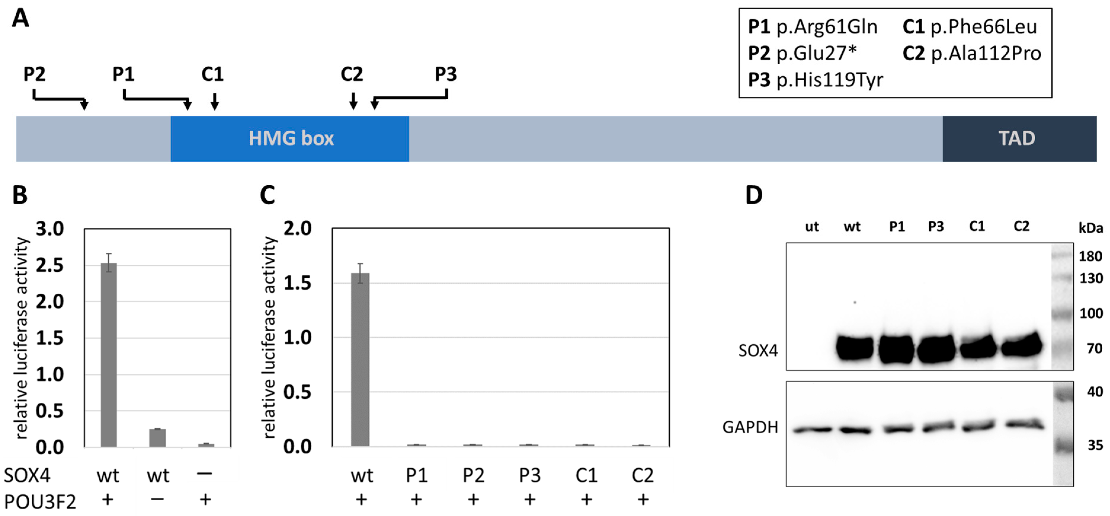
Using whole exome sequencing, we identified three novel variants of SOX4. Two of those were de novo (patient 1: c.182G>A, p.Arg61Gln, Figure 1A–E; and patient 2: c.79C>T, p.Glu27*, Figure 1F–J), while the third (patient 3: c.355C>T, p.His119Tyr) was inherited from an unaffected mother who was clinically re-evaluated. Phenotypic features of all new patients described here match with those described previously in other patients with variants of SOX4 and include intellectual disability, developmental delay, and behavioural concerns. In addition, patient 3 suffered from three to four seizures a week, while patients 1 and 2 did not experience recurrent seizures. Heart problems were reported for patients 1 and 2 but were not observed in patient 3. A detailed clinical description of all three patients is given in the Supplementary Materials, and clinical features are provided in Table S2. While variants identified in patients 1 and 3 affected highly conserved amino acids within the first (H1) and third α-helix (H3) of the HMG box (Figure 2A,B), the variant in patient 2 generated a premature stop codon and was predicted to result in a truncated protein lacking the HMG and transactivation domain. Before analysing the SOX4 variants localized at different positions within the HMG box or its N-terminal region (Figure 3A), synergistic interaction of SOX4 and POU3F2 was tested to assure the functionality of our assay. Single expression of wt SOX4 or POU3F2 did not significantly affect luciferase expression, whereas co-expression strongly activated reporter gene expression (Figure 3B). This synergistic effect was abolished for all three novel variants (p.Arg61Gln, p.Glu27*, p.His119Tyr), as well as for the two previously reported pathogenic variants (p.Phe66Leu, p.Ala112Pro) included as controls (Figure 3C). We investigated the physical interaction of wt and variant SOX4 as Large BiT fusion proteins with the complimentary POU3F2 as a Small BiT fusion protein. Both novel SOX4 variants reduced the protein–protein interactions of SOX4 and POU3F2 dramatically (Figure 4A). Unspecific protein–protein interactions were excluded by testing combinations of SOX4-Large BiT fusion protein and Small BiT peptide alone or POU3F2-Small BiT fusion protein and Large BiT peptide alone, respectively. We excluded protein instability or insufficient expression as causes of the observed effects by using Western blotting, confirming proper expression of the different SOX4 constructs except SOX4 p.Glu27*, which lacks the entire HMG domain and was too small for visualisation using SDS mini-gel (Figure 3D and Figure 4B). The weak bands of the SOX4 constructs shown in Figure 4B were expected and reflected the low-level expression due to the usage of the HSV-TK promoter.

3. Discussions

So far, only a few SOX4 variants have been described as affecting SOX4 function. We contribute to further clarification by reporting three novel SOX4 variants that resulted in a complete loss of transactivation activity in reporter gene assays. In addition, we provide functional data to show the effects of single amino acid substitutions on the formation of the SOX4 complex with its cofactor POU3F2.
SOX4 contains two functionally important domains; namely, an HMG box and a C-terminal transactivation domain (TAD). The HMG box facilitates DNA binding, bending, and nuclear trafficking [7,15], whereas the latter domain mediates the interaction with different cofactors. Truncated proteins missing the C-terminal TAD are not able to interact with their binding partners, resulting in a loss of their transactivation activity [5]. The variant c.79G>T (p.Glu27*) identified in patient 2 resulted in truncated SOX4, lacking both functional domains. Although early-truncating mutations often result in the absence of a gene product because of nonsense-mediated m-RNA decay (NMD), aberrant transcripts of SOX4 are not degraded by NMD because SOX4 is encoded by a single exon [16]. Therefore, the 66 amino acid residues spanning the N-terminus of SOX4 were included in our investigations. It was assumed that SOX4 p.Glu27* would no longer be able to facilitate any of its functions as the HMG domain and the TAD, both known to be crucial, were missing.
The exchange of an arginine at position 61 for glutamine results in a highly conserved residue within HMG domains of different proteins and species [8]. In addition to its location within a predicted bipartite nuclear localization signal [14], previous investigations have provided evidence of its role in DNA binding for Sox4, where it contacts the base pairs T11 and C7 of the Lama1 enhancer region, as well as other HMG box-containing proteins, such as Sox2, where it is involved in binding the FGF4 enhancer [13,17]. In addition to its importance for DNA binding, arginine 61 is also critically involved in the interaction between SOX4 and other proteins, such as GATA-3. In particular, binding of Sox4 to GATA-3 was abolished when Arg61 and Pro62 were both replaced by an alanine [18]. A minor contribution of the HMG domain was also shown for the protein–protein interactions of SOX2 and δEF3, a PAX6 homolog in chickens [19]. We also showed that alterations to amino acids within the HMG domain could prevent SOX4 complex formation with POU3F2. The variant identified here resulted in an exchange of a positively charged arginine for a neutral glutamine residue, which prevented SOX4–POU3F2 interaction in vitro and, additionally, might directly influence DNA binding or nuclear import, all of which affect SOX4 function in transcriptional regulation.
In addition, the second novel missense mutation (p.His119Tyr), which results in the exchange of a highly conserved residue in all SOX proteins of different species, completely abolished SOX4 activity in our reporter assays. For the protein–protein interactions of SOX4 and POU3F2, it was shown that the C-terminal transactivation domain is important, while influence from the HMG domain was not investigated [5]. In contrast to this, it was shown that, for the interaction of Sox2 with its binding partner Pou5f1, the C-terminal region of the HMG domain serves as an interaction platform and that the specific amino acid positions involved differ depending on the target gene. Even though the influence of His101 (corresponding to His119 in SOX4) was not assayed specifically, the surrounding amino acids reduced the ability for Sox2–Pouf51 interaction [17]. However, our results point to the importance of His119 for the interaction of SOX4 and POU3F2, thus indicating the involvement of the corresponding HMG domain region. Interestingly, this variant, identified in patient 3, was also identified in the asymptomatic mother. It is tempting to speculate that this variant might represent the first example of incomplete penetrance for “SOX4-related disorders”, a phenomenon already observed for other disorders caused by variants of SOX genes, such as SOX9-associated campomelic dysplasia and sex reversal [20,21,22], SOX11-associated coloboma [23], and SOX5-associated Lamb–Shaffer syndrome [24]. Whether the mother is mosaic for this variant, as another possible explanation of our observations, could not be absolutely excluded, although Sanger sequencing performed on DNA extracted from peripheral blood did not suggest somatic mosaicism in this tissue.

4. Material and Methods

Three novel variants of SOX4 were identified by whole-exome sequencing. Patients 1 and 3 were collected as part of national collaborations and patient 2 was identified via DECIPHER [25], corresponding to number 307135. Exome sequencing was performed at the respective institutions. Referring physicians provided detailed developmental, neurological, and behavioural histories of the patients. Patient information was anonymised before data sharing. Variants were described according to the SOX4 NM_003107.2 RefSeq transcript.
Wildtype (wt) and mutant (p.Arg61Gln and p.His119Tyr) SOX4 were amplified from control or patient genomic DNA and inserted into the pcDNA3.1(+)/myc-His B expression plasmid. The nonsense variant (p.Glu27*) and two previously reported pathogenic variants (p.Phe66Leu and p.Ala112Pro) [8] used as controls were generated by site-directed in vitro mutagenesis using the Q5 high-fidelity polymerase (Ipswich, MA, USA, New England Biolabs). Sequences of all primers are listed in Table S1. POU3F2 was expressed from pcDNA3.1+/C-(K)DYK-POU3F2 (OHu10724D, Piscataway, NJ, USA, GenScript Biotech Corporation). A modified luciferase-expressing plasmid was generated by inserting three repeats of the FXO-fragment containing binding sites for SOX4 and POU3F2 [5] into pGL4.23 (Madison, WI, USA, Promega Corporation). HEK293 cells were seeded in a 24-well plate to obtain around 70 % confluence at the day of transfection. The co-transfection was performed in triplicate using 125 ng of either wt- or mutant SOX4-expressing plasmid, together with the same amount of POU3F2-expressing plasmid and 250 ng of the luciferase reporter plasmid. One day after transfection, plates were analysed following the protocol for the Dual-Luciferase® Reporter System (Promega Coporation) and signals were detected using a Centro LB 960 luminometer (Bad Wildbad, Germany, Berthold Technologies GmbH & Co. KG). Vectors used for investigation of protein interaction were generated by amplification and insertion of the corresponding wt or mutant fragments from formerly generated expression vectors into modified NanoBiT® (Promega Corporation) expression plasmids containing either FLAG- or myc-tags. HEK293 cells were seeded in a 24-well plate to obtain around 70 % confluence at the day of transfection. A total of 250 ng of either wt or variant SOX4-Large BiT-expressing NanoBiT® vector (pBiT1.1-C-myc) was co-transfected with 250 ng of the complementary POU3F2-Small-BiT-expressing NanoBiT® vector (pBiT2.1-N-FLAG). Six hours after transfection, each well was split into 3 wells of a 96-well plate. One day after transfection, the plates were analysed following the protocol of the NanoBiT® PPI System (Promega Corporation) and signals were detected using a Centro LB 960 luminometer (Berthold Technologies GmbH & Co. KG). All experiments were conducted at least three times and pairwise significance was verified per each experiment independently using Welch’s unpaired t-test. To verify proper expression of SOX4 constructs, HEK293 cells were seeded in a six-well plate to obtain a confluence of 70 % at the day of transfection. A total of 2 µg of the corresponding plasmid was transfected and cells were harvested one day after transfection and lysed using RIPA buffer. The cell lysates were separated using a 10 % polyacrylamide gel, and SOX4 variants and GAPDH were detected using an antibody against myc-tag (Danvers, MA, USA, Cell Signaling Technology, #2272) or GAPDH (Cell Signaling Technology, #2118), respectively.
Sequence comparison of the SOX4 HMG boxes of different species was performed using the Clustal Omega alignment tool available at the UniProt website [26]. A model of the SOX4 HMG box interacting with DNA was generated with Swiss-Model utilizing the 3u2b.1 template [13,27].

5. Conclusions

In conclusion, we report three novel pathogenic variants of SOX4, all abolishing SOX4 function as a transcription factor in our functional assays. We extended our analyses by including additional known disease-causing variants and further affirm the necessity of in vitro analyses to evaluate the functional relevance of novel SOX4 variants identified in patients with Coffin–Siris spectrum disorders. By providing new insights into the functional relevance of disease-causing SOX4 variants, we address the importance of better characterization and classification of the rising numbers of variants obtained from NGS analyses.
Additionally, we obtained the first indications that incomplete penetrance of a novel SOX4 variant, affecting a highly conserved residue within the HMG domain, may be a genetic cause of neurodevelopmental delay, further highlighting the need for functional investigations to classify variants of unknown physiological significance identified by whole-exome or whole-genome sequencing analyses.

Supplementary Materials

The following supporting information can be downloaded at: https://www.mdpi.com/article/10.3390/ijms24043519/s1.

Author Contributions

Conceptualization, M.G. and F.J.K.; methodology, M.G.; validation, M.G.; formal analysis, M.G.; investigation, M.G.; resources, A.K., T.D., S.S., S.B.-W., M.B., T.B.H., C.G., E.M. and C.D.; data curation, M.G. and C.D., writing—original draft preparation, M.G., A.K., C.D. and F.J.K., writing—review and editing, M.G., A.K., C.D. and F.J.K., visualization, M.G.; supervision, M.G., C.D. and F.J.K.; project administration, M.G., C.D. and F.J.K. All authors have read and agreed to the published version of the manuscript.

Funding

This research received no external funding.

Institutional Review Board Statement

The study was conducted according to the guidelines of the Declaration of Helsinki.

Informed Consent Statement

Informed consent was obtained from all subjects involved in the study.

Data Availability Statement

The data presented in this study are available in the Supplementary Material.

Acknowledgments

The authors are grateful to the individuals and their families for generously donating samples and clinical information. The DDD study presents independent research commissioned by the Health Innovation Challenge Fund (grant number HICF-1009-003), a parallel funding partnership between Wellcome and the Department of Health, and the Wellcome Sanger Institute (grant number WT098051). The views expressed in this publication are those of the author(s) and not necessarily those of Wellcome or the Department of Health. The study has UK Research Ethics Committee approval (10/H0305/83, granted by the Cambridge South REC, and GEN/284/12, granted by the Republic of Ireland REC). The research team acknowledges the support of the National Institute for Health Research through the Comprehensive Clinical Research Network. This work was generated within the European Reference Network on Rare Congenital Malformations and Rare Intellectual Disability (ERN-ITHACA) (AK, FJK). This work was generated within the TRANSLATE-NAMSE project. We acknowledge support from the Open Access Publication Fund of the University of Duisburg-Essen.

Conflicts of Interest

The authors declare no conflict of interest.

References

  1. Bowles, J.; Schepers, G.; Koopman, P. Phylogeny of the SOX family of developmental transcription factors based on sequence and structural indicators. Dev. Biol. 2000, 227, 239–255. [Google Scholar] [CrossRef] [PubMed]
  2. Kamachi, Y.; Kondoh, H. Sox proteins: Regulators of cell fate specification and differentiation. Development 2013, 140, 4129–4144. [Google Scholar] [CrossRef]
  3. Geijsen, N.; Uings, I.J.; Pals, C.; Armstrong, J.; McKinnon, M.; Raaijmakers, J.A.; Lammers, J.-W.J.; Koenderman, L.; Coffer, P.J. Cytokine-specific transcriptional regulation through an IL-5Rα interacting protein. Science 2001, 293, 1136–1138. [Google Scholar] [CrossRef] [PubMed]
  4. Kuhlbrodt, K.; Herbarth, B.; Sock, E.; Enderich, J.; Hermans-Borgmeyer, I.; Wegner, M. Cooperative function of POU proteins and SOX proteins in glial cells. J. Biol. Chem. 1998, 273, 16050–16057. [Google Scholar] [CrossRef] [PubMed]
  5. Dy, P.; Penzo-Mendez, A.; Wang, H.; Pedraza, C.E.; Macklin, W.B.; Lefebvre, V. The three SoxC proteins—Sox4, Sox11 and Sox12—Exhibit overlapping expression patterns and molecular properties. Nucleic Acids Res. 2008, 36, 3101–3117. [Google Scholar] [CrossRef] [PubMed]
  6. Angelozzi, M.; Lefebvre, V. SOXopathies: Growing family of developmental disorders due to SOX mutations. Trends Genet. 2019, 35, 658–671. [Google Scholar] [CrossRef] [PubMed]
  7. van de Wetering, M.; Oosterwegel, M.; van Norren, K.; Clevers, H. Sox-4, an Sry-like HMG box protein, is a transcriptional activator in lymphocytes. EMBO J. 1993, 12, 3847–3854. [Google Scholar] [CrossRef]
  8. Zawerton, A.; Yao, B.; Yeager, J.P.; Pippucci, T.; Haseeb, A.; Smith, J.D.; Wischmann, L.; Kühl, S.J.; Dean, J.C.; Pilz, D.T. De novo SOX4 variants cause a neurodevelopmental disease associated with mild dysmorphism. Am. J. Hum. Genet. 2019, 104, 246–259. [Google Scholar] [CrossRef]
  9. Shim, S.; Kwan, K.Y.; Li, M.; Lefebvre, V.; Šestan, N. Cis-regulatory control of corticospinal system development and evolution. Nature 2012, 486, 74–79. [Google Scholar] [CrossRef] [PubMed]
  10. Tsurusaki, Y.; Koshimizu, E.; Ohashi, H.; Phadke, S.; Kou, I.; Shiina, M.; Suzuki, T.; Okamoto, N.; Imamura, S.; Yamashita, M. De novo SOX11 mutations cause Coffin–Siris syndrome. Nat. Commun. 2014, 5, 4011. [Google Scholar] [CrossRef] [Green Version]
  11. Angelozzi, M.; Karvande, A.; Molin, A.N.; Ritter, A.L.; Leonard, J.M.M.; Savatt, J.M.; Douglass, K.; Myers, S.M.; Grippa, M.; Tolchin, D.; et al. Consolidation of the clinical and genetic definition of a SOX4-related neurodevelopmental syndrome. J. Med. Genet. 2022. [Google Scholar] [CrossRef] [PubMed]
  12. Ghaffar, A.; Rasheed, F.; Rashid, M.; van Bokhoven, H.; Ahmed, Z.M.; Riazuddin, S.; Riazuddin, S. Biallelic in-frame deletion of SOX4 is associated with developmental delay, hypotonia and intellectual disability. Eur. J. Hum. Genet. 2022, 30, 243–247. [Google Scholar] [CrossRef] [PubMed]
  13. Jauch, R.; Ng, C.K.; Narasimhan, K.; Kolatkar, P.R. The crystal structure of the Sox4 HMG domain–DNA complex suggests a mechanism for positional interdependence in DNA recognition. Biochem. J. 2012, 443, 39–47. [Google Scholar] [CrossRef]
  14. Malki, S.; Boizet-Bonhoure, B.; Poulat, F. Shuttling of SOX proteins. Int. J. Biochem. Cell. Biol. 2010, 42, 411–416. [Google Scholar] [CrossRef] [PubMed]
  15. Hanover, J.A.; Love, D.C.; Prinz, W.A. Calmodulin-driven nuclear entry: Trigger for sex determination and terminal differentiation. J. Biol. Chem. 2009, 284, 12593–12597. [Google Scholar] [CrossRef]
  16. Wilke, C.O. Transcriptional robustness complements nonsense-mediated decay in humans. PLoS Genet. 2011, 7, e1002296. [Google Scholar] [CrossRef] [PubMed]
  17. Reményi, A.; Lins, K.; Nissen, L.J.; Reinbold, R.; Schöler, H.R.; Wilmanns, M. Crystal structure of a POU/HMG/DNA ternary complex suggests differential assembly of Oct4 and Sox2 on two enhancers. Genes Dev. 2003, 17, 2048–2059. [Google Scholar] [CrossRef]
  18. Kuwahara, M.; Yamashita, M.; Shinoda, K.; Tofukuji, S.; Onodera, A.; Shinnakasu, R.; Motohashi, S.; Hosokawa, H.; Tumes, D.; Iwamura, C. The transcription factor Sox4 is a downstream target of signaling by the cytokine TGF-β and suppresses TH 2 differentiation. Nat. Immunol. 2012, 13, 778–786. [Google Scholar] [CrossRef]
  19. Kamachi, Y.; Cheah, K.S.; Kondoh, H. Mechanism of regulatory target selection by the SOX high-mobility-group domain proteins as revealed by comparison of SOX1/2/3 and SOX9. Mol. Cell. Biol. 1999, 19, 107–120. [Google Scholar] [CrossRef]
  20. Cameron, F.J.; Hageman, R.M.; Cooke-Yarborough, C.; Kwok, C.; Goodwin, L.L.; Sillence, D.O.; Sinclair, A.H. A novel germ line mutation in SOX9 causes familial campomelic dysplasia and sex reversal. Hum. Mol. Genet. 1996, 5, 1625–1630. [Google Scholar] [CrossRef] [Green Version]
  21. Benko, S.; Gordon, C.T.; Mallet, D.; Sreenivasan, R.; Thauvin-Robinet, C.; Brendehaug, A.; Thomas, S.; Bruland, O.; David, M.; Nicolino, M. Disruption of a long distance regulatory region upstream of SOX9 in isolated disorders of sex development. J. Med. Genet. 2011, 48, 825–830. [Google Scholar] [CrossRef] [PubMed]
  22. Kwok, C.; Weller, P.A.; Guioli, S.; Foster, J.W.; Mansour, S.; Zuffardi, O.; Punnett, H.H.; Dominguez-Steglich, M.A.; Brook, J.D.; Young, I.D. Mutations in SOX9, the gene responsible for Campomelic dysplasia and autosomal sex reversal. Am. J. Hum. Genet. 1995, 57, 1028. [Google Scholar] [PubMed]
  23. Pillai-Kastoori, L.; Wen, W.; Wilson, S.G.; Strachan, E.; Lo-Castro, A.; Fichera, M.; Musumeci, S.A.; Lehmann, O.J.; Morris, A.C. Sox11 is required to maintain proper levels of Hedgehog signaling during vertebrate ocular morphogenesis. PLoS Genet. 2014, 10, e1004491. [Google Scholar] [CrossRef] [PubMed]
  24. Zawerton, A.; Mignot, C.; Sigafoos, A.; Blackburn, P.R.; Haseeb, A.; McWalter, K.; Ichikawa, S.; Nava, C.; Keren, B.; Charles, P. Widening of the genetic and clinical spectrum of Lamb–Shaffer syndrome, a neurodevelopmental disorder due to SOX5 haploinsufficiency. Genet. Med. 2020, 22, 524–537. [Google Scholar] [CrossRef]
  25. Firth, H.V.; Richards, S.M.; Bevan, A.P.; Clayton, S.; Corpas, M.; Rajan, D.; Van Vooren, S.; Moreau, Y.; Pettett, R.M.; Carter, N.P. DECIPHER: Database of chromosomal imbalance and phenotype in humans using ensembl resources. Am. J. Hum. Genet. 2009, 84, 524–533. [Google Scholar] [CrossRef] [PubMed]
  26. UniProt, C. UniProt: The universal protein knowledgebase in 2021. Nucleic Acids Res. 2021, 49, D480–D489. [Google Scholar] [CrossRef]
  27. Bienert, S.; Waterhouse, A.; de Beer, T.A.; Tauriello, G.; Studer, G.; Bordoli, L.; Schwede, T. The SWISS-MODEL Repository-new features and functionality. Nucleic Acids Res. 2017, 45, D313–D319. [Google Scholar] [CrossRef] [Green Version]
Figure 1. Photographs of patients carrying SOX4 variants. (AE) Patient 1 (c.182G>A; p.Arg61Gln). Her facial features are characterized by bilateral epicanthal folds, a small face, and a broad philtrum. Limb abnormalities include clinodactyly of the fifth finger and small hands and feet. (FJ) Patient 2 (c.79G>T; p.Glu27*). His facial features are characterized by hypertelorism, epicanthal folds, a smooth philtrum, a thin upper lip, low-set cup-shaped ears, and a prominent forehead. Limb abnormalities include bilateral clinodactyly and hypoplasia of the second and fifth digits.
Ijms 24 03519 g001
Figure 2. In silico analysis of SOX4 missense variant localization. (A) Sequence alignment of the SOX4 HMG-domain of different species. Previously reported missense variants [8,11] and new missense variants from patients 1 and 3 (bold) are indicated. Variants investigated in the luciferase reporter assay are underlined. Sequences reflecting the α-helices and nuclear localization sites are underlined and indicated by H1–H3 and NLS, respectively [13,14]. (B) Model of the SOX4 HMG-domain forming a complex with DNA based on template 3u2b [13]. Amino acid residues affected in patients are highlighted. New variants from patients 1 and 3 are shown in bold and α-helices (H) of the HMG-domain are indicated by H1, H2, and H3, respectively.
Ijms 24 03519 g002
Figure 3. Localization and function of analysed SOX4 variants. (A) SOX4 protein structure. Arrows indicate localization of investigated variants. The high mobility group (HMG) box and the transactivation domain (TAD) are indicated. C1 and C2 served as negative controls [8]. The figure is not drawn to scale. (B) Activation of luciferase gene expression depends on the interaction of SOX4 and POU3F2. The presence and absence of either SOX4 or POU3F2 are indicated by + and −, respectively. The significance of pairwise differences between co-transfection of SOX4 and POU3F2 constructs and single transfection of either construct was verified for each single experiment using Welch’s unpaired t-test, giving a p < 0.02. (C) SOX4-mediated activation was abolished by all SOX4 variants. The presence of POU3F2 is indicated by +. Data shown in (B,C) were normalized for transfection efficiency using constitutive Rluc expression (pRL-TK). Experiments were conducted at least three times independently and the results of one of these experiments are shown. The significance of pairwise differences between the SOX4 wt and either SOX4 variant was verified for each single experiment using Welch’s unpaired t-test, giving a p < 0.02. (D) Western blot showing proper expression of all SOX4 constructs (SOX4Glu27* excluded). ut, untransfected; wt, wildtype.
Ijms 24 03519 g003
Figure 4. Effects of novel SOX4 variants on protein–protein interaction. (A) Novel SOX4 variants reduced SOX4–POU3F2 interaction dramatically. The presence of POU3F2 is indicated by +. Experiments were conducted three times independently and the results of one of these experiments are shown. The significance of pairwise differences between SOX4 wt and either SOX4 variant was verified for each single experiment using Welch’s unpaired t-test, giving a p < 0.02. (B) Western blot showing the proper expression of the transfected SOX4 constructs. AU, arbitrary unit; wt, wildtype; P1, p.Arg61Gln; P3, p.His119Tyr; ut, untransfected.
Ijms 24 03519 g004
Disclaimer/Publisher’s Note: The statements, opinions and data contained in all publications are solely those of the individual author(s) and contributor(s) and not of MDPI and/or the editor(s). MDPI and/or the editor(s) disclaim responsibility for any injury to people or property resulting from any ideas, methods, instructions or products referred to in the content.