Review

Circular RNAs: Biogenesis, Biological Functions, and Roles in Myocardial Infarction

Key Laboratory of Cellular Physiology at Shanxi Medical University, Ministry of Education, Key Laboratory of Cellular Physiology of Shanxi Province, Department of Physiology, Shanxi Medical University, Taiyuan 030001, China
*
Authors to whom correspondence should be addressed.
These authors contributed equally to this work.
Int. J. Mol. Sci. 2023, 24(4), 4233; https://doi.org/10.3390/ijms24044233
Submission received: 28 November 2022 / Revised: 8 February 2023 / Accepted: 14 February 2023 / Published: 20 February 2023
(This article belongs to the Special Issue Non-coding RNAs in Pathogens and Associated Diseases)

Abstract

:
Non-coding RNAs have been excavated as important cardiac function modulators and linked to heart diseases. Significant advances have been obtained in illuminating the effects of microRNAs and long non-coding RNAs. Nevertheless, the characteristics of circular RNAs are rarely mined. Circular RNAs (circRNAs) are widely believed to participate in cardiac pathologic processes, especially in myocardial infarction. In this review, we round up the biogenesis of circRNAs, briefly describe their biological functions, and summarize the latest literature on multifarious circRNAs related to new therapies and biomarkers for myocardial infarction.

1. Introduction

Myocardial infarction (MI) is an acute cardiovascular illness with an extremely high case fatality rate. Although there are existing clinical approaches, such as pharmacotherapy, coronary intervention reperfusion therapy, and stem cell transplantation [1,2], myocardial necrosis and remodeling after MI cause enormous damage to cardiac function, and there remains a considerable number of patients with undesirable outcomes [3].
MI is mainly caused by reduced coronary blood flow, which causes the damage and remodeling of the heart tissue. The critical pathogenesis is endothelial dysfunction induced by diverse etiologies, such as poor atherosclerotic plaque stability and various pathophysiological modifications such as atherosclerotic plaque rupture and erosion [4]. These lead to platelet aggregation and excitation, the excitation of endogenous and exogenous body coagulation mechanisms, the activation of a transition from fibrinogen to fibrin, and a combined range of actions, such as blood corpuscles [5]. The formation of thrombosis leads to the complete or partial obstruction of the coronary arteries in diseases and decreased myocardial perfusion, thus leading to MI [6]. The presence of thrombosis in an epicardial coronary artery, which supplies the myocardium, reduces or stops the blood flow in part of the heart, reduces myocardial perfusion, and leads to MI due to the apoptosis and necrosis of myocardial cells. After the onset of MI, the heart will also undergo a series of compensatory reactions, such as cardiac hypertrophy and fibrosis, which further lead to cardiac remodeling. In addition, the sustained excitability of the sympathetic nervous system (SNS) and the renin-angiotensin-aldosterone system (RAAS) after MI further worsens cardiac function.
In recent years, significant advances have been made in the study of microRNAs and long non-coding RNAs, while the serviceable impacts of circular RNAs have rarely been researched. Nevertheless, an increasing wealth of evidence supports the potential of the underlying mechanisms of circRNAs as new therapeutic targets and biomarkers for cardiovascular diseases, particularly MI. In this review, we summarize the present state of knowledge about circRNAs. We discuss their biological origins, mechanisms of cyclization, and potential as new biomarkers and therapeutic targets for heart disorders, emphasizing MI.

2. Biogenesis and Features of Circular RNAs

In 1976, Sanger et al. [7] indicated that viroids are single-stranded, covalently closed RNA molecules with an unexceptionable self-complementarity and base-pair-making capacity. For the first time, circRNAs were discovered in viruses. Still, due to the limitations of biotechnology and human cognition, circRNAs were deemed to be the by-products of abnormal splicing and were ignored. Soon after, in 1979, Hsu and Coca-Prados at Rockefeller University first detected the presence of circRNAs in human HeLa cells through electron microscopy [8]. In 1980, circRNAs were found in the mitochondrial genome of yeast [9]. In the following period, circRNAs were occasionally found in mammals [10,11]. Specific circRNAs derived from the sex determination region Y (SRY) gene were found and considered to have a possible function in mouse testes [12]. In 2012, Danan et al. [13] found that circRNAs were abundant in archaea and had specific biological functions. The rapid development of RNA sequencing tools has led to the verification of large quantities and varieties of circRNAs. Jeck et al., the authors of [14], identified at least 25,000 circRNAs in human fibroblasts. This suggests that circRNAs are also associated with various functions of the heart. Thus far, researchers have discovered many circRNAs in eukaryotes, and they have determined that these RNAs have tissue-specific, cell-specific, and developmental-stage-specific manifestations [14,15,16,17,18]. These numerous scientific discoveries have rendered circRNAs a new star in non-coding RNA molecules, following microRNAs and long-stranded non-coding RNAs.
CircRNAs are diffusely present in multifarious human cells, peripheral blood, saliva, and other body fluids. In some cases, they are significantly more abundant than the related linear RNAs [19]. In 2021, it was reported that the expression of a cardiac-necroptosis-associated circRNA (CNEACR) was downregulated in myocardial infarction [20], being closely related to the progression of myocardial infarction. Subsequently, in 2022, another study highlighted the selective expression of circSamd4 in fetal and neonatal cardiomyocytes [21]. It also plays a crucial role in myocardial infarction. This is indicative of the tissue specificity of circular RNAs. At the same time, researchers have demonstrated that circRNAs abound in tissues that divide slowly, such as nerve cells or other highly differentiated cells, and are low in tissues that divide rapidly, such as cancer cells [22].
CircRNAs are abundant, stable, widely distributed, and specifically expressed, and so are a potential ideal candidate for non-invasive biomarkers and a regulatory target for MI. The latest study noted that a total of 3,862 differentially expressed circRNAs were identified by the bioinformatics and high-throughput RNA-seq analysis of RNA from the peripheral blood. By calculating and creating the receiver operating characteristic (ROC) curve, the authors noted that circTMEM165, circUBAC2, circZNF609, circANKRD12, and circSLC8A1 have a high sensitivity and specificity in the determination of myocardial infarction [23].

3. Cyclization Form of Circular RNAs

The mechanisms of RNAs are related to pre-mRNA splicing [15,24]. In eukaryotes, pre-mRNAs undergo a process called “canonical splicing”. The main splicing form of “typical splicing” involves the splicing of introns of pre-mRNAs by spliceosomes and ligation of exons into a linear RNA transcriptome, known as mRNA. Parallel with linear mRNAs, pre-mRNAs are also precursors of circRNAs. The pre-mRNAs undergo a process called “back splicing.” “Back splicing” is opposed to “canonical splicing,” which forms linear mRNAs. “Back splicing” is a unique splicing mechanism for forming circRNAs. Based on the “back splicing” approach, circRNAs lack the typical 5′ caps and 3′ poly (A) tail and form a stable, covalent, closed-loop frame. There are three primary cyclization forms of circular RNAs (Figure 1).

3.1. Lariat-Driven Cyclization

Typically, precursor RNA removes the introns during processing, and the introns are degraded by exonuclease after a debranching reaction. However, when pre-RNA has a sequence rich in 7nt guanine (G) and uracil (U) (a Gu-rich sequence) near one exon and an 11nt cytosine (C)-rich sequence (C-rich sequence) near another exon, during the splice reaction that functions to produce the intron noose the introns can avoid debranching and cyclize to form firm circRNAs [25].

3.2. Intron-Pairing-Driven Cyclization

The introns upstream and downstream of exons contain reverse complementary cis-acting elements, such as the ALU sequence, which are paired directly through base complementation, so that the splicing sites are spatially close to each other, forming exon–intron circRNA without intron removal or exon circRNA with intron removal. The diversity of the ALU sequences renders the pairing selective and competitive, generating multiple carcasses in the same gene [24].

3.3. RNA-Binding-Protein (RBP)-Driven Cyclization

The pre-mRNAs can be driven to circRNAs by RNA-binding protein (RBP). RBP can recognize and anchor specific gene sequences in introns and create splicing sites at both ends of exons that are close to each other through protein interaction or dimer formation, forming covalent links between the splicing acceptor and splicing donor, and, finally, cyclization, leading to the formation of cercariae [26].
Based on their molecular mechanisms of biogenesis, circRNAs are usually categorized into three types: (1) intronic circRNAs (ciRNAs), which refer to those circRNAs composed only of introns, which are located in the nucleus; (2) exonic circRNAs (EcircRNAs), which refer to the circRNAs composed only of exons, mainly existing in the cytoplasm; and (3) exon–intron circRNAs (EiciRNAs), which refer to the circRNAs that retain introns between their exons, principally existing in the nucleus.
The parental gene’s location can be used to separate circRNAs into intragenic circRNAs and intergenic circRNAs [27]. Each EcircRNA, EiciRNA, and circRNA molecule is derived from the splicing of different exons and/or introns within the same parent gene and, therefore, belongs to the intra-gene circRNA. In addition, circRNAs derived from genomic intervals between different genes are called inter-gene circRNAs [24].

4. Biological Functions of CircRNAs

4.1. CircRNAs as miRNA Sponges

Recently, the regulatory connection between circRNAs and microRNAs has drawn the attention of many researchers [28]. The interactive regulation of circRNAs and microRNAs is a focus of current research.
The sponging of miRNAs refers to the fact that microRNAs are connected to the 3’ untranslated region of mRNA, thereby inhibiting the translation of mRNA. CircRNAs comprise miRNA response elements (MREs), which are miRNA-integrating sites. The sponge adsorption of miRNAs by circRNAs is mainly accomplished through the MREs. CircRNAs have multiple miRNA-binding sites, and single circRNAs have many good or excellent miRNA-binding sites. Numerous discoveries have shown that circRNAs have the function of a miRNA sponge. As molecular sponges of miRNAs, circRNAs play a biological role in regulating miRNAs. Consistent with the miRNA sponge, the molecular sponge role of circRNAs means that it can bind to miRNA as a binding site of competitive endogenous RNA and restrain the biological viability of miRNA, thus indirectly managing the manifestation of downstream target molecules. The expression level of the corresponding target gene increases when the circRNAs are overexpressed, while the expression level of the corresponding target gene decreases when they are inhibited.

4.2. CircRNAs as Protein Sponges

CircRNAs occupy not only miRNA-binding elements but also bonding sites for protein. Research has indicated that partial circRNAs have protein-bonding sites and serve as protein sponges. CircRNA–protein interactions have been shown to impact protein expression, biogenesis, and pathophysiological progress [29,30]. Most circRNAs are derived from genes that encode proteins, and their frames can include exon sequences. CircRNAs binding to their target proteins will cause the functional suppression of these proteins [31].

4.3. CircRNAs Directly Translate Proteins

Translating a gene into a protein requires a translation initiation complex. The formation of this complex requires the recognition of the 5’ cap structure of mRNAs [32]. Interestingly, studies have shown that circRNAs can be translated into proteins.
Research indicates that circRNAs with internal ribosome entry site (IRES) elements can initiate protein translation. The first circRNA identified as a protein translator was a single-stranded circRNA found in the hepatitis C virus that produces a protein with 122 amino acids [33]. Yang et al. [34] identified several peptides translated by circRNAs in human fibroblasts and found that when adenosine is methylated, the common motif modified by 6-methyladenine upstream of the start codon promotes the translation ability of circRNAs. Other studies have shown that CIRCZNF609 can bind to the polyribosomes in mouse and human myoblast cells and perform protein translation in a splice-dependent and cap-dependent manner [35]. Circ-FBXW7 can be translated into the protein F-Box and WD repeat domain-containing 7 (FBXW7)-185aa, but the roles of this protein remain largely undiscovered [36]. The circRNAs of the virusoid include internal ribosome entry sites and show an emerging coding ability.
Although circRNA is understood to have a specific translation capacity, it has been demonstrated that the translation productivity could be higher owing to the influence of its unique ring structure. The functions of circRNA translation products are still unclear and warrant further investigation.

4.4. CircRNAs Directly Affect Gene Expression

CircRNAs are released into the cytoplasm and extracellularly, giving them the capacity to act as biomarkers. However, to a lesser extent, sectional circRNAs exist in the nucleus. These nuclear circRNAs can respond to RNA polymerase Ⅱ in the promoter region of the host genes and modulate transcription [37].
CircRNAs participate firsthand in the modulation of gene regulation by adjusting the transcription of linear RNA. When pre-mRNA contains the translation initiation site, if cyclization and non-linear splicing occur, the transcribed mRNA will be reduced, reducing the number of downstream proteins for translation. This effect is called the “mRNA trap” [38].

4.5. CircRNAs Regulate mRNA Stability

A few circRNAs can similarly regulate mRNA stability. Hansen et al. [39] found that the circular RNA derived from the cerebellar-degeneration-related protein 1 (CDR1) gene can form a double-stranded structure with CDR1 mRNA to enhance its stability. The Circ-Ras-GEF domain family member 1B (Ras-GEF1B) in murine macrophages enhances the strength of intercellular cell adhesion molecule-1 (ICAM-1) mRNA, causing the increased expression of ICAM-1 in the lipopolysaccharide (LPS)/toll-like receptor 4 (TLR4) inflammatory signaling pathway [40]. CircFndc3b stabilizes mRNA fused in the sarcoma (FUS), because circFndc3b is mainly gathered in the cytoplasm [41].

4.6. CircRNAs Regulate DNA Methylation

The latest controlled mechanism of circRNAs to have been discovered is that circRNA adjusts gene regulation by influencing the DNA methylation of downstream genes. For example, circRNA ACR activates PTEN-induced putative kinase 1 (Pink1) expression by directly binding to DNA Methyltransferase 3 Beta (Dnmt3B) and deterring the Dnmt3B-mediated DNA methylation of the Pink1 promoter [42].

4.7. CircRNAs Act as a Retrotransposon to Mediate Pseudo-Genetic Production

A recent study found that circRNA antilock can form pseudogenes [43]. The exon sequence of pseudogenes derived from linear mRNA is consistent with that of the normal gene. Still, the exon connection sequence of pseudogenes derived from circRNA is opposite to that of the common gene. This indicates that circRNAs can serve as reverse transposons to alter the structure of the genome and adjust gene expression.
In addition to the functions mentioned above, circRNAs are likewise involved in the modulation of transcriptional, post-transcriptional, and translation processes such as RNA processing, genome rearrangement, chromosome modification, and X chromosome silencing [44]. It can be seen that circRNAs are powerful and worthy of further study.

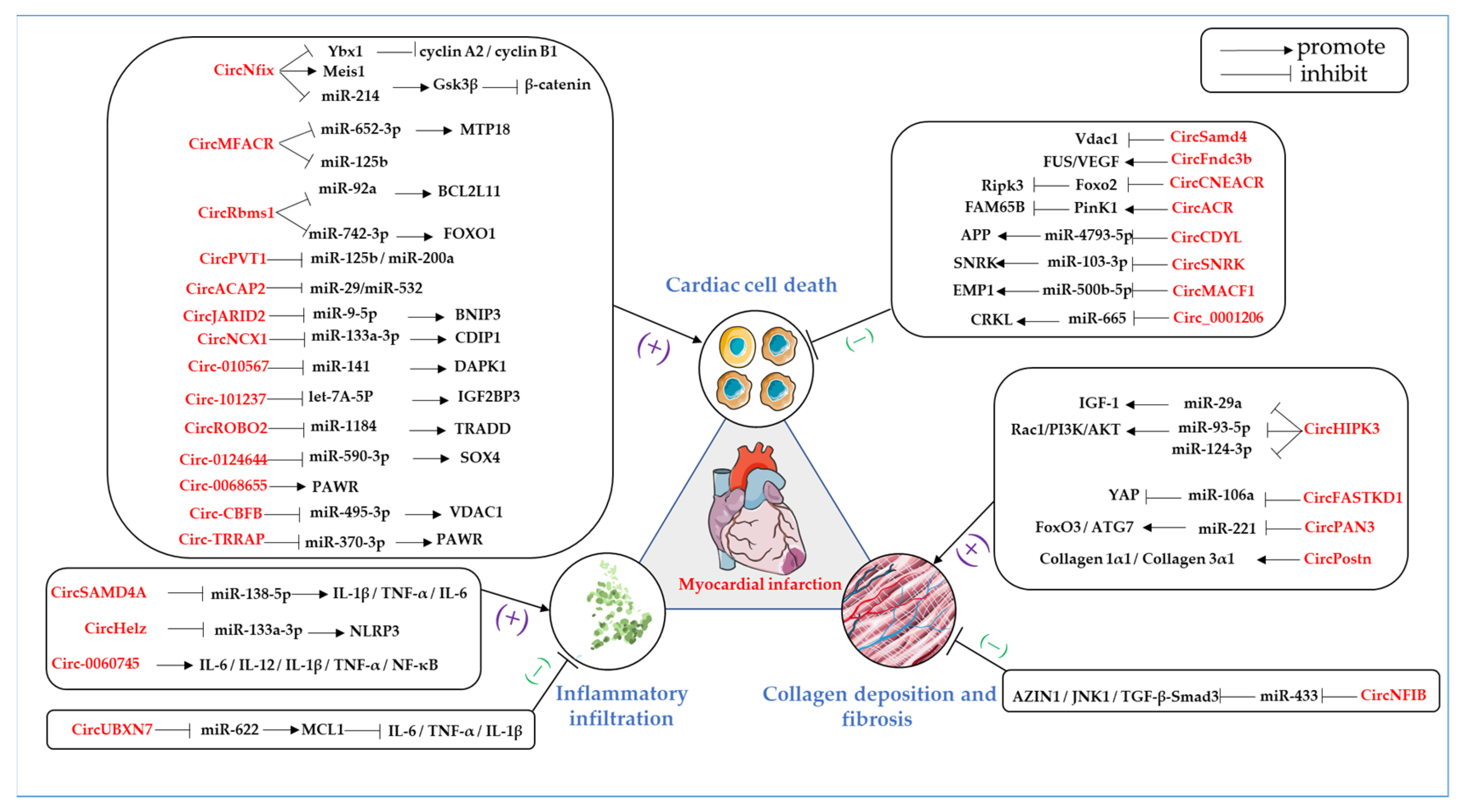
5. Conclusions on the Specific Functions of Circular RNAs in Myocardial Infarction

Circular RNAs are distributed extensively throughout the cardiovascular system and vary dramatically in their expression levels in the context of different cardiac disorders, such as acute MI, angina pectoris, and myocarditis, and function as regulators of heart development, cardiac function, and stress response. Plenty of research has indicated that the critical features of circular RNAs are related to MI [45,46,47,48,49,50,51,52,53,54,55,56,57,58,59,60,61,62,63,64,65,66,67,68,69,70,71,72,73,74,75,76,77]. These circRNAs with altered levels have been found to regulate cardiac cell death after the onset of MI, such as apoptosis, autophagy, necrosis, and inflammatory infiltration, and also regulate myocardial collagen deposition and fibrosis during the healing stage of MI. Here, we seek to describe the circular RNAs that are closely related to MI in terms of the promotion or suppression of the pathology of MI (Table 1 and Figure 2).

5.1. Role of Circular RNAs in Cardiac Cell Death and MI

Since adult mammalian cardiomyocytes are terminally differentiated cells with a very limited proliferative capacity, cardiomyocyte death during MI can result in the permanent loss of the cardiac functional unit. We attempted to summarize the role of circRNAs in promoting MI by regulating apoptosis, autophagy, and necrosis to provide a more comprehensive reference base for circRNAs involved in regulating cardiomyocyte death during MI.

5.1.1. CircRNAs Promote Cardiac Cell Death in MI

  • CircNfix
CircNfix has been demonstrated to be deeply involved in the MI regulation network. Huang et al. [45] conducted the first comprehensive analysis of the relationship between the super-enhancers and circRNA networks in the heart. They identified conserved cardiac circRNAs regulated by the super-enhancer Nfix, namely CircNfix. They found that the downregulation of circNfix can induce cardiomyocyte proliferation, promote angiogenesis, and inhibit cardiac apoptosis. Further research has shown that circNfix promotes myocardial infarction through the following mechanisms. CircNfix strengthens the mutual effects between Y-box-binding protein 1 (Ybx1) and NEDD4-like E3 ubiquitin-protein ligase (Nedd4L), facilitates Ybx1 ubiquitin-dependent degradation, and suppresses the expression of cyclin A2 and cyclin B1. CircNfix inhibits cardiomyocyte proliferation and angiogenesis after MI by promoting Ybx1 degradation mediated by ubiquitination, which promotes cardiomyocyte apoptosis and impairs cardiac function. CircNfix-associated super-enhancers were found to promote circNfix expression by promoting angiogenesis through myeloid Homeobox 1 (Meis1), a prominent transcription factor that controls cardiac cell cycle arrest. Meis1 knockdown significantly represses glycogen synthase kinase 3β (Gsk3β) expression. Gsk3β is well known to inhibit cardiomyocyte proliferation via the degradation of β-catenin. In addition, circNfix may function as a miR-214 sponge to upregulate Gsk3β expression and dampen β-catenin activity. CircNfix impairs cardiomyocyte proliferation and the Meis1/Gsk3β pathways. Interestingly, the expression levels of some circRNAs are inversely regulated by their roles, which appear to be compensatory transformations. A study conducted by Cui et al. [46] showed that circNfix promotes cardiomyocyte apoptosis, but the expression of circNfix exhibits a downward trend in apoptotic cardiomyocytes and ischemic hearts. Whether upregulated or downregulated, circNfix is a pro-apoptotic factor in MI and a potential biomarker and therapeutic target of MI.
  • CircACAP2
CircRNA ACAP2 was found to be increased in rat hearts subjected to MI and can mediate cardiac apoptosis by sponging miR-29, thereby promoting MI [47]. In 2021, it was reported that ArfGAP with coiled-coil, ankyrin repeat, and PH domains 2 (ACAP2) and mature miR-532, rather than premature miR-532, are significantly increased in plasma from patients with MI [48]. In vitro experiments similarly showed that the expression levels of ACAP2 and mature miR-532 are increased in AC16 cells under hypoxia conditions. Nevertheless, ACAP2 and mature miR-532 are significantly and negatively related. The enforced expression of ACAP2 promotes hypoxia-induced apoptosis by interrupting the maturation of miR-532.
  • CircMFACR
When exposed to hypoxia/reoxygenation (H/R) experimental conditions, circRNAs, named mitochondrial-fission-and apoptosis-related circRNA (MFACR), are substantially increased [50]. MFACR promotes MI progression by sponging miR-652-3p and favors the upregulation of the mitochondrial 18 kDa (MTP18) protein, which positively regulates mitochondrial fission and apoptosis in cardiomyocytes [78]. Another study revealed that the expression level of MFACR shows an upward trend in plasma samples from MI patients, MI animal models, and AC16 cells under hypoxia. At the same time, miR-125b is remarkably downregulated [49]. The authors further found that the overexpression of MFACR reduces the miR-125b levels, arising from the increased methylation of the miR-125b gene, thereby exacerbating apoptosis and deteriorating MI. The many recent findings on MFACR mark a new chapter in the study of circRNAs regulating mitochondrial dynamics and cardiomyocyte apoptosis.
  • CircPVT1
The circRNA PVT1 (circPVT1) is highly expressed in MI tissues and H/R-treated cardiomyocytes [51]. CircPVT1 functions as a competing endogenous RNA by competitively combining with both miR-125b and miR-200a. CircPVT1 plays a protective role in myocardium derived from MI and H/R damage by attenuating miR-125b- and miR-200a-mediated apoptotic signaling pathways, including the p53/TNF-receptor-associated factor 6 (TRAF6), Sirtuin 7 (SIRT7), Kelch-like ECH-associated protein 1 (KEAP1)/nuclear factor erythroid-2-related factor 2 (Nrf2), and programmed cell death 4 (PDCD4) pathways.
  • CircNCX1
The circRNA transcribed from the sodium/calcium exchanger 1 (NCX1) gene is circNCX1. CircNCX1 is significantly upregulated and exposed to hypoxia/reoxygenation (H/R) and promotes cardiac apoptosis [52]. Furthermore, CircNCX1 has been demonstrated to bind and inactivate miR-133a-3p, which aggravates ischemic myocardial injury induced by the pro-apoptotic protein cell-death-inducing P53 target 1 (CDIP1). The CircNCX1/miR-133a-3p/CDIP1 axis could provide a potential approach to facilitate treatment decisions for ischemic heart disease.
  • CircJARID2
It has been reported that hypoxia amplifies the upregulation of circJARID2 expression in H9C2 cells. The knockdown of circJARID2 inhibits hypoxia-treated apoptotic cell death by releasing miR-9-5p to downregulate BCL2 interacting protein 3 (BNIP3) expression [53]. In conclusion, circJARID2 promotes hypoxia-induced cardiomyocyte injury during the pathogenesis of MI.
  • CircROBO2
CircROBO2 is markedly upregulated in myocardial samples in response to ischemic damage. CircROBO2 enhances the expression of TNFRSF1A-associated death domain protein (TRADD) by sponging miR-1184, which promotes apoptotic cell death and exacerbates cardiac dysfunction after myocardial infarction [54]. This study revealed a novel regulation model of AMI consisting of CircROBO2, miR-1184, and TRADD.
  • CircRbms1
The CircRbms1 expression levels are upregulated in I/R mice and H2O2-treated H9c2 cells. The upregulated circRbms1 induces cardiomyocyte apoptosis and ROS release via the sponging of miR-92a. The authors showed that after knocking down circRbms1, miR-92a expression was upregulated, and the protein level of BCL2L11 in the heart tissue of the I/R mice was significantly reduced, alleviating cardiomyocyte apoptosis and cardiac function [55]. The circRbms1/miR-92a/BCL2L11 axis may be a potential biomarker for AMI therapy. In addition, other studies have confirmed the results of this work. CircRbms1 is upregulated in MI mouse cardiac tissue and hypoxia-induced cardiomyocytes. The overexpression of circRbms1 further aggravates hypoxia-induced cardiomyocyte damage. Studies have shown that circRbms1, by sponging miR-742-3p, promotes hypoxia-induced cardiomyocyte damage. FOXO1 is a target of miR-742-3p, and its expression is positively regulated by circRbms1 [56]. The CircRbms1/miR-742-3p/FOXO1 axis is also a critical pathway for the regulation of cardiomyocyte death.
  • CircCBFB
The study found that the expression of circ-CBFB was upregulated in a hypoxia/reoxygenation (H/R) injury cardiomyocyte model. Circ-CBFB inversely regulates miR-495-3p expression by acting as a competing endogenous RNA. Interestingly, the study identified that voltage-dependent anion channel 1(VDAC1) as a functional target of miR-495-3p that is positively regulated by circ-CBFB. The absence of circ-CBFB significantly inhibited H/R-induced oxidative stress in the cardiomyocytes, enhanced cell viability, and inhibited apoptosis [57]. Circ-CBFB/miR-495-3p/VDAC1 is an innovative axis in H/R challenge cardiomyocyte injury, providing new ideas for the management of acute myocardial infarction.
  • CircTRRAP
The expression of circTRRAP was significantly increased after hypoxia treatment in AC16 cells. Circ-TRRAP can target miR-370-3p. PAWR is a target of miR-370-3p, which is regulated by the circ-TRRAP/miR-370-3p axis. The protective effect of miR-370-3p is achieved by downregulating PAWR expression in hypoxia-treated AC16 cells. The downregulation of circ-TRRA inhibits cardiomyocyte apoptosis, inflammation, and oxidative stress [58].
In addition to the circRNAs mentioned above, many unnamed circRNAs promote apoptosis through the sponging of miRNAs.
Research has demonstrated that circRNA 010567 is increased and miR-141 is decreased in H9C2 cells treated with hypoxia. CircRNA 010567 increased the expression of DAPK1 by sponging miR-141, thus inhibiting cell proliferation, promoting apoptosis, and aggravating hypoxia-induced cardiac injury [59]. Furthermore, circRNA_101237 is a novel circRNA produced by exons 10 to 12 of the cyclin-dependent kinase 8 (CDK8) gene. Anoxia/reoxygenation (A/R) treatment led to a gradual time-dependent increase in the expression level of circRNA_101237. CircRNA_101237 induces the upregulation of insulin-like growth factor 2 MRNA-binding protein 3 (IGF2BP3) by sponging let-7A-5P, which activates autophagy to mediate cardiomyocyte apoptosis [60]. Circ_0124644 is highly expressed in acute MI patients and hypoxia-induced AC16 cells. Circ_0124644 acts as a ceRNA, sponging miR-590-3p to accelerate cardiomyocyte damage. It is also worth noting the opposite effect of miR-590-3p, which alleviates cardiomyocyte damage by targeting SRY-box transcription factor 4 (SOX4). The Circ_0124644/miR-590-3p/SOX4 axis has been demonstrated to be a novel pathway that regulates AMI onset and progression [61]. Circ_0068655 is upregulated in AMI myocardial tissue and cardiomyocytes induced by hypoxia. Circ_0068655 is pivotal in accelerating cardiac apoptosis by upregulating the PRKC apoptosis regulator (PAWR) [62]. These studies suggest that a large group of circRNAs contribute to MI regulation, and further exploration is required.

5.1.2. CircRNAs Suppress Cardiac Cell Death in MI

  • CircCNEACR
Cardiac-necroptosis-associated circRNA (CNEACR) is decreased in cardiomyocytes exposed to H/R and the hearts of mice in response to I/R. Following overexpression, CNEACR immediately combines with histone deacetylase 7 (HDAC7) in the cytoplasm, disturbing its entry into the nucleus, which causes the attenuation of the HDAC7-dependent suppression of forkhead box protein A2 (Foxo2) transcription. The CNEACR-mediated upregulation of Foxo2 inhibits the receptor-interacting-serine/threonine-kinase-3 (Ripk3)-dependent programmed necrosis of cardiomyocytes. The CNEACR/HDAC7/Foxo2/RIPK3 axis has a decreased MI area and ameliorated cardiac function [20].
  • CircSamd4
CircSamd4 is a circular RNA that is located in the mitochondria. Studies have highlighted the selective expression of circSamd4 in fetal and neonatal cardiomyocytes. The overexpression of circSamd4 can induce myocardial cell proliferation after myocardial infarction and inhibit cardiomyocyte apoptosis, reducing myocardial fibrosis. In light of this, the authors noted that circSamd4 downregulates Vdac1 expression and prevents the mitochondrial permeability transition pore (mPTP) from opening, enabling it to function [21].
  • CircACR
In response to I/R injury, a type of autophagy-related circular RNA (ACR) is significantly reduced [42]. The forced expression of ACR protects the heart from I/R injury, including a reduction in the myocardial area and improvement in cardiac function by inhibiting autophagic cell death. Pink1 regulates the core process of autophagy, in which a family with the sequence similarity 65 member B (FAM65B) has been identified as the target of Pink1. ACR activates Pink1 expression by immediately combining with DNA methyltransferase 3 beta (Dnmt3B), which obstructs the Dnmt3B-mediated DNA methylation of the Pink1 promoter. The discovery of ACR has revealed a new role of circRNAs in adjusting autophagy, and the ACR/PinK1/FAM65B axis may offer a prospective therapeutic target for the treatment of cardiovascular diseases.
  • CircCDYL
CircCDYL is a new regulator of myocardial generation after MI, which may improve cardiac function. CircCDYL is significantly downregulated in cardiac tissue and hypoxic cardiomyocytes. The overexpression of circCDYL can increase the expression level of the amyloid precursor protein by sponging miR-4793-5p, thus inducing myocardial regeneration after MI [72]. This suggests that circCDYL is likely to be a valuable therapeutic target in improving the prognosis of MI.
  • CircFndc3b
CircFndc3b is dramatically downregulated in the cardiac tissues of patients with ischemic cardiomyopathy and mice with MI. Recently, Garikipati et al. [41] demonstrated that the upregulation of circFndc3b provides protection against cardiac apoptosis and enhances angiogenesis and left ventricular function after MI via the FUS/vascular endothelial growth factor (VEGF) signaling axis.
  • CircSNRK
CircRNA SNRK is downregulated in response to MI. Upon MI, the enforced expression of circSNRK reduces cardiomyocyte apoptosis, promotes cardiomyocyte proliferation, enhances angiogenesis, and improves cardiac function. In addition, circSNRK plays a cardioprotective role by absorbing miR-103-3p and increasing the level of SNF-related kinase (SNRK), which can combine with GSK3β to modulate its phosphorylated activity [73].
  • CircMACF1
CircMACF1, as a sponge of miR-500b-5p, upregulates the expression of epithelial membrane protein 1 (EMP1), which effectively restores cardiomyocyte apoptosis in MI [74]. These results suggest that circMACF1 might inhibit ischemic damage and improve cardiac function after MI by repressing cardiomyocyte apoptosis.
  • Circ_0001206
Circ_0001206 expression is decreased significantly in H9C2 treated with H/R. The overexpression of circ_0001206, a sponge of miR-665, upregulates the expression of CRK-like proto-oncogene adaptor protein (CRKL) [75]. Circ_0001206 regulates the miR-665/CRKL axis, promotes cell viability, and inhibits cardiomyocyte apoptosis.

5.2. The Role of Circular RNAs in Inflammatory Infiltration and MI

Cardiac cell death is often accompanied by inflammatory infiltration, which is essential for cardiac repair and significantly impacts cardiac function after myocardial injury. The circRNAs that regulate inflammatory infiltration in MI are summarized below.

5.2.1. CircRNAs Promote Inflammatory Infiltration in MI

  • CircSAMD4A
The circular SAMD4A (circSAMD4A) levels are overexpressed in the acute MI and H9C2 cells of mice hearts with H/R. By downregulating miR-138-5p, circSAMD4A activates the pro-apoptotic gene Bax and silences the anti-apoptotic gene Bcl-2, thereby promoting apoptosis, and dramatically increases the levels of interleukin-1 beta (IL-1β), tumor necrosis factor (TNF)-α, and interleukin-6 (IL-6) [63].
  • CircHelz
CircHelz, a new circRNA transcribed from the zinc finger (Helz) gene, is markedly overexpressed in the ischemic myocardium and neonatal mouse ventricular cardiomyocytes under hypoxia conditions. CircHelz triggers a NOD-like receptor thermal-protein-domain-associated protein 3 (NLRP3) inflammasome-mediated, pro-inflammatory response in the cardiomyocytes by disturbing miR-133a-3p function, which leads to cardiac damage in MI [64].
  • Circ_0060745
Circ_0060745 is dramatically increased in the cardiomyocytes of MI mice, mostly in the myocardial fibroblasts. The knockdown of circ_0060745 inhibits the infiltration and migration of inflammatory cells and apoptosis of cardiomyocytes by modulating the deliverability of inflammatory cytokines, including IL-6, interleukin-12 (IL-12), IL-1β, TNF-α, and nuclear factor kappa B (NF-κB), in the myocardial fibroblasts after MI or under hypoxic conditions [65].

5.2.2. CircRNAs Inhibit Inflammatory Infiltration in MI

CircUBXN7 is dramatically reduced in patients and mouse models of acute MI. MiR-622/myeloid cell leukemia 1 (MCL1) has been identified as a novel target axis of circUBXN7 in the cardiomyocytes. The overexpression of circUBXN7 mitigates cardiomyocyte apoptosis and the secretion of inflammatory factors, including IL-6, TNF-α, and IL-1β, leading to an inflammatory reaction in the context of H/R damage by targeting miR-622 and preserving MCL1 expression [76].

5.3. The Roles of Circular RNAs in Collagen Deposition and Fibrosis after MI

Collagen deposition and fibrosis play stimulating roles in the remodeling process of MI hearts. The appropriate anti-fibrosis effect is critical in preventing catastrophic outcomes such as heart failure after myocardial infarction. The following paragraphs discuss some of the circRNAs that play essential roles in cardiac fibrosis after MI.

5.3.1. CircRNAs Promote Collagen Deposition and Fibrosis in the Healing Stage of MI

  • CircFASTKD1
CircFASTKD1 is highly expressed in human cardiac microvascular endothelial cells (HCMECs). Under hypoxic conditions, circFASTKD1 is significantly upregulated in HCMECs. The overexpression of circFastKD1 noticeably reduces the activity of HUVECs, including the mobility and formation of vascular structures. CircFASTKD1 serves as an endogenous sponge of miR-106a in endothelial cells and inhibits the YAP signaling pathway in the course of vascular endothelial progression [66].
  • CircPAN3
CircPAN3 is upregulated in rat hearts subjected to MI and TGFβ1-stimulated cardiac fibroblasts (CFs). CircPAN3 enhances the expression of Foxo3 and ATG7 via the sponging of miR-221, which activates the autophagy process to promote fibrosis after MI [67]. By knocking down circPAN3, autophagic cell death and cardiac fibrosis are suppressed.
  • CircHIPK3
CircHIPK3 is diffusely expressed in many diseases, such as pre-eclampsia, diabetic osteoblasts, and retinal vascular dysfunction [79]. CircHIPK3 is significantly increased in infarct hearts and hypoxia-pretreated cardiomyocytes. Upregulated circHIPK3 can aggravate myocardial I/R damage by absorbing miR-124-3p [68]. Exosomal circHIPK3 excreted by hypoxic cardiomyocytes influences the oxidative injury of cardiac microvascular endothelial cells via the miR-29a/IGF-1 axis [69]. Research has shown that the silencing of circHIPK3 can reduce collagen deposition in the infarct area and improve cardiac function. CircHIPK3 serves as a ceRNA that sponges miR-93-5p, thereby encouraging the excitation of the Rac1/PI3K/AKT pathway [80].
  • CircPostn
At first, circRNAs Postn (circPostn) was investigated in regard to its function in cancer progression [70]. In 2020, circPostn was further demonstrated to be significantly increased in the plasma of MI patients, MI mouse hearts, and H/R-induced human cardiomyocytes. A deficiency in circPostn decreases the expression of collagen 1α1 and collagen 3α1, which inactivates the atrial natriuretic peptide (ANP) and brain natriuretic peptide (BNP) in the ventricular tissues, reduces the infarct size, and significantly alleviates myocardial injury in mice affected by MI [71].

5.3.2. CircRNAs Suppress Collagen Deposition and Fibrosis in the Healing Stage of MI

The expression of circNFIB is reduced in post-MI heart tissues, TGF-β-treated NIH/3T3 cell lines, and primary adult cardiac fibroblasts. The overexpression of circNFIB can resolve the pro-fibrotic effects of antizyme inhibitor 1(AZIN1), c-Jun N-terminal kinase 1 (JNK1), and the transforming growth factor-β (TGF-β) SMAD family member 3 (Smad3) signaling pathway by competitively inhibiting miR-433 [77].
Compared with the circRNAs, which promote MI, current studies on the circ-RNAs which suppress MI damage are still limited, indicating that more cardiac protective circRNAs and their underlying mechanisms must be identified in the future.

6. Circular RNAs as Potential Diagnostic Markers of Myocardial Infarction

When MI occurs, the occlusion of the coronary artery over a long time can lead to myocardial necrosis, eventually leading to cardiac insufficiency, malignant arrhythmias, and, indeed, sudden cardiac death. The development of urgent percutaneous coronary intervention and speed coronary artery bypass grafting has effectively decreased the mortality of patients with myocardial infarction [81]. Nevertheless, the early diagnosis and prediction of potential future MI may help to prevent or reduce the severe consequences of the disease.
The typical clinical manifestation of MI is chest pain, which also occurs in acute pericarditis and aortic dissection. Thus far, invasive coronary angiography has usually been adopted as the gold criterion when diagnosing MI. Typical ECG manifestations and blood biomarkers such as CK-MB and cTnT are key indicators for the diagnosis of MI [82,83]. Serum CK-MB begins to rise at 4 to 9 h after onset, which is too late for acute MI [84]. Some patients with hyperacute MI can present without typical ECG changes and troponin elevation [85]. This kind of patient is easily overlooked during diagnosis in the clinic. In particular, the elderly often cannot accurately describe the symptoms of chest pain. If a patient has an atypical ECG with no significant or a slight elevation of troponin, it is difficult to differentiate MI without CT coronary angiography. Early diagnosis is crucial for improving survival among these patients [86]. In addition, inadequate conditions in some hospitals can limit clinicians’ judgment of the severity of coronary artery stenosis. Therefore, there is an urgent need for a new, stable biomarker that can be used to diagnose MI. If a new biomarker that can not only diagnose MI in the early stage but also reflect the severity of coronary artery stenosis is identified in the future, this will foster convenience in the diagnosis and treatment of patients.
CircRNAs are steady and highly maintainable in different species and are significant biomarkers for the analysis of diseases [87]. They are extraordinarily stable, because the ribonuclease enzymes do not easily degrade the unexposed ends. Intriguingly, circRNAs have been observed in the bloodstream, which raises the possibility that they might enable the formation of a novel biomarker library [88]. The initial research on circRNA and MI indicated that MI-related circRNA (MICRA) in the peripheral blood could forecast the left ventricular function of patients with MI. Vausort et al. [89] identified circRNA MICRA connected with MI in peripheral blood and discovered that low MICRA levels are typical of MI and deteriorate left heart function. Cuimei Zhao et al. [90] employed a circRNA array to detect the expression level of circRNA in coronary blood samples from MI patients and normal subjects. In the blood of the MI patients, Hsa_circRNA_001654 and hsa_circRNA_091761 were significantly increased, as observed in differential expression analyses, enabling them to act as a potential biomarker for the early diagnosis of MI. Lin et al. [91] employed chip technology to identify the expression of circRNAs in the peripheral blood of acute MI patients and ordinary subjects and obtained a total of 266 diversely expressed circRNAs, of which 121 were upregulated and 145 were downregulated. The abovementioned studies indicate that the variously expressed circRNAs may be markers of the genesis and progression of MI.
In addition, peripheral blood is easier to collect, and coupled with the stability of circRNAs, this means that circRNAs in the peripheral blood are likely to contribute to the diagnosis and prognosis of myocardial infarction. Therefore, we suspect that circRNAs are a viable biomarker of MI. However, the potential role of circRNAs as a biomarker requires more specific knowledge in order to be verified.

7. Perspectives

MI is a severe human health burden worldwide. However, to improve the diagnosis and treatment of MI patients and actively improve their cardiac prognosis, we must confront immense challenges in the early diagnosis and filtration of MI biomarkers.
CircRNAs function by modulating gene expression, protein translation, and production. CircRNAs also have the features of universality, conservation, tissue features, and firmness, providing them with significant capabilities as markers of disease filtration and therapies. Due to the fast progress in high-throughput sequencing and molecular bioinformatics, circRNAs could become a novel type of disease biomarker in the future. CircRNAs have been demonstrated to be pivotal regulators of the pathogenesis of MI, and their anomalous expressions can clearly influence disease development [27]. In the treatment of cardiac hypertrophy, research teams have proactively designed artificial circRNA sponges to target miR-132 and miR-212. Studies have shown that miR-132 and miR-212 promote cardiac hypertrophy. At the same time, circRNAs have been shown to competitively inhibit miR-132 and miR-212 activity and exhibit better stability than linear sponges [92]. The synthesis of MI-related circRNAs is expected to improve the survival rate of MI patients. Some circRNAs regulate MI through multiple signaling pathways, and abundant circRNAs have distinct expression levels in the plasma, tissues, and cardiomyocytes of MI patients. Due to the progress in high-throughput sequencing technology and bioinformatics, researchers can identify the circRNAs associated with MI more rapidly and easily, further revealing the molecular mechanisms that intercede in the progression of MI. The mystery of circRNAs will be revealed, offering greater insights into, and enabling the timely prevention, innovative treatment, and accurate prognosis of, myocardial infarction.
CircRNAs also play essential roles in cardiovascular disorders such as cardiac hypertrophy, atherosclerosis, and arrhythmias. Studies have reported that circRNAs are associated with cardiovascular diseases. For example, heart-related circRNA (HRCR) inhibits cardiac hypertrophy and heart failure by adsorbing miR-233 [93], while circ-Foxo3 facilitates cardiac aging [94] and circ-ANRIL is concerned with atherosclerosis [95]. Moreover, circRNA-284 can act as a biomarker of carotid plaque rupture [96].
Currently, there is no unified naming system for circRNAs, and standardized methods for detecting the levels of circRNAs in body fluids and tissues are required. In addition, the countless biological roles of circRNAs must be explored, and the particular mechanisms concerning various diseases remain unclear and must be clarified. This suggests that there is still much work to be done with respect to circRNAs.

8. Conclusions

We discussed the biogenesis, cyclization forms, and proven biological functions of circular RNAs. We focused on the positive and negative regulation of circular RNAs in myocardial infarction, primarily referring to cell death, inflammatory infiltration, and fibrosis. Moreover, this review emphasized circular RNAs as new potential biomarkers of myocardial infarction. Our review contributes to a comprehensive understanding of circular RNAs, providing a new perspective for understanding the mechanism of myocardial infarction and novel targets for the prevention and treatment of MI. In addition, the review provides references and directions for subsequent studies of the effects of circular RNAs on cardiac diseases such as myocardial infarction, cardiac hypertrophy, atherosclerosis, and arrhythmias.

Author Contributions

Writing—Original Draft, J.L. and Y.H.; Visualization, J.L., Y.H., S.W. and X.W.; Writing—Review & Editing, T.S. and J.C.; Conceptualization, T.S. and J.C. All authors have read and agreed to the published version of the manuscript.

Funding

This work was supported by the National Natural Science Foundation of China (82170294, 81800268, 82170523), Central Government Guiding Local Science and Technology Development Fund Project (YDZJSX2022A061), Innovation Project for Postgraduates in Shanxi Province (2021Y375), and partially by Shanxi “1331 Project” Quality and Efficiency Improvement Plan (XK201708).

Institutional Review Board Statement

Not applicable.

Informed Consent Statement

Not applicable.

Data Availability Statement

Not applicable.

Conflicts of Interest

The authors declare no conflict of interest.

References

  1. Wereski, R.; Kimenai, D.M.; Bularga, A.; Taggart, C.; Lowe, D.J.; Mills, N.L.; Chapman, A.R. Risk factors for type 1 and type 2 myocardial infarction. Eur. Heart J. 2022, 43, 127–135. [Google Scholar] [CrossRef] [PubMed]
  2. Sagris, M.; Antonopoulos, A.S.; Theofilis, P.; Oikonomou, E.; Siasos, G.; Tsalamandris, S.; Antoniades, C.; Brilakis, E.S.; Kaski, J.C.; Tousoulis, D.; et al. Risk factors profile of young and older patients with myocardial infarction. Cardiovasc. Res. 2022, 118, 2281–2292. [Google Scholar] [CrossRef] [PubMed]
  3. Anderson, J.L.; Morrow, D.A. Acute Myocardial Infarction. N. Engl. J. Med. 2017, 376, 2053–2064. [Google Scholar] [CrossRef] [PubMed] [Green Version]
  4. Gidlöf, O.; Smith, J.G.; Miyazu, K.; Gilje, P.; Spencer, A.; Blomquist, S.; Erlinge, D. Circulating cardio-enriched microRNAs are associated with long-term prognosis following myocardial infarction. BMC Cardiovasc. Disord. 2013, 13, 12. [Google Scholar] [CrossRef] [PubMed] [Green Version]
  5. Bulluck, H.; Hammond-Haley, M.; Fontana, M.; Knight, D.S.; Sirker, A.; Herrey, A.S.; Manisty, C.; Kellman, P.; Moon, J.C.; Hausenloy, D.J. Quantification of both the area-at-risk and acute myocardial infarct size in ST-segment elevation myocardial infarction using T1-mapping. J. Cardiovasc. Magn. Reson. 2017, 19, 57. [Google Scholar] [CrossRef] [Green Version]
  6. Joshi, N.V.; Toor, I.; Shah, A.S.; Carruthers, K.; Vesey, A.T.; Alam, S.R.; Sills, A.; Hoo, T.Y.; Melville, A.J.; Langlands, S.P.; et al. Systemic Atherosclerotic Inflammation Following Acute Myocardial Infarction: Myocardial Infarction Begets Myocardial Infarction. J. Am. Heart Assoc. 2015, 4, e001956. [Google Scholar] [CrossRef] [Green Version]
  7. Sanger, H.L.; Klotz, G.; Riesner, D.; Gross, H.J.; Kleinschmidt, A.K. Viroids are single-stranded covalently closed circular RNA molecules existing as highly base-paired rod-like structures. Proc. Natl. Acad. Sci. USA 1976, 73, 3852–3856. [Google Scholar] [CrossRef] [Green Version]
  8. Hsu, M.T.; Coca-Prados, M. Electron microscopic evidence for the circular form of RNA in the cytoplasm of eukaryotic cells. Nature 1979, 280, 339–340. [Google Scholar] [CrossRef]
  9. Arnberg, A.C.; Van Ommen, G.J.; Grivell, L.A.; Van Bruggen, E.F.; Borst, P. Some yeast mitochondrial RNAs are circular. Cell 1980, 19, 313–319. [Google Scholar] [CrossRef]
  10. Cocquerelle, C.; Daubersies, P.; Majérus, M.A.; Kerckaert, J.P.; Bailleul, B. Splicing with inverted order of exons occurs proximal to large introns. EMBO J. 1992, 11, 1095–1098. [Google Scholar] [CrossRef]
  11. Zaphiropoulos, P.G. Circular RNAs from transcripts of the rat cytochrome P450 2C24 gene: Correlation with exon skipping. Proc. Natl. Acad. Sci. USA 1996, 93, 6536–6541. [Google Scholar] [CrossRef] [PubMed] [Green Version]
  12. Capel, B.; Swain, A.; Nicolis, S.; Hacker, A.; Walter, M.; Koopman, P.; Goodfellow, P.; Lovell-Badge, R. Circular transcripts of the testis-determining gene Sry in adult mouse testis. Cell 1993, 73, 1019–1030. [Google Scholar] [CrossRef] [PubMed]
  13. Danan, M.; Schwartz, S.; Edelheit, S.; Sorek, R. Transcriptome-wide discovery of circular RNAs in Archaea. Nucleic Acids Res. 2012, 40, 3131–3142. [Google Scholar] [CrossRef] [PubMed]
  14. Jeck, W.R.; Sorrentino, J.A.; Wang, K.; Slevin, M.K.; Burd, C.E.; Liu, J.; Marzluff, W.F.; Sharpless, N.E. Circular RNAs are abundant, conserved, and associated with ALU repeats. RNA 2013, 19, 141–157. [Google Scholar] [CrossRef] [Green Version]
  15. Ashwal-Fluss, R.; Meyer, M.; Pamudurti, N.R.; Ivanov, A.; Bartok, O.; Hanan, M.; Evantal, N.; Memczak, S.; Rajewsky, N.; Kadener, S. circRNA biogenesis competes with pre-mRNA splicing. Mol. Cell 2014, 56, 55–66. [Google Scholar] [CrossRef] [Green Version]
  16. Patop, I.L.; Wüst, S.; Kadener, S. Past, present, and future of circRNAs. EMBO J. 2019, 38, e100836. [Google Scholar] [CrossRef]
  17. Haddad, G.; Lorenzen, J.M. Biogenesis and Function of Circular RNAs in Health and in Disease. Front. Pharmacol. 2019, 10, 428. [Google Scholar] [CrossRef]
  18. Zhou, W.Y.; Cai, Z.R.; Liu, J.; Wang, D.S.; Ju, H.Q.; Xu, R.H. Circular RNA: Metabolism, functions and interactions with proteins. Mol. Cancer 2020, 19, 172. [Google Scholar] [CrossRef]
  19. Memczak, S.; Jens, M.; Elefsinioti, A.; Torti, F.; Krueger, J.; Rybak, A.; Maier, L.; Mackowiak, S.D.; Gregersen, L.H.; Munschauer, M.; et al. Circular RNAs are a large class of animal RNAs with regulatory potency. Nature 2013, 495, 333–338. [Google Scholar] [CrossRef]
  20. Gao, X.Q.; Liu, C.Y.; Zhang, Y.H.; Wang, Y.H.; Zhou, L.Y.; Li, X.M.; Wang, K.; Chen, X.Z.; Wang, T.; Ju, J.; et al. The circRNA CNEACR regulates necroptosis of cardiomyocytes through Foxa2 suppression. Cell Death Differ. 2022, 29, 527–539. [Google Scholar] [CrossRef]
  21. Zheng, H.; Huang, S.; Wei, G.; Sun, Y.; Li, C.; Si, X.; Chen, Y.; Tang, Z.; Li, X.; Chen, Y.; et al. CircRNA Samd4 induces cardiac repair after myocardial infarction by blocking mitochondria-derived ROS output. Mol. Ther. 2022, 30, 3477–3498. [Google Scholar] [CrossRef] [PubMed]
  22. Mehta, S.L.; Dempsey, R.J.; Vemuganti, R. Role of circular RNAs in brain development and CNS diseases. Prog Neurobiol. 2020, 186, 101746. [Google Scholar] [CrossRef] [PubMed]
  23. Li, Q.; Wang, Y.; An, Y.; Wang, J.; Gao, Y. The Particular Expression Profiles of Circular RNA in Peripheral Blood of Myocardial Infarction Patients by RNA Sequencing. Front. Cardiovasc. Med. 2022, 9, 810257. [Google Scholar] [CrossRef] [PubMed]
  24. Caba, L.; Florea, L.; Gug, C.; Dimitriu, D.C.; Gorduza, E.V. Circular RNA-Is the Circle Perfect? Biomolecules 2021, 11, 1755. [Google Scholar] [CrossRef]
  25. Eger, N.; Schoppe, L.; Schuster, S.; Laufs, U.; Boeckel, J.N. Circular RNA Splicing. Adv. Exp. Med. Biol. 2018, 1087, 41–52. [Google Scholar] [CrossRef]
  26. Conn, S.J.; Pillman, K.A.; Toubia, J.; Conn, V.M.; Salmanidis, M.; Phillips, C.A.; Roslan, S.; Schreiber, A.W.; Gregory, P.A.; Goodall, G.J. The RNA binding protein quaking regulates formation of circRNAs. Cell 2015, 160, 1125–1134. [Google Scholar] [CrossRef] [PubMed] [Green Version]
  27. Altesha, M.A.; Ni, T.; Khan, A.; Liu, K.; Zheng, X. Circular RNA in cardiovascular disease. J. Cell. Physiol. 2019, 234, 5588–5600. [Google Scholar] [CrossRef]
  28. Panni, S.; Lovering, R.C.; Porras, P.; Orchard, S. Non-coding RNA regulatory networks. Biochim. Biophys. Acta Gene Regul. Mech. 2020, 1863, 194417. [Google Scholar] [CrossRef]
  29. Ulshöfer, C.J.; Pfafenrot, C.; Bindereif, A.; Schneider, T. Methods to study circRNA-protein interactions. Methods 2021, 196, 36–46. [Google Scholar] [CrossRef]
  30. Aufiero, S.; Reckman, Y.J.; Pinto, Y.M.; Creemers, E.E. Circular RNAs open a new chapter in cardiovascular biology. Nat. Rev. Cardiol. 2019, 16, 503–514. [Google Scholar] [CrossRef]
  31. Ho-Xuan, H.; Glažar, P.; Latini, C.; Heizler, K.; Haase, J.; Hett, R.; Anders, M.; Weichmann, F.; Bruckmann, A.; Van den Berg, D.; et al. Comprehensive analysis of translation from overexpressed circular RNAs reveals pervasive translation from linear transcripts. Nucleic Acids Res. 2020, 48, 10368–10382. [Google Scholar] [CrossRef] [PubMed]
  32. Welden, J.R.; Stamm, S. Pre-mRNA structures forming circular RNAs. Biochim. Biophys. Acta Gene Regul. Mech. 2019, 1862, 194410. [Google Scholar] [CrossRef] [PubMed]
  33. Kos, A.; Dijkema, R.; Arnberg, A.C.; van der Meide, P.H.; Schellekens, H. The hepatitis delta (delta) virus possesses a circular RNA. Nature 1986, 323, 558–560. [Google Scholar] [CrossRef] [PubMed]
  34. Di Timoteo, G.; Dattilo, D.; Centron-Broco, A.; Colantoni, A.; Guarnacci, M.; Rossi, F.; Incarnato, D.; Oliviero, S.; Fatica, A.; Morlando, M.; et al. Modulation of circRNA Metabolism by m6A Modification. Cell Rep. 2020, 31, 107641. [Google Scholar] [CrossRef]
  35. Legnini, I.; Di Timoteo, G.; Rossi, F.; Morlando, M.; Briganti, F.; Sthandier, O.; Fatica, A.; Santini, T.; Andronache, A.; Wade, M.; et al. Circ-ZNF609 Is a Circular RNA that Can Be Translated and Functions in Myogenesis. Mol. Cell 2017, 66, 22–37.e29. [Google Scholar] [CrossRef] [Green Version]
  36. Yang, Y.; Gao, X.; Zhang, M.; Yan, S.; Sun, C.; Xiao, F.; Huang, N.; Yang, X.; Zhao, K.; Zhou, H.; et al. Novel Role of FBXW7 Circular RNA in Repressing Glioma Tumorigenesis. J. Natl. Cancer Inst. 2018, 110, 304–315. [Google Scholar] [CrossRef] [Green Version]
  37. Cortés-López, M.; Miura, P. Emerging Functions of Circular RNAs. Yale J. Biol. Med. 2016, 89, 527–537. [Google Scholar]
  38. Sinha, T.; Panigrahi, C.; Das, D.; Chandra Panda, A. Circular RNA translation, a path to hidden proteome. Wiley Interdiscip. Rev. RNA 2022, 13, e1685. [Google Scholar] [CrossRef]
  39. Rossi, J.J. A novel nuclear miRNA mediated modulation of a non-coding antisense RNA and its cognate sense coding mRNA. EMBO J. 2011, 30, 4340–4341. [Google Scholar] [CrossRef]
  40. Ng, W.L.; Marinov, G.K.; Liau, E.S.; Lam, Y.L.; Lim, Y.Y.; Ea, C.K. Inducible RasGEF1B circular RNA is a positive regulator of ICAM-1 in the TLR4/LPS pathway. RNA Biol. 2016, 13, 861–871. [Google Scholar] [CrossRef] [Green Version]
  41. Garikipati, V.N.S.; Verma, S.K.; Cheng, Z.; Liang, D.; Truongcao, M.M.; Cimini, M.; Yue, Y.; Huang, G.; Wang, C.; Benedict, C.; et al. Circular RNA CircFndc3b modulates cardiac repair after myocardial infarction via FUS/VEGF-A axis. Nat. Commun. 2019, 10, 4317. [Google Scholar] [CrossRef] [PubMed] [Green Version]
  42. Zhou, L.Y.; Zhai, M.; Huang, Y.; Xu, S.; An, T.; Wang, Y.H.; Zhang, R.C.; Liu, C.Y.; Dong, Y.H.; Wang, M.; et al. The circular RNA ACR attenuates myocardial ischemia/reperfusion injury by suppressing autophagy via modulation of the Pink1/FAM65B pathway. Cell Death Differ. 2019, 26, 1299–1315. [Google Scholar] [CrossRef] [PubMed]
  43. An, Y.; Furber, K.L.; Ji, S. Pseudogenes regulate parental gene expression via ceRNA network. J. Cell Mol. Med. 2017, 21, 185–192. [Google Scholar] [CrossRef] [PubMed]
  44. Akhter, R. Circular RNA and Alzheimer’s Disease. Adv. Exp. Med. Biol. 2018, 1087, 239–243. [Google Scholar] [CrossRef] [PubMed]
  45. Huang, S.; Li, X.; Zheng, H.; Si, X.; Li, B.; Wei, G.; Li, C.; Chen, Y.; Chen, Y.; Liao, W.; et al. Loss of Super-Enhancer-Regulated circRNA Nfix Induces Cardiac Regeneration After Myocardial Infarction in Adult Mice. Circulation 2019, 139, 2857–2876. [Google Scholar] [CrossRef]
  46. Cui, X.; Dong, Y.; Li, M.; Wang, X.; Jiang, M.; Yang, W.; Liu, G.; Sun, S.; Xu, W. A circular RNA from NFIX facilitates oxidative stress-induced H9c2 cells apoptosis. In Vitro Cell. Dev. Biol. Anim. 2020, 56, 715–722. [Google Scholar] [CrossRef]
  47. Liu, X.; Wang, M.; Li, Q.; Liu, W.; Song, Q.; Jiang, H. CircRNA ACAP2 induces myocardial apoptosis after myocardial infarction by sponging miR-29. Minerva Med. 2022, 113, 128–134. [Google Scholar] [CrossRef]
  48. Zhang, J.; Tang, Y.; Zhang, J.; Wang, J.; He, J.; Zhang, Z.; Liu, F. CircRNA ACAP2 Is Overexpressed in Myocardial Infarction and Promotes the Maturation of miR-532 to Induce the Apoptosis of Cardiomyocyte. J. Cardiovasc. Pharmacol. 2021, 78, 247–252. [Google Scholar] [CrossRef]
  49. Wang, S.; Li, L.; Deng, W.; Jiang, M. CircRNA MFACR Is Upregulated in Myocardial Infarction and Downregulates miR-125b to Promote Cardiomyocyte Apoptosis Induced by Hypoxia. J. Cardiovasc. Pharmacol. 2021, 78, 802–808. [Google Scholar] [CrossRef]
  50. Wang, K.; Gan, T.Y.; Li, N.; Liu, C.Y.; Zhou, L.Y.; Gao, J.N.; Chen, C.; Yan, K.W.; Ponnusamy, M.; Zhang, Y.H.; et al. Circular RNA mediates cardiomyocyte death via miRNA-dependent upregulation of MTP18 expression. Cell Death Differ. 2017, 24, 1111–1120. [Google Scholar] [CrossRef] [Green Version]
  51. Luo, C.; Ling, G.X.; Lei, B.F.; Feng, X.; Xie, X.Y.; Fang, C.; Li, Y.G.; Cai, X.W.; Zheng, B.S. Circular RNA PVT1 silencing prevents ischemia-reperfusion injury in rat by targeting microRNA-125b and microRNA-200a. J. Mol. Cell. Cardiol. 2021, 159, 80–90. [Google Scholar] [CrossRef] [PubMed]
  52. Li, M.; Ding, W.; Tariq, M.A.; Chang, W.; Zhang, X.; Xu, W.; Hou, L.; Wang, Y.; Wang, J. A circular transcript of ncx1 gene mediates ischemic myocardial injury by targeting miR-133a-3p. Theranostics 2018, 8, 5855–5869. [Google Scholar] [CrossRef] [PubMed]
  53. Cai, X.; Li, B.; Wang, Y.; Zhu, H.; Zhang, P.; Jiang, P.; Yang, X.; Sun, J.; Hong, L.; Shao, L. CircJARID2 Regulates Hypoxia-Induced Injury in H9c2 Cells by Affecting miR-9-5p-Mediated BNIP3. J. Cardiovasc. Pharmacol. 2021, 78, e77–e85. [Google Scholar] [CrossRef] [PubMed]
  54. Chen, T.P.; Zhang, N.J.; Wang, H.J.; Hu, S.G.; Geng, X. Knockdown of circROBO2 attenuates acute myocardial infarction through regulating the miR-1184/TRADD axis. Mol. Med. 2021, 27, 21. [Google Scholar] [CrossRef] [PubMed]
  55. Jin, L.; Zhang, Y.; Jiang, Y.; Tan, M.; Liu, C. Circular RNA Rbms1 inhibited the development of myocardial ischemia reperfusion injury by regulating miR-92a/BCL2L11 signaling pathway. Bioengineered 2022, 13, 3082–3092. [Google Scholar] [CrossRef]
  56. Liu, B.; Guo, K. CircRbms1 knockdown alleviates hypoxia-induced cardiomyocyte injury via regulating the miR-742-3p/FOXO1 axis. Cell. Mol. Biol. Lett. 2022, 27, 31. [Google Scholar] [CrossRef]
  57. Chen, Y.E.; Yang, H.; Pang, H.B.; Shang, F.Q. Circ-CBFB exacerbates hypoxia/reoxygenation-triggered cardiomyocyte injury via regulating miR-495-3p in a VDAC1-dependent manner. J. Biochem. Mol. Toxicol. 2022, 36, e23189. [Google Scholar] [CrossRef]
  58. Zhang, Y.; Li, Z.; Wang, J.; Chen, H.; He, R.; Wu, H. CircTRRAP Knockdown Has Cardioprotective Function in Cardiomyocytes via the Signal Regulation of miR-370-3p/PAWR Axis. Cardiovasc. Ther. 2022, 2022, 7125602. [Google Scholar] [CrossRef]
  59. Zhao, Q.; Li, W.; Pan, W.; Wang, Z. CircRNA 010567 plays a significant role in myocardial infarction via the regulation of the miRNA-141/DAPK1 axis. J. Thorac. Dis. 2021, 13, 2447–2459. [Google Scholar] [CrossRef]
  60. Gan, J.; Yuan, J.; Liu, Y.; Lu, Z.; Xue, Y.; Shi, L.; Zeng, H. Circular RNA_101237 mediates anoxia/reoxygenation injury by targeting let-7a-5p/IGF2BP3 in cardiomyocytes. Int. J. Mol. Med. 2020, 45, 451–460. [Google Scholar] [CrossRef] [Green Version]
  61. Tan, J.; Pan, W.; Chen, H.; Du, Y.; Jiang, P.; Zeng, D.; Wu, J.; Peng, K. Circ_0124644 Serves as a ceRNA for miR-590-3p to Promote Hypoxia-Induced Cardiomyocytes Injury via Regulating SOX4. Front. Genet. 2021, 12, 667724. [Google Scholar] [CrossRef] [PubMed]
  62. Chai, Q.; Zheng, M.; Wang, L.; Wei, M.; Yin, Y.; Ma, F.; Li, X.; Zhang, H.; Liu, G. Circ_0068655 Promotes Cardiomyocyte Apoptosis via miR-498/PAWR Axis. Tissue Eng. Regen. Med. 2020, 17, 659–670. [Google Scholar] [CrossRef] [PubMed]
  63. Hu, X.; Ma, R.; Cao, J.; Du, X.; Cai, X.; Fan, Y. CircSAMD4A aggravates H/R-induced cardiomyocyte apoptosis and inflammatory response by sponging miR-138-5p. J. Cell. Mol. Med. 2022, 26, 1776–1784. [Google Scholar] [CrossRef] [PubMed]
  64. Bian, Y.; Pang, P.; Li, X.; Yu, S.; Wang, X.; Liu, K.; Ju, J.; Wu, H.; Gao, Y.; Liu, Q.; et al. CircHelz activates NLRP3 inflammasome to promote myocardial injury by sponging miR-133a-3p in mouse ischemic heart. J. Mol. Cell. Cardiol. 2021, 158, 128–139. [Google Scholar] [CrossRef] [PubMed]
  65. Zhai, C.; Qian, G.; Wu, H.; Pan, H.; Xie, S.; Sun, Z.; Shao, P.; Tang, G.; Hu, H.; Zhang, S. Knockdown of circ_0060745 alleviates acute myocardial infarction by suppressing NF-κB activation. J. Cell. Mol. Med. 2020, 24, 12401–12410. [Google Scholar] [CrossRef] [PubMed]
  66. Gao, W.Q.; Hu, X.M.; Zhang, Q.; Yang, L.; Lv, X.Z.; Chen, S.; Wu, P.; Duan, D.W.; Lang, Y.H.; Ning, M.; et al. Downregulation of circFASTKD1 ameliorates myocardial infarction by promoting angiogenesis. Aging 2020, 13, 3588–3604. [Google Scholar] [CrossRef]
  67. Li, F.; Long, T.Y.; Bi, S.S.; Sheikh, S.A.; Zhang, C.L. circPAN3 exerts a profibrotic role via sponging miR-221 through FoxO3/ATG7-activated autophagy in a rat model of myocardial infarction. Life Sci. 2020, 257, 118015. [Google Scholar] [CrossRef]
  68. Bai, M.; Pan, C.L.; Jiang, G.X.; Zhang, Y.M.; Zhang, Z. CircHIPK3 aggravates myocardial ischemia-reperfusion injury by binding to miRNA-124-3p. Eur. Rev. Med. Pharmacol. Sci. 2019, 23, 10107–10114. [Google Scholar] [CrossRef]
  69. Wang, Y.; Zhao, R.; Liu, W.; Wang, Z.; Rong, J.; Long, X.; Liu, Z.; Ge, J.; Shi, B. Exosomal circHIPK3 Released from Hypoxia-Pretreated Cardiomyocytes Regulates Oxidative Damage in Cardiac Microvascular Endothelial Cells via the miR-29a/IGF-1 Pathway. Oxidative Med. Cell. Longev. 2019, 2019, 7954657. [Google Scholar] [CrossRef]
  70. Long, N.; Chu, L.; Jia, J.; Peng, S.; Gao, Y.; Yang, H.; Yang, Y.; Zhao, Y.; Liu, J. CircPOSTN/miR-361-5p/TPX2 axis regulates cell growth, apoptosis and aerobic glycolysis in glioma cells. Cancer Cell Int. 2020, 20, 374. [Google Scholar] [CrossRef]
  71. Cheng, N.; Wang, M.Y.; Wu, Y.B.; Cui, H.M.; Wei, S.X.; Liu, B.; Wang, R. Circular RNA POSTN Promotes Myocardial Infarction-Induced Myocardial Injury and Cardiac Remodeling by Regulating miR-96-5p/BNIP3 Axis. Front. Cell Dev. Biol. 2020, 8, 618574. [Google Scholar] [CrossRef] [PubMed]
  72. Zhang, M.; Wang, Z.; Cheng, Q.; Wang, Z.; Lv, X.; Wang, Z.; Li, N. Circular RNA (circRNA) CDYL Induces Myocardial Regeneration by ceRNA After Myocardial Infarction. Med. Sci. Monit. 2020, 26, e923188. [Google Scholar] [CrossRef] [Green Version]
  73. Zhu, Y.; Zhao, P.; Sun, L.; Lu, Y.; Zhu, W.; Zhang, J.; Xiang, C.; Mao, Y.; Chen, Q.; Zhang, F. Overexpression of circRNA SNRK targets miR-103-3p to reduce apoptosis and promote cardiac repair through GSK3β/β-catenin pathway in rats with myocardial infarction. Cell Death Discov. 2021, 7, 84. [Google Scholar] [CrossRef] [PubMed]
  74. Zhao, B.; Li, G.; Peng, J.; Ren, L.; Lei, L.; Ye, H.; Wang, Z.; Zhao, S. CircMACF1 Attenuates Acute Myocardial Infarction Through miR-500b-5p-EMP1 Axis. J. Cardiovasc. Transl. Res. 2021, 14, 161–172. [Google Scholar] [CrossRef] [PubMed]
  75. Wang, D.; Tian, L.; Wang, Y.; Gao, X.; Tang, H.; Ge, J. Circ_0001206 regulates miR-665/CRKL axis to alleviate hypoxia/reoxygenation-induced cardiomyocyte injury in myocardial infarction. ESC Heart Fail. 2022, 9, 998–1007. [Google Scholar] [CrossRef] [PubMed]
  76. Wang, S.; Cheng, Z.; Chen, X.; Lu, G.; Zhu, X.; Xu, G. CircUBXN7 mitigates H/R-induced cell apoptosis and inflammatory response through the miR-622-MCL1 axis. Am. J. Transl. Res. 2021, 13, 8711–8727. [Google Scholar] [PubMed]
  77. Zhu, Y.; Pan, W.; Yang, T.; Meng, X.; Jiang, Z.; Tao, L.; Wang, L. Upregulation of Circular RNA CircNFIB Attenuates Cardiac Fibrosis by Sponging miR-433. Front. Genet. 2019, 10, 564. [Google Scholar] [CrossRef] [Green Version]
  78. Tondera, D.; Czauderna, F.; Paulick, K.; Schwarzer, R.; Kaufmann, J.; Santel, A. The mitochondrial protein MTP18 contributes to mitochondrial fission in mammalian cells. J. Cell Sci. 2005, 118, 3049–3059. [Google Scholar] [CrossRef] [PubMed] [Green Version]
  79. Wen, J.; Liao, J.; Liang, J.; Chen, X.P.; Zhang, B.; Chu, L. Circular RNA HIPK3: A Key Circular RNA in a Variety of Human Cancers. Front. Oncol. 2020, 10, 773. [Google Scholar] [CrossRef]
  80. Wu, Y.; Wu, M.; Yang, J.; Li, Y.; Peng, W.; Wu, M.; Yu, C.; Fang, M. Silencing CircHIPK3 Sponges miR-93-5p to Inhibit the Activation of Rac1/PI3K/AKT Pathway and Improves Myocardial Infarction-Induced Cardiac Dysfunction. Front. Cardiovasc. Med. 2021, 8, 645378. [Google Scholar] [CrossRef]
  81. Kommineni, N.; Saka, R.; Khan, W.; Domb, A.J. Non-polymer drug-eluting coronary stents. Drug Deliv. Transl. Res. 2018, 8, 903–917. [Google Scholar] [CrossRef] [PubMed]
  82. White, H.D.; Thygesen, K.; Alpert, J.S.; Jaffe, A.S. Clinical implications of the Third Universal Definition of Myocardial Infarction. Heart 2014, 100, 424–432. [Google Scholar] [CrossRef] [PubMed]
  83. Labugger, R.; Organ, L.; Collier, C.; Atar, D.; Van Eyk, J.E. Extensive troponin I and T modification detected in serum from patients with acute myocardial infarction. Circulation 2000, 102, 1221–1226. [Google Scholar] [CrossRef] [PubMed] [Green Version]
  84. Pervaiz, S.; Waskiewicz, D.; Anderson, F.P.; Lawson, C.J.; Lohmann, T.P.; Feng, Y.J.; Contois, J.H.; Wu, A.H. Comparative analysis of cardiac troponin I and creatine ki-nase-MB as markers of acute myocardial infarction. Clin. Cardiol. 1997, 20, 269–271. [Google Scholar] [CrossRef] [PubMed]
  85. Eisen, A.; Giugliano, R.P.; Braunwald, E. Updates on Acute Coronary Syndrome: A Review. JAMA Cardiol. 2016, 1, 718–730. [Google Scholar] [CrossRef]
  86. Schernthaner, C.; Lichtenauer, M.; Wernly, B.; Paar, V.; Pistulli, R.; Rohm, I.; Jung, C.; Figulla, H.R.; Yilmaz, A.; Cadamuro, J.; et al. Multibiomarker analysis in patients with acute myocardial infarction. Eur. J. Clin. Investig. 2017, 47, 638–648. [Google Scholar] [CrossRef]
  87. Jahani, S.; Nazeri, E.; Majidzadeh-A, K.; Jahani, M.; Esmaeili, R. Circular RNA; a new biomarker for breast cancer: A systematic review. J. Cell. Physiol. 2020, 235, 5501–5510. [Google Scholar] [CrossRef]
  88. Koh, W.; Pan, W.; Gawad, C.; Fan, H.C.; Kerchner, G.A.; Wyss-Coray, T.; Blumenfeld, Y.J.; El-Sayed, Y.Y.; Quake, S.R. Noninvasive in vivo monitoring of tissue-specific global gene expression in humans. Proc. Natl. Acad. Sci. USA 2014, 111, 7361–7366. [Google Scholar] [CrossRef] [Green Version]
  89. Vausort, M.; Salgado-Somoza, A.; Zhang, L.; Leszek, P.; Scholz, M.; Teren, A.; Burkhardt, R.; Thiery, J.; Wagner, D.R.; Devaux, Y. Myocardial Infarction-Associated Circular RNA Predicting Left Ventricular Dysfunction. J. Am. Coll. Cardiol. 2016, 68, 1247–1248. [Google Scholar] [CrossRef]
  90. Marinescu, M.C.; Lazar, A.L.; Marta, M.M.; Cozma, A.; Catana, C.S. Non-Coding RNAs: Prevention, Diagnosis, and Treatment in Myocardial Ischemia-Reperfusion Injury. Int. J. Mol. Sci. 2022, 23, 2728. [Google Scholar] [CrossRef]
  91. Lin, F.; Yang, Y.; Guo, Q.; Xie, M.; Sun, S.; Wang, X.; Li, D.; Zhang, G.; Li, M.; Wang, J.; et al. Analysis of the Molecular Mechanism of Acute Coronary Syndrome Based on circRNA-miRNA Network Regulation. Evid.-Based Complement. Altern. Med. eCAM 2020, 2020, 1584052. [Google Scholar] [CrossRef] [PubMed]
  92. Lavenniah, A.; Luu, T.D.A.; Li, Y.P.; Lim, T.B.; Jiang, J.; Ackers-Johnson, M.; Foo, R.S. Engineered Circular RNA Sponges Act as miRNA Inhibitors to Attenuate Pressure Overload-Induced Cardiac Hypertrophy. Mol. Ther. 2020, 28, 1506–1517. [Google Scholar] [CrossRef] [PubMed]
  93. Wang, K.; Long, B.; Liu, F.; Wang, J.X.; Liu, C.Y.; Zhao, B.; Zhou, L.Y.; Sun, T.; Wang, M.; Yu, T.; et al. A circular RNA protects the heart from pathological hypertrophy and heart failure by targeting miR-223. Eur. Heart J. 2016, 37, 2602–2611. [Google Scholar] [CrossRef] [PubMed] [Green Version]
  94. Du, W.W.; Yang, W.; Chen, Y.; Wu, Z.K.; Foster, F.S.; Yang, Z.; Li, X.; Yang, B.B. Foxo3 circular RNA promotes cardiac senescence by modulating multiple factors associated with stress and senescence responses. Eur. Heart J. 2017, 38, 1402–1412. [Google Scholar] [CrossRef] [PubMed] [Green Version]
  95. Holdt, L.M.; Kohlmaier, A.; Teupser, D. Molecular functions and specific roles of circRNAs in the cardiovascular system. Non-Coding RNA Res. 2018, 3, 75–98. [Google Scholar] [CrossRef]
  96. Bazan, H.A.; Hatfield, S.A.; Brug, A.; Brooks, A.J.; Lightell, D.J., Jr.; Woods, T.C. Carotid Plaque Rupture Is Accompanied by an Increase in the Ratio of Serum circR-284 to miR-221 Levels. Circ. Cardiovasc. Genet. 2017, 10, e001720. [Google Scholar] [CrossRef] [Green Version]
Figure 1. The cyclization forms of circular RNAs. A gene produces linear messenger and circular RNAs through two different splicing methods. The cyclization forms of circular RNAs are described in detail in the text. (i). Lariat-driven cyclization forms ciRNA. (ii). Intron-pairing-driven cyclization forms EicircRNA. (iii). RNA-binding-protein (RBP)-driven cyclization forms ciRNA. In this diagram of the cyclization forms of circular RNAs, exon 3 is associated with the upstream exon 2 rather than exon 4, as observed in linear splicing.
Ijms 24 04233 g001
Figure 2. The role of circular RNAs in the pathogenesis of myocardial infarction, including cardiac cell death, inflammatory infiltration, and collagen deposition and fibrosis. All circular RNAs are labeled in red. They are described in detail in the text. The arrows in the figure represent the role of promotion. The T bars indicate the effect of inhibition [20,21,41,42,45,46,47,48,49,50,51,52,53,54,55,56,57,58,59,60,61,62,63,64,65,66,67,68,70,71,72,73,74,75,76,77].
Ijms 24 04233 g002
Table 1. Specific and multiple roles of circular RNA in the pathogenesis of myocardial infarction, including cardiomyocyte death, inflammatory infiltration, and fibrosis. They are described in detail in the text.
NameExpressionFunctionRef.
CircNfixUpregulated
Downregulated
Pro-apoptotic;
inhibits angiogenesis
[45,46]
CircACAP2UpregulatedPro-apoptotic[47,48]
CircMFACRUpregulatedPro-apoptotic[49,50]
CircPVT1UpregulatedPro-apoptotic[51]
CircNCX1UpregulatedPro-apoptotic[52]
CircJARID2UpregulatedPro-apoptotic[53]
CircROBO2UpregulatedPro-apoptotic[54]
CircRbms1UpregulatedPro-apoptotic[55,56]
Circ-CBFBUpregulatedPro-apoptotic[57]
Circ-TRRAPUpregulatedPro-apoptotic[58]
Circ-010567UpregulatedPro-apoptotic[59]
Circ-101237UpregulatedPro-apoptotic;
promotes autophagy
[60]
Circ-0124644UpregulatedPro-apoptotic[61]
Circ-0068655UpregulatedPro-apoptotic[62]
CircSAMD4AUpregulatedPro-inflammatory infiltration; pro-apoptotic[63]
CircHelzUpregulatedPro-inflammatory infiltration[64]
Circ_0060745UpregulatedPro-inflammatory infiltration; pro-apoptotic[65]
CircFASTKD1UpregulatedInhibits angiogenesis[66]
CircPAN3UpregulatedPromotes autophagy;
promotes fibrosis
[67]
CircHIPK3UpregulatedPromotes collagen deposition; promotes fibrosis[68,69]
CircPOSTNUpregulatedPromotes collagen deposition; promotes fibrosis[70,71]
CircCNEACRDownregulatedInhibits programmed necrosis[20]
CircSamd4DownregulatedInhibits apoptosis; inhibits fibrosis[21]
CircACRDownregulatedInhibits autophagy[42]
CircCDYLDownregulatedInducing myocardial regeneration[72]
CircFndc3bDownregulatedInhibits apoptosis; promotes angiogenesis[41]
CircSNRKDownregulatedInhibits apoptosis; promotes angiogenesis[73]
CircMACF1DownregulatedInhibits apoptosis[74]
Circ_0001206DownregulatedInhibits apoptosis[75]
CircUBXN7DownregulatedInhibits apoptosis;
inhibits inflammatory infiltration
[76]
CircNFIBDownregulatedInhibits fibrosis[77]
Disclaimer/Publisher’s Note: The statements, opinions and data contained in all publications are solely those of the individual author(s) and contributor(s) and not of MDPI and/or the editor(s). MDPI and/or the editor(s) disclaim responsibility for any injury to people or property resulting from any ideas, methods, instructions or products referred to in the content.

