Review

Multiple Roles of the Stress Sensor GCN2 in Immune Cells

1
State Key Laboratory of Membrane Biology, Institute of Zoology, Chinese Academy of Sciences, Beijing 100101, China
2
University of Chinese Academy of Sciences, Beijing 100049, China
3
Beijing Institute for Stem Cell and Regenerative Medicine, Beijing 100101, China
*
Author to whom correspondence should be addressed.
These authors contributed equally to this work.
Int. J. Mol. Sci. 2023, 24(5), 4285; https://doi.org/10.3390/ijms24054285
Submission received: 4 January 2023 / Revised: 16 February 2023 / Accepted: 17 February 2023 / Published: 21 February 2023
(This article belongs to the Special Issue Cytokines in Inflammatory Signaling)

Abstract

:
The serine/threonine-protein kinase general control nonderepressible 2 (GCN2) is a well-known stress sensor that responds to amino acid starvation and other stresses, making it critical to the maintenance of cellular and organismal homeostasis. More than 20 years of research has revealed the molecular structure/complex, inducers/regulators, intracellular signaling pathways and bio-functions of GCN2 in various biological processes, across an organism’s lifespan, and in many diseases. Accumulated studies have demonstrated that the GCN2 kinase is also closely involved in the immune system and in various immune-related diseases, such as GCN2 acts as an important regulatory molecule to control macrophage functional polarization and CD4+ T cell subset differentiation. Herein, we comprehensively summarize the biological functions of GCN2 and discuss its roles in the immune system, including innate and adaptive immune cells. We also discuss the antagonism of GCN2 and mTOR pathways in immune cells. A better understanding of GCN2′s functions and signaling pathways in the immune system under physiological, stressful, and pathological situations will be beneficial to the development of potential therapies for many immune-relevant diseases.

1. Introduction

General control nonderepressible 2 (GCN2), which is encoded by eukaryotic translation initiation factor 2 alpha (eIF-2α) kinase 4 (EIF2AK4), is a serine/threonine-protein kinase that senses amino acid deficiencies through binding to uncharged transfer RNA (tRNA). It plays a key role in modulating amino acid metabolism in response to nutrient deprivation. Upon amino acid starvation, GCN2 promotes the phosphorylation of eIF2α, which leads to a repression of general translation and the initiation of gene reprogramming to facilitate adaptation to nutrient stress. In response to diverse stress stimuli, eukaryotic cells activate a common adaptive pathway, termed the integrated stress response (ISR), to restore cellular homeostasis [1]. ISR is a common cellular stress response that is initiated upon phosphorylation of eIF-2α at the residue serine-51 and which is critical for translational control to maintain cellular homeostasis and in response to various stress conditions in eukaryotes [2]. The four well-known sensors of ISR in mammals include GCN2, double-stranded RNA-dependent protein kinase K (PKR), heme-regulated inhibitor, and PKR-like endoplasmic reticulum kinase [3]. GCN2 activation and the phosphorylation of eIF-2α mainly inhibit the general translation of proteins not important for cell survival, promote the translation of proteins important for cell survival, and induce the transcription of several stress-responsive genes, including those involved in amino acid transport and antioxidation [4]. Recent studies have shown that GCN2 plays important roles in various physiological and pathological processes, in addition to maintaining organismal homeostasis under nutrient deprivation and other stresses. GCN2 mutations impair vascular and parenchymal remodeling during pulmonary fibrosis in rats and humans [5,6]. In this review, we summarize the recent progress on the signaling pathways and biological functions of GCN2 in mammals, with a primary focus on its diverse roles across different immune cell subpopulations.

2. Molecular Structure, Expression and Activation of GCN2

GCN2 was first discovered in the yeast Saccharomyces cerevisiae [7]. GCN2 is one of three eIF-2α kinases in yeast and one of four characterized eIF2α kinases in mammals [2,8,9]. All distinct eIF2α kinases share extensive identity in the kinase catalytic domain. In addition to the 12 conserved subdomains found in most protein kinases, they have other features including an insertion between subdomains IV and V that distinguish them from other serine/threonine kinases. Their regulatory regions share little similarity compared to the catalytic domain, which contributes to the different stress signals controlling each eIF2α kinase [10]. As an eIF-2α kinase conserved across all eukaryotes and which primarily responds to starvation of different amino acids, GCN2 is a multidomain protein. It possesses a typical eukaryotic protein kinase catalytic moiety and a paratactic domain homologous to histidyl-tRNA synthetase (HisRS), mediating the activation of GCN2 through direct interactions with multiple uncharged tRNAs that accumulate during a state of amino acid limitation [11,12]. In addition, GCN2 also contains RING finger and WD repeat containing and DEAD-like helicases (RWD) domain, partial kinase domain and C-terminal ribosome domain (CTD). RWD domain associates with the activator protein General control nonderepressible 1 (GCN1). Partial kinase domain is required for GCN2 activation. CTD is required for uncharged tRNA binding which facilitates GCN2 dimerization and ribosome association [8,13]. Recent studies have shown that GCN1 is essential for both the GCN2-dependent stress response and GCN2-independent cell cycle regulation [14]. The high concentrations of uncharged tRNAs that accumulate in starved cells favor binding to the HisRS-like domain and CTDs, which alters the conformation of GCN2, relieves inhibitory interactions between the HisRS-like domain, CTDs and the protein kinase domain and result in autophosphorylation of the activation loop of the enzyme, thereby ultimately activating GCN2 [13,15,16]. It should be noted that GCN2 isoforms lacking functional RWD domains are expressed in some organisms, such as in the parasite T. gondii, the malaria parasite P. falciparum and Dictyostelium, and can nevertheless be activated [17,18,19], indicating that these GCN2 isoforms are activated in a GCN1-independent way. This issue remains to be further addressed in mammals in the future.
GCN2 expression and activation can be induced by many conditions and factors (Figure 1). The activation of GCN2 has been extensively studied, primarily in yeast, in response to amino acid starvation, including essential amino acids and some nonessential amino acids. It has recently been reported that methionine deprivation acts via a GCN2-independent mechanism, while all other essential amino acids are sensed by GCN2 in Drosophila [20]. Upon starvation, accumulated uncharged tRNAs bind to the HisRS-like domain of GCN2 and induce an activating conformational change [8,11,15]. Later research has demonstrated that GCN2 can also be activated by glucose deprivation, purine starvation, and a high-salt environment, as well as by stressors unrelated to nutrients, including UV irradiation, osmotic stress, high levels of urea, oxidative stress (H2O2), high salinity (NaCl), tryptophanol and other anticancer drugs that inhibit proteasomes or histone deacetylases, and viral infections [4,8,21,22,23,24,25,26] (Figure 1). Small-molecule kinase inhibitors represent a broad class of cancer therapeutics. Some Food and Drug Administration (FDA)-approved inhibitors, such erlotinib and sunitinib, can bound and activate GCN2 [27]. It has been reported that tRNA binding is required for GCN2 activation in response to UV-induced stress or oxidative stress [28]. However, ongoing translation is not required for UV or oxidative stress-induced GCN2 activation [28]. The detailed mechanisms by which these nutrient-unrelated stresses activate GCN2 need to be addressed in the future.
GCN2 can be activated by stress conditions that inhibit aminoacyl-tRNA synthetases without amino acid starvation, such as intracellular acidification, DNA damage and some antitumor drugs, ultimately leading to uncharged tRNA accumulation [29,30,31]. The inhibition of proteasome activity by MG132 or Bortezomib decreases free cysteine, asparagine, and aspartate levels in the cytoplasm, resulting in the accumulation of deacylated tRNAs and the subsequent activation of GCN2 in mammalian cells, which can be rescued by adding these amino acids to the medium. Thus, blocking proteasome function leads to GCN2 activation [32].
It is also worth noting that changes unrelated to intracellular tRNA levels can also lead to GCN2 activation. Methylglyoxal, an endogenous metabolite derived from glycolysis and harmful to cells at high concentrations, can activate GCN2 with no detectable alteration to uncharged methionyl tRNAs [33,34,35]. Rapamycin activates GCN2 without increasing uncharged methionyl tRNAs or histidyl-tRNAs [35,36,37]. Rapamycin-mediated mammalian target of rapamycin (mTOR) inhibition increases the affinity of GCN2 for amino-deacylated tRNAs [36]. The phosphorylation of eIF-2α caused by leucine starvation is dependent on maintained mechanistic target of rapamycin complex 1 (mTORC1) activity [38]. In addition, GCN2 can be activated through serine/threonine-protein phosphatase PP1-1 (Sit4) phosphatase by the treatment of cells with rapamycin, the inhibition of mTOR, or culturing cells in a poor nitrogen source, such as γ-aminobutyric acid [39]. GCN2 activity is also regulated by the phosphoinositide 3-kinase (PI3K)/protein kinase B (PKB)/glycogen synthase kinase 3 beta (GSK-β) pathway in neurons and fibroblasts [40]. Inhibition of PI3K decreases GCN2 activity, eIF-2α phosphorylation, and levels of activating transcription factor 4 (ATF4) [40]. PI3K activation inhibits GSK-3β phosphorylation, and the inhibition of GSK-3β increases GCN2 auto-phosphorylation. In turn, eIF2α phosphorylation activates the PI3K pathway indirectly [41], potentially forming a feedback loop that enables crosstalk between the PI3K pathways and general amino acid control.
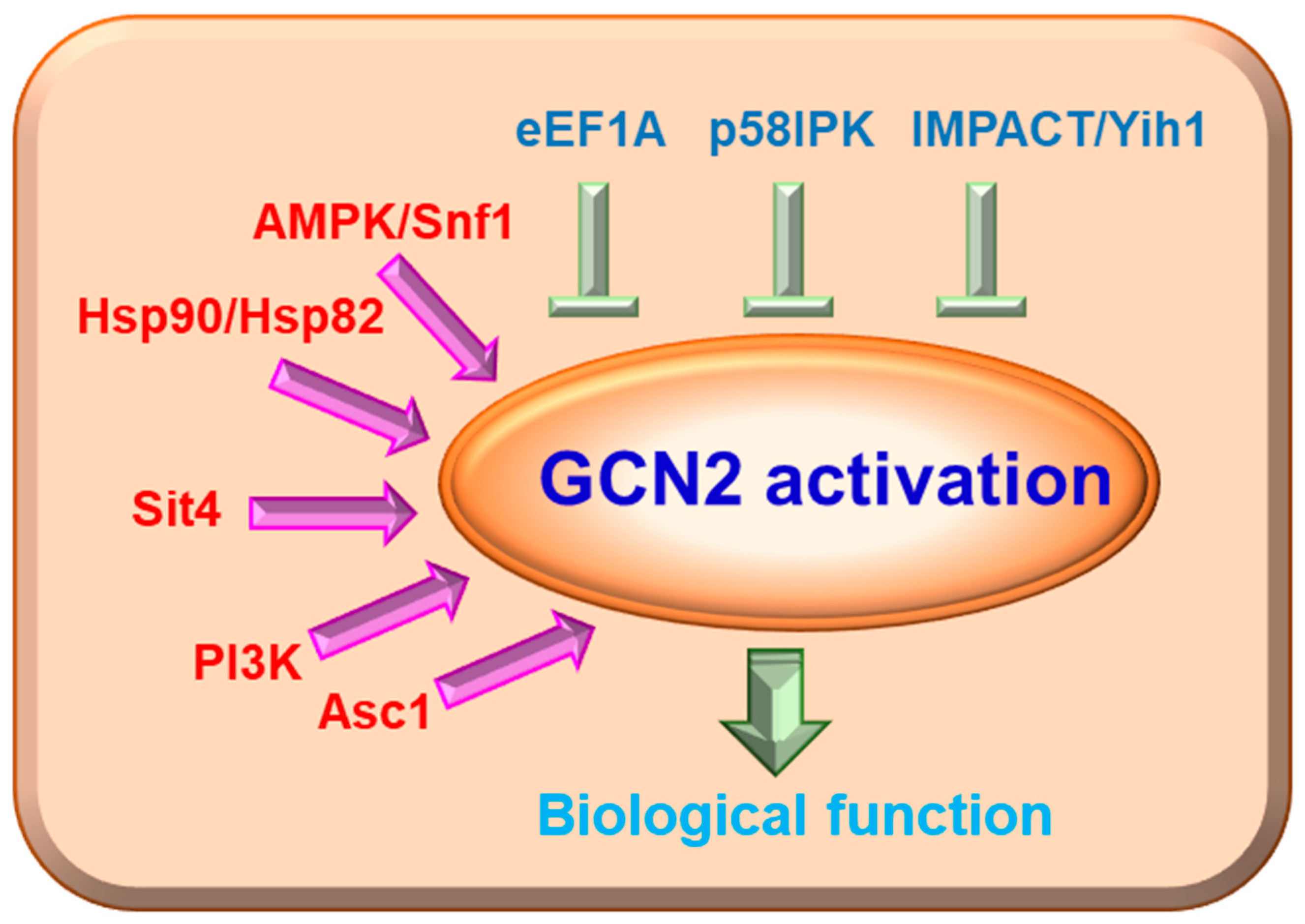
GCN2 activity in cells must be effectively inhibited under normal circumstances to allow for the maximum rate of protein synthesis. Many molecules have been recognized to directly or indirectly modulate GCN2 activation (Figure 2). Researchers have identified several endogenous inhibitors of GCN2 kinase in yeast and mammals, including eukaryotic translation elongation factor 1A (eEF1A), yeast impact RWD domain protein (IMPACT) homolog 1 (Yih1), IMPACT and p58IPK. eEF1A has been confirmed as an inhibitor of GCN2 in Saccharomyces cerevisiae cells. Visweswaraiah et al. proved that the essential translation elongation factor eEF1A can interact with the CTD of GCN2, and this interaction can be attenuated by uncharged tRNAs when cells are faced with amino acid scarcity. Meanwhile, eEF1A reduces the ability of GCN2 to phosphorylate its substrate, eIF2α, possibly through preventing GCN2 from binding eIF2α; however, it does not affect the autophosphorylation of GCN2 [42]. IMPACT and its homolog in yeast, Yih1, are potent suppressors of GCN2. Both IMPACT and Yih1 contain an RWD domain that competes with GCN2 for GCN1 binding, which is vital for GCN2 activation [43,44]. IMPACT and its homolog function mainly through the GCN2 signaling pathway. For example, IMPACT has been found to be preferentially expressed in the nervous system, where it may modulate neurite outgrowth in a GCN2-dependent way [45]; Rafael C. Ferraz et al. found that knockdown of the IMPACT homolog impt-1 in C. elegans activates the ISR pathway and increases both lifespan and stress resistance in a GCN2-dependent manner [46]. In addition, p58IPK is another inhibitor of GCN2, which can suppress GCN2 phosphorylation and prolong protein synthesis under endoplasmic reticulum deficiency, hypothermic challenge, and prolonged culture stress conditions [47].
Chaperone proteins, such as heat shock protein 82 (Hsp82) in yeast and mammalian heat shock protein 90 (Hsp90), may play an essential role in the maturation of GCN2 to an active kinase [42,48]. A highly conserved AMP-activated serine/threonine protein kinase (AMPK) in mammals and sucrose non-fermenting 1 (Snf1) in yeast, is activated when the AMP/ATP ratio increases. This process will switch off energy-consuming anabolic pathways while turning on ATP-producing pathways [49]. Snf1 and GCN2 directly interact with each other. Snf1 is activated by amino acid starvation and is required for GCN2 auto-phosphorylation and eIF-2α phosphorylation [50]. Resistance to glucose repression protein 1 (REG1) is a negative regulator of Snf1, and REG1 deletion increases the phosphorylation of GCN2 Thr-822 and eIF-2α independent of Snf1 under amino acid starvation [51]. Receptor for activated C-kinase (Rack1) in mammalian cells and its orthologue activating signal cointegrator–1 (Asc1) in S. cerevisiae are highly conserved proteins consisting of seven tryptophan-aspartate repeats [52] that act as scaffolds for proteins in various signaling pathways and play essential roles in regulating a wide array of biological processes. Asc1 is required for starvation-induced GCN2 auto-phosphorylation and eIF-2α phosphorylation promoting expression of amino acid biosynthesis genes in Schizosaccharomyces pombe under amino acid starvation [53].

3. GCN2 and Its Associated Signaling Pathways

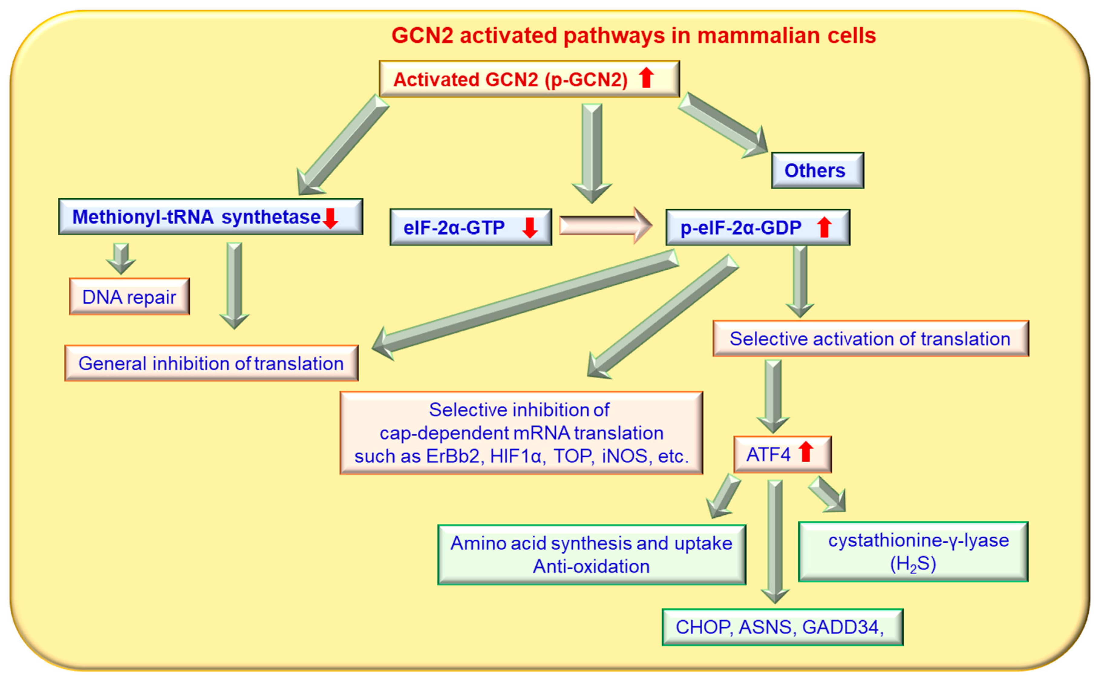
The eIF-2α molecule is the most important known target of GCN2 identified to date (Figure 3). The GCN2 protein can phosphorylate eIF2α in Drosophila and mice in vitro [10,54,55]. After being activated in response to nutritional deprivation or other stresses, GCN2 reduces eukaryotic initiation factor 2 (eIF-2) activity by phosphorylating the α subunit of eIF-2 at serine 51, which impairs the rate of GDP-GTP exchange catalyzed by the initiation factor eukaryotic translation initiation factor 2B (eIF2B) and blocks eIF-2 recycling [11,56,57]. Phosphorylated eIF-2α is unable to fulfill its normal role in helping the 40S ribosome subunit acquire methionyl initiator tRNA, and formation of the eIF2/tRNAiMet/GTP ternary complex, which is required for polysome formation and translation initiation, is decreased [58,59]. Furthermore, temperately increased phosphorylation of eIF-2α can completely inhibit protein synthesis initiation while favoring selective translation of some messenger RNA (mRNAs), including general control nondepressible 4 (GCN4) in yeast and ATF4 in mammals [60] (Figure 3).
In yeast, phosphorylation of eIF-2α by phosphorylated GCN2 decreases eIF-2-GTP levels and eliminates the (ternary complex) TC’s inhibitory effects of the upstream open reading frames (uORFs) of GCN4 mRNA, thereby promoting GCN4 translation and blocking general translation [61]. GCN4 is a transcriptional activator of the general amino acid control pathway and promotes the expression of genes encoding amino acid biosynthetic enzymes to remedy nutrient deprivation [15]. Similarly, phosphorylation of eIF-2α by GCN2 in mammalian cells induces the translation of ATF4 mRNA, which be classified a transcription factor in the basic leucine zipper family, which includes Gcn4. The mechanism of ATF4 mRNA translation induced by GCN2 in mammals is similar to that of GCN4 in yeast, which is dependent on two ORFs associated with its mRNA [62,63,64]. ATF4 is a transcription factor that binds an amino acid response element located in the promoters of specific genes and induces their transcription [65]. In the mouse liver, GCN2 deletion attenuates high fat diet-induced eIF-2α phosphorylation and the induction of ATF4-C/EBP homologous protein (CHOP) to decrease the expression of peroxisome proliferator-activated receptor gamma (PPARγ), fatty acid synthase and metallothionein [66]. In addition to eIF-2α, methionyl-tRNA synthetase has also been identified as a substrate of GCN2 (Figure 4). Methionyl-tRNA synthetase plays essential roles in initiating translation by transferring methionyl to initiator tRNA. Under UV irradiation conditions, GCN2 can phosphorylate methionyl-tRNA synthetase, which inhibits its binding to methionyl-tRNA and contributes to a down-regulation of general translation [67]. A recent study using quantitative phosphoproteomics showed that GCN2 also targets auxiliary effectors to modulate translation [68]. In addition to phosphorylation of eIF2a, GCN2 also phosphorylates the beta-subunit of the trimeric eukaryotic translation initiation factor 2 gamma (eIF2G) protein complex to promote its association with eukaryotic translation initiation factor 5 (eIF5) and, subsequently, to restrict recycling of the initiator methionyl-tRNA-bound eIF2-GDP ternary complex in amino acid-starved cells [68].
The protein kinase GCN2 also regulates mRNA translation in mammals in an ATF4-independent manner (Figure 4). The translation of inducible nitric oxide synthase is negatively regulated by GCN2 upon UV irradiation (22). GCN2 negatively regulates translation of the oncogene erb-b2 receptor tyrosine kinase 2 (ErBb2) and of hypoxia inducible factor 1α (HIF1α), both of which are involved in the cell cycle and cell survival [69,70]. Another direct GCN2 substrate-regulating protein is methionyl-tRNA synthetase [42]. Activated GCN2 phosphorylates methionyl-tRNA synthetase at serine 662, which inhibits methionyl-tRNA synthetase activity and activates the ataxia-telangiectasia, mutated protein kinases (ATM)/ ATM and Rad3-related protein kinases (ATR) system for DNA repair [42]. GCN2 negatively controls the translation of 5′-terminal oligopyrimidine tracts (TOP) mRNAs, which encode protein biosynthesis factors, under amino acid starvation, and this process is dependent on the stress granule-associated proteins T-cell intracellular antigen 1 (TIA-1) and TIA1 cytotoxic granule-associated RNA binding protein-like 1 (TIAR) [71]. This regulation will re-direct TOP mRNAs from polysomes to stress granules, thereby allowing modulation of limited resource usage according to amino acid availability [71]. GCN2 can directly regulate forkhead box O class protein (Foxo) activity in human cells and in Drosophila [72]. However, protein kinase RNA-like ER kinase (PERK) can compensate for potential deficiencies in Foxo activity when GCN2 is absent in human cells [72]. GCN2 can directly interact with Foxo3 in cells and phosphorylate Foxo3 to promote its nuclear translocation and transcriptional activation in skeletal muscle [73]. Under certain conditions, IFN-γ has the ability to promote cancer initiation and progression [74]. Importantly, IFN-γ accelerates arginine depletion and induces malignant transformation through the NF-κB/GCN2/eIF-2α pathway in vitro and in vivo [75], and arginine addition can rescue these effects of IFN-γ [76]. GCN2 activation reduces the activity of succinate dehydrogenase, an iron-sulfur mitochondrial enzyme, and promotes the nuclear localization of the transcription factor DNA-binding transcription factor 1 (Aft1) in the yeast Saccharomyces cerevisiae [77]. In normal situations, an excess of amino acids would promote low levels of GCN2 activity, leading to inhibition of Aft1 and iron transporters. This repression of iron transporters could help prevent intracellular iron from reaching toxic levels due to reactive oxygen species (ROS) production [77]. GCN2 is responsible for pro-apoptotic TNF-related apoptosis-inducing ligand receptor 2 (TRAIL-R2) upregulation, caspase-8 activation, and extrinsic apoptotic cell death in tumor cells under glutamine or methionine starvation [78].
In addition, the GCN2/ATF4 pathway is involved in amino acid starvation-induced expression of the stress-inducible gene p8 through an amino acid response element in the promoter of this gene [79]. Importantly, GCN2 also plays roles in regulating the amino acid starvation response in the nucleolus, in addition to its function in the cytosol. The nucleolar localization of GCN2 regulates small RNA transcripts, which may serve as alternative stress-sensing machinery for nutrient deficiency. Depletion of GCN2 by siRNA increases small RNA transcripts and induces activation of the p53 pathway, which is dependent on B-related factor 1 [80].

4. Biological Functions of GCN2

GCN2-deficient mice are viable and fertile. These mice display no obvious phenotypic abnormalities unless fed a diet lacking a single amino acid [81,82]. However, GCN2 has a wide array of biological functions across different cells and species [3,8,26,83,84]. GCN2β isoform (GCN2β), which is highly abundant in unfertilized mouse eggs, is active and elevates the phosphorylation of eIF-2α; however, phosphorylation of eIF-2α is reduced drastically after fertilization, suggesting that GCN2-mediated translational control may contribute to the regulation of oocyte maturation [85]. GCN2-mediated phosphorylation of eIF-2α is elevated in old/aging cells, potentially contributing to global translation reduction and lifespan extension [86]. GCN2 deficiency attenuates induced hepatic steatosis and insulin resistance in mice fed a high-fat diet, partially through shifting lipolysis to lipogenesis, as well as by decreasing oxidative and endoplasmic reticulum stress [66]. GCN2 is required for the growth of prostate cancer cells and regulates the expression of over 60 solute-carrier genes, including those involved in amino acid transport [87]. Inhibiting GCN2 in arginine-deprived hepatocellular carcinoma cells promotes a senescent phenotype, rendering these cells vulnerable to senolytic compound-induced death [88]. Heritable pulmonary veno-occlusive disease is related to biallelic mutations in EIF2AK4 which encodes GCN2. GCN2 deletion impairs the ability of hematopoietic stem cells to repopulate and regenerate by regulating metabolic alterations [89]. The GCN2 activator halofuginone significantly prevents cell proliferation and inhibits expression of anti-HLA class I antibodies-induced IL-8, monocyte chemoattractive protein-1, and transforming growth factor-beta 1 in human glomerular endothelial cells, suggesting that GCN2 activation may have a protective effect against antibody-mediated graft rejection [90]. Herein, we will briefly summarize the involvement of GCN2 in oxidative stress, cell survival, autophagy, and cell metabolism.

4.1. GCN2 and the Oxidative Stress Response

Excessive ROS production or impaired ROS reduction efficiency can lead to oxidative stress. Oxidative stress is associated with cytotoxic effects and has been implicated in the etiology of various diseases. The oxidative stress response is highly associated with amino acid supply. GCN2 is a potential regulator of redox homeostasis. Arriazu et al. demonstrated that amino acid deprivation decreases intracellular ROS levels in hepatic stellate cells in vitro, with this effect shown to be dependent on GCN2 but not on downstream eIF-2α [91]. Deletion or pharmacological inhibition of GCN2 significantly delays collective cell migration and wound closure by impairing the maintenance of intracellular free amino acids, particularly cysteine, as well as disrupting Ras-related C3 botulinum toxin substrate 1 (RAC1)-GTP-driven ROS generation, lamellipodia formation, and focal adhesion dynamics in human epidermal keratinocytes during wound healing [92]. Furthermore, GCN2 and its downstream signaling pathway play important roles in protection against oxidative injuries induced by an amino acid imbalanced diet [93].

4.2. GCN2 and Cell Survival, Cell Cycle and Cell Differentiation

The GCN2 kinase has a broad impact on cell survival. The GCN2/eIF-2α/ATF4 signaling pathway can mediate cell survival [94]. GCN2 can sense stalled ribosomes and activate the eIF-2α/ATF4 downstream signaling pathway to promote ISR, which protects neurons against ribosome stalling-mediated cell death [95]. Cai et al. reported that the GCN2/eIF-2α/ATF3 signaling pathway is important for cells in the renal medulla during urea stress. The loss of GCN2 increases the sensitivity of cells to urea stress and promotes activated caspase-3 expression to decrease cell survival [24]. GCN2 deficiency decreases doxorubicin-induced cardiotoxicity by ameliorating apoptosis and oxidative stress through an eIF-2α/CHOP-dependent pathway [96].
The GCN2-associated signaling pathway seems required for development and lifespan extension. GCN2 deficiency impairs translational control and the expression of differentiation-associated genes, eventually inhibiting normal epidermal differentiation in organotypic skin culture [97]; these findings indicate that the GCN2/eIF-2α signaling pathway promotes the translational control and differential protein expression required for normal keratinocyte differentiation. Under conditions of amino acid restriction, activated GCN2 and its downstream transcription factor ATF4 mediate transcription of 4E-BP, which regulates lifespan extension in Drosophila and is important for the normal development of certain tissues [98,99].
Xiao et al. found that the inhibited cell proliferation and increased apoptosis of breast cancer cells observed under leucine deprivation is dependent on fatty acid synthase (FASN). Leucine deprivation decreases Fasn expression by activating the GCN2/eIF-2α signaling pathway, which can inhibit sterol regulatory element-binding protein 1C (SREBP1C), a transcription factor that directly binds to the Fasn promoter and regulates Fasn mRNA abundance [100]. However, the specific mechanism by which the GCN2 signaling pathway regulates SREBP1C requires further investigation. GCN2 can also exert proapoptotic functions in cancer cells through posttranslational mechanisms. GCN2 protein levels critically determine the sensitivity of cancer cells to Na+, K+-ATPase ligand-induced apoptosis, both in vitro and in vivo. Na+,K+-ATPase ligand treatment triggers phosphorylation of GCN2 at threonine 899, which increases GCN2 protein expression by disrupting formation of the GCN2-beta-arrestin-NEDD4-like E3 ubiquitin protein ligase (NEDD4L) ternary complex. These increased GCN2 levels, in turn, aggravate Na+,K+-ATPase ligand-induced cancer cell apoptosis, which is largely dependent on a molecule downstream of GCN2, CHOP [101]. In eukaryotic cells, GCN2 plays an important role in regulation of the cell cycle under various stresses. DNA-damaging agents or nitrogen starvation induce cell arrest in the G1 phase, accompanied by phosphorylation of eIF-2α [102,103]. Hamanaka et al. demonstrated that PERK and GCN2 function cooperatively to regulate eIF-2α phosphorylation and cyclin D1 translation in fibroblasts after unfolded protein response (UPR) activation [104]. In addition, activation of the GCN2/eIF-2α signaling pathway can selectively up-regulate translational expression of a p21 transcript variant that contributes to cell cycle arrest at the G1/S phase and promotes cell survival [105].

4.3. GCN2 and Autophagy

The GCN2 signaling pathway plays a major role in autophagy and the regulation of stress-induced autophagy gene expression. The GCN2 kinase and its downstream transcription factors, ATF4 and CHOP, are required to increase transcription of a set of genes implicated in the formation, elongation, and function of the autophagosome [106]. Fougeray et al. reported that GCN2 can mediate IFN-γ-induced autophagy in human renal epithelial cells. During this process, IFN-γ induces tryptophan metabolism, then activates the GCN2 kinase and downstream eIF2α, an activator of autophagy [107]. During the lactation cycle of the bovine mammary gland, autophagy is induced in bovine mammary epithelial cells (BMECs) as a survival mechanism to maintain cellular homeostasis. IFN-γ-induced autophagy in primary BMECs promotes dramatic primary BMEC transformation in vivo and in vitro [75]. GCN2/eIF-2α signaling pathway-mediated IFN-γ-induced autophagy in BMECs reduces milk synthesis, while arginine supplementation can attenuate IFN-γ-induced autophagy and restore milk protein and fat synthesis to some extent [108].
Autophagy and its regulatory mechanisms are involved in intestinal homeostasis and repair, supporting intestinal barrier function in response to cellular stress through tight junction regulation and protection from cell death [109]. Acute amino acid starvation-induced autophagy inhibits intestinal inflammation, a key mediator of the ISR, through a mechanism dependent on GCN2 [110].

4.4. GCN2, mTOR and Metabolism

GCN2 and mTORC1 are two amino acid-sensing kinases in mammalian cells, and their combined effects orchestrate cellular adaptation to amino acid levels. GCN2 kinase is typically activated by increased levels of uncharged tRNAs during amino acid scarcity. The mTORC1 kinase can be activated by amino acid sufficiency, and enhanced mTORC1 activity promotes high levels of translation to maintain cell growth and proliferation by phosphorylating two important translational regulators, the S6 kinase (S6K) and eIF4E-binding protein (4E-BP1) [111,112]. GCN2 and mTORC1 signaling pathways are both regulated by amino acids and share some common functions.
These two amino acid-sensing systems are linked. mTORC1 is inactivated by certain nutrient deprivation conditions. GCN2 is involved in the inhibition of mTORC1; however, GCN2 alone is not sufficient to inhibit mTORC1 activity upon leucine or arginine deprivation, indicating the existence of additional mechanisms by which leucine and arginine regulate mTORC1 activity. Moreover, GCN2 inhibits mTORC1 activity through phosphorylation of eIF2α but independently of the downstream transcription factor ATF4 [113]. In contrast, Ye et al. reported that GCN2 inhibits mTORC1 activity through ATF4 [114]. Deprivation of various amino acids can activate GCN2, up-regulate ATF4 expression and promote expression of the stress response protein Sestrin2, which is required to sustain mTORC1 repression by blocking its lysosomal localization [114]. Furthermore, mTORC1 activity also seems to impact GCN2 activity. Pharmacological inhibition of mTORC1 leads to GCN2 activation and eIF-2α phosphorylation dependent on the catalytic subunit of protein phosphatase 6 (PP6C) [115]. Inhibition of mTORC1, either by rapamycin, TOR2 mutation or nitrogen deprivation, induces GCN2-dependent phosphorylation of eIF-2α [9]. In response to UV irradiation and oxidative stress, GCN2 is fully activated without any detectable change in target of rapamycin 2(TOR2) activity. However, during amino acid starvation, activation of GCN2 is dependent on maintained TOR2 activity, and GCN2 is required for timely inactivation of the mTOR pathway. Deprivation of glutamine, arginine, methionine, or lysine, but not of 16 other amino acids, has been shown to induce PKB activation in non-small cell lung cancer cells via GCN2-ATF4-regulated in development and DNA damage responses 1 (REDD1) axis-mediated mechanistic target of rapamycin complex 2 (mTORC2) activation [116]. Thus, the GCN2 and mTOR signaling pathways likely act in a coordinated manner to prevent the translational machinery from using excessive amounts of vital resources during nutrient-limited conditions. Nevertheless, the relationship between GCN2 and mTOR is complicates, and more research is needed to clearly define the regulatory interplay between GCN2 and mTOR.
GCN2 is closely linked to various metabolism-associated processes, including fatty acid and amino acid synthesis, gluconeogenesis and the hexosamine biosynthetic pathway. In primary human CD4+ T cells, indolamine 2,3-dioxygenase (IDO) activates GCN2 kinase through tryptophan depletion, resulting in down-regulation of key enzymes directly and indirectly involved in fatty acid synthesis [117]. The GCN2/ATF4 pathway can up-regulate serine synthesis enzymes in response to amino acid deprivation, which synergizes with PKM2-dependent accumulation of glycolytic precursors to maintain serine synthesis [118]. Gluconeogenesis normally plays a key role in the maintenance of peripheral glucose homeostasis. GCN2-deficient mice exhibit reduced gluconeogenesis upon administration of pyruvate, a gluconeogenic substrate. Xu et al. reported that GCN2 is important for maintaining gluconeogenesis in the liver through regulating expression of CCAAT enhancer-binding protein-beta (C/EBPβ) [119].

5. GCN2 in the Immune System

Amino acid catabolism is closely related to innate and adaptive immunity. GCN2 plays vital roles in the immune system and in the maintenance of immune homeostasis [3]. For example, GCN2/ATF4 signaling pathway-induced 4E-BP contributes to innate immunity by biasing mRNA translation toward cap-independent mechanisms, thus enhancing AMP synthesis during infection [120]. GCN2-mediated ISR impacts immune homeostasis in the intestine. GCN2 controls intestinal inflammation by suppressing inflammasome activation in response to amino acid starvation. During amino acid starvation or intestinal inflammation, GCN2-mediated ISR is activated in intestinal antigen presenting cells (APCs) and epithelial cells. GCN2 deficiency in CD11c+ APCs or intestinal epithelial cells leads to reduced autophagy and increased ROS levels, promoting inflammasome activation and interleukin (IL)-1β production and ultimately enhancing intestinal inflammation and T helper 17 cell (Th17) responses [110]. The regulatory roles of GCN2 in various immune cells will be discussed in the following sections.

5.1. GCN2 in Innate Cells

GCN2 participates in the protective innate immunity and in organismal stress responses caused by pathogen-induced amino acid starvation. During the process of bacterial infection, such as by Shigella or Salmonella, damage to the host cell membrane triggers an acute intracellular amino acid starvation response, then activates the GCN2/eIF-2α/ATF4/ATF3 ISR pathway and simultaneously decreases mTOR activity [121]. A systems biology approach revealed that there is a significant correlation between GCN2 gene expression in peripheral blood mononuclear cells and CD8+ T cell responses in humans vaccinated with the yellow fever vaccine YF-17D [122]. The increase in GCN2 gene expression in PBMCs is positively correlated with the increase of the proportion of activated CD8+ T cells, and the authors use experiments to confirm that after treatment of PBMCs with YF-17D, eIF2α phosphorylation is enhanced over time and Stress granules appear. It shows that in the time sequence, the expression of GCN2 in PBMC increases first, and then the activation of CD8+T increases [122]. Furthermore, Ravindran et al. demonstrated that YF-17D-induced GCN2 activation in dendritic cells initiates autophagy and enhances antigen presentation ability to both CD4+ and CD8+ T cells [123]. No studies addressing the direct role of GCN2 in granulocytes have been reported to date. Julia et al. provide a perspective on the signaling of pattern recognition receptors (PRRs) and cytokine receptors recruits nutrient-sensing and autophagic machinery to shape the immune response to microbes. Amino acid starvation during Shigella flexneri activates GCN2, which results in an Activating transcription factor 3 (ATF3)-dependent transcriptional signature, e.g., increased CHOP.
Ravindran et al. recently reported that mice deficient in GCN2 exhibit substantially high levels of ROS and, subsequently, of the proinflammatory mediator IL-1β in response to cellular stress. This phenomenon is likely due to a lack of autophagy in GCN2-deficient mice, whereas mice fed a reduced-amino-acid diet show significantly lower levels of oxidative stress and of inflammatory responses to cellular stress [123]. Further mechanistic insights reveal that GCN2-induced autophagy interferes with oxidative stress and inflammasome activation, thereby controlling inflammation (Figure 2). It has recently been reported that GCN2 is a key driver of the induction of anti-inflammatory macrophage functional polarization and myeloid-derived suppressor cells (MDSCs) in the tumor microenvironment, depending on ATF4 and altered oxidative metabolism and myeloid-lineage deletion of GCN2 can changes in the immune microenvironment with increased proinflammatory activation of macrophages and MDSCs and IFNγ expression in intratumoral CD8+ T cells [6]. It has been reported that both IL-6 and GCN2 are required to promote neovascularization in IFN-γ-induced IDO1-expressing asialo-GM1highCD11c+Gr-1+CD11blow cells with high autofluorescence [124]. Treatment with a GCN2 inhibitor alleviates MDSCs-related T cell suppression and restores T cell proliferation to enhance host anti-tumor immunity in mice [125]. In response to amino acid abundance, mTOR activates host protein translation, a mechanism that coronaviruses use for their own protein synthesis and replication. In contrast, the amino acid starvation sensor GCN2 activates pathways that limit inflammation and viral replication [126].
On the other hand, GCN2 signaling in myeloid cells can also promote inflammatory reactions in certain cases. The stress response kinase GCN2 promotes macrophage inflammation both in vitro and in vivo. Amino acid deficiency enhances the sensitivity of macrophages to lipopolysaccharides, as evidenced by increased IL-6 production dependent on the GCN2-ATF4 signaling pathway. In a mouse model of septicemia, mice with a myeloid cell-specific GCN2 deficiency were shown to exhibit reduced inflammatory responses and mortality, accompanied by decreased levels of TNF-α, IL-6, and IL-12 [127]. In mice, the GCN2 kinase is a key regulator of fibrogenesis and of acute and chronic liver injury induced by carbon tetrachloride. GCN2 plays roles in hepatic fibrogenesis and in the response to acute or chronic liver injury. Upon deficiency of the essential amino acid histidine, GCN2 is activated in primary and immortalized human hepatic stellate cells, resulting in decreased mRNA and protein expression of collagen type I, which is important for fibrogenesis. GCN2-deficient mice exhibit increased susceptibility to CCl4-induced acute or chronic liver damage [128].
GCN2 can be activated by IDO-driven tryptophan consumption to mediate the biological effects of IDO1 in immune cells. In vitro studies have shown that GCN2 kinase is required for IL-6 production by IDO-expressing macrophages in response to LPS stimulation in tryptophan-free RPMI 1640 medium [127]. Deletion of GCN2 specifically in myeloid cells decreases LPS-induced mouse septic mortality and decreases LPS-induced IL-6 levels, although not TNF-α production [127]. Clearance and tolerance to autoantigens produced by apoptotic cells are important to the maintenance of homeostasis and to prevent the initiation of inflammatory autoimmunity. Increased cell death and defective clearance can lead to the initiation and development of the autoimmune disease systemic lupus erythematosus (SLE) [129]. As a type of innate scavenging cell, macrophages are important for the clearance of apoptotic cell-associated self-antigens and the maintenance of self-tolerance. The tryptophan catabolizing enzyme IDO1 limits innate and adaptive immunity to apoptotic self-antigens, and IDO inhibits the inflammatory pathology caused by systemic autoimmune diseases [130]. The protein kinase GCN2, a primary downstream effector of IDO1, is involved in apoptotic cell-driven immune suppression. Upon activation of the GCN2 signaling pathway in macrophages, IDO1 enhances production of the apoptotic cell-driven anti-inflammatory cytokine IL-10 while reducing production of the proinflammatory cytokine IL-12 [130]. IDO1 fails to promote IL-10 protein expression in GCN2-deleted macrophages due to alterations in ribosomal association with cytokine mRNA transcripts [130]. Myeloid-specific deletion of GCN2 abrogates regulatory cytokine production and promotes inflammatory T-cell responses to apoptotic cell antigens and failure of tolerance induction. Consistently, myeloid deletion of GCN2 in lupus-prone mice results in increased immune cell activation, humoral autoimmunity, renal pathology, and mortality, while activation of GCN2 was found to significantly reduce these symptoms of systemic autoimmune disease [131]. Compared to wild-type mice, GCN2-deficient mice display resistance to anemia during a number of stress conditions, including hemolysis, amino acid deficiency and hypoxia. GCN2-deleted liver macrophages exhibit defects in the ATF4- NRF2 pathway that impair erythrophagocytosis and lysosome maturation, indicating that GCN2 is an important regulator of red blood cell clearance and iron recycling in liver macrophages [132].
GCN2 deletion in macrophages blocks leucine deprivation-induced browning and lipolysis in mouse white adipose tissue [133]. Further studies have revealed that GCN2 activation in macrophages reduces monoamine oxidase A expression and increases secretion of norepinephrine from macrophages to adipocytes, enhancing white adipose tissue browning and lipolysis under conditions of leucine deprivation [133]. Injection of the beta3-adrenergic receptor agonist CL316,243 and inhibition of monoamine oxidase A were both shown to increase norepinephrine levels, enhancing browning and lipolysis in white adipose tissue under leucine deprivation conditions [133]. Therefore, GCN2 expression in macrophages is involved in the balance between white and brown adipose tissues.

5.2. GCN2 in T Cells

GCN2 activation is important for T cell proliferation, activation, and differentiation. It has been reported that the type of amino acid transporter can dictate T cell differentiation fate and that T cells lacking the leucine transporter solute carrier family 7 member 5 (SLC7A5) are unable to evoke a robust response to antigen exposure or differentiate into effector T cells [134]. IDO inhibits T cell proliferation through the activation of GCN2 involves many pathways, such as GCN2 down-regulating TCR-complex ζ-chain, c-Myc, aerobic glycolysis and glutaminolysis levels by reducing levels of lactate dehydrogenase A (LDH-A) and glutaminase, GCN2-mediated down-regulation of key enzymes involved in fatty acid synthesis and GCN2 activated to decreasing glucose influx, and altering key enzymes involved in metabolism to decrease aerobic glycolysis and glutaminolysis [117,135,136]. In this way, IDO could be a constraining factor for alloreactive T cell proliferation and differentiation into effector T cell subtypes [136]. However, Sonner et al. found that GCN2 deficiency in T cells did not affect immunity to B16 tumors [137]. IDO-expressing plasmacytoid DCs suppress T cell proliferation through GCN2 activation in responding T cells via tryptophan catabolism. T cell-specific GCN2 deletion has been shown to weaken IDO-mediated suppression and eliminate IDO-mediated T cell anergy in vitro [138]. Consistently, proliferation of GCN2-deficient T cells is not inhibited by IDO-expressing DCs in vivo [138]. However, it has been reported that GCN2 is not involved in the suppression of anti-tumor T cell responses via tryptophan catabolism in melanoma-bearing mice [137]. It cannot be ignored the kynenurine (KYN) pathway metabolites KYN (catalyzed by IDO1) are endogenous agonists of the aryl hydrocarbon receptor (AhR). AhR can promote the development of type 1 regulatory T cells; another major source of IL-10. IL-10 is a potent anti-inflammatory cytokine that promotes differentiation, proliferation and maintenance of Treg cells, which impair CD8+ T cell maturation and cytotoxicity. IL10 also promote the development and proliferation of tolerogenic DCs, tumor-associated macrophages (TAMs), and MDSCs that populate the tumor microenvironment—to support angiogenesis immune escape of cancer cells [139]. The TME is the environment around a tumor. Tumors can affect their microenvironment by releasing cell signaling molecules, promote angiogenesis around the tumor and induce immune tolerance, while immune cells in the microenvironment can affect the growth and development of cancer cells [140].
A well-known animal model of experimental autoimmune encephalomyelitis (EAE) is widely employed to study the mechanisms and pathogenesis of multiple sclerosis. The invasion of Th1 and Th17 cells leads to central nervous system (CNS) demyelination and lesion formation during the peak of murine EAE. Spontaneous remission of EAE may occur after the peak of the disease, during which regulatory T cells (Treg) accumulate in the inflamed CNS, suppress effector T cell responses and reduce inflammation. GCN2 participates in the remission phase of EAE by affecting a variety of T cells, including effector T cells and Treg cells. Orsini et al. reported that the peak of the disease is characterized by high IDO expression and a high frequency of plasmacytoid DCs in the CNS of wild-type C57BL/6 mice. However, GCN2-deficient mice with EAE fail to reach the remission phase and are characterized by higher levels of CNS inflammation, accompanied by the increased presence of effector T cells (Th1/Th17) and a lower frequency of Treg cells [141]. Consistently, Keil et al. reported that GCN2-deficiency in mice exacerbates chronic disease progression during the remission period of EAE, mainly due to the low frequency of Treg cells. They demonstrated that GCN2 deficiency does not impact the survival, proliferation or suppressive capacity of Treg cells; rather, it decreases the infiltration of Treg cells into the inflamed CNS by reducing their ability to respond to CCL2 gradients [142]. However, GCN2-deficiency does not impact the migration of effector T cells. A subsequent study proved that the decreased infiltration capacity of Treg cells to the CNS in GCN2-deficient mice is not due to reduced surface receptor expression. These results suggest that, under an IDO-mediated immunoregulatory environment, the GCN2 kinase may be activated to restrict effector T cell responses and promote the accumulation of Treg cells in the inflamed CNS, mediating the remission phase of EAE. On the other hand, we recently demonstrated that GCN2 controls Th9 cell differentiation through a HIF1α-dependent glycolytic pathway [143]. GCN2-deficient mice are resistant to OVA-induced allergic airway inflammation [143]. Further studies should focus on the intrinsic roles of GCN2 in the differentiation and function of different T cell subsets.
The GCN2-mediated amino acid starvation response pathway is important for Th17 cell differentiation. Th17 cells, which mainly produce IL-17, have been recognized as a type of pro-inflammatory CD4+ T cell and implicated in various autoimmune and inflammatory disorders. Sundrud et al. reported that halofuginone can selectively inhibit mouse and human Th17 differentiation by activating the GCN2-mediated amino acid starvation response [144,145]. Consistently, the addition of excess amino acids rescued the inhibition of Th17 differentiation by halofuginone. This halofuginone-induced GCN2-mediated amino acid starvation response significantly reduces Th17 cell differentiation and alleviates Th17-associated EAE in vivo [144]. Halofuginone can activate the GCN2-mediated amino acid starvation response by binding glutamyl-prolyl-tRNA synthetase and inhibiting prolyl-tRNA synthetase activity; this effect can be reversed by the addition of exogenous proline or glutamyl-prolyl-tRNA synthetase [146]. Other natural product derivatives in the febrifugine family have also been shown to inhibit glutamyl-prolyl-tRNA synthetase [146]. In addition, halofuginone blocks IL-23-induced signal transducer and activator of transcription 3 (STAT3) phosphorylation and IL-23-dependent proinflammatory cytokine expression in CCR6+ Th17 cells via activation of the amino acid starvation response pathway [147]. Thus, targeting GCN2 is an effective approach to autoimmunity prevention, a critical rationale for the development of tools to manipulate GCN2 function for the treatment of inflammatory immune disease.
In murine models of glioblastoma, GCN2 deficiency limits CD8+ T cell activation and cytotoxic marker expression. Adoptive transfer of GCN2-deleted antigen-specific CD8+ T cells failed to control tumor burden compared to wild-type CD8+ T cells. This is likely because GCN2-eficient CD8+ T cells are sensitized to become rapidly necrotic when faced by reduced levels of PKCθ and p-PKCθ. This study demonstrates the importance of GCN2 to CD8+ T cell function and survival in mice [148]. GCN2-deficient CD8+ T cells, but not CD4+ T cells, exhibit defects in proliferation and trafficking in vitro and in vivo, indicating that GCN2 is required for efficient cytotoxic T cell function [149]. However, using an experimental B16 melanoma model with T cell-specific GCN2 knockout mice, Sonner et al. showed that GCN2 in T cells did not affect immunity against B16 tumors, suggesting that GCN2 is not intrinsically involved in the functional alteration of tumor-infiltrating T cells [137].

6. Conclusions

As a kinase that can sense amino acid scarcity and other stresses, the GCN2 kinase plays important roles in a variety of biological processes and diseases. Due to the important roles of GCN2 in many immunities, researchers continue to explore GCN2 inhibitors and activators. Several GCN2 inhibitors and activators have been approved by Food and Drug Administration (FDA), including erlotinib and sunitinib [27]. Neratinib has the ability to bind and activate GCN2 [27]. Inhibition of GCN2 activity in TAM and tumor cells in the tumor microenvironment can play an anti-tumor effect. The majority of existing studies focus on the effects of GCN2 deficiency or GCN2 activation under stress conditions, and the detailed molecular mechanisms have yet to be fully illuminated. The role of GCN2 in T cells and macrophages has been the focus of current research, but little is known about its role in neutrophils. In future studies, more attention should be paid to the antagonism of GCN2 and mTOR in different immune systems. Future research efforts should emphasize the ATF4-independent signaling pathways involved in GCN2-mediated biological regulation. Further studies should also seek to define in more detail the biological functions of GCN2 in the immune system and in immune-related diseases.

Author Contributions

All authors participated in the discussion, writing and correction of the manuscript. All authors have read and agreed to the published version of the manuscript.

Funding

This work was supported by the National Natural Science Foundation (31930041, Y.Z.), the Knowledge Innovation Program of the Chinese Academy of Sciences (XDA16030301, Y.Z.), and the National Key Research and Development Program of China (2017YFA0105002, 2017YFA0104402, Y.Z.).

Data Availability Statement

Data sharing is not applicable to this article, as no datasets were generated or analyzed in the current study.

Acknowledgments

We would like to thank Zhanfeng Liang and Bo Sun for their critical reading of the manuscript, as well as Ling Li for her excellent laboratory management.

Conflicts of Interest

The authors declare no conflict of interest.

Abbreviations

4E-BP1eIF4E-binding protein
Aft1DNA-binding transcription factor 1
AhRaryl hydrocarbon receptor
AMPKAMP-activated serine/threonine protein kinase
APCsAntigen presenting cells
Asc1Activating signal cointegrator–1
ATF4Activating transcription factor 4
ATMAtaxia-telangiectasia, mutated protein kinases
ATRATM and Rad3-related protein kinases
BMECsBovine mammary epithelial cells
C/EBPβCCAAT enhancer-binding protein-beta
CHOPC/EBP homologous protein
CNSCentral nervous system
CTDC-terminal ribosome domain
EAEExperimental autoimmune encephalomyelitis
eEF1AEukaryotic translation elongation factor 1A
eIF-2Eukaryotic initiation factor 2
eIF2BEukaryotic translation initiation factor 2B
eIF2GEukaryotic translation initiation factor 2 gamma
eIF-2αEukaryotic translation initiation factor 2 alpha
eIF5Eukaryotic translation initiation factor 5
ErBb2Erb-b2 receptor tyrosine kinase 2
FASNFatty acid synthase
FDAFood and Drug Administration
FoxoForkhead box O class
GCN1General control nonderepressible 1
GCN2General control nonderepressible 2
Gcn4pGeneral control nondepressible 4 protein
GSK-βGlycogen synthase kinase 3 beta
HisRSHistidyl-tRNA synthetase
Hsp82Heat shock protein 82
Hsp90Heat shock protein 90
IDOIndolamine 2,3-dioxygenase
IMPACTImpact RWD domain protein
ISRIntegrated stress response
MDSCsMyeloid derived suppressor cells
mRNAMessenger RNA
mTORMammalian target of rapamycin
mTORC1Mechanistic target of rapamycin complex 1
NEDD4LNEDD4 like E3 ubiquitin protein ligase
NRF2Nuclear factor erythroid 2–related factor 2
ORFsOpen reading frames
PERKProtein kinase RNA-like ER kinase
PI3KPhosphoinositide 3-kinase
PKBProtein kinase B
PKRDouble-stranded RNA-dependent protein kinase
PP6CProtein phosphatase 6
PPARγPeroxisome proliferator-activated receptor gamma
RAC1Ras-related C3 botulinum toxin substrate 1
Rack1Receptor for activated C-kinase
REDD1Regulated in development and DNA damage responses 1
REG1Resistance to glucose repression protein 1
ROSReactive oxygen species
RWDRING finger and WD repeat containing
S6KS6 kinase
Sit4Serine/threonine-protein phosphatase PP1-1
SLC7A5Solute carrier family 7 member 5
SLESystemic lupus erythematosus
Snf1Sucrose non-fermenting 1
SREBP1CSterol regulatory element-binding protein 1C
STAT3Signal transducer and activator of transcription 3
TIA-1T-cell intracellular antigen 1
TIARTIA1 cytotoxic granule-associated RNA binding protein like 1
TMETumor [141] microenvironment
TOPTranslation of 5′-terminal oligopyrimidine tracts
TOR2Target of rapamycin 2
TRAIL-R2TNF-related apoptosis-inducing ligand receptor 2
TregRegulatory T cells
tRNATransfer RNA
UPRUnfolded protein response
Yih1IMPACT homolog 1

References

  1. Pakos-Zebrucka, K.; Koryga, I.; Mnich, K.; Ljujic, M.; Samali, A.; Gorman, A.M. The integrated stress response. EMBO Rep. 2016, 17, 1374–1395. [Google Scholar] [CrossRef] [Green Version]
  2. Taniuchi, S.; Miyake, M.; Tsugawa, K.; Oyadomari, M.; Oyadomari, S. Integrated stress response of vertebrates is regulated by four eIF2alpha kinases. Sci. Rep. 2016, 6, 32886. [Google Scholar] [CrossRef] [PubMed]
  3. Donnelly, N.; Gorman, A.M.; Gupta, S.; Samali, A. The eIF2alpha kinases: Their structures and functions. Cell. Mol. Life Sci. 2013, 70, 3493–3511. [Google Scholar] [CrossRef] [PubMed]
  4. Grallert, B.; Boye, E. GCN2, an old dog with new tricks. Biochem. Soc. Trans. 2013, 41, 1687–1691. [Google Scholar] [CrossRef] [Green Version]
  5. Santos-Ribeiro, D.; Lecocq, M.; De Beukelaer, M.; Verleden, S.; Bouzin, C.; Ambroise, J.; Dorfmuller, P.; Yakoub, Y.; Huaux, F.; Quarck, R.; et al. Disruption of GCN2 Pathway Aggravates Vascular and Parenchymal Remodeling During Pulmonary Fibrosis. Am. J. Respir. Cell Mol. Biol. 2023. [Google Scholar] [CrossRef] [PubMed]
  6. Halaby, M.J.; Hezaveh, K.; Lamorte, S.; Ciudad, M.T.; Kloetgen, A.; MacLeod, B.L.; Guo, M.; Chakravarthy, A.; Medina, T.D.S.; Ugel, S.; et al. GCN2 drives macrophage and MDSC function and immunosuppression in the tumor microenvironment. Sci. Immunol. 2019, 4, eaax8189. [Google Scholar] [CrossRef]
  7. Hinnebusch, A.G. Mechanisms of gene regulation in the general control of amino acid biosynthesis in Saccharomyces cerevisiae. Microbiol. Rev. 1988, 52, 248–273. [Google Scholar] [CrossRef]
  8. Baird, T.D.; Wek, R.C. Eukaryotic initiation factor 2 phosphorylation and translational control in metabolism. Adv. Nutr. 2012, 3, 307–321. [Google Scholar] [CrossRef] [Green Version]
  9. Valbuena, N.; Rozalen, A.E.; Moreno, S. Fission yeast TORC1 prevents eIF2alpha phosphorylation in response to nitrogen and amino acids via Gcn2 kinase. J. Cell. Sci. 2012, 125, 5955–5959. [Google Scholar] [CrossRef] [Green Version]
  10. Berlanga, J.J.; Santoyo, J.; De Haro, C. Characterization of a mammalian homolog of the GCN2 eukaryotic initiation factor 2alpha kinase. Eur. J. Biochem. 1999, 265, 754–762. [Google Scholar] [CrossRef]
  11. Wek, S.A.; Zhu, S.; Wek, R.C. The histidyl-tRNA synthetase-related sequence in the eIF-2 alpha protein kinase GCN2 interacts with tRNA and is required for activation in response to starvation for different amino acids. Mol. Cell. Biol. 1995, 15, 4497–4506. [Google Scholar] [CrossRef] [PubMed] [Green Version]
  12. Zhu, S.; Sobolev, A.Y.; Wek, R.C. Histidyl-tRNA synthetase-related sequences in GCN2 protein kinase regulate in vitro phosphorylation of eIF-2. J. Biol. Chem. 1996, 271, 24989–24994. [Google Scholar] [CrossRef] [Green Version]
  13. Dong, J.; Qiu, H.; Garcia-Barrio, M.; Anderson, J.; Hinnebusch, A.G. Uncharged tRNA activates GCN2 by displacing the protein kinase moiety from a bipartite tRNA-binding domain. Mol. Cell 2000, 6, 269–279. [Google Scholar] [CrossRef] [Green Version]
  14. Yamazaki, H.; Kasai, S.; Mimura, J.; Ye, P.; Inose-Maruyama, A.; Tanji, K.; Wakabayashi, K.; Mizuno, S.; Sugiyama, F.; Takahashi, S.; et al. Ribosome binding protein GCN1 regulates the cell cycle and cell proliferation and is essential for the embryonic development of mice. PLoS Genet. 2020, 16, e1008693. [Google Scholar] [CrossRef]
  15. Hinnebusch, A.G. Translational regulation of GCN4 and the general amino acid control of yeast. Annu. Rev. Microbiol. 2005, 59, 407–450. [Google Scholar] [CrossRef]
  16. Lageix, S.; Zhang, J.; Rothenburg, S.; Hinnebusch, A.G. Interaction between the tRNA-binding and C-terminal domains of Yeast Gcn2 regulates kinase activity in vivo. PLoS Genet. 2015, 11, e1004991. [Google Scholar] [CrossRef] [Green Version]
  17. Fennell, C.; Babbitt, S.; Russo, I.; Wilkes, J.; Ranford-Cartwright, L.; Goldberg, D.E.; Doerig, C. PfeIK1, a eukaryotic initiation factor 2alpha kinase of the human malaria parasite Plasmodium falciparum, regulates stress-response to amino-acid starvation. Malar. J. 2009, 8, 99. [Google Scholar] [CrossRef] [Green Version]
  18. Konrad, C.; Wek, R.C.; Sullivan, W.J., Jr. GCN2-like eIF2alpha kinase manages the amino acid starvation response in Toxoplasma gondii. Int. J. Parasitol. 2014, 44, 139–146. [Google Scholar] [CrossRef] [Green Version]
  19. Rai, M.; Xiong, Y.; Singleton, C.K. Disruption of the ifkA and ifkB genes results in altered cell adhesion, morphological defects and a propensity to form pre-stalk O cells during development of Dictyostelium. Differentiation 2006, 74, 583–595. [Google Scholar] [CrossRef] [PubMed]
  20. Srivastava, A.; Lu, J.; Gadalla, D.S.; Hendrich, O.; Gronke, S.; Partridge, L. The Role of GCN2 Kinase in Mediating the Effects of Amino Acids on Longevity and Feeding Behaviour in Drosophila. Front. Aging 2022, 3, 944466. [Google Scholar] [CrossRef] [PubMed]
  21. Deng, J.; Harding, H.P.; Raught, B.; Gingras, A.C.; Berlanga, J.J.; Scheuner, D.; Kaufman, R.J.; Ron, D.; Sonenberg, N. Activation of GCN2 in UV-irradiated cells inhibits translation. Curr. Biol. 2002, 12, 1279–1286. [Google Scholar] [CrossRef] [Green Version]
  22. Peidis, P.; Papadakis, A.I.; Rajesh, K.; Koromilas, A.E. HDAC pharmacological inhibition promotes cell death through the eIF2alpha kinases PKR and GCN2. Aging 2010, 2, 669–677. [Google Scholar] [CrossRef]
  23. Yang, R.; Wek, S.A.; Wek, R.C. Glucose limitation induces GCN4 translation by activation of Gcn2 protein kinase. Mol. Cell. Biol. 2000, 20, 2706–2717. [Google Scholar] [CrossRef] [PubMed] [Green Version]
  24. Cai, Q.; Brooks, H.L. Phosphorylation of eIF2alpha via the general control kinase, GCN2, modulates the ability of renal medullary cells to survive high urea stress. Am. J. Physiol. Renal Physiol. 2011, 301, F1202–F1207. [Google Scholar] [CrossRef] [Green Version]
  25. Eleftheriadis, T.; Tsogka, K.; Pissas, G.; Antoniadi, G.; Liakopoulos, V.; Stefanidis, I. Activation of general control nonderepressible 2 kinase protects human glomerular endothelial cells from harmful high-glucose-induced molecular pathways. Int. Urol. Nephrol. 2016, 48, 1731–1739. [Google Scholar] [CrossRef] [PubMed]
  26. Castilho, B.A.; Shanmugam, R.; Silva, R.C.; Ramesh, R.; Himme, B.M.; Sattlegger, E. Keeping the eIF2 alpha kinase Gcn2 in check. Biochim. Biophys. Acta 2014, 1843, 1948–1968. [Google Scholar] [CrossRef] [Green Version]
  27. Tang, C.P.; Clark, O.; Ferrarone, J.R.; Campos, C.; Lalani, A.S.; Chodera, J.D.; Intlekofer, A.M.; Elemento, O.; Mellinghoff, I.K. GCN2 kinase activation by ATP-competitive kinase inhibitors. Nat. Chem. Biol. 2022, 18, 207–215. [Google Scholar] [CrossRef] [PubMed]
  28. Anda, S.; Zach, R.; Grallert, B. Activation of Gcn2 in response to different stresses. PLoS ONE 2017, 12, e0182143. [Google Scholar] [CrossRef] [PubMed] [Green Version]
  29. Ghavidel, A.; Kislinger, T.; Pogoutse, O.; Sopko, R.; Jurisica, I.; Emili, A. Impaired tRNA nuclear export links DNA damage and cell-cycle checkpoint. Cell 2007, 131, 915–926. [Google Scholar] [CrossRef] [Green Version]
  30. Miyamoto, Y.; Machida, K.; Mizunuma, M.; Emoto, Y.; Sato, N.; Miyahara, K.; Hirata, D.; Usui, T.; Takahashi, H.; Osada, H.; et al. Identification of Saccharomyces cerevisiae isoleucyl-tRNA synthetase as a target of the G1-specific inhibitor Reveromycin A. J. Biol. Chem. 2002, 277, 28810–28814. [Google Scholar] [CrossRef] [Green Version]
  31. Habibi, D.; Ogloff, N.; Jalili, R.B.; Yost, A.; Weng, A.P.; Ghahary, A.; Ong, C.J. Borrelidin, a small molecule nitrile-containing macrolide inhibitor of threonyl-tRNA synthetase, is a potent inducer of apoptosis in acute lymphoblastic leukemia. Invest. New Drugs 2012, 30, 1361–1370. [Google Scholar] [CrossRef] [PubMed]
  32. Suraweera, A.; Munch, C.; Hanssum, A.; Bertolotti, A. Failure of amino acid homeostasis causes cell death following proteasome inhibition. Mol. Cell 2012, 48, 242–253. [Google Scholar] [CrossRef] [Green Version]
  33. Rabbani, N.; Thornalley, P.J. Methylglyoxal, glyoxalase 1 and the dicarbonyl proteome. Amino Acids 2012, 42, 1133–1142. [Google Scholar] [CrossRef]
  34. Nomura, W.; Maeta, K.; Kita, K.; Izawa, S.; Inoue, Y. Role of Gcn4 for adaptation to methylglyoxal in Saccharomyces cerevisiae: Methylglyoxal attenuates protein synthesis through phosphorylation of eIF2alpha. Biochem. Biophys. Res. Commun. 2008, 376, 738–742. [Google Scholar] [CrossRef] [PubMed]
  35. Nomura, W.; Maeta, K.; Kita, K.; Izawa, S.; Inoue, Y. Methylglyoxal activates Gcn2 to phosphorylate eIF2alpha independently of the TOR pathway in Saccharomyces cerevisiae. Appl. Microbiol. Biotechnol. 2010, 86, 1887–1894. [Google Scholar] [CrossRef] [PubMed] [Green Version]
  36. Cherkasova, V.A.; Hinnebusch, A.G. Translational control by TOR and TAP42 through dephosphorylation of eIF2alpha kinase GCN2. Genes Dev. 2003, 17, 859–872. [Google Scholar] [CrossRef] [PubMed] [Green Version]
  37. Kubota, H.; Obata, T.; Ota, K.; Sasaki, T.; Ito, T. Rapamycin-induced translational derepression of GCN4 mRNA involves a novel mechanism for activation of the eIF2 alpha kinase GCN2. J. Biol. Chem. 2003, 278, 20457–20460. [Google Scholar] [CrossRef] [Green Version]
  38. Rodland, G.E.; Tvegard, T.; Boye, E.; Grallert, B. Crosstalk between the Tor and Gcn2 pathways in response to different stresses. Cell Cycle 2014, 13, 453–461. [Google Scholar] [CrossRef] [Green Version]
  39. Staschke, K.A.; Dey, S.; Zaborske, J.M.; Palam, L.R.; McClintick, J.N.; Pan, T.; Edenberg, H.J.; Wek, R.C. Integration of general amino acid control and target of rapamycin (TOR) regulatory pathways in nitrogen assimilation in yeast. J. Biol. Chem. 2010, 285, 16893–16911. [Google Scholar] [CrossRef] [Green Version]
  40. Lewerenz, J.; Baxter, P.; Kassubek, R.; Albrecht, P.; Van Liefferinge, J.; Westhoff, M.A.; Halatsch, M.E.; Karpel-Massler, G.; Meakin, P.J.; Hayes, J.D.; et al. Phosphoinositide 3-kinases upregulate system xc(-) via eukaryotic initiation factor 2alpha and activating transcription factor 4—A pathway active in glioblastomas and epilepsy. Antioxid. Redox Signal 2014, 20, 2907–2922. [Google Scholar] [CrossRef] [Green Version]
  41. Kazemi, S.; Mounir, Z.; Baltzis, D.; Raven, J.F.; Wang, S.; Krishnamoorthy, J.L.; Pluquet, O.; Pelletier, J.; Koromilas, A.E. A novel function of eIF2alpha kinases as inducers of the phosphoinositide-3 kinase signaling pathway. Mol. Biol. Cell 2007, 18, 3635–3644. [Google Scholar] [CrossRef]
  42. Visweswaraiah, J.; Lageix, S.; Castilho, B.A.; Izotova, L.; Kinzy, T.G.; Hinnebusch, A.G.; Sattlegger, E. Evidence that eukaryotic translation elongation factor 1A (eEF1A) binds the Gcn2 protein C terminus and inhibits Gcn2 activity. J. Biol. Chem. 2011, 286, 36568–36579. [Google Scholar] [CrossRef] [Green Version]
  43. Sattlegger, E.; Swanson, M.J.; Ashcraft, E.A.; Jennings, J.L.; Fekete, R.A.; Link, A.J.; Hinnebusch, A.G. YIH1 is an actin-binding protein that inhibits protein kinase GCN2 and impairs general amino acid control when overexpressed. J. Biol. Chem. 2004, 279, 29952–29962. [Google Scholar] [CrossRef] [PubMed] [Green Version]
  44. Pereira, C.M.; Sattlegger, E.; Jiang, H.Y.; Longo, B.M.; Jaqueta, C.B.; Hinnebusch, A.G.; Wek, R.C.; Mello, L.E.; Castilho, B.A. IMPACT, a protein preferentially expressed in the mouse brain, binds GCN1 and inhibits GCN2 activation. J. Biol. Chem. 2005, 280, 28316–28323. [Google Scholar] [CrossRef] [PubMed] [Green Version]
  45. Roffe, M.; Hajj, G.N.; Azevedo, H.F.; Alves, V.S.; Castilho, B.A. IMPACT is a developmentally regulated protein in neurons that opposes the eukaryotic initiation factor 2alpha kinase GCN2 in the modulation of neurite outgrowth. J. Biol. Chem. 2013, 288, 10860–10869. [Google Scholar] [CrossRef] [Green Version]
  46. Ferraz, R.C.; Camara, H.; De-Souza, E.A.; Pinto, S.; Pinca, A.P.; Silva, R.C.; Sato, V.N.; Castilho, B.A.; Mori, M.A. IMPACT is a GCN2 inhibitor that limits lifespan in Caenorhabditis elegans. BMC Biol. 2016, 14, 87. [Google Scholar] [CrossRef] [Green Version]
  47. Roobol, A.; Roobol, J.; Bastide, A.; Knight, J.R.; Willis, A.E.; Smales, C.M. p58IPK is an inhibitor of the eIF2alpha kinase GCN2 and its localization and expression underpin protein synthesis and ER processing capacity. Biochem. J. 2015, 465, 213–225. [Google Scholar] [CrossRef] [PubMed] [Green Version]
  48. Donze, O.; Picard, D. Hsp90 binds and regulates Gcn2, the ligand-inducible kinase of the alpha subunit of eukaryotic translation initiation factor 2 [corrected]. Mol. Cell. Biol. 1999, 19, 8422–8432. [Google Scholar] [CrossRef] [Green Version]
  49. Hardie, D.G. AMP-activated/SNF1 protein kinases: Conserved guardians of cellular energy. Nat. Rev. Mol. Cell Biol. 2007, 8, 774–785. [Google Scholar] [CrossRef]
  50. Cherkasova, V.; Qiu, H.; Hinnebusch, A.G. Snf1 promotes phosphorylation of the alpha subunit of eukaryotic translation initiation factor 2 by activating Gcn2 and inhibiting phosphatases Glc7 and Sit4. Mol. Cell. Biol. 2010, 30, 2862–2873. [Google Scholar] [CrossRef] [Green Version]
  51. Hedbacker, K.; Carlson, M. SNF1/AMPK pathways in yeast. Front. Biosci. 2008, 13, 2408–2420. [Google Scholar] [CrossRef] [Green Version]
  52. Adams, D.R.; Ron, D.; Kiely, P.A. RACK1, A multifaceted scaffolding protein: Structure and function. Cell Commun. Signal. 2011, 9, 22. [Google Scholar] [CrossRef] [PubMed] [Green Version]
  53. Tarumoto, Y.; Kanoh, J.; Ishikawa, F. Receptor for activated C-kinase (RACK1) homolog Cpc2 facilitates the general amino acid control response through Gcn2 kinase in fission yeast. J. Biol. Chem. 2013, 288, 19260–19268. [Google Scholar] [CrossRef] [PubMed] [Green Version]
  54. Sood, R.; Porter, A.C.; Olsen, D.A.; Cavener, D.R.; Wek, R.C. A mammalian homologue of GCN2 protein kinase important for translational control by phosphorylation of eukaryotic initiation factor-2alpha. Genetics 2000, 154, 787–801. [Google Scholar] [CrossRef]
  55. Santoyo, J.; Alcalde, J.; Mendez, R.; Pulido, D.; de Haro, C. Cloning and characterization of a cDNA encoding a protein synthesis initiation factor-2alpha (eIF-2alpha) kinase from Drosophila melanogaster. Homology To yeast GCN2 protein kinase. J. Biol. Chem. 1997, 272, 12544–12550. [Google Scholar] [CrossRef] [Green Version]
  56. Merrick, W.C. Mechanism and regulation of eukaryotic protein synthesis. Microbiol. Rev. 1992, 56, 291–315. [Google Scholar] [CrossRef]
  57. Hershey, J.W. Translational control in mammalian cells. Annu. Rev. Biochem. 1991, 60, 717–755. [Google Scholar] [CrossRef]
  58. Hinnebusch, A.G. The scanning mechanism of eukaryotic translation initiation. Annu. Rev. Biochem. 2014, 83, 779–812. [Google Scholar] [CrossRef] [PubMed]
  59. Kedersha, N.; Chen, S.; Gilks, N.; Li, W.; Miller, I.J.; Stahl, J.; Anderson, P. Evidence that ternary complex (eIF2-GTP-tRNA(i)(Met))-deficient preinitiation complexes are core constituents of mammalian stress granules. Mol. Biol. Cell 2002, 13, 195–210. [Google Scholar] [CrossRef] [Green Version]
  60. Holcik, M.; Sonenberg, N. Translational control in stress and apoptosis. Nat. Rev. Mol. Cell Biol. 2005, 6, 318–327. [Google Scholar] [CrossRef] [PubMed]
  61. Sonenberg, N.; Hinnebusch, A.G. Regulation of translation initiation in eukaryotes: Mechanisms and biological targets. Cell 2009, 136, 731–745. [Google Scholar] [CrossRef] [PubMed] [Green Version]
  62. Harding, H.P.; Novoa, I.; Zhang, Y.; Zeng, H.; Wek, R.; Schapira, M.; Ron, D. Regulated translation initiation controls stress-induced gene expression in mammalian cells. Mol. Cell 2000, 6, 1099–1108. [Google Scholar] [CrossRef] [PubMed]
  63. Lu, P.D.; Harding, H.P.; Ron, D. Translation reinitiation at alternative open reading frames regulates gene expression in an integrated stress response. J. Cell Biol. 2004, 167, 27–33. [Google Scholar] [CrossRef] [PubMed]
  64. Vattem, K.M.; Wek, R.C. Reinitiation involving upstream ORFs regulates ATF4 mRNA translation in mammalian cells. Proc. Natl. Acad. Sci. USA 2004, 101, 11269–11274. [Google Scholar] [CrossRef] [Green Version]
  65. Averous, J.; Bruhat, A.; Jousse, C.; Carraro, V.; Thiel, G.; Fafournoux, P. Induction of CHOP expression by amino acid limitation requires both ATF4 expression and ATF2 phosphorylation. J. Biol. Chem. 2004, 279, 15706. [Google Scholar] [CrossRef]
  66. Liu, S.; Yuan, J.; Yue, W.; Bi, Y.; Shen, X.; Gao, J.; Xu, X.; Lu, Z. GCN2 deficiency protects against high fat diet induced hepatic steatosis and insulin resistance in mice. Biochim. Biophys. Acta Mol. Basis. Dis. 2018, 1864, 3257–3267. [Google Scholar] [CrossRef]
  67. Kwon, N.H.; Kang, T.; Lee, J.Y.; Kim, H.H.; Kim, H.R.; Hong, J.; Oh, Y.S.; Han, J.M.; Ku, M.J.; Lee, S.Y.; et al. Dual role of methionyl-tRNA synthetase in the regulation of translation and tumor suppressor activity of aminoacyl-tRNA synthetase-interacting multifunctional protein-3. Proc. Natl. Acad. Sci. USA 2011, 108, 19635–19640. [Google Scholar] [CrossRef] [Green Version]
  68. Dokladal, L.; Stumpe, M.; Pillet, B.; Hu, Z.; Garcia Osuna, G.M.; Kressler, D.; Dengjel, J.; De Virgilio, C. Global phosphoproteomics pinpoints uncharted Gcn2-mediated mechanisms of translational control. Mol. Cell 2021, 81, 1879–1889.e1876. [Google Scholar] [CrossRef]
  69. Sequeira, S.J.; Wen, H.C.; Avivar-Valderas, A.; Farias, E.F.; Aguirre-Ghiso, J.A. Inhibition of eIF2alpha dephosphorylation inhibits ErbB2-induced deregulation of mammary acinar morphogenesis. BMC Cell Biol. 2009, 10, 64. [Google Scholar] [CrossRef] [Green Version]
  70. Lou, J.J.; Chua, Y.L.; Chew, E.H.; Gao, J.; Bushell, M.; Hagen, T. Inhibition of hypoxia-inducible factor-1alpha (HIF-1alpha) protein synthesis by DNA damage inducing agents. PLoS ONE 2010, 5, e10522. [Google Scholar] [CrossRef]
  71. Damgaard, C.K.; Lykke-Andersen, J. Translational coregulation of 5’TOP mRNAs by TIA-1 and TIAR. Genes Dev. 2011, 25, 2057–2068. [Google Scholar] [CrossRef] [Green Version]
  72. You, S.; Li, H.; Hu, Z.; Zhang, W. eIF2alpha kinases PERK and GCN2 act on FOXO to potentiate FOXO activity. Genes Cells 2018, 23, 786–793. [Google Scholar] [CrossRef] [Green Version]
  73. Guo, Y.; Wang, H.; Tang, Y.; Wang, Y.; Zhang, M.; Yang, Z.; Nyirimigabo, E.; Wei, B.; Lu, Z.; Ji, G. GCN2 deficiency protects mice from denervation-induced skeletal muscle atrophy via inhibiting FoxO3a nuclear translocation. Protein Cell 2018, 9, 966–970. [Google Scholar] [CrossRef] [Green Version]
  74. Zaidi, M.R.; Merlino, G. The two faces of interferon-gamma in cancer. Clin. Cancer Res. 2011, 17, 6118–6124. [Google Scholar] [CrossRef] [Green Version]
  75. Xia, X.J.; Gao, Y.Y.; Zhang, J.; Wang, L.; Zhao, S.; Che, Y.Y.; Ao, C.J.; Yang, H.J.; Wang, J.Q.; Lei, L.C. Autophagy mediated by arginine depletion activation of the nutrient sensor GCN2 contributes to interferon-gamma-induced malignant transformation of primary bovine mammary epithelial cells. Cell Death Discov. 2016, 2, 15065. [Google Scholar] [CrossRef] [Green Version]
  76. Ren, W.; Li, Y.; Xia, X.; Guo, W.; Zhai, T.; Jin, Y.; Che, Y.; Gao, H.; Duan, X.; Ma, H.; et al. Arginine inhibits the malignant transformation induced by interferon-gamma through the NF-kappaB-GCN2/eIF2alpha signaling pathway in mammary epithelial cells in vitro and in vivo. Exp. Cell Res. 2018, 368, 236–247. [Google Scholar] [CrossRef]
  77. Caballero-Molada, M.; Planes, M.D.; Benlloch, H.; Atares, S.; Naranjo, M.A.; Serrano, R. The Gcn2-eIF2alpha pathway connects iron and amino acid homeostasis in Saccharomyces cerevisiae. Biochem. J. 2018, 475, 1523–1534. [Google Scholar] [CrossRef] [PubMed]
  78. Yerbes, R.; Mora-Molina, R.; Fernandez-Farran, F.J.; Hiraldo, L.; Lopez-Rivas, A.; Palacios, C. Limiting glutamine utilization activates a GCN2/TRAIL-R2/Caspase-8 apoptotic pathway in glutamine-addicted tumor cells. Cell Death Dis. 2022, 13, 906. [Google Scholar] [CrossRef] [PubMed]
  79. Averous, J.; Lambert-Langlais, S.; Cherasse, Y.; Carraro, V.; Parry, L.; B’Chir, W.; Jousse, C.; Maurin, A.C.; Bruhat, A.; Fafournoux, P. Amino acid deprivation regulates the stress-inducible gene p8 via the GCN2/ATF4 pathway. Biochem. Biophys. Res. Commun. 2011, 413, 24–29. [Google Scholar] [CrossRef] [PubMed]
  80. Nakamura, A.; Kimura, H. A new role of GCN2 in the nucleolus. Biochem. Biophys. Res. Commun. 2017, 485, 484–491. [Google Scholar] [CrossRef]
  81. Zhang, P.; McGrath, B.C.; Reinert, J.; Olsen, D.S.; Lei, L.; Gill, S.; Wek, S.A.; Vattem, K.M.; Wek, R.C.; Kimball, S.R.; et al. The GCN2 eIF2alpha kinase is required for adaptation to amino acid deprivation in mice. Mol. Cell. Biol. 2002, 22, 6681–6688. [Google Scholar] [CrossRef] [Green Version]
  82. Anthony, T.G.; McDaniel, B.J.; Byerley, R.L.; McGrath, B.C.; Cavener, D.R.; McNurlan, M.A.; Wek, R.C. Preservation of liver protein synthesis during dietary leucine deprivation occurs at the expense of skeletal muscle mass in mice deleted for eIF2 kinase GCN2. J. Biol. Chem. 2004, 279, 36553–36561. [Google Scholar] [CrossRef] [PubMed] [Green Version]
  83. Murguia, J.R.; Serrano, R. New functions of protein kinase Gcn2 in yeast and mammals. IUBMB Life 2012, 64, 971–974. [Google Scholar] [CrossRef]
  84. Balasubramanian, M.N.; Butterworth, E.A.; Kilberg, M.S. Asparagine synthetase: Regulation by cell stress and involvement in tumor biology. Am. J. Physiol. Endocrinol. Metab. 2013, 304, E789–E799. [Google Scholar] [CrossRef] [PubMed] [Green Version]
  85. Alves, V.S.; Motta, F.L.; Roffe, M.; Delamano, A.; Pesquero, J.B.; Castilho, B.A. GCN2 activation and eIF2alpha phosphorylation in the maturation of mouse oocytes. Biochem. Biophys. Res. Commun. 2009, 378, 41–44. [Google Scholar] [CrossRef]
  86. Hu, Z.; Xia, B.; Postnikoff, S.D.; Shen, Z.J.; Tomoiaga, A.S.; Harkness, T.A.; Seol, J.H.; Li, W.; Chen, K.; Tyler, J.K. Ssd1 and Gcn2 suppress global translation efficiency in replicatively aged yeast while their activation extends lifespan. Elife 2018, 7, e35551. [Google Scholar] [CrossRef] [PubMed]
  87. Cordova, R.A.; Misra, J.; Amin, P.H.; Klunk, A.J.; Damayanti, N.P.; Carlson, K.R.; Elmendorf, A.J.; Kim, H.G.; Mirek, E.T.; Elzey, B.D.; et al. GCN2 eIF2 kinase promotes prostate cancer by maintaining amino acid homeostasis. Elife 2022, 11, e81083. [Google Scholar] [CrossRef]
  88. Missiaen, R.; Anderson, N.M.; Kim, L.C.; Nance, B.; Burrows, M.; Skuli, N.; Carens, M.; Riscal, R.; Steensels, A.; Li, F.; et al. GCN2 inhibition sensitizes arginine-deprived hepatocellular carcinoma cells to senolytic treatment. Cell Metab. 2022, 34, 1151–1167.e1157. [Google Scholar] [CrossRef]
  89. Li, C.; Wu, B.; Li, Y.; Chen, J.; Ye, Z.; Tian, X.; Wang, J.; Xu, X.; Pan, S.; Zheng, Y.; et al. Amino acid catabolism regulates hematopoietic stem cell proteostasis via a GCN2-eIF2alpha axis. Cell Stem Cell 2022, 29, 1119–1134.e7. [Google Scholar] [CrossRef]
  90. Eleftheriadis, T.; Pissas, G.; Crespo, M.; Filippidis, G.; Antoniadis, N.; Liakopoulos, V.; Stefanidis, I. The effect of anti-HLA class I antibodies on the immunological properties of human glomerular endothelial cells and their modification by mTOR inhibition or GCN2 kinase activation. Mol. Med. Rep. 2021, 23, 355. [Google Scholar] [CrossRef]
  91. Arriazu, E.; Perez de Obanos, M.P.; Lopez-Zabalza, M.J.; Herraiz, M.T.; Iraburu, M.J. Amino acid deprivation decreases intracellular levels of reactive oxygen species in hepatic stellate cells. Cell. Physiol. Biochem. 2010, 26, 281–290. [Google Scholar] [CrossRef]
  92. Miles, R.R.; Amin, P.H.; Diaz, M.B.; Misra, J.; Aukerman, E.; Das, A.; Ghosh, N.; Guith, T.; Knierman, M.D.; Roy, S.; et al. The eIF2 kinase GCN2 directs keratinocyte collective cell migration during wound healing via coordination of reactive oxygen species and amino acids. J. Biol. Chem. 2021, 297, 101257. [Google Scholar] [CrossRef]
  93. Chaveroux, C.; Lambert-Langlais, S.; Parry, L.; Carraro, V.; Jousse, C.; Maurin, A.C.; Bruhat, A.; Marceau, G.; Sapin, V.; Averous, J.; et al. Identification of GCN2 as new redox regulator for oxidative stress prevention in vivo. Biochem. Biophys. Res. Commun. 2011, 415, 120–124. [Google Scholar] [CrossRef]
  94. Sahu, D.; Gupta, S.; Hau, A.M.; Nakashima, K.; Leivo, M.Z.; Searles, S.C.; Elson, P.; Bomalaski, J.S.; Casteel, D.E.; Boss, G.R.; et al. Argininosuccinate Synthetase 1 Loss in Invasive Bladder Cancer Regulates Survival through General Control Nonderepressible 2 Kinase-Mediated Eukaryotic Initiation Factor 2alpha Activity and Is Targetable by Pegylated Arginine Deiminase. Am. J. Pathol. 2016, 18, 200–213. [Google Scholar] [CrossRef] [PubMed] [Green Version]
  95. Ishimura, R.; Nagy, G.; Dotu, I.; Chuang, J.H.; Ackerman, S.L. Activation of GCN2 kinase by ribosome stalling links translation elongation with translation initiation. Elife 2016, 5, e14295. [Google Scholar] [CrossRef]
  96. Wang, Y.; Lei, T.; Yuan, J.; Wu, Y.; Shen, X.; Gao, J.; Feng, W.; Lu, Z. GCN2 deficiency ameliorates doxorubicin-induced cardiotoxicity by decreasing cardiomyocyte apoptosis and myocardial oxidative stress. Redox Biol. 2018, 17, 25–34. [Google Scholar] [CrossRef]
  97. Collier, A.E.; Wek, R.C.; Spandau, D.F. Human Keratinocyte Differentiation Requires Translational Control by the eIF2alpha Kinase GCN2. J. Invest. Dermatol. 2017, 137, 1924–1934. [Google Scholar] [CrossRef] [PubMed] [Green Version]
  98. Kang, M.J.; Vasudevan, D.; Kang, K.; Kim, K.; Park, J.E.; Zhang, N.; Zeng, X.; Neubert, T.A.; Marr, M.T., 2nd; Ryoo, H.D. 4E-BP is a target of the GCN2-ATF4 pathway during Drosophila development and aging. J. Cell Biol. 2017, 216, 115–129. [Google Scholar] [CrossRef] [Green Version]
  99. Zid, B.M.; Rogers, A.N.; Katewa, S.D.; Vargas, M.A.; Kolipinski, M.C.; Lu, T.A.; Benzer, S.; Kapahi, P. 4E-BP extends lifespan upon dietary restriction by enhancing mitochondrial activity in Drosophila. Cell 2009, 139, 149–160. [Google Scholar] [CrossRef] [PubMed] [Green Version]
  100. Xiao, F.; Wang, C.; Yin, H.; Yu, J.; Chen, S.; Fang, J.; Guo, F. Leucine deprivation inhibits proliferation and induces apoptosis of human breast cancer cells via fatty acid synthase. Oncotarget 2016, 7, 63679–63689. [Google Scholar] [CrossRef]
  101. Wei, C.; Lin, M.; Jinjun, B.; Su, F.; Dan, C.; Yan, C.; Jie, Y.; Jin, Z.; Zi-Chun, H.; Wu, Y. Involvement of general control nonderepressible kinase 2 in cancer cell apoptosis by posttranslational mechanisms. Mol. Biol. Cell 2015, 26, 1044–1057. [Google Scholar] [CrossRef] [Green Version]
  102. Krohn, M.; Skjolberg, H.C.; Soltani, H.; Grallert, B.; Boye, E. The G1-S checkpoint in fission yeast is not a general DNA damage checkpoint. J. Cell Sci. 2008, 121, 4047–4054. [Google Scholar] [CrossRef] [PubMed] [Green Version]
  103. Martin, R.; Berlanga, J.J.; de Haro, C. New roles of the fission yeast eIF2alpha kinases Hri1 and Gcn2 in response to nutritional stress. J. Cell Sci. 2013, 126, 3010–3020. [Google Scholar] [PubMed] [Green Version]
  104. Hamanaka, R.B.; Bennett, B.S.; Cullinan, S.B.; Diehl, J.A. PERK and GCN2 contribute to eIF2alpha phosphorylation and cell cycle arrest after activation of the unfolded protein response pathway. Mol. Biol. Cell 2005, 16, 5493–5501. [Google Scholar] [CrossRef] [PubMed]
  105. Lehman, S.L.; Cerniglia, G.J.; Johannes, G.J.; Ye, J.; Ryeom, S.; Koumenis, C. Translational Upregulation of an Individual p21Cip1 Transcript Variant by GCN2 Regulates Cell Proliferation and Survival under Nutrient Stress. PLoS Genet. 2015, 11, e1005212. [Google Scholar] [CrossRef] [Green Version]
  106. B’Chir, W.; Maurin, A.C.; Carraro, V.; Averous, J.; Jousse, C.; Muranishi, Y.; Parry, L.; Stepien, G.; Fafournoux, P.; Bruhat, A. The eIF2alpha/ATF4 pathway is essential for stress-induced autophagy gene expression. Nucleic Acids Res. 2013, 41, 7683–7699. [Google Scholar] [CrossRef] [Green Version]
  107. Fougeray, S.; Mami, I.; Bertho, G.; Beaune, P.; Thervet, E.; Pallet, N. Tryptophan depletion and the kinase GCN2 mediate IFN-gamma-induced autophagy. J. Immunol. 2012, 189, 2954–2964. [Google Scholar] [CrossRef] [Green Version]
  108. Xia, X.; Che, Y.; Gao, Y.; Zhao, S.; Ao, C.; Yang, H.; Liu, J.; Liu, G.; Han, W.; Wang, Y.; et al. Arginine Supplementation Recovered the IFN-gamma-Mediated Decrease in Milk Protein and Fat Synthesis by Inhibiting the GCN2/eIF2alpha Pathway, Which Induces Autophagy in Primary Bovine Mammary Epithelial Cells. Mol. Cells 2016, 39, 410–417. [Google Scholar]
  109. Foerster, E.G.; Mukherjee, T.; Cabral-Fernandes, L.; Rocha, J.D.B.; Girardin, S.E.; Philpott, D.J. How autophagy controls the intestinal epithelial barrier. Autophagy 2022, 18, 86–103. [Google Scholar] [CrossRef]
  110. Ravindran, R.; Loebbermann, J.; Nakaya, H.I.; Khan, N.; Ma, H.; Gama, L.; Machiah, D.K.; Lawson, B.; Hakimpour, P.; Wang, Y.C.; et al. The amino acid sensor GCN2 controls gut inflammation by inhibiting inflammasome activation. Nature 2016, 531, 523–527. [Google Scholar] [CrossRef] [Green Version]
  111. Fingar, D.C.; Salama, S.; Tsou, C.; Harlow, E.; Blenis, J. Mammalian cell size is controlled by mTOR and its downstream targets S6K1 and 4EBP1/eIF4E. Genes Dev. 2002, 16, 1472–1487. [Google Scholar] [CrossRef] [PubMed] [Green Version]
  112. Dowling, R.J.; Topisirovic, I.; Alain, T.; Bidinosti, M.; Fonseca, B.D.; Petroulakis, E.; Wang, X.; Larsson, O.; Selvaraj, A.; Liu, Y.; et al. mTORC1-mediated cell proliferation, but not cell growth, controlled by the 4E-BPs. Science 2010, 328, 1172–1176. [Google Scholar] [CrossRef] [PubMed] [Green Version]
  113. Averous, J.; Lambert-Langlais, S.; Mesclon, F.; Carraro, V.; Parry, L.; Jousse, C.; Bruhat, A.; Maurin, A.C.; Pierre, P.; Proud, C.G.; et al. GCN2 contributes to mTORC1 inhibition by leucine deprivation through an ATF4 independent mechanism. Sci. Rep. 2016, 6, 27698. [Google Scholar] [CrossRef] [PubMed]
  114. Ye, J.; Palm, W.; Peng, M.; King, B.; Lindsten, T.; Li, M.O.; Koumenis, C.; Thompson, C.B. GCN2 sustains mTORC1 suppression upon amino acid deprivation by inducing Sestrin2. Genes Dev. 2015, 29, 2331–2336. [Google Scholar] [CrossRef] [Green Version]
  115. Wengrod, J.; Wang, D.; Weiss, S.; Zhong, H.; Osman, I.; Gardner, L.B. Phosphorylation of eIF2alpha triggered by mTORC1 inhibition and PP6C activation is required for autophagy and is aberrant in PP6C-mutated melanoma. Sci. Signal. 2015, 8, ra27. [Google Scholar] [CrossRef] [Green Version]
  116. Jin, H.O.; Hong, S.E.; Kim, J.Y.; Jang, S.K.; Park, I.C. Amino acid deprivation induces AKT activation by inducing GCN2/ATF4/REDD1 axis. Cell Death Dis. 2021, 12, 1127. [Google Scholar] [CrossRef]
  117. Eleftheriadis, T.; Pissas, G.; Antoniadi, G.; Liakopoulos, V.; Stefanidis, I. Indoleamine 2,3-dioxygenase depletes tryptophan, activates general control non-derepressible 2 kinase and down-regulates key enzymes involved in fatty acid synthesis in primary human CD4+ T cells. Immunology 2015, 146, 292–300. [Google Scholar] [CrossRef] [Green Version]
  118. Ye, J.; Mancuso, A.; Tong, X.; Ward, P.S.; Fan, J.; Rabinowitz, J.D.; Thompson, C.B. Pyruvate kinase M2 promotes de novo serine synthesis to sustain mTORC1 activity and cell proliferation. Proc. Natl. Acad. Sci. USA 2012, 109, 6904–6909. [Google Scholar] [CrossRef] [Green Version]
  119. Xu, X.; Hu, J.; McGrath, B.C.; Cavener, D.R. GCN2 regulates the CCAAT enhancer binding protein beta and hepatic gluconeogenesis. Am. J. Physiol. Endocrinol. Metab. 2013, 305, E1007–E1017. [Google Scholar] [CrossRef] [PubMed] [Green Version]
  120. Vasudevan, D.; Clark, N.K.; Sam, J.; Cotham, V.C.; Ueberheide, B.; Marr, M.T., 2nd; Ryoo, H.D. The GCN2-ATF4 Signaling Pathway Induces 4E-BP to Bias Translation and Boost Antimicrobial Peptide Synthesis in Response to Bacterial Infection. Cell Rep. 2017, 21, 2039–2047. [Google Scholar] [CrossRef] [PubMed] [Green Version]
  121. Tattoli, I.; Sorbara, M.T.; Vuckovic, D.; Ling, A.; Soares, F.; Carneiro, L.A.; Yang, C.; Emili, A.; Philpott, D.J.; Girardin, S.E. Amino acid starvation induced by invasive bacterial pathogens triggers an innate host defense program. Cell Host Microbe 2012, 11, 563–575. [Google Scholar] [CrossRef] [Green Version]
  122. Querec, T.D.; Akondy, R.S.; Lee, E.K.; Cao, W.; Nakaya, H.I.; Teuwen, D.; Pirani, A.; Gernert, K.; Deng, J.; Marzolf, B.; et al. Systems biology approach predicts immunogenicity of the yellow fever vaccine in humans. Nat. Immunol. 2009, 10, 116–125. [Google Scholar] [CrossRef] [PubMed] [Green Version]
  123. Ravindran, R.; Khan, N.; Nakaya, H.I.; Li, S.; Loebbermann, J.; Maddur, M.S.; Park, Y.; Jones, D.P.; Chappert, P.; Davoust, J.; et al. Vaccine activation of the nutrient sensor GCN2 in dendritic cells enhances antigen presentation. Science 2014, 343, 313–317. [Google Scholar] [CrossRef] [PubMed] [Green Version]
  124. Dey, S.; Mondal, A.; DuHadaway, J.B.; Sutanto-Ward, E.; Laury-Kleintop, L.D.; Thomas, S.; Prendergast, G.C.; Mandik-Nayak, L.; Muller, A.J. IDO1 Signaling through GCN2 in a Subpopulation of Gr-1(+) Cells Shifts the IFNgamma/IL6 Balance to Promote Neovascularization. Cancer Immunol. Res. 2021, 9, 514–528. [Google Scholar] [CrossRef]
  125. Jackson, J.J.; Shibuya, G.M.; Ravishankar, B.; Adusumilli, L.; Bradford, D.; Brockstedt, D.G.; Bucher, C.; Bui, M.; Cho, C.; Colas, C.; et al. Potent GCN2 Inhibitor Capable of Reversing MDSC-Driven T Cell Suppression Demonstrates In Vivo Efficacy as a Single Agent and in Combination with Anti-Angiogenesis Therapy. J. Med. Chem. 2022, 65, 12895–12924. [Google Scholar] [CrossRef]
  126. Tomé, D. Amino acid metabolism and signalling pathways: Potential targets in the control of infection and immunity. Nutr. Diabetes 2021, 11, 20. [Google Scholar] [CrossRef] [PubMed]
  127. Liu, H.; Huang, L.; Bradley, J.; Liu, K.; Bardhan, K.; Ron, D.; Mellor, A.L.; Munn, D.H.; McGaha, T.L. GCN2-dependent metabolic stress is essential for endotoxemic cytokine induction and pathology. Mol. Cell. Biol. 2014, 34, 428–438. [Google Scholar] [CrossRef] [Green Version]
  128. Arriazu, E.; Ruiz de Galarreta, M.; Lopez-Zabalza, M.J.; Leung, T.M.; Nieto, N.; Iraburu, M.J. GCN2 kinase is a key regulator of fibrogenesis and acute and chronic liver injury induced by carbon tetrachloride in mice. Lab. Investig. 2013, 93, 303–310. [Google Scholar] [CrossRef] [Green Version]
  129. Colonna, L.; Lood, C.; Elkon, K.B. Beyond apoptosis in lupus. Curr. Opin. Rheumatol. 2014, 26, 459–466. [Google Scholar] [CrossRef] [Green Version]
  130. Ravishankar, B.; Liu, H.; Shinde, R.; Chandler, P.; Baban, B.; Tanaka, M.; Munn, D.H.; Mellor, A.L.; Karlsson, M.C.; McGaha, T.L. Tolerance to apoptotic cells is regulated by indoleamine 2,3-dioxygenase. Proc. Natl. Acad. Sci. USA 2012, 109, 3909–3914. [Google Scholar] [CrossRef] [Green Version]
  131. Ravishankar, B.; Liu, H.; Shinde, R.; Chaudhary, K.; Xiao, W.; Bradley, J.; Koritzinsky, M.; Madaio, M.P.; McGaha, T.L. The amino acid sensor GCN2 inhibits inflammatory responses to apoptotic cells promoting tolerance and suppressing systemic autoimmunity. Proc. Natl. Acad. Sci. USA 2015, 112, 10774–10779. [Google Scholar] [CrossRef] [PubMed] [Green Version]
  132. Toboz, P.; Amiri, M.; Tabatabaei, N.; Dufour, C.R.; Kim, S.H.; Fillebeen, C.; Ayemoba, C.E.; Khoutorsky, A.; Nairz, M.; Shao, L.; et al. The amino acid sensor GCN2 controls red blood cell clearance and iron metabolism through regulation of liver macrophages. Proc. Natl. Acad. Sci. USA 2022, 119, e2121251119. [Google Scholar] [CrossRef]
  133. Wang, F.; Xiao, F.; Du, L.; Niu, Y.; Yin, H.; Zhou, Z.; Jiang, X.; Jiang, H.; Yuan, F.; Liu, K.; et al. Activation of GCN2 in macrophages promotes white adipose tissue browning and lipolysis under leucine deprivation. FASEB J. 2021, 35, e21652. [Google Scholar] [CrossRef]
  134. Sinclair, L.V.; Rolf, J.; Emslie, E.; Shi, Y.B.; Taylor, P.M.; Cantrell, D.A. Control of amino-acid transport by antigen receptors coordinates the metabolic reprogramming essential for T cell differentiation. Nat. Immunol. 2013, 14, 500–508. [Google Scholar] [CrossRef] [Green Version]
  135. Eleftheriadis, T.; Pissas, G.; Antoniadi, G.; Tsogka, K.; Sounidaki, M.; Liakopoulos, V.; Stefanidis, I. Indoleamine 2,3-dioxygenase downregulates T-cell receptor complex zetachain and cMyc, and reduces proliferation, lactate dehydrogenase levels and mitochondrial glutaminase in human Tcells. Mol. Med. Rep. 2016, 13, 925–932. [Google Scholar] [CrossRef] [Green Version]
  136. Eleftheriadis, T.; Pissas, G.; Yiannaki, E.; Markala, D.; Arampatzis, S.; Antoniadi, G.; Liakopoulos, V.; Stefanidis, I. Inhibition of indoleamine 2,3-dioxygenase in mixed lymphocyte reaction affects glucose influx and enzymes involved in aerobic glycolysis and glutaminolysis in alloreactive T-cells. Hum. Immunol. 2013, 74, 1501–1509. [Google Scholar] [CrossRef] [PubMed]
  137. Sonner, J.K.; Deumelandt, K.; Ott, M.; Thome, C.M.; Rauschenbach, K.J.; Schulz, S.; Munteanu, B.; Mohapatra, S.; Adam, I.; Hofer, A.C.; et al. The stress kinase GCN2 does not mediate suppression of antitumor T cell responses by tryptophan catabolism in experimental melanomas. Oncoimmunology 2016, 5, e1240858. [Google Scholar] [CrossRef] [PubMed] [Green Version]
  138. Munn, D.H.; Sharma, M.D.; Baban, B.; Harding, H.P.; Zhang, Y.; Ron, D.; Mellor, A.L. GCN2 kinase in T cells mediates proliferative arrest and anergy induction in response to indoleamine 2,3-dioxygenase. Immunity 2005, 22, 633–642. [Google Scholar] [CrossRef] [PubMed] [Green Version]
  139. Cheong, J.E.; Sun, L. Targeting the IDO1/TDO2-KYN-AhR Pathway for Cancer Immunotherapy—Challenges and Opportunities. Trends Pharmacol. Sci. 2018, 39, 307–325. [Google Scholar] [CrossRef]
  140. Anderson, N.M.; Simon, M.C. The tumor microenvironment. Curr. Biol. 2020, 30, R921–R925. [Google Scholar] [CrossRef]
  141. Orsini, H.; Araujo, L.P.; Maricato, J.T.; Guereschi, M.G.; Mariano, M.; Castilho, B.A.; Basso, A.S. GCN2 kinase plays an important role triggering the remission phase of experimental autoimmune encephalomyelitis (EAE) in mice. Brain Behav. Immun. 2014, 37, 177–186. [Google Scholar] [CrossRef] [PubMed] [Green Version]
  142. Keil, M.; Sonner, J.K.; Lanz, T.V.; Oezen, I.; Bunse, T.; Bittner, S.; Meyer, H.V.; Meuth, S.G.; Wick, W.; Platten, M. General control non-derepressible 2 (GCN2) in T cells controls disease progression of autoimmune neuroinflammation. J. Neuroimmunol. 2016, 297, 117–126. [Google Scholar] [CrossRef] [PubMed]
  143. Wang, P.; Xu, Y.; Zhang, J.; Shi, L.; Lei, T.; Hou, Y.; Lu, Z.; Zhao, Y. The amino acid sensor general control nonderepressible 2 (GCN2) controls TH9 cells and allergic airway inflammation. J. Allergy Clin. Immunol. 2019, 144, 1091–1105. [Google Scholar] [CrossRef]
  144. Sundrud, M.S.; Koralov, S.B.; Feuerer, M.; Calado, D.P.; Kozhaya, A.E.; Rhule-Smith, A.; Lefebvre, R.E.; Unutmaz, D.; Mazitschek, R.; Waldner, H.; et al. Halofuginone inhibits TH17 cell differentiation by activating the amino acid starvation response. Science 2009, 324, 1334–1338. [Google Scholar] [CrossRef] [Green Version]
  145. Domblides, C.; Lartigue, L.; Faustin, B. Control of the Antitumor Immune Response by Cancer Metabolism. Cells 2019, 8, 104. [Google Scholar] [CrossRef] [PubMed] [Green Version]
  146. Keller, T.L.; Zocco, D.; Sundrud, M.S.; Hendrick, M.; Edenius, M.; Yum, J.; Kim, Y.J.; Lee, H.K.; Cortese, J.F.; Wirth, D.F.; et al. Halofuginone and other febrifugine derivatives inhibit prolyl-tRNA synthetase. Nat. Chem. Biol. 2012, 8, 311–317. [Google Scholar] [CrossRef] [Green Version]
  147. Carlson, T.J.; Pellerin, A.; Djuretic, I.M.; Trivigno, C.; Koralov, S.B.; Rao, A.; Sundrud, M.S. Halofuginone-induced amino acid starvation regulates Stat3-dependent Th17 effector function and reduces established autoimmune inflammation. J. Immunol. 2014, 192, 2167–2176. [Google Scholar] [CrossRef] [Green Version]
  148. Rashidi, A.; Miska, J.; Lee-Chang, C.; Kanojia, D.; Panek, W.K.; Lopez-Rosas, A.; Zhang, P.; Han, Y.; Xiao, T.; Pituch, K.C.; et al. GCN2 is essential for CD8(+) T cell survival and function in murine models of malignant glioma. Cancer Immunol. Immunother. 2020, 69, 81–94. [Google Scholar] [CrossRef]
  149. Van de Velde, L.A.; Guo, X.J.; Barbaric, L.; Smith, A.M.; Oguin, T.H., 3rd; Thomas, P.G.; Murray, P.J. Stress Kinase GCN2 Controls the Proliferative Fitness and Trafficking of Cytotoxic T Cells Independent of Environmental Amino Acid Sensing. Cell Rep. 2016, 17, 2247–2258. [Google Scholar] [CrossRef] [Green Version]
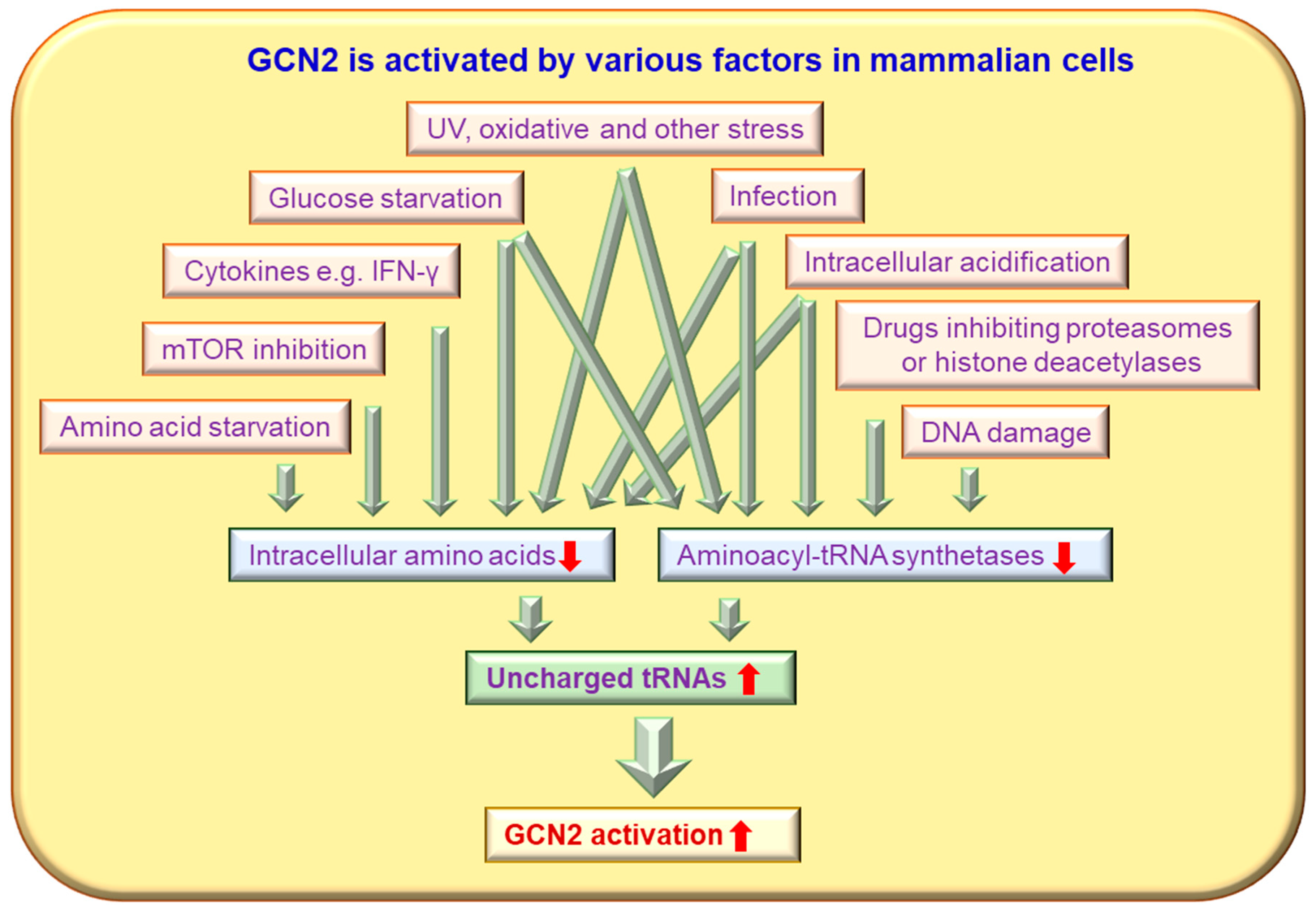
Figure 1. GCN2 is activated by various factors in mammalian cells. Many stimuli, such as nutrient starvation, glucose deprivation, purine starvation, high salt environment, UV irradiation, osmotic stress, high levels of urea, oxidative stress (H2O2), high salinity (NaCl), tryptophanol, anticancer drugs, and viral infections, can trigger enhanced GCN2 activity via uncharged tRNA-dependent pathways.
Ijms 24 04285 g001
Figure 2. GCN2 activity is positively and negatively regulated by various molecules in mammalian cells and in yeast. AMPK/Snf1, Hsp90/Hsp82, Sit4, PI3K and Asc1 up-regulate GCN2 activity, whereas eEF1A, p58IPK and IMPACT/Yih1 inhibit GCN2 activity.
Ijms 24 04285 g002
Figure 3. GCN2 orchestrates protein synthesis in mammalian cells and yeast. Many stimuli, such as nutrient starvation, will trigger enhanced GCN2 activity, which will subsequently inhibit global protein synthesis and promote the integrated stress response.
Ijms 24 04285 g003
Figure 4. Activated GCN2 impacts many intracellular pathways in mammalian cells. Enhanced GCN2 activity can enhance methionyl-tRNA synthetase, eIF-2α and other molecules to redistribute protein translation via many pathways.
Ijms 24 04285 g004
Disclaimer/Publisher’s Note: The statements, opinions and data contained in all publications are solely those of the individual author(s) and contributor(s) and not of MDPI and/or the editor(s). MDPI and/or the editor(s) disclaim responsibility for any injury to people or property resulting from any ideas, methods, instructions or products referred to in the content.