Article

The Phaseolus vulgaris Receptor-Like Kinase PvFER1 and the Small Peptides PvRALF1 and PvRALF6 Regulate Nodule Number as a Function of Nitrate Availability

1
Departamento de Biología Molecular de Plantas, Instituto de Biotecnología, Universidad Nacional Autónoma de México, Avenida Universidad 2001, Colonia Chamilpa, Cuernavaca, Morelos 62210, Mexico
2
Departamento de Docencia, Universidad Autónoma Agraria Antonio Narro, Saltillo, Coahuila 25315, Mexico
3
Centro de Ciencias Genómicas, Universidad Nacional Autónoma de México, Avenida Universidad 2001, Colonia Chamilpa, Cuernavaca, Morelos 62210, Mexico
*
Author to whom correspondence should be addressed.
Int. J. Mol. Sci. 2023, 24(6), 5230; https://doi.org/10.3390/ijms24065230
Submission received: 7 January 2023 / Revised: 18 February 2023 / Accepted: 20 February 2023 / Published: 9 March 2023

Abstract

:
Legumes associate with Gram-negative soil bacteria called rhizobia, resulting in the formation of a nitrogen-fixing organ, the nodule. Nodules are an important sink for photosynthates for legumes, so these plants have developed a systemic regulation mechanism that controls their optimal number of nodules, the so-called autoregulation of nodulation (AON) pathway, to balance energy costs with the benefits of nitrogen fixation. In addition, soil nitrate inhibits nodulation in a dose-dependent manner, through systemic and local mechanisms. The CLE family of peptides and their receptors are key to tightly controlling these inhibitory responses. In the present study, a functional analysis revealed that PvFER1, PvRALF1, and PvRALF6 act as positive regulators of the nodule number in growth medium containing 0 mM of nitrate but as negative regulators in medium with 2 and 5 mM of nitrate. Furthermore, the effect on nodule number was found to be consistent with changes in the expression levels of genes associated with the AON pathway and with the nitrate-mediated regulation of nodulation (NRN). Collectively, these data suggest that PvFER1, PvRALF1, and PvRALF6 regulate the optimal number of nodules as a function of nitrate availability.

1. Introduction

Nitrogen-fixing nodules are specialized organs that contain bacteria generically called rhizobia, which colonize the roots of legumes in a symbiotic interaction. Inside this organ, atmospheric dinitrogen (N2) is converted into ammonium (NH4+) through the activity of the nitrogenase complex present in differentiated bacteroids, providing plant hosts with this important source of bioavailable nitrogen [1]. Legumes attract compatible partners by exuding flavonoids into the rhizosphere, which are specifically detected by a bacterial membrane-associated protein, NodD [2]. After this perception, the rhizobia express the nodulation (nod) genes, resulting in the biosynthesis and secretion of the Nod factors [3]. Nod factors are in turn specifically perceived by a receptor complex located in the root hair tip membrane, which in Lotus japonicus L. includes NOD FACTOR RECEPTOR 1 (LjNFR1), LjNFR5, and SYMBIOTIC RECEPTOR-LIKE KINASE (LjSYMRK) [4,5]. Subsequently, several genes are expressed in the legume, including NODULE INCEPTION (NIN), which is a master regulator of nodulation necessary for infection, organogenesis, control of the nodule number, metabolism, and senescence [6,7,8].
Nodule organogenesis and nitrogen fixation require high levels of energy. To optimize the cost of energy with the benefits of nitrate uptake, legumes have developed local and systemic regulatory mechanisms to control the optimal number of nodules formed [9]. In Glycine max (L.) Merr., L. japonicus, and Medicago truncatula Gaertn., a systemic regulatory mechanism called autoregulation of nodulation (AON) controls the optimal number of nodules. During this process, RHIZOBIA-INDUCED CLAVATA3/ENDOSPERM-SURROUNDING REGION (CLE) (GmRIC1) and GmRIC2 (which are respectively known as LjCLE-R1 and LjCLE-R2 in L. japonicus and MtCLE12 and MtCLE13 in M. truncatula) are biosynthesized in the roots of these legumes, induced by the rhizobia [10,11]. GmRIC1 and GmRIC2 are transported to the aerial tissues and perceived by a leucine-rich repeat (LRR)-RLK receptor NODULE AUTOREGULATION RECEPTOR KINASE/SUPER NUMERIC NODULES/HYPERNODULATION AND ABERRANT ROOT (GmNARK/MtSUNN/LjHAR) [12,13,14]. The perception of these peptides leads to a reduction in the level of a mobile microRNA, miR2111, which promotes the accumulation of the mRNA of TOO MUCH LOVE (TML) in the roots, encoding an F-box protein that negatively regulates nodulation [15,16]. NIN is known to bind directly to the promoter sequences of the genes encoding LjCLE-RS1 and LjCLE-RS2 in L. japonicus or MtCLE13 in M. truncatula, activating their expression [6,8].
Nitrogen (N) is essential for plant development, and the presence of nitrate in the soil affects nodulation in a concentration-dependent manner through both local and systemic mechanisms [17,18]. In G. max, the production of the NITRATE-INDUCED CLE1 (GmNIC1) peptide is induced in roots in the presence of nitrate and is perceived by GmNARK in the root, inhibiting nodulation through an unknown local mechanism [10]. In M. truncatula, nitrate was reported to inhibit rhizobia-mediated NIN transcription through the action of NIN-like proteins (NLPs). At high nitrate levels, NIN expression is reduced and NLP proteins accumulate in the nucleus, bind to the few remaining NIN proteins, and suppress the expression of the NIN-activated genes, locally inhibiting nodule formation [19]. NLP1 in M. truncatula and its homolog NITRATE UNRESPONSIVE SYMBIOSIS1 (NRSYM1) in L. japonicus accumulate in the nucleus in response to high levels of nitrate and activate the expression of CLE peptides (CLE35 in M. truncatula and CLE-RS2 in L. japonicus) by directly binding to their promoter sequences, mediating the inhibition of nodulation through the AON pathway [20,21,22].
In addition to CLEs, other peptides are required for the legume–rhizobia symbiosis. Members of the C-terminally encoded peptide (CEP) family are important for the systemic induction of nodulation under low-nitrate conditions [23]. Nodule-specific cysteine-rich peptides (NCRs), exclusively present in legumes of the inverted repeat–lacking clade (IRLC), are essential for the differentiation of bacteroids [24]. Rapid alkalinization factors (RALFs) are another family of peptides involved in nodulation; for example, in M. truncatula, the overexpression of MtRALF1 caused an increase in aborted infections, a reduction in the number of nodules, and aberrant nodulation [25]. The mechanism by which mtRALF1 inhibits nodulation and whether this mechanism is conserved in other legumes are yet to be determined. Using a phosphoproteomic assay in Arabidopsis thaliana (L.) Heynh, [26] showed that FERONIA (FER), an RLK of the CrRLK1L (Catharanthus roseus (L.) G. Don RLK1L) subfamily, is a RALF1 receptor [26]. Furthermore, in a recent report, FER was shown to be required for the perception and function of more than half of the RALFs in A. thaliana [27], suggesting the involvement of FER in the regulation of nodulation through its perception of MtRALF1 in M. truncatula.
The FER–RALF module is known to regulate plant growth during biotic and abiotic stress conditions, including metal ion, salinity, drought, and mechanical stresses [28,29,30]. Interestingly, the plant pathogenic fungi Golovinomyces orontii and Magnaporthe oryzae, as well as the plant-parasitic nematode Meloidogyne incognita, produce RALF-like peptides (F-RALFs and MiRALFs, respectively) that bind to FER during their infection of A. thaliana, increasing their pathogenicity [31,32,33]. Recently, we reported that different CrRLK1Ls, including the closest FER homolog, are expressed in the nodules of four legumes [34], suggesting a possible role for the FER–RALF module during this symbiotic process.
Herein, we found that PvFER1, PvRALF1, and PvRALF6 are expressed in Phaseolus vulgaris L. nodules, which respond differentially to Rhizobium tropici inoculation under low- versus high-nitrate conditions. We also investigated the role of these three genes in the regulation of the optimal number of nodules as a function of a suitable or low-nitrate concentrations in the growth medium. Through RNA interference (RNAi) silencing and overexpression of PvFER1, PvRALF1, or PvRALF6, these genes were found to positively or negatively regulate the expression of downstream genes involved in the nitrate-mediated regulation of nodulation (NRN) and AON pathways under nothing and high-N conditions, respectively.

2. Results

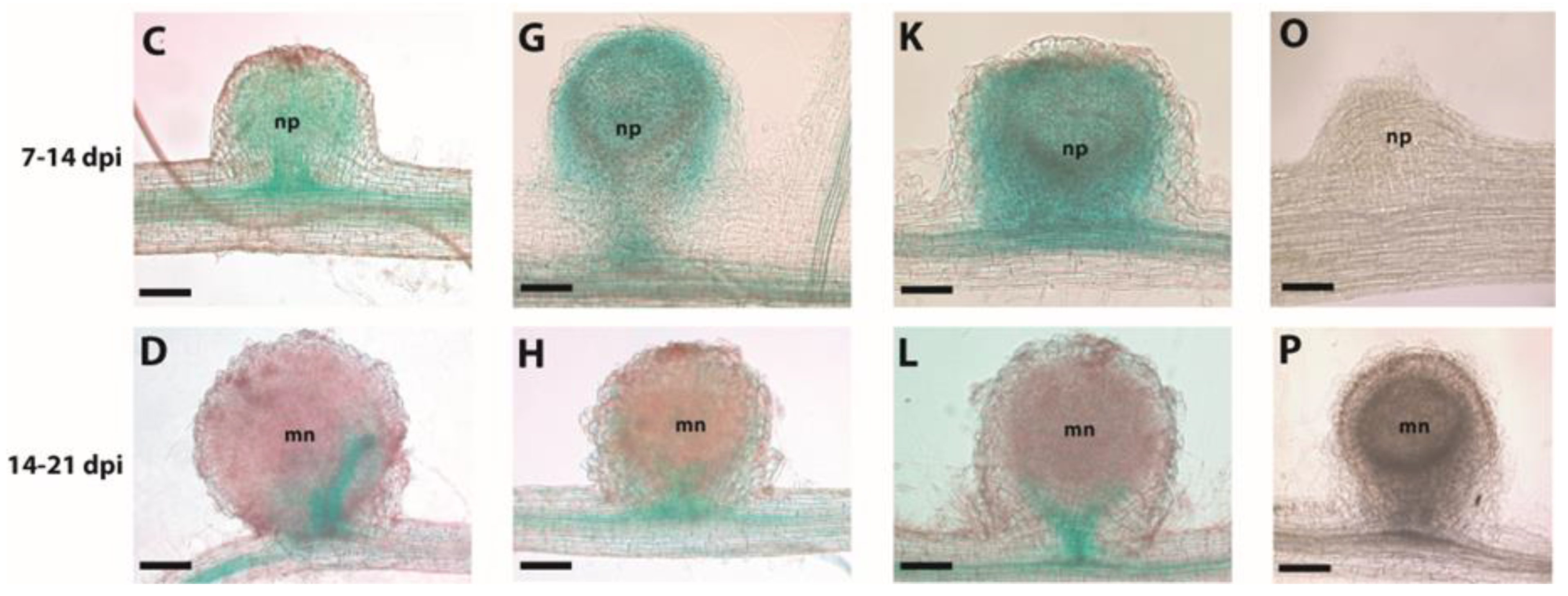
2.1. PvFER1 Is a P. vulgaris RLK Expressed in Nodules

Recently, we identified 33 CrRLK1L genes in the P. vulgaris genome, eight of which are expressed in roots [34]. A comparison of CrRLK1Ls expression in four legume species (P. vulgaris, L. japonicus, G. max, and M. truncatula) indicated that a gene homologous to the A. thaliana gene FER was expressed in all of their nodules [34]. Using a qPCR analysis, we also demonstrated that PvFER1 (Phvul.008G08100) was expressed in P. vulgaris roots inoculated with R. tropici, throughout various stages of nodule development [34]. An in-silico analysis of the primary structure of PvFER1 showed that it possesses a transmembrane domain, a characteristic extracellular malectin-like domain, and a cytoplasmic kinase domain (Figure S1), as is reported for FER in A. thaliana. These data prompted us to evaluate the role of PvFER1 in nodulation in P. vulgaris.
The spatial expression patterns of PvFER1 were investigated in transgenic P. vulgaris roots and nodules by analyzing the expression of a GUS reporter driven by the PvFER1 promoter (proPvFER1::GUS). GUS activity was analyzed in the hairy roots and nodules of the composite plants grown in 0 mM nitrate. PvFER1 promoter activity was observed in the apices and the central cylinder of the main and lateral roots (Figure S2A). In inoculated roots, the promoter activity was observed in the primordia of nodules during all stages evaluated (Figure 1A–C), as well as in the vascular bundles of the mature nodules (Figure 1D). Collectively, these data suggest that PvFER1 is a RLK expressed in the meristematic zones and vasculature of P. vulgaris roots and nodules.

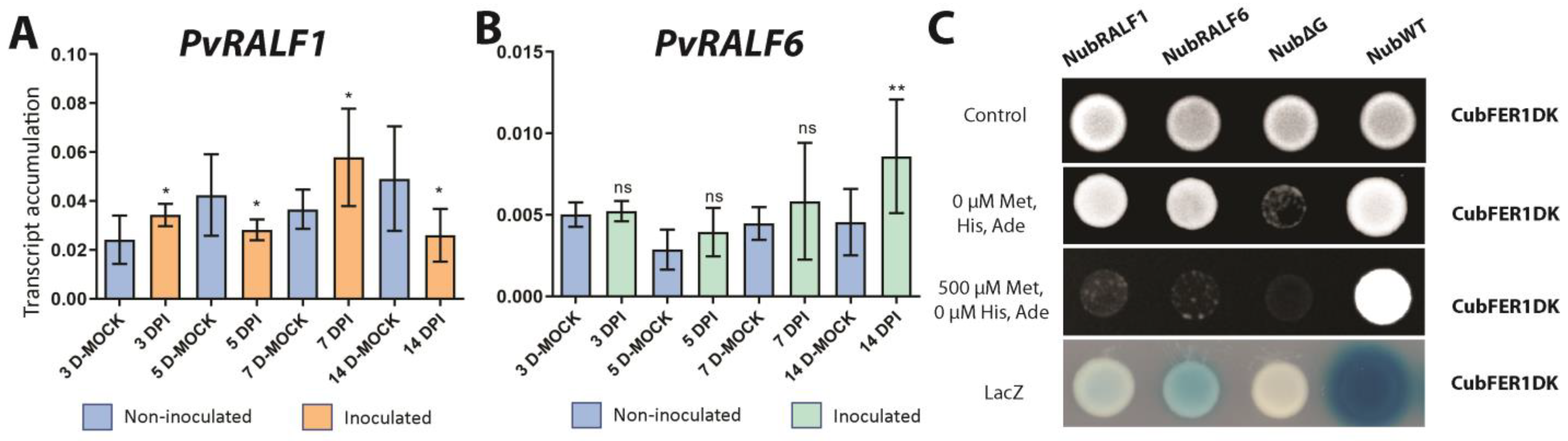
2.2. PvRALF1 and PvRALF6 Are Expressed in Nodules and Are Cysteine-Rich Peptides That Interact with PvFER1

It is well known that RALF peptides are ligands for CrRLK1L receptors, and there have also been reports of interaction of RALFs of a given plant species with a CrRLK1L of a different one [27,35,36,37,38]. To assess whether RALF contributes to the regulation of bean nodule number, we first examined the RALF gene family in P. vulgaris, revealing nine members that encode peptides with high similarity to RALF1 from A. thaliana. All nine PvRALFs showed conserved RRXL and YISY signatures, as well as a conserved secretion signal and four cysteine residues (Figure S3). A phylogenetic analysis of RALF amino acid sequences from A. thaliana, P. vulgaris, M. truncatula, and G. max and the RALF from the fungal plant pathogen Fusarium proliferatum grouped the nine PvRALFs into three of the four major clades formed (Figure S4); clade 1 contains four PvRALFs, clade 2 contains two PvRALFs, clade 3 contains three PvRALFs, while clade 4 contains no PvRALFs. These data are consistent with previous analyses of RALF peptides in 52 different plant species [39].
The expression profiles of the nine PvRALF genes were obtained from the P. vulgaris Gene Expression Atlas [40]. Five of the genes, PvRALF2, PvRALF4, PvRALF5, PvRALF6, and PvRALF8, show little or no expression, while PvRALF1, PvRALF3, PvRALF7, and PvRALF9 were expressed at high levels in all tissues tested (Figure S5). Among the four most expressed genes, PvRALF1 (Phvul.007G197000), the closest homolog of A. thaliana RALF1, was the most abundant in inoculated roots and nodules. Interestingly, PvRALF6 (Phvul.001G266400), the closest homolog of MtRALF1, showed the highest expression levels in nodules at 5 dpi. RT-qPCR analysis of P. vulgaris roots inoculated with R. tropici or a mock solution and grown in 0 mM nitrate confirmed that the PvRALF1 and PvRALF6 transcripts accumulated in differing patterns following the bacterial inoculation (Figure 2A,B); the PvRALF1 transcript abundance increased in the inoculated roots at 3 and 7 dpi and decreased at 5 and 14 dpi (Figure 2A), while PvRALF6 only showed an increase at 14 dpi (Figure 2B) compared with the mock-inoculated roots. These results suggest that RALF genes could fine-tune the different stages of nodulation.
The promoter activity of each of these two genes was analyzed by evaluating the spatial expression patterns of proPvRALF1::GUS and proPvRALF6::GUS using a histochemical analysis of GUS activity in P. vulgaris hairy roots. Similar to the results observed in the analysis of proPvFER1::GUS, the GUS activity of proPvRALF1::GUS and proPvRALF6::GUS was observed in the root apices, the central cylinder of the roots (Figure S2B,C), and in the nodules during the evaluated times (Figure 1E–L). These observations, together with previous reports in A. thaliana posing FER as the main receptor for most RALF peptides [27], strongly suggest that PvRALF1 and/or PvRALF6 could interact with PvFER1 in roots and nodules. This prompted us to confirm whether PvRALF1 and/or PvRALF6 interact(s) with PvFER1 using the split ubiquitin system. The observed growth of yeast indicated that Cub-PvFER1ΔK (a truncated version of PvFER1 that lacks the kinase domain to avoid undesirable effects in yeast) was able to restore spliced ubiquitinase function when NubWT, PvRALF1-Nub, or PvRALF6-Nub was present, but not when a mutated version of the Nub protein was used (NubΔG) (Figure 2C). These results confirm that PvRALF1 and PvRALF6 interact with PvFER1, strongly suggesting that these three proteins together could participate in nodulation in P. vulgaris.

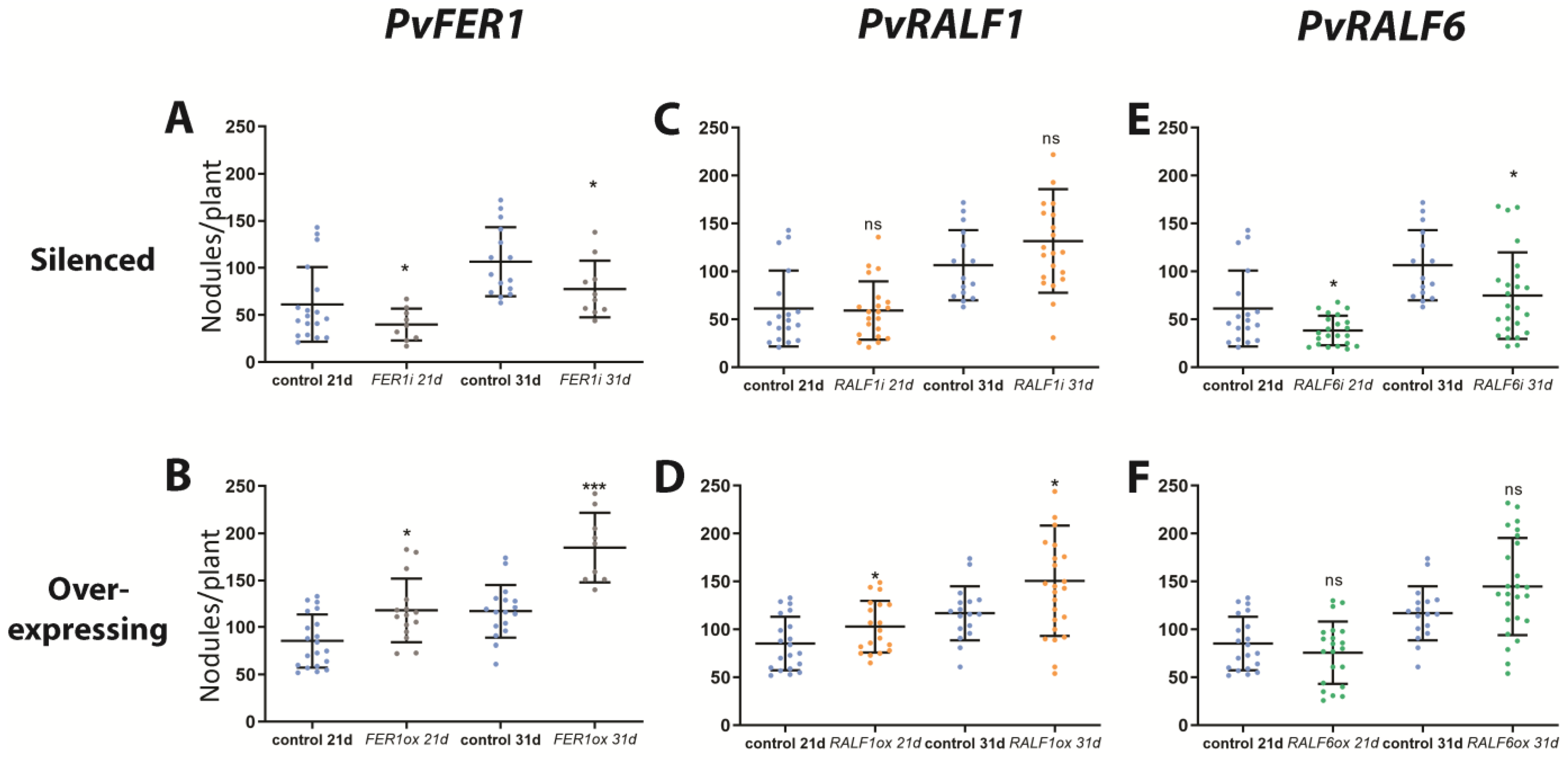
2.3. PvFER1, PvRALF1, and PvRALF6 Contribute to Determining the Number of Nodules Produced by P. vulgaris Roots

To assess the role of PvFER1 in nodulation, the effects of the RNAi-mediated downregulation and overexpression of this gene in P. vulgaris hairy roots grown under 0 mM of nitrate were investigated. The expression levels of this gene in the PvFER1-silenced (PvFER1i) and PvFER1-overexpressing (PvFER1ox) lines were first confirmed using a qPCR analysis, revealing an approximately 50% reduction in the PvFER1 transcripts in the PvFER1i roots and a five-fold increase in the PvFER1ox roots (Figure S6A,B). Importantly, no differences were observed in the length of the PvFER1i or PvFER1ox roots compared with the controls (Figure S7A,B). An analysis of the number of nodules in these lines revealed significant differences in the total number of nodules generated by these composite plants (Figure 3A,B); PvFER1i produced fewer nodules than the control roots at 21 and 31 dpi (Figure 3A), while the opposite was true for PvFER1ox (Figure 3B).
Similar to PvFER1, composite bean plants with PvRALF1 RNAi (PvRALF1i), PvRALF6 RNAi (PvRALF6i), PvRALF1-overexpressing (PvRALF1ox), or PvRALF6-overexpressing (PvRALF6ox) roots and their respective controls were inoculated with R. tropici-GUS and grown in the absence of nitrate. An RT-qPCR analysis showed approximately 70% and 80% reductions in the transcript accumulation levels of the PvRALF1i and PvRALF6i roots, respectively, and 35- and 200-fold increases in the PvRALF1ox and PvRALF6ox roots, respectively (Figure S6A,B). Similar to PvFER1, the PvRALF1/PvRALF6-silenced and -overexpressing roots showed no difference in root length to the controls (Figure S7A,B). PvRALF6-silenced lines inoculated with R. tropici showed a reduction in the number of nodules at 21 and 31 dpi compared with the control, while PvRALF1i showed no significant difference (Figure 3C,E). In contrast, PvRALF1ox displayed an increased number of nodules at 21 and 31 dpi compared to the control and transgenic roots overexpressing PvRALF6 which showed no changes (Figure 3D,F).
To assess the additional effects of these different levels of PvFER1, PvRALF1i, and PvRALF6i transcript accumulation on nodule organogenesis and nitrogenase activity, we analyzed the diameters and acetylene reduction of the nodules produced by these different transgenic roots. Only small differences in nodule diameters were found in both conditions compared with the control nodules (Figure S8A,B). Furthermore, non-significant differences in acetylene reduction were observed in the nodules of roots overexpressing PvRALF1 and PvRALF6 or silenced in PvFER1 (Figure S9A,B).

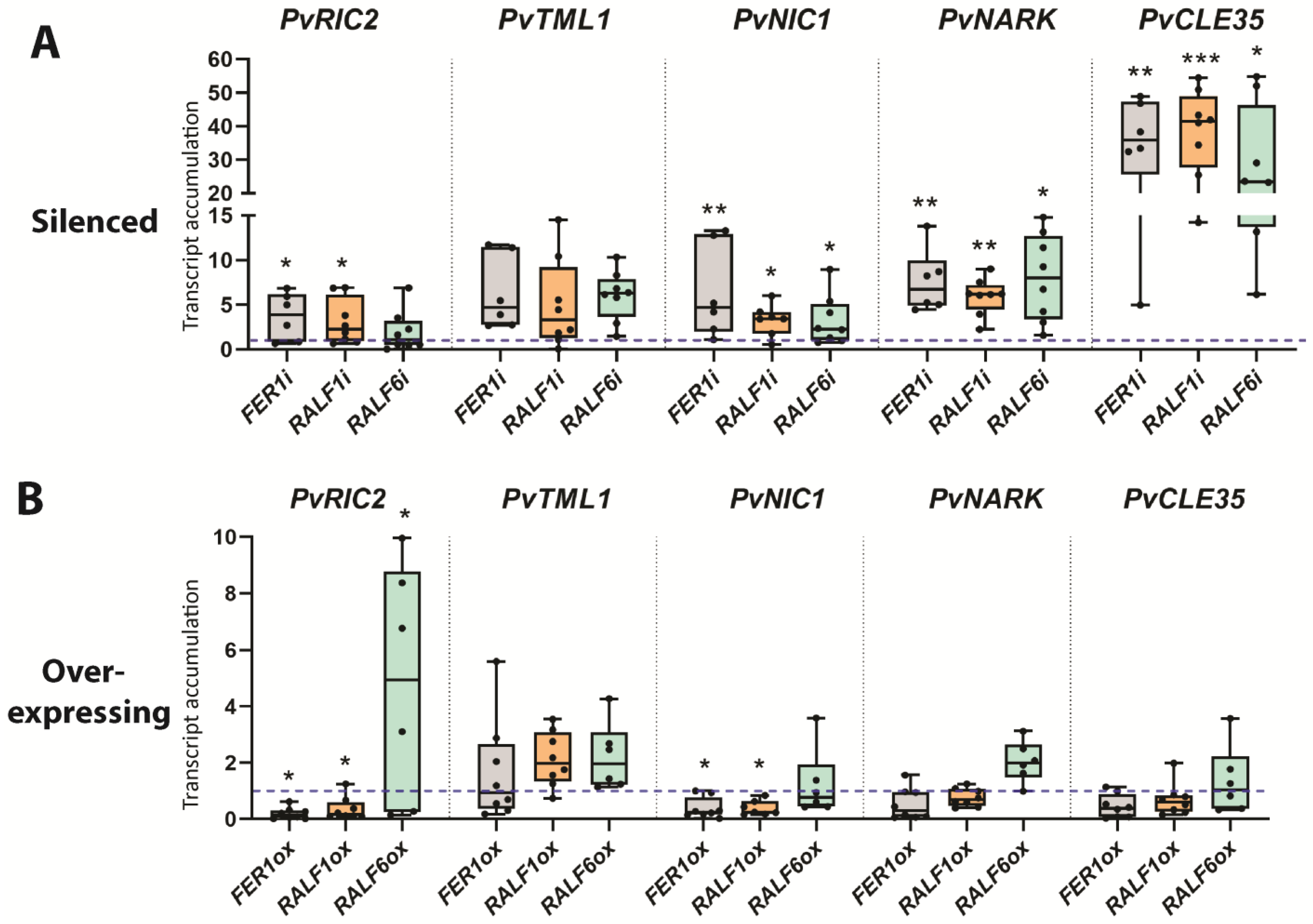
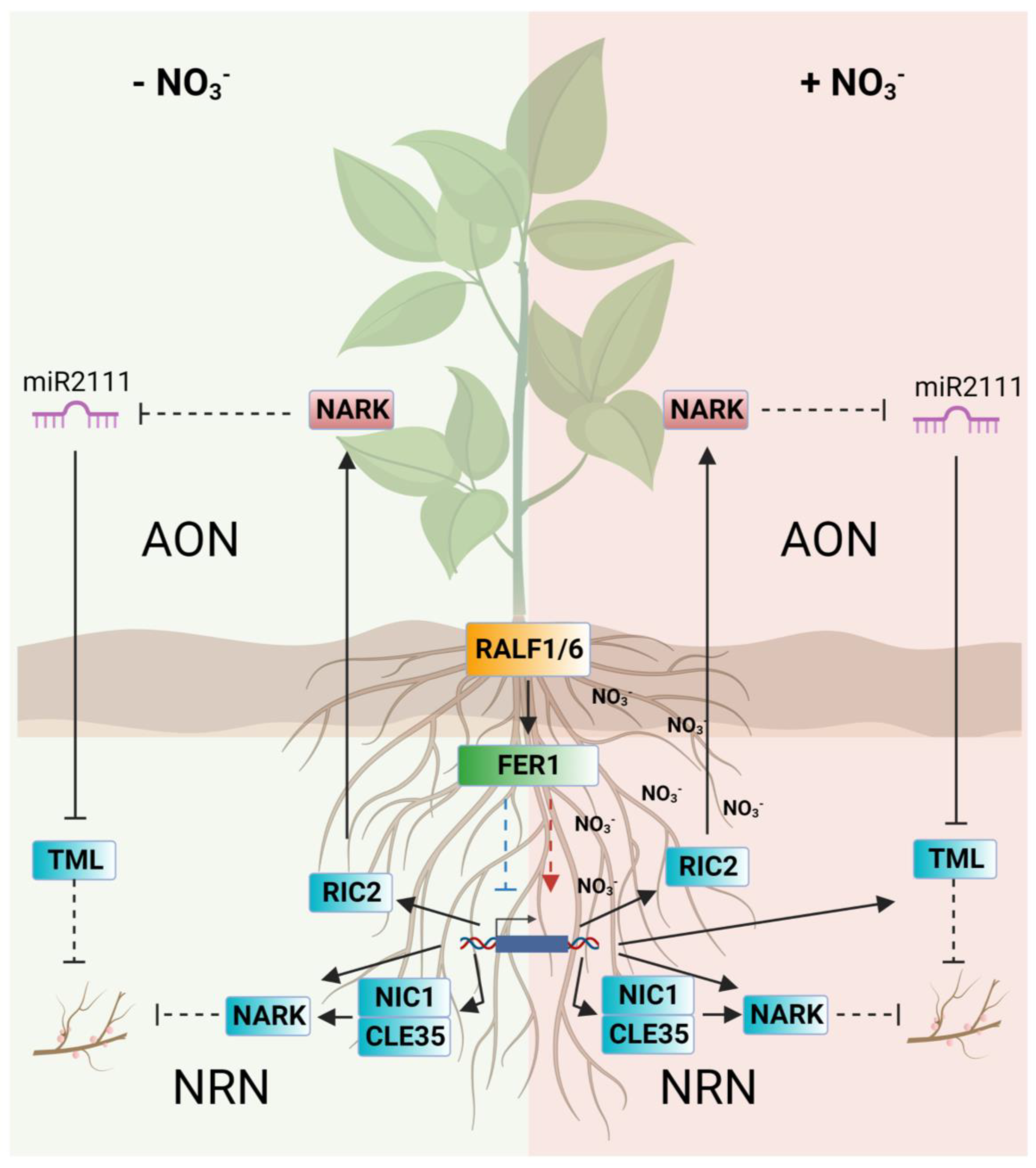
2.4. PvFER1, PvRALF1, and PvRALF6 Regulate the Expression of AON-Related Genes

To estimate the involvement of PvFER, PvRALF1 and PvRALF6 in nodulation signaling pathways, we examined the effect of silencing and overexpressing of these genes on the expression of key genes related to nodule organogenesis (nodule inception (PvNIN) and leghemoglobin (PvLEG)), nodule metabolism (sucrose synthase (PvSS) and glutamine synthetase (PvGS)), and AON (PvRIC2 and too much love (PvTML)). When comparing the expression of genes involved in organogenesis and metabolism of nodules in the PvFER1i and PvFER1ox roots with their controls, no differences were observed in the expression of the PvNIN and PvSS genes; however, PvLEG and PvGS were increased and decreased in FER1i and PvFER1ox roots, respectively (Figure S10A–D). No significant differences in the expression levels of PvNIN, PvLEG, PvGS, or PvSS were detected in the PvRALF1i/PvRALF6i and PvRALF1ox/PvRALF6ox roots (Figure S10A–D).
The expression of the AON marker gene PvRIC2 was increased in PvFER1i and reduced in the PvFER1ox roots compared with the controls (Figure 4A,B), while the accumulation of PvTML transcripts was similar between the PvFER1i and PvFER1ox roots and the controls (Figure 4A,B). PvRIC2 expression was increased in the PvRALF1i roots and reduced in the PvRALF1ox roots compared with the controls, whereas PvRIC2 expression was higher in the PvRALF6ox roots (Figure 4A,B). On the other hand, no change in PvTML expression was detected in the PvRALF1i/PvRALF6i and PvRALF1ox/PvRALF6ox lines (Figure 4A,B). Collectively, these data suggest that PvFER1, PvRALF1, and PvRALF6 participate in the regulation of the optimal number of nodules in bean roots by somehow modulating the expression of PvRIC2, while no clear effect was observed on the nodule organogenesis or metabolism genes.

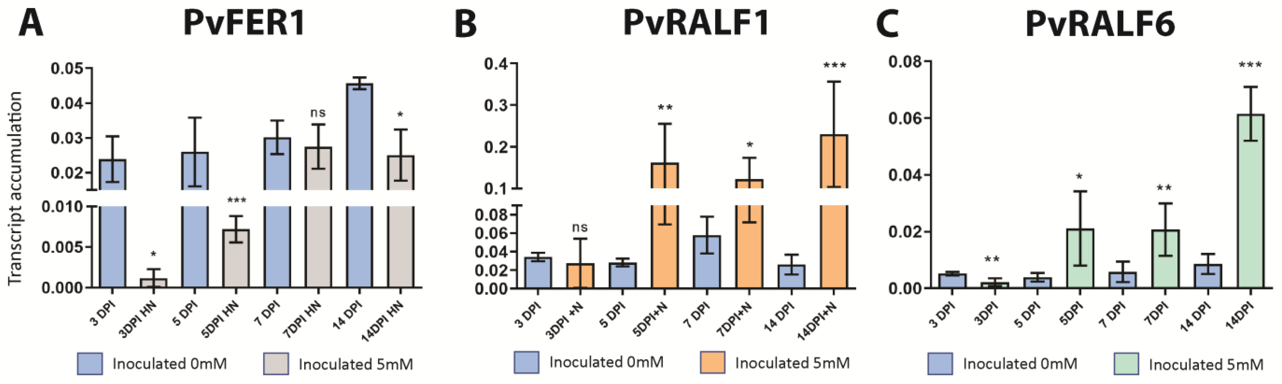
2.5. PvFER1, PvRALF1, and PvRALF6 Respond to Symbiosis under High-Nitrate Conditions

Previously, it was reported that FER and RALF1 regulate A. thaliana growth under high-C/N relation conditions and nitrate starvation [41,42]. This led us to evaluate the effect of nitrate on the accumulation of PvFER1, PvRALF1, and PvRALF6 transcript levels in P. vulgaris. A qPCR analysis of roots inoculated with R. tropici and grown at 5 mM of nitrate revealed a greater accumulation of PvRALF1 and PvRALF6 transcripts at 5, 7, and 14 dpi (Figure 5B,C) compared with plants grown under 0 mM nitrate (Figure 2A,B). In contrast, PvFER1 showed reduced levels of transcript accumulation under high-nitrate conditions (5 mM) at 3, 5, and 14 dpi (Figure 5A). These observations indicate that the expression of PvFER1, PvRALF1, and PvRALF6 are regulated in response to both nitrate and rhizobia, suggesting a role for these genes in regulating nodulation under high-nitrate conditions.

2.6. PvFER1, PvRALF1, and PvRALF6 Regulate the Expression of NRN-Related Genes

To investigate the potential involvement of the three analyzed genes in the NRN pathway, the effects of silencing and overexpressing PvFER1, PvRALF1, and PvRALF6 on the expression of the NRN marker genes PvCLE35, NODULE-INDUCED CLE1 (PvNIC1), and PvNARK under 0 mM nitrate were examined. All genes tested showed increased expression levels in transgenic roots in which either PvFER1, PvRALF1, or PvRALF6 were silenced, whereas the overexpression of PvFER1 and PvRALF1 reduced the expression of PvNIC1; no significant changes were detected in the transcript levels of PvCLE35 or PvNARK in the PvFER1-, PvRALF1-, or PvRALF6-overexpressing roots (Figure 4A, B). These results suggest that PvFER1, PvRALF1, and PvRALF6 also contribute to the regulation of the expression of the genes in the NRN pathway.

2.7. PvFER1, PvRALF1, and PvRALF6 Regulate the Number of Nodules Produced by P. vulgaris under High-Nitrate Conditions

To examine a probable role by which PvFER1, PvRALF1, and PvRALF6 regulate nodule number as a function of nitrogen availability, transgenic roots in which these genes were either silenced or overexpressed were challenged by growth under high-nitrate conditions. The PvFER1i, PvRALF1i, and PvRALF6i lines developed more nodules than the control roots under 5 mM of nitrate at 21 and 31 dpi (Figure 6A–C), whereas transgenic roots that overexpressed PvRALF1 or PvRALF6, but not PvFER1, showed fewer nodules than the control roots under these same conditions (Figure 6D–F). Similar results were observed with 2 mM of nitrate in both the silenced and overexpressed roots; however, the inhibition of the nodule number was reduced (Figure S11). No significant differences in nodule diameters were observed in any of these transgenic lines grown under 2 or 5 mM of nitrate (Figure S12).
The expression of the nodule organogenesis marker genes (PvNIN and PvLEG) remained virtually unchanged in the roots in which PvFER1, PvRALF1, or PvRALF6 was silenced or overexpressed when grown under high-nitrate conditions (Figure S13A–D). In PvRALF1ox roots, an increase in the expression of PvGS and PvSS was observed; however, their expression was not affected in the PvFER1ox or PvRALF6ox lines, nor in PvFER1i, PvRALF1i, or PvRALF6i (Figure S13A–D). The expression of the AON marker gene PvRIC2 was elevated in the PvFER1- and PvRALF1-silenced roots (Figure 7A), while the PvTML transcripts were reduced in the PvFER1-, PvRALF1-, and PvRALF6-silenced roots under this nodulation-inhibitory condition (Figure 7A). When PvRALF1 was overexpressed, an increase in the accumulation of PvRIC2 and PvTML transcripts was detected (Figure 7B). PvNARK transcript levels were reduced in the silenced roots of any of the three genes; however, no changes were detected in the overexpressing roots (Figure 7A,B). The transcript levels of the NRN marker gene PvNIC1 showed a reduction in the PvRALF1-silenced roots and an increase in the PvRALF1-overexpressed roots (Figure 7A,B). Finally, the expression of PvCLE35 did not change in the silenced roots of any of the three genes examined, but was increased in the roots that overexpressed PvFER1 or PvRALF1 (Figure 7A,B). These data strongly suggest that PvFER1, PvRALF1, and PvRALF6 participate in the control of the number of nodules by regulating the expression of genes of the AON and NRN pathways at high-nitrate concentrations (5 mM).

3. Discussion

The RALF family of peptides and FER, which is a member of the CrRLK1L receptor subfamily, are highly conserved and have, in the last decade, been shown to be involved in a plethora of processes. The interaction between RALF1 and FER was first described in the inhibition of root elongation in A. thaliana [43]. Since then, there has been a rapid increase in the number of processes in which these ligand–receptor complexes have been reported to be involved, as recent review articles show [28,29,30]. Despite this research interest, the role of the FER–RALF ligand–receptor complex during symbiotic associations, such as legume–rhizobia symbioses, has been poorly studied. Here, we report that PvFER1, PvRALF1, and PvRALF6 participate in the regulation of nodule number in P. vulgaris as a function of nitrate availability.
It is well known that RALF1 sensing by FER promotes the inhibition of root elongation and root hair tip growth [26,43,44,45,46]. This is important for nodulation because root hairs constitute the main site at which rhizobia enter legume roots during symbiosis. In the present work, we found that PvFER1, PvRALF1, and PvRALF6 are expressed in P. vulgaris roots, and the activity of their promoters was also detected in the central cylinder and root apices (Figure S2), similar to what was reported for their homologs in A. thaliana and rice (Oryza sativa L.) [44,47]. These observations suggest that these genes may have a role in root development in P. vulgaris. Haruta et al. (2014) showed that the exogenous addition of synthetic RALF inhibits root expansion in A. thaliana, a process that depends on the function of FER; however, in transgenic P. vulgaris roots with either silenced or overexpressed PvFER1, PvRALF1, or PvRALF6 genes, we observed no differences in root growth compared with the control transgenic roots (Figure S7). Considering that there are 17 CrRLK1Ls and 33 RALFs in A. thaliana [26,27,39,48], and 37 CrRLK1Ls and nine PvRALFs in the P. vulgaris genome [34,39], the apparent lack of phenotype in P. vulgaris roots lacking or overexpressing PvFER1, PvRALF1, or PvRALF6 transcripts is likely due to the overlapping functions of the remaining CrRLK1Ls and RALF genes. An alternative explanation could be that the remaining transcript levels in the silenced roots (Figure S6) may be sufficient to mediate normal root growth, although no phenotype was observed in the hairy root system control used here. An analysis of P. vulgaris plants lacking or overexpressing multiple RALFs and CrRLK1Ls could help answer this question and may also reveal novel functions for the other RALF and CrRLK1L genes in P. vulgaris.
In M. truncatula, MtRALF1 expression was reported to be induced during nodulation, and its overexpression impairs the symbiosis between this legume and the bacterium Sinorhizobium meliloti [25]. In line with this, during a comparative analysis of the expression of CrRLK1Ls in four legumes, a cluster of CrRLK1L genes expressed in nodules was found, with the expression of the closest homolog of A. thaliana FER detected in the nodules of all legumes tested [34]. An analysis of the expression and promoter activity of PvFER1, PvRALF1, and PvRALF6 in inoculated P. vulgaris roots revealed that they are induced in nodules during various stages of their organogenesis (Figure 1 and Figure 2A,B). Several reports support the interaction between the RALF peptides and CrRLK1Ls; nearly half of the A. thaliana RALFs (16 of 33) can be perceived by FER, leading to the inhibition of root elongation [27]. Along the same line, RALF peptides from different plant species and those from fungi and M. incognita can interact with FER from A. thaliana [31,32,33]. Through protein–protein interaction analyses, we found that PvRALF1 and PvRALF6 interact with PvFER1 in P. vulgaris (Figure 2C), suggesting a role of the PvRALF1/6 perception by PvFER1 at different stages of nodulation in P. vulgaris.
Reverse genetic analysis revealed a role for PvFER1, PvRALF1, and PvRALF6 in regulating the number of nodules under low- and high-nitrate conditions (Figure 3 and Figure 6); however, little or no effect on nodule development and function was observed (Figures S8, S9 and S12). In M. truncatula, it was previously reported that the overexpression of MtRALF1 impairs the symbiosis between this legume and S. meliloti, affecting not only the number of nodules, as we observed in P. vulgaris, but also impacting their function and decreasing their bacteroid content [25]. The differences observed in nodulation between these two legumes could be due to the different types of nodules that each of them forms; namely, indeterminate nodules in M. truncatula and determinate nodules in P. vulgaris. It is well known that there are significant differences at the genetic, morphological, and physiological levels between these two types of nodules [49,50]. A further exploration of the role of the FER and RALF genes in other legumes could answer the questions of whether there is a conserved role of these two genes in nodulation and whether they have additional functions in the development of indeterminate nodules.
Many genes have been reported to be associated with the regulation of nodule number, participating in both the local and systemic signaling pathways involved in the induction or repression of nodulation. The loss of function of the CLEs, NARK, or TML genes in several legumes induced an increase in the number of nodules [10,11,12,13,14], contrary to what was observed with the silencing of PvFER1, PvRALF1, and PvRALF6 but similar to their overexpression under 0 mM of nitrate (Figure 3 and Figure 6). Several authors have reported that the RALF1–FER complex regulates the transcription, splicing, and translation of genes associated with root development, root hair growth, and the responses to biotic and abiotic stresses [51,52,53,54]. Here, we describe that PvFER1, PvRALF1, and PvRALF6 are involved in regulating the expression of NARK, TML, and certain CLEs (RIC2, NIC1 and CLE35) under 0 and 5 mM of nitrate (Figure 4 and Figure 7). In agreement with this, various authors [54,55,56] reported that among the many differentially expressed genes in A. thaliana fer1 and fer4 mutants, or overexpressing RALF23, numerous genes encoding PvNARK-like LRR receptors were differentially expressed (Table S2). Furthermore, three of the seven A. thaliana TML genes were upregulated in fer1, one was upregulated and one downregulated in fer4, and none were differentially expressed in RALF23ox when compared with the wild-type control. Interestingly, none of the seven CLE genes in A. thaliana showed changes in expression in any of the lines tested (Table S2) [54,55,56]. These data suggest that the transcriptional regulation of the NARKs and TML by PvRALF1, PvRALF6, and PvFER1 observed in the present work is conserved in A. thaliana, whereas the regulation of CLE gene expression is likely associated with nodulation in legumes. A more detailed analysis of the transcriptional regulation of the AON and NRN pathway genes by FER and RALF in other legumes will confirm whether this mechanism is conserved during the nodulation process. Furthermore, a global assessment of the effect of changes in RALF and FER gene expression in P. vulgaris and other legumes, determined using an RNA-seq analysis, will lead to a deeper understanding of the role of these genes in nodulation.
FER is known to participate in the regulation of plant growth under low-nitrate conditions by interacting with the TOR/RAPTOR complex and with the E3 ubiquitin ligase ARABIDOPSIS TOXICOS EN LEVADURA 6 (ATL6) [41,42]. A comparative analysis of the effect of silencing and overexpressing PvFER1, PvRALF1, and PvRALF6 on the number of nodules showed contrasting effects under different nitrate concentrations, indicating that the PvRALF1/6–PvFER1 complex may be positively or negatively involved in regulating the number of nodules, depending on the nutritional status of the plant (Figure 3 and Figure 6). Similar opposite functions have been previously documented for the RALFs and CrRLK1Ls; for instance, the RALF1–FER complex promotes root hair growth and inhibits root elongation [26,44]. Under biotic stress, the RALF17–FER complex acts as a positive regulator of plant defense responses, while RALF23/33/34–FER functions as a negative regulator [57]. In abiotic stress conditions, FER and the CrRLK1Ls HERCULES1, HERCULES2, THESEUS1, and MEDOS1–4 can act as positive or negative regulators of root development, but have an opposite function in the hypocotyls, in response to metal ions [58,59].
In the present study, we identified some differences in the nodule number and expression of genes related to the AON and NRN pathways when PvFER1, PvRALF1, and PvRALF6 were differentially expressed. There was a direct association between changes in the expression of PvCLE35, PvRIC2, PvNIC1, PvNARK, or PvTML and the reduction in the number of nodules in silenced PvFER1 and PvRALF6 in 0 mM of nitrate, and overexpressing PvRALF1 in roots grown with 5 mM of nitrate. Similarly, the effect of overexpressing PvFER1 or PvRALF1 in 0 mM of nitrate and silencing PvFER1, PvRALF1, or PvRALF6 grown with 5 mM of nitrate had a consistent effect on increasing the number of nodules and changes in the expression of those genes from the AON and NRN pathways. In the remaining conditions (silencing of PvRALF1 or overexpressing PvRALF6 in 0 mM nitrate, and overexpression of PvFER1 or PvRALF6 at 5 mM nitrate), however, there is no obvious explanation for the observed phenotype based on the expression of the genes related to AON and NRN (Figure 4 and Figure 7). These observations strongly suggest the existence of additional participants or additional pathways involved in the regulation of the nodule number by PvFER1, PvRALF1, and PvRALF6 in P. vulgaris. This is not surprising as the versatility of FER functions in plant development has long been known [28,29,30]; for instance, the regulation of hormonal signaling by the RALF–FER complex may be an additional mechanism by which RALF and FER could regulate the number of nodules in P. vulgaris, since the importance of these signaling molecules has been reported in nodulation [60,61]. Another possibility could be based on the relationship between gene expression patterns and function. In this sense, by modifying the expression levels of PvFER1, PvRALF1, and PvRALF6 in the roots, the normal expression patterns (the specific zone, level and/or condition of expression) of the genes of the AON and NRN pathways, could be altered. Therefore, it can be argued that beyond its function in nodules, there may be unexpected effects due to the changes in its expression patterns. For example, it is known that the function of gibberellins is specific as a function of time, and in the early stages of nodulation, functions as an inducer of nodule organogenesis, but its latter function is as an inhibitor of nodulation [61]. A similar effect may be happening due to changes in the genes evaluated here. Specific silencing or overexpression of PvFER1, PvRALF1, and PvRALF6 will shed light on the mechanism by which these genes exert their function on nodulation through the AON and NRN pathways.
AON-related genes are also involved in the regulation of the number of nodules in P. vulgaris under low-phosphate conditions [62]. Furthermore, it was recently reported that RALF23 is induced by PHOSPHATE STARVATION RESPONSE 1 (PHR1) upon phosphate starvation in A. thaliana and, together with FER, participates in the suppression of plant immunity under low-phosphate conditions [56]. It would therefore be interesting to analyze whether PvFER1, PvRALF1, and PvRALF6, or other CrRLK1L and RALF genes in P. vulgaris, play a role in the regulation of nodule number as a function of phosphate concentration. The functional analysis of FER and RALF during nodulation under low-phosphate conditions, and in other different nutritional conditions, will shed light on the possible role of these ligand–receptor complexes as key regulators of the number of nodules required in legumes in relation to various nutritional environments.
In summary, in this work we demonstrated that PvFER1, PvRALF1, and PvRALF6 are expressed during the nodulation process and presumably regulate the number of nodules in P. vulgaris as a function of nitrate levels. Based on these results, we propose a model that connects the functions in nodulation of PvRALF1, PvRALF6, and PvFER1 in relation to nitrate availability (Figure 8), probably mediated by its modulation of the expression levels of CLE peptides and their NARK receptors in the roots.

4. Materials and Methods

4.1. Identification, Bioinformatic Analysis, and Phylogeny of P. vulgaris FER and RALFs

Following the same strategy used to identify CrRLK1L proteins in more than 50 plant species [34], RALF peptides from A. thaliana, P. vulgaris, G. max, and M. truncatula were identified using a BLASTP search of the Phytozome v12 database (https://phytozome.jgi.doe.gov, accessed on 10 October 2022) [63], using the RALF1 peptide sequence of A. thaliana as a query.
The peptide alignment of RALF amino acid sequences from the four aforementioned plant species was performed using the MUSCLE algorithm within the AliView alignment editor [64], followed by a manual optimization of misaligned regions. A maximum-likelihood phylogenetic tree [65] was constructed for the sequence alignment using IQ-TREE v1.6.12 [66] and the JTT+F+R10 substitution model, with 1000 bootstraps and default parameters.
The conservation of the protein motifs present in the four plants tested was determined using the MEME analyzer (http://meme-suite.org, accessed on 28 May 2020) [67]. This was carried out using the full-length amino acid sequences, setting the maximum number to 15 motifs, the number of motifs expected to any number of repeats, and the length of the motif as 10 to 200 amino acids. The other parameters were kept as default.

4.2. In Silico Expression Profile Analysis of RALF Genes in P. vulgaris

Expression profiles of the nine members of the P. vulgaris RALF gene family were retrieved from the P. vulgaris Gene Expression Atlas PvGEA (https://plantgrn.noble.org/PvGEA/, accessed on 5 March 2022) [40]. To better visualize these expression patterns, a heat map was made of the expression profiles of the nine genes. The distribution and abundance of differentially expressed genes were also presented as heatmaps using Heatmap Illustrator v1.0 (HemI) [68].

4.3. Plant Growth Conditions

P. vulgaris cv. Negro Jamapa seeds were surface-sterilized and incubated for 2 days at 28 °C in the dark. At 2 days post-germination (dpg), the seedlings were planted in pots of sterile vermiculite and inoculated with R. tropici CIAT 899 at an OD600 of 0.05 or with mock solution in the case of the control plants. The plants were irrigated with Fahraeus medium [69] with either 0 mM nitrate or 2 or 5 mM nitrate (nodule-inhibitory conditions) to analyze the expression, promoter activity, and function of the genes of interest. Roots at 3, 5, 7 and 14 days post-inoculation (dpi) were harvested, frozen in liquid nitrogen, and then stored at −75 °C until required for RNA extraction.

4.4. Composite Plant Generation

P. vulgaris seedlings (2 dpg) were inoculated with a selected clone of Agrobacterium rhizogenes carrying the plasmid construct of interest for promoter, silencing, or overexpression analysis of the gene of interest, as well as their respective controls. The inoculated seedlings were placed at the top of 15 mL Falcon tubes, with the cotyledon and the root outside and submerged into Fahraeus medium, respectively. Then, the seedling-containing tubes were introduced into 50 mL glass test tubes, filling with Fahraeus medium below the cotyledon to maintain the humidity. At 12 dpi with A. rhizogenes, transgenic roots expressing the fluorescent reporter protein were selected, and non-transformed and non-fluorescent transgenic roots were eliminated. Composite plants were transplanted into pots with vermiculite and inoculated with R. tropici CIAT899 (OD600 of 0.05) or mock-inoculated for further analysis.

4.5. RNA Extraction and qPCR Analysis

RNA was isolated from the frozen tissues using Trizol reagent, following the manufacturer’s instructions (MilliporeSigma, Burlington, MA, USA). The RNA integrity was verified using electrophoresis and its concentration was assessed using a NanoDrop2000 spectrophotometer (Thermo Fisher Scientific, Waltham, MA, USA). Any genomic DNA contamination was removed by incubating the samples with RNase-free DNase (10 U/µL; Roche, Basel, Switzerland) at 37 °C for 30 min. The cDNA was synthesized from 200 ng RNA using RevertAid Reverse Transcriptase (200 U/µL; Thermo Fisher Scientific). Next, a qPCR assay was performed using a Maxima SYBR Green/ROX qPCR kit (Thermo Fisher Scientific) on the Applied Biosystems QuantStudio 5 qPCR system (Thermo Fisher Scientific), following the manufacturer’s instructions. The thermal cycling conditions were as follows: 95 °C for 10 min, followed by 40 cycles of 95 °C for 15 s and 60 °C for 60 s. The melting curve stage was evaluated under the following thermal conditions: 95 °C for 15 s, 60 °C for 60 s, and 96 °C for 5 s. Relative expression values were calculated using the 2−ΔCt method [70] with the widely used elongation factor 1α (PvEF1α) and the P. vulgaris insulin-degrading enzyme (PvIDE) as internal references [71,72]. Similar results were obtained using either PvIDE or PvEF1α as the reference gene. Only the PvIDE reference gene was selected to normalize the data in the figures. Three biological replicates were performed with three technical repeats. The gene-specific oligonucleotides used for the RT-qPCR assay are listed in Table S1.

4.6. Plasmid Design and Construction

To analyze the activity of each promoter, at least 2000 bp of the promoter sequence upstream of the PvFER1, PvRALF1, or PvRALF6 translation start site were amplified from P. vulgaris genomic DNA and then cloned into the pENTR/SD/D-TOPO vector (Thermo Fisher Scientific). A Gateway LR reaction was performed between the entry vector (pENTR-pFER1, pENTR-pRALF1, or pENTR-pRALF6) and the destination vector (pBGWSF7.0) [73] according to the manufacturer’s instructions (Thermo Fisher Scientific). The resulting expression vectors (pBG-pFER1, pBG-pRALF1, and pBG-pRALF6) were cloned into A. rhizogenes K599. The pBGWSF7.0 vector allows the expression of the GUS reporter under the control of the promoter under evaluation (proPvFER1::GUS, proPvRALF1::GUS, and proPvRALF6::GUS) to indirectly determine promoter activity through GUS enzymatic activity. An empty version of the pBGWSF7.0 vector was used as the control for promoter activity analysis, as previously reported [72,73].
For the gene overexpression analyses, the coding sequences (CDSs) of PvFER1, PvRALF1, and PvFER6 were amplified from 2 dpg P. vulgaris root cDNA. The fragment containing each CDS was cloned into pENTR/SD/D-TOPO vectors (pENTR-FER1CDS, pENTR-RALF1CDS, or pENTR-RALF6CDS) and then recombined with the vector pH7FWG2D [73] using the Gateway system. The generated constructs (pH7-PvFER1CDS, pH7-PvRALF1CDS, and pH7-PvRALF6CDS) were introduced into A. rhizogenes K599. The pH7FWG2D vector is suitable for driving the expression of a chimeric protein fused with green fluorescent protein (GFP), under the control of the 35S promoter. As a control vector, an empty version of the pH7FWG2D vector (identified as GUS in results), which expresses a GFP-GUS fusion was used for all the overexpression analyses [72,73].
For the RNAi constructs, an amplified 120-bp fragment of the PvFER1, PvRALF1, or PvRALF6 5′ untranslated region (UTR) was first cloned into the pENTR vector (pENTR-FER1i, pENTR-RALF1i, or pENTR-RALF6i) and then recombined with the ptdT-DC-RNAi vector (ptdT-FER1i, ptdT-RALF1i, or ptdT-RALF6i) [74]. ptdT-DC-RNAi allows the expression of a stem-loop RNA structure containing a sequence complementary to the target, which is processed by the RNA-induced silencing complex and then results in the silencing of the target. As the control vector, ptdT-DC-RNAi containing a scrambled DNA sequence (identified as SAC in results) was used for all the RNAi analyses, as reported in previous works [72,74,75].
All generated vectors were confirmed by sequencing. The sequences of the different oligonucleotides used are listed in Table S1.

4.7. Analysis of the Promoter Activities

The promoter activities of the genes of interest were evaluated using the GUS staining protocol [76]. Roots containing one of the vectors for promoter analysis (pBG-pFER1, pBG-pRALF1, and pBG-pRALF6) were harvested at 7, 14 or 21 dpi. To examine the GUS activity, the roots were clarified, rehydrated, and mounted as described by Jefferson (1987). Treatment times were up to 1, 2, and 6 h for pBG-pFER1, pBG-pRALF1, and pBG-pRALF6 composite plants, respectively. To rule out a false positive due to the GUS endophytic activity, control transgenic roots carrying the empty vector (EV) with no promoter to mediate GUS report expression, were also subjected to GUS staining for up to 24 h. No staining was observed, ruling out false positive.
The stained roots were observed using bright-field microscopy (02552240; AmScope, Irvine, CA, USA).

4.8. Analysis of the Number and Diameter of Nodules in Transgenic Roots

Composite plants expressing any of the different silencing, overexpressing, or control (pTdT-SAC, pH7FWG2D EV) constructs were inoculated with R. tropici CIAT899 GUS. These transgenic roots were harvested at 21 and 31 dpi under 0, 2, and 5 mM of nitrate conditions. The number and diameter of the nodules were analyzed using FIJI software [77,78].

4.9. Nitrogenase Activity Analysis

Nitrogenase activity was determined by measuring acetylene reduction [79,80]. Transgenic roots nodulated with R. tropici CIAT899 WT (21 dpi) were placed in 160 mL glass vials. After sealing the vials with rubber stoppers, 2 mL of air was withdrawn with a syringe and the same amount of acetylene was injected into each of the vials. The samples were incubated for 2 h at room temperature, and the ethylene production was measured in a gas chromatograph (Varian model 3300; Agilent Technologies, Santa Clara, CA, USA), as described by Ramírez et al. (1999) [80]. The nodules were then removed and dehydrated to measure their dry weight. Acetylene reduction was expressed as the µmol of ethylene h−1 g of nodule dry weight−1.

4.10. Protein Interaction Analysis

To examine whether the PvFER1 peptide interacts with PvRALF1 or PvRALF6, vectors were first constructed to perform the split ubiquitin system [81]. A modified version of the PvFER1 CDS lacking the kinase domain was cloned into the pENTR/SD/D-TOPO vector (pENTR-FER1ΔK). The previously described pENTR-RALF1CDS and pENTR-RALF6CDS vectors were recombined with the destination vector MetYC_GW (Cub destination vector), while pENTR-FER1ΔK was combined with pXN22_GW (NubG destination vector) [82] for the split-ubiquitin system assays.
Yeast strains THY.AP4 (MATa ura3, leu2, lexA::LacZ::trp1 lexA::HIS3 lexA::ADE2) and THY.AP5 (MATα URA3, leu2, trp1, his3 loxP::ade2) were transformed with the PvFER1ΔK-Cub, PvRALF1-Nub, and PvRALF6-Nub constructs using the LiAc protocol previously described by Lalonde et al. (2010) [81]. Fusions that did not interact with soluble NubWT, which has a high affinity for the Cub domain, were considered false negatives, while those that interacted with NubΔG (which has a reduced affinity for Cub) corresponded to false positives [81].

4.11. Statistical Analysis

To establish the significance of the results obtained, differential statistical analyses were performed. Transcript accumulation data for all evaluated genes, the number of nodules, and the acetylene reduction data were analyzed using a non-parametric Mann–Whitney test. The diameters of the nodules were analyzed using a Kruskal–Wallis test, followed by Dunn’s multiple comparisons. All statistical tests were carried out using GraphPad Software version 8.2.263 (GraphPad Software, San Diego, CA, USA).

Supplementary Materials

The supporting information can be downloaded at https://www.mdpi.com/article/10.3390/ijms24065230/s1.

Author Contributions

C.Q., L.C. and J.S.-M. conceptualized the study. J.S.-M. analyzed the data. N.N. and J.S.-M. collected plant tissues, extracted and purified RNA, and carried out the cDNA synthesis. J.S.-M. and M.A.J.-V. generated the vector constructs. J.S.-M. performed the RT-qPCR experiments. J.S.-M. and P.R. performed the protein–protein interaction experiments. A.L.-S. and J.S.-M. performed the acetylene reduction assays. J.S.-M. accomplished the promoter activity experiments. C.Q. and J.S.-M. drafted, edited, and revised the manuscript. All authors have read and agreed to the published version of the manuscript.

Funding

This work was supported by UNAM-PAPIIT (IN201118 and IN203021 to CQ) and by Consejo Nacional de Ciencia y Tecnología in Mexico (CONACyT; Fronteras de la Ciencia FC-2016/1503 to CQ and fellowship 463767 to JSM). We also thank PAPIIT DGAPA for grant IV200519 for the acquisition of the QuantStudio 5 qPCR system (Applied Biosystems, Waltham, MA, USA), and CONACyT for grant 253247, which enabled the acquisition of the Nikon Eclipse Ti confocal microscope (Nikon Instruments, Tokyo, Japan), both used in this work.

Acknowledgments

We are very grateful to Citlali Fonseca and Miguel Lara for their critical reading of the manuscript.

Conflicts of Interest

The authors declare no conflict of interest.

References

  1. Roy, S.; Liu, W.; Nandety, R.S.; Crook, A.; Mysore, K.S.; Pislariu, C.I.; Frugoli, J.; Dickstein, R.; Udvardi, M.K. Celebrating 20 Years of Genetic Discoveries in Legume Nodulation and Symbiotic Nitrogen Fixation. Plant Cell 2020, 32, 15–41. [Google Scholar] [CrossRef] [PubMed] [Green Version]
  2. Mulligan, J.T.; Long, S.R. Induction of Rhizobium meliloti nodC expression by plant exudate requires nodD. Proc. Natl. Acad. Sci. USA 1985, 82, 6609–6613. [Google Scholar] [CrossRef] [PubMed] [Green Version]
  3. Dénarié, J.; Cullimore, J. Lipo-oligosaccharide nodulation factors: A new class of signaling molecules mediating recognition and morphogenesis. Cell 1993, 74, 951–954. [Google Scholar] [CrossRef] [PubMed]
  4. Radutoiu, S.; Madsen, L.H.; Madsen, E.B.; Felle, H.H.; Umehara, Y.; Grønlund, M.; Sato, S.; Nakamura, Y.; Tabata, S.; Sandal, N.; et al. Plant recognition of symbiotic bacteria requires two LysM receptor-like kinases. Nature 2003, 425, 585–592. [Google Scholar] [CrossRef]
  5. Wang, L.; Wang, L.; Tan, Q.; Fan, Q.; Zhu, H.; Hong, Z.; Zhang, Z.; Duanmu, D. Efficient Inactivation of Symbiotic Nitrogen Fixation Related Genes in Lotus japonicus Using CRISPR-Cas9. Front. Plant Sci. 2016, 7, 1333. [Google Scholar] [CrossRef] [Green Version]
  6. Soyano, T.; Hirakawa, H.; Sato, S.; Hayashi, M.; Kawaguchi, M. NODULE INCEPTION creates a long-distance negative feedback loop involved in homeostatic regulation of nodule organ production. Proc. Natl. Acad. Sci. USA 2014, 111, 14607–14612. [Google Scholar] [CrossRef] [Green Version]
  7. Vernié, T.; Kim, J.; Frances, L.; Ding, Y.; Sun, J.; Guan, D.; Niebel, A.; Gifford, M.L.; de Carvalho-Niebel, F.; Oldroyd, G.E.D. The NIN Transcription Factor Coordinates Diverse Nodulation Programs in Different Tissues of the Medicago truncatula Root. Plant Cell 2015, 27, 3410–3424. [Google Scholar] [CrossRef] [Green Version]
  8. Laffont, C.; Ivanovici, A.; Gautrat, P.; Brault, M.; Djordjevic, M.A.; Frugier, F. The NIN transcription factor coordinates CEP and CLE signaling peptides that regulate nodulation antagonistically. Nat. Commun. 2020, 11, 3167. [Google Scholar] [CrossRef]
  9. Ferguson, B.J.; Mens, C.; Hastwell, A.H.; Zhang, M.; Su, H.; Jones, C.H.; Chu, X.; Gresshoff, P.M. Legume nodulation: The host controls the party. Plant, Cell Environ. 2019, 42, 41–51. [Google Scholar] [CrossRef] [Green Version]
  10. Reid, D.E.; Ferguson, B.J.; Gresshoff, P.M. Inoculation- and Nitrate-Induced CLE Peptides of Soybean Control NARK-Dependent Nodule Formation. Mol. Plant-Microbe Interactions 2011, 24, 606–618. [Google Scholar] [CrossRef] [Green Version]
  11. Hastwell, A.H.; de Bang, T.C.; Gresshoff, P.M.; Ferguson, B.J. CLE peptide-encoding gene families in Medicago truncatula and Lotus japonicus, compared with those of soybean, common bean and Arabidopsis. Sci. Rep. 2017, 7, 9384. [Google Scholar] [CrossRef] [PubMed]
  12. Krusell, L.; Madsen, L.H.; Sato, S.; Aubert, G.; Genua, A.; Szczyglowski, K.; Duc, G.; Kaneko, T.; Tabata, S.; De Bruijn, F.; et al. Shoot control of root development and nodulation is mediated by a receptor-like kinase. Nature 2002, 420, 422–426. [Google Scholar] [CrossRef] [PubMed]
  13. Searle, I.R.; Men, A.E.; Laniya, T.S.; Buzas, D.M.; Iturbe-Ormaetxe, I.; Carroll, B.J.; Gresshoff, P.M. Long-Distance Signaling in Nodulation Directed by a CLAVATA1-Like Receptor Kinase. Science 2003, 299, 109–112. [Google Scholar] [CrossRef] [Green Version]
  14. Schnabel, E.; Journet, E.-P.; de Carvalho-Niebel, F.; Duc, G.; Frugoli, J. The Medicago truncatula SUNN Gene Encodes a CLV1-like Leucine-rich Repeat Receptor Kinase that Regulates Nodule Number and Root Length. Plant Mol. Biol. 2005, 58, 809–822. [Google Scholar] [CrossRef] [PubMed]
  15. Takahara, M.; Magori, S.; Soyano, T.; Okamoto, S.; Yoshida, C.; Yano, K.; Sato, S.; Tabata, S.; Yamaguchi, K.; Shigenobu, S.; et al. TOO MUCH LOVE, a Novel Kelch Repeat-Containing F-box Protein, Functions in the Long-Distance Regulation of the Legume–Rhizobium Symbiosis. Plant Cell Physiol. 2013, 54, 433–447. [Google Scholar] [CrossRef] [PubMed] [Green Version]
  16. Gautrat, P.; Laffont, C.; Frugier, F. Compact Root Architecture 2 Promotes Root Competence for Nodulation through the miR2111 Systemic Effector. Curr. Biol. 2020, 30, 1339–1345.e3. [Google Scholar] [CrossRef] [PubMed]
  17. Barbulova, A.; Rogato, A.; D’Apuzzo, E.; Omrane, S.; Chiurazzi, M. Differential Effects of Combined N Sources on Early Steps of the Nod Factor–Dependent Transduction Pathway in Lotus japonicus. Mol. Plant-Microbe Interact. 2007, 20, 994–1003. [Google Scholar] [CrossRef] [Green Version]
  18. Gühl, K.; Holmer, R.; Xiao, T.T.; Shen, D.; Wardhani, T.A.K.; Geurts, R.; van Zeijl, A.; Kohlen, W. The Effect of Exogenous Nitrate on LCO Signalling, Cytokinin Accumulation, and Nodule Initiation in Medicago truncatula. Genes 2021, 12, 988. [Google Scholar] [CrossRef]
  19. Lin, J.-S.; Li, X.; Luo, Z.; Mysore, K.S.; Wen, J.; Xie, F. NIN interacts with NLPs to mediate nitrate inhibition of nodulation in Medicago truncatula. Nat. Plants 2018, 4, 942–952. [Google Scholar] [CrossRef]
  20. Nishida, H.; Tanaka, S.; Handa, Y.; Ito, M.; Sakamoto, Y.; Matsunaga, S.; Betsuyaku, S.; Miura, K.; Soyano, T.; Kawaguchi, M.; et al. A NIN-LIKE PROTEIN mediates nitrate-induced control of root nodule symbiosis in Lotus japonicus. Nat. Commun. 2018, 9, 499. [Google Scholar] [CrossRef] [Green Version]
  21. Luo, Z.; Lin, J.-S.; Zhu, Y.; Fu, M.; Li, X.; Xie, F. NLP1 reciprocally regulates nitrate inhibition of nodulation through SUNN-CRA2 signaling in Medicago truncatula. Plant Commun. 2021, 2, 100183. [Google Scholar] [CrossRef]
  22. Moreau, C.; Gautrat, P.; Frugier, F. Nitrate-induced CLE35 signaling peptides inhibit nodulation through the SUNN receptor and miR2111 repression. Plant Physiol. 2021, 185, 1216–1228. [Google Scholar] [CrossRef] [PubMed]
  23. Imin, N.; Mohd-Radzman, N.A.; Ogilvie, H.A.; Djordjevic, M.A. The peptide-encoding CEP1 gene modulates lateral root and nodule numbers in Medicago truncatula. J. Exp. Bot. 2013, 64, 5395–5409. [Google Scholar] [CrossRef] [PubMed]
  24. Van de Velde, W.; Zehirov, G.; Szatmari, A.; Debreczeny, M.; Ishihara, H.; Kevei, Z.; Farkas, A.; Mikulass, K.; Nagy, A.; Tiricz, H.; et al. Plant Peptides Govern Terminal Differentiation of Bacteria in Symbiosis. Science 2010, 327, 1122–1126. [Google Scholar] [CrossRef] [PubMed]
  25. Combier, J.-P.; Küster, H.; Journet, E.-P.; Hohnjec, N.; Gamas, P.; Niebel, A. Evidence for the Involvement in Nodulation of the Two Small Putative Regulatory Peptide-Encoding Genes MtRALFL1 and MtDVL1. Mol. Plant-Microbe Interactions 2008, 21, 1118–1127. [Google Scholar] [CrossRef] [PubMed] [Green Version]
  26. Haruta, M.; Sabat, G.; Stecker, K.; Minkoff, B.B.; Sussman, M.R. A Peptide Hormone and Its Receptor Protein Kinase Regulate Plant Cell Expansion. Science 2014, 343, 408–411. [Google Scholar] [CrossRef] [Green Version]
  27. Abarca, A.; Franck, C.M.; Zipfel, C. Family-wide evaluation of RAPID ALKALINIZATION FACTOR peptides. Plant Physiol. 2021, 187, 996–1010. [Google Scholar] [CrossRef]
  28. Zhang, X.; Yang, Z.; Wu, D.; Yu, F. RALF–FERONIA Signaling: Linking Plant Immune Response with Cell Growth. Plant Commun. 2020, 1, 100084. [Google Scholar] [CrossRef]
  29. Ji, D.; Chen, T.; Zhang, Z.; Li, B.; Tian, S. Versatile Roles of the Receptor-Like Kinase Feronia in Plant Growth, Development and Host-Pathogen Interaction. Int. J. Mol. Sci. 2020, 21, 7881. [Google Scholar] [CrossRef]
  30. Solis-Miranda, J.; Quinto, C. The CrRLK1L subfamily: One of the keys to versatility in plants. Plant Physiol. Biochem. 2021, 166, 88–102. [Google Scholar] [CrossRef]
  31. Masachis, S.; Segorbe, D.; Turrà, D.; Leon-Ruiz, M.; Fürst, U.; El Ghalid, M.; Leonard, G.; López-Berges, M.S.; Richards, T.A.; Felix, G.; et al. A fungal pathogen secretes plant alkalinizing peptides to increase infection. Nat. Microbiol. 2016, 1, 16043. [Google Scholar] [CrossRef]
  32. Thynne, E.; Saur, I.M.L.; Simbaqueba, J.; Ogilvie, H.A.; Gonzalez-Cendales, Y.; Mead, O.; Taranto, A.; Catanzariti, A.-M.; McDonald, M.C.; Schwessinger, B.; et al. Fungal phytopathogens encode functional homologues of plant rapid alkalinization factor (RALF) peptides. Mol. Plant Pathol. 2017, 18, 811–824. [Google Scholar] [CrossRef] [PubMed]
  33. Zhang, X.; Peng, H.; Zhu, S.; Xing, J.; Li, X.; Zhu, Z.; Zheng, J.; Wang, L.; Wang, B.; Chen, J.; et al. Nematode-Encoded RALF Peptide Mimics Facilitate Parasitism of Plants through the FERONIA Receptor Kinase. Mol. Plant 2020, 13, 1434–1454. [Google Scholar] [CrossRef] [PubMed]
  34. Solis-Miranda, J.; Fonseca-García, C.; Nava, N.; Pacheco, R.; Quinto, C. Genome-Wide Identification of the CrRLK1L Subfamily and Comparative Analysis of Its Role in the Legume-Rhizobia Symbiosis. Genes 2020, 11, 793. [Google Scholar] [CrossRef] [PubMed]
  35. Scheer, J.M.; Pearce, G.; Ryan, C.A. LeRALF, a plant peptide that regulates root growth and development, specifically binds to 25 and 120 kDa cell surface membrane proteins of Lycopersicon peruvianum. Planta 2005, 221, 667–674. [Google Scholar] [CrossRef] [PubMed]
  36. Morato do Canto, A.; Ceciliato, P.H.O.; Ribeiro, B.; Morea, F.A.O.; Garcia, A.A.F.; Silva-Filho, M.C.; Moura, D.S. Biological activity of nine recombinant AtRALF peptides: Implications for their perception and function in Arabidopsis. Plant Physiol. Biochem. 2014, 75, 45–54. [Google Scholar] [CrossRef]
  37. Pearce, G.; Moura, D.S.; Stratmann, J.; Ryan, C.A. RALF, a 5-kDa ubiquitous polypeptide in plants, arrests root growth and development. Proc. Natl. Acad. Sci. USA 2001, 98, 12843–12847. [Google Scholar] [CrossRef] [Green Version]
  38. Covey, P.A.; Subbaiah, C.C.; Parsons, R.L.; Pearce, G.; Lay, F.T.; Anderson, M.A.; Ryan, C.A.; Bedinger, P.A. A Pollen-Specific RALF from Tomato That Regulates Pollen Tube Elongation. Plant Physiol. 2010, 153, 703–715. [Google Scholar] [CrossRef] [Green Version]
  39. Campbell, L.; Turner, S.R. A Comprehensive Analysis of RALF Proteins in Green Plants Suggests There Are Two Distinct Functional Groups. Front. Plant Sci. 2017, 8, 37. [Google Scholar] [CrossRef] [Green Version]
  40. O’Rourke, J.A.; Iniguez, L.P.; Fu, F.; Bucciarelli, B.; Miller, S.S.; Jackson, S.A.; McClean, P.E.; Li, J.; Dai, X.; Zhao, P.X.; et al. An RNA-Seq based gene expression atlas of the common bean. BMC Genom. 2014, 15, 866. [Google Scholar] [CrossRef] [Green Version]
  41. Xu, G.; Chen, W.; Song, L.; Chen, Q.; Zhang, H.; Liao, H.; Zhao, G.; Lin, F.; Zhou, H.; Yu, F. FERONIA phosphorylates E3 ubiquitin ligase ATL6 to modulate the stability of 14-3-3 proteins in response to the carbon/nitrogen ratio. J. Exp. Bot. 2019, 70, 6375–6388. [Google Scholar] [CrossRef] [PubMed]
  42. Song, L.; Xu, G.; Li, T.; Zhou, H.; Lin, Q.; Chen, J.; Wang, L.; Wu, D.; Li, X.; Wang, L.; et al. The RALF1-FERONIA complex interacts with and activates TOR signaling in response to low nutrients. Mol. Plant 2022, 15, 1120–1136. [Google Scholar] [CrossRef] [PubMed]
  43. Du, C.; Li, X.; Chen, J.; Chen, W.; Li, B.; Li, C.; Wang, L.; Li, J.; Zhao, X.; Lin, J.; et al. Receptor kinase complex transmits RALF peptide signal to inhibit root growth in Arabidopsis. Proc. Natl. Acad. Sci. USA 2016, 113, E8326–E8334. [Google Scholar] [CrossRef] [PubMed] [Green Version]
  44. Duan, Q.; Kita, D.; Li, C.; Cheung, A.Y.; Wu, H.-M. FERONIA receptor-like kinase regulates RHO GTPase signaling of root hair development. Proc. Natl. Acad. Sci. USA 2010, 107, 17821–17826. [Google Scholar] [CrossRef] [Green Version]
  45. Li, C.; Yeh, F.-L.; Cheung, A.Y.; Duan, Q.; Kita, D.; Liu, M.-C.; Maman, J.; Luu, E.J.; Wu, B.W.; Gates, L.; et al. Glycosylphosphatidylinositol-anchored proteins as chaperones and co-receptors for FERONIA receptor kinase signaling in Arabidopsis. Elife 2015, 4, 1–21. [Google Scholar] [CrossRef]
  46. Campos, W.F.; Dressano, K.; Ceciliato, P.H.O.; Guerrero-Abad, J.C.; Silva, A.L.; Fiori, C.S.; Do Canto, A.M.; Bergonci, T.; Claus, L.A.N.; Silva-Filho, M.C.; et al. Arabidopsis thaliana rapid alkalinization factor 1-mediated root growth inhibition is dependent on calmodulin-like protein 38. J. Biol. Chem. 2018, 293, 2159–2171. [Google Scholar] [CrossRef] [Green Version]
  47. Wang, L.; Wang, D.; Yang, Z.; Jiang, S.; Qu, J.; He, W.; Liu, Z.; Xing, J.; Ma, Y.; Lin, Q.; et al. Roles of FERONIA-like receptor genes in regulating grain size and quality in rice. Sci. China Life Sci. 2021, 64, 294–310. [Google Scholar] [CrossRef]
  48. Cao, J.; Shi, F. Evolution of the RALF Gene Family in Plants: Gene Duplication and Selection Patterns. Evol. Bioinform. 2012, 8, 271–292. [Google Scholar] [CrossRef]
  49. Ferguson, B.J.; Indrasumunar, A.; Hayashi, S.; Lin, M.-H.; Lin, Y.-H.; Reid, D.E.; Gresshoff, P.M. Molecular Analysis of Legume Nodule Development and Autoregulation. J. Integr. Plant Biol. 2010, 52, 61–76. [Google Scholar] [CrossRef]
  50. Larrainzar, E.; Villar, I.; Rubio, M.C.; Pérez-Rontomé, C.; Huertas, R.; Sato, S.; Mun, J.-H.; Becana, M. Hemoglobins in the legume– Rhizobium symbiosis. New Phytol. 2020, 228, 472–484. [Google Scholar] [CrossRef]
  51. Li, C.; Liu, X.; Qiang, X.; Li, X.; Li, X.; Zhu, S.; Wang, L.; Wang, Y.; Liao, H.; Luan, S.; et al. EBP1 nuclear accumulation negatively feeds back on FERONIA-mediated RALF1 signaling. PLoS Biol. 2018, 16, e2006340. [Google Scholar] [CrossRef] [Green Version]
  52. Wang, L.; Yang, T.; Wang, B.; Lin, Q.; Zhu, S.; Li, C.; Ma, Y.; Tang, J.; Xing, J.; Li, X.; et al. RALF1-FERONIA complex affects splicing dynamics to modulate stress responses and growth in plants. Sci. Adv. 2020, 6, eaaz1622. [Google Scholar] [CrossRef] [PubMed]
  53. Zhu, S.; Estévez, J.M.; Liao, H.; Zhu, Y.; Yang, T.; Li, C.; Wang, Y.; Li, L.; Liu, X.; Pacheco, J.M.; et al. The RALF1–FERONIA Complex Phosphorylates eIF4E1 to Promote Protein Synthesis and Polar Root Hair Growth. Mol. Plant 2020, 13, 698–716. [Google Scholar] [CrossRef] [PubMed]
  54. Wang, P.; Clark, N.M.; Nolan, T.M.; Song, G.; Bartz, P.M.; Liao, C.-Y.; Montes-Serey, C.; Katz, E.; Polko, J.K.; Kieber, J.J.; et al. Integrated omics reveal novel functions and underlying mechanisms of the receptor kinase FERONIA in Arabidopsis thaliana. Plant Cell 2022, 34, 2594–2614. [Google Scholar] [CrossRef] [PubMed]
  55. Guo, H.; Nolan, T.M.; Song, G.; Liu, S.; Xie, Z.; Chen, J.; Schnable, P.S.; Walley, J.W.; Yin, Y. FERONIA Receptor Kinase Contributes to Plant Immunity by Suppressing Jasmonic Acid Signaling in Arabidopsis thaliana. Curr. Biol. 2018, 28, 3316–3324.e6. [Google Scholar] [CrossRef] [Green Version]
  56. Tang, J.; Wu, D.; Li, X.; Wang, L.; Xu, L.; Zhang, Y.; Xu, F.; Liu, H.; Xie, Q.; Dai, S.; et al. Plant immunity suppression via PHR1-RALF-FERONIA shapes the root microbiome to alleviate phosphate starvation. EMBO J. 2022, 41, e109102. [Google Scholar] [CrossRef]
  57. Stegmann, M.; Monaghan, J.; Smakowska-Luzan, E.; Rovenich, H.; Lehner, A.; Holton, N.; Belkhadir, Y.; Zipfel, C. The receptor kinase FER is a RALF-regulated scaffold controlling plant immune signaling. Science 2017, 355, 287–289. [Google Scholar] [CrossRef] [Green Version]
  58. Richter, J.; Ploderer, M.; Mongelard, G.; Gutierrez, L.; Hauser, M.-T. Role of CrRLK1L Cell Wall Sensors HERCULES1 and 2, THESEUS1, and FERONIA in Growth Adaptation Triggered by Heavy Metals and Trace Elements. Front. Plant Sci. 2017, 8, 1554. [Google Scholar] [CrossRef] [Green Version]
  59. Richter, J.; Watson, J.M.; Stasnik, P.; Borowska, M.; Neuhold, J.; Berger, M.; Stolt-Bergner, P.; Schoft, V.; Hauser, M.-T. Multiplex mutagenesis of four clustered CrRLK1L with CRISPR/Cas9 exposes their growth regulatory roles in response to metal ions. Sci. Rep. 2018, 8, 12182. [Google Scholar] [CrossRef] [Green Version]
  60. Xiao, G.; Zhao, P.; Zhang, Y. A Pivotal Role of Hormones in Regulating Cotton Fiber Development. Front. Plant Sci. 2019, 10, 87. [Google Scholar] [CrossRef] [Green Version]
  61. Velandia, K.; Reid, J.B.; Foo, E. Right time, right place: The dynamic role of hormones in rhizobial infection and nodulation of legumes. Plant Commun. 2022, 100327. [Google Scholar] [CrossRef] [PubMed]
  62. Isidra-Arellano, M.C.; Pozas-Rodríguez, E.A.; del Rocío Reyero-Saavedra, M.; Arroyo-Canales, J.; Ferrer-Orgaz, S.; del So-corro Sánchez-Correa, M.; Cardenas, L.; Covarrubias, A.A.; Valdés-López, O. Inhibition of legume nodulation by Pi deficiency is dependent on the autoregulation of nodulation (AON) pathway. Plant J. 2020, 103, 1125–1139. [Google Scholar] [CrossRef] [PubMed]
  63. Goodstein, D.M.; Shu, S.; Howson, R.; Neupane, R.; Hayes, R.D.; Fazo, J.; Mitros, T.; Dirks, W.; Hellsten, U.; Putnam, N.; et al. Phytozome: A comparative platform for green plant genomics. Nucleic Acids Res. 2012, 40, D1178–D1186. [Google Scholar] [CrossRef] [PubMed]
  64. Larsson, A. AliView: A fast and lightweight alignment viewer and editor for large datasets. Bioinformatics 2014, 30, 3276–3278. [Google Scholar] [CrossRef] [Green Version]
  65. Guindon, S.; Dufayard, J.-F.; Lefort, V.; Anisimova, M.; Hordijk, W.; Gascuel, O. New Algorithms and Methods to Estimate Maximum-Likelihood Phylogenies: Assessing the Performance of PhyML 3.0. Syst. Biol. 2010, 59, 307–321. [Google Scholar] [CrossRef] [Green Version]
  66. Nguyen, L.-T.; Schmidt, H.A.; Von Haeseler, A.; Minh, B.Q. IQ-TREE: A Fast and Effective Stochastic Algorithm for Estimating Maximum-Likelihood Phylogenies. Mol. Biol. Evol. 2015, 32, 268–274. [Google Scholar] [CrossRef]
  67. Bailey, T.L.; Boden, M.; Buske, F.A.; Frith, M.; Grant, C.E.; Clementi, L.; Ren, J.; Li, W.W.; Noble, W.S. MEME SUITE: Tools for motif discovery and searching. Nucleic Acids Res. 2009, 37, W202–W208. [Google Scholar] [CrossRef]
  68. Deng, W.; Wang, Y.; Liu, Z.; Cheng, H.; Xue, Y. HemI: A Toolkit for Illustrating Heatmaps. PLoS ONE 2014, 9, e111988. [Google Scholar] [CrossRef]
  69. Vincet, J.M. A Manual for the Practical Study of Root-Nodule Bacteria; Blackwell Scientific Publications: Oxford, UK, 1970; 164p. [Google Scholar]
  70. Livak, K.J.; Schmittgen, T.D. Analysis of relative gene expression data using real-time quantitative PCR and the 2−ΔΔCT Method. Methods 2001, 25, 402–408. [Google Scholar] [CrossRef]
  71. Borges, A.; Tsai, S.M.; Caldas, D.G.G. Validation of reference genes for RT-qPCR normalization in common bean during biotic and abiotic stresses. Plant Cell Rep. 2012, 31, 827–838. [Google Scholar] [CrossRef]
  72. Montiel, J.; Nava, N.; Cárdenas, L.; Sánchez-López, R.; Arthikala, M.-K.; Santana, O.; Sánchez, F.; Quinto, C. A Phaseolus vulgaris NADPH Oxidase Gene is Required for Root Infection by Rhizobia. Plant Cell Physiol. 2012, 53, 1751–1767. [Google Scholar] [CrossRef] [PubMed] [Green Version]
  73. Karimi, M.; Inzé, D.; Depicker, A. GATEWAY vectors for Agrobacterium-mediated plant. Trends Plant Sci. 2002, 7, 193–195. [Google Scholar] [CrossRef] [PubMed]
  74. Valdés-López, O.; Arenas Huertero, C.; Ramírez, M.; Girard, L.; Sánchez, F.; Vance, C.P.; Luis Reyes, J.; Hernández, G. Essential role of MYB transcription factor: PvPHR1 and microRNA: PvmiR399 in phosphorus-deficiency signalling in common bean roots. Plant Cell Environ. 2008, 31, 1834–1843. [Google Scholar] [CrossRef] [PubMed]
  75. Montiel, J.; Arthikala, M.-K.; Cárdenas, L.; Quinto, C. Legume NADPH Oxidases Have Crucial Roles at Different Stages of Nodulation. Int. J. Mol. Sci. 2016, 17, 680. [Google Scholar] [CrossRef] [PubMed] [Green Version]
  76. Jefferson, R.A. Assaying chimeric genes in plants: The GUS gene fusion system. Plant Mol. Biol. Rep. 1987, 5, 387–405. [Google Scholar] [CrossRef]
  77. Schindelin, J.; Arganda-Carreras, I.; Frise, E.; Kaynig, V.; Longair, M.; Pietzsch, T.; Preibisch, S.; Rueden, C.; Saalfeld, S.; Schmid, B.; et al. Fiji: An open-source platform for biological-image analysis. Nat. Methods 2012, 9, 676–682. [Google Scholar] [CrossRef] [Green Version]
  78. Rueden, C.T.; Schindelin, J.; Hiner, M.C.; Dezonia, B.E.; Walter, A.E.; Arena, E.T.; Eliceiri, K.W. ImageJ2: ImageJ for the next generation of scientific image data. BMC Bioinform. 2017, 18, 529. [Google Scholar] [CrossRef] [Green Version]
  79. Vessey, J.K. Measurement of nitrogenase activity in legume root nodules: In defense of the acetylene reduction assay. Plant Soil 1994, 158, 151–162. [Google Scholar] [CrossRef]
  80. Ramírez, M.; Valderrama, B.; Arredondo-Peter, R.; Soberón, M.; Mora, J.; Hernández, G. Rhizobium etli Genetically Engineered for the Heterologous Expression of Vitreoscilla sp. Hemoglobin: Effects on Free-Living and Symbiosis. Mol. Plant-Microbe Interactions 1999, 12, 1008–1015. [Google Scholar] [CrossRef] [Green Version]
  81. Obrdlik, P.; El-Bakkoury, M.; Hamacher, T.; Cappellaro, C.; Vilarino, C.; Fleischer, C.; Ellerbrok, H.; Kamuzinzi, R.; Ledent, V.; Blaudez, D.; et al. K+ channel interactions detected by a genetic system optimized for systematic studies of membrane protein interactions. Proc. Natl. Acad. Sci. USA 2004, 101, 12242–12247. [Google Scholar] [CrossRef] [Green Version]
  82. Lalonde, S.; Sero, A.; Pratelli, R.; Pilot, G.; Chen, J.; Sardi, M.I.; Parsa, S.A.; Kim, D.-Y.; Acharya, B.R.; Stein, E.V.; et al. A membrane protein/signaling protein interaction network for Arabidopsis version AMPv2. Front. Physiol. 2010, 1, 24. [Google Scholar] [CrossRef] [PubMed] [Green Version]
Figure 1. PvFER1, PvRALF1, and PvRALF6 gene promoters are active during nodule organogenesis. Bright-field images of common bean transgenic roots expressing proFER1::GUS (AD), proRALF1::GUS (EH), or proRALF6::GUS (IL). An empty vector (EV) was used as a control to rule out false positives (MP). Promoter activity was visualized by GUS staining at different times after rhizobia inoculations (3–5, 5–7, 7–14, and 14–21 dpi). r, rhizodermis; c, cortex; np, nodule primordium; mn, mature nodule. The scale bar represents 500 μm.
Ijms 24 05230 g001aIjms 24 05230 g001b
Figure 2. PvRALF1 and PvRALF6 respond to rhizobial inoculation at 0 mM and the corresponding peptide products interact with PvFER1ΔK. Accumulation levels of PvRALF1 (A) and PvRALF6 transcripts (B) in inoculated and non-inoculated wild-type roots under 0 mM nitrate. Data are means ± SD (n = 9). A non-parametric Mann–Whitney test was used to assess significant differences. * p < 0.05, ** p < 0.01; ns = not significant. The transcript accumulation of the IDE gene was used as a reference. (C) Split-ubiquitin system assays showing interaction between PvFERΔK and PvRALF1 or PvRALF6. NubWT and NubΔG were used as positive and negative controls, respectively. The experiments were repeated twice with similar results.
Ijms 24 05230 g002
Figure 3. PvFER1, PvRALF1, and PvRALF6 have a positive effect in regulating the nodule number, in 0 mM nitrate. (AF) Quantification of the number of nodules in PvFER1- (gray dots, A), PvRALF1- (orange dots, C), and PvRALF6-silenced (green dots, E), and PvFER1- (gray dots, B), PvRALF1- (orange dots, D), and PvRALF6-overexpressing (green dots, F) transgenic roots at 21 and 31 dpi and under 0 mM nitrate, and their corresponding control roots (SAC for silenced and GUS for overexpressing roots, blue dots). Lines represent the mean ± SD. Data from three independent experiments are plotted as dots (n ≥ 9). A non-parametric Mann–Whitney test was used to assess significant differences. * p < 0.05, *** p < 0.001; ns = not significant.
Ijms 24 05230 g003
Figure 4. PvFER1, PvRALF1, and PvRALF6 negatively regulate the expression of genes related to AON and NRN under low-nitrate conditions. (A,B) Transcript accumulation levels of AON (PvRIC2 and PvTML) and NRN (PvNIC, PvNARK, and PvCLE35) marker genes in PvFER1- (gray boxes), PvRALF1- (orange boxes), and PvRALF6-silenced (green boxes) (A) or -overexpressing (B) transgenic roots at 21 dpi under low-nitrate conditions. The blue dotted lines represent the levels of the transcripts in the control roots for the five genes analyzed. The upper and lower edges of the boxes delimit the first to third quartiles, the horizontal line within the box represents the median, and the whiskers indicate the smallest and largest outlier in the data set. Data from three independent experiments are plotted as dots (n ≥ 8). Accumulation levels of IDE transcript were used as the reference. A non-parametric Mann–Whitney test was used to assess significant differences (* p < 0.05, ** p < 0.01, *** p < 0.001).
Ijms 24 05230 g004
Figure 5. PvFER1, PvRALF1, and PvRALF6 respond to rhizobial inoculation in roots at 5 mM. (AC) Accumulation levels of PvFER1 (gray bars, A), PvRALF1 (orange bars, B), and PvRALF6 (green bars, C) transcripts in roots inoculated in low (blue bars) and high nitrate. Data are means ± SD (n ≥ 9). A non-parametric Mann–Whitney test was used to assess significant differences. * p < 0.05, ** p < 0.01, *** p < 0.001; ns = not significant. The transcript accumulation of the IDE gene was used as a reference.
Ijms 24 05230 g005
Figure 6. PvFER1, PvRALF1, and PvRALF6 have a negative effect in regulating the nodule number in 5 mM nitrate. (AF) Effect of silencing and overexpression of PvFER1 (gray dots, A,D), PvRALF1 (orange dots, B,E) and PvRALF6 (green dots, C,F) on the number of nodules at 21 and 31 dpi. Blue dots are the controls in each case (SAC for silenced and GUS for overexpressing roots). The lines represent the mean ± SD. Data from three independent experiments are represented as dots (n ≥ 9). A non-parametric Mann–Whitney test was used to assess significant differences. * p < 0.05, ** p < 0.01, *** p < 0.001; ns = not significant.
Ijms 24 05230 g006
Figure 7. Positive regulation of genes related to AON and NRN by PvFER1, PvRALF1, and PvRALF6 under high-nitrate conditions. (A,B) Accumulation levels of AON (PvRIC2 and PvTML) and NRN (PvNIC, PvNARK, and PvCLE35) marker gene transcripts in PvFER1- (gray boxes), PvRALF1- (orange boxes), and PvRALF6-silenced (green boxes) (A) and -overexpressed (B) transgenic roots at 21 dpi under 5 mM nitrate. The blue dotted line represents the levels of the transcripts in the control roots for the five genes analyzed. The upper and lower edges of the boxes delimit the first to third quartiles, the horizontal line within the box represents the median, and the whiskers indicate the smallest and largest outlier in the data set. Data from three independent experiments are represented as points (n ≥ 8). A non-parametric Mann–Whitney test was used to assess significant differences. * p < 0.05. The IDE transcript accumulation was used as the internal reference gene.
Ijms 24 05230 g007
Figure 8. Proposed model for the regulation of the number of nodules in common bean by the PvRALF1/6-PvFER1 complex as a function of nitrate levels. In the absence of nitrate (left panel), RALF1 and RALF6 (orange box), and probably other RALFs, are perceived by FER1 (green box) and mediate the downregulation of the AON and NRN pathways, inducing the formation of more nodules. When nitrate is present (right panel), the RALF peptides are recognized by FER1 and mediate the upregulation of the AON and NRN pathways, leading to an inhibition of additional nodule formation. Solid lines indicate direct and dotted lines indicates indirect interaction. The arrowheads represent activation, and line endings indicate a repression. NO2, nitrate; RALF1/6, RAPID ALKALINIZATION FACTOR 1/6; FER1, FERONIA 1; CLE35, CLAVATA3/ENDOSPERM-SURROUNDING REGION (CLE) 35; NIC1, NITRATE-INDUCED CLE 1; NARK, NODULE AUTOREGULATION RECEPTOR KINASE; RIC2, RHIZOBIA-INDUCED CLE 2; TML, TOO MUCH LOVE; AON, autoregulation of nodulation; NRN, nitrate-mediated regulation of nodulation.
Ijms 24 05230 g008
Disclaimer/Publisher’s Note: The statements, opinions and data contained in all publications are solely those of the individual author(s) and contributor(s) and not of MDPI and/or the editor(s). MDPI and/or the editor(s) disclaim responsibility for any injury to people or property resulting from any ideas, methods, instructions or products referred to in the content.

