Article

Pharmacogenetics of the Primary and Metastatic Osteosarcoma: Gene Expression Profile Associated with Outcome

by
Alini Trujillo-Paolillo
1,2,*,
Francine Tesser-Gamba
1,
Maria Teresa Seixas Alves
3,
Reynaldo Jesus Garcia Filho
4,
Renato Oliveira
5,
Antonio Sergio Petrilli
6 and
Silvia Regina Caminada Toledo
1,2,*
1
Genetics Laboratory, Pediatric Oncology Institute (IOP/GRAACC), Federal University of Sao Paulo, Rua Botucatu, Vila Clementino, Sao Paulo 04023-062, SP, Brazil
2
Department of Clinical and Experimental Oncology, Federal University of Sao Paulo, Rua Dr. Diogo de Faria, Vila Clementino, Sao Paulo 04037-003, SP, Brazil
3
Department of Pathology, Federal University of Sao Paulo, Rua Botucatu, Vila Clementino, Sao Paulo 04023-062, SP, Brazil
4
Department of Orthopedic Surgery and Traumatology, Federal University of Sao Paulo, Rua Borges Lagoa, Vila Clementino, Sao Paulo 04038-031, SP, Brazil
5
Department of Thoracic Surgery, Federal University of Sao Paulo, Rua Napoleao de Barros, Vila Clementino 04024-002, SP, Brazil
6
Pediatric Oncology Institute (IOP/GRAACC), Department of Pediatrics, Federal University of Sao Paulo, Rua Botucatu, Vila Clementino, Sao Paulo 04023-062, SP, Brazil
*
Authors to whom correspondence should be addressed.
Int. J. Mol. Sci. 2023, 24(6), 5607; https://doi.org/10.3390/ijms24065607
Submission received: 26 December 2022 / Revised: 8 March 2023 / Accepted: 8 March 2023 / Published: 15 March 2023
(This article belongs to the Section Molecular Pathology, Diagnostics, and Therapeutics)

Abstract

:
Osteosarcoma (OS) is the most common malignant bone tumor in children and adolescents. In recent decades, OS treatment has reached a plateau and drug resistance is still a major challenge. Therefore, the present study aimed to analyze the expression of the genes related to pharmacogenetics in OS. The expression of 32 target genes in 80 paired specimens (pre-chemotherapeutic primary tumor, post-chemotherapeutic primary tumor and pulmonary metastasis) obtained from 33 patients diagnosed with OS were analyzed by the real-time PCR methodology. As the calibrators (control), five normal bone specimens were used. The present study identified associations between the OS outcome and the expression of the genes TOP2A, DHFR, MTHFR, BCL2L1, CASP3, FASLG, GSTM3, SOD1, ABCC1, ABCC2, ABCC3, ABCC5, ABCC6, ABCC10, ABCC11, ABCG2, RALBP1, SLC19A1, SLC22A1, ERCC1 and MSH2. In addition, the expression of the ABCC10, GGH, GSTM3 and SLC22A1 genes were associated with the disease event, and the metastasis specimens showed a high expression profile of ABCC1, ABCC3 and ABCC4 genes and a low expression of SLC22A1 and ABCC10 genes, which is possibly an important factor for resistance in OS metastasis. Therefore, our findings may, in the future, contribute to clinical management as prognostic factors as well as possible therapeutic targets.

1. Introduction

Osteosarcoma (OS) is the most common primary malignant bone tumor in children and adolescents [1]. The overall survival probabilities have not improved during the last 30 years. Since then, the treatment has consisted of complete tumor resection after neoadjuvant chemotherapy, followed by adjuvant chemotherapy [2]. According to the EURAMOS-1 study results, the MAP regimen (Methotrexate, Adriamycin—doxorubicin, Platinol—cisplatin) must be considered the standard chemotherapy treatment for high-grade osteosarcoma [3].
Equivalent chemotherapeutic drug doses may lead to wide interpatient variability in treatment response, and it may be due to pharmacokinetic (Absorption, Distribution, Metabolism and Elimination—ADME) and pharmacodynamic (receptors and targets) differences in drugs [4]. The biological mechanisms involved in genetic variability are the differences in gene expression, epigenetics and genetic polymorphism [5]. Pharmacogenetics investigations have been explored in OS to understand the variability in treatment outcomes among patients [6,7]. Many pharmacogenomic studies have been conducted in OS and are beginning to yield insights into how to modify and improve chemotherapeutic approaches [8]. However, these studies have been focused merely on single nucleotide polymorphisms (SNPs) [9,10]. Moreover, a major priority in OS management is pulmonary metastasis, as this is the primary cause of death [11].
Therefore, the aim of the present study was to investigate the gene expression profile in a pharmacogenetic context, to the best of our knowledge, for the first time in paired OS specimens. The gene panel was designed based on MAP pharmacokinetic and pharmacodynamic modeling, as well as cell death and DNA damage repair processes that could be related to MAP response and OS tumorigenesis. The present study has investigated 32 genes involved in many processes, such as apoptosis—B-cell lymphoma 2 like 1 (BCL2L1), caspase 3 (CASP3) and Fas ligand (FASLG); cell cycle—cyclin-dependent kinase 1 (CDK1); damage recognition—high mobility group box 1 (HMGB1); DNA repair—excision repair cross-complementing 1 (ERCC1), excision repair cross-complementing 2 (ERCC2) and mutS homolog 2 (MSH2); detoxification—glutathione S-transferase (GSTM1, GSTM3, GSTP1 and GSTT1) and superoxide dismutase 1 (SOD1); doxorubicin pathway—DNA topoisomerase II alpha (TOP2A); folate pathway—dihydrofolate reductase (DHFR) gamma-glutamyl hydrolase (GGH) and methylenetetrahydrofolate reductase (MTHFR); influx transport—solute carrier family (SLC19A1, SLC22A1 and SLC31A1); and efflux transport—ATP binding cassette (ABCB1, ABCC1, ABCC2, ABCC3, ABCC4, ABCC5, ABCC6, ABCC10, ABCC11 and ABCG2), ATPase copper transporting beta (ATP7B) and ralA binding protein 1 (RALBP1).

2. Results

2.1. Gene Expression Profile in Primary OS, Metastatic OS and Normal Bone

The expression of 32 target genes was investigated in OS and normal bone specimens. In some cases, this expression was different when comparing pre-chemotherapy with post-chemotherapy specimens, as well as in the primary and metastatic OS. The present investigation did not detect the expression of the HMGB1 gene, neither in the OS nor in the normal bone specimens.
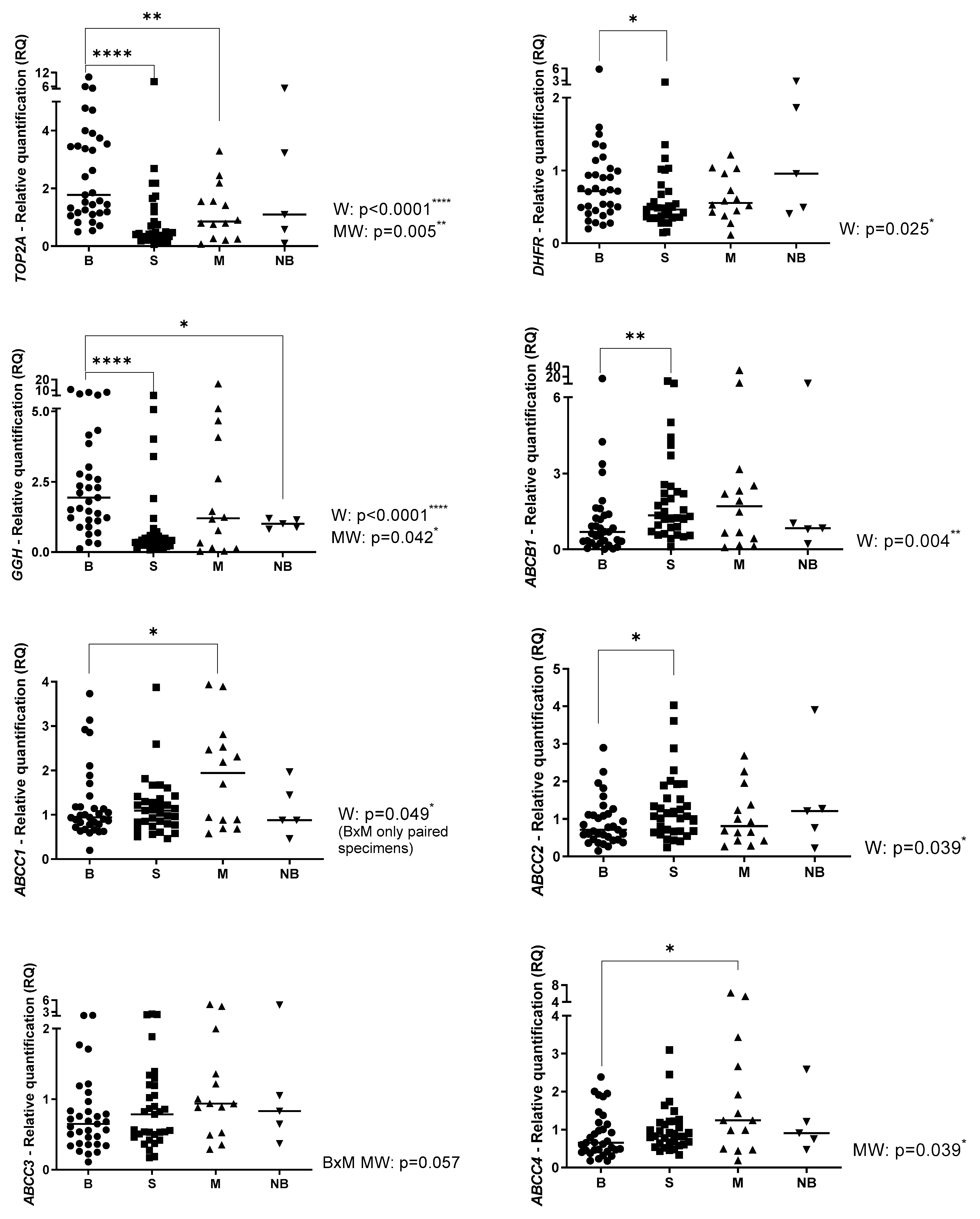
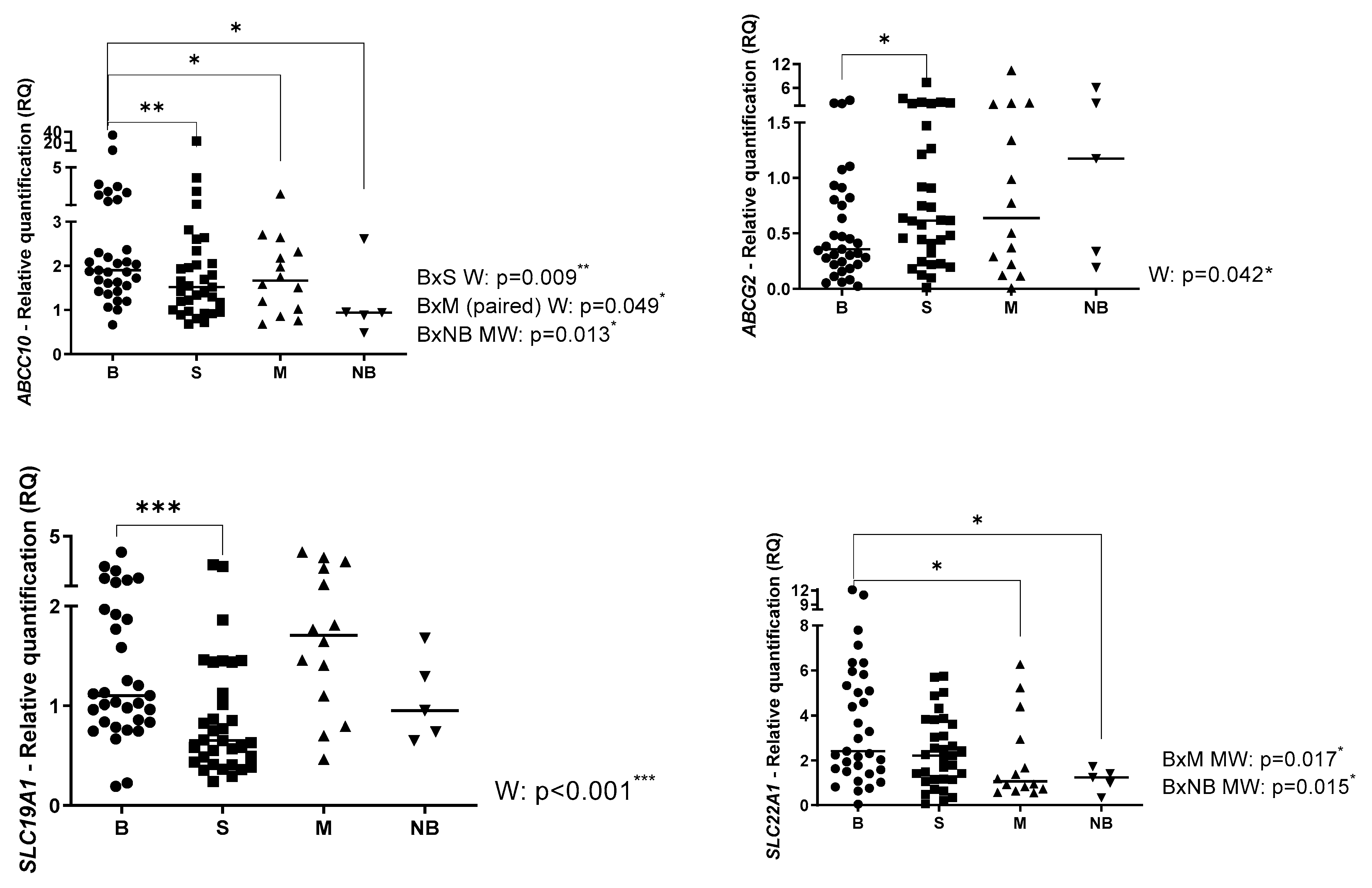
The relative quantification (RQ) of the target genes with statistically significant results in all analyzed specimens: pre-chemotherapy (B), post-chemotherapy (S), metastasis (M) and normal bone (NB) are presented in Figure 1. The RQ of the target genes with non-statistically significant results is presented in the Supplementary Figure S1. Regarding the comparisons between primary OS and normal bone, evaluated by the Mann-Whitney test, higher expression of GSTM3, GGH, ABCC10 and SLC22A1 genes in OS was observed. (p = 0.037; p = 0.042; p = 0.013; and p = 0.015, respectively).
The comparisons between pre- and post-chemotherapy specimens were analyzed using the Wilcoxon test since all these samples were paired. The post-chemotherapy specimens presented higher expression of the BCL2L1, FASLG, ABCB1, ABCC2 and ABCG2 genes than the pre-chemotherapy specimens (p = 0.012; p = 0.037; p = 0.004; p = 0.039; and p = 0.042, respectively). The post-chemotherapy specimens presented lower expression of the CASP3, CDK1, MSH2, GSTM3, SOD1, TOP2A, DHFR, GGH, ABCC10 and SLC19A1 genes than the pre-chemotherapy specimens (p = 0.037; p < 0.001; p = 0.001; p < 0.001; p = 0.004; p < 0.0001; p = 0.025; p < 0.0001; p = 0.009; and p < 0.001, respectively).
The gene expression in metastasis specimens was compared with the pre-chemotherapy specimens using the Wilcoxon and Mann-Whitney test (paired and non-paired samples, respectively) because only 14 patients had developed metastasis. The metastasis specimens presented higher expression of ABCC1, ABCC3 and ABCC4 genes (p = 0.049; p = 0.057 (trend); and p = 0.039, respectively) and lower expression of ERCC2, MSH2, SOD1, TOP2A, ABCC10 and SLC22A1 genes (p = 0.043; p = 0.043; p = 0.048; p = 0.005; p = 0.049; and p = 0.017, respectively).

2.2. Gene Expression Profile Associated with Clinical Parameters

The clinical parameters were associated with gene expression, evaluated by the Mann-Whitney test (Table 1). Tumors from patients who were metastatic at diagnosis presented higher expression of ABCB1, ABCC6, ABCC10, BCL2L1 and SLC19A1 genes (p = 0.039; p = 0.048; p = 0.048; p = 0.026; and p = 0.010, respectively) than tumors from patients who were non-metastatic at diagnosis. Poor responders’ tumors presented higher expression of ERCC1 and TOP2A genes (p = 0.021 and p = 0.036, respectively) and lower expression of ABCC3, FASLG and SLC22A1 genes (p = 0.031; p = 0.017; and p = 0.014, respectively) than good responders’ tumors. The sizes of tumors resected in surgery were also associated with the expression of the investigated genes. Large tumors of 12 cm or more presented lower expression of ABCG2, CASP3 and MSH2 genes (p = 0.027; p = 0.033; and p = 0.045, respectively) than small tumors. The local control was conducted with surgery that could be either conservative or an amputation. Tumors from patients who underwent amputation presented higher expression of ABCC11, DHFR, ERCC1, GSTM3, SLC19A1 and TOP2A genes (p = 0.002; p = 0.007; p = 0.0042; p = 0.022; p = 0.002. and p = 0.010, respectively) and lower expression of FASLG, MTHFR and SLC22A1 genes (p < 0.0001; p = 0.003; and p = 0.024, respectively) than tumors from patients who underwent conservative surgery. In the relapse analyses, one patient was excluded because he had disease progression during the first treatment and died before he reached remission. Tumors from patients who relapsed presented higher expression of TOP2A (p = 0.038) and lower expression of ABCC3, ABCC5 and FASLG genes (p = 0.026; e p = 0.051; p = 0.050, respectively) than patients who not relapsed. Regarding the ABCC5 result, this was only a trend of statistical significance.

2.3. Gene Expression Profile Associated with OAS and EFS

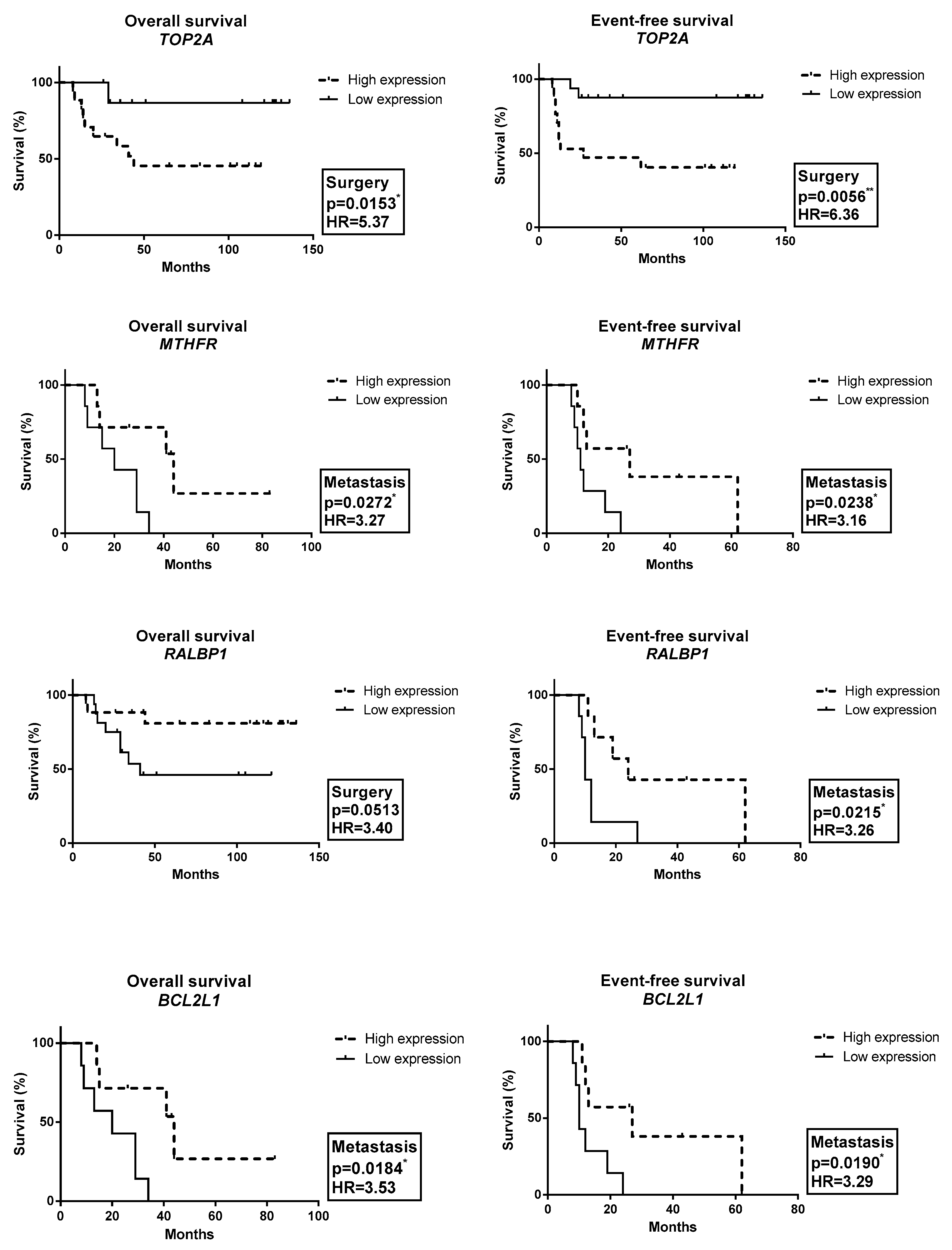
As shown in Figure 2, patients with high expression of the ABCC5 and BCL2L1 genes in the pre-chemotherapy biopsy had a trend towards worse OAS (p = 0.051; HR = 3.42) and EFS (p = 0.058; HR = 3.27), respectively, compared with patients with low expression of the ABCC5 and BCL2L1 genes. Moreover, patients with high expression of the ABCC3 gene in the pre-chemotherapy biopsy had worse EFS compared with patients with low expression of the ABCC3 gene (p = 0.048; HR = 3.41). Patients with high expression of the TOP2A gene in the post-chemotherapy specimens had worse OAS (p = 0.015; HR = 5.37) and EFS (p = 0.005; HR = 6.36) compared with patients with low expression of the TOP2A gene. Furthermore, patients with low expression of the RALBP1A gene in the post-chemotherapy specimens had a trend towards worse OAS compared with patients with high expression of the RALBP1 gene (p = 0.051; HR = 3.40). Patients with low expression of the BCL2L1 and MTHFR genes in the metastasis had worse OAS (p = 0.018, HR = 3.53; and p = 0.027, HR = 3.27, respectively) and worse EFS (p = 0.019, HR = 3.29; and p = 0.024, HR = 3.16, respectively) compared with patients with high expression of the BCL2L1 and MTHFR genes. Moreover, patients with low expression of the ABCC2, RALBP1 and SOD1 genes in the metastasis specimens had worse EFS (p = 0.048, HR = 3.16; p = 0.022, HR = 3.26; and p = 0.027, HR = 3.14, respectively) compared with patients with high expression of the ABCC2, RALBP1 and SOD1 genes.

3. Discussion

The expression of genes analyzed in the present study was investigated for the first time in paired OS specimens. When comparing OS samples obtained pre-and post-treatment, as well as from metastases, we detected different levels of expression of the selected genes. Moreover, this study showed that the genes related to a treatment response could be associated with OS tumorigenesis.
TOP2A is a target for several anticancer agents, such as doxorubicin, and a variety of mutations in this gene have been associated with the development of drug resistance. This nuclear enzyme is involved in processes such as chromosome condensation, chromatid separation and the relief of torsional stress that occurs during DNA transcription and replication [12]. A meta-analysis showed that high TOP2A expression is associated with a worse prognosis in many types of cancer [13]. In OS, the presence of TOP2A amplification tends to relate to a worse overall survival rate [14]. The present study showed an association between high expression of TOP2A and poor response, amputation and relapse. Moreover, high expression was also associated with worse OAS and EFS.
DHFR, GGH and MTHFR are genes involved in the methotrexate pathway and response [15]. Methotrexate resistance in human OS cells is associated with an amplification and/or overexpression of its target, the DHFR [6]. We observed that patients who underwent amputation presented metastasis with higher DHFR expression than patients who underwent conservative surgery. Increased levels of GGH led to a decreased accumulation of polyglutamated MTX and MTX resistance [16]. In OS, the ratio between the patients and the controls for the polymorphisms GGH_452T/C, GGH_401T/C and GGH_16T/C was greater than 1.5. The GGH_401C/T variant enhanced promoter activity, increasing protein expression [9]. In the present study, it was observed that OS presented higher GGH expression than in normal bone. The rs1801133 polymorphism of the MTHFR has been the most frequently studied in OS and leads to a C to T substitution, resulting in decreased enzymatic activity. In OS, the TT genotype was significantly associated with toxicity [15]. However, we observed that low expression in OS metastasis was associated with worse OAS and EFS. Moreover, the patients who underwent amputation had lower MTHFR expression in the primary tumor compared with the patients who underwent conservative surgery.
Regarding apoptosis, BCL2L1, CASP3 and FASLG were investigated in the present study. The longer isoform of BCL2L1 acts as an apoptotic inhibitor and the shorter isoform acts as an apoptotic activator [17]. We found that high BCL2L1 expression in the primary tumor was associated with metastasis at diagnosis and a worse EFS. Nevertheless, in the metastatic tumor, low expression was associated with worse OAS and EFS. This discrepancy in our results could be explained by the theory that the primary tumor expresses the longer isoform (anti-apoptosis) and a metastatic tumor expresses the shorter isoform (pro-apoptosis), since chemotherapy drugs stimulate the production of the shorter isoform [18]. The G allele of the variant rs2720376, linked with lower CASP3 expression, was associated with a lower EFS in OS [10]. In this study, low CASP3 expression was associated with large tumors. The variant rs763110, linked to a lower FasL expression, was associated with a lower EFS in OS [10]. We found that low FASLG expression was associated with a poor response, amputation and relapse.
Regarding genes related to detoxification of the chemotherapeutic drugs, GSTM1 and GSTP1 presented no association with the outcome in OS. The polymorphism in GSTM3 (AA versus BB) has been associated with OS risk [19]. The present study observed an association between high GSTM3 expression and amputation. Moreover, OS presented higher GSTM3 expression than normal bone. The GSTM3 polymorphism could confer different efficiencies in the metabolism of carcinogens and has been shown to modulate various cancers’ risk [20]. The null GSTT1 genotype was associated with OS risk [19]. Resistant cell lines of OS showed lower SOD1 expression than their parental cells [21]. We found that metastasis presented lower SOD1 expression than the primary tumor, and patients with low expression of the SOD1 gene in the metastasis had worse EFS.
Genes involved in the repair of DNA adducts induced by cisplatin, which thereby influence cisplatin efficacy, have been investigated by the largest number of studies on OS [7]. ERCC1 positivity has presented an association with poor EFS and OAS in OS [22]. The present study showed an association between high expression and poor response and amputation. The ERCC2 rs1799793 polymorphism was related to the high risk of OS development [23]. This study showed that metastasis presented lower ERCC2 expression than primary tumors. We found that low MSH2 expression was associated with large primary tumors. Metastasis specimens presented lower expression than primary tumors. In OS, the variant rs4638843 in MSH2 was associated with a worse EFS [10]. Moreover, a wide investigation of childhood cancers found germline mutations of MSH2 in OS [24]. Taken together, our results showed that metastasis in OS presents low expression of the ERCC2 and MSH2 genes compared with pre-chemotherapy biopsy, which could be related to decreased ability to repair DNA damage in metastasis, possibly resulting in genetic alterations accumulation and more aggressive cancer [25].
High efflux transporter gene expression and low influx transporter gene expression are the main resistance mechanisms related to cisplatin, doxorubicin and methotrexate, in vitro. Moreover, many polymorphisms in these genes have been related to treatment response in OS [6,7,26,27,28]. The present study, for the first time, investigated the expression of transporter genes in paired specimens. The results showed that the tumor biopsy presented high ABCC6 and ABCC10 expression, and metastasis presented high ABCB1 expression when metastasis was present at diagnosis. The patients with high ABCC3 and ABCC5 expression in biopsy presented worse EFS and OAS, respectively, and patients with low ABCC2 expression in metastasis presented worse EFS.The patients with low RALBP1 expression in surgery and metastasis presented worse OAS and EFS, respectively.
OS presented higher ABCC10 and SLC22A1 expression than normal bone. However, low SLC22A1 expression was associated with a poor response and amputation, probably due to its influx function. This is the first investigation regarding SLC22A1 and OS. SLC22A1 could be activated by miR-21, which is overexpressed in OS and was associated with tumorigenesis [29]. Moreover, metastasis presented higher ABCC1, ABCC3 and ABCC4 expression and lower SLC22A1 and ABCC10 expression than the primary tumor. This pattern could contribute to the lower intracellular concentration of the chemotherapeutic drugs. Consequently, it could contribute to the mechanism of resistance in metastasis, which is the main cause of death in OS patients. Therefore, with the knowledge of the metastasis profile, it is possible to develop new strategies for these patients. CBT-1® is an adjunct to chemotherapy in all cancer types with multi-drug resistance. Eight clinical trials are evaluating CBT-1® in patients with many cancer types, such as acute myelogenous leukemia, breast, non-Hodgkin’s lymphoma, Hodgkin’s disease, lung, and sarcoma [30,31]. Moreover, CBT-1® was able to revert the ABCB1/ABCC1-mediated resistance against doxorubicin in OS cell lines [32]. In the future, it could be interesting to evaluate CBT-1® in metastatic OS patients
In conclusion, the present study identified associations between OS outcome and expression of the genes TOP2A, DHFR, MTHFR, BCL2L1, CASP3, FASLG, GSTM3, SOD1, ABCB1, ABCC2, ABCC3, ABCC5, ABCC6, ABCC10, ABCC11, ABCG2, RALBP1, SLC19A1, SLC22A1, ERCC1 and MSH2. In addition, the pre-chemotherapy biopsy from OS patients had higher gene expression of ABCC10, GGH, GSTM3 and SLC22A1 compared with bone specimens obtained from healthy subjects, and the metastasis specimens showed a high expression profile of ABCC1, ABCC3 and ABCC4 and low expression of SLC22A1 and ABCC10, which is possibly an important factor for resistance in OS metastasis. In summary, we found that the expression of genes related MAP pharmacokinetic and pharmacodynamic modeling, as well as cell death and DNA damage repair processes are associated with OS tumorigenesis and MAP response in OS patients. Therefore, in the future, our findings may contribute to clinical management as prognostic markers and also as possible therapeutic targets.

4. Materials and Methods

4.1. Patients and Specimens

We investigated 80 paired specimens obtained from 33 patients with diagnoses of OS. These patients were admitted to the Pediatric Oncology Institute (IOP/GRAACC/UNIFESP) between 2006 and 2016. The average age at diagnosis was 13 years old. Of 33 OS patients, 14 patients presented pulmonary metastasis. Thus, we investigated 33 biopsy specimens (pre-chemotherapy), 33 surgery specimens (post-chemotherapy) and 14 pulmonary metastasis specimens. Five normal bone tissues were used as a control; they were obtained from orthopedic surgeries of five healthy individuals that underwent trauma and did not present either genetic disorders or bone diseases. This study had the Research Ethics Committee approval from the Federal University of Sao Paulo (N° 0189/2016), and all patients agreed to participate by informed consent. All patients were treated following the GLATO (Grupo Latino Americano de Tratamento de Osteossarcoma—Latin American Group of Osteosarcoma Treatment) protocol of 2006, which is based on high doses of cisplatin, doxorubicin and methotrexate. All clinical data are summarized in Table 2.

4.2. Gene Expression (qRT-PCR)

The expression of 32 genes involved with pharmacogenetics was measured by a quantitative reverse transcription PCR (qRT-PCR). All frozen tissues were submitted to an RNA extraction using TRIzol® Reagent (Thermo Fisher Scientific, Waltham, MA, USA). The cDNA was synthesized using SuperScript® Vilo™ Master Mix (Invitrogen, Waltham, MA, USA). The qRT-PCR was performed in triplicate using TaqMan® Gene Expression Assays (Thermo Fisher Scientific, Waltham, MA, USA) (Table 3). The ACTB and GAPDH genes were used as endogenous controls. Normal bone was used as a calibrator.

4.3. Statistical Analyses

Data analyses were performed using GraphPad Prism version 6.0 for Windows (GraphPad Software, San Diego, CA, USA). The gene expression measured by relative quantification was compared using nonparametric tests: the Wilcoxon and Mann-Whitney tests. The overall survival (OAS) and event-free survival (EFS) were calculated using the Kaplan-Meier method and the survival curves were compared using the log-rank test. The time of relapse was considered the time from the OS diagnosis until the relapse event. For OAS and EFS analyses, the median value of each gene and specimen type (biopsy, surgery or metastasis) was the cut-off that defined high or low expression. Statistical significance was considered when p < 0.05.

Supplementary Materials

The following supporting information can be downloaded at: https://www.mdpi.com/article/10.3390/ijms24065607/s1.

Author Contributions

Conceptualization, A.T.-P., F.T.-G. and S.R.C.T.; Methodology, A.T.-P.; Validation, A.T.-P.; Formal analysis, A.T.-P.; Investigation, A.T.-P., F.T.-G., A.S.P. and S.R.C.T.; Resources, A.T.-P., F.T.-G., M.T.S.A., R.J.G.F. and R.O.; Data curation, A.T.-P.; Writing – original draft, A.T.-P.; Writing – review & editing, A.T.-P., F.T.-G. and S.R.C.T.; Visualization, A.T.-P.; Supervision, S.R.C.T.; Project administration, A.T.-P. and S.R.C.T.; Funding acquisition, A.T.-P. and S.R.C.T. All authors have read and agreed to the published version of the manuscript.

Funding

This research was funded by FAPESP (São Paulo Research Foundation), grant number 2016/01718-0, CNPq (National Council for Scientific and Technological Development) grant number 140540/2016-4 and GRAACC (Support Group for Adolescents and Children with Cancer). The APC was funded by FAPESP (São Paulo Research Foundation), grant number 2023/02839-0.

Institutional Review Board Statement

The study was conducted in accordance with the Declaration of Helsinki, and approved by the Ethics Committee of Federal University of Sao Paulo (N° 0189/2016, approved on 19-Apr-2016).

Informed Consent Statement

Informed consent was obtained from all subjects involved in the study.

Data Availability Statement

The data presented in this study are available on request from the corresponding author.

Conflicts of Interest

The authors declare no conflict of interest.

References

  1. Valery, P.C.; Laversanne, M.; Bray, F. Bone cancer incidence by morphological subtype: A global assessment. Cancer Causes Control 2015, 26, 1127–1139. [Google Scholar] [CrossRef]
  2. Kager, L.; Tamamyan, G.; Bielack, S. Novel insights and therapeutic interventions for pediatric osteosarcoma. Future Oncol. 2017, 13, 357–368. [Google Scholar] [CrossRef] [PubMed]
  3. Whelan, J.S.; Bielack, S.S.; Marina, N.; Smeland, S.; Jovic, G.; Hook, J.M.; Krailo, M.; Anninga, J.; Butterfass-Bahloul, T.; Böhling, T.; et al. EURAMOS-1, an international randomised study for osteosarcoma: Results from pre-randomisation treatment. Ann. Oncol. 2015, 26, 407–414. [Google Scholar] [CrossRef] [PubMed]
  4. Deenen, M.J.; Cats, A.; Beijnen, J.H.; Schellens, J.H. Part 2: Pharmacogenetic variability in drug transport and phase I anticancer drug metabolism. Oncologist 2011, 16, 820–834. [Google Scholar] [CrossRef] [PubMed] [Green Version]
  5. Deenen, M.J.; Cats, A.; Beijnen, J.H.; Schellens, J.H. Part 1: Background, methodology, and clinical adoption of pharmacogenetics. Oncologist 2011, 16, 811–819. [Google Scholar] [CrossRef] [PubMed] [Green Version]
  6. Serra, M.; Hattinger, C.M. The pharmacogenomics of osteosarcoma. Pharm. J. 2017, 17, 11–20. [Google Scholar] [CrossRef]
  7. Vos, H.I.; Coenen, M.J.; Guchelaar, H.J.; Te Loo, D.M. The role of pharmacogenetics in the treatment of osteosarcoma. Drug Discov. Today 2016, 21, 1775–1786. [Google Scholar] [CrossRef]
  8. Gianferante, D.M.; Mirabello, L.; Savage, S.A. Germline and somatic genetics of osteosarcoma—Connecting aetiology, biology and therapy. Nat. Rev. Endocrinol. 2017, 13, 480–491. [Google Scholar] [CrossRef]
  9. Hattinger, C.M.; Biason, P.; Iacoboni, E.; Gagno, S.; Fanelli, M.; Tavanti, E.; Serena, V.; Stefano, F.; Andrea, R.; Rossana, R.; et al. Candidate germline polymorphisms of genes belonging to the pathways of four drugs used in osteosarcoma standard chemotherapy associated with risk, survival and toxicity in non-metastatic high-grade osteosarcoma. Oncotarget 2016, 7, 61970–61987. [Google Scholar] [CrossRef] [Green Version]
  10. Hagleitner, M.M.; Coenen, M.J.; Gelderblom, H.; Makkinje, R.R.; Vos, H.I.; de Bont, E.S.; van der Graaf, W.T.; Schreuder, H.; Flucke, U.; van Leeuwen, F.; et al. A First Step toward Personalized Medicine in Osteosarcoma: Pharmacogenetics as Predictive Marker of Outcome after Chemotherapy-Based Treatment. Clin. Cancer Res. 2015, 21, 3436–3441. [Google Scholar] [CrossRef] [Green Version]
  11. Whelan, J.S.; Davis, L.E. Osteosarcoma, chondrosarcoma, and chordoma. J. Clin. Oncol. 2018, 36, 188–193. [Google Scholar] [CrossRef] [PubMed]
  12. Available online: https://www.ncbi.nlm.nih.gov/gene/7153 (accessed on 10 December 2018).
  13. Ren, L.; Liu, J.; Gou, K.; Xing, C. Copy number variation and high expression of DNA topoisomerase II alpha predict worse prognosis of cancer: A meta-analysis. J. Cancer 2018, 9, 2082–2092. [Google Scholar] [CrossRef] [PubMed]
  14. Nguyen, A.; Lasthaus, C.; Guerin, E.; Marcellin, L.; Pencreach, E.; Gaub, M.P.; Guenot, D.; Entz-Werle, N. Role of Topoisomerases in Pediatric High Grade Osteosarcomas: TOP2A Gene Is One of the Unique Molecular Biomarkers of Chemoresponse. Cancers 2013, 5, 662–675. [Google Scholar] [CrossRef]
  15. Hattinger, C.M.; Tavanti, E.; Fanelli, M.; Vella, S.; Picci, P.; Serra, M. Pharmacogenomics of genes involved in antifolate drug response and toxicity in osteosarcoma. Expert Opin. Drug Metab. Toxicol. 2017, 13, 245–257. [Google Scholar] [CrossRef]
  16. Hegyi, M.; Arany, A.; Semsei, A.F.; Csordas, K.; Eipel, O.; Gezsi, A.; Kutszegi, N.; Csoka, M.; Muller, J.; Erdelyi, D.; et al. Pharmacogenetic analysis of high-dose methotrexate treatment in children with osteosarcoma. Oncotarget 2017, 8, 9388–9398. [Google Scholar] [CrossRef] [PubMed] [Green Version]
  17. Available online: https://www.ncbi.nlm.nih.gov/gene/598 (accessed on 10 December 2018).
  18. Shkreta, L.; Froehlich, U.; Paquet, E.R.; Toutant, J.; Elela, S.A.; Chabot, B. Anticancer drugs affect the alternative splicing of Bcl-x and other human apoptotic genes. Mol. Cancer Ther. 2008, 7, 1398–1409. [Google Scholar] [CrossRef] [PubMed] [Green Version]
  19. Wang, Z.; Xu, H.; He, M.; Wu, H.; Zhu, Y.; Su, Z. The association of glutathione S-transferase polymorphisms in patients with osteosarcoma: Evidence from a meta-analysis. Eur. J. Cancer Care (Engl.) 2015, 24, 417–424. [Google Scholar] [CrossRef]
  20. Xu, Y.; Wang, J.; Dong, W. GSTM3 A/B polymorphism and risk for head and neck cancer: A meta-analysis. PLoS ONE 2014, 9, e83851. [Google Scholar] [CrossRef] [Green Version]
  21. Tao, Y.; Ou, Y.; Yin, H.; Chen, Y.; Zhong, S.; Gao, Y.; Zhao, Z.; He, B.; Huang, Q.; Deng, Q. Establishment and characterization of human osteosarcoma cells resistant to pyropheophorbide-α methyl ester-mediated photodynamic therapy. Int. J. Oncol. 2017, 51, 1427–1438. [Google Scholar] [CrossRef] [Green Version]
  22. Hattinger, C.M.; Michelacci, F.; Sella, F.; Magagnoli, G.; Benini, S.; Gambarotti, M.; Palmerini, E.; Picci, P.; Serra, M.; Ferrari, S. Excision repair cross-complementation group 1 protein expression predicts survival in patients with high-grade, non-metastatic osteosarcoma treated with neoadjuvant chemotherapy. Histopathology 2015, 67, 338–347. [Google Scholar] [CrossRef]
  23. Chen, X.J.; Tong, Z.C.; Kang, X.; Wang, Z.C.; Huang, G.L.; Yang, T.M.; Dong, L. ERCC polymorphisms and risk of osteosarcoma: A meta-analysis. Eur. Rev. Med. Pharmacol. Sci. 2018, 22, 6658–6666. [Google Scholar] [PubMed]
  24. Gröbner, S.N.; Worst, B.C.; Weischenfeldt, J.; Buchhalter, I.; Kleinheinz, K.; Rudneva, V.A.; Johann, P.; Balasubramanian, G.P.; Segura-Wang, M.; Brabetz, S.; et al. The landscape of genomic alterations across childhood cancers. Nature 2018, 555, 321–327. [Google Scholar] [CrossRef] [Green Version]
  25. Hanahan, D.; Weinberg, R.A. Hallmarks of cancer: The next generation. Cell 2011, 144, 646–674. [Google Scholar] [CrossRef] [PubMed] [Green Version]
  26. Siddik, Z.H. Cisplatin: Mode of cytotoxic action and molecular basis of resistance. Oncogene 2003, 22, 7265–7279. [Google Scholar] [CrossRef] [PubMed] [Green Version]
  27. Chou, A.J.; Gorlick, R. Chemotherapy resistance in osteosarcoma: Current challenges and future directions. Expert Rev. Anticancer. Ther. 2006, 6, 1075–1085. [Google Scholar] [CrossRef]
  28. Hattinger, C.M.; Pasello, M.; Ferrari, S.; Picci, P.; Serra, M. Emerging drugs for high-grade osteosarcoma. Expert Opin. Emerg. Drugs 2010, 15, 615–634. [Google Scholar] [CrossRef]
  29. Nakka, M.; Allen-Rhoades, W.; Li, Y.; Kelly, A.J.; Shen, J.; Taylor, A.M.; Barkauskas, D.; Yustein, J.; Andrulis, I.; Wunder, J.; et al. Biomarker significance of plasma and tumor miR-21, miR-221, and miR-106a in osteosarcoma. Oncotarget 2017, 8, 96738–96752. [Google Scholar] [CrossRef] [Green Version]
  30. Available online: http://www.cbapharma.com/cbt1/cbt1.html (accessed on 10 December 2018).
  31. Available online: https://www.cancer.gov/about-cancer/treatment/clinical-trials/intervention/mdr-modulator-cbt-1 (accessed on 10 December 2018).
  32. Fanelli, M.; Hattinger, C.M.; Vella, S.; Tavanti, E.; Michelacci, F.; Gudeman, B.; Barnett, D.; Picci, P.; Serra, M. Targeting ABCB1 and ABCC1 with their Specific Inhibitor CBT-1® can Overcome Drug Resistance in Osteosarcoma. Curr. Cancer Drug Targets 2016, 16, 261–274. [Google Scholar] [CrossRef]
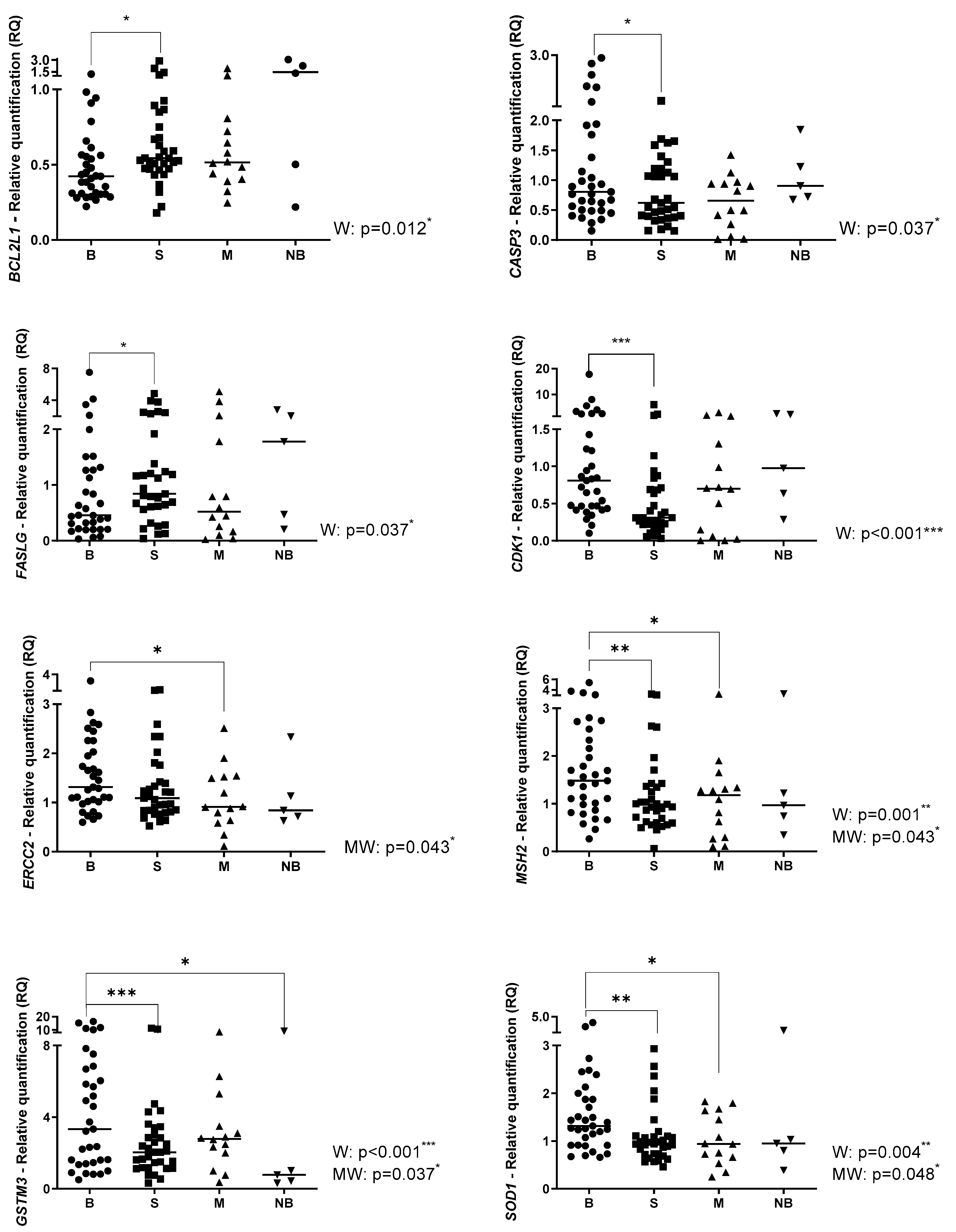
Figure 1. The relative quantification (RQ) in the pre-chemotherapy (B), post-chemotherapy (S), metastasis (M) and normal bone (NB) specimens. W: Wilcoxon Test; MW: Mann-Whitney Test. * p < 0.05; ** p < 0.01; *** p < 0.001; **** p < 0.0001.
Ijms 24 05607 g001aIjms 24 05607 g001bIjms 24 05607 g001c
Figure 2. Association between gene expression and survival in OS. Pre-chemotherapy (Biopsy); Post-chemotherapy (Surgery); and Metastasis (M). * p < 0.05; ** p < 0.01.
Ijms 24 05607 g002aIjms 24 05607 g002b
Table 1. Association between gene expression and clinical parameters.
Clinical ParameterGeneExpressionSpecimenp
Metastasis at diagnosis * vs. Non-metastasis at diagnosisABCB1M0.039
ABCC6B0.048
ABCC10B0.048
BCL2L1S0.026
SLC19A1S0.010
*** Poor responder * vs. Good responderABCC3S0.031
ERCC1B0.021
FASLGS0.017
SLC22A1B0.014
TOP2AS0.036
Large tumor (>12 cm) * vs. Small tumorABCG2S0.027
CASP3B0.033
MSH2B0.045
Amputation * vs. Conservative surgeryABCC11M0.042
DHFRM0.007
ERCC1M0.042
FASLGS<0.0001
M0.042
GSTM3S0.022
MTHFRS0.003
SLC19A1B0.002
S0.016
SLC22A1B0.024
TOP2AS0.010
Relapse * vs. Non-relapseABCC3M0.026
ABCC5M0.051 **
FASLGS0.041
TOP2AS0.038
Higher gene expression (↑); Lower gene expression (↓); Pre-chemotherapy—Biopsy (B), Post-chemotherapy—Surgery (S); Metastasis (M). * Clinical parameter associated with higher or lower gene expression. Per example, patients metastatic at diagnosis had higher gene expression of ABCB1 gene in the metastasis specimens compared with the non-metastatic at diagnosis patients. ** Trend of statistical significance. *** Poor responder: <90% of necrosis post-neoadjuvant therapy.
Table 2. Clinical features of OS patients.
Clinical FeaturesN (%)
Gender
Male64%
Female36%
Age
≤1342%
>1358%
Metastasis at diagnosis
Yes33
No67
Primary site
Femur67
Tibia24
Humerus9
Surgery
Conservative85
Amputation15
Histology
Mixed24
Osteoblastic55
Condroblastic9
Fibroblastic3
Telangiectatic3
Not identified6
Tumor size
<12 cm55
≥12 cm42
Not identified3
Grade of tumor necrosis
≤90%49
>90%45
Not identified6
Pulmonary metastasis
Yes42
No57
Relapse
Yes33
No67
Death
Yes33
No67
N: Number of patients.
Table 3. TaqMan® Gene Expression Assays.
FunctionGeneAssay
ApoptosisBCL2L1Hs00236329_m1
CASP3Hs00234387_m1
FASLHs00181226_g1
Cell cycleCDK1Hs00938777_m1
Damage recognitionHMGB1Hs01923466_g1
DNA repairERCC1Hs01012158_m1
ERCC2Hs00361161_m1
MSH2Hs00953527_m1
DetoxificationGSTM1Hs01683722_gH
GSTM3Hs00356079_m1
GSTP1Hs00943350_g1
GSTT1Hs02512069_s1
SOD1Hs00533490_m1
Doxorubicin pathwayTOP2AHs01032137_m1
EndogenousACTBHs01060665_g1
GAPDHHs02758991_g1
Folate pathwayDHFRHs00758822_s1
GGHHs00914163_m1
MTHFRHs01114487_m1
TransportABCB1Hs00184500_m1
ABCC1Hs01561502_m1
ABCC2Hs00166123_m1
ABCC3Hs00978452_m1
ABCC4Hs00988717_m1
ABCC5Hs00981089_m1
ABCC6Hs00184566_m1
ABCC10Hs01056200_m1
ABCC11Hs01090758_m1
ABCG2Hs01053790_m1
ATP7BHs00163739_m1
RALBP1Hs01034984_g1
SLC19A1Hs00953344_m1
SLC22A1Hs00427552_m1
SLC31A1Hs00977266_g1
Disclaimer/Publisher’s Note: The statements, opinions and data contained in all publications are solely those of the individual author(s) and contributor(s) and not of MDPI and/or the editor(s). MDPI and/or the editor(s) disclaim responsibility for any injury to people or property resulting from any ideas, methods, instructions or products referred to in the content.