Article

Whole Genome Resequencing Revealed the Effect of Helicase yqhH Gene on Regulating Bacillus thuringiensis LLP29 against Ultraviolet Radiation Stress

1
State Key Laboratory of Ecological Pest Control for Fujian and Taiwan Crops, Key Laboratory of Biopesticide and Chemical Biology of Ministry of Education & Ministerial and Provincial Joint Innovation Centre for Safety Production of Cross-Strait Crops, College of Life Science, Fujian Agriculture and Forestry University, Fuzhou 350002, China
2
College of Plant Protection, Fujian Agriculture and Forestry University, Fuzhou 350002, China
3
College of Life Science, Fujian Agriculture and Forestry University, Fuzhou 350002, China
*
Author to whom correspondence should be addressed.
Int. J. Mol. Sci. 2023, 24(6), 5810; https://doi.org/10.3390/ijms24065810
Submission received: 27 January 2023 / Revised: 23 February 2023 / Accepted: 1 March 2023 / Published: 18 March 2023
(This article belongs to the Section Molecular Genetics and Genomics)

Abstract

:
Bacillus thuringiensis (Bt) is a widely used microbial pesticide. However, its duration of effectiveness is greatly shortened due to the irradiation of ultraviolet rays, which seriously hinders the application of Bt preparations. Therefore, it is of great importance to study the resistance mechanism of Bt to UV at the molecular level to improve the UV-resistance of Bt strains. In order to know the functional genes in the UV resistance, the genome of UV-induced mutant Bt LLP29-M19 was re-sequenced and compared with the original strain Bt LLP29. It was shown that there were 1318 SNPs, 31 InDels, and 206 SV between the mutant strain and the original strain Bt LLP29 after UV irradiation, which were then analyzed for gene annotation. Additionally, a mutated gene named yqhH, a member of helicase superfamily II, was detected as an important candidate. Then, yqhH was expressed and purified successfully. Through the result of the enzymatic activity in vitro, yqhH was found to have ATP hydrolase and helicase activities. In order to further verify its function, the yqhH gene was knocked out and complemented by homologous recombinant gene knockout technology. The survival rate of the knockout mutant strain Bt LLP29-ΔyqhH was significantly lower than that of the original strain Bt LLP29 and the back-complemented strain Bt LLP29-ΔyqhH-R after treated with UV. Meanwhile, the total helicase activity was not significantly different on whether Bt carried yqhH or not. All of these greatly enrich important molecular mechanisms of Bt when it is in UV stress.

1. Introduction

Bacillus thuringiensis (Bt), a gram-positive bacterium, is commonly used in microbial pesticides with the advantages of safety and good insecticidal effect [1]. As a popular biopesticide, Bt produces insecticidal crystal proteins (ICPs) that are effective against a variety of insect species, including Lepidoptera, Hemiptera, and Diptera [2,3,4]. However, the structure of insecticidal crystal proteins and the mechanism of action with pests need to be studied in depth [5]. As a biogenic insecticide, it has significant advantages over chemical insecticides [6]. External factors such as ultraviolet radiation can damage or degrade Bt strains and their toxin proteins, reducing the duration of Bt preparations and consequently limiting their effectiveness [3,7]. Therefore, developing transgenic Bt crops, screening of strains with strong resistance to UV, or improving melanin production are important for the application of Bt [8,9,10].
Whole genome resequencing is widely used in the research of plants and microorganisms [11,12]. Whole genome data have been reported for a wide range of Bt strains, providing a large amount of information for the research of Bt [13,14]. However, bioinformatics analysis of Bt in stress environments, especially UV stress environments, is still scarce. Therefore, the re-sequencing of highly UV stress-resistant strains and the discovery of variation information in gene sequences by sequence comparison can provide an effective method for screening UV resistance genes.
Ultraviolet (UV) is an invisible spectrum spanning wavelengths from 200 to 400 nm. UV radiation directly affects the survival of microorganisms. Therefore, it is essential to investigate the UV resistance mechanism of microorganisms. In Bacillus, melanin can attenuate the effect of UV exposure on Bt insecticide products and has been used as a protective agent [9]. Currently, several strains have been screened for the production of high levels of melanin, which can effectively increase the UV resistance of the strain [15,16,17]. In addition, algae and cyanobacteria have evolved various avoidance and repair mechanisms to protect themselves against the damaging effects of UV radiation to acclimate to enhanced UV-B radiation. For example, cyanobacteria can provide effective resistance to UV stress through secondary metabolites [18,19].
Ultraviolet radiation results in extensive DNA damage, with double-strand breaks (DSBs) as the most prominent formed of damage. Studies have shown that helicase have an important function in the repair of DNA damage. Helicase, a key macromolecular substance in the process of biomolecular metabolism, exists widely in various organisms. Helicase can convert double-stranded nucleic acids or structured nucleic acid strands to single-stranded nucleic acid, thereby mediating the metabolic activities of DNA or RNA [20,21]. It had been found that helicase had various biological functions. In cells, they were involved in DNA replication, repair, transcriptional recombination, RNA splicing, protein translation, genome stability, and other important molecular activities process [22,23,24]. According to its sequences, as well as structural and mechanistic features, helicase can be classified into six families [25]. Among them, the two largest families are called superfamily I (SFI) and superfamily II (SFII). SFI and SFII are monomeric, and the other four families are hexameric [26]. After being reported as a new gene involved in recombination and repair, DNA helicase was found in many bacteria, such as Escherichia coli, Pseudomonas, Neisseria meningitides, and B. subtilis [27,28,29,30]. DNA helicase had been found to participate in the stress response of Bt. The DNA helicase recG in B. thuringiensis is associated with UV resistance, and recG knockout strains are more sensitive to UV light [31]. A variety of RNA helicases have been identified in B. cereus, which can participate in stress reactions under heat, oxidation, and pH stress [32,33]. Whether other types of helicases also regulate the stress response of Bt remains to be determined.
In our previous study, UV-resistant Bt LLP29-M19 was obtained after the moquitocidal Bt strain LLP29 was exposed to UV for 19 generations. Bt LLP29-M19 showed re-sistance to UV radiation for up to 60 min [8]. In this study, the UV mutant strain Bt LLP29-M19 with high UV resistance was re-sequenced and compared with its original strain Bt LLP29. We found a helicase superfamily II member, yqhH, mutated in Bt LLP29-M19. To determine the function of yqhH in Bt UV resistance, the in vitro activity of yqhH was measured, and the strains of Bt LLP29 with and without yqhH were compared for UV resistance, as well as their DNA unwinding activities in different UV-B treatments. The present results could not only enrich the genomic data of Bt but also enhance the understanding of the UV resistance mechanism of Bt and provide an important theoretical basis for the field application of Bt.

2. Results

2.1. Sequencing Quality Control and Assembly of Bt LLP29-M19

Genomic DNA was extracted from the samples, and eligible libraries were sequenced by Illumina Hiseq 2500. A total of 7,302,812 raw reads were measured for the Bt LLP29-M19 strain. After quality control, 7,301,420 clean reads were obtained, and the GC content of the measured data was 35.61%. Using BWA software (version 0.7.10-r789), the obtained clean reads were matched to the Bt LLP29 reference genome; 98.45% of the reads were mapped to the reference genome; and 98.04% of the reads were properly mapped (Table S1). By analyzing the length of the inserted fragment, its distribution conforms to the normal distribution, indicating that the construction of the sequencing data library was not abnormal (Figure S1).

2.2. Differential Analysis of Resequencing Results

The Bt LLP29-M19 genome and LLP29 sequencing data were analyzed and compared in this study. Compared with the existing reference genome, the genome-wide heterozygosity ratio of Bt LLP29-M19 strain was 0.17%. Compared with the reference genome, the total number of SNP site variations was 1318; the sum of InDel mutations was 31; and the total number of SV-annotated variants was 206 (Table 1 and Table S2). For further analysis, the mutated genes were annotated into the database, resulting in 149 mutated genes. In addition, it was found that in most of the variant types, the variation mainly existed in the exonic region.

2.3. Functional Annotation of Variant Genes at the DNA Level

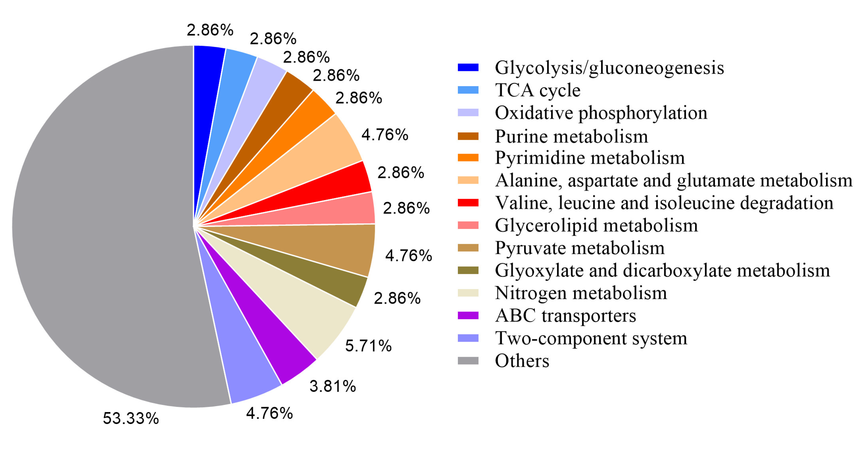
Variations in the CDS region may cause changes in gene function, and the variant genes in Bt LLP29-M19 were annotated. A total of 567 variant genes were annotated by COG, among which the functional classes of genes with more variants were metabolic process (110), catalytic activity (105), cellular process (76) and binding (58) (Figure 1). At the same time, the obtained variant genes were subjected to KEGG analysis, and a total of 105 were annotated into different signaling pathways. The main annotated metabolic pathways of these variant genes were ABC transporters (173), two-component system (88), purine metabolism (70), and pyrimidine metabolism (64) (Figure 2).

2.4. UV-Related Gene Screening

Environmental changes can cause extensive DNA damage, and double-strand breaks (DSBs) are the most critical form of biological damage. In bacteria, homologous recombination repair is the most important repair method for DNA double-strand breaks. Studies have identified a number of helicase capable of participating in nucleotide excision repair, such as pcrA and recQ [34,35]. By GO and KEGG analysis, a total of 149 genes were annotated in the variant genes. We focused on the helicase genes. We identified four helicase genes mutated in Bt LLP29-M19 by sequence comparison. We found that one of the genes had a base mutation from C to T at the 265th position, and the amino acid changed from glutamine to terminator (Figure S2). By gene annotation, it was named yqhH, which belongs to the SNF2 family of DNA decapping enzymes. Therefore, to investigate the function of the helicase in Bt anti-UV, we selected the yqhH gene for in-depth analysis.

2.5. Cloning and Expression of yqhH Gene

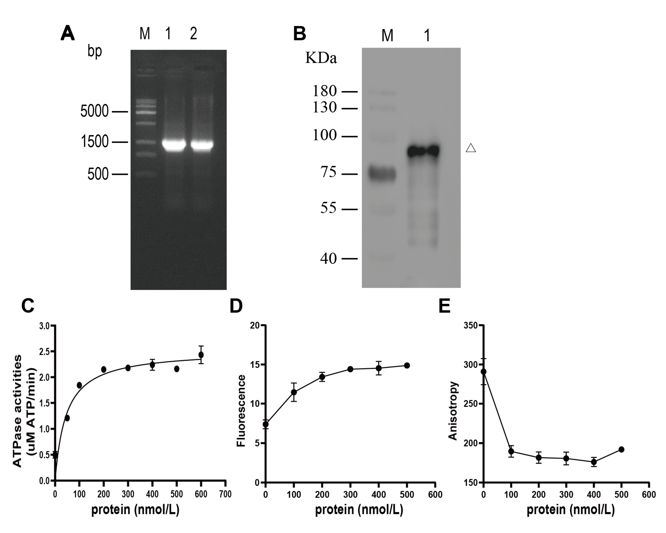
To analyze the function of the yqhH gene in vitro, a yqhH gene of size 1.5 Kb was first obtained by PCR (Figure 3A). Then, the expressed yqhH was purified by protein Ni-NTA resin kit after induction by 0.5 mM IPTG. To test the accuracy of the purified protein, the results were analyzed by Western blot, which showed that the molecular weight of the yqhH fusion protein was about 80 kDa, as expected (Figure 3B).

2.6. ATP Hydrolysis Activity of yqhH Helicase

To elucidate the function of yqhH, an ATP hydrolysis activity assay was first performed. In presence of ATP, yqhH protein could catalyze the hydrolysis of ATP, which indicated that yqhH helicase was a DNA-dependent ATPase. Meanwhile, the ATP hydrolysis activity increased in a gradient with the increase in enzyme concentration. When the helicase concentration reached a certain amount, the ATP hydrolysis activity reached saturation (Figure 3C). These results indicate that yqhH expressed in vitro can hydrolyze ATP and have ATP hydrolysis activity.

2.7. Unwinding Activity of yqhH Helicase

In the helicase kinetics assay, different concentrations of yqhH helicase were added into the unwinding reaction, respectively. As the concentration of yqhH increased, the fluorescence intensity of the substrate donor increased while the relative intensity of fluorescence polarization decreased (Figure 3D,E). In addition, the helicase activity increased with the increase in yqhH concentration. When the enzyme concentration reached a certain amount (400 nmol), the helicase activity reached a maximum, indicating that the helicase activity was concentration-dependent, which suggested that yqhH expressed in vitro had helicase activity.

2.8. Knocking out yqhH in Bt LLP29

To knock out the yqhH gene of Bt LLP29, experiments were performed by homologous recombination technique (Figure 4A). The recombinant plasmid was transformed into Bt LLP29 competent cells by electroporation, and the strain with Kana resistance but no Erm resistance was reconstituted into by high temperature (Figure 4B). The knockout strain was further characterized by expanded culture, and the Bt LLP29-ΔyqhH strain could be grown normally in a Kana antibiotic-containing medium (Figure 4C). To confirm the successful knockout of yqhH, total DNA of the mutant strain was extracted and examined by PCR using specific primers (Figure 4D). It was also indicated that yqhH was successfully knocked out in strain Bt LLP29-ΔyqhH by sequence alignment.

2.9. Complementation of yqhH in Bt LLP29-ΔyqhH

To further investigate the function of yqhH, the gene was also restored in this study. The recombinant plasmid was desalinized and electroporated into Bt LLP29-ΔyqhH competent cells. After high-temperature recombination screening, a new strain was obtained that was resistant to neither Erm nor Kana (Figure 5A). The complementation strain was then further characterized by expanded culture, and the Bt LLP29-ΔyqhH-R strain could only grow normally in antibiotic-free medium (Figure 5B). To confirm the successful complementation of target gene yqhH in Bt LLP29-ΔyqhH, the total DNA of selected strain was extracted, and the fragments of F1*R1*, and F2*R2* were amplified and detected (Figure 5C,D). After sequencing analysis, the amplified sequence had a homology of 99% with the previously reported sequence, indicating that the complementary strain Bt LLP29-ΔyqhH-R was successfully obtained.

2.10. Growth Curves and UV-B Treatment Phenotype

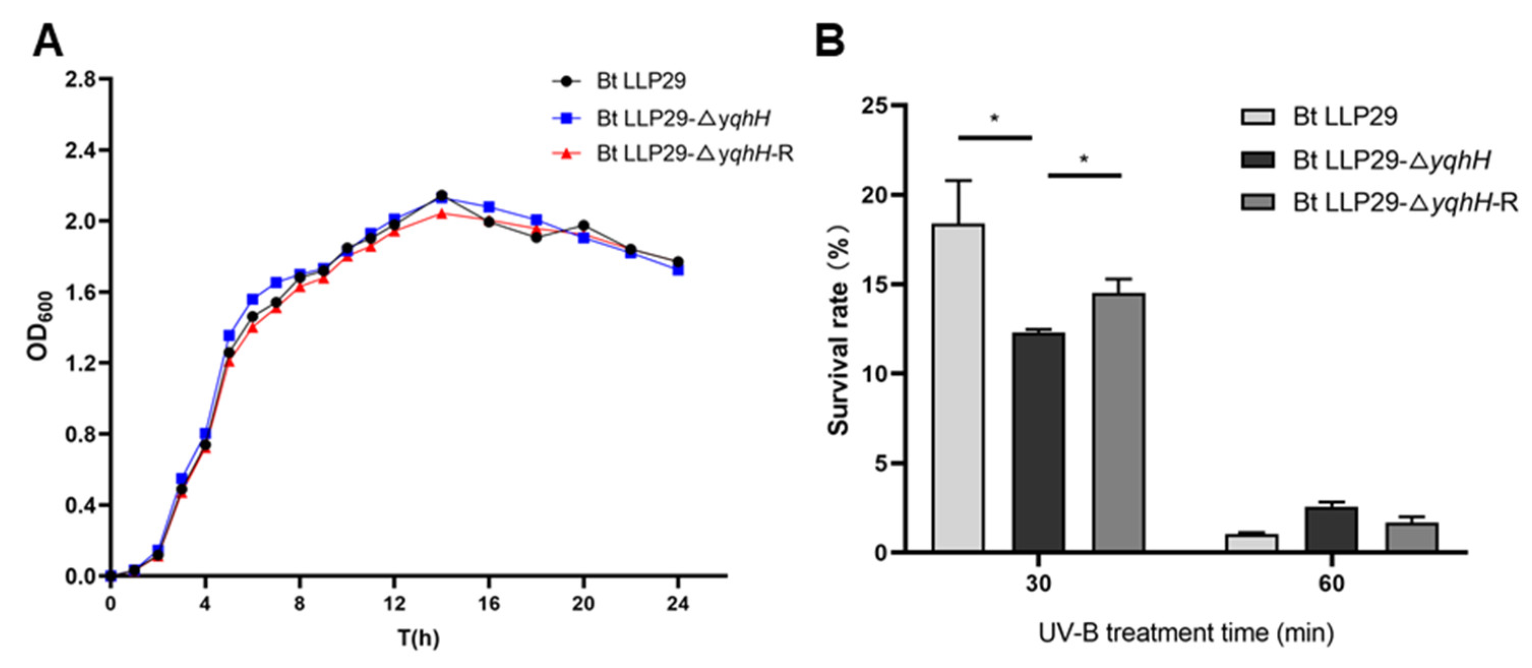
To detect the effect of yqhH on the growth of Bt, the growth curves of three strains were first analyzed. The results showed that similar growth curves were detected in the different tested Bt strains with minimal difference. At the early growth stage, Bt LLP29-ΔyqhH grew slightly faster than Bt LLP29 and Bt LLP29-ΔyqhH-R. However, three tested strains entered a stationary phase with an OD600 of about 2.0 after 14 h (Figure 6A).
To test the effect of yqhH on Bt in UV, the following experiments were performed. Three strains, Bt LLP29, Bt LLP29-ΔyqhH, and Bt LLP29-ΔyqhH-R, were treated with UV irradiation to observe the survival rate of the strains for 0 min, 30 min, and 60 min, respectively. The survival rates of three tested strains decreased with the increase in UV treatment time. However, the survival rate of Bt LLP29-ΔyqhH was significantly lower than both of Bt LLP29 and Bt LLP29-ΔyqhH-R when UV treatment was performed for 30 min. After UV treatment for 60 min, the survival rate of three strains were all lower than 5%, but the survival rate of Bt LLP29-ΔyqhH was slightly higher. The results indicate that the yqhH gene can regulate the ability of Bt strains to resistance to UV (Figure 6B).

2.11. Unwinding Activity In Vivo

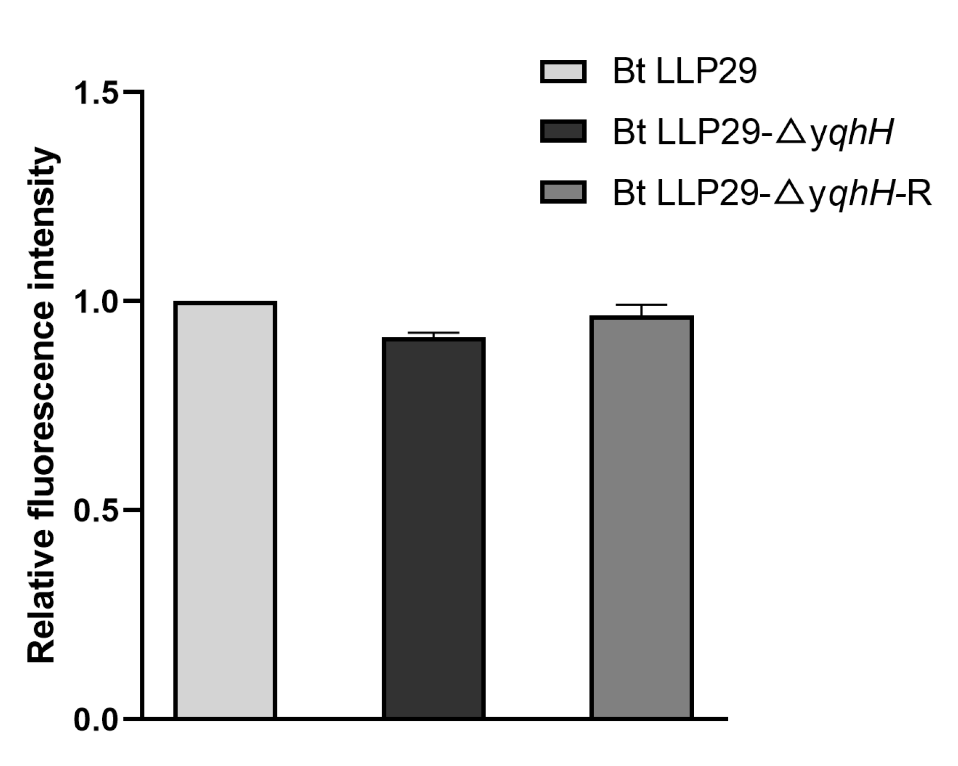
To detect the helicase activity of yqhH, an unwinding activity comparison was carried out in the tested Bt strains. However, no significant differences in the unwinding ability were found between the knockout and wild-type strains and the complementary strains carrying yqhH. After deletion of yqhH, the helicase activity of Bt LLP29 was similar to that of Bt LLP29 and its complementary strain Bt LLP29-ΔyqhH-R (p > 0.05). The helicase activity of Bt LLP29-ΔyqhH-R was also not found to be significantly different when yqhH was complemented (Figure 7). These results suggest that yqhH is not significantly involved in the helicase activity of Bt LLP29. However, there may be other factors besides yqhH that have helicase activity.

3. Discussion

Bacillus thuringiensis is a widely used microbial insecticide. The adverse external environment, such as ultraviolet radiation, can destroy or degrade Bt strains and their toxin proteins, which can shorten the shelf life of Bt products and greatly limit their application. How to effectively select the critical genes for UV resistance will provide invaluable information for understanding the molecular mechanism of UV stress resistance in Bt [15,36]. With the development of sequencing technology, genome resequencing has offered a convenient way to identify mutated genes between resistant strains and original strains [37].
A large number of differential genes can be identified by gene sequencing, which provides an effective method for identifying critical genes that regulate stress accordingly [38,39]. In this study, the Illumina Hiseq 2500 was used to re-sequence the genome of UV-resistant strain LLP29-M19, and 149 mutated genes were found. These genes were functionally annotated and were mainly associated with genes for glucose conversion and synthesis, signal transduction, nucleotide excision repair, and cellular components. These results suggest that UV light affects the growth of Bt through the synthesis of glucose in Bt and the DNA replication pathway.
UV has been shown to cause direct damage to DNA molecules [40]. Previous studies have found that unwinding enzymes play an important role in DNA damage repair. Helicase is present in most organisms and plays various functions such as DNA replication, repair, recombination, and transcription [23,41]. Many of the helicases in B. subtilis and E. coli have been studied intensively [42,43]. In B. subtilis, it had been proved that pcrA, recQ, and recG helicases had ATPase and DNA unwinding activities and played an essential role in maintaining the stability of genes [29,35,44]. In this study, we found three helicase genes that were mutated after UV treatment, suggesting that these genes may be involved in the UV response. Additionally, the yqhH gene was selected for functional analysis. The enzymatic activity of yqhH in vitro was assayed by FRET, and it was in agreement with the gene annotation results that yqhH was able to hydrolyze ATP and unwind double-stranded DNA (Figure 3C–E). However, the specific mechanism of yqhH protein deconvolution needed to be further investigated. We further knocked down the yqhH gene in Bt and examined the changes in total protein activity in the different tested strains, while no significant changes were observed in the unwinding enzyme activity (Figure 7). Therefore, we speculate that other similar unwinding enzymes exist in vivo that could complement the function of yqhH knockdown.
The helicase regulates the microbial response to stress, and the same family helicases can perform different functions [31,32]. In the UV treatment test, the survival rate of tested Bt strains decreased after UV stress. UV was found to be able to significantly reduce the survival rate of Bt strains, but there were variations between different Bt strains (Figure 6B). Consistent with previous results, UV could affect the growth of microorganisms [17,36], which indicated that yqhH helicase could play a role in the repair of cells after damage. However, the knockout mutant was more sensitive to UV and grew more slowly than the other Bt strains harboring yqhH (Figure 6B). This result indicates that the cells are subjected to UV irradiation, and the deletion of yqhH affected the normal unwinding of DNA, stalling replication and affecting cell growth. Additionally, the survival of the knockout mutant was obviously reduced when it was treated with UV for 30 min, indicating that the unwinding enzyme might be involved in the response to stress at the early stage of stress.
In addition, there are some genes among the mutated genes that might affect the ability of Bt to function in the UV environment. We found that the gene encoding the insecticidal crystal protein cry4B also had an SV mutation, suggesting that UV may affect the insecticidal ability of Bt, and the exact mechanism needs to be investigated. In addition, a regulatory protein, ylbF, had an SV mutation. Studies in Bacillus subtilis, suggest that ylbF is required for both the sensory state and spore formation. ylbF deletion leads to a significant reduction in the expression of comK, a key regulator of the sensory state [45]. In strain Bt LLP29-M19, the tyrosine recombinase xerC gene was also mutated in SV. In most bacteria, xerC and xerD act on the replication endpoint region of the chromosome to dissociate dimers into monomers in a homologous recombination manner [46,47]. Some antioxidant genes were also mutated among the mutant genes, such as gltA and glsA. In conclusion, all of these results could not only enrich the genomic information of Bt but also provide the basis for further study of the regulatory mechanism of UV resistance.

4. Materials and Methods

4.1. Tested Strains

Bt LLP29 was isolated from Magnolia denudate in the laboratory [48]. Bt LLP29-M19 is a high UV-resistant Bt strain with stable genetic characteristics [8]. E. coli PMAD and E. coli PSTK were kindly provided by the Institute of Plant Protection, Chinese Academy of Agricultural Sciences.

4.2. Sequencing, Assembly and Analysis of Genome Sequences

Genomic DNA of acceptable quality was fragmented by ultrasonic broken method. The fragmented DNA was then subjected to fragment purification, end-repair, 3′- end addition of A, and ligation of sequencing connectors, followed by agarose gel electrophoresis for fragment size selection and PCR amplification to form sequencing libraries. The quality-checked libraries were sequenced by Illumina HiSeq 2500. Reads with adapters are removed; low quality values are filtered to ensure data quality. The qualified reads were analyzed for mapping by Burrows-Wheeler Aligner (BWA) software (version 0.7.10-r789) with Bt LLP29 genome as reference [49]. SNP and small InDel detection and annotation were conducted using GATK software (version 3.4) and SnpEff software (version 4.3), respectively [50,51]. The BLAST variant genes were compared with functional databases such as GO and KEGG to analyze the functions of these genes [52,53].

4.3. Cloning, Expression, and Purification of yqhH

To generate the yqhH fragment for cloning, the DNA was amplified using the Bt LLP29 genome. The target fragment was cloned by a TA cloning kit to obtain the positive strain, and the plasmid was extracted, identified by PCR and enzyme digestion, and the correctly sequenced plasmid was named pMD19T-yqhH. Additionally, the pMD19T-yqhH recombinant plasmid was digested with BamHI/EcoRI and then ligated into the Pet-32a expression vector. Sequenced clones were transformed into E. coli BL21 cell to express recombinant yqhH. After induced by 0.5 mM IPTG, the target protein was purified using a Protein Ni-NTA Resin reagent kit (TransGen Biotech, Beijing, China). Finally, the purified yqhH protein was detected by SDS-PAGE, and the protein concentration was determined by BCA method.

4.4. ATPase Activity Assay

ATPase activity was measured using the ATPase assay kit (Innova Biosciences, Cambridge, Britain): 100 μg of the extracted protein solution was added to 100 μL of substrate buffer (0.1 M Tris-HCl, 5 mM MgCl2, 1 mM ATP, 4 nM dsDNA). The reaction was carried out at 37 °C for 20 min, and the Goldmix was added to terminate the ATP hydrolysis reaction. Then, the stabilizer was added after 2 min. After 30 min at room temperature, the absorption value was measured at 635 nm wavelength. The enzymatic activity (μM/min) of the helicase was defined as the amount of enzyme required to catalyze the hydrolysis of 1 mol of substrate per minute.

4.5. Unwinding Activity Assay

The DNA-melting activity of the enzyme was analyzed by Fluorescence Resonance Energy Transfer (FRET) [54]. Experiments were performed referring to previously described methods with modifications [55]. The reaction buffer (25 mmol/L Tris-HCl, 1 mmol/L MgCl2, 50 mmol/L NaCl, 0.1 mM DTT, 1 mmol/L ATP, pH 7.5) with 2 nmol/L dsDNA (5′H-A/3′F-B) was added into different concentrations of yqhH enzyme (total 200 μL), and then the reaction was conducted at room temperature for 20 min; finally, the fluorescence of the sample was detected at a 525 nm wavelength by a microplate reader signals. All quantitative results were obtained from triplicate assays and analyzed using GraphPad Prism 8.

4.6. Construction of the Gene Knockout Vector

To construct the yqhH gene knockout vector, standard cloning procedures were used. E. coli pMAD was selected as the knockout vector, and the Kana resistance gene was used to replace the target gene to construct a yqhH gene knockout strain. According to the Bt LLP29 genome, specific primers yqhH-A1, yqhH-A2, yqhH-B1, and yqhH-B2 were designed. Primers yqhH-K1 and yqhH-K2 were designed for kanamycin sequences in E. coli PSTK (Table S3). Using Bt LLP29 total DNA and E. coli PSTK total DNA as templates, the specific primers yqhH-A1/A2 and yqhH-B1/B2 were used to amplify the fragment A and B of yqhH, and primer yqhH-K1/K2 was employed to amplify the resistant fragment K. After purification, the PCR products were ligated by ClonExpress II One Step Cloning Kit (Vazyme, Nanjing, China), and the gene knockout vector PMAD-ΔyqhH-AKB was successfully constructed. The positive cloned plasmids were confirmed by enzyme digestion and sequencing and then transferred to E. coli Trans110 competent cells for demethylation, and the plasmid PMAD-ΔyqhH-AKB-Trans110 was extracted for verification and desalinization.

4.7. Preparation and Transformation of Bt LLP29 Competent Cells

Preparation and transformation of competent Bacillus bacteria were performed as previously described [56]. The activated Bt LLP29 strain was transferred to a 200 mL LB medium and then incubated at 30 °C until the value of OD600 was from 0.5 to 0.7. Cells were placed on ice for 30 min and collected by centrifugation at 4 °C and 7000 rpm for 10 min. After 4 washes with SG (0.5 m/L Mannitol + 10% Glycerol), the pellet was gently pipetted with 200 uL SG and then transferred to a 1.5 mL EP tube. Taking 20 μL plasmid to 100 μL competent cells, the mixture was placed on ice for 10 min before electroporation. Transfection was performed by electroporation using a MicroPulser Electroporator (Bio-Rad, Shanghai, china). After successfully shocked, the pellet was added to 1800 μL of LB medium. After incubated at 30 °C and 150 rpm for 3 h, it was centrifuged at 12,000 rpm for 3 min; the supernatant was discarded; and then the cells was spread on an LB solid medium (Kana + Erm), following culturing at 30 °C for 24 h.

4.8. Homologous Recombination and Identification

Knockout gene homologous recombination and identification of knockout strains were performed as previously described [31]. The mutant strain was obtained by high temperature homologous recombination reaction. Positive clones were activated and then transferred to an LB medium without antibiotics at 43 °C. When the value of OD600 was 0.6, the cells were again transferred to a fresh LB medium without antibiotics, and this was repeated 4 times. The bacterial solution was spread on a Kan plate and incubated at 30 °C for 12 h. Individual clones were then selected and loaded onto Kan and Erm plates for screening. To verify the mutant strain, yqhH-F1, yqhH-R1, yqhH-F2, and yqhH-R2 primers were used to amplify the fragments of yqhH-F1/R1, yqhH-F2/R2 (Table S3). Total DNA of Bt LLP29 was used as a negative control. PCR products were sequenced and analyzed (Sangon Biotech, Shanghai, China).

4.9. Construction and Verification of yqhH Deletion Complementary Strain

The deletion-complementing strain Bt LLP29-ΔyqhH-R was constructed according to the method of gene knockout. Using Bt LLP29 total DNA as a template and yqhH-A1/yqhH-B2 as primers, the target sequence yqhH-AB was amplified and then connected to construct a recombinant vector PMAD-yqhH-AB-R. The verified correct plasmid was demethylated and desalinated, and then the plasmid was electroporated into the competent cells of the defective Bt LLP29-ΔyqhH. The positive transformants were screened for complementary strains Bt LLP29-ΔyqhH-R after high temperature recombination at 43 °C. For further verification, the Bt LLP29-ΔyqhH-R total DNA was used as the template, and the yqhH-R-F1*, yqhH-R-R1* and yqhH-R-F2*, yqhH-R-R2* primers were used to amplify the fragment F1*/R1*and F2*/R2*, respectively. The PCR product was verified by sequencing analysis.

4.10. Growth Curve Assay

To detect the effect of the knockdown of yqhH on the growth of Bt, the growth curves of Bt LLP29, Bt LLP29-ΔyqhH, and Bt LLP29-ΔyqhH-R were measured. The tested strains were grown in 100 mL LB media at 30 °C to an OD600 of 1.0 and then transferred to 250 mL LB media at 1% and cultured in a shaker at 30 °C. The OD600 was measured every 1 h from 1 to 12 h and every 2 h from 12 to 24 h. Finally, the growth curve was plotted, with the growth time as the abscissa and the OD600 as the ordinate.

4.11. UV-B Treatment Experiment

The Bt LLP29, Bt LLP29-ΔyqhH, and Bt LLP29-ΔyqhH-R strains were activated and transferred to 100 mL liquid LB for incubation until the stable phase. According to the previous method [31], 2 mL of each strain was placed in a 6-well plate and treated with UV-B in a Scientz03-II UV Crosslinker for 0, 20, and 40 min. Samples were removed, and the plates were coated with a 104 dilution of the bacterial solution. Three parallel replicates were set up, and the experiment was repeated three times. The survival rate of the three strains treated with UV at different stress times was calculated by CFU counting.

Supplementary Materials

The following supporting information can be downloaded at: https://www.mdpi.com/article/10.3390/ijms24065810/s1.

Author Contributions

Conceptualization, W.M. and L.Z.; formal analysis, W.M. and Y.M.; writing—original draft preparation, W.M. and L.Z.; writing—review and editing, X.G. and L.Z. All authors have read and agreed to the published version of the manuscript.

Funding

This work was funded by National Key R&D Program of China (grant no. 2022 YFD1400700), the United Fujian Provincial Health and Education Project for Tackling Key Research (Grant No. 2019-WJ-29), Natural Science Foundation of Fujian Province (Grant No. 2020J01550), Fujian Province Foreign Cooperation Project (2022I0029 & 2020I0031), Fujian Agriculture and Forestry University Construction Project for Technological Innovation and Service System of Tea Industry Chain (K1520005A03), The first-class discipline construction project of the ecology of Fujian Agriculture and Forestry University, The Special Fund for Scientific and Technological Innovation of Fujian Agriculture and Forestry University (KFA20124A and CXZX2020128B).

Institutional Review Board Statement

Not applicable.

Informed Consent Statement

Not applicable.

Data Availability Statement

Not applicable.

Acknowledgments

We are very grateful to Huang Enjiong (Fuzhou International Travel HealthCare Center) and Tan Weilong (Nanjing Bioengineering (Gene) Technology Center for Medicines) for carefully reading the manuscript.

Conflicts of Interest

The authors declare no conflict of interest.

References

  1. Lemaux, P.G. Genetically Engineered Plants and Foods: A Scientist’s Analysis of the Issues (Part I). Annu. Rev. Plant Biol. 2008, 59, 771–812. [Google Scholar] [CrossRef] [PubMed] [Green Version]
  2. Höfte, H.; Whiteley, H.R. Insecticidal crystal proteins of Bacillus thuringiensis. Microbiol. Rev. 1989, 53, 242–255. [Google Scholar] [CrossRef] [PubMed]
  3. Zhang, L.; Zhang, X.; Zhang, Y.; Wu, S.; Gelbič, I.; Xu, L.; Guan, X. A new formulation of Bacillus thuringiensis: UV protection and sustained release mosquito larvae studies. Sci. Rep. 2016, 6, 39425. [Google Scholar] [CrossRef] [PubMed] [Green Version]
  4. Letowski, J.; Bravo, A.; Brousseau, R.; Masson, L. Assessment of cry1 gene contents of Bacillus thuringiensis strains by use of DNA microarrays. Appl. Environ. Microbiol. 2005, 71, 5391–5398. [Google Scholar] [CrossRef] [PubMed] [Green Version]
  5. Chakrabarty, S.; Jin, M.; Wu, C.; Chakraborty, P. Bacillus thuringiensis vegetative insecticidal protein family Vip3A and mode of action against pest Lepidoptera. Pest Manag. Sci. 2020, 76, 1612–1617. [Google Scholar] [CrossRef] [PubMed]
  6. Chakrabarty, S.; Chakraborty, P.; Islam, T.; Aminul Islam, A.K.M.; Datta, J.; Bhattacharjee, T.; Minghui, J.; Xiao, Y. Bacillus thuringiensis proteins: Structure, mechanism and biological control of insect pests. In Bacilli and Agrobiotechnology: Plant Stress Tolerance, Bioremediation, and Bioprospecting; Springer Nature: Cham, Switzerland, 2022. [Google Scholar]
  7. Pozsgay, M.; Fast, P.; Kaplan, H.; Carey, P.R. The effect of sunlight on the protein crystals from Bacillus thuringiensis var. kurstaki HD1 and NRD12: A Raman spectroscopic study. J. Invertebr. Pathol. 1987, 50, 246–253. [Google Scholar] [CrossRef]
  8. Zhang, L.; Zhang, X.; Batool, K.; Hu, X.; Chen, M.; Xu, J.; Wang, J.; Pan, X.; Huang, T.; Xu, L.; et al. Comparison and Mechanism of the UV-Resistant Mosquitocidal Bt Mutant LLP29-M19. J. Med. Entomol. 2018, 55, 210–216. [Google Scholar] [CrossRef] [PubMed]
  9. Ruan, L.; Yu, Z.; Fang, B.; He, W.; Wang, Y.; Shen, P. Melanin pigment formation and increased UV resistance in Bacillus thuringiensis following high temperature induction. Syst. Appl. Microbiol. 2004, 27, 286–289. [Google Scholar] [CrossRef] [PubMed] [Green Version]
  10. Cao, Z.L.; Tan, T.T.; Jiang, K.; Mei, S.Q.; Hou, X.Y.; Cai, J. Complete genome sequence of Bacillus thuringiensis L-7601, a wild strain with high production of melanin. J. Biotechnol. 2018, 275, 40–43. [Google Scholar] [CrossRef]
  11. Nishito, Y.; Osana, Y.; Hachiya, T.; Popendorf, K.; Toyoda, A.; Fujiyama, A.; Itaya, M.; Sakakibara, Y. Whole genome assembly of a natto production strain Bacillus subtilis natto from very short read data. BMC Genom. 2010, 11, 243. [Google Scholar] [CrossRef] [Green Version]
  12. Dong, Z.; Alam, M.K.; Xie, M.; Yang, L. Mapping of a major QTL controlling plant height using a high-density genetic map and QTL-seq methods based on whole-genome resequencing in Brassica napus. G3 2021, 11, jkab118. [Google Scholar] [CrossRef] [PubMed]
  13. Hollensteiner, J.; Poehlein, A.; Sproer, C.; Bunk, B.; Sheppard, A.E.; Rosenstiel, P.; Schulenburg, H.; Liesegang, H. Complete genome sequence of the nematicidal Bacillus thuringiensis MYBT18247. J. Biotechnol. 2017, 260, 48–52. [Google Scholar] [CrossRef] [PubMed]
  14. Li, Q.; Zou, T.; Ai, P.; Pan, L.; Fu, C.; Li, P.; Zheng, A. Complete genome sequence of Bacillus thuringiensis HS18-1. J. Biotechnol. 2015, 214, 61–62. [Google Scholar] [CrossRef] [PubMed]
  15. Zhang, J.T.; Yan, J.P.; Zheng, D.S.; Sun, Y.J.; Yuan, Z.M. Expression of mel gene improves the UV resistance of Bacillus thuringiensis. J. Appl. Microbiol. 2008, 105, 151–157. [Google Scholar] [CrossRef]
  16. Sansinenea, E.; Salazar, F.; Ramirez, M.; Ortiz, A. An Ultra-Violet Tolerant Wild-Type Strain of Melanin-Producing Bacillus thuringiensis. Jundishapur J. Microbiol. 2015, 8, e20910. [Google Scholar] [CrossRef] [Green Version]
  17. Zhu, L.; Chu, Y.; Zhang, B.; Yuan, X.; Wang, K.; Liu, Z.; Sun, M. Creation of an Industrial Bacillus thuringiensis Strain With High Melanin Production and UV Tolerance by Gene Editing. Front. Microbiol. 2022, 13, 913715. [Google Scholar] [CrossRef]
  18. Xue, L.; Zhang, Y.; Zhang, T.; An, L.; Wang, X. Effects of enhanced ultraviolet-B radiation on algae and cyanobacteria. Crit. Rev. Microbiol. 2005, 31, 79–89. [Google Scholar] [CrossRef]
  19. Gao, Q.; Garcia-Pichel, F. Microbial ultraviolet sunscreens. Nat. Rev. Microbiol. 2011, 9, 791–802. [Google Scholar] [CrossRef]
  20. Enemark, E.J.; Joshua-Tor, L. On helicases and other motor proteins. Curr. Opin. Struct. Biol. 2008, 18, 243–257. [Google Scholar] [CrossRef] [PubMed] [Green Version]
  21. Lohman, T.M.; Tomko, E.J.; Wu, C.G. Non-hexameric DNA helicases and translocases: Mechanisms and regulation. Nat. Rev. Mol. Cell Biol. 2008, 9, 391–401. [Google Scholar] [CrossRef] [PubMed]
  22. Rezazadeh, S. RecQ helicases; at the crossroad of genome replication, repair, and recombination. Mol. Biol. Rep. 2012, 39, 4527–4543. [Google Scholar] [CrossRef]
  23. Bernstein, K.A.; Gangloff, S.; Rothstein, R. The RecQ DNA helicases in DNA repair. Annu. Rev. Genet. 2010, 44, 393–417. [Google Scholar] [CrossRef] [PubMed] [Green Version]
  24. Sharma, S.; Doherty, K.M.; Brosh, R.M., Jr. Mechanisms of RecQ helicases in pathways of DNA metabolism and maintenance of genomic stability. Biochem. J. 2006, 398, 319–337. [Google Scholar] [CrossRef] [PubMed]
  25. Gorbalenya, A.E.; Koonin, E.V. Helicases: Amino acid sequence comparisons and structure-function relationships. Curr. Opin. Struct. Biol. 1993, 3, 419–429. [Google Scholar] [CrossRef]
  26. Patel, S.S.; Picha, K.M. Structure and function of hexameric helicases. Annu. Rev. Biochem. 2000, 69, 651–697. [Google Scholar] [CrossRef]
  27. Beyene, G.T.; Balasingham, S.V.; Frye, S.A.; Namouchi, A.; Homberset, H.; Kalayou, S.; Riaz, T.; Tønjum, T. Characterization of the Neisseria meningitidis Helicase RecG. PLoS ONE 2016, 11, e0164588. [Google Scholar] [CrossRef] [Green Version]
  28. Steiner, P.; Sauer, U. Overexpression of the ATP-dependent helicase RecG improves resistance to weak organic acids in Escherichia coli. Appl. Microbiol. Biotechnol. 2003, 63, 293–299. [Google Scholar] [CrossRef] [Green Version]
  29. Torres, R.; Romero, H.; Rodríguez-Cerrato, V.; Alonso, J.C. Interplay between Bacillus subtilis RecD2 and the RecG or RuvAB helicase in recombinational repair. DNA Repair 2017, 55, 40–46. [Google Scholar] [CrossRef]
  30. Yeom, J.; Lee, Y.; Park, W. ATP-dependent RecG helicase is required for the transcriptional regulator OxyR function in Pseudomonas species. J. Biol. Chem. 2012, 287, 24492–24504. [Google Scholar] [CrossRef] [Green Version]
  31. Xu, J.; Wu, C.; Yang, Z.; Liu, W.; Chen, H.; Batool, K.; Yao, J.; Fan, X.; Wu, J.; Rao, W.; et al. For: Pesticide biochemistry and physiology recG is involved with the resistance of Bt to UV. Pestic. Biochem. Physiol. 2020, 167, 104599. [Google Scholar] [CrossRef]
  32. Pandiani, F.; Chamot, S.; Brillard, J.; Carlin, F.; Nguyen-the, C.; Broussolle, V. Role of the five RNA helicases in the adaptive response of Bacillus cereus ATCC 14579 cells to temperature, pH, and oxidative stresses. Appl. Environ. Microbiol. 2011, 77, 5604–5609. [Google Scholar] [CrossRef] [Green Version]
  33. Français, M.; Carlin, F.; Broussolle, V.; Nguyen-Thé, C. Bacillus cereus cshA Is Expressed during the Lag Phase of Growth and Serves as a Potential Marker of Early Adaptation to Low Temperature and pH. Appl. Environ. Microbiol. 2019, 85, e00486-19. [Google Scholar] [CrossRef] [Green Version]
  34. Newman, J.A.; Gileadi, O. RecQ helicases in DNA repair and cancer targets. Essays Biochem. 2020, 64, 819–830. [Google Scholar] [CrossRef]
  35. Moreno-Del Alamo, M.; Torres, R.; Manfredi, C.; Ruiz-Masó, J.A.; Del Solar, G.; Alonso, J.C. Bacillus subtilis PcrA Couples DNA Replication, Transcription, Recombination and Segregation. Front. Mol. Biosci. 2020, 7, 140. [Google Scholar] [CrossRef]
  36. Jalali, E.; Maghsoudi, S.; Noroozian, E. Ultraviolet protection of Bacillus thuringiensis through microencapsulation with Pickering emulsion method. Sci. Rep. 2020, 10, 20633. [Google Scholar] [CrossRef] [PubMed]
  37. Bansal, V. A statistical method for the detection of variants from next-generation resequencing of DNA pools. Bioinformatics 2010, 26, i318–i324. [Google Scholar] [CrossRef] [PubMed] [Green Version]
  38. Yang, Q.; Yang, Y.; Tang, Y.; Wang, X.; Chen, Y.; Shen, W.; Zhan, Y.; Gao, J.; Wu, B.; He, M.; et al. Development and characterization of acidic-pH-tolerant mutants of Zymomonas mobilis through adaptation and next-generation sequencing-based genome resequencing and RNA-Seq. Biotechnol. Biofuels 2020, 13, 144. [Google Scholar] [CrossRef] [PubMed]
  39. Chen, H.; Zeng, X.; Yang, J.; Cai, X.; Shi, Y.; Zheng, R.; Wang, Z.; Liu, J.; Yi, X.; Xiao, S.; et al. Whole-genome resequencing of Osmanthus fragrans provides insights into flower color evolution. Hortic. Res. 2021, 8, 98. [Google Scholar] [CrossRef] [PubMed]
  40. Slade, D.; Radman, M. Oxidative stress resistance in Deinococcus radiodurans. Microbiol. Mol. Biol. Rev. 2011, 75, 133–191. [Google Scholar] [CrossRef] [PubMed] [Green Version]
  41. Dillingham, M.S. Superfamily I helicases as modular components of DNA-processing machines. Biochem. Soc. Trans. 2011, 39, 413–423. [Google Scholar] [CrossRef] [PubMed] [Green Version]
  42. Moreno-Del Álamo, M.; Carrasco, B.; Torres, R. Bacillus subtilis PcrA Helicase Removes Trafficking Barriers. Cells 2021, 10, 935. [Google Scholar] [CrossRef]
  43. Tuteja, N.; Tuteja, R. Prokaryotic and eukaryotic DNA helicases. Essential molecular motor proteins for cellular machinery. Eur. J. Biochem. 2004, 271, 1835–1848. [Google Scholar] [CrossRef] [PubMed]
  44. Qin, W.; Liu, N.N.; Wang, L.; Zhou, M.; Ren, H.; Bugnard, E.; Liu, J.L.; Zhang, L.H.; Vendôme, J.; Hu, J.S.; et al. Characterization of biochemical properties of Bacillus subtilis RecQ helicase. J. Bacteriol. 2014, 196, 4216–4228. [Google Scholar] [CrossRef] [PubMed] [Green Version]
  45. Tortosa, P.; Albano, M.; Dubnau, D. Characterization of ylbF, a new gene involved in competence development and sporulation in Bacillus subtilis. Mol. Microbiol. 2000, 35, 1110–1119. [Google Scholar] [CrossRef] [PubMed]
  46. Huber, K.E.; Waldor, M.K. Filamentous phage integration requires the host recombinases XerC and XerD. Nature 2002, 417, 656–659. [Google Scholar] [CrossRef] [PubMed]
  47. Jouan, L.; Szatmari, G. Interactions of the Caulobacter crescentus XerC and XerD recombinases with the E. coli dif site. FEMS Microbiol. Lett. 2003, 222, 257–262. [Google Scholar] [CrossRef] [Green Version]
  48. Zhang, L.; Huang, E.; Lin, J.; Gelbic, I.; Zhang, Q.; Guan, Y.; Huang, T.; Guan, X. A novel mosquitocidal Bacillus thuringiensis strain LLP29 isolated from the phylloplane of Magnolia denudata. Microbiol. Res. 2010, 165, 133–141. [Google Scholar] [CrossRef]
  49. Li, H.; Durbin, R. Fast and accurate short read alignment with Burrows-Wheeler transform. Bioinformatics 2009, 25, 1754–1760. [Google Scholar] [CrossRef] [Green Version]
  50. McKenna, A.; Hanna, M.; Banks, E.; Sivachenko, A.; Cibulskis, K.; Kernytsky, A.; Garimella, K.; Altshuler, D.; Gabriel, S.; Daly, M.; et al. The Genome Analysis Toolkit: A MapReduce framework for analyzing next-generation DNA sequencing data. Genome Res. 2010, 20, 1297–1303. [Google Scholar] [CrossRef] [Green Version]
  51. Cingolani, P.; Platts, A.; Wang le, L.; Coon, M.; Nguyen, T.; Wang, L.; Land, S.J.; Lu, X.; Ruden, D.M. A program for annotating and predicting the effects of single nucleotide polymorphisms, SnpEff: SNPs in the genome of Drosophila melanogaster strain w1118; iso-2; iso-3. Fly 2012, 6, 80–92. [Google Scholar] [CrossRef] [Green Version]
  52. Kanehisa, M.; Goto, S.; Kawashima, S.; Okuno, Y.; Hattori, M. The KEGG resource for deciphering the genome. Nucleic Acids Res. 2004, 32, D277–D280. [Google Scholar] [CrossRef] [Green Version]
  53. Ashburner, M.; Ball, C.A.; Blake, J.A.; Botstein, D.; Butler, H.; Cherry, J.M.; Davis, A.P.; Dolinski, K.; Dwight, S.S.; Eppig, J.T.; et al. Gene ontology: Tool for the unification of biology. The Gene Ontology Consortium. Nat. Genet. 2000, 25, 25–29. [Google Scholar] [CrossRef] [PubMed] [Green Version]
  54. Zhang, X.D.; Dou, S.X.; Xie, P.; Wang, P.Y.; Xi, X.G. RecQ helicase-catalyzed DNA unwinding detected by fluorescence resonance energy transfer. Acta Biochim. Biophys. Sin. 2005, 37, 593–600. [Google Scholar] [CrossRef] [PubMed] [Green Version]
  55. Bizebard, T.; Ferlenghi, I.; Iost, I.; Dreyfus, M. Studies on three E. coli DEAD-box helicases point to an unwinding mechanism different from that of model DNA helicases. Biochemistry 2004, 43, 7857–7866. [Google Scholar] [CrossRef] [PubMed]
  56. Li, X.; Zhang, X.; Hu, X.; Ding, X.; Xia, L.; Hu, S. Elaboration of an Electroporation Protocol for Bacillus Thuringiensis. J. Nat. Sci. Hunan Norm. Univ. 2013, 36, 63–67. [Google Scholar]
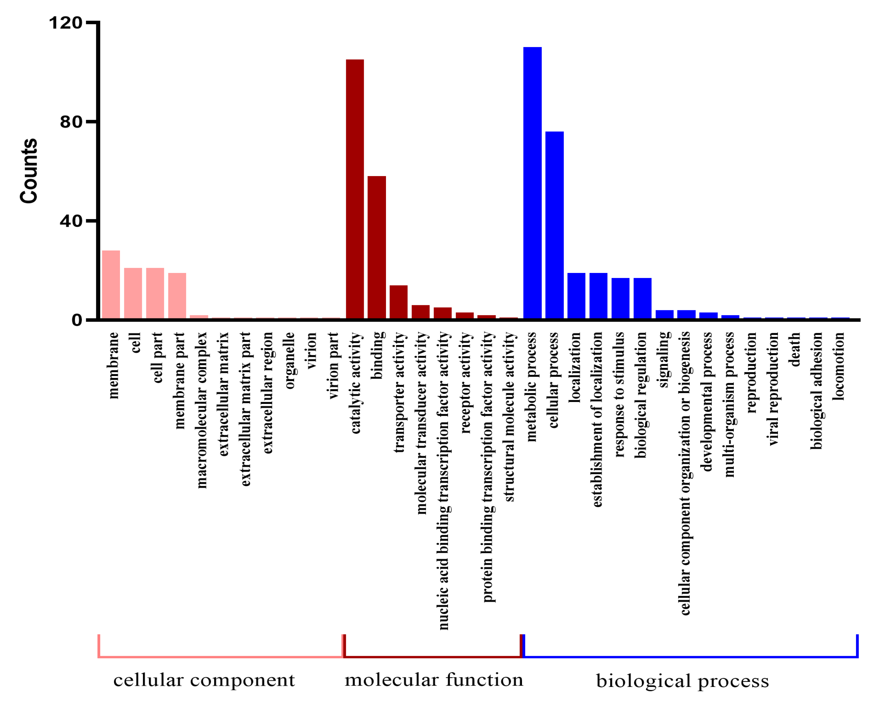
Figure 1. Variation gene GO annotation clustering.
Ijms 24 05810 g001
Figure 2. Classification of gene variations compared with KEGG database by blast.
Ijms 24 05810 g002
Figure 3. Cloning, protein expression and activity analysis of yqhH gene. (A) PCR product of fragment yqhH. Lane M: 15K DNA marker; lane 1, 2: yqhH PCR product. (B) Western blot analysis of yqhH helicase. Lane M: Protein mark; lane 1: Purified yqhH. (C) ATP hydrolysis activity assay of yqhH helicase. (D) Changes in the fluorescence intensity of the substrate donor in the presence of ATP. (E) Fluorescence resonance energy transfer changes in yqhH unwound substrates in the presence of ATP. In (CE), data are the mean values of three technical replicates ±SD.
Ijms 24 05810 g003
Figure 4. Construction of knockout strains. (A) Flow chart of Bt LLP29-△yqhH defective strain. (B) Kana and Erm plates screen for defective strains. (C) Kana and Erm liquid LB verification defective strains. (D) PCR cross-validation of Bt LLP29-ΔyqhH; lane M: 15 K DNA marker; The templates for 1–3 are Bt LLP29-ΔyqhH,Bt LLP29,H2O, and the primers are F1/R1; 4–6 templates are in the same order as 1–3 templates, and the primers are F2/R2.
Ijms 24 05810 g004
Figure 5. Screening and validation of Bt LLP29-△yqhH-R strain. (A) Kana and Erm plates screen for defective strains. (B) Kana and Erm liquid LB verification defective strains. (C) PCR cross-validation of Bt LLP29-ΔyqhH-R; lane M: 15K DNA marker; The templates for 1–3 are Bt LLP29-ΔyqhH-R,Bt LLP29,H2O, and the primers are F1*/R1*. (D) PCR cross-validation of Bt LLP29-ΔyqhH-R; lane M: 15K DNA marker; The templates for 1–3 are Bt LLP29-ΔyqhH-R,Bt LLP29,H2O, and the primers are F2*/R2*.
Ijms 24 05810 g005
Figure 6. The growth curve and phenotypes of different Bt strains. (A) The growth curve. (B) The UV-B assay. Data are the mean values of three technical replicates ± SD. * p < 0.05 (t-test) indicates a significant difference between the original strain and mutants.
Ijms 24 05810 g006
Figure 7. Unwinding activity of different strains. Data are the mean values of three technical replicates ± SD.
Ijms 24 05810 g007
Table 1. Comparative analysis of genome variation in the Bt LLP29-M19 strain.
Variation TypeNumber of VariantsSum
SNP annotation
Genome-wide heterozygous ratio (%)0.177
Total number of SNP loci1318
InDel annotation
Sum of insertion mutations1431
Total deletion mutation17
SV annotation
Deletion35206
Inversion145
inversion26
Disclaimer/Publisher’s Note: The statements, opinions and data contained in all publications are solely those of the individual author(s) and contributor(s) and not of MDPI and/or the editor(s). MDPI and/or the editor(s) disclaim responsibility for any injury to people or property resulting from any ideas, methods, instructions or products referred to in the content.