Article

Comparative Analysis of the Alkaline Proteolytic Enzymes of Yarrowia Clade Species and Their Putative Applications

1
Department of Biotechnology and Food Microbiology, Wrocław University of Environmental and Life Sciences, 51-630 Wrocław, Poland
2
SPO, Univ Montpellier, INRAE, Institut Agro, 34060 Montpellier, France
*
Authors to whom correspondence should be addressed.
Int. J. Mol. Sci. 2023, 24(7), 6514; https://doi.org/10.3390/ijms24076514
Submission received: 13 February 2023 / Revised: 19 March 2023 / Accepted: 28 March 2023 / Published: 30 March 2023
(This article belongs to the Section Biochemistry)

Abstract

:
Proteolytic enzymes are commercially valuable and have multiple applications in various industrial sectors. The most studied proteolytic enzymes produced by Yarrowia lipolytica, extracellular alkaline protease (Aep) and extracellular acid protease (Axp), were shown to be good candidates for different biotechnological applications. In this study, we performed a comprehensive analysis of the alkaline proteolytic enzymes of Yarrowia clade species, including phylogenetic studies, synteny analysis, and protease production and application. Using a combination of comparative genomics approaches based on sequence similarity, synteny conservation, and phylogeny, we reconstructed the evolutionary scenario of the XPR2 gene for species of the Yarrowia clade. Furthermore, except for the proteolytic activity of the analyzed Yarrowia clade strains, the brewers’ spent grain (BSG) was used as a substrate to obtain protein hydrolysates with antioxidant activity. For each culture, the degree of hydrolysis was calculated. The most efficient protein hydrolysis was observed in the cultures of Y. lipolytica, Y. galli, and Y. alimentaria. In contrast, the best results obtained using the 2,2-azinobis (3-ethyl-benzothiazoline-6-sulfonic acid (ABTS) method were observed for the culture medium after the growth of Y. divulgata, Y. galli, and Y. lipolytica on BSG.

1. Introduction

Nearly two decades ago, the genus Yarrowia was still limited to Yarrowia lipolytica species [1,2]. In 2004, Péter et al. [3] described Candida galli, a sister species of Y. lipolytica, which was later renamed Y. galli. In 2005, Cletus Kurtzman [4] defined the Yarrowia clade as a taxonomic group, including Y. lipolytica and C. galli, as well as Candida hispaniensis and Candida bentonensis. Since then, numerous species have been assigned to the Yarrowia clade primarily based on marker sequencing, which has become a common tool in taxonomic studies. Today, the Yarrowia clade comprises Y. lipolytica, Yarrowia alimentaria, Yarrowia bubula, Yarrowia brassicae, Yarrowia deformans, Yarrowia divulgata, Y. galli, Yarrowia hollandica, Yarrowia keelungensis, Yarrowia osloensis, Yarrowia parophonii, Yarrowia phangngaensis, Yarrowia porcina, Yarrowia yakushimensis, and C. hispaniensis [5]. The species listed above share many morphological and physiological characteristics of Y. lipolytica. One is the ability to exist as yeast cells, pseudomycelium, or true septate hyphae. However, the best known feature of all the species is their high capacity for lipid accumulation, which are the carbon storage compounds accumulated during nitrogen-limiting conditions [6,7]. Yarrowia clade species vary in terms of the maximum growth temperature, ranging from 21 °C for Y. alimentaria to 37 °C for Y. phangngaensis [7].
Most of the species belonging to the Yarrowia clade originate from European, American, or Asian regions, being isolated from various natural habitats, which are also common for Y. lipolytica, such as dairy and meat products (Y. alimentaria, Y. osloensis, Y. divulgata, Y. galli, Y. bubula, and Y. porcina), mangrove or marine waters (Y. keelungensis, Y. phangngaensis and other strains of Y. divulgata), guts of insects or vertebrates (Y. parophonii, Y. yakushimensis, and Y. hollandica) and from fermented vegetable food (Y. brassicae). Numerous concerns about the safety of using Yarrowia clade are related to Y. deformans and some strains of Y. galli, which have been isolated from human tissues [8].
However, based on the aerobic nature and the rare ability to grow at 37 °C, Y. lipolytica has been classified as generally recognized as safe (GRAS) by the American Food and Drug Administration (FDA) for particular applications. It was also considered a “safe-to-use” organism based on an exhaustive literature assessment [9]. Furthermore, the unique biochemical characteristics of Y. lipolytica contributed to the irrefutable interest of scientists in that species and its large-scale use [10]. Nowadays, Y. lipolytica is commercially used to produce microbial proteins, single cell oil, organic acids, and polyhydroxy alcohols. In addition, several enzymes like RNases, phosphatases, esterases, lipases, and proteases are produced using Y. lipolytica cultures [11]. The high proteolytic and lipolytic activity contributed to the widespread distribution of Y. lipolytica in the commercial production of meat and dairy products [12].
The most studied proteolytic enzymes produced by Y. lipolytica, i.e., extracellular alkaline protease (Aep) and extracellular acid protease (Axp), are synthesized under starvation conditions at the end of the exponential growth phase in protein-rich media [13,14].
Studies by Enderlin and Ogrydziak (1994) classified Aep as a 32 kDa subtilisin-like protein encoded by the gene, secreted in the form of a 55 kDa glycosylated precursor [15]. To develop the mature form of this enzyme, a 15 amino acid long presequence had to be removed, starting with the cleavage of the dipeptides (X-Ala or X-Pro) followed by hydrolysis of the peptide bond between Lys and Arg [16]. Aep secretion by Y. lipolytica growing in a yeast extract peptone dextrose (YPD) medium (pH 6.8) reaches around 1–2 g/L [17]. Furthermore, as shown by Akpınar and colleagues (2011), skimmed milk-based medium is appropriate for Aep secretion, and no negative impact of additional carbon (glucose) or nitrogen (glutamine) sources was determined [18].
Early studies on the nature of the Axp protease conducted by Yamada and Ogrydziak (1983) indicated the formation of three mature forms of this enzyme, which differed in their molecular weights—28, 32, and 36 kDa [19]. However, Young and colleagues (1996) showed the formation of a 37 kDa enzyme, sometimes subject to partial hydrolysis. Consequently, Axp was finally classified as a pepsin-like protein, composed of 397 amino acids with a 44 amino acid long prepro-region. The enzyme, secreted as a precursor with a 42 kDa molecular mass, is converted into its mature form in acidic environments [20].
In contrast to Y. lipolytica, the information about the biotechnological applications of other Yarrowia clade members are very limited. For example, studies about erythritol, mannitol, and arabitol production, with pure or crude glycerol as well as with glucose or fructose as carbon sources, are still in the laboratory research phase [21,22]. Similarly, studies related to lipid biosynthesis in a medium based on switch grass hydrolysate using Yarrowia clade species were performed on a small scale [6].
According to our current knowledge, studies performed by Ozturk [23] are the only ones related to the proteolytic activity of Yarrowia species other than Y. lipolytica. These studies showed the ability of Y. galli, Y. deformans, and Y. alimentaria to gelatin liquefaction, their high proteolytic activity, and their ability to secrete proteases. Therefore, due to the presumably high application potential of the produced proteases, it is necessary to expand our knowledge about these enzymes and their properties.
The selection of a substrate in protease research is crucial for commercial application. The cost of the growth medium represents about 30–40% of the price of industrial enzymes [24]. The brewers’ spent grain (BSG), the major solid by-product in the brewing industry, due to the high protein content (19–30% w/w) and low price of approximately EURO 35 per ton, is a prospective substrate. Moreover, the average annual global production of BSG, estimated to be about 39 million tons, with 3.4 million tons produced in the European Union, forces the search for new valorization methods for this agro-industrial by-product [25].
Our research provides a comprehensive analysis of the alkaline proteolytic enzymes of Yarrowia clade species, including phylogenetic studies, synteny analysis, and protease production and application. Furthermore, in order to address the economic aspects of protease production, a protein-rich spent grains medium was selected. Finally, considering the potential applications of Yarrowia proteases, we established a link between the high degree of protein hydrolysis and antioxidant activity of the obtained hydrolysates.

2. Results and Discussion

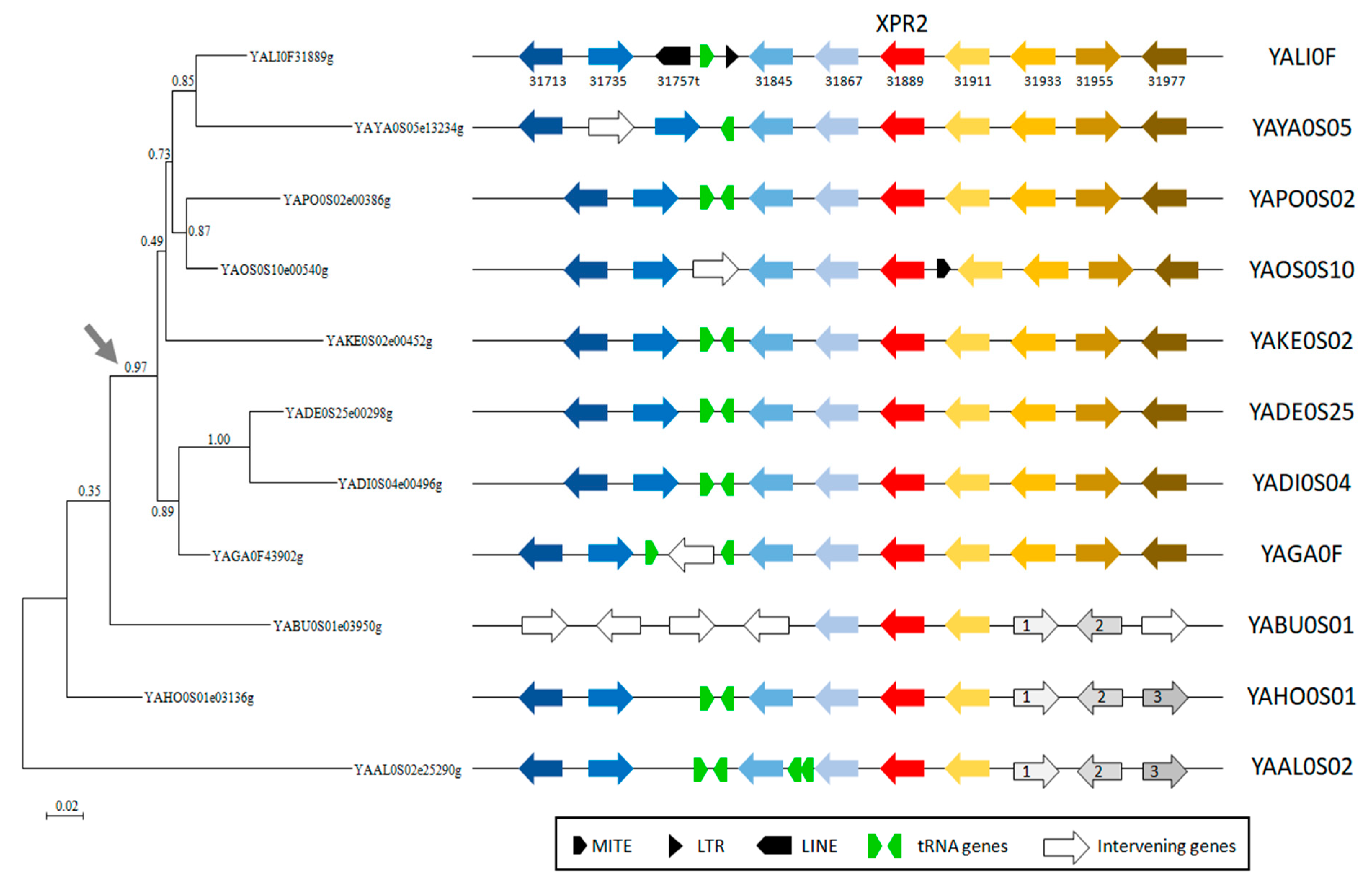
2.1. Comparative Analysis of Yarrowia Alkaline Proteases from a Genomic Point of View

The search for alkaline proteases encoding genes in the genome of the different Yarrowia species showed the presence of 5 (Y. phangngaensis) to 21 (Y. galli) genes (Table 1). Five genes were also found in C. hispaniensis, which has a basal position in the clade. Due to the lack of access, genomes of Y. brassicae and Y. parophonii were not examined in terms of alkaline protease genes. Instead, three strains of Y. lipolytica were analyzed: the Polish strain A101, the German strain H222, and the laboratory strain E150 (CLIB 122) issued from a crossing between the French W29 and the American ATCC 18942 followed by many back-crosses [8]. The genome of E150 was the first fully sequenced genome and is still the reference for genome assembly and annotation of new Y. lipolytica genomes.
Based on sequence similarity, we defined 13 different groups of putative alkaline proteases as shown on the phylogenetic tree (Figure 1).
Most groups contained one Y. lipolytica representative except for three groups with two proteins with multiple counterparts that could not be separated based on sequence similarity. One of these groups corresponds to the XPR2 group, i.e., the Aep encoded by YALI0F31889g, part of which (highlighted by a blue curve in Figure 1) gathered one protein per species except for Y. phangngaensis and C. hispaniensis for which no ortholog was found. The genetic distances between these genes are small, as shown by the short branches in the tree. The other proteins in the XPR2 group exhibited very long branches representing proteins emanating from successive duplication events, which occurred in Y. osloensis, Y. porcina, Y. galli, and Y. yakushimensis. Synteny analysis of these genes showed different insertion sites suggesting that these duplications occurred independently (Figure S1).
One exception to this rule is the conservation of synteny in the upstream region of YAOS0S01e24608g and YAPO0S01e09780g genes, which are probably derived from the same event (Figure S1). In Y. yakushimensis, YAYA0S05e13234g, the syntenic copy of XPR2, was successively duplicated three times on scaffold YAYA0S6. These duplication events involved not only XPR2 but also the homolog of YALI0F31845g (F31845 in Figure S1).
In contrast, there is extensive synteny conservation in the region of XPR2 close homologs (part of the XPR2 group with short branches) with some insertions of tRNA genes, transposable elements, and intervening genes (Figure 2). Nevertheless, we observed a rearrangement occurring one gene upstream of the XPR2 homolog (upstream of YALI0F31867g) in Y. bubula. An additional rearrangement generated a loss of synteny one gene downstream of XPR2 (downstream from YALI0F31911g). As synteny is conserved in Y. bubula, Y. hollandica, and Y. alimentaria, which are basal species of the genus Yarrowia, the most parsimonious explanation is that the rearrangement occurred in the other species (grey arrow in Figure 2), leading to the actual order of genes in Y. lipolytica and closely related species.

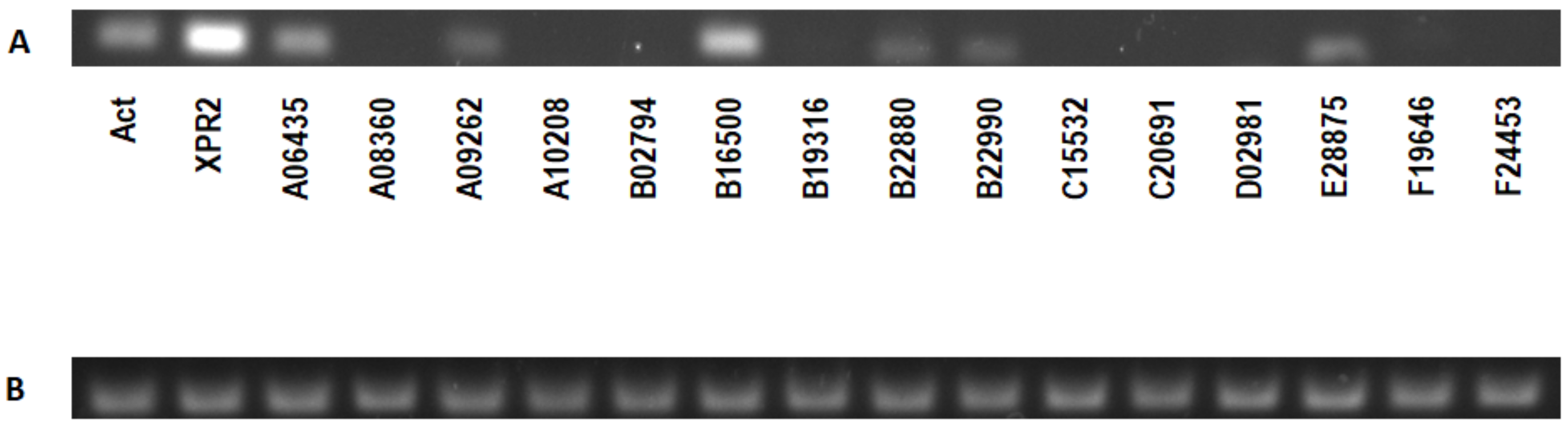
2.2. Expression Analysis of Y. lipolytica W29 Alkaline Proteases in Various Culture Conditions and Signal Peptide Prediction

To verify which of the 16 putative proteases may play an important role in protein hydrolysis by the analyzed strains, we took advantage of previously published RNA-seq data of Y. lipolytica W29 [26] and unpublished data performed on a derivative of E150 (CLIB 122) (genotype: E150, ku70::URA3, LEU2). Twelve putative protease encoding genes showed low or no expression (Table 2). The XPR2 gene is highly expressed in minimal medium with glucose but poorly in oleic acid and tributyrin. YALI0A06435g and YALI0A08360g were moderately expressed on the three tested carbon sources in yeast nitrogen base (YNB) medium. Surprisingly, YALI0B16500g, a homolog of Saccharomyces cerevisiae PRB1- protease B, is highly expressed in YPD medium at high and low pH. Previous studies showed that XPR2 is induced by pH above 6 and/or by the presence of peptone [27]. Thus, we checked the expression of all putative protease encoding genes in these conditions and, as expected, XPR2 was expressed on the YPD medium, although moderately, probably due to the genotype of E150 (CLIB 122) derivative (his-1, ura3-302, leu2-270, xpr2-322, ku70::URA3, LEU2). In turn, low expression of XPR2 was observed on YPD glucose at pH 4 (Table 2). The highest expression was observed for YALI0B16500g on both YPD and YPD at pH 4. Furthermore, the functional annotation of YALI0B16500g predicts it to be localized in the vacuole. Thus, we checked all proteases for the presence of a putative signal peptide. It appeared that all of the putative proteases, including YALI0B16500p, could potentially be secreted (Table 2).

2.3. Analysis of Expression of XPR2Alkaline Protease in the BSG Medium by Y. lipolytica

Expression analysis of all the genes encoding putative proteases in the BSG-based medium was performed. Due to the large number of genes to be tested, we analyzed the expression only in the Y. lipolytica W29 strain. Out of the 16 genes encoding putative proteases, 7 were expressed under the analyzed conditions. The highest expression was observed for XPR2, followed by the YALI0B16500 gene (Figure 3). These data are, however, in contrast with the proteomic studies, in which the analyzed bands on SDS-PAGE were covered mostly with Aep protein (details see Section 2.7). These results were further verified by overexpression of the YALI0B16500 gene in the PO1d strain (deleted for the XPR2 gene), where no protease activity was detected on skimmed milk agar medium at pH 6.8.

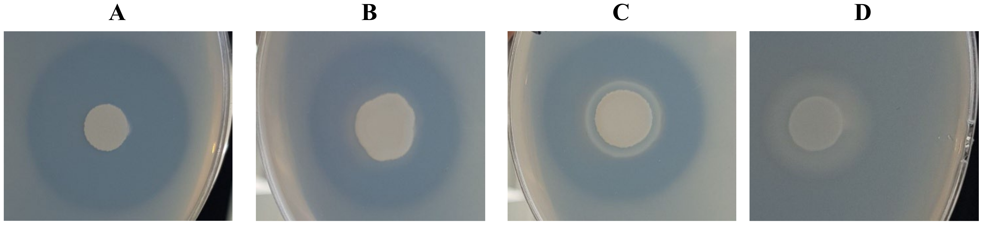
2.4. Investigation of the Proteolytic Activity of Yarrowia Clade Species

The proteolytic activity of the Yarrowia clade was analyzed using skimmed milk agar plates. Results showed huge diversity of the clade members in terms of their abilities to secrete proteases (Table 3).
Y. phangngaensis, Y. deformans, and C. hispaniensis showed no proteolytic activity, indicated by the lack of proteolysis zones on the skimmed milk agar (Table 3). The highest proteolytic activity was observed for Y. yakushimensis (15.05 ± 0.52), followed by Y. lipolytica (14.80 ± 0.40), Y. keelungensis (14.02 ± 0.04), and Y. alimentaria (13.51 ± 1.01) (Figure 4).
The proteolytic activity of fungal, yeast, and bacterial strains derived from an artisanal raw ewe’s milk cheese was investigated by Ozturkoglu-Budak and colleagues [28]. Among the five tested yeast species, only Y. lipolytica AF_335977 and Debaryomyces hansenii AF_210327 were able to secrete proteases. However, the proteolytic activity of AF_335977 was described as medium. At the same time, it was the best result among tested yeast species. In the studies by Gardini and colleagues [29], several yeast species isolated at different stages of Pecorino Crotonese production and ripening were characterized by their technological properties. However, only five strains, identified as Y. lipolytica and D. hansenii, were able to hydrolyze skimmed milk proteins.
Furthermore, Y. lipolytica and numerous other yeast species isolated from starter-free raw cow’s milk cheeses were investigated for extracellular proteolytic activity. The highest activity of proteolytic enzymes was observed for Y. lipolytica and K. lactis, while the highest caseinolytic activity was presented by Y. lipolytica and P. guilliermondii [30]. Similarly, the proteolytic activity of yeast species isolated at different stages during the processing of pastirma (the most popular dry-cured meat product in Turkey) was investigated [23]. The yeast isolates, classified as Candida zeylanoides (the predominant species), C. deformans, C. galli as well as C. alimentaria (currently classified as Yarrowia genus), showed clear zones surrounding the colonies. Our results clearly confirm the potential of some Yarrowia species to secrete high amounts of proteases into the growth medium.

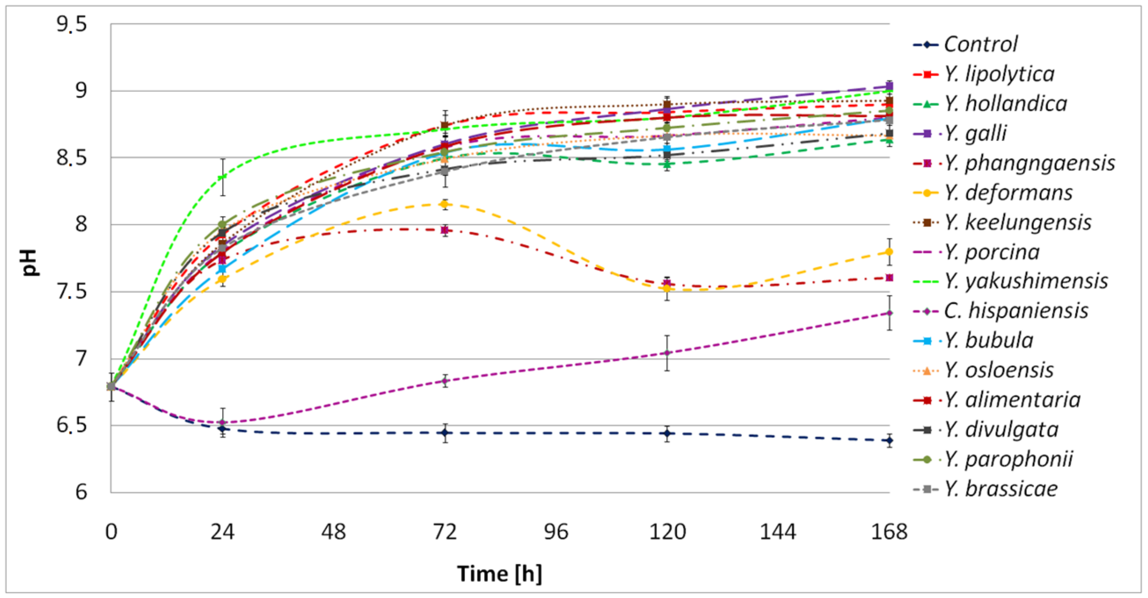
2.5. Characterization of the Proteolytic Activity of Yarrowia Clade Growing on BSG

The Yarrowia clade members differed significantly in terms of their proteolytic abilities. Y. phangngaensis, Y. deformans, and C. hispaniensis showed no proteolytic activity under the analyzed conditions (Figure 5). In Y. lipolytica W29, XPR2 is the highest expressed gene in the BSG medium (Figure 3). The absence of proteolytic activity in Y. phangngaensis and C. hispaniensis could be explained by the loss of the XPR2 homolog (Figure 1), which is, however, not the case in Y. deformans. The latter species was analyzed for the expression of XPR2 ortholog (YADE0S25e00298g) in BSG medium. However, no transcript was detected.
The proteolytic activity of Y. hollandica, Y. porcina, Y. yakushimensis, Y. bubula, Y. osloensis, Y. divulgata, and Y. brassicae did not exceed 0.5 U/mL (Figure 5). The highest proteolytic activity was detected for Y. lipolytica W29 with 2.47 U/mL/min (Figure 5). Other Yarrowia species showed strong proteolytic activities: Y. galli (1.96 U/mL/min), Y. alimentaria (1.38 U/mL/min), Y. keelungensis (1.07 U/mL/min), and Y. parophonii (0.80 U/mL/min). These results allow us to consider the above-mentioned strains as interesting producers of proteolytic enzymes with potential industrial applications in the future.
In the cultures of Y. lipolytica, Y. alimentaria, and Y. parophonii, the proteolytic activity remained stable throughout the process, indicating continuous enzyme secretion and protein hydrolysis. In turn, in cultures of Y. galli and Y. keelungensis, a significant decrease in protease activity after reaching the maximum point was observed. It is possible that Y. galli and Y. keelungensis secreted a significant amount of enzymes at the beginning of the culture, allowing rapid protein hydrolysis and providing sufficient nutrients to decrease protease secretion.
The proteolytic activity of Y. lipolytica W29 and Y. lipolytica IMUFRJ 50862, isolated from an estuary near Rio de Janeiro, were analyzed in the research of Braga and colleagues [31], who investigated the effect of lipase secretion on the γ-decalactone production. In a medium optimized for lipase production, the proteolytic activity reached 0.0034 U/mL/min and 0.00151 U/mL/min for W29 and IMUFRJ 50862, respectively. The difference in results obtained in our work, caused by the medium composition, clearly indicates the potential for using BSG as a substrate.
In the present work, high proteolytic activity was associated with significant hydrolysis of BSG protein. Furthermore, it caused the accumulation of hydrolysis products, expressed as free α-amino groups. In the culture of Y. lipolytica, Y. galli, Y. alimentaria, and Y. keelungensis, characterized already as leading proteolytic strains, the concentration of α-amino groups reached 4367μg/mL, 4245 μg/mL, 3939 μg/mL, and 3885 μg/mL, respectively (Figure 6).
Interestingly, in our previous work using proteolytic bacteria, the concentration of free α-amino groups in the same BSG-based medium never exceeded 2600 μg/mL [32]. This result clearly shows the benefits of using Yarrowia clade yeast when applying BSG.
Eight strains of Y. lipolytica, isolated from goat/water buffalo mozzarella cheese, were previously studied by Suzzi and colleagues [33] for their ability to breakdown milk proteins. Y. lipolytica isolates showed weak proteolytic activity (expressed as leucine concentration [mg] per 100 mL of milk) after 8 days of the process. However, between the 8th to the 14th day, a huge increase in proteolytic activity was observed. After 14 days, the concentration of leucine reached 300–600 mg/100 mL, while in our research, the concentration of free α-amino groups in the BSG medium reached a maximum of 350–450 mg/100 mL for the most active strains (Figure 6). It is worth pointing out that the time of culture in our research was only 5 days. This result further confirms the proteolytic abilities of Yarrowia clade members. For six out of fifteen tested species, the degree of BSG protein hydrolysis (DH%) exceeded 50% (Figure 7).
The best results were observed for Y. lipolytica with 72.78% hydrolyzed proteins. Therefore, we conclude that the hydrolysis performed by Yarrowia species enabled a major protein breakdown. Furthermore, in similar studies with Rhizopus sp., DH% of the BSG protein reached 59.0 ± 5.4% [34]. This further confirms that BSG proteins are strong inducers of the Yarrowia clade proteolytic enzymes and, thus, that it is an appropriate substrate for optimization studies. Moreover, given that filamentous fungi are common producers of hydrolytic enzymes, the results obtained by Y. lipolytica, Y. galli, Y. alimentaria, and Y. keelungensis are more than satisfactory.
The protein hydrolysis in the cultures of Yarrowia clade members was associated with environmental alkalization (Figure 8). As a result, the final pH fluctuated between 8.5 and 9. Only in the Y. phangngaensis, Y. deformans, and C. hispaniensis culture, where a lack of proteolytic activity and no hydrolysis of the BSG proteins were observed, did the pH not exceed 8.
Medium alkalization in the cultures of Y. lipolytica confirmed the previous studies in which K. lactis, D. hansenii, Pichia guilliermondii, Pichia fermentans, S. cerevisiae, Candida spp., and Rhodotorula spp., as well as 21 strains of Y. lipolytica, were analyzed for their properties influencing cheese production [30]. As a result, 16/21 and 10/21 of Y. lipolytica strains showed alkalizing properties on citrate agar and litmus milk, respectively.
To summarize the current information, species such as Y. lipolytica, Y. alimentaria, Y. galli, and Y. keelungensis are the most efficient and universal (in terms of medium composition) proteolytic enzyme producers.

2.6. Profile of Yarrowia Clade Proteolytic Enzymes Secreted in BSG-Based Medium

The gelatin zymography allowed for the detection of several proteolytic enzymes produced extracellularly during the growth of Yarrowia clade species in BSG medium (Figure S2). Molecular masses of proteolysis bands observed on zymography gel are summarized in Table 4. It is important to underline that some proteolysis bands represent protease complexes formed as a result of interactions between enzyme monomers. This can be confirmed in the Y. lipolytica example. The molecular weight of Y. lipolytica proteases is 40–90 kDa. Therefore, producing an enzyme with a molecular weight of 110 or 130 kDa is impossible. Nevertheless, based on obtained results, it is possible to define the enzymes/enzymes complex involved in BSG protein hydrolysis.
As shown, Y. lipolytica and Y. alimentaria were the most active species in proteolytic activity and the most efficient in BSG protein hydrolysis. The protease profile of Y. lipolytica and Y. alimentaria was represented by two main proteolytic bands at 110 and 130 kDa (Table 4). However, a proteolytic band at 130 kDa was also detected in Y. brassicae and Y. divulgata culture, where low proteolytic activity and DH% < 30 were estimated. Therefore, we suggest a key role of the 110 kDa protein complex in BSG protein hydrolysis.
Furthermore, strong proteolytic activity in BSG medium as well as significant DH% of BSG protein, was demonstrated for Y. galli. The profile of proteolytic enzymes of Y. galli was represented by two bands with a molecular mass of 37–39 kDa and 40 kDa (Table 4). Nevertheless, the production of 40 kDa protease of low proteolytic activity (<0.5 U/mL) was determined in the Y. osloensis culture medium. Following this, protease of 37–39 kDa seems significant for BSG protein hydrolysis. It is important to underline that the same enzyme profile as Y. galli was observed for Y. parophonii, in which proteolytic activity (0.80 U/mL/min) was almost twice as low as Y. galli (1.96 U/mL/min). An explanation for this is the difference in the expression level of the 37–39 kDa enzyme.
Finally, a protease of molecular mass 30 kDa, secreted in Y. keelungensis (Table 4), is a crucial factor affecting BSG protein hydrolysis as indicated by high proteolytic activity and significant DH%. Production of proteolytic enzymes of 30 kDa was also observed in the Y. porcina culture medium. However, based on the results of the proteomic analysis presented in Section 2.7, enzymes of Y. keelungensis and Y. porcina are, with a high probability, different compounds of similar molecular weight.
Production of proteolytic enzymes in Y. lipolytica was already reported. An alkaline extracellular protease of 35 kDa was produced by Y. lipolyticaYITun15 [35]. This enzyme showed significant activity in a wide spectrum of factors tested. A Y. lipolytica coconut copra isolate produced protease of 61.5 kDa. The fish flower was applied as a substrate and enzyme production inductor [36]. In other studies, a 28 kDa protease was secreted by an auxotrophic recombinant strain of Y. lipolytica Po1d (JMY 1852). The enzyme was produced as an undesirable factor affecting human interferon alpha 2b production [37]. Another example involved the production of the intercellular protease of 97 kDa by Y. lipolytica strain IPN-MI-1 of the yogurt origin. The enzyme showed stable activity in the pH range of 5.0–9.0, and 37 °C was the optimal temperature [38].
In order to conduct proteomic analysis of Yarrowia clade enzymes, protein electrophoreses were performed. Unfortunately, despite a significant increase in protease concentration obtained using acetone precipitation, some of the proteases detected by gelatin zymography did not appear on the electrophoretic gel. Therefore, only the samples presented in Table S1 were subjected to proteomic investigation.

2.7. Protein Sequence Coverage of Yarrowia Clade Proteases to Other Fungal Enzymes

Proteomic analysis of the enzymes expressed in the BSG medium culture of Y. lipolytica, Y. keelungensis, and Y. alimentaria showed high protein sequence similarity to Aep of Y. lipolytica E150 (CLIB122) encoded by XPR2 (Table S1). The amino acid sequence of the Y. lipolytica enzyme complex of 130 kDa and 110 kDa was 40% and 44% aligned to the sequence of Aep, respectively. Moreover, the protein sequence coverage of the Y. alimentaria enzyme complex (130 kDa) and Y. keelungensis enzyme (30 kDa) to Aep reached 42% and 35%, respectively.
In the analysis of these results, two factors were important. First, we used public databases of fungal proteins to analyze the proteomic data, but the proteomes of Yarrowia species are not available, except those of Y. lipolytica strains. Despite this, the results of the proteomic analysis confirmed close affinity between Yarrowia species. Moreover, matched peptide sequences were found in the position range of 158–454, corresponding to the proper chain of Aep, which is converted into the active form of the enzyme upon signal peptide and propeptide cleavage.
The proteolytic enzyme of Y. osloensis (65 kDa) and Y. parophonii (65 kDa) were, to a great extent, 32% and 26%, respectively, aligned to alkaline extracellular protease of Yarrowia sp. E02 and Yarrowia sp. B02, while sequence coverage of Y. porcina (30 kDa) protease to the Carboxypeptidase of Y. lipolytica YB392 coded by B0I71DRAFT_162965 gene reached 25%. The full set of Yarrowia clade enzymes proteomic analysis results is available in the Supplementary Materials (Table S1).

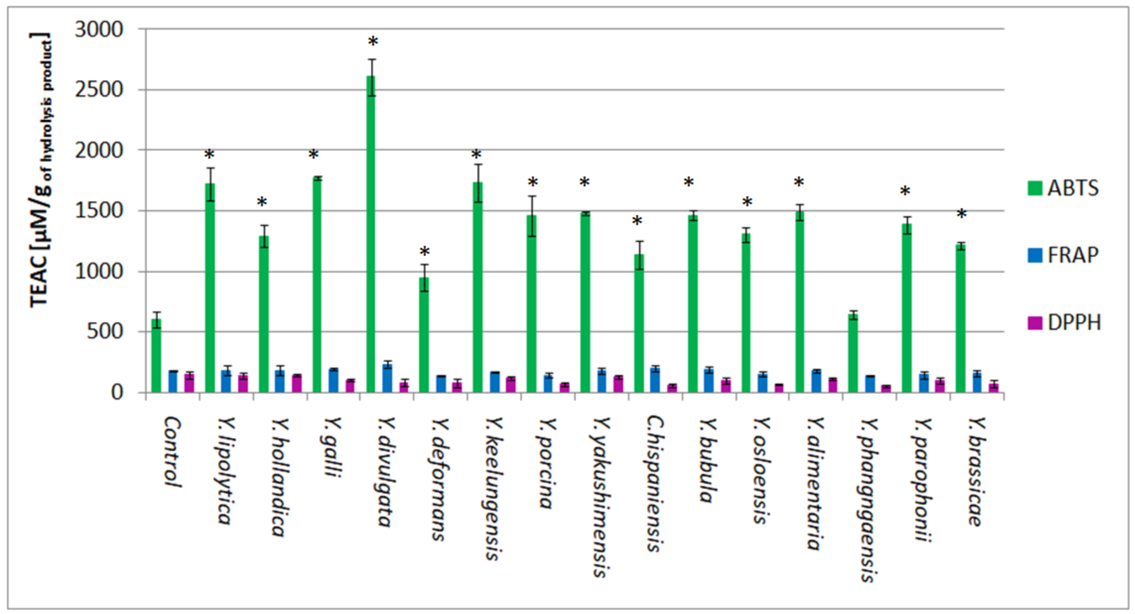
2.8. Antioxidant Activity of BSG Protein Hydrolysates

Three methods were applied in order to estimate the antioxidant activity of BSG protein hydrolysates obtained in the cultures of Yarrowia clade species. Significant antioxidant activity was detected using the ABTS method. The highest activity, reaching 2606.13 μM TEAC/g, was detected in the Y. divulgata culture medium (Figure 9). This result was followed by the activity of Y. galli, Y. keelungensis, and Y. lipolytica samples, reaching 1771.87 μM TEAC/g, 1730.73 μM TEAC/g, and 1722.63 μM TEAC/g, respectively. It was more than twice higher than the activity of the control sample (603.73 μM TEAC/g). According to our previous research, the antioxidant activity of the control samples is caused by the partial hydrolysis of BSG protein occurring during media sterilization [32]. High antioxidant activity of post-culture media of Y. galli, Y. keelungensis, and Y. lipolytica strains resulted mainly from the efficient hydrolysis of spent grain protein (DH% > 60%), which is not the case of Y. divulgata, where DH% did not exceed 30%.
In studies where protein hydrolysis is carried out directly in microbial cultures, the antioxidant activity of hydrolysates may be affected by the products of microbial metabolism. BSG is a waste material containing, in addition to protein, cellulose (17–25%), non-cellulosic carbohydrates (25–35%), lignin (8–28%), lipids (10%), and ash (5%) [39]. Therefore, due to the hydrolysis, other compounds, such as phenolics, may be exposed or released to the culture medium affecting the antioxidant activity of hydrolysate [40]. Y. divulgata is the object of extensive studies concerning erythritol production [22,41]. According to den Hartog et al. (2010), erythritol was recognized to possess strong antioxidant activity [42]. Therefore, simultaneous production of erythritol as a process accompanying BSG protein hydrolysis should be considered possible.
In our previous work, the antioxidant activity of BSG protein hydrolysates, obtained using the ABTS method directly in proteolytic bacteria cultures, ranged from 345.18 to 1621.31 μM TEAC/g peptides [32]. In this work, most of the analyzed samples showed a total activity of about 1500 μM TEAC/g. Therefore, using some Yarrowia clade yeast proteolytic enzymes allowed us to obtain hydrolysates with higher antioxidant activity compared to bacterial enzymes. It is likely related to DH%, reaching higher values in the yeast cultures, showing that high antioxidant activity is strongly associated with a high degree of protein breakdown.
The other two methods did not demonstrate the significant antioxidant activity of hydrolysates compared to the control. The best result obtained using the DPPH method is 144.90 μM TEAC/g in the culture medium of Y. hollandica, i.e., almost 10 times lower than the activity detected using the ABTS method (Figure 9). The antioxidant activity of Yarrowia clade culture samples did not reach the control activity (147.80 μM TEAC/g). However, these results align with the work of Wołosiak and colleagues [43], in which the limitation in the solubility of peptides and amino acids in the system of DPPH assay (free radical resuspended in ethanol solution) is underlined. In addition, numerous problems related to the formation of iridescence in the presence of amide compounds were described. Therefore, the authors recommended the ABTS method as appropriate for the research on the samples containing significant quantities of peptides. On the other hand, due to large differences in the DPPH and ABTS systems, when possible, it is still advisable to apply both methods.
The antioxidant activity of BSG protein hydrolysates was evaluated using a ferric reducing antioxidant power (FRAP) assay in the work of Vieira and colleagues [44]. Activities of hydrolysates reached 0.062 mg TE/mg dw (247.71 84 μM TEAC/g), 0.098 mg TE/mg dw (391.55 μM TEAC/g), and 0.101 mg TE/mg dw (403.53 μM TEAC/g) when using brewery spent yeast extract, neutrase, and alcalase, respectively, for hydrolysis purposes. According to Vieira and colleagues, FRAP of protein hydrolysates depends not only on the molecular size of peptides but on the presence of specific peptide sequences and the amino acid composition of the hydrolysis products.
Antioxidant activities measured in Yarrowia clade cultures ranged between 140–240 μM TEAC/g using the FRAP method, with the highest result obtained for the hydrolysate of Y. divulgata (233.13 μM TEAC/g) (Figure 9). In addition, the obtained activities were not significantly different from the control sample (181.46 μM TEAC/g). Presumably, due to their chemical properties, the peptides in our samples did not precipitate in the reduction reaction of Fe3+ into Fe2+ as electron donors, which is the basis of the FRAP method.
Nowadays, antioxidant peptides are increasingly applied in the production of nutraceuticals and functional food ingredients [45], pharmaceuticals [46], and in the field of dermatology [47]. As an aging population, we search for ingredients of increased potency. Therefore, peptides of the activity that result in diminished lines and wrinkles, smoother skin texture, and reduced redness and skin discoloration are highly sought after in the skincare industry [47]. Moreover, the unstable and reactive nature of free radicals results in cell damage leading to diseases such as hypertension, cardiovascular disease, cancer, diabetes mellitus, and neurodegenerative and inflammatory diseases. Therefore, antioxidant peptides protect the body by scavenging free radicals and inhibiting lipid peroxidation reactions, thus preventing oxidative damage [46]. In addition, antioxidant peptides are also applied in food systems to preserve food products against lipid oxidation leading to the development of undesirable off-flavors, odors, and potentially toxic reaction products [45]. Therefore, we believe that the peptides obtained as a result of BSG protein hydrolysis can be used in the food, pharmaceutical, and medical industries.

3. Materials and Methods

3.1. Chemicals, Microorganisms, and Materials

Ninhydrin (analytical grade), Trizma® base (≥99.9%), Trichloroacetic acid (TCA, ≥99.0%), Triton X-100 (analytical grade), Sodium Dodecyl Sulfate (SDS; ≥99%), Bromophenol Blue (analytical grade), 2,2′-azino-di-(3-ethylbenzthiazoline sulfonic acid) (ABTS, ≥98%)), potassium peroxodisulfate (≥99%), 2,2-diphenyl-1-picrylhydrazyl (DPPH), 6-hydroxy-2,5,7,8-tetramethylchroman-2-carboxylic acid (Trolox, ≥97%), iron(III) chloride hexahydrate (FeCl3 × 6H2O, ≥97%),2,4,6-Tris(2-pyridyl)-S-triazine (TPTZ, ≥98%), Coomassie Brilliant Blue R 250 were purchased from Sigma (Sigma-Aldrich, Saint-Louis, MO, USA) and 4×Laemmli Sample Buffer was purchased from Bio-Rad (Bio-Rad Laboratories, Hercules, CA, USA). All other chemicals used in the following studies were of analytical grade.
Yarrowia clade species: Y. lipolytica W29, Y. hollandica CBS 4855, Y. galli CBS 9722, Y. phangngaensis CBS 10407, Y. deformans CBS 2071, Y. keelungensis CBS 11062, Y. porcina CBS 12935, Y. yakushimensis CBS 10254, Candida hispaniensis CBS 9996, Y. bubula CBS 12934, Y. osloensis CBS 10146, Y. alimentaria CBS 10151, Y. divulgata CBS 11013, Y. parophonii CBS 12427, and Y. brassicae CBS 15225 were purchased from the Centraalbureau voor Schimmel cultures (CBS, Utrecht, The Netherlands), stored in YPD(agar (15 g/L), bacteriological peptone (20 g/L), glucose (20 g/L), yeast extract (10 g/L)) slants and cultured in YPD broth at 25 °C, 180 rpm for 24 h before use.
BSG, used as a nutrient source and factor inducing the proteolytic enzyme secretion, was purchased from the Polish brewery Browar Stu Mostów (Wroclaw, Poland), where it was collected after wort production. Initial wort was obtained from the malt composed of 30% barley and 70% wheat grains and therefore contained various proteins. Initially, in order to protect the raw material from the development of contaminating microflora, raw BSG was dried at 50 °C for approximately 48 h. During this process, the material was periodically mixed and controlled in terms of humidity.

3.2. Bioinformatic Analysis of Yarrowia Clade Proteolytic Enzymes

Protease sequences were extracted from the Yarrowia genomes previously published by Červenák and colleagues [48] by sequence homology to alkaline extracellular protease XPR2 (YALIA101S03e00364g) from Y. lipolytica A101. The blastp threshold for E-value was set to 1 × 10⁻²⁰. Protein sequences were aligned using Clustal Omega [49] in the multi-platform graphical user interface Seaview version 4.6.2 [50]. Gblocks [51] was used to select blocks of conserved sites useful for phylogenetic analysis. Trees were reconstructed by maximum likelihood using PhyML 3.0 [52] with an LG evolutionary model. Robustness of the trees was assessed by the approximate likelihood ratio test approach (aLRT). Synteny analyses were done manually using Artemis genome browser [53].

3.3. Analysis of Alkaline Proteases Expression in Y. lipolytica Grown in Various Culture Conditions and Prediction of Putative Signal Peptide

Previously published data [26] were used to estimate the expression level of each Y. lipolytica W29 alkaline protease on yeast nitrogen base (YNB) with oleic acid, glucose, and tributyrin. Additional unpublished data from cDNA sequence analysis (Neuvéglise and colleagues) were used for analyzing the expression of the same genes in YPD and YPD at pH 4.0. Signal peptide predictions were performed with SignalP-5.0 [54] and DeepLoc-1.0 [55].

3.4. Analysis of Alkaline Proteases Expression in Y. lipolytica Grown on Spent Grains

The analysis of gene expression encoding verified and putative alkaline protease in Y. lipolytica was performed in the yeast cells growing on BSG according to the methodology described in Section 3.5. After 72 h of culture, the biomass was filtered through Whatman filters and collected into 50 mL Falcon tubes. The biomass was divided into 2 mL samples, centrifuged at 15,000 rpm for 1 min, washed 3 times with sterile distilled water, and frozen in liquid nitrogen. RNA was extracted from three biological replicates using RNeasy Micro Kit (Qiagen; Santa Clarita, CA, USA) following the manufacturer’s instructions. The obtained RNA was DNase digested (Thermo Fisher, Waltham, MI, USA) for reverse transcription reaction using Maxima First Strand cDNA Synthesis Kit (Thermo Fisher Scientific, Inc., Waltham, MA, USA). The obtained cDNA was a template for PCR reaction using Phire Green Hot Start II PCR Master Mix (Thermo Fisher Scientific, Inc., Waltham, MA, USA) and primers listed in Table 5. Similarly, expression of Y. deformans YADE0S25e00298g gene was analyzed following the above-described methodology. Actin gene (ACT1, YALI0D08272g) was chosen as a housekeeping gene in this analysis.

3.5. Cultivation of Yarrowia Clade Species in the BSG Medium

The analyzed species belonging to the Yarrowia clade were cultured in a minimal medium supplemented with BSG as carbon and nitrogen sources to stimulate proteolytic enzyme secretion. Yeast cultures were performed in 250 mL Erlenmeyer flasks containing 50 mL of the medium composed of BSG (50 g/L), MgSO4 (1 g/L), KH2PO4 (0.1 g/L), K2HPO4 (0.13 g/L), CaCl2 (0.5 g/L), and FeSO4 × H2O (0.01 g/L). After sterilization, the pH of the medium was adjusted to 7.0 by adding sterile 5 M NaOH (addition of approx. 320 μL per flask). Before inoculating the main culture, yeast was grown in YPD medium for 24 h under180 rpm agitation at 25 °C, centrifuged for 10 min at 7500 rpm, and washed with distilled water. The optical density was standardized to reach OD600 = 0.25 in the BSG medium. Non-inoculated medium was cultured under the same conditions and used as a control. Each culture was cultivated for 168 h at 25 °C on a rotary agitation (180 rpm). One milliliter samples were taken every 48 h and analyzed for proteolytic activity, and the concentration of free α-amino groups was estimated. As pH is an important factor affecting the type of proteases produced by Yarrowia clade species, appropriate pH measurements were performed.

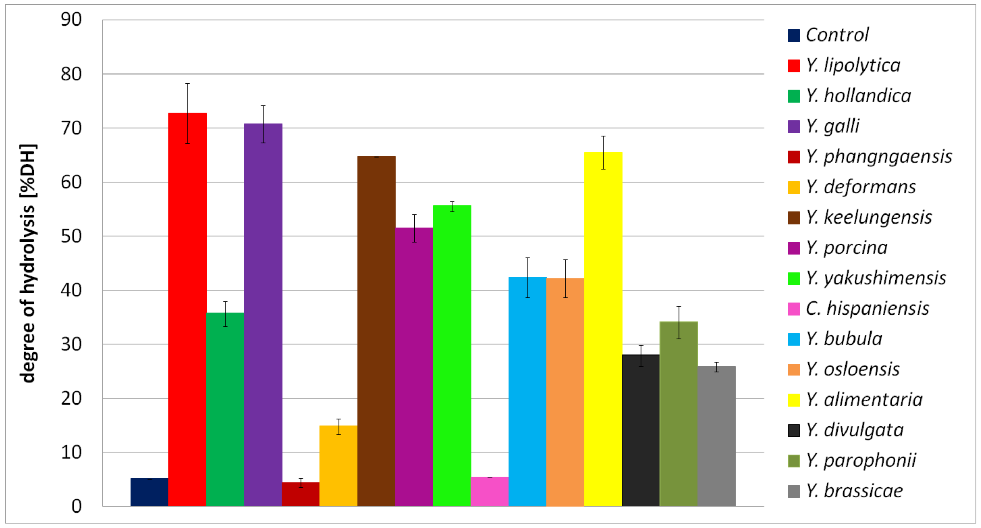
3.6. Determination of Hydrolysis Efficiency

In order to define involvement of proteases secreted by Yarrowia clade species on the availability and solubilization of BSG proteins, %DH was estimated. For this purpose, the concentration of free α-amino groups was determined using ninhydrin method [56]. The maximum concentration of α-amino groups detected for each culture was compared to the data obtained for the protein hydrolysis using hydrochloric acid (total hydrolysis), performed as described previously [32]. The %DH was calculated according to the following equation:
% D H = B H × 100 / T H
where TH (μg/mL) and BH (μg/mL) are the average concentration of α-amino groups obtained, respectively, as a result of the total hydrolysis (TH) or in the yeast culture (BH) calculated per 1 g of BSG sample.

3.7. Analysis of the Proteolytic Activity

In order to evaluate the proteolytic activity of the yeast species, the method of Anson was applied [57]. Shortly, proteolytic activity was estimated by mixing 0.45 mL of casein (1.1% in 50 mM Tris-HCl buffer, pH 7.5) with 0.5 mL of 50 mM Tris-HCl buffer (pH 7.5) and 0.05 mL of the analyzed sample. Reaction mixtures were incubated at 55 °C for 10 min, followed by adding 0.5 mL 8% (w/v) TCA to terminate the reaction. For each sample, appropriate negative controls were performed (no enzyme addition). As a positive control, protease from Bacillus licheniformis (Sigma-Aldrich, Germany) was applied. Finally, samples were centrifuged at 12,000 rpm for 15 min, and the absorbance of supernatants was determined at 280 nm. Tyrosine was used as a standard in the concentration range of 0–2 mM/L. One unit of enzyme activity was defined as the amount of enzyme required to release 1 μM of tyrosine per 1 mL of culture medium within 1 min under the experimental conditions.
The proteolytic activity of the Yarrowia clade species was also detected using skimmed milk agar plates. For this purpose, the standardized biomass solutions (OD600 = 1.0) were spotted (5 μL) on the agar plates containing 2% (w/v) of agar and 10% of sterile skimmed milk (0.5%) (w/v). Inoculated plates were air dried under sterile conditions followed by 24 h incubation at 25 °C. Finally, the proteolysis zones were measured. As a control, non-inoculated medium was spotted on the agar plates and incubated under the same conditions. Both types of proteolytic activity measurements were performed in triplicate.

3.8. Estimation of Proteolytic Enzyme Profiles

3.8.1. Determination of the Profile and Molecular Mass of Yarrowia Clade Proteases Using Gelatin Zymography and Electrophoresis

Gelatin zymography was carried out according to the methodology described previously [32]. The samples, collected after 72 h of Yarrowia clade cultivation in BSG medium, were filtered through 0.45 μm membrane and concentrated using Amicon Centrifugal Filters in order to apply approximately 5μg of proteins on a gel well. Finally, samples were mixed 1:1 with a Sample buffer (62.5 mM Tris–HCl (pH 6.8), 0.01% bromophenol blue, 2% SDS, and 25% glycerol), cooled down, and subjected to SDS-PAGE with a 10% gel containing 0.1% gelatin. Thermo Scientific™ PageRuler™ Prestained Protein Ladder (Thermo Fisher Scientific, Cleveland, OH, USA), 10 to 180 kDa was used as a molecular weight standard. The non-inoculated BSG medium was used as a control.
In order to conduct proteomic analysis of secreted proteases, SDS-PAGE electrophoresis was performed. Samples were collected at 72 h, and proteins were precipitated using acetone. Each sample, 400 μL, was cooled down on ice and mixed with 1600 μL of acetone, previously cooled to −80 °C. Mixtures were shaken vigorously and incubated at −80 °C for 1 h. Samples were centrifuged at 13,000 rpm for 15 min. Supernatants were discarded, and final protein precipitates were evaporated using vacuum evaporator to remove residual acetone. Protein precipitates were dissolved in the 50 μL of 50 mM Tris-HCl buffer, pH 7.4, in order to obtain 8 times concentrated samples. For strains Y. divulgata, Y. hollandica, Y. bubula, Y. brassicae, and Y. yakushimensis, the described procedure was not enough to obtain positive result of electrophoresis. The process was thus repeated with 1 mL of the medium concentrated 20-fold.
In order to run the SDS-PAGE, 8- and 20-times concentrated samples were mixed with 4×Laemmli Sample Buffer and loaded on 10% gel. Gels were run at 150 V followed by staining with Coomassie Brilliant Blue R250 (40% methanol, 10% acetic acid, 0.1% Coomassie blue) for 1 h and destained overnight (40% methanol, 10% acetic acid). Thermo Scientific™ PageRuler™ Prestained Protein Ladder, 10 to 180 kDa, was applied as a molecular weight standard. Uninoculated BSG medium was used as a control.

3.8.2. Comparative Proteomic Analysis of the Yarrowia Clade Proteolytic Enzymes

Protein bands cut out from the SDS-PAGE gels were analyzed in the Laboratory of Mass Spectrometry, Institute of Biochemistry and Biophysics of Polish Academy of Sciences (Warsaw, Poland). Gel pieces were dried with ACN and reduced (treated) with 10 mM DTT in 100 mM NH4HCO3 for 30 min at 57 °C. Cysteines were then alkylated with 0.5 M iodoacetamide in 100 mM NH4HCO3 (45 min in dark at room temperature). Proteins were digested overnight at 37 °C with 10 ng/μL of trypsin in 25 mM NH4HCO3 (Promega). Afterward, samples were concentrated and desalted on an RP-C18 pre-column (Waters), and further peptide separation was achieved on a nano-Ultra Performance Liquid Chromatography (UPLC) RP-C18 column (Waters, BEH130 C18 column, 75 μm i.d., 250 mm long). NanoACQUITY UPLC system, using a 45 min linear acetonitrile gradient, was applied. Column outlet was directly coupled to the Electrospray ionization (ESI) ion source of the Orbitrap Elite mass spectrometer (Thermo), working in the regime of data-dependent MS to MS/MS switch with HCD peptide fragmentation. The raw data files were pre-processed with Mascot Distiller software (version 2.7, MatrixScience). The obtained peptide masses and fragmentation spectra were compared to libraries using the National Center Biotechnology Information (NCBI) non-redundant database nr 20210206 (349124773 sequences; 126215745164 residues), with “other Fungi” filter (16225947 sequences) using the Mascot search engine (Mascot Server v. 2.7, MatrixScience). The following search parameters were applied: enzyme specificity was set to trypsin, fixed modifications: Carbamidomethyl (C), variable modification: Oxidation (M), and one missed cleavage allowed. Finally, offline mass recalibration of peptide and fragment masses in individual LC-MS/MS runs was performed to reduce mass errors, with resulting parent mass window of around 5 ppm and fragment mass window of around 0.01 Da. Decoy Mascot option was activated, but the FDR computations were unsuccessful due to vastness of database and low number of spectra originating from samples of limited complexity. Therefore Mascot-provided score threshold of 45 was used for peptide/protein identification.

3.9. Estimation of Antioxidant Activity of BSG Protein Hydrolysates

The antioxidant activity of the hydrolysates was investigated using two methods based on the depletion of the radical cation, ABTS and DPPH assay, and the FRAP method involving reduction of colorless, oxidized Fe3+ form of iron into a blue-colored Fe2+ form. All determinations were performed according to Ciurko et al. [58]. Despite a similar mechanism of action, there are significant differences between ABTS and 2,2-diphenyl-1-picrylhydrazyl (DPPH) method, mostly related to the solubility of free radicals. In this research, ABTS radical was dissolved in water. Therefore, it was appropriate to estimate activity of water-soluble antioxidants, while DPPH radical, dissolved in ethanol solution, is more suitable to estimate activity of hydrophobic compounds. According to the work of Wołosiak and colleagues [43], deactivation of DPPH radical occurs predominantly via single electron transfer (SET) and uncommonly by hydrogen atom transfer (HAT), which has a dominant in the neutralization of ABTS radical. Differences in the described inactivation system cause recommendations for using both methods to determine the antioxidant activity of tested compounds. The antioxidant activity was estimated in samples after 120 h of the culture when DH% reached maximum value. Some modifications of DPPH assay were made: 10 μL samples were mixed with 190 μL of 0.1 mM DPPH radical solution in Eppendorf tubes (1.5 mL). Samples were incubated for 30 min in dark conditions, followed by centrifugation at 12,000 rpm for 6 min. After centrifugation, 100 μL of each sample was transferred to the microplate wells, and the absorbance at 517 nm was measured using Tecan instrument (Tecan Group Ltd., Männedorf, Switzerland). As a control, uninoculated medium incubated in the same conditions was used. Trolox (a synthetic analog of alpha-tocopherol) was used to estimate Trolox equivalent antioxidant capacity. Standard curves were prepared for 0–2000 μM, 0–1000 μM, and 0–400 μM for ABTS, DPPH, and FRAP methods, respectively. The obtained results were calculated per gram of protein hydrolysis products. For this purpose, peptide concentration in every sample was analyzed according to the Lowry method [59], with some modifications. A sample with a value of 10 μL was mixed with 50 μL AB solution and incubated for 10 min at room temperature. AB solution was prepared by mixing reagent A, composed of 0.1 M NaOH and 0.2 M Na2CO3, with reagent B containing 1% sodium citrate in a ratio of 50:1. After 10 min, 5 μL of Folin–Ciocalteau reagent was added to the mixture and incubated for additional 30 min at room temperature. Finally, the absorbance of each sample was measured at 750 nm using Tecan microplate reader. Peptide concentration was measured using peptide digest standard (Thermo Fisher Scientific, Cleveland, OH, USA) applied in a concentration range of 0–1000 μg/mL. The results were expressed as mean ± SD. Statistical significance was determined using Student’s t-test. The significance level was set at p < 0.05.

4. Conclusions

The major conclusion of this work is that species of the Yarrowia clade showed an important diversity of alkaline proteases with different secretion abilities. In addition, the obtained data showed that the BSG could be used as a substrate for Yarrowia clade yeasts to obtain protein hydrolysates with antioxidant activity. Results presented in our work clearly confirm the potential of Yarrowia clade species to secrete high amounts of proteases into the growth medium.
The presented research is the basis for scientists who would like to develop knowledge about the proteolytic enzymes of the Yarrowia clade leading to their widespread commercial use. Based on the data presented here, further research can be performed on optimizing protease or bioreactor scale production. Another direction of further research may be the development of protease purification technology. In this study, proteolytic enzymes were used to produce bioactive peptides as a consequence of BSG protein breakdown, and hydrolysates with significant antioxidant activity were obtained. We believe that the presented research can contribute to the development of a system of biopeptide production, applied as a nutraceutical and functional food ingredient or as pharmaceutical and active cosmetic ingredients for the skin care industry.

Supplementary Materials

The following supporting information can be downloaded at: https://www.mdpi.com/article/10.3390/ijms24076514/s1.

Author Contributions

D.C.: conceptualization, investigation, formal analysis, validation, visualization, methodology, writing—original draft. C.N.: conceptualization, investigation, formal analysis, methodology, writing—review, and editing. M.S.: investigation. Z.L.: formal analysis, methodology, supervision, writing—review, and editing. T.J.: conceptualization, resources, supervision, investigation, writing—review and editing. All authors have read and agreed to the published version of the manuscript.

Funding

The Article Processing Charge (APC) was financed by Wroclaw University of Environmental and Life Sciences. Z.L. received financial support from the Polish National Agency for Academic Exchange within International Academic Partnership Programme under the project: Interdisciplinary international cooperation as the key to excellence in science and education (INCREaSE), Grant No: PPI/APM/2018/1/00013/U/00.

Institutional Review Board Statement

Not applicable.

Informed Consent Statement

Not applicable.

Data Availability Statement

The data presented in this study are available on request from the corresponding author.

Conflicts of Interest

The authors declare no competing interests.

References

  1. Van der Walt, J.P.; von Arx, J.A. The yeast genus Yarrowia gen. nov. Antonie Van Leeuwenhoek 1980, 46, 517–521. [Google Scholar] [CrossRef]
  2. Sugita, T.; Takashima, M. Yeasts, A Taxonomic Study. Med. Mycol. J. 2011, 52, 927–929. [Google Scholar] [CrossRef] [Green Version]
  3. Péter, G.; Dlauchy, D.; Vasdinyei, R.; Tornai-Lehoczki, J.; Deák, T. Candida galli sp. nov., a new yeast from poultry. Antonie van Leeuwenhoek 2004, 86, 105–110. [Google Scholar] [CrossRef] [PubMed]
  4. Kurtzman, C.P. New species and a new combination in the Hyphopichia and Yarrowia yeast clades. Antonie van Leeuwenhoek Int. J. Gen. Mol. Microbiol. 2005, 88, 121–130. [Google Scholar] [CrossRef]
  5. Péter, G.; Nagy, E.S.; Dlauchy, D. Systematics, diversity and ecology of the genus Yarrowia and the methanol-assimilating yeasts. In Non-Conventional Yeasts: From Basic Research to Application; Springer: Berlin/Heidelberg, Germany, 2019; pp. 297–339. [Google Scholar] [CrossRef]
  6. Quarterman, J.; Slininger, P.J.; Kurtzman, C.P.; Thompson, S.R.; Dien, B.S. A survey of yeast from the Yarrowia clade for lipid production in dilute acid pretreated lignocellulosic biomass hydrolysate. Appl. Microbiol. Biotechnol. 2017, 101, 3319–3334. [Google Scholar] [CrossRef] [PubMed]
  7. Michely, S.; Gaillardin, C.; Nicaud, J.M.; Neuvéglise, C. Comparative Physiology of Oleaginous Species from the Yarrowia Clade. PLoS ONE 2013, 8, e63356. [Google Scholar] [CrossRef]
  8. Madzak, C. Yarrowia lipolytica strains and their biotechnological applications: How natural biodiversity and metabolic engineering could contribute to cell factories improvement. J. Fungi 2021, 7, 548. [Google Scholar] [CrossRef]
  9. Groenewald, M.; Boekhout, T.; Neuvéglise, C.; Gaillardin, C.; Van Dijck, P.W.M.; Wyss, M. Yarrowia lipolytica: Safety assessment of an oleaginous yeast with a great industrial potential. Crit. Rev. Microbiol. 2014, 40, 187–206. [Google Scholar] [CrossRef] [PubMed]
  10. Park, Y.K.; Ledesma-Amaro, R. What makes Yarrowia lipolytica well suited for industry? Trends Biotechnol. 2022, 41, 242–254. [Google Scholar] [CrossRef]
  11. Liu, H.H.; Ji, X.J.; Huang, H. Biotechnological applications of Yarrowia lipolytica: Past, present and future. Biotechnol. Adv. 2015, 33, 1522–1546. [Google Scholar] [CrossRef] [PubMed]
  12. Zinjarde, S.S. Food-related applications of Yarrowia lipolytica. Food Chem. 2014, 152, 1–10. [Google Scholar] [CrossRef]
  13. Glover, D.J.; McEwen, R.K.; Thomas, C.R.; Young, T.W. pH-regulated expression of the acid and alkaline extracellular proteases of Yarrowia lipolytica. Microbiology 1997, 143, 3045–3054. [Google Scholar] [CrossRef] [Green Version]
  14. Ogrydziak, D.M.; Demain, A.L.; Tannenbaum, S.R. Regulation of extracellular protease production in Candida lipolytica. BBA Gen. Subj. 1977, 497, 525–538. [Google Scholar] [CrossRef] [PubMed]
  15. Matoba, S.; Ogrydziak, D.M. A novel location for dipeptidyl aminopeptidase processing sites in the alkaline extracellular protease of Yarrowia lipolytica. J. Biol. Chem. 1989, 264, 6037–6043. [Google Scholar] [CrossRef] [PubMed]
  16. Enderlin, C.S.; Ogrydziak, D.M. Cloning, nucleotide sequence and functions of XPR6, which codes for a dibasic processing endoprotease from the yeast Yarrowia lipolytica. Yeast 1994, 10, 67–79. [Google Scholar] [CrossRef]
  17. Ogrydziak, D.M. Yeast extracellular proteases. Crit. Rev. Biotechnol. 1993, 13, 1–55. [Google Scholar] [CrossRef] [PubMed]
  18. Akpınar, O.; Uçar, F.; Yalçın, H.T. Screening and regulation of alkaline extracellular protease and ribonuclease production of Yarrowia lipolytica strains isolated and identified from different cheeses in Turkey. Ann. Microbiol. 2011, 61, 907–915. [Google Scholar] [CrossRef]
  19. Yamada, T.; Ogrydziak, D.M. Extracellular acid proteases produced by Saccharomycopsis lipolytica. J. Bacteriol. 1983, 154, 23–31. [Google Scholar] [CrossRef] [Green Version]
  20. Young, T.W.; Wadeson, A.; Glover, D.J.; Quincey, R.V.; Butlin, M.J.; Kamei, E.A. The extracellular acid protease gene of Yarrowia lipolytica: Sequence and pH-regulated transcription. Microbiology 1996, 142, 2913–2921. [Google Scholar] [CrossRef] [Green Version]
  21. Rakicka-Pustułka, M.; Miedzianka, J.; Jama, D.; Kawalec, S.; Liman, K.; Janek, T.; Skaradziński, G.; Rymowicz, W.; Lazar, Z. High value-added products derived from crude glycerol via microbial fermentation using Yarrowia clade yeast. Microb. Cell Fact. 2021, 20, 1–18. [Google Scholar] [CrossRef]
  22. Rakicka, M.; Kieroń, A.; Hapeta, P.; Neuvéglise, C.; Lazar, Z. Sweet and sour potential of yeast from the Yarrowia clade. Biomass Bioenergy 2016, 92, 48–54. [Google Scholar] [CrossRef]
  23. Ozturk, I. Presence, changes and technological properties of yeast species during processing of pastirma, a Turkish dry-cured meat product. Food Control. 2015, 50, 76–84. [Google Scholar] [CrossRef]
  24. Joo, H.S.; Kumar, C.G.; Park, G.C.; Paik, S.R.; Chang, C.S. Oxidant and SDS-stable alkaline protease from Bacillus clausii I-52: Production and some properties. J. Appl. Microbiol. 2003, 95, 267–272. [Google Scholar] [CrossRef] [PubMed]
  25. Lynch, K.M.; Steffen, E.J.; Arendt, E.K. Brewers’ spent grain: A review with an emphasis on food and health. J. Inst. Brew. 2016, 122, 553–568. [Google Scholar] [CrossRef]
  26. Meunchan, M.; Michely, S.; Devillers, H.; Nicaud, J.M.; Marty, A.; Neuvéglise, C. Comprehensive analysis of a yeast lipase family in the Yarrowia clade. PLoS ONE 2015, 10, e63356. [Google Scholar] [CrossRef]
  27. Śnieżewski, Ł.; Walczak, E.; Lazar, Z.; Robak, M. Low peptone dose as inductor of alkaline protease promoter used for invertase gene expression in Yarrowia lipolytica. J. Life Sci. 2012, 6, 1100–1108. [Google Scholar]
  28. Ozturkoglu-Budak, S.; Wiebenga, A.; Bron, P.A.; de Vries, R.P. Protease and lipase activities of fungal and bacterial strains derived from an artisanal raw ewe’s milk cheese. Int. J. Food Microbiol. 2016, 237, 17–27. [Google Scholar] [CrossRef] [PubMed]
  29. Gardini, F.; Tofalo, R.; Belletti, N.; Iucci, L.; Suzzi, G.; Torriani, S.; Guerzoni, M.E.; Lanciotti, R. Characterization of yeasts involved in the ripening of Pecorino Crotonese cheese. Food Microbiol. 2006, 23, 641–648. [Google Scholar] [CrossRef] [PubMed]
  30. Atanassova, M.R.; Fernández-Otero, C.; Rodríguez-Alonso, P.; Fernández-No, I.C.; Garabal, J.I.; Centeno, J.A. Characterization of yeasts isolated from artisanal short-ripened cows’ cheeses produced in Galicia (NW Spain). Food Microbiol. 2016, 53, 172–181. [Google Scholar] [CrossRef] [PubMed]
  31. Braga, A.; Gomes, N.; Belo, I. Lipase induction in Yarrowia lipolytica for castor oil hydrolysis and its effect on γ-decalactone production. JAOCS J. Am. Oil Chem. Soc. 2012, 89, 1041–1047. [Google Scholar] [CrossRef] [Green Version]
  32. Ciurko, D.; Łaba, W.; Żarowska, B.; Janek, T. Enzymatic hydrolysis using bacterial cultures as a novel method for obtaining antioxidant peptides from brewers’ spent grain. RSC Adv. 2021, 11, 4688–4700. [Google Scholar] [CrossRef] [PubMed]
  33. Suzzi, G.; Lanorte, M.T.; Galgano, F.; Andrighetto, C.; Lombardi, A.; Lanciotti, R.; Guerzoni, M.E. Proteolytic, lipolytic and molecular characterisation of Yarrowia lipolytica isolated from cheese. Int. J. Food Microbiol. 2001, 69, 69–77. [Google Scholar] [CrossRef]
  34. Ibarruri, J.; Cebrián, M.; Hernández, I. Solid State Fermentation of Brewer’s Spent Grain Using Rhizopus sp. to Enhance Nutritional Value. Waste Biomass Valorization 2019, 10, 3687–3700. [Google Scholar] [CrossRef]
  35. Bessadok, B.; Masri, M.; Breuck, T.; Sadok, S. Characterization of the Crude Alkaline Extracellular Protease of Yarrowia lipolytica YlTun15. J. Fish. 2017, 11, 19–24. [Google Scholar] [CrossRef]
  36. Hernández-Martínez, R.; Sancho-Solano, A.; Loera-Corral, O.; Rojo-Domínguez, A.; Regalado-González, C.; Huerta-Ochoa, S.; Prado-Barragán, L.A. Purification and characterization of a thermostable alkaline protease produced by Yarrowia lipolytica. Rev. Mex. Ing. Quim. 2011, 10, 333–341. [Google Scholar]
  37. Gasmi, N.; Ayed, A.; Nicaud, J.M.; Kallel, H. Design of an efficient medium for heterologous protein production in Yarrowia lipolytica: Case of human interferon alpha 2b. Microb. Cell Fact. 2011, 10, 38. [Google Scholar] [CrossRef] [Green Version]
  38. Hernández-Montañez, Z.; Araujo-Osorio, J.; Noriega-Reyes, Y.; Chávez-Camarillo, G.; Villa-Tanaca, L. The intracellular proteolytic system of Yarrowia lipolytica and characterization of an aminopeptidase. FEMS Microbiol. Lett. 2007, 268, 178–186. [Google Scholar] [CrossRef] [PubMed] [Green Version]
  39. Del Río, J.C.; Prinsen, P.; Gutiérrez, A. Chemical composition of lipids in brewer’s spent grain: A promising source of valuable phytochemicals. J. Cereal Sci. 2013, 58, 248–254. [Google Scholar] [CrossRef] [Green Version]
  40. Verni, M.; Pontonio, E.; Krona, A.; Jacob, S.; Pinto, D.; Rinaldi, F.; Verardo, V.; Díaz-de-Cerio, E.; Coda, R.; Rizzello, C.G. Bioprocessing of Brewers’ Spent Grain Enhances Its Antioxidant Activity: Characterization of Phenolic Compounds and Bioactive Peptides. Front. Microbiol. 2020, 11, 1831. [Google Scholar] [CrossRef]
  41. Eszterbauer, E.; Németh, Á. Optimization of Erythritol Fermentation by High-Throughput Screening Assays. Hung. J. Ind. Chem. 2022, 50, 7–10. [Google Scholar] [CrossRef]
  42. Den Hartog, G.J.M.; Boots, A.W.; Adam-Perrot, A.; Brouns, F.; Verkooijen, I.W.C.M.; Weseler, A.R.; Haenen, G.R.M.M.; Bast, A. Erythritol is a sweet antioxidant. Nutrition 2010, 26, 449–458. [Google Scholar] [CrossRef]
  43. Wołosiak, R.; Drużyńska, B.; Derewiaka, D.; Piecyk, M.; Majewska, E.; Ciecierska, M.; Worobiej, E.; Pakosz, P. Verification of the conditions for determination of antioxidant activity by abts and dpph assays—A practical approach. Molecules 2022, 27, 50. [Google Scholar] [CrossRef] [PubMed]
  44. Vieira, E.F.; da Silva, D.D.; Carmo, H.; Ferreira, I.M.P.L.V.O. Protective ability against oxidative stress of brewers’ spent grain protein hydrolysates. Food Chem. 2017, 228, 602–609. [Google Scholar] [CrossRef] [PubMed]
  45. Sila, A.; Bougatef, A. Antioxidant peptides from marine by-products: Isolation, identification and application in food systems. A review. J. Funct. Foods 2016, 21, 10–26. [Google Scholar] [CrossRef]
  46. Danquah, M.; Agyei, D. Pharmaceutical applications of bioactive peptides. OA Biotechnol. 2012, 1, 5. [Google Scholar] [CrossRef] [Green Version]
  47. Fields, K.; Falla, T.J.; Rodan, K.; Bush, L. Bioactive peptides: Signaling the future. J. Cosmet. Dermatol. 2009, 8, 8–13. [Google Scholar] [CrossRef]
  48. Červenák, F.; Juríková, K.; Devillers, H.; Kaffe, B.; Khatib, A.; Bonnell, E.; Sopkovičová, M.; Wellinger, R.J.; Nosek, J.; Tzfati, Y.; et al. Identification of telomerase RNAs in species of the Yarrowia clade provides insights into the co-evolution of telomerase, telomeric repeats and telomere-binding proteins. Sci. Rep. 2019, 9, 13365. [Google Scholar] [CrossRef] [Green Version]
  49. Dineen, S.A.; Gibson, D.; Karplus, T.J.; Li, K.; Lopez, W.; McWilliam, R.; Remmert, H.; Söding, M.; Thompson, J.; Higgins, J.D.; et al. Fast, scalable generation of high-quality protein multiple sequence alignments using Clustal Omega. Mol. Syst. Biol. 2011, 7, 539. [Google Scholar]
  50. Gouy, M.; Guindon, S.; Gascuel, O. Sea view version 4: A multiplatform graphical user interface for sequence alignment and phylogenetic tree building. Mol. Biol. Evol. 2010, 27, 221–224. [Google Scholar] [CrossRef] [Green Version]
  51. Castresana, J. Selection of conserved blocks from multiple alignments for their use in phylogenetic analysis. Mol. Biol. Evol. 2000, 17, 540–552. [Google Scholar] [CrossRef] [Green Version]
  52. Guindon, S.; Dufayard, J.F.; Lefort, V.; Anisimova, M.; Hordijk, W.; Gascuel, O. New algorithms and methods to estimate maximum-likelihood phylogenies: Assessing the performance of PhyML 3.0. Syst. Biol. 2010, 59, 307–321. [Google Scholar] [CrossRef] [Green Version]
  53. Carver, T.; Harris, S.R.; Berriman, M.; Parkhill, J.; McQuillan, J.A. Artemis: An integrated platform for visualization and analysis of high-throughput sequence-based experimental data. Bioinformatics 2012, 28, 464–469. [Google Scholar] [CrossRef] [PubMed] [Green Version]
  54. Armenteros, J.J.A.; Tsirigos, K.D.; Sønderby, C.K.; Petersen, T.N.; Winther, O.; Brunak, S.; von Heijne, G.; Nielsen, H. SignalP 5.0 improves signal peptide predictions using deep neural networks. Nat. Biotechnol. 2019, 37, 420–423. [Google Scholar] [CrossRef] [PubMed] [Green Version]
  55. Thumuluri, V.; Armenteros, J.J.A.; Johansen, A.R.; Nielsen, H.; Winther, O. DeepLoc 2.0: Multi-label subcellular localization prediction using protein language models. Nucleic Acids Res. 2022, 50, W228–W234. [Google Scholar] [CrossRef] [PubMed]
  56. Sun, S.W.; Lin, Y.C.; Weng, Y.M.; Chen, M.J. Efficiency improvements on ninhydrin method for amino acid quantification. J. Food Compos. Anal. 2006, 19, 112–117. [Google Scholar] [CrossRef]
  57. Anson, M.L. The estimation of pepsin, trypsin, papain, and cathepsin with hemoglobin. J. Gen. Physiol. 1938, 22, 79–89. [Google Scholar] [CrossRef]
  58. Ciurko, D.; Czyżnikowska, Z.; Kancelista, A.; Łaba, W.; Janek, T. Sustainable Production of Biosurfactant from Agro-Industrial Oil Wastes by Bacillus subtilis and Its Potential Application as Antioxidant and ACE Inhibitor. Int. J. Mol. Sci. 2022, 23, 10824. [Google Scholar] [CrossRef]
  59. Walker, J.M.; Kruger, N.J. The Bradford Method for Protein Quantitation. In The Protein Protocols Handbook; Springer: Berlin/Heidelberg, Germany, 2009; pp. 17–24. [Google Scholar] [CrossRef]
Figure 1. Unrooted circular phylogenetic tree showing the relationship between the alkaline proteases found in the genomes of Yarrowia clade species. The thirteen groups of proteins are depicted by grey clouds. The tree was constructed with phyML with a LG evolutionary model. Proteins closely related to XPR2 are highlighted by a blue curve. Protein-coding genes of Y. lipolytica are in large characters, and those of C. hispaniensis are in red.
Ijms 24 06514 g001
Figure 2. Synteny analysis of alkaline extracellular protease XPR2 from different Yarrowia species. The phylogenetic tree was constructed from an alignment of 446 amino acids with phyML with a LG evolutionary model. XPR2 orthologs are in red and flanking genes are in different blue or yellow to brown colors reflecting orthologs in the different species. Transposable elements (MITE, LTR, LINE) and tRNA genes are represented as described in the legend. The grey arrow close to the phylogenetic tree indicates the rearrangement event leading to the actual gene order in Y. lipolytica and closely related species.
Ijms 24 06514 g002
Figure 3. (A) Expression of alkaline proteases in Y. lipolytica W29 growing on BSG medium, (B) Verification of primers on genomic DNA. The numbers correspond to the analyzed genes, XPR2 refers to the alkaline extracellular protease, and Act refers to actin gene.
Ijms 24 06514 g003
Figure 4. Proteolysis zone observed on the skimmed milk agar plate culture of (A) Y. yakushimensis, (B) Y. keelungensis, (C) Y. alimentaria presented strong proteolytic properties, and (D) Y. brassicae of the lower activity.
Ijms 24 06514 g004
Figure 5. Proteolytic activity of Yarrowia clade species grown in BSG protein medium as a function of time. The results are presented as mean values ± SD. n = 3.
Ijms 24 06514 g005
Figure 6. Free α-amino groups concentration in Yarrowia clade cultures grown in BSG protein medium as a function of time. The results are presented as mean values ± SD. n = 3.
Ijms 24 06514 g006
Figure 7. Degree of BSG protein hydrolysis (DH%) in the culture of Yarrowia clade species. The results are presented as mean values ± SD. n = 3.
Ijms 24 06514 g007
Figure 8. Kinetics of pH in the Yarrowia clade culture performed in BSG medium as a function of time. The results are presented as mean values ± SD. n = 3.
Ijms 24 06514 g008
Figure 9. Comparison of antioxidant activity of BSG protein hydrolysates obtained in different yeast species cultures belonging to the Yarrowia clade. The results are presented as mean values ± SD. n = 3. The significance level was set at * p < 0.05.
Ijms 24 06514 g009
Table 1. Number of alkaline proteases identified in the genomes of Yarrowia clade species.
No.Species and StrainNumber of Alkaline Protease
1Y. lipolytica A10115
2Y. lipolytica E150 (CLIB 122)16
3Y. lipolytica H22216
4Y.alimentaria CBS 1015119
5Y. bubula CBS 1293416
6Y. deformans CBS 207115
7Y. divulgata CBS 1101315
8Y. galli CBS 972221
9Y. hollandica CBS 485515
10Y. keelungensis CBS 1106217
11Y. osloensis CBS 1014619
12Y. phangngaensis CBS 104075
13Y. porcina CBS 1293519
14Y. yakushimensis CBS 1025415
15C. hispaniensis CBS 99965
Table 2. Predictable expression of the alkaline proteases of Y. lipolytica in various culture conditions and prediction of putative signal peptide.
YALI ProteaseYNBExpression YPDExpression on YPD at pH 4Functional AnnotationSignal Peptide with SignalP-5.0Signal Peptide with DeepLoc-1.0Type of Signal Sequence
Rank of Expression OARank of Expression GLURank of Expression TB
YALI0A06435g94137954250moderatelowsimilar to uniprot|P09232 S. cerevisiae YEL060C (ohnolog of YOR003W) PRB1 protease B, vacuolar0.99110.9966pre-pro?
YALI0A08360g383133605852lowlowweakly similar to uniprot|P09230 Y. lipolytica XPR2 Alkaline extracellular protease precursor0.98940.999pre-pro?
YALI0A09262g602660386189lowlowuniprot|F2Z604/Q6H9N6 Y. lipolytica YALI0A09262g AEP2 Alkaline extracellular protease precursor0.98580.9988pre-pro?
YALI0A10208gNENENENENEsimilar to uniprot|P09230 Y. lipolytica XPR2 Alkaline extracellular protease precursor0.9960.9989
YALI0B02794g543155135001lowlowsimilar to uniprot|P25381 S. cerevisiae YCR045C Probable subtilisin-family protease with a role in the formation of the dityrosine layer of the spore0.99830.9914
YALI0B16500g213308246highhighsimilar to uniprot|P09232 S. cerevisiae YEL060C (ohnolog of YOR003W) PRB1 protease B, vacuolar0.90680.8928pre-pro
YALI0B19316g616261815646lowlowsimilar to uniprot|P09230 Y. lipolytica XPR2 Alkaline extracellular protease precursor0.91920.9999pre-pro
YALI0B22880g577257945898lowlowsimilar to uniprot|P09230 Y. lipolytica XPR2 Alkaline extracellular protease precursor0.99850.9992pre-pro
YALI0B22990g595358386124lowlowweakly similar to uniprot|P09230 Y. lipolytica XPR2 Alkaline extracellular protease precursor0.99420.997
YALI0C15532gNENENENENEsome similarities with uniprot|P09230 Y. lipolytica XPR2 Alkaline extracellular protease precursor0.98970.6785pre-pro
YALI0C20691g521355205477lowlowsimilar to uniprot|P09230 Y. lipolytica XPR2 Alkaline extracellular protease precursor0.99120.9998pre-pro
YALI0D02981g546955615850lowlowhighly similar to uniprot|P09230 Y. lipolytica XPR2 Alkaline extracellular protease precursor0.980.9994degenerate pre-pro
YALI0E28875g537751825176lowlowsimilar to uniprot|P09230 Y. lipolytica XPR2 Alkaline extracellular protease precursor0.96680.9995
YALI0F19646g620162076117NENEweakly similar to uniprot|P58371 Magnaporthe grisea Subtilisin-like proteinase Spm1 precursor0.99390.9997degenerate pre-pro
YALI0F24453g598960465675lowlowweakly similar to uniprot|P25381 S. cerevisiae YCR045c similarity to serin proteases0.99160.9996
XPR25263225880moderatelowuniprot|P09230 Y. lipolytica Alkaline extracellular protease0.98330.9995degenerate pre-pro
Expression on YNB medium with OA, GLU, and TB was performed with W29 [26], whereas expression on YPD and YPD at pH 4 was performed with a derivative of E150 (CLIB 122) of genotype E150, ku70::URA3, LEU2 (Neuvéglise, unpublished). OA—oleic acid; GLU—glucose; TB—tributyrin.
Table 3. Proteolysis zone diameter of Yarrowia clade species growing on skimmed milk agar plates. The results are presented as mean values ± SD. n = 3.
Tested SpeciesControlY. lipolyticaY. hollandicaY. galliY. phangngaensisY. deformansY. keelungensisY. porcina
Proteolytic zone diameter [mm]-14.80
(±0.40)
8.83
(±0.76)
11.14 (±1.97)--14.02
(±0.04)
12.90
(±0.86)
Tested speciesY. yakushimensisC. hispaniensisY. bubulaY. osloensisY. alimentariaY. divulgataY. parophoniiY. brassicae
Proteolytic zone diameter [mm]15.05
(±0.52)
-9.18
(±0.32)
9.05 (±0.08)13.51
(±1.01)
9.52
(±1.32)
9.73
(±0.64)
8.33
(±1.52)
Table 4. Molecular masses of Yarrowia clade enzymes observed on gelatin gel.
HostMolecular Mass [kDa]Reference to Figure S2
303537–39405565100110130180
Y. lipolytica ×× 9A, lane 3
Y. alimentaria ×× 9A, lane 6
Y. hollandica × × 9C, lane 11
Y. divulgata ××9B, lane 10
Y. brassicae ××9B, lane 8
Y. porcina× ×9C, lane 12
Y. keelungensis× 9A, lane 5
Y. galli ×× 9A, lane 4
Y. parophonii ×× 9B, lane 7
Y. osloensis × ×× 9C, lane 13
Y. bubula × 9B, lane 9
Y. yakushimensis × 9C, lane 14
Table 5. Primers used for alkaline proteases expression analysis.
NoGenePrimer NameSequence
1YALI0F31889qXPR2-FAATCCAGAAGCTGGACTCTC
qXPR2-RCTCGGGGAGAGACACAATG
2YALI0A06435qA06435-FTGCTTGGAAGCTGCGATATG
qA06435-RAGGGAGCGTCCAGTTCTTC
3YALI0A08360qA08360-FCATCTTCAAGGACGGTGTTTC
qA08360-RTGGTGACCAAGTCGGAGAAC
4YALI0A09262qA09262-FGTATCAAGACCGCCTTTGAC
qA09262-RGTACCCTGGTACACGTTCTG
5YALI0A10208qA10208-FCCAACAATACTGCAGGAAACG
qA10208-RAAATACGAGCCAGGTGATAGG
6YALI0B02794qB02794-FCGATCGACTCTGGCATTC
qB02794-RTTCGACAGCGAAATCACCTC
7YALI0B16500qB16500-FCTCACGAGTTGTGGGTTGAG
qB16500-RGTAGGCAATGTCCTTGTGTC
8YALI0B19316qB19316-FGGATACCACGGGCTGTTTG
qB19316-RCCGGTTGACCTGGTTATCTC
9YALI0B22880qB22880-FGTCGACGCGTTCACTGATG
qB22880-RACTGGAAGTCTCGCTTGTG
10YALI0B22990qB22990-FCGAGTCTGCTGTTGACATTG
qB22990-RACCCGAATATTTTGAAGCCATC
11YALI0C15532qC15532-FAAGGCATCACTGCTGACTTC
qC15532-RGGGAGCTGATCACAGTAGAC
12YALI0C20691qC20691-FCTGACCTCGACTCTGACATC
qC20691-RAAAGGCATTGAGAGCATGTG
13YALI0D02981qD02981-FCGAAGAGGACCGGTACATTG
qD02981-RGGTTCACGATGGTGGAGTTG
14YALI0E28875qE28875-FAGACTCGAGAGGAGCACTAC
qE28875-RCTGTGTCGGCAGAGAACTTG
15YALI0F19646qF19646-FACGTCGGAACCTTTTCCAAC
qF19646-RGGGTGTTTCGAGGGTTCTG
16YALI0F24453qF24453-FTGCGTTCGGCAAGTTTAAAG
qF24453-RGGACAGTTCCAGAGACATCATC
17YALI0D08272qAct1-FCAGTCCAAGCGAGGTATCC
qAct1-RTCTTCTCTCGGTTGGACTTG
18YADE0S25e00298g1_1qDE-00298-FTCGCCGGAAAGTTCAACTC
qDE-00298-RTAGGCGTAGCTGCTTGC
19YADE0S07e01684g1_1qDE-Act1-FGCACCACACCTTCTACAAC
qDE-Act1-RGAGAAACCGGAGTAGATGGG
Disclaimer/Publisher’s Note: The statements, opinions and data contained in all publications are solely those of the individual author(s) and contributor(s) and not of MDPI and/or the editor(s). MDPI and/or the editor(s) disclaim responsibility for any injury to people or property resulting from any ideas, methods, instructions or products referred to in the content.

