Article

MemophenolTM Prevents Amyloid-β Deposition and Attenuates Inflammation and Oxidative Stress in the Brain of an Alzheimer’s Disease Rat

1
Department of Chemical, Biological, Pharmaceutical and Environmental Sciences, University of Messina, Viale F. Stagno D’Alcontres 31, 98166 Messina, Italy
2
Department of Biomedical and Biotechnological Sciences, University of Catania, Via Santa Sofia 97, 95123 Catania, Italy
3
Department of Biomedical, Dental and Morphological and Functional Imaging, University of Messina, Via Consolare Valeria, 98125 Messina, Italy
4
Department of Pharmaceutical Chemistry, College of Pharmacy, King Saud University, Riyadh 11451, Saudi Arabia
5
Institut für Ernährungswissenschaft, Justus Liebig Universitat Giessen, 35390 Giessen, Germany
6
Department of Veterinary Sciences, University of Messina, Viale SS Annunziata, 98168 Messina, Italy
*
Author to whom correspondence should be addressed.
These authors contributed equally to this work.
Int. J. Mol. Sci. 2023, 24(8), 6938; https://doi.org/10.3390/ijms24086938
Submission received: 22 March 2023 / Revised: 4 April 2023 / Accepted: 5 April 2023 / Published: 8 April 2023
(This article belongs to the Special Issue The Influence of Natural Products in Neurodegenerative Disorders)

Abstract

:
Alzheimer’s disease (AD) is the most common cause of dementia, and its prevalence rises with age. Inflammation and altered antioxidant systems play essential roles in the genesis of neurodegenerative diseases. In this work, we looked at the effects of MemophenolTM, a compound rich in polyphenols derived from French grape (Vitis vinifera L.) and wild North American blueberry (Vaccinium angustifolium A.) extracts, in a rat model of AD. Methods: For 60 days, the animals were administered with AlCl3 (100 mg/kg, orally) and D-galactose (60 mg/kg, intraperitoneally), while from day 30, MemophenolTM (15 mg/kg) was supplied orally for 30 consecutive days. AlCl3 accumulates mainly in the hippocampus, the main part of the brain involved in memory and learning. Behavioral tests were performed the day before the sacrifice when brains were collected for analysis. Results: MemophenolTM decreased behavioral alterations and hippocampus neuronal degeneration. It also lowered phosphorylated Tau (p-Tau) levels, amyloid precursor protein (APP) overexpression, and β-amyloid (Aβ) buildup. Furthermore, MemophenolTM reduced the pro-oxidative and pro-inflammatory hippocampus changes caused by AD. Our finding, relevant to AD pathogenesis and therapeutics, suggests that MemophenolTM, by modulating oxidative and inflammatory pathways and by regulating cellular brain stress response mechanisms, protects against the behavioral and histopathological changes associated with AD.

1. Introduction

Alzheimer’s disease (AD) is among the most common senile dementias that occur in later life, representing a leading cause of disability and death in the elderly. The lengthening of life has led to the aging of the world population; therefore, it is estimated that every 20 years, the number of people affected by AD will double from the current number of 26.6 million to 106.8 million by 2050 [1]. From a neuropathological point of view, AD is characterized by the deposition of β-amyloid (Aβ) peptides, the formation of neurofibrillary tangles, cerebral angiopathy, astrocyte and microglia activation, and neuronal loss, leading to progressive cognitive and memory impairment [2]. According to the most accredited hypothesis, the key pathogenetic event responsible for the degeneration of neurons and for morphological, functional, and cognitive modifications is an excessive formation or accumulation of amyloid-genetic peptides [3].
Furthermore, the Aβ peptide would seem to have a crucial role in biological fluids and in the cells they contain [4]. Human erythrocytes, for example, show a loss of oxygen-dependent metabolic modulation following exposure to the Aβ peptide [5,6].
Neuroinflammation and oxidative stress have been shown to be significant contributors to AD disease progression and chronicity [7,8,9]. Therefore, the main strategies that should be used to prevent or treat the disease should be aimed at reducing inflammation, oxidative imbalance, and the accumulation of Aβ in the brain. AD still remains an incurable disease. In fact, despite the considerable progress made in recent years by biomedical research, there is still no therapeutic intervention that has been shown to be capable of reversing or stopping the underlying pathological process of this disease, but rather only of acting on the symptoms [10,11]. Furthermore, considering that many treatments currently used cause significant side effects, it is not surprising that today much attention is paid to the positive aspects that foods have, among other things, also on increasingly widespread neurological diseases in the modern world. Precisely because neurodegenerative diseases are characterized by a long preclinical phase, it is possible to go and act with nutrition to prevent the onset or slow down the progression of the disease. In fact, nutrients act as “nutraceuticals”, that is, as food principles that have beneficial effects on health [12,13,14,15,16].
Epidemiological studies have shown that consuming diets rich in anti-inflammatory and antioxidant agents, such as those found in fruits and vegetables, can reduce the risk of developing age-related neurodegenerative diseases [17,18,19,20]. Previous studies have revealed that daily consumption of grape and blueberry juice for a total of 12 weeks improved memory in elderly volunteers [21,22]. In another study, it was shown that the combined consumption of a grape and blueberry extract for 8 weeks was able to prevent memory decline in old mice [23]. The neuroprotective effects of these compounds seem to be linked to the high content of polyphenols, in particular of monomers and proanthocyanidins of flavanols, which have also been shown to have a protective effect in a study on cognitive impairment and cerebral aging induced by D-galactose. Based on the notions learned from the literature, in our study we evaluated the effect of the integration of a compound consisting of extracts of French grape and wild North American blueberry known as MemophenolTM on the molecular and cognitive alterations of AD in the aluminum-induced rat model.

2. Results

2.1. Effects of MemophenolTM on Behavioral and Histological Alterations

AD is a disease characterized by cognitive alterations which are in turn due to changes that occur in neurons, especially in the hippocampus. For this reason, we investigated the effect of MemophenolTM both on the behavior and on the tissue alteration of the CA1 region of the hippocampus in AlCl3-treated rats.
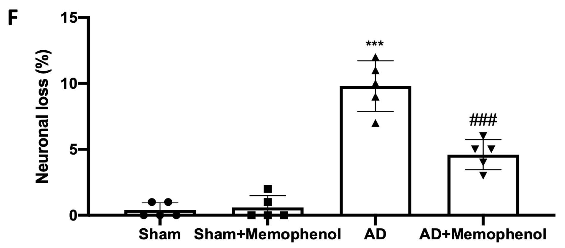
On day four of the Morris Water Maze (MWM) test’s training period, when compared to day one, the animals in all groups demonstrated a diminishing trend in escape latency time (Figure 1A). MemophenolTM increased animal persistence in the target quadrant in the searching experiment, indicating an increase in memory consolidation as compared to the AD group (Figure 1B). The MemophenolTM-treated rats showed a decrease in the time of transfer latency in initial acquisition latency (IAL) and retention transfer latency (RTL) in the Elevated Plus Maze (EPM) test, indicating an improvement in memory retention when compared to the AD group (Figure 1C). The MemophenolTM treatment significantly enhanced the recognition index % (RI) in the novel object recognition (NOR) test, indicating an improvement in cognitive function as compared to the AD group (Figure 1D). The control group’s brain samples revealed normal tissue organization in the CA1 hippocampus region (Figure 1E,F). Tissues from the AD group, on the other hand, exhibited substantially more severe neuronal degeneration, with fewer dark basophilic neurons in the CA1 hippocampal pyramidal and polymorphic layers (Figure 1E,F). MemophenolTM treatment greatly decreased AlCl3-induced CA1 neuronal degeneration (Figure 1E,F). Furthermore, both behavioral (Figure 1A–D) and histological analysis (Figure 1E,F) revealed no difference between the Sham and Sham + MemophenolTM groups; hence, a molecular study on the control animals administered with MemophenolTM was omitted.

2.2. Effects of MemophenolTM Treatment on Aβ Deposition and APP and p-Tau Over-Expression

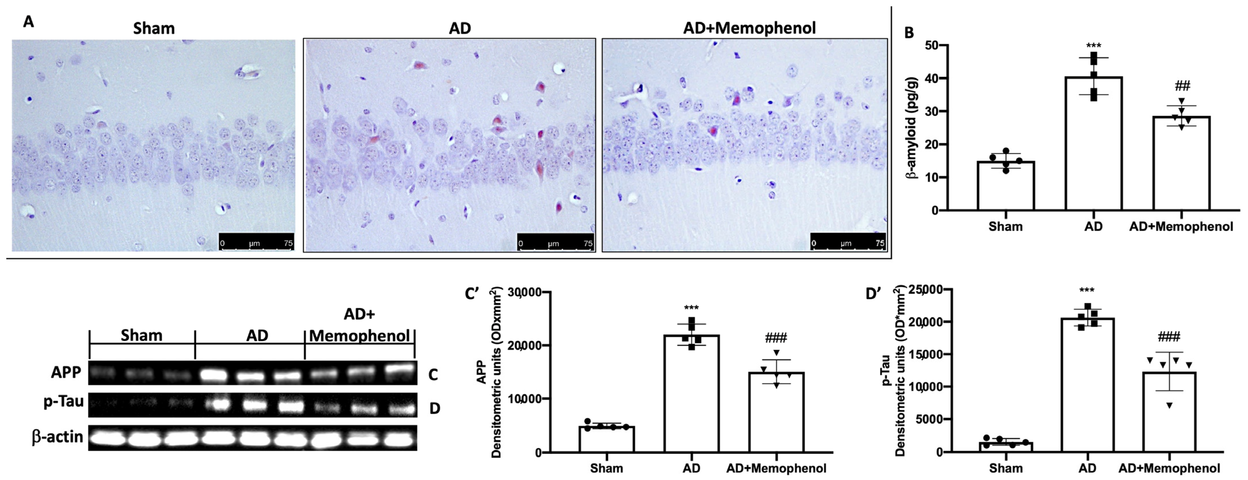
To demonstrate that MemophenolTM had action on amyloidosis typical of AD, we performed Congo red staining. The staining results showed that more Aβ deposits were present in the hippocampus of the AD group animals, while these deposits were significantly reduced after MemophenolTM treatment (Figure 2A). The result was further confirmed by an analysis of Aβ levels with an ELISA kit as presented in Figure 2B. Furthermore, we wanted to evaluate whether MemophenolTM was also able to act on two other specific markers of AD disease such as APP and p-Tau. APP (Figure 2C,C’) and p-Tau (Figure 2D,D’) expression levels were higher in the hippocampi from the AD group compared to the Sham group. Administration of MemophenolTM considerably lowered both levels (Figure 2C,C’,D,D’).

2.3. MemophenolTM Treatment Effects on Oxidative Hippocampal Modifications

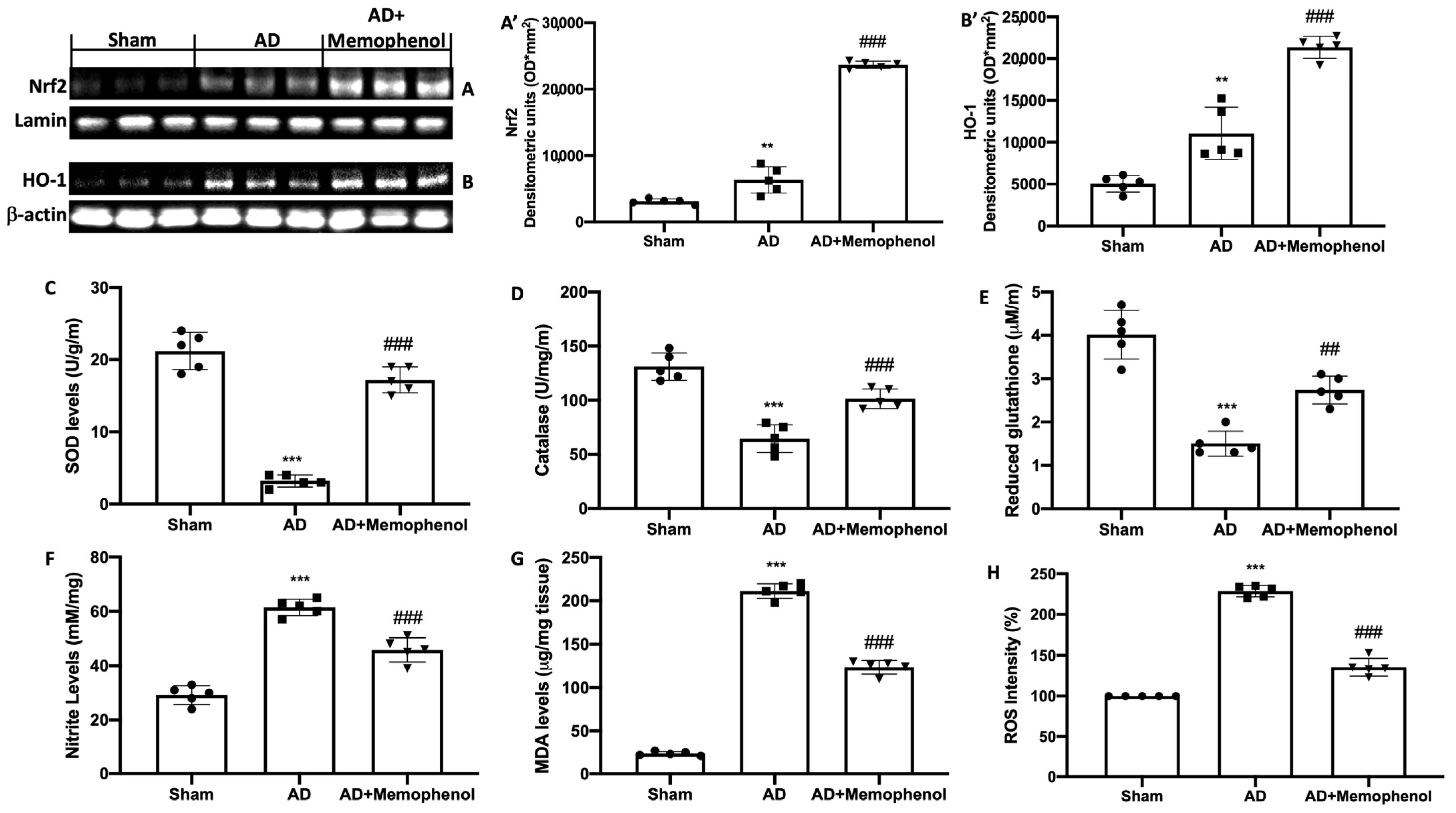
It is known that oxidative stress is an important risk factor for this pathology. In this regard, we wanted to evaluate the antioxidant activity of MemophenolTM by Western blot analysis and biochemical tests on the hippocampus. Increased nuclear factor erythroid 2–related factor 2 (Nrf2) expression was found in the hippocampi of MemophenolTM-treated rats as compared to the AD and Sham groups (Figure 3A,A’). The same trend was also observed for heme oxygenase-1 (HO-1) (Figure 3B,B’). Furthermore, biochemical analysis revealed that MemophenolTM-treated rats had improved antioxidant defenses. Superoxide dismutase (SOD) levels (Figure 3C), catalase (CAT) activity (Figure 3D), and glutathione (GSH) levels (Figure 3E) were all significantly higher in the MemophenolTM group than in the AD group. In contrast, nitrite (Figure 3F), lipid peroxidation (MDA) (Figure 3G), and reactive oxygen species (ROS) (Figure 3H) levels in the AD group were considerably higher than in the Sham group. Treatment with MemophenolTM significantly reduced nitrite levels, lipid peroxidation, and ROS levels in the hippocampus.

2.4. MemophenolTM Treatment Effects on Pro-Inflammatory Markers

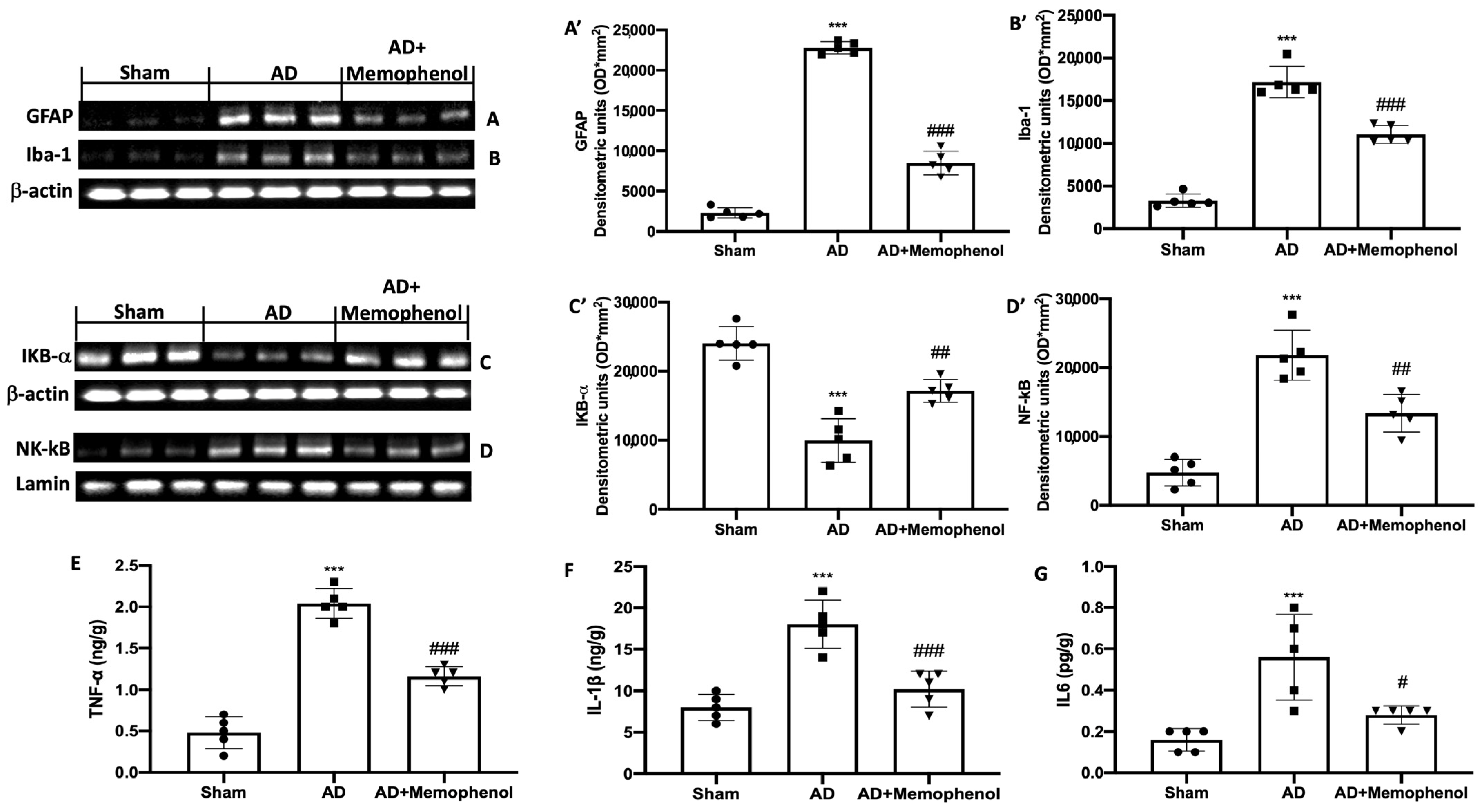
Another fact implicated in the progression of AD is neuroinflammation. For this reason, in addition to the antioxidant action of MemophenolTM, we also wanted to investigate its anti-inflammatory properties. Western blot investigations on hippocampus tissue for glial fibrillary acid protein (GFAP) and ionized calcium-binding adaptor molecule 1 (Iba-1) expression were used to examine astrocyte and microglial cell activity in connection to AD. The GFAP and Iba-1 expressions were low in the Sham group but significantly higher in the AD-treated rats. MemophenolTM treatment reduced the elevated expression of GFAP and Iba-1 under these conditions (Figure 4A,A’ for GFAP and Figure 4B,B’ for Iba-1). In addition, the Western blot analysis revealed a significant downregulation of the NF-κB pathway, which was activated by AlCl3 injection. The AD rat samples c α (IkB-α) expression in the cytoplasm (Figure 4C,C’), and enhanced NF-κB nuclear localization (Figure 4D,D’). Treatment with MemophenolTM boosted IkB-α expression while restoring NF-κB expression to baseline levels. MemophenolTM administration also lowered tumor necrosis factor-α (TNF-α) (Figure 4E), interleukin-1β (IL-1β) (Figure 4F), and interleukin-6 (IL-6) (Figure 4G) levels, which were elevated in the AD group due to the activity of the NF-κB pathway.

3. Discussion

Many chronic diseases, including Alzheimer’s, are determined by both heredity and environment. The genetic abnormalities of the APP and presenilin genes account for just 5% of the overall number of AD patients (familial instances), but the majority of AD patients are most likely due to environmental and other genetic variables affecting Aβ clearance [3]. Major environmental influences are likely to include an excess or deficit of dietary ingredients with bioactivity in key pathways that are taken on a regular basis. Our understanding of how food and drink might potentially impact the development of AD will aid in the development and implementation of medicines to battle this deadly illness. MemophenolTM is derived from French grape and wild blueberry extracts and has a specific mix of essential polyphenols that has been clinically demonstrated to increase learning and memory functions. These polyphenols appear to have a threefold impact for a synergistic protective effect on the brain, acting in two ways: by enhancing both neurogenesis and sympathetic plasticity. In this regard, a clinical study was conducted on 215 elderly people which demonstrated that chronic integration of MemophenolTM improves both short- and long-term memory [24]. In addition, a preclinical study showed that supplementation with a diet rich in polyphenols derived from grape and blueberry extracts prevented spatial locomotor impairments in middle-aged mice [23]. Another preclinical study was also conducted which highlighted the ability of polyphenol-rich grape and blueberry extracts to attenuate cognitive decline and improve neuronal function in aged mice [25]. In this study, an increase in the neurogenerative process was observed in elderly mice that assumed a diet rich in polyphenols extracted from grapes and blueberries compared to mice of the same age that were not supplemented with the same diet. Furthermore, some of the polyphenols included in the extract have been found in the brain in their native forms or as metabolites. This indicates that polyphenols may act directly centrally, while they may impact mouse survival through a potential systemic effect [25]. Given the observed effects on memory, it was decided to test this compound in an animal model of AD. In particular, in this study, we looked at the impact of MemophenolTM supplementation on reactive oxygen species and the inflammatory processes that define AD. Several studies have demonstrated the role of oxidative stress in the growth and development of disease. According to recent research, AD has a latent phase before symptoms appear and a diagnosis is obtained. When compared to healthy individuals, the development of AD is preceded by a moderate cognitive impairment phase with a minor increase in Aβ deposition but with a considerable oxidative imbalance [26,27]. A significant amount of research has revealed that excessive ROS generation causes neuronal death and other pathological alterations in AD [28,29]. Oxidative damage is associated with the abnormal accumulation of Aβ and the overexpression of APP and p-Tau [8,30]. Elevated APP levels are linked to decreased hippocampus neurogenesis and, as a result, poorer cognitive function [31,32]. In fact, some evidence suggests that hippocampal plasticity is connected to memory consolidation, learning, and cognitive function [33,34]. Animal studies have conclusively shown that AlCl3 neurotoxicity is involved in the development of neurodegenerative illnesses such as Alzheimer’s. By aggregating Tau proteins, it enhances the development of Aβ protein plaques in the brain. AlCl3 has also been connected to the neurodegeneration and modifications associated with aging. AlCl3 toxicity, according to [35], is produced by increased ROS release, which causes oxidative damage in the hippocampus. Although aluminum is not a transition metal and cannot catalyze redox reactions, AlCl3 can induce neurotoxicity by generating free radicals [36,37]. Aluminum ions have a strong affinity for bio-membranes and can exacerbate the cellular oxidative environment by strengthening transition metal pro-oxidant properties [38]. It has also been associated with mitochondrial function impairment in vitro and in vivo, as well as impairment of the antioxidant defense system, which may lead to the development of oxidative stress [39,40,41]. AlCl3 treatment mostly accumulates in the hippocampus, which is known to be particularly vulnerable to AD and to play an important role in learning and memory processes [42]. For these reasons, hippocampus tissue was subjected to histological, biochemical, and molecular studies. MemophenolTM inhibited the course of AD by lowering Tau hyperphosphorylation, APP levels, and Aβ buildup. From a behavioral standpoint, it significantly decreased cognitive deficits. Histologically, it decreased the chronic hippocampus neuron loss and degeneration features of AD. These behavioral and histological effects might be attributed to the molecular characteristics of MemophenolTM. It boosted cellular defenses against ROS by boosting the Nrf2/HO-1 pathway. Nrf2 oversees genes that code for endogenous antioxidant enzymes, redox balance factors, and stress response proteins [28,43,44]. It specifically stimulated phase II detoxification enzymes such as CAT, SOD, and GSH. Furthermore, MemophenolTM lowered nitrite levels, lipid peroxidation, and ROS levels that were elevated by AD [45]. These anti-oxidative stress effects resulted in a decrease in the pro-inflammatory macroenvironment. We found that reducing the activity of the NF-κB pathway has significant anti-inflammatory properties. NF-κB is a key transcription factor in pro-inflammatory signaling [46,47]. In healthy settings, NF-κB is bound to its inhibitor IkB-α and is sequestered within the cytoplasm [48]. The inhibitor is destroyed during inflammation, and NF-kB translocates into the nucleus to encode pro-inflammatory proteins [49]. Our findings demonstrated restored cytoplasmic levels of IkB-α and decreased NF-κB nuclear expression of associated target pro-inflammatory mediators such as TNF-α, IL-1β, and IL-6. The aggregation of Aβ plaques can also lead to the activation of astrocytes and microglia, which are not only secondary players in pathological processes, but seem to contribute to synaptic and neuronal loss and to the accumulation of pathogenic proteins even in the early stages of disease [50,51,52]. Our treatment with MemophenolTM also demonstrated the ability to reduce AlCl3-induced astrogliosis and microgliosis by reducing hippocampal GFAP and Iba-1 expression.

4. Materials and Methods

4.1. Tested Product: MemophenolTM

MemophenolTM provided by the company Activ’Inside is a standardized polyphenol-rich extract from grapes and blueberries; it is abundant in bioavailable flavonoids and can operate locally on cognitive functioning by bridging the blood–brain barrier. Total flavonoids (flavan-3-ols, flavonols, and anthocyanins): >43%, flavan-3-ols monomers; ≥20%, oligomers (DP ≤ 4); ≥22%, flavonols (quercetin and glycosylated derivatives); ≥0.15%, anthocyanins: ≥0.10%).

4.2. Animals

Male Wistar rats (Envigo, Milan, Italy) were used (age: six to eight weeks, weight: 250–280 g). The animals were kept in a confined space and fed standard rodent chow (Envigo, Teklad Rodent Diet T.2018.12) and water. The study was approved by the University of Messina’s Review Board for Animal Care (OPBA). All animal experiments were conducted in accordance with new Italian legislation (D.Lgs 2014/26), EU legislation (EU Directive 2010/63), and the ARRIVE guidelines.

4.3. Experimental Protocol

Aluminum (AlCl3) is a popular AD model [53]. For 60 days the rats were treated with AlCl3 (100 mg/kg, orally) and D-galactose (60 mg/kg, intraperitoneally) [35,54,55].

Experimental Groups

The rats were randomly divided into the following groups (n = 20 for each group):
-
Sham group: saline was administered to the rats;
-
Sham + MemophenolTM group: saline was administered to the rats, and MemophenolTM (15 mg/kg) was administered orally for 30 consecutive days;
-
AD group: as previously mentioned, the rats were treated with AlCl3 (100 mg/kg, orally) and D-galactose (60 mg/kg, intraperitoneally) for 60 days;
-
AD + MemophenolTM group: as previously documented, the rats were treated with AlCl3 (100 mg/kg, orally) and D-galactose (60 mg/kg, intraperitoneally) for 60 days, and MemophenolTM (15 mg/kg) was supplied orally by gavage for 30 consecutive days.
The dose of MemophenolTM was based on previous studies performed in the laboratory in which the compound was administered in increasing doses for 3 months. Behavioral test training was completed prior to the experiment’s conclusion date. The animals were sacrificed at the end of the trial after behavioral changes were examined. Brain tissues were collected for further analysis.

4.4. Behavioral Assessment

4.4.1. MWM

To assess spatial learning and memory consolidation, the MWM test was used [18,56]. The percentage of distance walked and the amount of time spent in the target quadrant were both recorded.

4.4.2. EPM

The EPM exam was used to assess memory-related activities. The behavioral test was carried out as previously reported [57,58].

4.4.3. NOR

The NOR test was used to measure cognitive function abnormalities induced by Alzheimer’s disease. The behavioral test was carried out as previously reported [59]. The RI was used to record the time spent studying the unfamiliar object. It was calculated by dividing the amount of time spent examining a novel object (TN) by the amount of time spent exploring a familiar object (TF), [RI = TN/(TN + TF)]. An RI % larger than 50% implies more time spent finding the TN, whereas an RI % less than 50% indicates more time spent investigating the TF [60].

4.5. Histological Analysis and Congo Red Staining

Brain samples were taken and processed, and slices (7 μm) were cut into longitudinal sections and stained with hematoxylin and eosin (H&E) [61,62]. The necrosis percentages of necrotic neurons out of total neurons were manually counted along the ipsilateral hippocampus CA1 region [63]. Furthermore, some sections, after being deparaffinized and dehydrated by alcohol gradients, were stained with Highman Congo red staining solution for 5–10 min as described by Xia Zhao et al. [61]. An experienced histopathologist examined the sections under a Leica DM6 microscope (Leica Microsystems SpA, Milan, Italy) with a motorized stage and Leica LAS X Navigator 7.31 software (Leica Microsystems SpA, Milan, Italy).

4.6. Western Blot Analysis

Western blots on the hippocampi were performed as previously described [46,64]. Specific primary antibodies were used, such as anti-IkB-α (Santa Cruz Biotechnology, sc-1643), anti-NF-κB p65 (Santa Cruz Biotechnology, sc-8008), anti-Nrf2 (Santa Cruz Biotechnology, sc-36594), anti-HO-1 (Santa Cruz Biotechnology, sc-136960), anti-p-Tau (Santa Cruz Biotechnology, sc-32275), anti-APP (Santa Cruz Biotechnology, sc-32277), anti-GFAP (Cell Signaling Technology, Danvers, MA, USA), and anti-Iba1 (Santa Cruz Biotechnology, sc-32725), and they were mixed in a 5% w/v non-fat dried milk solution and were incubated at 4 °C overnight. The blots were then incubated for 1 h at room temperature with a peroxidase-conjugated bovine anti-mouse IgG secondary antibody or a peroxidase-conjugated goat anti-rabbit IgG secondary antibody (Jackson Immuno Research, West Grove, PA, USA). To ensure that the quantities of protein were similar, the membranes were additionally treated with an antibody against β-actin and Lamin (Santa Cruz Biotechnology, Dallas, TX, USA). Signals were detected using a Super-Signal West Pico Chemiluminescent Substrate (Biogenerica, Pedara, Italy) enhanced chemiluminescence detection system reagent [44]. The relative expression of the protein bands was measured using densitometry and was standardized to β-actin and Lamin levels using Bio-Rad ChemiDoc XRS 2.1.1 software [48]. The blot signal images were input into analysis software (Image Quant TL, v2003, Bio-rad, Segrate, Italy).

4.7. Biochemical Analysis

Biochemical analyses were conducted on the hippocampi:

4.7.1. Measurement of SOD Activity

After homogenizing the samples in the Tris buffer, they were centrifuged at 13,000 rpm. The solution was then incubated at 4 °C for 10 min before being centrifuged again. The absorbance of the samples was measured every 60 s for 10 min at 420 nm [57,65].

4.7.2. Measurement of CAT Activity

After homogenizing the samples in the phosphate buffer, hydrogen peroxide was added. Enzyme levels were expressed as CAT activity in U/mg protein, and the absorbance (240 nm) was measured for 0–10 min at 240 min [66].

4.7.3. GSH Levels

A trichloroacetic acid solution was administered after homogenizing the samples with the phosphate buffer. After centrifuging the solution, 5,5′-dithiobis-(2-nitrobenzoic acid) was added. Using a microplate reader, the GSH levels were measured at 412 nm [57].

4.7.4. Measurement of Nitrite Levels

After homogenizing the samples in the phosphate buffer, the Griess reagent was applied. For 30 min, the solution was incubated. At 548 nm, the absorbance was measured [67].

4.7.5. Measurement of MDA

The evaluation of thiobarbituric acid-reactant substances, a suitable indication of lipid peroxidation, was performed on the samples. At 532 nm, the absorbance of the supernatant was measured [68].

4.7.6. Measurement of ROS

After homogenizing the samples in the phosphate buffer, they were exposed to 1 mM dichlorofluorescein diacetate for 10 min at room temperature in the dark (DCFH-DA). The esterase activity used to convert non-fluorescent DCFH-DA to the highly fluorescent product 20,70-dichlorofluorescein (DCF) was used to monitor the presence of peroxides caused by the oxidative burst in the brain [69].

4.7.7. Cytokines and Aβ Measurement

An ELISA kit was used to measure IL-6, TNF-α, IL-1β, and Aβ levels in the hippocampus (Diaclone Research, Biosource Europe, USCN life Sciences; Abcam, Milan, Italy) [35].

4.8. Statistical Evaluation

All values are expressed as mean ± standard error of the mean (SD) of N observations. N denotes the number of animals utilized in in vivo studies. One-way ANOVA was used to examine the data, followed by a Bonferroni post hoc test for multiple comparisons. A p-value less than 0.05 was regarded as significant.

5. Conclusions

In summary, oral treatment with MemophenolTM at a dose of 15 mg/kg, by acting on oxidative stress and inflammatory processes, was able to manage AD features such as behavioral changes related to cognitive functions and memory, phosphorylated Tau levels, and the aberrant overexpression of APP, the accumulation of β-amyloid, and neuronal degeneration. Certainly, taking MemophenolTM cannot cure Alzheimer’s disease, but it is a nutritional supplement that may slow the course of the illness and alleviate symptoms connected with this pathology.

Author Contributions

Conceptualization: V.C. and R.D.P.; data curation: R.S., V.C., R.D.P. and A.S.A.; formal analysis: M.T. and M.C.; investigation: D.I., R.S. and U.W.; methodology: R.D. and R.F.; supervision: S.C., V.C. and R.D.P.; validation: V.C.; writing—original draft: R.S. and M.T.; writing—review and editing: V.C. and R.D.P. All authors have read and agreed to the published version of the manuscript.

Funding

This study was supported by grants from “Piano di incentivi per la Ricerca, Linea Intervento 2 PIACERI, 2020–2022”, the University of Catania, Italy, and Researchers Supporting Project number (RSPD2023R750), King Saud University, Riyadh, Saudi Arabia.

Institutional Review Board Statement

The study was approved by the University of Messina’s Review Board for Animal Care (OPBA). All animal experiments were conducted in accordance with new Italian legislation (D.Lgs 2014/26), EU legislation (EU Directive 2010/63), and the ARRIVE guidelines.

Informed Consent Statement

Not applicable.

Data Availability Statement

Based on the rules of our laboratory, the datasets used in the current study are available from the corresponding author ([email protected]) upon reasonable request.

Acknowledgments

We acknowledge helpful discussions with Line Pourtau and David Gaudout from Activ’Inside.

Conflicts of Interest

The authors declare no conflict of interest.

References

  1. Brookmeyer, R.; Johnson, E.; Ziegler-Graham, K.; Arrighi, H.M. Forecasting the global burden of Alzheimer’s disease. Alzheimers Dement. 2007, 3, 186–191. [Google Scholar] [CrossRef] [Green Version]
  2. Thomas, P.; Fenech, M. A review of genome mutation and Alzheimer’s disease. Mutagenesis 2007, 22, 15–33. [Google Scholar] [CrossRef] [Green Version]
  3. Hardy, J.; Selkoe, D.J. The amyloid hypothesis of Alzheimer’s disease: Progress and problems on the road to therapeutics. Science 2002, 297, 353–356. [Google Scholar] [CrossRef] [PubMed] [Green Version]
  4. Seubert, P.; Vigo-Pelfrey, C.; Esch, F.; Lee, M.; Dovey, H.; Davis, D.; Sinha, S.; Schlossmacher, M.; Whaley, J.; Swindlehurst, C.; et al. Isolation and quantification of soluble Alzheimer’s beta-peptide from biological fluids. Nature 1992, 359, 325–327. [Google Scholar] [CrossRef] [PubMed]
  5. Clementi, M.E.; Giardina, B.; Colucci, D.; Galtieri, A.; Misiti, F. Amyloid-beta peptide affects the oxygen dependence of erythrocyte metabolism: A role for caspase 3. Int. J. Biochem. Cell Biol. 2007, 39, 727–735. [Google Scholar] [CrossRef] [PubMed]
  6. Carelli-Alinovi, C.; Pirolli, D.; Giardina, B.; Misiti, F. Protein kinase C mediates caspase 3 activation: A role for erythrocyte morphology changes. Clin. Hemorheol. Microcirc. 2015, 59, 345–354. [Google Scholar] [CrossRef]
  7. Heneka, M.T.; O’Banion, M.K. Inflammatory processes in Alzheimer’s disease. J. Neuroimmunol. 2007, 184, 69–91. [Google Scholar] [CrossRef]
  8. Butterfield, D.A.; Boyd-Kimball, D. Oxidative Stress, Amyloid-beta Peptide, and Altered Key Molecular Pathways in the Pathogenesis and Progression of Alzheimer’s Disease. J. Alzheimer’s Dis. 2018, 62, 1345–1367. [Google Scholar] [CrossRef] [Green Version]
  9. Trovato, A.; Siracusa, R.; Di Paola, R.; Scuto, M.; Ontario, M.L.; Bua, O.; Di Mauro, P.; Toscano, M.A.; Petralia, C.C.T.; Maiolino, L.; et al. Redox modulation of cellular stress response and lipoxin A4 expression by Hericium Erinaceus in rat brain: Relevance to Alzheimer’s disease pathogenesis. Immun. Ageing 2016, 13, 23. [Google Scholar] [CrossRef] [Green Version]
  10. Wang, Y.J.; Zhou, H.D.; Zhou, X.F. Clearance of amyloid-beta in Alzheimer’s disease: Progress, problems and perspectives. Drug Discov. Today 2006, 11, 931–938. [Google Scholar] [CrossRef]
  11. Frozza, R.L.; Lourenco, M.V.; De Felice, F.G. Challenges for Alzheimer’s Disease Therapy: Insights from Novel Mechanisms Beyond Memory Defects. Front. Neurosci. 2018, 12, 37. [Google Scholar] [CrossRef] [PubMed]
  12. Makkar, R.; Behl, T.; Bungau, S.; Zengin, G.; Mehta, V.; Kumar, A.; Uddin, M.S.; Ashraf, G.M.; Abdel-Daim, M.M.; Arora, S.; et al. Nutraceuticals in Neurological Disorders. Int. J. Mol. Sci. 2020, 21, 4424. [Google Scholar] [CrossRef] [PubMed]
  13. Rigacci, S.; Stefani, M. Nutraceuticals and amyloid neurodegenerative diseases: A focus on natural phenols. Expert Rev. Neurother. 2015, 15, 41–52. [Google Scholar] [CrossRef] [PubMed]
  14. Fumia, A.; Cicero, N.; Gitto, M.; Nicosia, N.; Alesci, A. Role of nutraceuticals on neurodegenerative diseases: Neuroprotective and immunomodulant activity. Nat. Prod. Res. 2022, 36, 5916–5933. [Google Scholar] [CrossRef]
  15. Jabir, N.R.; Khan, F.R.; Tabrez, S. Cholinesterase targeting by polyphenols: A therapeutic approach for the treatment of Alzheimer’s disease. CNS Neurosci. Ther. 2018, 24, 753–762. [Google Scholar] [CrossRef]
  16. Havsteen, B.H. The biochemistry and medical significance of the flavonoids. Pharm. Ther. 2002, 96, 67–202. [Google Scholar] [CrossRef]
  17. Ontario, M.L.; Siracusa, R.; Modafferi, S.; Scuto, M.; Sciuto, S.; Greco, V.; Bertuccio, M.P.; Trovato Salinaro, A.; Crea, R.; Calabrese, E.J.; et al. Potential prevention and treatment of neurodegenerative disorders by olive polyphenols and hidrox. Mech. Ageing Dev. 2022, 203, 111637. [Google Scholar] [CrossRef]
  18. Cordaro, M.; Trovato Salinaro, A.; Siracusa, R.; D’Amico, R.; Impellizzeri, D.; Scuto, M.; Ontario, M.L.; Crea, R.; Cuzzocrea, S.; Di Paola, R.; et al. Hidrox((R)) Roles in Neuroprotection: Biochemical Links between Traumatic Brain Injury and Alzheimer’s Disease. Antioxidants 2021, 10, 818. [Google Scholar] [CrossRef]
  19. Siracusa, R.; Scuto, M.; Fusco, R.; Trovato, A.; Ontario, M.L.; Crea, R.; Di Paola, R.; Cuzzocrea, S.; Calabrese, V. Anti-inflammatory and Anti-oxidant Activity of Hidrox((R)) in Rotenone-Induced Parkinson’s Disease in Mice. Antioxidants 2020, 9, 824. [Google Scholar] [CrossRef]
  20. Cordaro, M.; Modafferi, S.; D’Amico, R.; Fusco, R.; Genovese, T.; Peritore, A.F.; Gugliandolo, E.; Crupi, R.; Interdonato, L.; Di Paola, D.; et al. Natural Compounds Such as Hericium erinaceus and Coriolus versicolor Modulate Neuroinflammation, Oxidative Stress and Lipoxin A4 Expression in Rotenone-Induced Parkinson’s Disease in Mice. Biomedicines 2022, 10, 2505. [Google Scholar] [CrossRef]
  21. Krikorian, R.; Nash, T.A.; Shidler, M.D.; Shukitt-Hale, B.; Joseph, J.A. Concord grape juice supplementation improves memory function in older adults with mild cognitive impairment. Br. J. Nutr. 2010, 103, 730–734. [Google Scholar] [CrossRef] [Green Version]
  22. Krikorian, R.; Shidler, M.D.; Nash, T.A.; Kalt, W.; Vinqvist-Tymchuk, M.R.; Shukitt-Hale, B.; Joseph, J.A. Blueberry supplementation improves memory in older adults. J. Agric. Food Chem. 2010, 58, 3996–4000. [Google Scholar] [CrossRef] [PubMed] [Green Version]
  23. Bensalem, J.; Servant, L.; Alfos, S.; Gaudout, D.; Laye, S.; Pallet, V.; Lafenetre, P. Dietary Polyphenol Supplementation Prevents Alterations of Spatial Navigation in Middle-Aged Mice. Front. Behav. Neurosci. 2016, 10, 9. [Google Scholar] [CrossRef] [PubMed] [Green Version]
  24. Bensalem, J.; Dudonne, S.; Etchamendy, N.; Pellay, H.; Amadieu, C.; Gaudout, D.; Dubreuil, S.; Paradis, M.E.; Pomerleau, S.; Capuron, L.; et al. Polyphenols From Grape and Blueberry Improve Episodic Memory in Healthy Elderly with Lower Level of Memory Performance: A Bicentric Double-Blind, Randomized, Placebo-Controlled Clinical Study. J. Gerontol. A Biol. Sci. Med. Sci. 2019, 74, 996–1007. [Google Scholar] [CrossRef]
  25. Bensalem, J.; Dudonne, S.; Gaudout, D.; Servant, L.; Calon, F.; Desjardins, Y.; Laye, S.; Lafenetre, P.; Pallet, V. Polyphenol-rich extract from grape and blueberry attenuates cognitive decline and improves neuronal function in aged mice. J. Nutr. Sci. 2018, 7, e19. [Google Scholar] [CrossRef] [Green Version]
  26. Markesbery, W.R.; Schmitt, F.A.; Kryscio, R.J.; Davis, D.G.; Smith, C.D.; Wekstein, D.R. Neuropathologic substrate of mild cognitive impairment. Arch. Neurol. 2006, 63, 38–46. [Google Scholar] [CrossRef] [PubMed] [Green Version]
  27. O’Brien, R.J.; Resnick, S.M.; Zonderman, A.B.; Ferrucci, L.; Crain, B.J.; Pletnikova, O.; Rudow, G.; Iacono, D.; Riudavets, M.A.; Driscoll, I.; et al. Neuropathologic studies of the Baltimore Longitudinal Study of Aging (BLSA). J. Alzheimer’s Dis. 2009, 18, 665–675. [Google Scholar] [CrossRef] [Green Version]
  28. Wang, X.; Wang, W.; Li, L.; Perry, G.; Lee, H.G.; Zhu, X. Oxidative stress and mitochondrial dysfunction in Alzheimer’s disease. Biochim. Biophys. Acta 2014, 1842, 1240–1247. [Google Scholar] [CrossRef] [Green Version]
  29. Cassidy, L.; Fernandez, F.; Johnson, J.B.; Naiker, M.; Owoola, A.G.; Broszczak, D.A. Oxidative stress in alzheimer’s disease: A review on emergent natural polyphenolic therapeutics. Complement. Ther. Med. 2020, 49, 102294. [Google Scholar] [CrossRef]
  30. Huang, W.J.; Zhang, X.; Chen, W.W. Role of oxidative stress in Alzheimer’s disease. Biomed. Rep. 2016, 4, 519–522. [Google Scholar] [CrossRef] [Green Version]
  31. Reddy, P.H.; Manczak, M.; Mao, P.; Calkins, M.J.; Reddy, A.P.; Shirendeb, U. Amyloid-beta and mitochondria in aging and Alzheimer’s disease: Implications for synaptic damage and cognitive decline. J. Alzheimer’s Dis. 2010, 20 (Suppl. S2), S499–S512. [Google Scholar] [CrossRef] [PubMed] [Green Version]
  32. Zhao, J.; O’Connor, T.; Vassar, R. The contribution of activated astrocytes to Abeta production: Implications for Alzheimer’s disease pathogenesis. J. Neuroinflamm. 2011, 8, 150. [Google Scholar] [CrossRef] [Green Version]
  33. Amaral, D.G.; Witter, M.P. The three-dimensional organization of the hippocampal formation: A review of anatomical data. Neuroscience 1989, 31, 571–591. [Google Scholar] [CrossRef] [PubMed]
  34. Nakashiba, T.; Young, J.Z.; McHugh, T.J.; Buhl, D.L.; Tonegawa, S. Transgenic inhibition of synaptic transmission reveals role of CA3 output in hippocampal learning. Science 2008, 319, 1260–1264. [Google Scholar] [CrossRef] [PubMed] [Green Version]
  35. Ali, A.A.; Ahmed, H.I.; Abu-Elfotuh, K. Modeling stages mimic Alzheimer’s disease induced by different doses of aluminum in rats: Focus on progression of the disease in response to time. J. Alzheimer’s Park. Dement. 2016, 1, 2. [Google Scholar]
  36. Sethi, P.; Jyoti, A.; Singh, R.; Hussain, E.; Sharma, D. Aluminium-induced electrophysiological, biochemical and cognitive modifications in the hippocampus of aging rats. Neurotoxicology 2008, 29, 1069–1079. [Google Scholar] [CrossRef]
  37. Moumen, R.; Ait-Oukhatar, N.; Bureau, F.; Fleury, C.; Bougle, D.; Arhan, P.; Neuville, D.; Viader, F. Aluminium increases xanthine oxidase activity and disturbs antioxidant status in the rat. J. Trace Elem. Med. Biol. 2001, 15, 89–93. [Google Scholar] [CrossRef]
  38. Becaria, A.; Bondy, S.C.; Campbell, A. Aluminum and copper interact in the promotion of oxidative but not inflammatory events: Implications for Alzheimer’s disease. J. Alzheimer’s Dis. 2003, 5, 31–38. [Google Scholar] [CrossRef] [Green Version]
  39. Niu, P.Y.; Niu, Q.; Zhang, Q.L.; Wang, L.P.; He, S.E.; Wu, T.C.; Conti, P.; Di Gioacchino, M.; Boscolo, P. Aluminum impairs rat neural cell mitochondria in vitro. Int. J. Immunopathol Pharm. 2005, 18, 683–689. [Google Scholar] [CrossRef] [Green Version]
  40. Kumar, V.; Bal, A.; Gill, K.D. Impairment of mitochondrial energy metabolism in different regions of rat brain following chronic exposure to aluminium. Brain Res. 2008, 1232, 94–103. [Google Scholar] [CrossRef]
  41. Kumar, V.; Bal, A.; Gill, K.D. Susceptibility of mitochondrial superoxide dismutase to aluminium induced oxidative damage. Toxicology 2009, 255, 117–123. [Google Scholar] [CrossRef]
  42. Calabrese, V.; Cornelius, C.; Dinkova-Kostova, A.T.; Calabrese, E.J.; Mattson, M.P. Cellular stress responses, the hormesis paradigm, and vitagenes: Novel targets for therapeutic intervention in neurodegenerative disorders. Antioxid. Redox Signal. 2010, 13, 1763–1811. [Google Scholar] [CrossRef] [Green Version]
  43. Bellezza, I.; Giambanco, I.; Minelli, A.; Donato, R. Nrf2-Keap1 signaling in oxidative and reductive stress. Biochim. Biophys. Acta Mol. Cell Res. 2018, 1865, 721–733. [Google Scholar] [CrossRef]
  44. Fusco, R.; Cordaro, M.; Siracusa, R.; D’Amico, R.; Genovese, T.; Gugliandolo, E.; Peritore, A.F.; Crupi, R.; Impellizzeri, D.; Cuzzocrea, S.; et al. Biochemical Evaluation of the Antioxidant Effects of Hydroxytyrosol on Pancreatitis-Associated Gut Injury. Antioxidants 2020, 9, 781. [Google Scholar] [CrossRef]
  45. Limon-Pacheco, J.; Gonsebatt, M.E. The role of antioxidants and antioxidant-related enzymes in protective responses to environmentally induced oxidative stress. Mutat. Res. 2009, 674, 137–147. [Google Scholar] [CrossRef] [PubMed]
  46. Impellizzeri, D.; Siracusa, R.; Cordaro, M.; Peritore, A.F.; Gugliandolo, E.; Mancuso, G.; Midiri, A.; Di Paola, R.; Cuzzocrea, S. Therapeutic potential of dinitrobenzene sulfonic acid (DNBS)-induced colitis in mice by targeting IL-1beta and IL-18. Biochem. Pharm. 2018, 155, 150–161. [Google Scholar] [CrossRef] [PubMed]
  47. Di Paola, R.; Impellizzeri, D.; Fusco, R.; Cordaro, M.; Siracusa, R.; Crupi, R.; Esposito, E.; Cuzzocrea, S. Ultramicronized palmitoylethanolamide (PEA-um((R))) in the treatment of idiopathic pulmonary fibrosis. Pharm. Res. 2016, 111, 405–412. [Google Scholar] [CrossRef] [PubMed]
  48. Cordaro, M.; Impellizzeri, D.; Siracusa, R.; Gugliandolo, E.; Fusco, R.; Inferrera, A.; Esposito, E.; Di Paola, R.; Cuzzocrea, S. Effects of a co-micronized composite containing palmitoylethanolamide and polydatin in an experimental model of benign prostatic hyperplasia. Toxicol. Appl. Pharm. 2017, 329, 231–240. [Google Scholar] [CrossRef]
  49. Esposito, E.; Impellizzeri, D.; Bruschetta, G.; Cordaro, M.; Siracusa, R.; Gugliandolo, E.; Crupi, R.; Cuzzocrea, S. A new co-micronized composite containing palmitoylethanolamide and polydatin shows superior oral efficacy compared to their association in a rat paw model of carrageenan-induced inflammation. Eur. J. Pharm. 2016, 782, 107–118. [Google Scholar] [CrossRef]
  50. Hong, S.; Beja-Glasser, V.F.; Nfonoyim, B.M.; Frouin, A.; Li, S.; Ramakrishnan, S.; Merry, K.M.; Shi, Q.; Rosenthal, A.; Barres, B.A.; et al. Complement and microglia mediate early synapse loss in Alzheimer mouse models. Science 2016, 352, 712–716. [Google Scholar] [CrossRef] [Green Version]
  51. Shi, Y.; Yamada, K.; Liddelow, S.A.; Smith, S.T.; Zhao, L.; Luo, W.; Tsai, R.M.; Spina, S.; Grinberg, L.T.; Rojas, J.C.; et al. ApoE4 markedly exacerbates tau-mediated neurodegeneration in a mouse model of tauopathy. Nature 2017, 549, 523–527. [Google Scholar] [CrossRef] [PubMed]
  52. Sosna, J.; Philipp, S.; Albay, R., 3rd; Reyes-Ruiz, J.M.; Baglietto-Vargas, D.; LaFerla, F.M.; Glabe, C.G. Early long-term administration of the CSF1R inhibitor PLX3397 ablates microglia and reduces accumulation of intraneuronal amyloid, neuritic plaque deposition and pre-fibrillar oligomers in 5XFAD mouse model of Alzheimer’s disease. Mol. Neurodegener. 2018, 13, 11. [Google Scholar] [CrossRef]
  53. Walton, J.R.; Wang, M.X. APP expression, distribution and accumulation are altered by aluminum in a rodent model for Alzheimer’s disease. J. Inorg. Biochem. 2009, 103, 1548–1554. [Google Scholar] [CrossRef] [PubMed]
  54. Cordaro, M.; Salinaro, A.T.; Siracusa, R.; D’Amico, R.; Impellizzeri, D.; Scuto, M.; Ontario, M.L.; Cuzzocrea, S.; Di Paola, R.; Fusco, R.; et al. Key Mechanisms and Potential Implications of Hericium erinaceus in NLRP3 Inflammasome Activation by Reactive Oxygen Species during Alzheimer’s Disease. Antioxidants 2021, 10, 1664. [Google Scholar] [CrossRef] [PubMed]
  55. Zhang, Y.; Pi, Z.; Song, F.; Liu, Z. Ginsenosides attenuate d-galactose- and AlCl(3)-inducedspatial memory impairment by restoring the dysfunction of the neurotransmitter systems in the rat model of Alzheimer’s disease. J. Ethnopharmacol. 2016, 194, 188–195. [Google Scholar] [CrossRef]
  56. Mahmoodzadeh, T.; Kashani, M.H.K.; Ramshini, H.; Moslem, A.; Mohammad-Zadeh, M. Effect of Camellia sinensis on spatial memory in a rat model of Alzheimer’s disease. J. Biomed. 2016, 1, e5340. [Google Scholar] [CrossRef] [Green Version]
  57. Mutlu, O.; Akar, F.; Celikyurt, I.K.; Tanyeri, P.; Ulak, G.; Erden, F. 7-NI and ODQ Disturbs Memory in the Elevated Plus Maze, Morris Water Maze, and Radial Arm Maze Tests in Mice. Drug Target Insights 2015, 9, 1–8. [Google Scholar] [CrossRef] [Green Version]
  58. Paterniti, I.; Campolo, M.; Siracusa, R.; Cordaro, M.; Di Paola, R.; Calabrese, V.; Navarra, M.; Cuzzocrea, S.; Esposito, E. Liver X receptors activation, through TO901317 binding, reduces neuroinflammation in Parkinson’s disease. PLoS ONE 2017, 12, e0174470. [Google Scholar] [CrossRef] [Green Version]
  59. Impellizzeri, D.; D’Amico, R.; Fusco, R.; Genovese, T.; Peritore, A.F.; Gugliandolo, E.; Crupi, R.; Interdonato, L.; Di Paola, D.; Di Paola, R.; et al. Acai Berry Mitigates Vascular Dementia-Induced Neuropathological Alterations Modulating Nrf-2/Beclin1 Pathways. Cells 2022, 11, 2616. [Google Scholar] [CrossRef] [PubMed]
  60. Mudo, G.; Frinchi, M.; Nuzzo, D.; Scaduto, P.; Plescia, F.; Massenti, M.F.; Di Carlo, M.; Cannizzaro, C.; Cassata, G.; Cicero, L.; et al. Anti-inflammatory and cognitive effects of interferon-beta1a (IFNbeta1a) in a rat model of Alzheimer’s disease. J. Neuroinflamm. 2019, 16, 44. [Google Scholar] [CrossRef] [Green Version]
  61. Zhao, X.; Li, S.; Gaur, U.; Zheng, W. Artemisinin Improved Neuronal Functions in Alzheimer’s Disease Animal Model 3xtg Mice and Neuronal Cells via Stimulating the ERK/CREB Signaling Pathway. Aging Dis. 2020, 11, 801–819. [Google Scholar] [CrossRef]
  62. Petrosino, S.; Campolo, M.; Impellizzeri, D.; Paterniti, I.; Allara, M.; Gugliandolo, E.; D’Amico, R.; Siracusa, R.; Cordaro, M.; Esposito, E.; et al. 2-Pentadecyl-2-Oxazoline, the Oxazoline of Pea, Modulates Carrageenan-Induced Acute Inflammation. Front. Pharm. 2017, 8, 308. [Google Scholar] [CrossRef] [Green Version]
  63. Zhang, J.; Huang, W.; Xu, F.; Cao, Z.; Jia, F.; Li, Y. Iron Dyshomeostasis Participated in Rat Hippocampus Toxicity Caused by Aluminum Chloride. Biol. Trace Elem. Res. 2020, 197, 580–590. [Google Scholar] [CrossRef]
  64. Cordaro, M.; Siracusa, R.; Crupi, R.; Impellizzeri, D.; Peritore, A.F.; D’Amico, R.; Gugliandolo, E.; Di Paola, R.; Cuzzocrea, S. 2-Pentadecyl-2-Oxazoline Reduces Neuroinflammatory Environment in the MPTP Model of Parkinson Disease. Mol. Neurobiol. 2018, 55, 9251–9266. [Google Scholar] [CrossRef]
  65. Cordaro, M.; Trovato Salinaro, A.; Siracusa, R.; D’Amico, R.; Impellizzeri, D.; Scuto, M.; Ontario, M.L.; Interdonato, L.; Crea, R.; Fusco, R.; et al. Hidrox((R)) and Endometriosis: Biochemical Evaluation of Oxidative Stress and Pain. Antioxidants 2021, 10, 720. [Google Scholar] [CrossRef] [PubMed]
  66. D’Amico, R.; Trovato Salinaro, A.; Cordaro, M.; Fusco, R.; Impellizzeri, D.; Interdonato, L.; Scuto, M.; Ontario, M.L.; Crea, R.; Siracusa, R.; et al. Hidrox((R)) and Chronic Cystitis: Biochemical Evaluation of Inflammation, Oxidative Stress, and Pain. Antioxidants 2021, 10, 1046. [Google Scholar] [CrossRef] [PubMed]
  67. Cordaro, M.; Paterniti, I.; Siracusa, R.; Impellizzeri, D.; Esposito, E.; Cuzzocrea, S. KU0063794, a Dual mTORC1 and mTORC2 Inhibitor, Reduces Neural Tissue Damage and Locomotor Impairment After Spinal Cord Injury in Mice. Mol. Neurobiol. 2017, 54, 2415–2427. [Google Scholar] [CrossRef] [PubMed]
  68. Genovese, T.; D’Amico, R.; Fusco, R.; Impellizzeri, D.; Peritore, A.F.; Crupi, R.; Interdonato, L.; Gugliandolo, E.; Cuzzocrea, S.; Paola, R.D.; et al. Acai (Euterpe Oleraceae Mart.) Seeds Regulate NF-kappaB and Nrf2/ARE Pathways Protecting Lung against Acute and Chronic Inflammation. Cell Physiol. Biochem. 2022, 56, 1–20. [Google Scholar] [CrossRef]
  69. Zizzo, M.G.; Frinchi, M.; Nuzzo, D.; Jinnah, H.A.; Mudo, G.; Condorelli, D.F.; Caciagli, F.; Ciccarelli, R.; Di Iorio, P.; Mule, F.; et al. Altered gastrointestinal motility in an animal model of Lesch-Nyhan disease. Auton. Neurosci. 2018, 210, 55–64. [Google Scholar] [CrossRef]
Figure 1. The administration of MemophenolTM reduced behavioral and hippocampal alterations. MWM test: training (A); probe trial (B); EPM test (C); NOR test (D); histological analysis: Sham, Sham + Memophenol, AD, AD + Memophenol (E); quantification of necrotic neurons (F). Scale bar 75 μm. For the behavioral and histological investigations, n = 5 rats were used from each group and for each analysis. A p-value less than 0.05 was regarded as significant. *** p < 0.001 versus Sham, ### p < 0.001 versus AD.
Ijms 24 06938 g001aIjms 24 06938 g001b
Figure 2. MemophenolTM reduced Aβ deposition in AD rats. Congo red staining (labeled amyloidosis) in the hippocampus (A); ELISA analysis for β-amyloid levels (B); Western blot analyses for APP (C) and p-Tau (D) with related densitometric analysis (C’,D’); scale bar 75 μm. For the Western blot and ELISA investigations, n = 5 rats from each group and for each analysis were employed. A p-value of less than 0.05 was considered significant. *** p < 0.001 versus Sham, ## p < 0.01 versus AD, ### p < 0.001 versus AD.
Ijms 24 06938 g002
Figure 3. MemophenolTM administration decreased pro-oxidative modifications in the hippocampus. Western blot analysis of Nrf2 expression (A) and HO-1 (B) with related densitometric analysis (A’,B’), and biochemical analysis of SOD levels (C), CAT activity (D), GSH levels (E), nitrite levels (F), MDA levels (G), and ROS levels (H). For both analyses, n = 5 rats from each group and for each analysis were employed. A p-value of less than 0.05 was considered significant. ** p < 0.01 versus Sham, *** p < 0.001 versus Sham, ## p < 0.01 versus AD, ### p < 0.001 versus AD.
Ijms 24 06938 g003
Figure 4. The effects of MemophenolTM on the activation of astrocytes and microglia and on the NF-κB pathway following AlCl3 injection. Western blot analyses of GFAP (A), Iba-1 (B), IkB-α (C), and NF-κB (D) expression with related densitometric analysis (A’–D’); ELISA kit of TNF-α (E), IL-1β (F), and IL-6 (G) levels. For the Western blot and ELISA analyses, n = 5 rats from each group and for each analysis were employed. A p-value of less than 0.05 was considered significant. *** p < 0.001 versus Sham, # p < 0.05 versus AD, ## p < 0.01 versus AD, ### p < 0.001 versus AD.
Ijms 24 06938 g004
Disclaimer/Publisher’s Note: The statements, opinions and data contained in all publications are solely those of the individual author(s) and contributor(s) and not of MDPI and/or the editor(s). MDPI and/or the editor(s) disclaim responsibility for any injury to people or property resulting from any ideas, methods, instructions or products referred to in the content.