Article

The Control of the Crossover Localization in Allium

by
Natalia Kudryavtseva
1,2,
Aleksey Ermolaev
1,2,
Anton Pivovarov
1,2,
Sergey Simanovsky
1,3,
Sergey Odintsov
1,2 and
Ludmila Khrustaleva
1,2,*
1
All-Russian Research Institute of Agricultural Biotechnology, 42 Timiryazevskaya Str., Moscow 127550, Russia
2
Center of Molecular Biotechnology, Russian State Agrarian University—Moscow Timiryazev Agricultural Academy, 49 Timiryazevskaya Str., Moscow 127550, Russia
3
Severtsov Institute of Ecology and Evolution, Russian Academy of Sciences, 33 Leninsky Prosp., Moscow 119071, Russia
*
Author to whom correspondence should be addressed.
Int. J. Mol. Sci. 2023, 24(8), 7066; https://doi.org/10.3390/ijms24087066
Submission received: 14 December 2022 / Revised: 5 April 2023 / Accepted: 8 April 2023 / Published: 11 April 2023
(This article belongs to the Special Issue Genetics and Multi-Omics for Crop Breeding)

Abstract

:
Meiotic crossovers/chiasmata are not randomly distributed and strictly controlled. The mechanisms behind crossover (CO) patterning remain largely unknown. In Allium cepa, as in the vast majority of plants and animals, COs predominantly occur in the distal 2/3 of the chromosome arm, while in Allium fistulosum they are strictly localized in the proximal region. We investigated the factors that may contribute to the pattern of COs in A. cepa, A. fistulosum and their F 1 diploid (2n = 2x = 8C + 8F) and F 1 triploid (2n = 3x = 16F + 8C) hybrids. The genome structure of F 1 hybrids was confirmed using genomic in situ hybridization (GISH). The analysis of bivalents in the pollen mother cells (PMCs) of the F 1 triploid hybrid showed a significant shift in the localization of COs to the distal and interstitial regions. In F 1 diploid hybrid, the COs localization was predominantly the same as that of the A. cepa parent. We found no differences in the assembly and disassembly of ASY1 and ZYP1 in PMCs between A. cepa and A. fistulosum, while F 1 diploid hybrid showed a delay in chromosome pairing and a partial absence of synapsis in paired chromosomes. Immunolabeling of MLH1 (class I COs) and MUS81 (class II COs) proteins showed a significant difference in the class I/II CO ratio between A. fistulosum (50%:50%) and A. cepa (73%:27%). The MLH1:MUS81 ratio at the homeologous synapsis of F 1 diploid hybrid (70%:30%) was the most similar to that of the A. cepa parent. F 1 triploid hybrid at the A. fistulosum homologous synapsis showed a significant increase in MLH1:MUS81 ratio (60%:40%) compared to the A. fistulosum parent. The results suggest possible genetic control of CO localization. Other factors affecting the distribution of COs are discussed.

1. Introduction

Crossover (recombination) is the exchange of DNA between maternal and paternal chromosomes during meiosis with the formation of new combinations of genetic material inherited in the next generation. Meiotic crossovers (COs) in plants are not randomly distributed along the length of individual chromosomes, and are tightly regulated [1,2,3,4,5]. COs often occur in the distal 2/3 of the chromosome arm, while suppression of recombination is observed in the proximal centromere regions in most eukaryotes [6,7,8]. The exceptions are several species with COs strictly localized in the proximal region adjacent to the centromere, and among them are Allium fistulosum [9,10,11]. The cytological manifestation of COs is chiasmata, the position of which determines the configuration of bivalents at metaphase I of meiosis. Previously, it was thought that distal chiasmata may have originated in more central regions of chromosomes and migrated by a process called terminalization to distal regions, but now it is known that chiasmata do not terminate, and therefore their location reflects their real place of origin [2,12,13]. COs promote accurate segregation of homologous chromosomes, and this is a key moment in the formation of the chromosome set of gametes. To prevent the appearance of non-exchange chromosomes, COs are strictly controlled. Obligating one CO per bivalent is a necessary condition for the correct segregation of chromosomes in meiosis [14,15,16]. COs are initiated by DNA double-strand breaks (DSBs) formed by the topoisomerase-like protein SPO11, along with a cohort of partner subunits [3]. The number of DSBs occurs in excess compared to crossovers, likely to ensure that at least one crossover will be formed per homolog pair [17,18,19,20]. DSBs that do not become crossovers are repaired to give non-crossovers. The phenomenon of interference discovered at the beginning of the last century [21,22] is one of the pieces of evidence indicating the existence of control over the formation of COs. The term "interference" refers to the observation that a CO in one region of a chromosome interferes with another CO being formed nearby [17].
The meiotic recombination pathway begins with each chromosome binding to a protein axis that includes ASY1, the lateral element of the synaptonemal complex (SC), at leptotene [23]. In asy mutants of Arabidopsis thaliana in absence of ASY1, telomere-led recombination becomes dominant, limiting spaced crossover formation along the chromosomes [24]. The ZYP1 protein, the component of a central element of SC, plays the role of a molecular zipper to bring homologous chromosomes in close apposition and synapsis formation [25,26,27]. Recent studies in plants have shown that ZYP1 may be a key player in controlling the number and location of meiotic crossovers [25,28].
There are two major pathways for meiotic crossing-over. One pathway requires many proteins, including the ZMM group (ZYP1-4, MSH4-5, MER3) and the MutL homolog 1 (MLH1)/MLH3 complex [29,30]. COs produced by this pathway, named class I COs, are subject to interference. The other pathway does not require ZMM proteins, but depends on the MUS81/Mms4 endonuclease complex in budding yeast [31,32] and the MUS81/EME1 complex in plants [33,34,35]. MUS81 indicates COs that may be formed by the processing of non-Holliday junction (HJ) intermediates and involved in a secondary subset of meiotic crossovers that are interference insensitive [33,36,37]. COs produced by this pathway are named class II COs. It has recently been found in A. thaliana that class II COs can be generated by at least two parallel pathways which depend on either the structure-specific endonuclease AtMUS81, or a homolog of Fanconi Anemia Complementation Group D2 (AtFANCD2) that promotes noninterfering COs [38].
For cytological study, immunolabeling of MLH1 and MUS81 is usually used to mark designated sites of class I and II COs in plants [39,40,41,42,43,44]. The frequency ratio of MLH1 and MUS81 ranged between 75–85% (MLH1) and 15–25% (MUS81), observed on A. thaliana [35], Solanum lycopersicum [41,42] and allotetraploid (AABB) durum wheat (Triticum turgidum subsp. durum) [43]. Class II COs predominantly occur in pericentromeric heterochromatin, as shown on the tomato [42]. There is a complex interaction between class I and II pathways to generate COs. A study of meiosis in mice has shown that loss of MUS81 results in a compensatory increase in MLH1-tagged nascent COs as a result of increased class I COs [45]. For the first time in plant cytogenetics, Anderson and coauthors [42] reported that classes I and II of COs are not independent. Using a combination of immunofluorescent localization of meiotic proteins and distribution of late recombination nodules (LNs) on the tomato SCs, the authors showed that classes I and II have different recombination profiles along the chromosome, and the two crossing-over pathways interact with each other.
All cytogenetic studies using immunodetection of proteins involved in the recombination and assembly of the synaptonemal complex were carried out on plants with distal chiasmata [35,41,42,43]. To our knowledge, plants with strictly localized chiasmata in the pericentromeric region, such as A. fistulosum, have never been studied in this aspect. However, this phenomenon has been exciting geneticists and cytologists since 1935. Emsweller and Jones [10] were intrigued by the different localization of chiasmata in two closely related Allium species. In A. cepa, the chiasmata are localized in the distal and interstitial regions and appear as ring bivalents, while in A. fistulosum, on the contrary, the chiasmata are strictly located in the proximal region and appear as cross-bivalents. Analyzing the meiosis of the F 1 hybrid between A. cepa and A. fistulosum, the authors found that all the bivalents were rings, as in A. cepa. It has been suggested that A. cepa possesses a dominant gene, and A. fistulosum a recessive gene, and possible genetic control of the distribution of chiasmata has been hypothesized. Later analysis of the synaptonemal complex in F 1 hybrid between A. cepa and A. fistulosum revealed a synapsis disorder in the centromere region, which could be the cause of a significant decrease in COs in the proximal region [46]. A. fistulosum is a rich reservoir of desirable traits for improving the bulb onion (A. cepa) gene pool [47,48,49]. Knowledge of CO location control can help to successfully introduce desirable traits of A. fistulosum into onions.
In this study, we employed immunodetection of proteins, involved in CO and SC formation (MLH1, MUS81, ASY1 and ZYP1) and analysis of chiasmata localization in two closely related Allium species, showing highly contrasting patterns of chiasmata distribution, and F 1 hybrids between them. Using genomic in situ hybridization (GISH), F 1 (A. cepa× A. fistulosum) hybrids with diploid (2n = 2x = 8F + 8C) and triploid (2n = 3x = 16F + 8C) sets of chromosomes were revealed. The availability of such a unique plant material allowed us to study the localization of chiasmata and the ratio of MLH1 and MUS81 on homeologous pairs A. cepa/A. fistulosum and on homologous pairs of A. fistulosum in the presence of a haploid set of A. cepa at pachytene in pollen mother cells (PMCs). The results suggest possible genetic control of CO localization.

2. Results

2.1. GISH Analysis of Genomic Structure of F 1 Hybrids (A. cepa × A. fistulosum)

Scoring of the chromosome number in F 1 hybrids on the mitotic metaphase spreads revealed individual plants with 16 and 24 chromosomes. GISH analysis showed that the F 1 hybrid with 16 chromosomes displayed 8 chromosomes of A. cepa (8C) and 8 chromosomes of A. fistulosum (8F) (Figure 1a). Karyotype analysis showed the presence of complete haploid sets of both parental species: 2n = 2x = 8F + 8C (Figure 1a ). GISH analysis of the F 1 triploid with 24 chromosomes displayed 8 chromosomes of A. cepa (8C) and 16 chromosomes of A. fistulosum (16F) (Figure 1b). Karyotype analysis showed the presence of a complete haploid set of A. cepa and complete diploid sets of A. fistulosum chromosomes: 2n = 3x = 16F + 8C (Figure 1b ). The GISH result indicates the formation of unreduced 2n-gametes during microspogenesis in A. fistulosum. The F 1 triploid hybrid is a unique plant material for studying the effect of the A. cepa haploid genome on the configuration of the A. fistulosum homologous bivalents, assembly and disassembly of the synaptonemal complex, and MLH1 and MUS81 protein immunodetection.

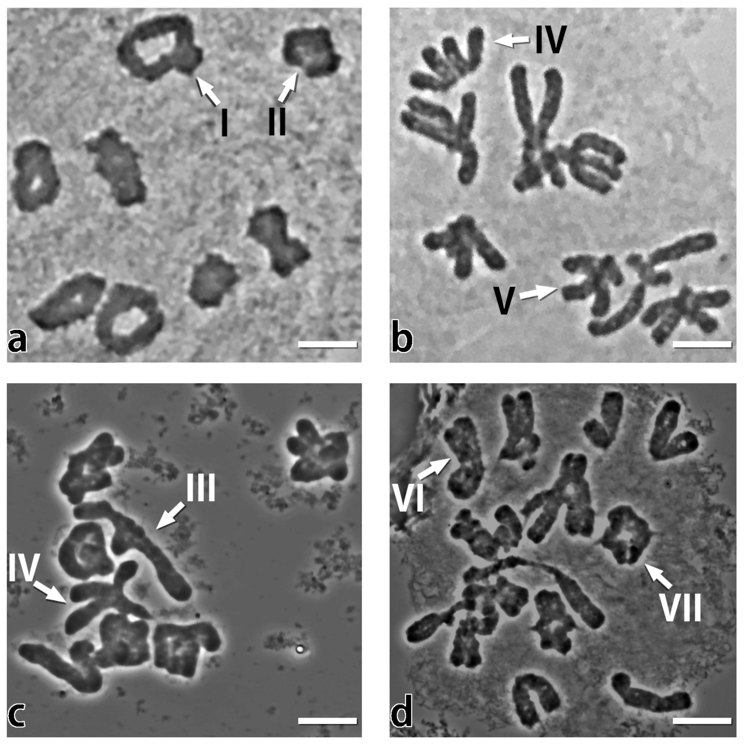
2.2. The Chiasmata Distribution in PMCs at Metaphase I

The acetocarmine stained squash preparations of pollen mother cells (PMCs) at metaphase I of A. cepa and A. fistulosum, and their F 1 diploid and triploid hybrids were analyzed.
In A. cepa, chiasmata occurred mainly in distal and interstitial bivalent regions and are very rare in proximal regions (Table 1). Chiasmata localized in the distal 2/3 of the chromosome arm accounted for 98.2%, and in the proximal 1/3 there were only 1.8%. The majority of metaphases contained eight bivalents with two distal chiasmata. Bivalents with three chiasmata were also common, especially in long chromosomes, in which two distal and one interstitial chiasmata often occurred (Figure 2a). The frequency of chiasmata per cell was 19.1.
The extreme localization of chiasmata to the proximal regions adjacently to the centromere of the A. fistulosum bivalents was evident, although occasionally, distal and interstitial chiasmata and univalents were found (Figure 2b). Chiasmata localized in the proximal 1/3 of the chromosome arm accounted for 97.1%, and in the distal 2/3 there were only 2.9% (Table 1). Open bivalents and univalents have been observed, which was reflected in the frequency COs per cell—15.4.
In diploid F 1 hybrid (A. cepa×A. fistulosum), carrying eight chromosomes of the A. cepa female parental species and eight chromosomes of the A. fistulosum male parental species, the chiasmata distribution was most similar to that of the A. cepa parent. No bivalents were observed with only proximally localized chiasmata as in A. fistulosum. The bivalents possessed chiasmata mostly in distal and interstitial regions. Homeologous chromosomes conjugated during prophase I of meiosis and formed chiasmata, indicating sufficient homology despite the difference in genome size between A. fistulosum and A. cepa. The genome size of A. cepa (1C = 16.4 Gb, [50]) has 4.8 Gb more DNA and correspondingly larger chromosomes than A. fistulosum (1C = 11.6 Gb, [51]). We have observed heteromorphic bivalents, open bivalents with one chiasma and univalent pair of unequal size (Figure 2c). In the F 1 hybrid, 47.7% of the chiasmata were located in distal regions of bivalents and 41.3% in interstitial ones, which is considerably higher than that of A. fistulosum (1.7% and 1.2%, correspondingly).
In the F 1 triploid hybrid, bivalents are mainly formed due to the pairing of homologous chromosomes of A. fistulosum in the presence of a complete haploid set of A. cepa chromosomes. Surprisingly, homomorphic bivalents of A. fistulosum showed pronounced chiasmata localization in the distal 2/3 of the chromosome arm (in distal region—34.5% and interstitial region—26.0%), and only 39.5% in the proximal region compared to A. fistulosum—97.1% (Table 1). Trivalents formed by two homologous chromosomes of A. fistulosum and one homeologous chromosome of A. cepa were observed very rarely (Figure S1).
The data of chiasmata distribution in A. cepa, A. fistulosum and their F 1 diploid and triploid hybrids presented in Table 1 were analyzed using a χ 2 statistic. Analysis of the contingency table containing the number of chiasmata on different locations in analyzed plant samples (Table 1) showed the presence of statistically significant differences in chiasmata locations among analyzed samples ( χ 2 = 1966.4, df = 6, p value < 0.001, Cramer’s V = 0.62). Pairwise comparisons showed the presence of statistically significant differences in chiasmata types between analyzed samples in all pairs (Table 2). Pairwise Cramer’s V values indicate that A. cepa and A. fistulosum have the most different patterns of chiasmata location (Cramer’s V = 0.95), while F 1 diploid and triploid hybrids’ chiasmata location patterns are the least different (Cramer’s V = 0.24). Both F 1 diploid and triploid hybrids’ chiasmata location patterns are more similar to A. cepa (Cramer’s V = 0.34 for F 1 diploid hybrid and Cramer’s V = 0.49 for triploid F 1 hybrid) rather than A. fistulosum (Cramer’s V = 0.87 for F 1 diploid hybrid and Cramer’s V = 0.72 for triploid F 1 hybrid), which is consistent with the hypothesis about dominant gene-controlling chiasmata distributions in A. cepa suggested by Emsweller and Jones [10].

2.3. Behavior of ASY1 and ZYP1 during SC Assembly and Disassembly in A. cepa, A. fistulosum, and their F 1 Diploid and Triploid Hybrids

Meiotic COs occur in association with synaptonemal complexes (SCs) that connect paired homologous chromosomes during pachytene. In A. thaliana, it was shown that SC have a key role in regulating CO numbers and distributions [24,25].
The dynamics of the SC during prophase I were monitored by immunolocalization of ASY1 (lateral elements of SC) and ZYP1 (transverse filaments of SC) in A. cepa, A. fistulosum and their F 1 diploid and triploid hybrids. A comparative analysis of ASY1 and ZYP1 behavior in the hybrids and their parental species was performed.
In A. cepa at leptotene, ASY1 loaded on the meiotic chromosome axes, while ZYP1 began to be loaded along the synapsis sites. At zygotene, ASY1 was detected as single thin tracks that corresponded to unpaired chromosome axes, while ZYP1 tracks became longer along synapsed chromosome axes. At pachytene, ZYP1 tracks have fully formed along the synapsed homologous chromosomes, while only a few unpaired regions can be identified by some weak ASY1 signals or as a remaining diffuse signal. During diplotene, ZYP1 undergoes degradation, indicating the beginning of disassembly of synaptonemal complexes. At diakinesis, ZYP1 signals appear as ball-like structures (Figure 3 and Figure S2).
In A. fistulosum, the behavior of ASY1 and ZYP1 was similar to that of A. cepa. We found no differences in the assembly and disassembly of these proteins between A. cepa and A. fistulosum (Figure 3 and Figure S3). However, ASY1 and ZYP1 of both A. cepa and A. fistulosum showed the peculiarities of loading and unloading during prophase I of meiosis. ZYP1 loaded only at the pairing regions of chromosomes, while ASY1 signals disappeared from them. Thus, the tripartite SC structure, combining two ASY1 linear structures and one ZYP1 core, has never been observed in A. cepa, A. fistulosum, and their F 1 hybrids (Figure 3a–d). The same ZYP1 loading pattern was shown in maize, wheat and rice, characterized by the absence of the tripartite SC structure [52,53,54,55]. In contrast, the tripartite SC structure is formed at pachytene in Arabidopsis, barley and rye [56,57,58,59].
In the F 1 diploid hybrid, synapsis was formed between homeologous chromosomes. At leptotene, comparative analysis of the F 1 diploid hybrid and parental species revealed no difference in ASY1 loading (Figure 3 and Figure S4). ASY1 was detected as single thin tracks forming along unpaired chromosome axes, while ZYP1 assembly delay was found, as evidenced by the lower number of ZYP1 tracks compared to parental species. At zygotene, a number of unpaired chromosomes with loaded ASY1 remained due to the above-mentioned delay in homeologous synapsis, while ZYP1 continued to load along the synapsed chromosome axes. In the middle of the pachytene, long, thick ASY1 tracks were observed, indicating that long regions of homeologous chromosomes had not yet synapsed. The number of ZYP1 tracks was lower compared to the parent species. At late pachytene, synapsis formation was accomplished, and long ZYP1 tracks were observed. Furthermore, the superimposition of DAPI and ZYP1 images revealed a partial absence of synapsis in paired chromosomes (Figure S4). These observations coincided with our data of chiasma analysis at metaphase I. As a result of the lack of synapsis in several sites between homeologous chromosomes, univalents were observed (Figure 2c). At diplotene and diakinesis, the disassembly of the synaptonemal complex did not differ from the parental species.
Comparison between the F 1 triploid hybrid and parental species revealed no difference in ASY1 and ZYP1 loading at leptotene (Figure 3 and Figure S5). This observation can be explained by the presence of a diploid set of A. fistulosum chromosomes that readily form homologous synapsis. At zygotene, long tracks of ASY1 were observed, which is probably associated with the presence of an unpaired set of A. cepa chromosomes (Figure S5), and ZYP1 long tracks were more likely formed between homologous chromosomes of A. fistulosum. At pachytene, ASY1 long tracks appeared along with ASY1 short tracks, which probably derived from abnormal synapsis of a single A. cepa chromosome with two homologous chromosomes of A. fistulosum (Figure S1). The ZYP1 long tracks coincided with paired DAPI stained chromosomes that indicates complete synapsis of homologous chromosomes of A. fistulosum (Figure 3 and Figure S5). At diplotene and diakinesis, the disassembly of the synaptonemal complex did not differ from the parental species.

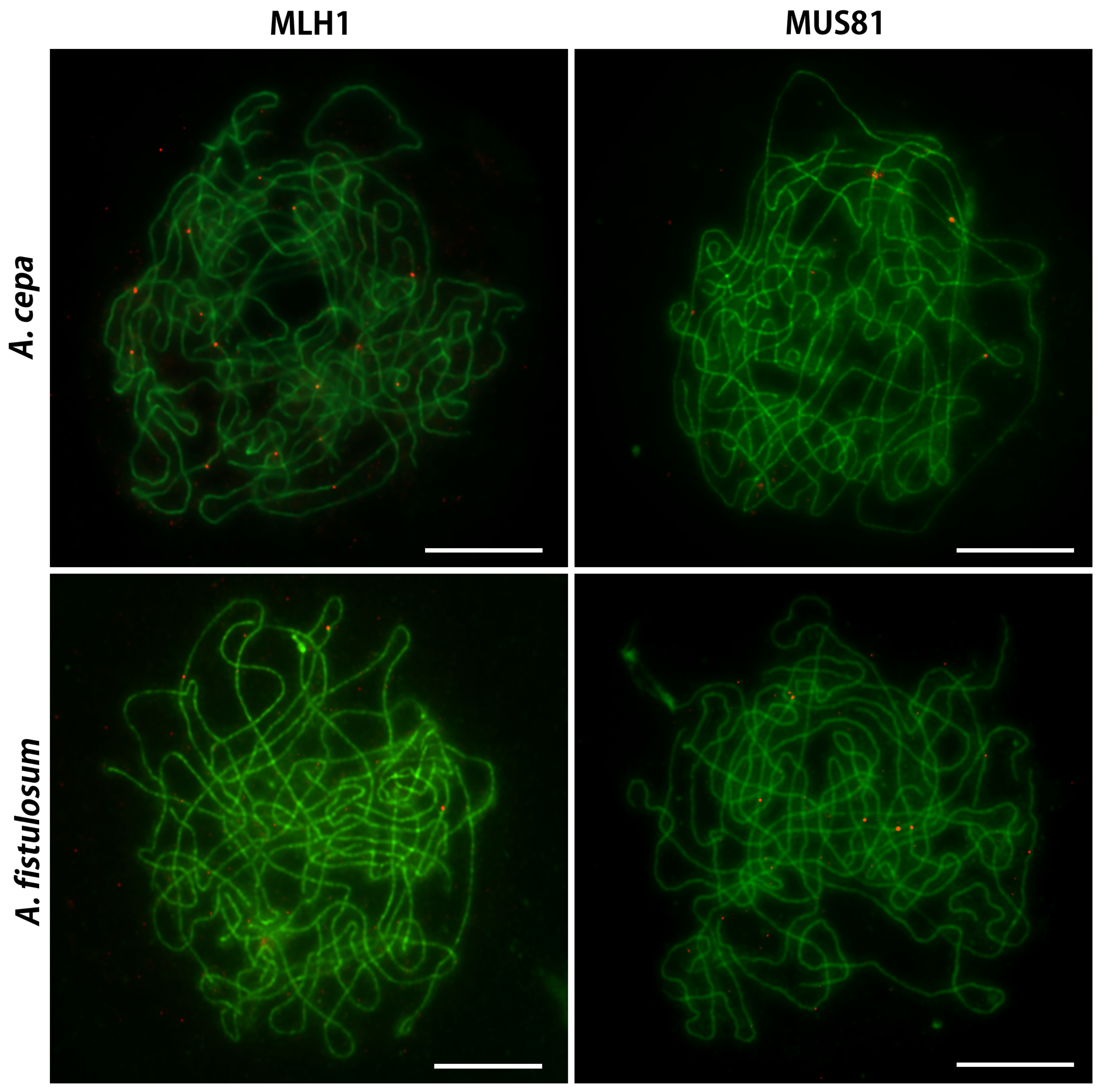
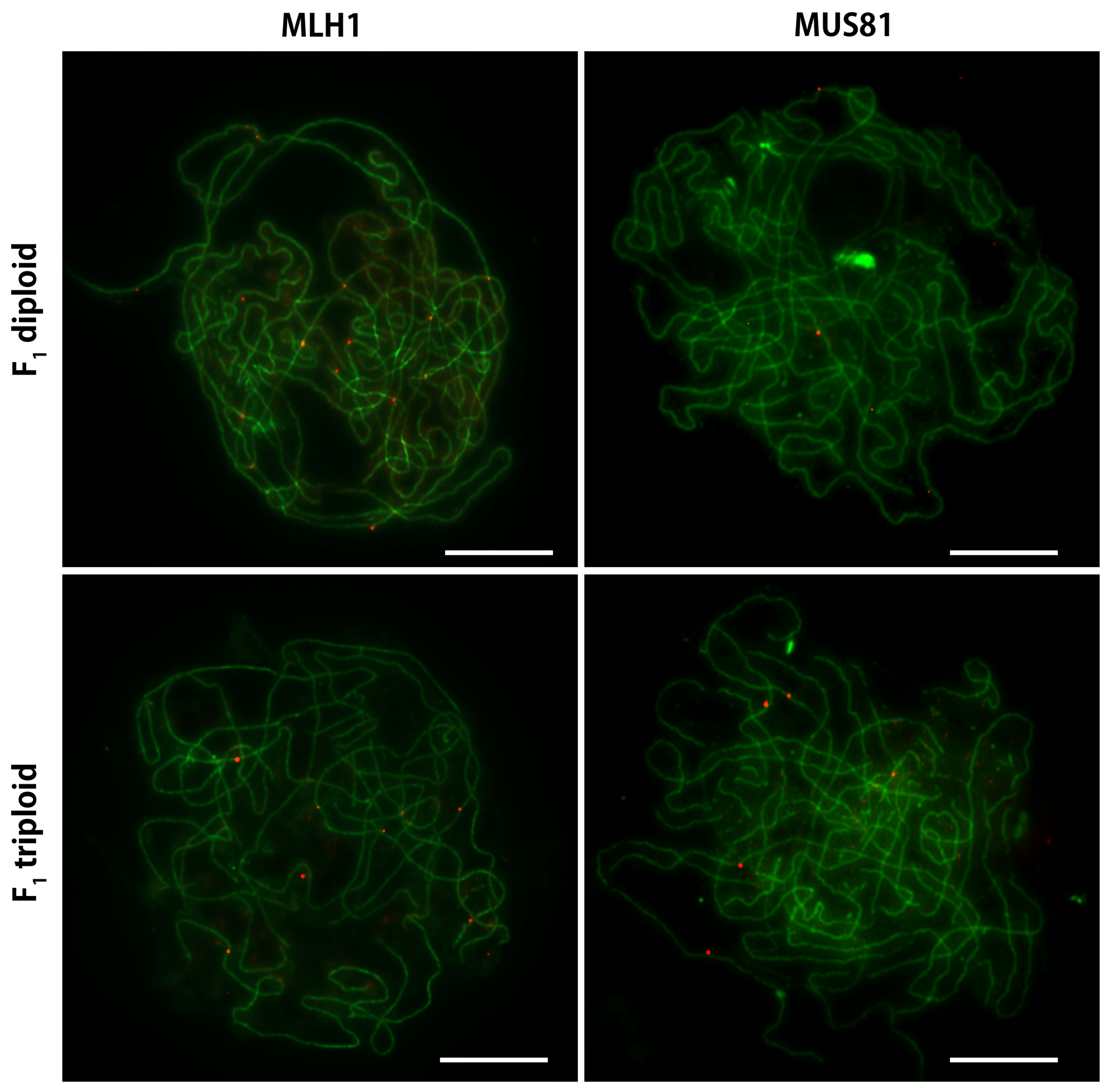
2.4. Immunocytological Detection of MLH1 and MUS81 on Pachytene Chromosomes of A. cepa and A. fistulosum and their F 1 Diploid and Triploid Hybrids

In order to lift the veil over the phenomenon of proximal chiasmata location, we (1) studied the features of ASY1 and ZYP1 during SC assembly and disassembly, and (2) identified the pathways of chiasmata (COs) origin via immunolocalization of MLH1 and MUS81 in A. cepa, A. fistulosum, and their F 1 diploid and triploid hybrids.
Scoring of the chiasmata distribution showed significant differences in A. cepa and A. fistulosum. In the F 1 diploid hybrid, chiasmata were predominantly located in distal and interstitial regions of homeologous bivalents, and in F 1 triploid hybrid a significant distal shift in homologous bivalents was observed. Chiasmata only indicates that a CO has occurred, not its class type. The MLH1 protein involved in meiotic recombination is commonly used as a marker for class I COs that are susceptible to interference, and another protein, MUS81, for class II COs that are indifferent to interference [30,32,42,43,60]. In order to find out the pathway of origin of COs, we performed immunolabelling of MLH1 and MUS81. For this purpose, polyclonal antibodies against the A. cepa protein sequences were prepared. We have counted the number of MLH1 and MUS81 foci per cell on ZYP1 tracks in prophase I of PMCs. Recently, it was shown that ZYP1 is required for class I CO formation in wild types of A. thaliana [26] and Brassica rapa [61]. However, in zyp mutants of A. thaliana, ZYP1 is dispensable for class I CO formation [26,35]. Immunolocalization of MUS81 in wild types of A. thaliana and wheat showed its association with ZYP1 [35,43,62]. Such studies have never been performed on onions.
In order to clearly distinguish the MLH1 and MUS81 signals from the background, only the fluorescent signal located on the ZYP1 track was considered as a true signal. Dual-color immunodetection of MLH1/ZYP1 and MUS81/ZYP1 was performed on different preparations of pachytene chromosomes. We could not carry out simultaneous detection of MLH1, MUS81 and ZYP1 on one preparation due to the unavailability of the three-color antibody system. Preparation of pachytene chromosomes was carried out on a population of PMCs from a single plant in each variant at the same time for MLH1 and MUS81. The MLH1 and MUS81 foci data per cell, obtained by counting on different slides, did not reveal significant differences between an artificial population (see Section 4) in which both MLH1 and MUS81 were expected to be counted in the same cell (Figure S6).
In A. cepa, dual immunodetection of MLH1 in conjunction with ZYP1 revealed that MLH1 formed an average of 11.1 foci per cell. The average number of MUS81 foci associated with ZYP1 was 4.1 (Table 3, Figure 4). The total number of MLH1 and MUS81 foci per cell was 15.2, which is less than the frequency of COs (19.1) established at metaphase I (Table 1). This minor discrepancy may be due to technical limitations related to the accessibility of antibodies to target proteins on the onion pachytene chromosomes. Although it is possible that there is a third pathway of CO formation, the recombinant proteins were not used as a marker for COs in this study. It has recently been shown in A. thaliana that AtFANCD2 is required for meiotic homologous recombination, promoting noninterfering COs in a MUS81-independent manner [62]. The FANCD2 protein is monoubiquinated in response to DNA damage, resulting in its localization to nuclear foci with other proteins (BRCA1 and BRCA2) involved in homology-directed DNA repair [63].
We have demonstrated via immunodetection of meiotic proteins during prophase I of A. cepa that MLH1 was essential for the majority of foci per cell (73%) compared to MUS81 (27%). This result is similar to the frequency ratio of MLH1 and MUS81 observed on A. thaliana [35], Solanum lycopersicum [41,42] and allotetraploid (AABB) durum wheat (Triticum turgidum subsp. durum) [43].
Dual immunodetection of MLH1 and MUS81 in A. fistulosum revealed an approximately equal mean number of foci per cell—6.5 both for MLH1 and MUS81 (Table 3, Figure 4). The number of MLH1 foci per cell was 4.6 less in A. fistulosum compared to A. cepa (p value <0.001, Table S1) and 2.4 more for MUS81 (p value <0.001, Table S1). The ratio of MLH1 and MUS81 foci per cell was 50%:50%, which is significantly different from A. cepa (73%:27%). The observed difference in ratio of MLH1 and MUS81 foci is in agreement with the distribution of chiasmata in these species. It is possible that MLH1, which is involved in the interference-dependent COs formation, has a limited ability to operate on the specifically organized A. fistulosum chromatin at the genomic and epigenomic levels. Under these circumstances, MUS81 should predominantly recruit for the interference-free CO formation to guarantee obligate recombination. This observation points to interference between class I and II COs that coincides with previously reported data on tomato [42].
In the F 1 diploid hybrid, we observed a mean of 8.5 MLH1 and 3.6 MUS81 foci per cell (Table 3, Figure 4). All detected signals of MLH1 and MUS81 were observed on the ZYP1 tracks formed between homeologous chromosomes A. cepa/A. fistulosum. Immunocytological analysis of the ZYP1 assembly in this study revealed the lack of ZYP1 in the short segments of paired homeologous chromosomes in F 1 diploid hybrid. In order to identify MLH1 and MUS81 signals at sites with missed ZYP1 on paired chromosomes, signal scoring was also performed on merged images of MLH1/ZYP1 and MUS81/ZYP1 with DAPI-stained chromatids. We did not detect any signal in ZYP1 missing sites. The total number of MLH1 and MUS81 foci per cell was 12.1, which is less than in parental species due to univalents and open bivalents, that were observed at metaphase I (Figure 2). However, the ratio of MLH1/MUS81 per cell (70%:30%) was almost equal to that of A. cepa (73%:27%). The result corresponds to the location of the chiasmata predominantly in the distal and proximal regions (Table 1, Figure 2). The observation of an A. cepa type of MLH1/MUS81 coincides with a predominantly A. cepa type of chiasmata location in this and previous studies [46].
In the F 1 triploid hybrid, we observed a mean of 8.5 MLH1 and 5.7 MUS81 foci per cell (Table 3, Figure 5). The total number of MLH1 and MUS81 foci per cell was 14.2 (Table 1). The frequency ratio of MLH1 and MUS81 is 60%:40%. In this F 1 triploid hybrid, CO formation occurs between the homologous chromosomes of A. fistulosum, and we may expect the MLH1/MUS81 ratio to be similar to A. fistulosum (50%:50%). However, the presence of the haploid set of A. cepa chromosomes modified the MLH1/MUS81 ratio. The increased number of MLH1 foci associated with an additional gene dosage in the F 1 triploid hybrid (p value < 0.001, Table S1) is probably one of the players responsible for the distal shift of chiasmata (Table 1, Figure 2).

3. Discussion

In this study, we set out to test the hypothesis of possible genetic control of COs/chiasmata distribution, and tried to determine the factors underlying the highly contrasting patterns of chiasmata localization in two closely related Allium species. To investigate this phenomenon, we performed immunological and cytogenetic assays of CO formation components that are detectable in the prophase I and metaphase I stages of meiosis. Additionally, the behavior of ASY1 and ZYP1 during the assembly and disassembly of the SCs was studied. We used a unique plant material, which made it possible to study the formation of COs on heteromorphic (nonhomologous) and homomorphic (homologous) meiotic pairs of chromosomes. A distal shift of chiasmata on the A. fistulosum homologous chromosome pairs in the presence of the A. cepa genome suggests genetic control of CO localization. Furthermore, this study has provided new insights into the interaction of two pathways of CO formation to maintain total CO levels per nucleus in heteromorphic and homomorphic meiotic pairs in interspecific hybrids. New knowledge of SC assembly/disassembly was obtained that has not previously been reported for Alliums.

3.1. Chromatin, Genome and the Highly Contrasting Chiasmata Localization in A. cepa and A. fistulosum

Studies of CO patterns in many organisms have shown high levels of COs in distal euchromatin and low levels of COs in pericentric heterochromatin, whether analyzed using chiasmata, genetic maps, genotyped recombinant inbred lines, recombinant nodules, or MLH1 foci [7,24,41,42,64,65,66]. It should be noted that all of these studies were carried out on organisms with a distal distribution of COs. Species with strictly proximally localized COs are rare among plants and animals. To our knowledge, among the plant species studied for recombination distribution, only four species are known with extreme localization of chiasmata, namely, Allium fistulosum [10], Allium porrum [67,68], and Allium altaicum [11,69], which are very close phylogenetically, and Fritillaria meleagris [70], belonging to the Liliaceae family. All four species belong to clade Liliopsida, subclass Petrosaviidae. The proximal localization of the chiasmata in Fritillaria meleagris is explained by an association between chiasmata localization and restricted synapsis [2], which is not typical for the Allium species [46]. What do we know about chromatin organization in A. fistulosum and A. cepa? The localization of constitutive heterochromatin along the entire chromosome is usually observed using differential C-band staining on mitotic metaphase chromosomes. Comparative analysis of the C-band pattern in A. fistulosum and A. cepa did not reveal significant differences [11]. In both species, all chromosomes possess terminal C-bands which are larger in A. fistulosum than those found in A. cepa, and centromeric heterochromatin is present in all the chromosomes in the form of very thin bands sometimes seen as paired dots. Comparing the C-band pattern with the distribution of chiasmata in species of the Allium genus, Vosa [11] concluded that the position of the C-bands seems to be independent of chiasmata localization. Moreover, COs may be distributed in a more random fashion than chiasmata in the more condensed state of the chromosome at metaphase I. Evidence for this assumption was obtained by analyzing AFLP linkages in Allium porrum [71]. It was shown that AFLP markers are not inherited in large linkage blocks, despite the cytogenetic observations of chiasmata occurring mainly in the proximal region. In A. fistulosum, the large terminal heterochromatin block consists of a 378 bp satellite repeats interspersed with Ty1-copia retrotransposone sequences and microsatellites [72]. Analysis of the distribution of DNA methylation pattern in A. fistulosum using specific antibodies against 5-methylcytosine (anti-5mC) showed highly methylated DNA blocks in the distal regions of all chromosomes [73]. We may suggest that the highly methylated distal heterochromatin blocks move chiasmata away from the telomeric ends in A. fistulosum.
Higgins et al. [74], studying the predominantly distal distribution of COs in barley, concluded that the reason for this phenomenon is spatiotemporal asymmetry of the meiotic program. The authors found that the distal euchromatin was the first to replicate, and the formation of DSBs in the distal regions occurred before the elaboration of a continuous chromosome axis in the interstitial/proximal regions. Thus, DSBs in interstitial and proximal regions occur later than distal events, and rarely progresses to form chiasmata. This is in contrast with observations in plants such as Arabidopsis and Brassica, in which the formation of the chromosome axes at late G2/leptotene is very rapid and recombination initiation is synchronous throughout the chromosomes. We may hypothesize that spatiotemporal asymmetry in DNA replication takes place in A. fistulosum, then the large terminal blocks of heterochromatin would replicate later than DNA in the interstitial/proximal regions, reducing the ability to form COs in these regions.
It is obvious that epigenetic modification can influence the CO location, as it was demonstrated on met1 and non-CG/H3K9me2 mutants of Arabidopsis [75,76]. Using whole-genome sequencing of mouse single sperm, providing a genome-wide map of crossovers, genetic and epigenetic factors that affect crossover probability have been identified [77]. An analogue to the single sperm sequencing approach outlined above is an already developed method of the single pollen cell sequencing [78,79], which will allow us to investigate meiotic recombination in plants at the DNA sequence level to directly explore the recombination landscape at the molecular level in an unbiased manner. Further sequencing and telomere-to-telomere assembly of the A. cepa and A. fistulosum genomes, together with advances in epigenetics, will help resolve this enigma of highly contrasting patterns of chiasmata localization in these closely related Allium species.

3.2. The A. cepa Type of Chiasmata Distribution on Homeologous Bivalents in F 1 Diploid Hybrid

Despite the difference in the amount of DNA and contrasting chiasmata location between A. fistulosum and A. cepa, the chromosome conjugation, SC formation and crossing over occurred in the F 1 diploid hybrid. Remarkably, all bivalents had predominantly distal and interstitial chiasmata as in A. cepa. Our results are in complete agreement with those previously obtained in F 1 hybrid between A. fistulosum and A. cepa [10,46]. It should be noted that we used A. cepa as the female parent and A. fistulosum as the pollinator, but in previous studies, analysis of bivalents configuration was performed using F 1 hybrids, in which the female parent was A. fistulosum and the pollinator was A. cepa. Thus, the influence of the cytoplasm on the configuration of bivalents can be excluded, at least in the case of these closely related species. Our data confirm the hypothesis of the presence of a dominant gene/genes that provide distal and interstitial COs between the homeologous chromosomes in the F 1 diploid hybrid, previously put forward by Emsweller and Jones [10]. However, such an assumption would be very simplistic, given the fact that the hybrid combines two genomes that differ in size [68], as well as intrinsic properties [11,72,73,80].
Using the immunodetection of ASY1 and ZYP1 in prophase I, we revealed a delay in chromosome pairing and the lack of synapsis in several sites between homeologous chromosomes in F 1 diploid hybrid. These results agree with data obtained by Albini and Jones [46] on SC spreads using electron microscopy. The authors observed only 80–90% of the synapsed chromosomes at late pachytene in F 1 hybrid between A. fistulosum and A. cepa. It was suggested that the factors involved in promoting the interstitial and distal location of chiasmata might be disturbances of synapsis in the centromeric regions of homeologous bivalents between A. cepa and A. fistulosum and, probably, this constraint, reduces proximal chiasmata frequency in the F 1 diploid hybrid. Using light epifluorescent microscopy, we were not able to determine whether the non-sinapsis sites are located in the pericentromeric regions of the pachytene chromosomes, which look like a tangled ball. Disturbances of synapsis may indicate sites of DNA differences where extra blocks of DNA occur in the A. cepa genome. We often observed long ZYP1 tracks that almost coincided with DAPI-stained chromatin. The adjustment of chromosomes axis lengths during synapsis of the parental chromosomes with different lengths in interspecific hybrids could take place [81,82]. However, how the correct homologous sequence is located efficiently among the bulk of chromatin embedded nuclear DNA remains unclear [83].

3.3. A Significant Shift in Chiasmata Localization and MLH1/MUS81 Ratio in F 1 Triploid Hybrid (2n = 3x = 16F + 8C) Is Evidence of Genetic Control of COs Distribution

Previously, Emsweller and Jones [9] suggested that A. cepa possesses a dominant gene, and A. fistulosum possesses a recessive one based on an analysis of bivalent configurations in F 1 and gene segregation in F 2 hybrids between A. cepa and A. fistulosum. The F 1 triploid hybrid, which has a complete diploid set of A. fistulosum chromosomes and eight chromosomes of A. cepa, is an ideal source for testing the genetic control hypothesis. In the F 1 triploid hybrid, bivalents were formed by the A. fistulosum homologous chromosomes that have no differences in the length and homology of sequences. Despite this, a significant shift in the localization of chiasmata to the distal and interstitial regions was found (Table 1, Figure 2d). This observation indicates that the genetic control of chiasmata distribution is possible, along with other factors that control crossing over.
Counting chiasmata in bivalents does not allow for determination of the pathway of CO origin. Immunolocalization of MLH1 (class I COs) and MUS81 (class II COs) in A. cepa, A. fistulosum and their F 1 diploid and triploid hybrids showed that the two CO pathways interact. The number of MLH1 foci per cell is not independent of the number of MUS81 foci (Table 3). Moreover, the MLH1/MUS81 ratio correlates with the distribution of chiasmata. We observed that a higher MLH1/MUS81 ratio was linked with an increase in the number of distal and interstitial chiasmata. In the F 1 triploid hybrid, a significant increase in MLH1 foci per cell compared to A. fistulosum was observed. A more pronounced difference in the MLH1/MUS81 ratio was found in the parental species. With this in mind, it was of interest to evaluate whether the protein sequences of MLH1 and MUS81 differ in A. cepa and A. fistulosum. We found that MLH1 is highly conserved between A. cepa and A. fistulosum. Transcripts of MLH1 (A. cepa: GBRO01051308.1, A. fistulosum: GFAM01047024.1) contains ORF (Open Reading Frame) encoding proteins of equal length (725 amino acids) and share 99.9% (724/725) amino acids identity with polymorphism in one residual. Thus, the mlh1 genes of A. cepa and A. fistulosum should produce almost identical proteins. In the F 1 triploid hybrid, MLH1 could be involved in recombination at an increased dosage if all three loci (two A. fistulosum and one A. cepa) express in equal proportions. However, in newly synthesized interspecific hybrids, a genome-wide expression level dominance and/or homeolog expression bias depending on species and/or tissue was shown [84].
Increasing the number of MLH1 foci per cell, the number of MUS81 foci per cell was significantly decreased in the F 1 triploid hybrid compared to A. fistulosum, that is consistent with the statement of COs homeostasis to maintain a fixed amount of COs per cell [18,85]. Interestingly, MUS81 is more polymorphic between A. cepa and A. fistulosum compared to MLH1. Transcripts of MUS81 (A. cepa: GBRQ01006735.1, A. fistulosum: GHMM01233510.1) contains ORF encoding proteins of equal length (604 amino acids) and share 93.5% (565/604) amino acids identity with polymorphism in 39 residuals with increasing identity in winged helix domain (96.2%; 102/106) and XPF-nuclease domain (99.3%; 145/146). MUS81 protein sequences showed considerable interspecific polymorphism between A. cepa and A. fistulosum even in functional domains. Whether this structural polymorphism of the MUS81 is associated with possible functional features and the dominant/recessive allelic stage is unknown.
In summary, we have shown, for the first time, (1) a significant shift in chiasmata localization on the A. fistulosum homologous chromosome pairs in the presence of haploid set of the A. cepa chromosomes in F 1 triploid hybrid, and (2) an increase in the number of MLH1 foci per cell was balanced by a decrease in the number of MUS81 foci in the F 1 triploid hybrid, which is consistent with CO homeostasis, (3) a significant difference in the MLH1/MUS81 ratio between A. cepa and A. fistulosum, and (4) in the F 1 diploid hybrid, the MLH1/MUS81 ratio was similar to that of A. cepa, and (5) there is no difference in assembly and disassembly of ASY1 and ZYP1 between A. cepa and A. fistulosum, which was similar to that of maize, wheat and rice, and (6) the ZYP1 assembly delay was found in the F 1 diploid hybrid. Finally, our results support the hypothesis of genetic control of CO localization. Given the biological importance and complexity of crossing over, the control of CO localization must be considered on many levels beyond genetics, including epigenetics, as the template’s chromatin environment, spatiotemporal DNA replication and the distribution of DSBs, impact of the protein modification and interaction, and others.

4. Materials and Methods

4.1. Plant Materials

Perennial plants of A. fistulosum (2n = 2x = 16), of the Ruskiy Zimniy variety grow on the experimental field of the Center for Molecular Biotechnology RSAU-MTAA. Bulbs of A. cepa (2n = 2x = 16), of the Chalcedon variety were planted in the field. F 1 hybrids between A. cepa and A. fistulosum were obtained by manual emasculation of anthers in unopened buds. Pollen from A. fistulosum plants were collected in Petri dishes and stored at +4 C until the pistil stigma ripened for pollination in emasculated buds of A. cepa. Pollination of each flower was carried out for 2 days by reapplying pollen on the stigma of the pistil with a brush. F 1 diploid hybrid (A. cepa×A. fistulosum) (2n = 2x = 8F + 8C) acc. 1–20 and F 1 triploid hybrid (A. cepa× A. fistulosum) (2n = 3x = 16F + 8C) acc. 7–20 were used in this study.

4.2. Genomic In Situ Hybridization (GISH)

Mitotic chromosomes of F 1 hybrids were prepared from young root meristems using the squash method according to Kudryavtseva et al. [86]. In situ hybridization, immunological detection, and counterstaining procedures were the same as previously described by Khrustaleva and Kik [87]. The hybridization mixture contained: 50% (v/v) formamide, 10% (w/v) dextran sulfate, 2 × SSC, 0.25% (w/v) sodium dodecyl sulfate (SDS), 50 ng/ μ L of labeled probe (DIG)-11-dUTP and 1500 ng/ μ L of blocking DNA. In the hybridization mixture, we used a ratio of 1:30 of probe and block DNA, and washes at 78% stringency were applied. In F 1 (A. cepa×A. fistulosum) (2n = 2x = 8F + 8C) the A. fistulosum genomic DNA was used as a probe and the A. cepa genomic DNA was used as a block. In F 1 (A. cepa× A. fistulosum) (2n = 3x = 16F + 8C) the A. cepa genomic DNA was used as a probe and the A. fistulosum genomic DNA was used as a block. In both cases, the labeled (DIG)-11-dUTP probe DNA was detected with anti-Dig-FITC raised in sheep (Roche diagnostics GmbH, Mannheim, Germany) and amplified with anti-sheep-FITC raised in rabbit (Vector Laboratories, CA, USA).
GISH preparations were visualized using a Zeiss AxioImager M2 microscope (www.zeiss.com, accessed on 15 November 2021) and a black–white sensitive digital Hamamatsu camera C13440-20CU (www.hamamatsu.com, accessed on 15 November 2021). The final optimization of images was performed using Adobe Photoshop (Adobe Inc., San Jose, CA, USA). Karyotype analysis and identification of individual chromosomes with fluorescent signals by the DRAWID program [88] were performed according to bulb onion nomenclature [89] and previously published karyotypes of closely related Allium species [90].

4.3. Analysis of the Chiasmata Distribution in PMCs

Fresh unfixed anthers containing pollen mother cells (PMCs) at metaphase I were tapped out in a drop of 1% acetocarmine on a glass slide, gently mixed, and then heated for 1 min at 60 C on a heating table. The cells were spread by tapping on the coverslip and gently squashed. Slides were examined under a Zeiss Axiolab 5 microscope (www.zeiss.com, accessed on 28 August 2021) using phase-contrast microscopy. The selected images were captured using a digital Axiocam 208 color camera (www.zeiss.com, accessed on 28 August 2021). Image processing was performed by using Zen 2.6 lite (blue edition), an image analysis software. Bivalent arms were arbitrarily divided into three regions of equal length (proximal, interstitial, distal). The position of chiasmata on chromosome arm was measured by a DRAWID program [88]. Only non-overlapping bivalents were used for measurements of positions of chiasmata.

4.4. Immunochemical Analysis

4.4.1. Antibody Production

Reference transcriptomes of A. cepa and A. fistulosum from GenBank (Transcriptome Shotgun Assembly) were used for the acquisition of ZYP1, MLH1 and MUS81 protein sequences. Sequences of these proteins from other species in order to use as a reference were identified based on literature data: ZYP1 (Zea mays, GenBank: ADM47598.1, [54]), MLH1 (Solanum lycopersicum, GenBank: EF071927.1, [41]), MUS81 (Arabidopsis thaliana, GenBank: AB177892.1, [91]). tBLASTn was used for identification transcript sequences of the corresponding proteins in Transcriptome Shotgun Assembly database (identity >50%, query cover > 70%). The longest Open Reading Frame (ORF) was translated into an amino acid sequence. The following steps, including identification of a peptide sequence for antibody production and antibody synthesis, were performed by the outsource company PrimeBioMed (www.primebiomed.ru, accessed on 9 February 2020). The anti-ASY1 polyclonal antibody (raised in rabbit against ASY1 of Arabidopsis thaliana) was described by Armstrong et al. [23].

4.4.2. Preparation of Meiotic Chromosome for Protein Immunolocalization

We developed an original method of meiotic chromosome preparation. Anthers at the desired stage of meiosis were fixed by Clark’s fixative (ethanol:acetic acid, 3:1, v/v) for the 1 h. The fixed anthers were washed in tap water for 30 min, and then in citrate buffer (10 mM sodium citrate, 10 mM citric acid, pH 4.8) for 10 min. A total of 5–8 anthers were transferred into 1.5 mL tubes with 50 μ L of 0.6% enzyme mixture (1:1:1) pectolyase Y-23 (Kikkoman, Tokyo, Japan), Cellulase Onozuka R-10 (Yakult Co., Ltd., Tokyo, Japan) and Cytohelicase (Sigma-Aldrich Co., LLC, St. Louis, MO 63103 USA) for 120 min at 37 C. After 120 min, the enzyme mixture was deleted by Pasteur Pipettes from the tubes. Anthers were gently transformed into a fine cell suspension in the tube using a dissecting needle. A total of 100 μ L of 60% acetic acid was added to the cell suspension. The tube with cell suspension was then heated for 5 min at 50 C. A total of 60 μ L of ethanol/acetic acid fixative at a ratio of 3:1 was added. A total of 10 μ L of cell suspension was dropped onto a slide, and by the time the surface became granule-like (10–15 s), 30 μ L of ethanol/acetic acid fixative at a ratio of 3:1 was added. Then the slides with cells were immediately dried with an airflow. The same day, the slides were used for immunochemical analysis. Some slides were kept in the fridge—70 C.

4.4.3. Immunochemical Detection of ASY1 and ZYP1

Slides were incubated with blocking buffer (5% BSA in 1 × PBS, 0.1% Tween-20, 1 mM EDTA, pH = 8.0) for 2 h at room temperature. Then, slides were incubated with rabbit anti-ASY1 (1:20) and rat anti-ZYP1 (1:20) in the blocking buffer for 24 h at 4 C. Slides were washed 4 × 30 min in 1 × PBS, 0.1% Tween-20, 1 mM EDTA (pH = 8) at room temperature. Slides were incubated with blocking buffer for 2 h at room temperature. The following secondary antibodies diluted in block buffer (1:100) were applied at 37 C for 1 h: for ASY1, the visualization was goat anti-rabbit Alexa Fluor 555, (red fluorescence) (Abcam plc, Cambridge, CB2 0AX, UK), and for ZYP1 it was goat anti-rat Alexa Fluor 488, (green fluorescence) (Abcam plc, Cambridge, CB2 0AX, UK). The slides were washed 4 × 30 min in 1 × PBS, 0.1% Tween-20, 1 mM EDTA (pH = 8) at room temperature and mounted in Vectashield antifade medium (Vector Laboratories) with 2 μ g/mL DAPI.

4.4.4. Immunochemical Detection of MLH1/ZYP1 and MUS81/ZYP1

The incubation and washing steps were the same as described above. The MLH1 immunodetection was with mouse anti-MLH1 (1:100), followed by the secondary goat anti-mouse antibody conjugated with Alexa Fluor 555, (red fluorescence) (Abcam plc, Cambridge, CB2 0AX, UK). The MUS81 immunodetection was with guinea pig anti-MUS81 (1:100), followed by the secondary goat anti-guinea pig Alexa Fluor 555 (red fluorescence) (Abcam plc, Cambridge, CB2 0AX, UK). Simultaneous ZYP1 immunodetection was with rat anti-ZYP1 (1:20), followed by goat anti-rat Alexa Fluor 488 (green fluorescence) (Abcam plc, Cambridge, CB2 0AX, UK).

4.4.5. Statistical Analysis

Statistical analysis of chiasmata "location types" distribution was performed applying χ 2 -statistics from a stats package using R programming language v4.2.1 [92]. Pairwise comparisons between A. cepa, A. fistulosum and F 1 diploid and triploid (A. cepa×A. fistulosum) hybrids chiasmata “location types” was performed using χ 2 -statistics. p values were adjusted using Bonferroni correction. Effect size (Cramer’s V) was calculated using a cramerV function from the rcompanion v2.4.21 package [93].
Identification of statistically significant differences in MLH1 and MUS81 foci number per cell among A. cepa, A. fistulosum, and F 1 diploid and triploid hybrids was performed using the Wilcoxon test in R programming language v4.2.1 [92]. Calculated p values were adjusted using the False Discovery Rate (FDR). A p value threshold < 0.05 was considered as statistically significant.
An estimation of the mean MLH1/MUS81 ratio per cell was performed via generation of all possible pairwise combinations of MUS81 and MLH1 foci (artificial population) in analyzed cells for each analyzed species (Tables S2 and S3). For each pair, the MLH1/MUS81 ratio was calculated. The weighted mean with inverse weights was calculated for each analyzed species in order to decrease the magnitude of influence of combinations with an extreme MLH1/MUS81 ratio.

4.4.6. Microscopy and Imaging

The slides were examined under a Zeiss AxioImager M2 microscope (www.zeiss.com, accessed on 15 November 2021). The selected images were captured using a digital Hamamatsu camera C13440-20CU (www.hamamatsu.com, accessed on 15 November 2021). Image processing was performed by Zen 2.6 (blue edition), an image analysis software.

Supplementary Materials

The supporting information can be downloaded at: https://www.mdpi.com/article/10.3390/ijms24087066/s1.

Author Contributions

N.K.—investigation, methodology, resources, verification, visualization; A.E.—formal analysis, visualization, investigation, writing—original draft; A.P.—investigation; S.S.—investigation; S.O.—investigation; L.K.—conceptualization, data curation, formal analysis, funding acquisition, investigation, methodology, project administration, supervision, writing—original draft. All authors have read and agreed to the published version of the manuscript.

Funding

This research was funded by the Russian Scientific Foundation, grant number 20-46-07005.

Institutional Review Board Statement

Not applicable.

Informed Consent Statement

Not applicable.

Data Availability Statement

Data available in a publicly accessible repository.

Acknowledgments

We thank Glyn Jenkins for the training course on the immunolocalization of meiotic proteins.

Conflicts of Interest

The authors declare no conflict of interest.

Abbreviations

The following abbreviations are used in this manuscript:
COCrossing Over
DSBDouble-Strand Breaks
PMCPollen Mother Cells
GISHGenomic in situ hybridization
SCSynaptonemal Complex
LNLate recombination Nodules
ORFOpen Reading Frame
HJHolliday Junction

References

  1. Darlington, C. Meiosis in diploid and tetraploid Primula sinensis. J. Genet. 1931, 24, 65–96. [Google Scholar] [CrossRef]
  2. Higgins, J.; Osman, K.; Jones, G.; Franklin, F. Factors underlying restricted crossover localization in barley meiosis. Annu. Rev. Genet. 2014, 48, 29–47. [Google Scholar] [CrossRef] [Green Version]
  3. Lam, I.; Keeney, S. Mechanism and regulation of meiotic recombination initiation. Cold Spring Harb. Perspect. Biol. 2015, 7, a016634. [Google Scholar] [CrossRef] [PubMed] [Green Version]
  4. Mercier, R.; Mézard, C.; Jenczewski, E.; Macaisne, N.; Grelon, M. The molecular biology of meiosis in plants. Annu. Rev. Plant Biol. 2015, 66, 297–327. [Google Scholar] [CrossRef]
  5. Gray, S.; Cohen, P.E. Control of meiotic crossovers: From double-strand break formation to designation. Annu. Rev. Genet. 2016, 50, 175. [Google Scholar] [CrossRef] [PubMed] [Green Version]
  6. Haenel, Q.; Laurentino, T.G.; Roesti, M.; Berner, D. Meta-analysis of chromosome-scale crossover rate variation in eukaryotes and its significance to evolutionary genomics. Mol. Ecol. 2018, 27, 2477–2497. [Google Scholar] [CrossRef] [Green Version]
  7. Gardiner, L.J.; Wingen, L.U.; Bailey, P.; Joynson, R.; Brabbs, T.; Wright, J.; Higgins, J.D.; Hall, N.; Griffiths, S.; Clavijo, B.J.; et al. Analysis of the recombination landscape of hexaploid bread wheat reveals genes controlling recombination and gene conversion frequency. Genome Biol. 2019, 20, 1–16. [Google Scholar] [CrossRef] [Green Version]
  8. Desjardins, S.D.; Simmonds, J.; Guterman, I.; Kanyuka, K.; Burridge, A.J.; Tock, A.J.; Sanchez-Moran, E.; Franklin, F.C.H.; Henderson, I.R.; Edwards, K.J.; et al. FANCM promotes class I interfering crossovers and suppresses class II non-interfering crossovers in wheat meiosis. Nat. Commun. 2022, 13, 3644. [Google Scholar] [CrossRef]
  9. Emsweller, S.; Jones, H. A Gene for Control of Interstitial Localization of Chiasmata in Allium fistulosum L. Science 1935, 81, 543–544. [Google Scholar] [CrossRef]
  10. Emsweller, S.; Jones, H. Meiosis in Allium fistulosum, Allium cepa, and their hybrid. Hilgardia 1935, 9, 275–294. [Google Scholar] [CrossRef] [Green Version]
  11. Vosa, C.G. Heterochromatic patterns in Allium. Heredity 1976, 36, 383–392. [Google Scholar] [CrossRef] [Green Version]
  12. Tease, C. Cytological detection of crossing-over in BUdR substituted meiotic chromosomes using the fluorescent plus Giemsa technique. Nature 1978, 272, 823–824. [Google Scholar] [CrossRef]
  13. Tease, C.; Jones, G. Analysis of exchanges in differentially stained meiotic chromosomes of Locusta migratoria after BrdU-substitution and FPG staining. Chromosoma 1978, 69, 163–178. [Google Scholar] [CrossRef]
  14. Bishop, D.K.; Zickler, D. Early decision: Meiotic crossover interference prior to stable strand exchange and synapsis. Cell 2004, 117, 9–15. [Google Scholar] [CrossRef] [PubMed] [Green Version]
  15. Hillers, K.J. Crossover interference. Curr. Biol. 2004, 14, R1036–R1037. [Google Scholar] [CrossRef] [PubMed] [Green Version]
  16. Kleckner, N.; Zickler, D.; Jones, G.H.; Dekker, J.; Padmore, R.; Henle, J.; Hutchinson, J. A mechanical basis for chromosome function. Proc. Natl. Acad. Sci. USA 2004, 101, 12592–12597. [Google Scholar] [CrossRef] [Green Version]
  17. Moens, P.B.; Kolas, N.K.; Tarsounas, M.; Marcon, E.; Cohen, P.E.; Spyropoulos, B. The time course and chromosomal localization of recombination-related proteins at meiosis in the mouse are compatible with models that can resolve the early DNA-DNA interactions without reciprocal recombination. J. Cell Sci. 2002, 115, 1611–1622. [Google Scholar] [CrossRef]
  18. Martini, E.; Diaz, R.L.; Hunter, N.; Keeney, S. Crossover homeostasis in yeast meiosis. Cell 2006, 126, 285–295. [Google Scholar] [CrossRef] [Green Version]
  19. Pazhayam, N.M.; Turcotte, C.A.; Sekelsky, J. Meiotic crossover patterning. Front. Cell Dev. Biol. 2021, 9, 681123. [Google Scholar] [CrossRef]
  20. Choi, K.; Zhao, X.; Kelly, K.A.; Venn, O.; Higgins, J.D.; Yelina, N.E.; Hardcastle, T.J.; Ziolkowski, P.A.; Copenhaver, G.P.; Franklin, F.C.H.; et al. Arabidopsis meiotic crossover hot spots overlap with H2A.Z nucleosomes at gene promoters. Nat. Genet. 2013, 45, 1327–1336. [Google Scholar] [CrossRef] [Green Version]
  21. Muller, H.J. The mechanism of crossing-over. Am. Nat. 1916, 50, 193–221. [Google Scholar] [CrossRef]
  22. Sturtevant, A.H. The behavior of the chromosomes as studied through linkage. Z. FÜR Indukt. Abstammungs Und Vererbungslehre 1915, 13, 234–287. [Google Scholar] [CrossRef]
  23. Armstrong, S.J.; Caryl, A.P.; Jones, G.H.; Franklin, F.C.H. Asy1, a protein required for meiotic chromosome synapsis, localizes to axis-associated chromatin in Arabidopsis and Brassica. J. Cell Sci. 2002, 115, 3645–3655. [Google Scholar] [CrossRef] [Green Version]
  24. Lambing, C.; Kuo, P.C.; Tock, A.J.; Topp, S.D.; Henderson, I.R. ASY1 acts as a dosage-dependent antagonist of telomere-led recombination and mediates crossover interference in Arabidopsis. Proc. Natl. Acad. Sci. USA 2020, 117, 13647–13658. [Google Scholar] [CrossRef] [PubMed]
  25. Capilla-Pérez, L.; Durand, S.; Hurel, A.; Lian, Q.; Chambon, A.; Taochy, C.; Solier, V.; Grelon, M.; Mercier, R. The synaptonemal complex imposes crossover interference and heterochiasmy in Arabidopsis. Proc. Natl. Acad. Sci. USA 2021, 118, e2023613118. [Google Scholar] [CrossRef] [PubMed]
  26. France, M.G.; Enderle, J.; Röhrig, S.; Puchta, H.; Franklin, F.C.H.; Higgins, J.D. ZYP1 is required for obligate cross-over formation and cross-over interference in Arabidopsis. Proc. Natl. Acad. Sci. USA 2021, 118, e2021671118. [Google Scholar] [CrossRef]
  27. Storlazzi, A.; Xu, L.; Schwacha, A.; Kleckner, N. Synaptonemal complex (SC) component Zip1 plays a role in meiotic recombination independent of SC polymerization along the chromosomes. Proc. Natl. Acad. Sci. USA 1996, 93, 9043–9048. [Google Scholar] [CrossRef] [Green Version]
  28. Lloyd, A. Crossover patterning in plants. Plant Reprod. 2022, 36, 55–72. [Google Scholar] [CrossRef]
  29. Lynn, A.; Soucek, R.; Börner, G.V. ZMM proteins during meiosis: Crossover artists at work. Chromosome Res. 2007, 15, 591–605. [Google Scholar] [CrossRef] [PubMed] [Green Version]
  30. Zakharyevich, K.; Tang, S.; Ma, Y.; Hunter, N. Delineation of joint molecule resolution pathways in meiosis identifies a crossover-specific resolvase. Cell 2012, 149, 334–347. [Google Scholar] [CrossRef] [PubMed] [Green Version]
  31. Hollingsworth, N.M.; Brill, S.J. The Mus81 solution to resolution: Generating meiotic crossovers without Holliday junctions. Genes Dev. 2004, 18, 117–125. [Google Scholar] [CrossRef] [PubMed] [Green Version]
  32. De Muyt, A.; Jessop, L.; Kolar, E.; Sourirajan, A.; Chen, J.; Dayani, Y.; Lichten, M. BLM helicase ortholog Sgs1 is a central regulator of meiotic recombination intermediate metabolism. Mol. Cell 2012, 46, 43–53. [Google Scholar] [CrossRef] [PubMed] [Green Version]
  33. Berchowitz, L.E.; Francis, K.E.; Bey, A.L.; Copenhaver, G.P. The role of AtMUS81 in interference-insensitive crossovers in A. thaliana. PLoS Genet. 2007, 3, e132. [Google Scholar] [CrossRef]
  34. Crismani, W.; Girard, C.; Froger, N.; Pradillo, M.; Santos, J.L.; Chelysheva, L.; Copenhaver, G.P.; Horlow, C.; Mercier, R. FANCM limits meiotic crossovers. Science 2012, 336, 1588–1590. [Google Scholar] [CrossRef] [PubMed] [Green Version]
  35. Higgins, J.D.; Buckling, E.F.; Franklin, F.C.H.; Jones, G.H. Expression and functional analysis of AtMUS81 in Arabidopsis meiosis reveals a role in the second pathway of crossing-over. Plant J. 2008, 54, 152–162. [Google Scholar] [CrossRef] [PubMed]
  36. Heyer, W.D.; Ehmsen, K.T.; Solinger, J.A. Holliday junctions in the eukaryotic nucleus: Resolution in sight? Trends Biochem. Sci. 2003, 28, 548–557. [Google Scholar] [CrossRef]
  37. Osman, F.; Dixon, J.; Doe, C.L.; Whitby, M.C. Generating crossovers by resolution of nicked Holliday junctions: A role for Mus81-Eme1 in meiosis. Mol. Cell 2003, 12, 761–774. [Google Scholar] [CrossRef]
  38. Kurzbauer, M.T.; Pradillo, M.; Kerzendorfer, C.; Sims, J.; Ladurner, R.; Oliver, C.; Janisiw, M.P.; Mosiolek, M.; Schweizer, D.; Copenhaver, G.P.; et al. Arabidopsis thaliana FANCD2 promotes meiotic crossover formation. Plant Cell 2018, 30, 415–428. [Google Scholar] [CrossRef] [Green Version]
  39. Chelysheva, L.; Grandont, L.; Vrielynck, N.; Le Guin, S.; Mercier, R.; Grelon, M. An easy protocol for studying chromatin and recombination protein dynamics during Arabidopsis thaliana meiosis: Immunodetection of cohesins, histones and MLH1. Cytogenet. Genome Res. 2010, 129, 143–153. [Google Scholar] [CrossRef]
  40. Martín, A.C.; Shaw, P.; Phillips, D.; Reader, S.; Moore, G. Licensing MLH1 sites for crossover during meiosis. Nat. Commun. 2014, 5, 1–5. [Google Scholar] [CrossRef] [Green Version]
  41. Lhuissier, F.G.; Offenberg, H.H.; Wittich, P.E.; Vischer, N.O.; Heyting, C. The mismatch repair protein MLH1 marks a subset of strongly interfering crossovers in tomato. Plant Cell 2007, 19, 862–876. [Google Scholar] [CrossRef] [PubMed] [Green Version]
  42. Anderson, L.K.; Lohmiller, L.D.; Tang, X.; Hammond, D.B.; Javernick, L.; Shearer, L.; Basu-Roy, S.; Martin, O.C.; Falque, M. Combined fluorescent and electron microscopic imaging unveils the specific properties of two classes of meiotic crossovers. Proc. Natl. Acad. Sci. USA 2014, 111, 13415–13420. [Google Scholar] [CrossRef] [PubMed] [Green Version]
  43. Desjardins, S.D.; Ogle, D.E.; Ayoub, M.A.; Heckmann, S.; Henderson, I.R.; Edwards, K.J.; Higgins, J.D. MutS homologue 4 and MutS homologue 5 maintain the obligate crossover in wheat despite stepwise gene loss following polyploidization. Plant Physiol. 2020, 183, 1545–1558. [Google Scholar] [CrossRef] [PubMed]
  44. Li, X.; Zhang, J.; Huang, J.; Xu, J.; Chen, Z.; Copenhaver, G.P.; Wang, Y. Regulation of interference-sensitive crossover distribution ensures crossover assurance in Arabidopsis. Proc. Natl. Acad. Sci. USA 2021, 118, e2107543118. [Google Scholar] [CrossRef] [PubMed]
  45. Holloway, J.K.; Booth, J.; Edelmann, W.; McGowan, C.H.; Cohen, P.E. MUS81 generates a subset of MLH1-MLH3–independent crossovers in mammalian meiosis. PLoS Genet. 2008, 4, e1000186. [Google Scholar] [CrossRef] [Green Version]
  46. Albini, S.; Jones, G. Synaptonemal complex spreading in Allium cepa and Allium fistulosum. III. The F1 hybrid. Genome 1990, 33, 854–866. [Google Scholar] [CrossRef]
  47. Netzer, D. Greenhouse technique to evaluate pink root disease caused by Pyrenochaeta terrestris. Euphytica 1985, 34, 385–391. [Google Scholar] [CrossRef]
  48. Galván, G.A.; Wietsma, W.; Putrasemedja, S.; Permadi, A.; Kik, C. Screening for resistance to anthracnose (Colletotrichum gloeosporioides Penz.) in Allium cepa and its wild relatives. Euphytica 1997, 95, 173–178. [Google Scholar] [CrossRef]
  49. Rabinowitch, H.D. Breeding alliaceous crops for pest resistance. In Proceedings of the I International Symposium on Edible Alliaceae 433, Mendoza, Argentina, 14–18 March 1994; pp. 223–246. [Google Scholar]
  50. Finkers, R.; van Kaauwen, M.; Ament, K.; Burger-Meijer, K.; Egging, R.; Huits, H.; Kodde, L.; Kroon, L.; Shigyo, M.; Sato, S.; et al. Insights from the first genome assembly of Onion (Allium cepa). G3 2021, 11, jkab243. [Google Scholar] [CrossRef]
  51. Baranyu, M.; Greilhuber, J. Genome size in Allium: In quest of reproducible data. Ann. Bot. 1999, 83, 687–695. [Google Scholar] [CrossRef] [Green Version]
  52. Wang, C.J.R.; Carlton, P.M.; Golubovskaya, I.N.; Cande, W.Z. Interlock formation and coiling of meiotic chromosome axes during synapsis. Genetics 2009, 183, 905–915. [Google Scholar] [CrossRef] [PubMed] [Green Version]
  53. Wang, M.; Wang, K.; Tang, D.; Wei, C.; Li, M.; Shen, Y.; Chi, Z.; Gu, M.; Cheng, Z. The central element protein ZEP1 of the synaptonemal complex regulates the number of crossovers during meiosis in rice. Plant Cell 2010, 22, 417–430. [Google Scholar] [CrossRef] [PubMed] [Green Version]
  54. Golubovskaya, I.N.; Wang, C.R.; Timofejeva, L.; Cande, W.Z. Maize meiotic mutants with improper or non-homologous synapsis due to problems in pairing or synaptonemal complex formation. J. Exp. Bot. 2011, 62, 1533–1544. [Google Scholar] [CrossRef] [PubMed] [Green Version]
  55. Khoo, K.H.; Able, A.J.; Able, J.A. The isolation and characterisation of the wheat molecular ZIPper I homologue, TaZYP1. Bmc Res. Notes 2012, 5, 1–13. [Google Scholar] [CrossRef] [PubMed] [Green Version]
  56. Higgins, J.D.; Sanchez-Moran, E.; Armstrong, S.J.; Jones, G.H.; Franklin, F.C.H. The Arabidopsis synaptonemal complex protein ZYP1 is required for chromosome synapsis and normal fidelity of crossing over. Genes Dev. 2005, 19, 2488–2500. [Google Scholar] [CrossRef] [PubMed] [Green Version]
  57. Mikhailova, E.; Phillips, D.; Sosnikhina, S.; Lovtsyus, A.; Jones, R.; Jenkins, G. Molecular assembly of meiotic proteins Asy1 and Zyp1 and pairing promiscuity in rye (Secale cereale L.) and its synaptic mutant sy10. Genetics 2006, 174, 1247–1258. [Google Scholar] [CrossRef] [PubMed] [Green Version]
  58. Phillips, D.; Nibau, C.; Wnetrzak, J.; Jenkins, G. High resolution analysis of meiotic chromosome structure and behaviour in barley (Hordeum vulgare L.). PLoS ONE 2012, 7, e39539. [Google Scholar] [CrossRef] [PubMed] [Green Version]
  59. Hesse, S.; Zelkowski, M.; Mikhailova, E.I.; Keijzer, C.J.; Houben, A.; Schubert, V. Ultrastructure and dynamics of synaptonemal complex components during meiotic pairing and synapsis of standard (A) and accessory (B) rye chromosomes. Front. Plant Sci. 2019, 10, 773. [Google Scholar] [CrossRef] [PubMed] [Green Version]
  60. Ehmsen, K.T.; Heyer, W.D. Biochemistry of meiotic recombination: Formation, processing, and resolution of recombination intermediates. In Recombination and Meiosis; Springer: Berlin/Heidelberg, Germany, 2008; pp. 91–164. [Google Scholar]
  61. Cuacos, M.; Lambing, C.; Pachon-Penalba, M.; Osman, K.; Armstrong, S.J.; Henderson, I.R.; Sanchez-Moran, E.; Franklin, F.C.H.; Heckmann, S. Meiotic chromosome axis remodelling is critical for meiotic recombination in Brassica rapa. J. Exp. Bot. 2021, 72, 3012–3027. [Google Scholar] [CrossRef] [PubMed]
  62. Knoll, A.; Higgins, J.D.; Seeliger, K.; Reha, S.J.; Dangel, N.J.; Bauknecht, M.; Schröpfer, S.; Franklin, F.C.H.; Puchta, H. The Fanconi anemia ortholog FANCM ensures ordered homologous recombination in both somatic and meiotic cells in Arabidopsis. Plant Cell 2012, 24, 1448–1464. [Google Scholar] [CrossRef] [PubMed] [Green Version]
  63. Paquin, K.L.; Mamrak, N.E.; Garzon, J.L.; Cantres-Velez, J.A.; Azzinaro, P.A.; Vuono, E.A.; Lima, K.E.; Camberg, J.L.; Howlett, N.G. FANCD2 binding to H4K20me2 via a methyl-binding domain is essential for efficient DNA cross-link repair. Mol. Cell. Biol. 2019, 39, e00194-19. [Google Scholar] [CrossRef] [Green Version]
  64. Lukaszewski, A.J. Unexpected behavior of an inverted rye chromosome arm in wheat. Chromosoma 2008, 117, 569–578. [Google Scholar] [CrossRef]
  65. Giraut, L.; Falque, M.; Drouaud, J.; Pereira, L.; Martin, O.C.; Mézard, C. Genome-wide crossover distribution in Arabidopsis thaliana meiosis reveals sex-specific patterns along chromosomes. PLoS Genet. 2011, 7, e1002354. [Google Scholar] [CrossRef] [Green Version]
  66. Bauer, E.; Falque, M.; Walter, H.; Bauland, C.; Camisan, C.; Campo, L.; Meyer, N.; Ranc, N.; Rincent, R.; Schipprack, W.; et al. Intraspecific variation of recombination rate in maize. Genome Biol. 2013, 14, 1–17. [Google Scholar] [CrossRef] [PubMed]
  67. Levan, A. The cytology of the species hybrid Allium cepa × fistulosum and its polyploid derivatives. Hereditas 1941, 27, 253–272. [Google Scholar] [CrossRef]
  68. Jones, G.; Khazanehdari, K.; Ford-Lloyd, B. Meiosis in the leek (Allium porrum L.) revisited. II. Metaphase I observations. Heredity 1996, 76, 186–191. [Google Scholar] [CrossRef] [Green Version]
  69. Inada, I.; Endo, M. C-banded karyotype analysis of Allium fistulosum and A. altaicum and their phylogenetic relationship. J. Jpn. Soc. Hortic. Sci. 1994, 63, 593–602. [Google Scholar] [CrossRef] [Green Version]
  70. Newton, W.; Darlington, C. Fritillaria meleagris: Chiasmaformation and distribution. J. Genet. 1930, 22, 1–14. [Google Scholar] [CrossRef]
  71. Smilde, W.; Van Heusden, A.; Kik, C. AFLPs in leek (Allium porrum) are not inherited in large linkage blocks. Euphytica 1999, 110, 127–132. [Google Scholar] [CrossRef]
  72. Fesenko, I.; Khrustaleva, L.; Karlov, G. Organization of the 378-bp satellite repeat in terminal heterochromatin of Allium fistulosum. Russ. J. Genet. 2002, 38, 745–753. [Google Scholar] [CrossRef]
  73. Khrustaleva, L.; Kirov, I.; Romanov, D.; Budylin, M.; Lapitskaya, I.; Kiseleva, A.; Fesenko, I.; Karlov, G. The chromosome organization of genes and some types of extragenic DNA in Allium. Acta Hortic. 2012, 969, 43–52. [Google Scholar] [CrossRef]
  74. Higgins, J.D.; Perry, R.M.; Barakate, A.; Ramsay, L.; Waugh, R.; Halpin, C.; Armstrong, S.J.; Franklin, F.C.H. Spatiotemporal asymmetry of the meiotic program underlies the predominantly distal distribution of meiotic crossovers in barley. Plant Cell 2012, 24, 4096–4109. [Google Scholar] [CrossRef] [PubMed] [Green Version]
  75. Mirouze, M.; Lieberman-Lazarovich, M.; Aversano, R.; Bucher, E.; Nicolet, J.; Reinders, J.; Paszkowski, J. Loss of DNA methylation affects the recombination landscape in Arabidopsis. Proc. Natl. Acad. Sci. USA 2012, 109, 5880–5885. [Google Scholar] [CrossRef] [PubMed] [Green Version]
  76. Underwood, C.J.; Choi, K.; Lambing, C.; Zhao, X.; Serra, H.; Borges, F.; Simorowski, J.; Ernst, E.; Jacob, Y.; Henderson, I.R.; et al. Epigenetic activation of meiotic recombination near Arabidopsis thaliana centromeres via loss of H3K9me2 and non-CG DNA methylation. Genome Res. 2018, 28, 519–531. [Google Scholar] [CrossRef] [Green Version]
  77. Hinch, A.G.; Zhang, G.; Becker, P.W.; Moralli, D.; Hinch, R.; Davies, B.; Bowden, R.; Donnelly, P. Factors influencing meiotic recombination revealed by whole-genome sequencing of single sperm. Science 2019, 363, eaau8861. [Google Scholar] [CrossRef]
  78. Dreissig, S.; Fuchs, J.; Cápal, P.; Kettles, N.; Byrne, E.; Houben, A. Measuring meiotic crossovers via multi-locus genotyping of single pollen grains in barley. PLoS ONE 2015, 10, e0137677. [Google Scholar] [CrossRef] [Green Version]
  79. Dreissig, S.; Fuchs, J.; Himmelbach, A.; Mascher, M.; Houben, A. Sequencing of single pollen nuclei reveals meiotic recombination events at megabase resolution and circumvents segregation distortion caused by postmeiotic processes. Front. Plant Sci. 2017, 8, 1620. [Google Scholar] [CrossRef] [Green Version]
  80. Kirov, I.V.; Kiseleva, A.V.; Van Laere, K.; Van Roy, N.; Khrustaleva, L.I. Tandem repeats of Allium fistulosum associated with major chromosomal landmarks. Mol. Genet. Genom. 2017, 292, 453–464. [Google Scholar] [CrossRef]
  81. Jenkins, G.; Rees, H. Synaptonemal complex formation in a Festuca hybrid. In Proceedings of the Kew Chromosome Conference II, Kew, UK, 1–4 September 1982; Alen & Unwin: London, UK, 1983; pp. 233–242. [Google Scholar]
  82. Jenkins, G.; White, J. Elimination of synaptonemal complex irregularities in a Lolium hybrid. Heredity 1990, 64, 45–53. [Google Scholar] [CrossRef]
  83. Renkawitz, J.; Lademann, C.A.; Jentsch, S. Mechanisms and principles of homology search during recombination. Nat. Rev. Mol. Cell Biol. 2014, 15, 369–383. [Google Scholar] [CrossRef]
  84. Zhang, L.; He, J.; He, H.; Wu, J.; Li, M. Genome-wide unbalanced expression bias and expression level dominance toward Brassica oleracea in artificially synthesized intergeneric hybrids of Raphanobrassica. Hortic. Res. 2021, 8, 246. [Google Scholar] [CrossRef] [PubMed]
  85. Cole, F.; Kauppi, L.; Lange, J.; Roig, I.; Wang, R.; Keeney, S.; Jasin, M. Homeostatic control of recombination is implemented progressively in mouse meiosis. Nat. Cell Biol. 2012, 14, 424–430. [Google Scholar] [CrossRef] [PubMed] [Green Version]
  86. Kudryavtseva, N.; Havey, M.J.; Black, L.; Hanson, P.; Sokolov, P.; Odintsov, S.; Divashuk, M.; Khrustaleva, L. Cytological evaluations of advanced generations of interspecific hybrids between Allium cepa and Allium fistulosum showing resistance to Stemphylium vesicarium. Genes 2019, 10, 195. [Google Scholar] [CrossRef] [PubMed] [Green Version]
  87. Khrustaleva, L.; Kik, C. Cytogenetical studies in the bridge cross Allium cepa × (A. fistulosum × A. roylei). Theor. Appl. Genet. 1998, 96, 8–14. [Google Scholar] [CrossRef]
  88. Kirov, I.; Khrustaleva, L.; Van Laere, K.; Soloviev, A.; Meeus, S.; Romanov, D.; Fesenko, I. DRAWID: User-friendly java software for chromosome measurements and idiogram drawing. Comp. Cytogenet. 2017, 11, 747. [Google Scholar] [CrossRef]
  89. De Vries, J. Onion chromosome nomenclature and homoeology relationships workshop report. Euphytica 1990, 49, 1–3. [Google Scholar] [CrossRef]
  90. De Vries, J.; Jongerius, M. Interstitial C-bands on the chromosomes of Allium-species from the section cepa. In Proceedings of the Allium symposium: EUCARPIA-Section Vegetables, Coventry, UK, 6–9 September 1988; Institute of Horticultural Research: Bengaluru, India, 1988. [Google Scholar]
  91. Mannuss, A.; Dukowic-Schulze, S.; Suer, S.; Hartung, F.; Pacher, M.; Puchta, H. RAD5A, RECQ4A, and MUS81 have specific functions in homologous recombination and define different pathways of DNA repair in Arabidopsis thaliana. Plant Cell 2010, 22, 3318–3330. [Google Scholar] [CrossRef] [Green Version]
  92. Team, R.C. R: A Language and Environment for Statistical Computing. R Foundation for Statistical Computing, Vienna, Austria. 2013. Available online: http://www.R-project.org/ (accessed on 10 January 2022).
  93. Mangiafico, S. Rcompanion: Functions to Support Extension Education Program Evaluation, R package version 2.4.21. 2023.
Figure 1. GISH on mitotic metaphase chromosomes of F 1 interspecific hybrids between A. cepa and A. fistulosum: (a) F 1 diploid hybrid contains 8 chromosomes of A. cepa and 8 chromosomes of A. fistulosum; (b) F 1 triploid hybrid contains 8 chromosomes of A. cepa and 16 chromosomes of A. fistulosum; (a ,b ) karyotypes of the F 1 diploid and F 1 triploid hybrids, respectively. Bars represent 10 μ m.
Ijms 24 07066 g001
Figure 2. Acetocarmine-stained squash preparations of PMCs at metaphase I: (a)—A. cepa, (b)—A. fistulosum, (c)—F 1 (A. cepa×A. fistulosum) diploid hybrid (2n = 2x = 8F + 8C), (d)—F 1 (A. cepa×A. fistulosum) triploid hybrid (2n = 2x = 16F + 8C). Roman numbers mark different types of bivalents: I—ring bivalent with two distal and single interstitial chiasmata, II—ring bivalent with two distal chiasmata, III—heteromorphic open bivalent with interstitial chiasma, IV—univalent, V—cross-bivalent, VI—H-form of cross-bivalent, VII—ring bivalent with two interstitial chiasmata. Bars represent 10 μ m.
Ijms 24 07066 g002
Figure 3. The behavior of ASY1 (red) and ZYP1 (green) during prophase I in A. cepa, A. fistulosum and their F 1 diploid and triploid hybrids. Chromatin was stained with DAPI. (ad) Arrows in zoomed regions are pointing at regions with absence of the ASY1 signal after loading ZYP1.
Ijms 24 07066 g003
Figure 4. Immunolocalization of the MLH1/ZYP1 or MUS81/ZYP1 at pachytenes of A. cepa and A. fistulosum. MLH1 (on the left) marks class I COs, and MUS81 (on the right) marks class II COs. Bar = 10 μ m.
Ijms 24 07066 g004
Figure 5. Immunolocalization of the MLH1/ZYP1 and MUS81/ZYP1 at pachytenes of F 1 diploid hybrid (2n = 2x = 8F + 8C) and F 1 triploid hybrid (2n = 3x = 16F + 8C). Bar = 10 μ m.
Ijms 24 07066 g005
Table 1. Chiasmata frequency and distribution in A. cepa, A. fistulosum, and the F 1 diploid and triploid hybrids (A. cepa× A. fistulosum).
Number of Cells ScoredMean PMC
Chiasma Frequency
Number of
Bivalents Analyzed
Chiasmata Location
Proximal %  (Total)Interstitial %  (Total)Distal
%  (Total)
A. cepa5419.14351.8  (18)20.0   (207)78.2  (808)
A. fistulosum4315.434097.1  (643)1.2  (8)1.7  (11)
F 1 (A. cepa × A. fistulosum)
(2n = 2x = 8F + 8C)3512.024811.4  (48)43.0  (180)45.6  (191)
F 1 (A. cepa × A. fistulosum)
(2n = 3x = 16F + 8C)2914.623139.5  (167)26.0  (110)34.5  (146)
Table 2. Pairwise comparisons of chiasmata type distribution in A. cepa, A. fistulosum, and the F 1 diploid and triploid hybrids (A. cepa× A. fistulosum).
Pairwise ComparisonCramer’s V χ 2 p Value *
A. cepa vs. A. fistulosum0.95<0.001
A. fistulosum vs. F 1 diploid hybrid0.87<0.001
A. fistulosum vs. F 1 triploid hybrid0.72<0.001
A. cepa vs. F 1 triploid hybrid0.49<0.001
A. cepa vs. F 1 diploid hybrid0.34<0.001
F 1 diploid hybrid vs. F 1 triploid hybrid0.24<0.001
* Bonferroni corrected.
Table 3. The frequency of MLH1 and MUS81 signals separating on ZYP1 tracks at the pachytene of A. cepa, A. fistulosum, and the F 1 diploid and triploid hybrids (A. cepa× A. fistulosum).
MLH1 MUS81 Foci per Cell *
Total CellsTotal SignalsMean Signals per Cell Total CellsTotal SignalsMean Signals per Cell
Allium cepa3740911.1 (73%) 391584.1 (27%)15.2 (100%)
Allium fistulosum402576.5 (50%) 412726.5 (50%)13.0 (100%)
F 1 (A. cepa × A. fistulosum)
(2n = 2x = 8F + 8C)383208.5 (70%) 371363.6 (30%)12.1 (100%)
F 1 (A. cepa × A. fistulosum)
(2n = 3x = 16F + 8C)363118.5 (60%) 382145.7 (40%)14.2 (100%)
* —The sum of the average values per cell
Disclaimer/Publisher’s Note: The statements, opinions and data contained in all publications are solely those of the individual author(s) and contributor(s) and not of MDPI and/or the editor(s). MDPI and/or the editor(s) disclaim responsibility for any injury to people or property resulting from any ideas, methods, instructions or products referred to in the content.

