Article

Genome-Wide Identification and Expression Analysis Reveals the B3 Superfamily Involved in Embryogenesis and Hormone Responses in Dimocarpus longan Lour.

Institute of Horticultural Biotechnology, Fujian Agriculture and Forestry University, Fuzhou 350002, China
*
Authors to whom correspondence should be addressed.
These authors contributed equally to this work.
Int. J. Mol. Sci. 2024, 25(1), 127; https://doi.org/10.3390/ijms25010127
Submission received: 25 October 2023 / Revised: 17 December 2023 / Accepted: 19 December 2023 / Published: 21 December 2023

Abstract

:
B3 family transcription factors play an essential regulatory role in plant growth and development processes. This study performed a comprehensive analysis of the B3 family transcription factor in longan (Dimocarpus longan Lour.), and a total of 75 DlB3 genes were identified. DlB3 genes were unevenly distributed on the 15 chromosomes of longan. Based on the protein domain similarities and functional diversities, the DlB3 family was further clustered into four subgroups (ARF, RAV, LAV, and REM). Bioinformatics and comparative analyses of B3 superfamily expression were conducted in different light and with different temperatures and tissues, and early somatic embryogenesis (SE) revealed its specific expression profile and potential biological functions during longan early SE. The qRT-PCR results indicated that DlB3 family members played a crucial role in longan SE and zygotic embryo development. Exogenous treatments of 2,4-D (2,4-dichlorophenoxyacetic acid), NPA (N-1-naphthylphthalamic acid), and PP333 (paclobutrazol) could significantly inhibit the expression of the DlB3 family. Supplementary ABA (abscisic acid), IAA (indole-3-acetic acid), and GA3 (gibberellin) suppressed the expressions of DlLEC2, DlARF16, DlTEM1, DlVAL2, and DlREM40, but DlFUS3, DlARF5, and DlREM9 showed an opposite trend. Furthermore, subcellular localization indicated that DlLEC2 and DlFUS3 were located in the nucleus, suggesting that they played a role in the nucleus. Therefore, DlB3s might be involved in complex plant hormone signal transduction pathways during longan SE and zygotic embryo development.

1. Introduction

The B3 superfamily is a large plant-specific transcription factor, named for the B3 domain [1]. The first B3 gene is the viviparous·1 (VPl) gene, which has transcriptional activity [2], and its encode proteins have three domains (B1, B2, and B3 domains). The B3 domain is a highly conserved domain that can specifically bind to DNA [3,4]. The B3 domain is composed of 100–120 aa, including seven-stranded open β-barrel and two α-helices, forming a structure that binds DNA and acts by mosaicism with the DNA sulcus [5]. The B3 superfamily plays an important regulatory role in plant growth [6,7]. According to the protein structure and functional characteristics, the B3 superfamily can be divided into four subfamilies: LAV (Leafy cotyledon2-Abscisic acid insensitive3-VAL) subfamily, ARF (auxin response factor) subfamily, RAV (related ABI3-VP1) subfamily, and reproduction meristem (REM) subfamily. The LAV family contains two subgroups, which are LEC2-ABI3 and VAL [7,8]. In Arabidopsis thaliana, except for the B3 structure, the structure of each subfamily is different. The ARF subfamily contains the auxin/IAA domain, the LAV subfamily contains the zf-CW domain, the RAV subfamily has the AP2 domain, and the REM subfamily only contains the B3 domain [8].
At present, the B3 superfamily has been identified and analyzed in many plants, involving 118 B3 genes in Arabidopsis thaliana [9], 91 B3 genes in rice [1], 78 in Phyllostachys edulis [8], 97 in Solanum lycopersicum [10], 72 in Citrus sinensis [11], 69 in pummelo [11], and 57 in Ananas comosus L. [12]. The B3 superfamily plays a vital role in plant embryonic development, growth, and stress resistance [13,14]. ARF transcription factors mediate auxin response by interacting with Aux/IAA transcription factors, thereby regulating the expression of auxin early response genes [7]. The ARF2 protein can bind to synthetic auxin response elements, which acted downstream of HLS1 when affected by ethylene and light [15]. In Arabidopsis thaliana, AtMYB77 interacted with the AtARF7 protein, and this interaction led to a decrease in the number of lateral roots [16]. In addition, some ARF family members participated in the regulation of germ growth and seed development [17,18]. AtARF6 promotes the maturation of flower organs and responds to stress [19]. Studies have shown that the overexpression of SlRAV1 improved tolerance to diseases caused by fungi and bacteria, while silencing this gene enhances disease susceptibility [20]. The overexpression of AtRAV1 accelerated leaf senescence [21], and it led to the delayed development of lateral roots and rosette leaves, while plants without AtRAV1 expression flowered earlier, indicating that RAV1 was involved in plant growth and development [22]. AtVRN1 was the first gene in the REM family to be functionally defined which could maintain the response to vernalization in Arabidopsis [23,24].
The LEC2 and FUSCA3 (FUS3) genes are in the LAV subfamily, and ABI3 also belongs to this subfamily; they are involved in the growth and development of seeds [25]. Yang et al. [26] found that the overexpression of ABI3 could up-regulate the LDP gene in the absence of FUS3, thereby promoting oil accumulation. FUS3 is a primary regulator of seed development, which promotes seed maturation and dormancy by regulating ABA/GA levels, and is also involved in the interaction of hormones in plants; it was highly expressed in the embryonic development stage [27]. In woody mangroves, the viviparous process included embryo formation and embryo germination. FUS3 was expressed in the embryonic tissues and stimulated viviparous germination through an interaction with ABA, GA, BR, and auxin, indicating that it played an important role in the occurrence of the viviparous process [28]. Furthermore, FUS3 regulated the downstream gene AIL6 to play a role in seed dormancy and lipid metabolism [29]. The LEC2 gene regulates the meristem and participates in the biosynthesis of plant hormones [7], and the increase in ABA inhibitors in the fus3 mutant showed an intolerance to high temperature [27]. In the fus3 mutant, BBM could not induce somatic embryogenesis, while in the lec2 mutant, BBM-induced somatic embryogenesis was significantly reduced [30,31]. BBM transcription factors induced somatic embryogenesis by regulating the LEC1-ABI3-FUS3-LEC2 network [32]. LEC2 regulates the transcriptional activity of LEC1, L1L, ABI3, and FUS3 and plays an important role in maintaining the morphology of the suspensor, cotyledon development, synthesis of storage proteins during embryo maturation, and inhibition of premature seed germination [31,33,34,35,36]. In cassava, MeLEC2 was highly expressed in somatic embryos, and the overexpression of LEC2 could produce embryonic cells on the surface of mature leaves [37].
Longan (Dimocarpus longan Lour.) is a principal tropical/subtropical fruit tree. The development of the longan embryo is closely related to the yield and fruit quality [38]. However, the zygotic embryos of longan are encased in its pulp, making them difficult to obtain, and the development status of early embryos cannot be observed [39]. Therefore, it is essential to study the embryonic development of longan. Somatic embryogenesis (SE) is highly similar to embryonic development [40]. Since Lai et al. [41,42] established the high-frequency occurrence of the somatic embryogenesis system, it has provided a prominent experimental system for studying the SE of woody plants. In recent years, the second-generation and third-generation sequencing of the longan genome has provided complete and comprehensive genomic information for the related research on longan SE [43,44]. At the same time, the molecular mechanism of longan development was further revealed by improving the genetic transformation system of longan [45,46]. At present, the B3 superfamily has been identified in several plants, while comprehensive data regarding the evolution and expression patterns of the B3 superfamily in longan are still unavailable. Therefore, the longan B3 superfamily was identified based on the longan genome database, and its expression patterns were analyzed under different exogenous hormone treatments, which laid a foundation for studying the regulatory mechanism of the B3 superfamily during longan SE.

2. Results

2.1. Genome-Wide Identification of B3 Genes in Longan

To identify B3 genes in the longan genome, the potential members of the B3 superfamily were obtained by using the Hidden Markov Model. In total, 113 and 80 DlB3s were obtained from the second-generation and third-generation sequences of the longan genome database, respectively. The second-generation members were compared with the third-generation members, and SMART and Pfam databases were used to analyze the conserved domains of candidate sequences; the members that did not include the domain were removed (Dlo025515, Dlo025507). Due to the large difference in protein sequence length, it was impossible to compare, so the protein sequence which was shorter than 100 was deleted (Dlo024586). For the same coding sequences, we retained one of them: the coding sequence of Dlo024135 was the same as Dlo024091 and the coding sequence of Dlo024139 was the same as Dlo024094, so we removed Dlo024091 and Dlo024094. Finally, 75 DlB3s were obtained. According to the similarity of the AtB3s sequence and the classification of the phylogenetic tree, 75 DlB3s were named (Table 1).
The basic properties of DlB3 genes, including protein length (aa), molecular weight (MW), pI, instability coefficient, and grand average of hydropathicity, are shown in Table 1. The length of the DlB3s ranged from 116 aa (DlREM33) to 1719 aa (DlREM42), with an average of 487.09 aa. The protein molecular weight ranged from 13.41 kD (DlREM33) to 192.57 kD (DlREM42), averaging 54.79 kD. The theoretical isoelectric points (pI) ranged from 4.50 to 10.85, and a total of 37 DlB3s were basic proteins. The instability coefficient was 8.92–73.08, and a total of 52 members were unstable proteins. The hydrophilicity was between −1.067 and 0.030, and only DlREM22 was a hydrophobic protein. In addition, subcellular localization analysis showed that DlB3 proteins were localized in different organelles: 62.67% of the DlB3 proteins were localized in the nucleus, 14.57% of the members were localized in the chloroplast, 12% of the members were localized in the cytoplasm, and four DlB3 proteins were localized in the plasma membrane. Furthermore, DlREM26/27 were localized in mitochondria, and DlREM10 and DlARF4 were localized in the peroxisome (Table 1).

2.2. Phylogenetic Relationship and Synteny Analysis of DlB3 Genes

A phylogenetic tree was constructed based on multiple sequence alignment between full-length protein sequences of 50 AtB3s and 75 DlB3s, using the ML method by MEGA5.05. According to the classification of Arabidopsis, DlB3s were mainly divided into four subgroups: ARF, RAV, LAV, and REM, with 18, 7, 8, and 42 members, respectively (Figure 1). The phylogenetic tree analysis showed that there were direct homologous gene pairs between longan and Arabidopsis: nine pairs of homologous genes in the ARF subfamily, four pairs of homologous genes in the LAV subfamily, and only one pair of direct homologous genes in the REM subfamily. Only DlREM4 had a separate branch, and most DlB3s have high similarity with Arabidopsis thaliana.
Chromosome localization of 75 DlB3 genes in longan was performed. DlB3 genes were unevenly distributed on the fifteen chromosomes of longan: three chromosomes (Chr3, Chr7, Chr9) contained one DlB3 gene, thirty-four DlB3 genes were located on Chr11, and DlREM40 and DlREM41 were located on the unknown chromosome (Figure 1). Chr11 showed tandem duplication, indicating hot spots for DlB3 gene distributions and gene duplication contributing to the amplification of the DlB3 family. To obtain collinearity gene pairs, TBtools software (V2.031) was utilized. The collinearity analysis of the DlB3 family revealed 20 segmental duplication events in 34 members. These results suggested that tandem and segmental duplication may have been the main driving force of the evolution of the DlB3 family.

2.3. Conserved Motifs, Structural Domains, and Gene Structure Analysis

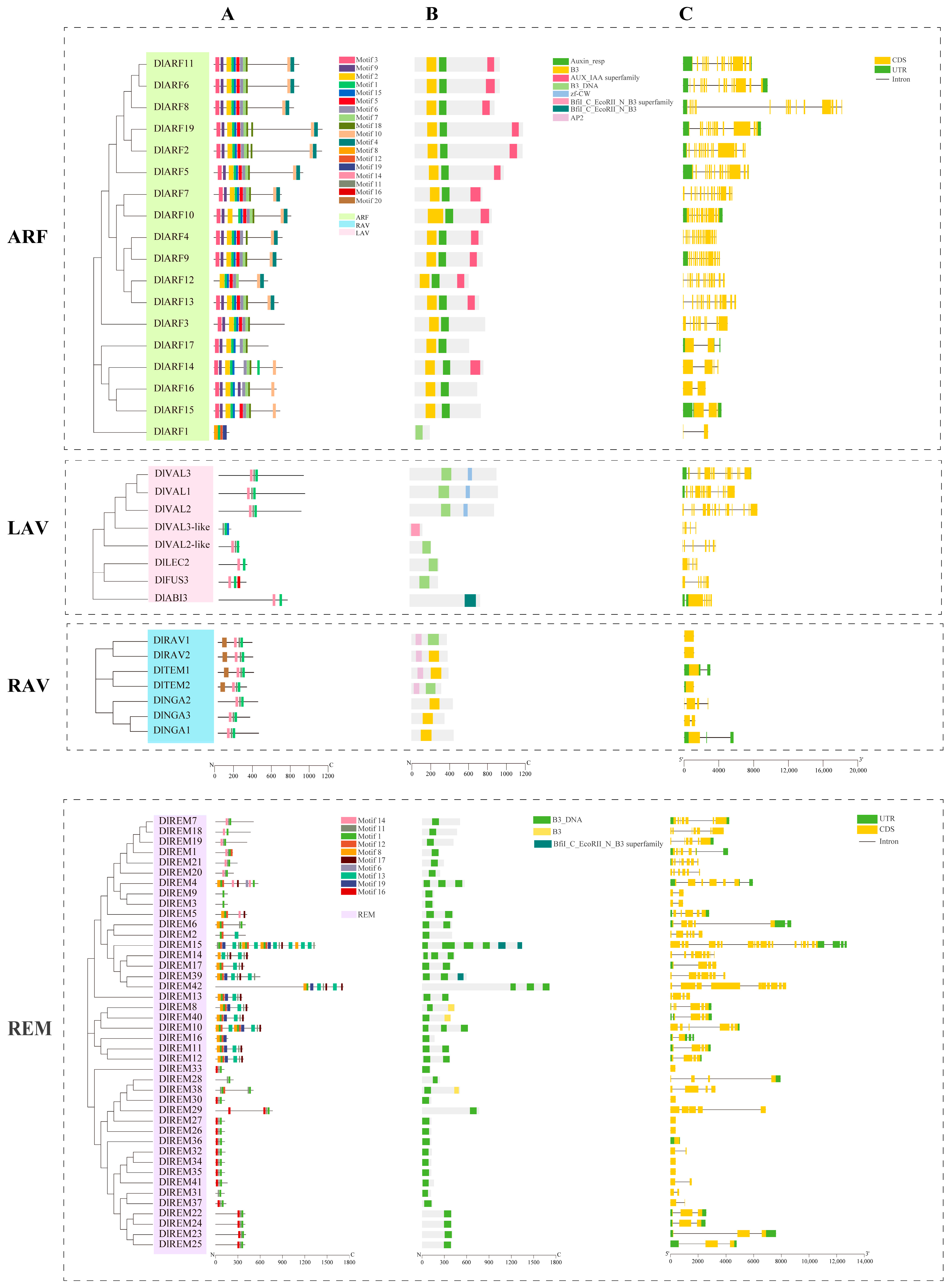
To identify the conserved structure of longan B3 protein, 20 motifs were predicted using MEME software (https://meme-suite.org/meme/tools/meme (accessed on 5 September 2023)). The structural domains and gene structure of the DlB3 family were examined and visualized using the TBtools software. Motif 1 was detected in all DlB3 proteins. In the ARF subfamily, most members contained motif 2, motif 7, motif 8, motif 10, and motif 17. Only DlARF2 and DlARF4 had motif 1. In the LAV subfamily, except for DlVAL3-like, all included motif 14 and all contained motif 6 except DlLEC2, DlFUS3, and DlABI3. All members of the RAV subfamily contained motif 1, motif 6, and motif 14. The motif distribution of the REM subfamily was significantly different, and motif 14 was only contained in DlREM1, DlREM4, DlREM5, DlREM7, DlREM18, DlREM19, DlREM20 and DlREM21. In conclusion, they were placed in the same group as they probably have similar functions (Figure 2A).
Domain analysis of the DlB3 protein sequence revealed that all members contained B3 or B3_DNA domains (Figure 2B). In the ARF subfamily, most members contained AUX_IAA and auxin domains; DlAVL1, DlAVL2, and DlAVL3 of the LAV subfamily contained the zf-CW domain. In the RAV subfamily, DlTEM1, DlTEM2, DlRAV1, and DlRAV2 contained the AP2 domain.
To further understand the composition of DlB3s, their gene structures were compared. In total, 49.3% of DlB3s contained UTR, and 50% of the REM subfamily contained UTR; a few members of the REM and RAV subfamilies did not contain introns (Figure 2C).

2.4. Analysis Related to Cis-Acting Elements and Transcription Start Site in the DlB3 Promoters

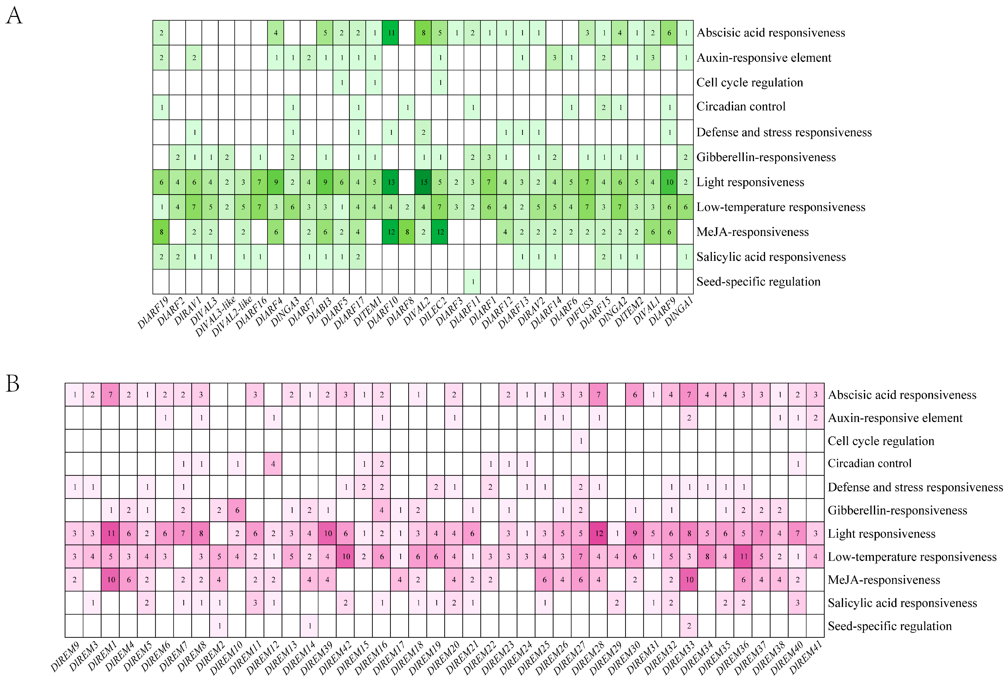
To understand the potential function of the DlB3 genes in various reactions, the 2 kb promoter region upstream of ATG was selected for cis-acting element analysis. These results suggest that DlB3s were associated with numerous phytohormone-related elements, including abscisic acid (ABA) response, gibberellin response, salicylic acid (SA) response, auxin response, and MeJA response (Figure 3A,B). With the exception of DlREM7, all DlB3s contained low-temperature-responsive elements, and 28 DlB3s contained defense- and stress-responsive elements. Additionally, nineteen DlB3s included circadian control cis-acting elements and cell cycle regulation cis-acting elements, and seed-specific regulation cis-acting elements were only present in the promoter of four DlB3 genes. In summary, DlB3s may affect hormone regulation and various stress responses.
To further analyze the function of the 5′ terminal regulatory sequence of DlB3s, the intron and transcription start site of the 2000 bp sequence upstream of the DlB3 initiation codon ATG were predicted. The results of prediction analysis showed that only 5 of the 75 members had introns in the 5′ untranslated region. DlARF1, DlARF6, DlVAL2, DlVAL2-like, DlREM1, DlREM3, DlREM9, DlREM16, DlREM21, DlREM26, and DlREM28 only contained one transcription start site, while the others all contained multiple transcription start sites. The number of transcription start sites of different members varied greatly. In general, there were four types of transcription start sites, A, T, C, and G, with A being the most common, followed by T and G (Supplementary Table S2).

2.5. Analysis of DlB3 Protein Interactions

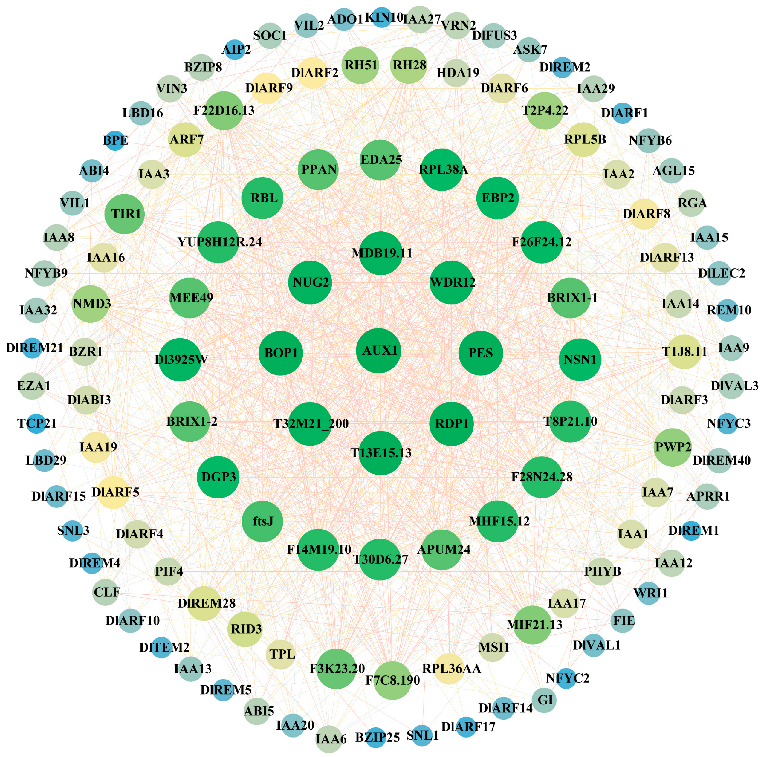
To further explore the interaction relationships within the DlB3 family and among other family members, the PPIs were analyzed using STRING online software (https://cn.string-db.org/cgi/input?sessionId=btJ9BcX3f0Pk&input_page_show_search=on (accessed on 6 December 2023)). The results showed that DlB3s not only had strong interactions between family members but also interacted with other proteins. AUX1 interacted with most proteins (DlARF2, DlARF3, DlARF4, DlARF5, DlARF6, DlARF8, DlARF9, DlARF10, DlARF13, DlARF14, DlARF15, DlARF17, DlABI3). DlABI3 had the most interactions with eight proteins, including ABI4, ABI5, AGL15, AIP2, APRR1, AUX1, BZIP25, and BZIP8, followed by DlREM28, which interacted with six proteins. DlFUS3 interacted with five proteins (ABI4, ABI5, AGL15, BZIP8, CLF); DlLEC2 interacted with four proteins (ABI4, ABI5, AGL15, CLF). Therefore, it was speculated that DlB3s were functionally diverse, and the division of labor among subfamily members was different (Figure 4).

2.6. RNA-Seq Revealed the Expression Profiles of Longan DlB3 Genes in Different Tissues and Treatments

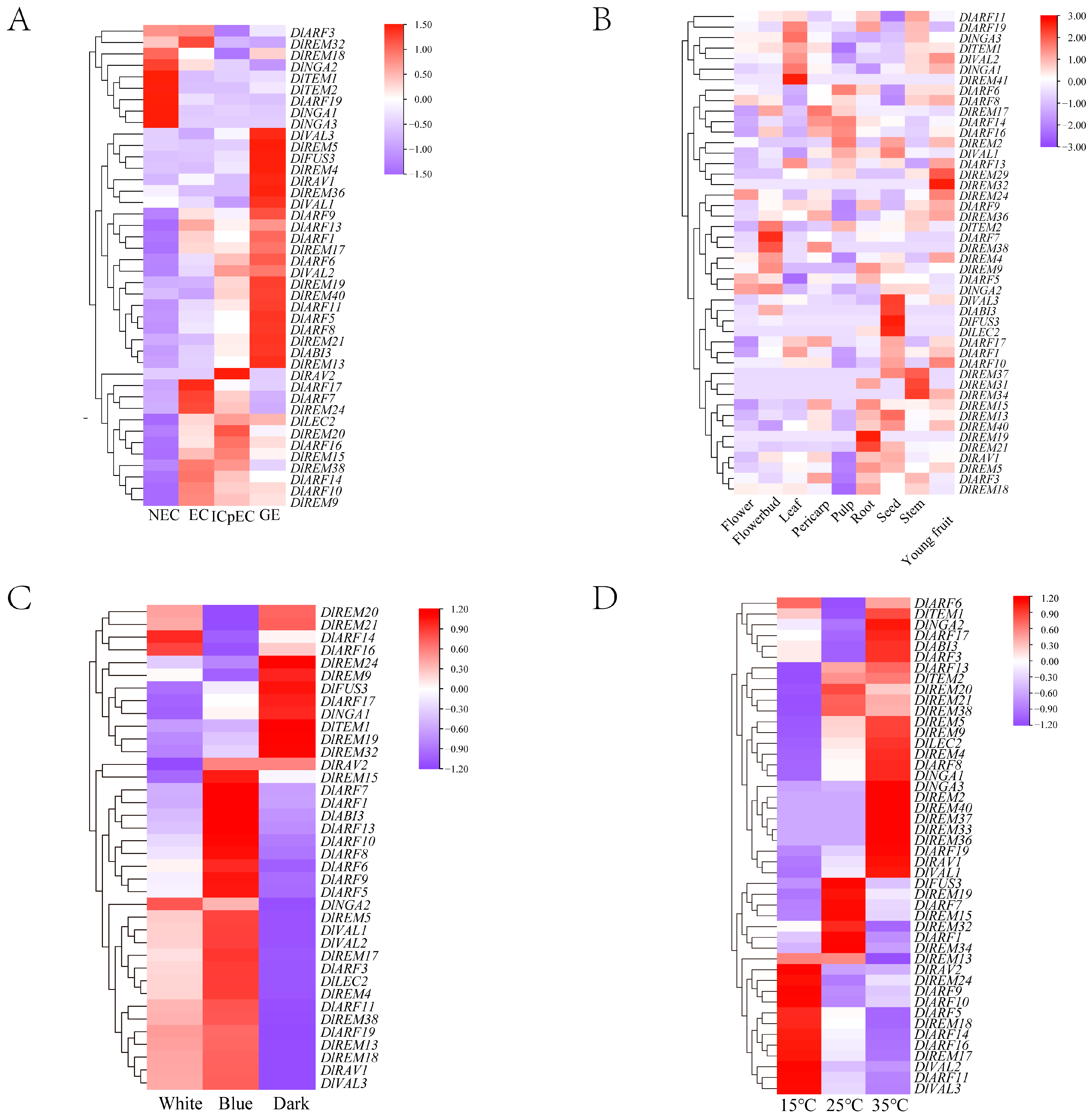
To further investigate the expression patterns of the B3 superfamily in longan, the FPKM values of DlB3s were extracted from the longan transcriptome database. Of the 75 DlB3 genes, 31 DlB3 genes were not detected (Figure 5A). The remaining DlB3s displayed three expression patterns, with high expression at the NEC, GE, and EC to ICpEC stages. The expressions of DlREM18, DlNGA1, DlNGA2, DlARF12, and DlTEM1 were high at the NEC stage, and they were less or even not expressed at the EC to GE stage. Twenty-two DlB3s had specific expressions at the GE stage, DlFUS3 was not expressed at the NEC stage, and DlFUS3, DlVAL3, DlARF5, and DlREM40 were expressed at a low level at the EC stage but increased gradually from the EC stage to GE stage, suggesting that these genes may significantly contribute to the early SE process. DlLEC2 was not expressed at the NEC stage but remained stable from the EC stage to the GE stage. DlARF7, DlARF10, DlARF14, and DlREM9 expression levels gradually decreased from the EC stage to the GE stage. It was demonstrated that these genes may promote the early SE process and maintain the embryonic state of EC.
Based on the transcriptome of longan in different tissues, RNA-seq data on the DlB3 family were analyzed. A total of 47 DlB3s were detected (Figure 5B). This study revealed that 47 genes displayed tissue-specific expression in all tissues. The results showed that DlARF5 and DlNGA2 were specifically expressed in flower buds and flowers, but they were rarely or not detected in other tissues, revealing that they were mainly involved in floral organ development and flowering induction. Notably, DlLEC2 and DlFUS3 were highly expressed in the seed and might be involved in longan seed dormancy and germination. DlREM19 and DlREM21 were highly expressed in the root; DlARF8, DlARF9, DlARF13, DlREM36, and DlNGA1 were highly expressed in the young fruit. These results suggested that DlB3s may be widely involved in the growth and development of longan, and the functions of multiple members were redundant.
There are two factors affecting longan SE, including light and temperature. According to the above, a series of core promoter elements were identified in the promoter sequences of DlB3s, which were involved in light and stress responsiveness. Hence, RNA-seq data were used to analyze the expression pattern of DlB3s under different treatments (Figure 5C). Most DlB3s responded to different light treatments, with higher expression under blue light. DlARF14, DlARF16, and DlNGA2 were specifically expressed under white light treatment. DlREM9/19/24/32, DlFUS3, DlARF17, DlNGA1, and DlTEM1 were highly expressed under dark conditions. It can be seen that the DlB3 family also plays an important role in light response.
The expression patterns of DlB3s were further studied under different temperature conditions by analyzing the RNA-seq data for the longan EC; a total of 49 DlB3s were detected (Figure 5D). These results indicated the high expression levels of 26 DlB3s at 35 °C, and 13 DlB3s responded to low temperature (15 °C). In contrast, DlFUS3, DlARF1, DlARF7, and DlREM15/19/32/34 were inhibited under high- or low-temperature treatment. In summary, the majority of the DlB3 family may play a role in the self-repair process under temperature stress.

2.7. Expression Analysis of the DlB3 Family during Early SE

The analysis of the FPKM found that DlARF5, DlARF16, DlTEM1, DlVAL2, DlLEC2, DlFUS3, DlREM9, and DlREM40 were significantly differentially expressed at the early stage. Therefore, the expression patterns of these genes were analyzed using qRT-PCR during longan early SE. The results showed that the qRT-PCR expression trends of DlARF5, DlTEM1, DlVAL2, DlFUS3, DlREM9, and DlREM40 were similar to the RNA-seq (Figure 6B). The above results indicated that the expression of DlB3s had a particular spatial and temporal expression specificity, and the specific function needed to be further verified. All genes except DlREM9 were highly expressed at the GE stage; it is concluded that they played a role in the GE stage; the transcription levels of DlVAL2 and DlREM40 were down-regulated from the EC stage to the ICpEC stage. DlREM9 was explicitly expressed at the EC stage and up-regulated from the ICpEC stage to the GE stage; this study revealed that DlREM9 significantly contributed to the induction and maintenance of longan EC. The expression of DlFUS3 increased gradually from the EC stage to the GE stage, indicating that it had a positive regulatory effect in the SE of longan. DlLEC2 was expressed from the EC to the GE stage and reached its peak value at the GE stage. These results suggested that the DlB3 family was involved in longan early SE and played different roles.

2.8. Analysis of the Expression Patterns of the DlB3 Family at Different Development Stages of Zygotic Embryos

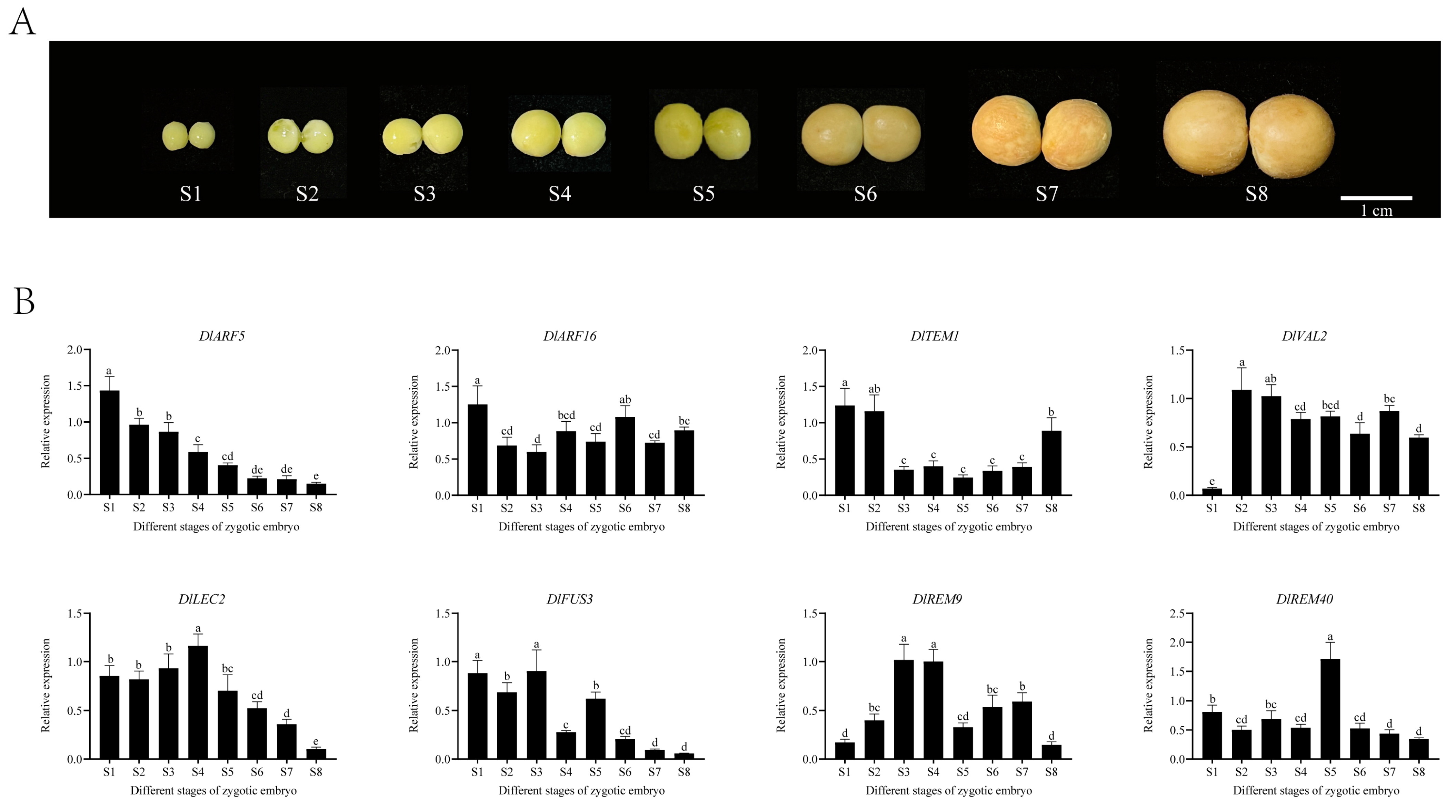
The qRT-PCR experiments showed that the expression levels of DlB3s were divided into different patterns (Figure 7). DlARF5 showed a gradual downward trend from S1 to S8; the expression level of DlLEC2 was high throughout the whole stage of zygotic embryo development, which initially increased and then decreased, and the expression level was highest at the S4 stage and lowest at the S8 stage. The trend of DlFUS3 and DlARF16 fluctuated significantly, and DlFUS3 showed a descending trend from the S5 to the S8 stage. DlTEM1 and DlREM9 exhibited opposite expression patterns, with DlTEM1 decreasing from the S2 stage and increasing at the S8 stage, while DlREM9 showed the opposite. DlVAL2 maintained high expression levels, with the lowest expression at the S1 stage, while DlREM40 maintained low expression levels and the highest expression at the S5 stage. Thus, DlB3s may serve a function in the development of longan zygotic embryos.

2.9. Analysis of the Expression Patterns of the DlB3 Family under Different Exogenous Hormone Treatments

This study demonstrated that exogenous 2,4-D (2,4-Dichlorophenoxyacetic acid) treatment significantly inhibited the expression of DlB3s (Figure 8A). The qRT-PCR analysis of the DlB3 family revealed that DlARF5, DlFUS3, and DlREM9 were significantly higher than the control group under different concentrations of exogenous IAA treatment, DlREM9 levels were significantly higher than those of the control, and the concentrations with the strongest promotion were 3 mg/L, 1.5 mg/L, and 0.5 mg/L (Figure 8B). However, under exogenous IAA treatment, the transcriptional levels of DlARF6, DlTEM1, DlVAL2, DlLEC2, and DlREM40 were inhibited to different degrees. Under the treatment with auxin inhibitor NPA, the different concentrations of NPA treatment significantly reduced DlB3 expression compared to the control group, indicating that exogenous NPA could significantly inhibit the expression of DlB3s (Figure 8C). Notably, the transcription levels of DlARF5, DlFUS3, and DlREM9 showed a reverse trend compared with those of IAA treatment. However, the expression levels of DlARF16, DlTEM1, DlVAL2, DlLEC2, and DlREM40 were consistent with a similar trend under IAA treatment, and their specific mechanisms still need to be further studied.
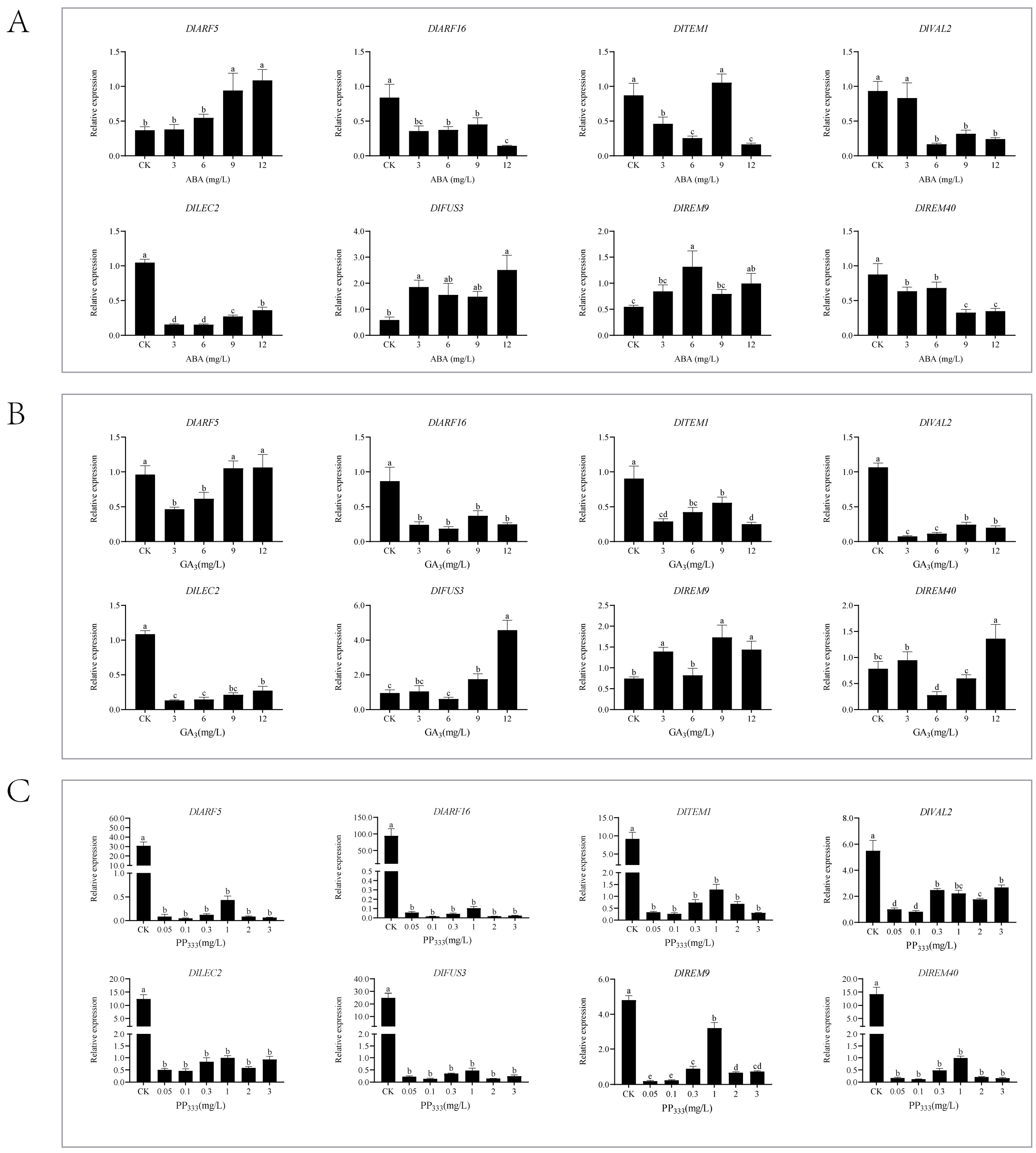
The treatment with 3, 6, 9, and 12 mg/L of ABA could promote the expression of DlARF5, DlFUS3, and DlREM9, demonstrating that exogenous ABA could promote the transcription (Figure 9A), whereas the expressions of DlARF16, DlLEC2, and DlREM40 were suppressed under exogenous ABA treatment. Under 9 mg/L ABA treatment, the expression of DlTEM1 was about 1.21-fold higher than that of the control, and DlVAL2 was not significantly different between the 3 mg/L ABA treatment and the control.
The qRT-PCR result showed that under different concentrations of exogenous GA3 treatment, DlARF16, DlTEM1, DlVAL2, and DlLEC2 were significantly lower than the control, suggesting that exogenous GA3 could inhibit their transcription levels (Figure 9B). Then, the transcription levels of DlFUS3 and DlREM9 were promoted to different degrees under GA3 treatment, and DlFUS3 had the most obvious promotion effect under 12 mg/L GA3 treatment. The transcription levels of DlARF5 and DlREM40 were promoted only at partial concentrations. The qRT-PCR results showed that DlB3 family expression was significantly lower under PP333 treatment concentrations, indicating that exogenous PP333 could significantly inhibit DlB3 expression (Figure 9C). The expressions of DlFUS3 and DlREM9 showed a significant difference under PP333 treatment compared to exogenous GA3 treatment. To sum up, DlB3s may play a primary role in the regulation of the hormone signal transduction pathway in longan SE.

2.10. Subcellular Localization of DlLEC2 and DlFUS3

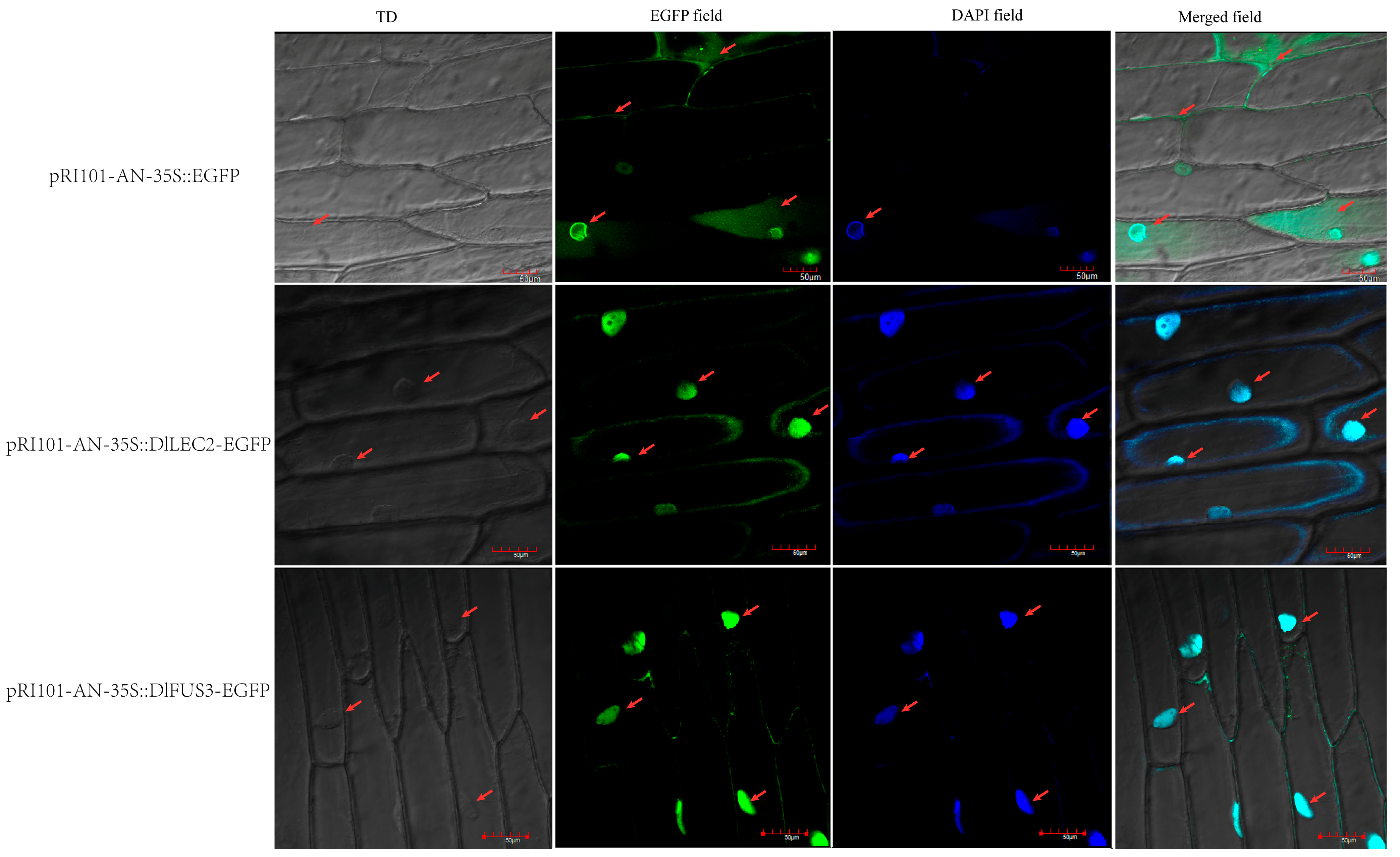
LEC2 and FUS3 are involved in plant embryogenesis. In order to further verify their potential functions in longan, their subcellular localization was verified. Based on the WoLF PSORT software (https://wolfpsort.hgc.jp/ (accessed on 3 October 2023)), DlLEC2 and DlFUS3 were predicted as nucleus localization proteins. Hence, the full-length coding sequences of DlLEC2 and DlFUS3 without a terminator codon (TGA/TAA) were fused with the EGFP (Enhanced Green Fluorescent Protein) and placed under the control of the 35S cauliflower mosaic virus (CaMV) promoter. The expression of pRI101-AN-35S::DlLEC2-EGFP and pRI101-AN-35S::DlFUS3-EGFP were observed with laser confocal microscopy using Agrobacterium infestation injected into onion epidermal cells, and 4’,6-diamidino-2-phenylindol (DAPI) was used for marking the nuclear localization. The results showed that onion epidermal cells transfected with the fusion expression vector containing the target fragments DlLEC2 and DlFUS3 showed green fluorescence mainly in the nucleus. In contrast, the fluorescence signal of the control group was distributed throughout the whole cell. After DAPI staining, pRI101-AN-35S::DlLEC2-EGFP, pRI101-AN-35S::DlFUS3-EGFP, and the positive control all showed fluorescence signals in the nucleus (Figure 10). Based on these results, DlLEC2 and DlFUS3 are nuclear transcription factors that play a role in regulating longan SE.

3. Discussion

3.1. DlB3 Family May Be Evolutionarily Conservative and Functionally Diverse

The B3 family has multiple functions in plant growth and development and the abiotic stress response and participates in the hormone signaling pathways [1,47]. In this study, 75 DlB3s were identified; the lengths of the DlB3s ranged from 116 aa to 1719 aa. Tong et al. [48] found that the number of amino acids of 88 maize B3 proteins was between 105 and 1152 aa. In soybean, the number of amino acids of 145 B3 proteins was 72-1136 aa [14], and the number of amino acids of 97 B3 members of tomato was between 92 and 1317 aa [10]. Therefore, the number of amino acids represented in B3 proteins is relatively conserved in plants. In this study, motif 1 was detected in all B3 proteins, and most of the members contained motif 1 in maize. In Gossypium hirsutum [49], the distribution of REM family motifs was also specific, suggesting that conserved motifs play a specific role in the evolution of longan. In the process of plant evolution, it is speculated that genes without introns may be more conservative [50,51]. In this study, the RAV and REM subfamilies have genes without introns, and the genes of the same subfamily have a similar motif composition and intron number. Therefore, it is speculated that their evolutionary origin and molecular function are similar. Promoter cis-acting elements play an important role in the regulation of gene expression [52], and the prediction of promoter cis-acting elements lays a foundation for analyzing the function of DlB3 genes. It was found that the promoter sequence of DlB3s contains many core elements, including stress response elements, hormone response elements, and light response elements. It is speculated that the DlB3 family may play a role in the growth and stress response of longan through different cis-acting elements.

3.2. DlB3s May Be Involved in Longan Embryogenesis and Organ Morphogenesis

The overexpression of AtLEC2 induced callus and somatic embryo formation in Arabidopsis [53], and FUS3 also played key roles in controlling embryo development [54]. In this study, some longan B3 genes were highly expressed from the EC stage to the GE stage and differentially expressed during the development of zygotic embryos. DlFUS3 gradually increased from the EC to the GE stage, while DlLEC2 was expressed from the EC to the GE stage, and both reached the peak in the GE stage, indicating that DlLEC2 and DlFUS3 might be involved in the process of longan SE. qRT-PCR is measured in the local region of the gene, and RNA-seq is measured in the full-length region of the gene [55,56,57]. The results of qRT-PCR in the early stage of longan SE were different from those of RNA-seq, presumably due to the fact that the samples were not from the same batch and the different detection regions. DlARF5, DlLEC2, and DlFUS3 maintained low expression levels from the S5 stage to the S8 stage. However, DlARF16 had high transcription levels from the S1 to the S8 stage. In summary, DlB3s played a role in the early embryo. AtARF5 was involved in the formation of shoots from Arabidopsis calli [58], CsARF19 was involved in the formation of the nucellar callus of citrus polyembryonic varieties, and CsREM9 might be functional in early embryogenesis [11]. LEC2 and FUS3 recognize the CME at the FLC site and interact with each other in the vernalization process [59], and ARF6 and ARF8 were also involved in the flowering process [60]. In Solanum lycopersicum, SlARF12 was highly expressed in the early stage of fruit development, indicating that it played a key role in fruit development and maturation [61]. RNA-seq indicated that 47 DlB3s showed tissue-specific expression at different levels in all tissues. DlTEM2, DlARF5, DlARF7, DlREM38, DlREM9, and DlNGA2 were highly expressed in the flower bud stage, which suggested that these genes were involved in the flowering process of longan. Meanwhile, DlREM24/29/32 were highly expressed in young fruit, and DlLEC2 and DlFUS3 were highly expressed in the seed. In summary, DlB3s are involved in the growth and development of longan plants.

3.3. DlB3s Were Involved in Longan SE through Hormones and Stress Response

Hormones play an essential role in regulating plant growth and development; auxin plays a vital role in the regulation of SE [62]. ABA can promote plant embryogenesis, seed dormancy, and fruit ripening, and the addition of an appropriate amount of ABA can promote the development of somatic embryos [63]. By mediating auxin biosynthesis and polar transport, ABA plays a role in the initiation of somatic embryogenesis by establishing auxin response patterns in the callus [64]. Abiotic stress has adverse effects on plant growth and development. Previous studies have shown that ABA, GA3, and auxin play a key role in plants under abiotic stress [65]. GA3 regulates the expression of transcription factors related to SE and participates in the process of SE [66]. It was found that LEC2 reduced ABA levels and promoted somatic embryogenesis by controlling ABA8′-hydroxylase (CYP707A1/2/3) that catabolizes ABA [67,68]. In our study, exogenous GA3 could significantly inhibit the expression of most members of the DlB3 family but up-regulate DlFUS3 and DlREM9 expression. Different concentrations of exogenous ABA promoted the transcription levels of DlARF5, DlFUS3, and DlREM9 but inhibited the transcription of other DlB3s. 2,4-D plays a vital role in the regulation of somatic embryogenesis [69] In carrots, embryogenic cells differentiate into somatic embryos in hormone-free medium, while in medium containing 2,4-D, there was no differentiation. Therefore, the presence of 2,4-D inhibits the maturation of embryonic cells [70]. The effect of 2,4-D is phasic, promoting the induction of somatic embryos and inhibiting embryonic development [71]. In rice, OsRAV9, OsRAV14, and OsRAV15 were down-regulated by exogenous IAA [72]. Studies have revealed that the expression of LEC2 is closely related to the application of exogenous IAA. In explants cultured without IAA, the overexpression of the LEC2 gene increased the content of endogenous IAA and promoted somatic embryogenesis, which proved that the LEC2 gene might affect embryogenesis through the level of endogenous auxin [73,74]. In this study, supplementary exogenous 2,4-D and NPA significantly inhibited the expression of DlB3s in longan EC, and IAA induced the expression of DlARF5, DlFUS3, and DlREM9, while IAA inhibited other DlB3s; it could be seen that DlB3s were involved in the auxin signal transduction pathway.
Appropriate stress can promote plant somatic embryogenesis [75,76]. It was found that sustained high temperature (34–38 °C) would lead to embryo development stagnation and small fruit formation [77], and the longan EC could not develop generally under 40 °C treatment [78]. The RAV family responded to various stresses [11,79]. Under different temperature treatments, the expression of the DlB3 family suggested that most DlB3s were promoted at high temperatures (35 °C). The expression of DlLEC2 was promoted under 35 °C treatment, while the expression of DlFUS3 was inhibited under temperature stress. In addition, the transcription levels of 14 DlB3s were promoted under low-temperature treatment. Above all, consistent with the study of Ren et al. [14], DlB3 genes may be involved in the response to abiotic stress and hormones during longan SE.

4. Materials and Methods

4.1. Plant Materials and Treatments

The ‘Hong He Zi’ (‘HHZ’) longan ECs, which involved the embryogenic callus (EC), incomplete compact pro-embryogenic cultures (ICpECs), and globular embryo (GE) were obtained as previously described by Lai et al. [41]. For exogenous hormone treatment, 2 g 18-day longan ECs were transferred to MS (Coolaber, Beijing, China) liquid medium (2% sucrose (Sinopharm, Beijing, China)) with 2,4-D (1.0 mg/L) (Yeasen, Shanghai, China) at 25 °C with 110 r·min−1 shaking in the dark for five days [80]. And then, the pre-cultured longan ECs were transferred to MS liquid basal medium (2% sucrose), supplemented with GA3 (3, 6, 9, and 12 mg/L) (Macklin, Shanghai, China), ABA (3, 6, 9, and 12 mg/L) (Yeasen, Shanghai, China), 2,4-D (0.5, 1.0, 1.5, and 2.0 mg/L), IAA (0.5, 1.0, 1.5, and 2.0 mg/L) (Coolaber, Beijing, China), N-1-Naphthylphthalamic acid (NPA: 5, 10, 20,30, 40, and 50 mg/L) (Coolaber, Beijing, China), and Paclobutrazol (PP333: 0.05, 0.1, 0.3, 1, 2, and 3 mg/L) (Solarbio, Beijing, China) with agitation at 120 rpm at 25 °C under dark conditions for 24 h, with three replicates [66]. Collection began in June when young fruits emerged from their cotyledon; the zygotic embryos of different developmental stages were collected from the cotyledon embryo stage of young fruit, and the zygotic embryos were collected every five days, which were labeled as S1, S2, S3, S4, S5, S6, S7, and S8 in turn [66,80]. All test materials were immediately frozen in liquid nitrogen and stored at −80 °C for subsequent tests.

4.2. Identification of the B3 Superfamily in Longan

The identification of the B3 superfamily was based on the second-generation sequence of longan from the NCBI Sequence Read Archive (SRA) database (SRR17675476) [43] and the third-generation sequence of longan from the NCBI database (PRJNA792504) [44]. The protein sequence of Arabidopsis thaliana was downloaded from https://www.arabidopsis.org/ (accessed on 4 September 2023) (Supplementary Text S1). The latest Hidden Markov Model (HMM) for the B3 superfamily (PF02363) (http://pfam.xfam.org/ (accessed on 15 July 2023)) was used, the potential members were obtained from the longan genome database with HMMER3.0, and the E-value was thresholded at 1 × 10⁻⁵. Then, all potential sequences were aligned using the DNAMAN software (V6 6.0.3.99) to eliminate repetitive proteins, combined with the results of NCBI analysis of the protein domain of the candidate sequence, and the members without the domain were removed. The molecular weight (MW), amino acid number (aa), isoelectric point (pI), average hydrophilicity (GRAVY), and instability coefficient were predicted using Expasy Protparam (https://web.expasy.org/protparam/ (accessed on 4 May 2022)). The subcellular localization of DlB3s was predicted with WOLF PSORT (https://wolfpsort.hgc.jp/ (accessed on 3 October 2023)), and members of the DlB3 family were named with reference to Arabidopsis.

4.3. Phylogenetic Evolution and Synteny Analysis of DlB3s

The protein sequences of 75 DlB3s and 50 AtB3s were aligned using the ClustalW by MEGA5.05. Then, the Maximum Likelihood (ML) method was used to construct the phylogenetic tree. The remaining parameters were designed: Bootstrap = 1000, Poisson model, and partial deletion (95%). The phylogenetic tree was perfected using the online software Chiplot (https://www.chiplot.online/ (accessed on 4 September 2023)). The diagrams of syntenic analysis were plotted using TBtools (V2.031, South China Agricultural University, Guangzhou, China) [81].

4.4. Analysis of Conserved Motifs, Structural Domains, and Gene Structure of DlB3s

The conserved motifs were predicted using the online software MEME (https://meme-suite.org/meme/tools/meme (accessed on 5 September 2023)) with the number of motifs set to 20, and the protein domain was predicted using NCBI (https://www.ncbi.nlm.nih.gov/Structure/bwrpsb/bwrpsb.cgi (accessed on 5 September 2023)). TBtools software was used to annotate the gene structure and visualize motifs and the conserved domains.

4.5. Analysis of Cis-Acting Elements and Transcription Start Site of DlB3s

A 2kb sequence upstream of the transcription start site of genes in the DlB3 gene family was extracted from the longan genome file, and PlantCARE (http://bioinformatics.psb.ugent.be/webtools/plantcare/html (accessed on 5 September 2023)) was used to predict the cis-acting elements. Finally, TBtools was used for visualization. Then, the BDGP (https://fruitfly.org/seq_tools/promoter.html (accessed on 6 December 2023)) was used to predict the transcription start site, and the minimum promoter score was set to 0.8.

4.6. Protein Interaction Analysis of DlB3s

The protein sequences of DlB3s were selected, and the protein–protein interactions (PPIs) of DlB3s were analyzed with STRING (https://cn.string-db.org/cgi/input?sessionId=btJ9BcX3f0Pk&input_page_show_search=on (accessed on 6 December 2023)). Arabidopsis Thaliana was used as the model plant, and the required score was set at 0.700 to analyze the protein interactions of DlB3 members.

4.7. Expression Analysis of the DlB3 Family at the Early Stage of SE, Different Tissues, Different Light Quality, and Different Temperatures

Excel software (12.1.0.16120) was used to extract the FPKM values of DlB3 family members from the transcriptomes of early SE (NEC, EC, ICpEC, and GE) (NCBI BioProject number: PRJNA891444) [43], different tissues (NCBI BioProject number: PRJNA326792) (young fruit, seed, flower, flower bud, leaf, pulp, root, and stem), different light qualities [82] (blue, white, dark as the control), and different temperatures (15 °C, 25 °C, and 35 °C) (NCBI BioProject number: PRJNA889670), normalized by log2FPKM. TBtools software was used to visualize and analyze the expression level of each member.

4.8. Subcellular Localization Analysis

The CDS sequences of DlLEC2 and DlFUS3 were selected to design subcellular localization primers by DNAMAN6 (Supplementary Table S1). The target gene sequence was amplified with PCR, the full-length coding sequences of DlFUS3 and DlLEC2 were inserted into pRI101-AN vector, and then the recombinant plasmid was transferred into Agrobacterium. pRI101-AN-35S::DlLEC2-EGFP and pRI101-AN-35S::DlFUS3-EGFP were transiently expressed in Allium cepa, whose epidermal cells were infiltrated by Agrobacterium. The onions were kept in a dark environment at 26 °C for three days; 4′,6-diamidino-2-phenylindol (DAPI) was used as a nuclear localization marker. The fluorescence signals of DlLEC2 and DlFUS3 proteins in cells were observed using an Olympus FV1200 confocal laser microscope (Tokyo, Japan), GFP wavelength 475 nm, and DAPI wavelength 450 nm.

4.9. RNA Extraction and qRT-PCR Analysis

Total RNA was extracted using a TransZolUp kit (TransGen Biotech, Beijing, China) for the material of the longan early SE (EC, ICpEC, GE), and the total RNA of different development stages of the zygotic embryo was extracted using a BioTeke kit (Cat#RP3301). The cDNA was carried out according to the instruction manual of Hifair®III 1st Strand cDNA Synthesis SuperMix for qPCR (gDNA digester plus) (Yeasen, Shanghai, China). Primer3 software (https://primer3.ut.ee/ (accessed on 9 September 2023))was used to design primers, and DlACTB, DlEF-la, and DlUBQ were used as internal control genes (Supplementary Table S1). The 20 μL reaction system contained the following: HRbioTM qPCR SYBR®Green Master Mix (No Rox) (Heruibio, Guangzhou, China), ddH2O 8.2 μL, 1 µL of 10-fold diluted cDNA, and 0.4 μL specific primer pairs. The operating parameters of the qRT-PCR were as follows: 95 °C for 30 s, followed by 40 cycles of 95 °C for 10 s, and 58 °C for 30 s. The relative expressions of DlB3s were calculated using the 2−ΔCT method [83,84], and the data were imported into SPSS software (R26.0.0.0) to analyze significant differences; different letters representing significant differences were assessed with one-way ANOVA and Duncan test (p < 0.05); Graphpad 8.0.2 was used for the draft.

5. Conclusions

In this study, the genome of the longan B3 superfamily was identified, and its expression in different stages of early somatic embryogenesis and exogenous hormones was analyzed. A total of 75 DlB3 genes were identified in longan, and their bioinformatics and FPKM values in different transcriptomes were comprehensively analyzed, which revealed their specific expression profiles and potential biological functions during longan early SE. Subcellular localization indicated that DlLEC2 and DlFUS3 were located in the nucleus, suggesting that they played a role in the nucleus. Exogenous treatments with 2,4-D, NPA, and PP333 could significantly inhibit the expression of the DlB3 family. Supplementary ABA, IAA, and GA3 suppressed the expressions of DlLEC2, DlARF16, DlTEM1, DlVAL2, and DlREM40, but DlFUS3, DlARF5, and DlREM9 showed an opposite trend. The results showed that DlB3 genes might be involved in the somatic embryo development and hormone response of longan, which could provide reference for the subsequent functional verification of the B3 superfamily and the study of the hormone response mechanism.

Supplementary Materials

The following supporting information can be downloaded at https://www.mdpi.com/article/10.3390/ijms25010127/s1.

Author Contributions

M.T., G.Z. and X.G. designed and performed the experiments and statistical analyses, produced most of the figures and tables, and wrote this manuscript. M.A., W.M., J.L. and B.Z. conducted some of the experiments and analyzed the data. Y.C., Y.L. and Z.L. revised this manuscript. Y.C. contributed to the creation of the concept and the funding acquisition. All authors have read and agreed to the published version of the manuscript.

Funding

This research was funded by the Technology Innovation Fund of Fujian Agriculture and Forestry University (KFb22022XA), the Natural Science Foundation of Fujian Province (2021J01086), and the Natural Science Foundation of Fujian Province (2020J01543).

Institutional Review Board Statement

Not applicable.

Informed Consent Statement

Not applicable.

Data Availability Statement

The original contributions presented in this study are included in the article material. Further inquiries can be directed to the corresponding authors.

Acknowledgments

Thanks to the Institute of Horticultural Biotechnology of Fujian Agriculture and Forestry University for providing conditions for this study; thanks to the workers who contributed to our research.

Conflicts of Interest

The authors declare no conflicts of interest.

Abbreviations

DlDimocarpus longan Lour.
aaamino acids
NECnon-embryogenic callus
ECembryogenic callus
ICpECincomplete compact pro-embryogenic cultures
GEglobular embryos
SEsomatic embryogenesis
DNAdeoxyribonucleic acid
RNAribonucleic acid
cDNAcomplementary DNA
qPCRquantitative real-time PCR
CDScoding sequence
UTRuntranslated regions
bpbase pairs
μLmicroliter
mLmilliliter
ggram
dday
hhour
minminute
ssecond
rpmrevolutions per minute
FPKMFragments Per Kilo-base of exon per Million fragments mapped
2,4-D2,4-Dichlorophenoxyacetic acid
IAAIndole-3-acetic acid
ABAabscisic acid
GAgibberellin
SAsalicylic acid
MeJamethyl jasmonate
NPAN-1-naphthylphthalamic acid
PP333paclobutrazol
DAPI4′,6-diamidino-2-phenylindol
EGFPEnhanced Green Fluorescent Protein
RNA-seqRNA sequencing

References

  1. Swaminathan, K.; Peterson, K.; Jack, T. The plant B3 superfamily. Trends Plant Sci. 2008, 13, 647–655. [Google Scholar] [CrossRef] [PubMed]
  2. McCarty, D.R.; Hattori, T.; Carson, C.B.; Vasil, V.; Lazar, M.; Vasil, I.K. The Viviparous-1 developmental gene of maize encodes a novel transcriptional activator. Cell 1991, 66, 895–905. [Google Scholar] [CrossRef] [PubMed]
  3. Suzuki, M.; Kao, C.Y.; McCarty, D.R. The conserved B3 domain of VIVIPAROUS1 has a cooperative DNA binding activity. Plant Cell 1997, 9, 799–807. [Google Scholar] [CrossRef] [PubMed]
  4. Liu, Y.; Dong, Z. The Function and Structure of Plant B3 Domain Transcription Factor. Mol. Plant Breed. 2017, 15, 1868–1873. [Google Scholar] [CrossRef]
  5. Yamasaki, K.; Kigawa, T.; Inoue, M.; Tateno, M.; Yamasaki, T.; Yabuki, T.; Aoki, M.; Seki, E.; Matsuda, T.; Tomo, Y.; et al. Solution structure of the B3 DNA binding domain of the Arabidopsis cold-responsive transcription factor RAV1. Plant Cell 2004, 16, 3448–3459. [Google Scholar] [CrossRef]
  6. Tsukagoshi, H.; Morikami, A.; Nakamura, K. Two B3 domain transcriptional repressors prevent sugar-inducible expression of seed maturation genes in Arabidopsis seedlings. Proc. Nat. Acad. Sci. USA 2007, 104, 2543–2547. [Google Scholar] [CrossRef]
  7. Guangyu, L.; Lingfei, Y.; Xinbo, C. Research progress of Arabidopsis B3 transcription factor gene superfamily. Chem. Life 2013, 33, 287–293. [Google Scholar] [CrossRef]
  8. Wu, J.; Yu, S.; Liu, Z.; Fu, Y.; Zhou, M. Genome Identification and Expression Pattern Analysis of Phyllostachys edulis B3 Family. J. Agric. Biotechnol. 2019, 27, 43–54. [Google Scholar]
  9. Romanel, E.; Schrago, C.G.; Couñago, R.M.; Russo, C.A.M.; Alves-Ferreira, M. Evolution of the B3 DNA Binding Superfamily: New Insights into REM Family Gene Diversification. PLoS ONE 2009, 4, e5791. [Google Scholar] [CrossRef]
  10. Sun, T.; Wang, D.; Gong, D.; Chen, L.; Chen, Y.; Sun, Y. Genome-wide Identification and Bioinformatic Analysis of B3 Superfamily in Tomato. J. Plant Genet. Resour. 2015, 16, 806–814. [Google Scholar] [CrossRef]
  11. Liu, Z.; Ge, X.-X.; Wu, X.-M.; Xu, Q.; Atkinson, R.G.; Guo, W.-W. Genome-wide analysis of the citrus B3 superfamily and their association with somatic embryogenesis. BMC Genom. 2020, 21, 305. [Google Scholar] [CrossRef] [PubMed]
  12. Ruan, C.C.; Chen, Z.; Hu, F.C.; Fan, W.; Wang, X.H.; Guo, L.J.; Fan, H.Y.; Luo, Z.W.; Zhang, Z.L. Genome-wide characterization and expression profiling of B3 superfamily during ethylene-induced flowering in pineapple (Ananas comosus L.). BMC Genom. 2021, 22, 561. [Google Scholar] [CrossRef] [PubMed]
  13. Verma, S.; Bhatia, S. A comprehensive analysis of the B3 superfamily identifies tissue-specific and stress-responsive genes in chickpea (Cicer arietinum L.). 3 Biotech 2019, 9, 346. [Google Scholar] [CrossRef] [PubMed]
  14. Ren, C.; Wang, H.; Zhou, Z.; Jia, J.; Zhang, Q.; Liang, C.; Li, W.; Zhang, Y.; Yu, G. Genome-wide identification of the B3 gene family in soybean and the response to melatonin under cold stress. Front. Plant Sci. 2023, 13, 1091907. [Google Scholar] [CrossRef] [PubMed]
  15. Ellis, C.M.; Nagpal, P.; Young, J.C.; Hagen, G.; Guilfoyle, T.J.; Reed, J.W. AUXIN RESPONSE FACTOR1 and AUXIN RESPONSE Factor2 regulate senescence and floral organ abscission in Arabidopsis thaliana. Development 2005, 132, 4563–4574. [Google Scholar] [CrossRef]
  16. Shin, R.; Burch, A.Y.; Huppert, K.A.; Tiwari, S.B.; Murphy, A.S.; Guilfoyle, T.J.; Schachtman, D.P. The Arabidopsis transcription factor MYB77 modulates auxin signal transduction. Plant Cell 2007, 19, 2440–2453. [Google Scholar] [CrossRef] [PubMed]
  17. Wenzel, C.L.; Marrison, J.; Mattsson, J.; Haseloff, J.; Bougourd, S.M. Ectopic divisions in vascular and ground tissues of Arabidopsis thaliana result in distinct leaf venation defects. J. Exp. Bot. 2012, 63, 5351–5364. [Google Scholar] [CrossRef] [PubMed]
  18. Lianzhe, W.; Lijun, X.; Jiankang, L.; Yuan, W.; Shuai, Z.; Bingbing, L. Cloning and Expression Analysis of B3 Group Transcription Factor REM-1 Gene in Wheat. Mol. Plant Breed. 2019, 17, 4853–4858. [Google Scholar] [CrossRef]
  19. Wu, M.-F.; Tian, Q.; Reed, J.W. Arabidopsis microRNA167 controls patterns of ARF6 and ARF8 expression, and regulates both female and male reproduction. Development 2006, 133, 4211–4218. [Google Scholar] [CrossRef]
  20. Chandan, R.; Kumar, R.; Swain, D.; Ghosh, S.; Bhagat, P.; Patel, S.; Bagler, G.; Sinha, A.; Jha, G. RAV1 family members function as transcriptional regulators and play a positive role in plant disease resistance. Plant J. 2023, 114, 39–54. [Google Scholar] [CrossRef]
  21. Woo, H.; Kim, J.; Kim, J.-Y.; Kim, J.; Lee, U.; Song, I.-J.; Kim, J.-H.; Lee, H.-Y.; Nam, H.; Lim, P. The RAV1 transcription factor positively regulates leaf senescence in Arabidopsis. J. Exp. Bot. 2010, 61, 3947–3957. [Google Scholar] [CrossRef] [PubMed]
  22. Hu, Y.X.; Wang, Y.H.; Liu, X.F.; Li, J.Y. Arabidopsis RAV1 is down-regulated by brassinosteroid and may act as a negative regulator during plant development. Cell Res. 2004, 14, 8–15. [Google Scholar] [CrossRef] [PubMed]
  23. Mylne, J.; Mas, C.; Hill, J. NMR assignment and secondary structure of the C-terminal DNA binding domain of Arabidopsis thaliana VERNALIZATION1. Biomol. NMR Assign. 2011, 6, 5–8. [Google Scholar] [CrossRef]
  24. Levy, Y.Y.; Mesnage, S.; Mylne, J.S.; Gendall, A.R.; Dean, C. Multiple roles of Arabidopsis VRN1 in vernalization and flowering time control. Science 2002, 297, 243–246. [Google Scholar] [CrossRef] [PubMed]
  25. Sugliani, M.; Brambilla, V.; Clerkx, E.J.M.; Koornneef, M.; Soppe, W.J.J. The conserved splicing factor SUA controls alternative splicing of the developmental regulator ABI3 in Arabidopsis. Plant Cell 2010, 22, 1936–1946. [Google Scholar] [CrossRef] [PubMed]
  26. Yang, Z.; Liu, X.; Wang, K.; Li, Z.; Jia, Q.; Zhao, C.; Zhang, M. ABA-INSENSITIVE 3 with or without FUSCA3 highly up-regulates lipid droplet proteins and activates oil accumulation. J. Exp. Bot. 2021, 73, 2077–2092. [Google Scholar] [CrossRef] [PubMed]
  27. Chiu, R.S.; Nahal, H.; Provart, N.J.; Gazzarrini, S. The role of the Arabidopsis FUSCA3transcription factor during inhibition of seed germination at high temperature. BMC Plant Biol. 2012, 12, 15. [Google Scholar] [CrossRef]
  28. Zhou, X.; Weng, Y.; Su, W.; Ye, C.; Qu, H.; Li, Q. Uninterrupted embryonic growth leading to viviparous propagule formation in woody mangrove. Front. Plant Sci. 2023, 13, 1061747. [Google Scholar] [CrossRef]
  29. Liu, X.; Li, N.; Chen, A.; Saleem, N.; Jia, Q.; Zhao, C.; Li, W.; Zhang, M. FUSCA3-induced AINTEGUMENTA-like 6 manages seed dormancy and lipid metabolism. Plant Physiol. 2023, 193, 1091–1108. [Google Scholar] [CrossRef]
  30. Horstman, A.; Bemer, M.; Boutilier, K. A transcriptional view on somatic embryogenesis. Regeneration 2017, 4, 201–216. [Google Scholar] [CrossRef]
  31. Shiyi, W.; Yizi, H.; Zhouyang, L.; Huahong, H.; Erpei, L. Research progress in plant somatic embryogenesis and its molecular regulation mechanism. J. Zhejiang A F Univ. 2022, 39, 223–232. [Google Scholar]
  32. Horstman, A.; Li, M.; Heidmann, I.; Weemen, M.; Chen, B.; Muino, J.M.; Angenent, G.C.; Boutilier, K. The BABY BOOM Transcription Factor Activates the LEC1-ABI3-FUS3-LEC2 Network to Induce Somatic Embryogenesis. Plant Physiol. 2017, 175, 848–857. [Google Scholar] [CrossRef] [PubMed]
  33. Wang, H.; Guo, J.; Lambert, K.N.; Lin, Y. Developmental control of Arabidopsis seed oil biosynthesis. Planta 2007, 226, 773–783. [Google Scholar] [CrossRef] [PubMed]
  34. Suzuki, M.; Wang, H.; McCarty, D.R. Repression of the LEAFY COTYLEDON 1/B3 Regulatory Network in Plant Embryo Development by VP1/ABSCISIC ACID INSENSITIVE 3-LIKE B3 Genes1. Plant Physiol. 2006, 143, 902–911. [Google Scholar] [CrossRef] [PubMed]
  35. Zhao, X.; Liu, T.; Huang, M.J.; Zhang, J.W.; Tuluhong, G.; Zhang, X. Cloning and Activity Assay of Somatic Embryogenesis-specific Gene GbLEC2 Promoter in Gossypium barbadense. J. Agric. Biotechnol. 2023, 31, 695–703. [Google Scholar]
  36. Braybrook, S.A.; Harada, J.J. LECs go crazy in embryo development. Trends Plant Sci. 2008, 13 12, 624–630. [Google Scholar] [CrossRef]
  37. Brand, A.; Quimbaya, M.A.; Tohme, J.; Chavarriaga-Aguirre, P. Arabidopsis LEC1 and LEC2 Orthologous Genes Are Key Regulators of Somatic Embryogenesis in Cassava. Front. Plant Sci. 2019, 10, 673. [Google Scholar] [CrossRef]
  38. Wenyu, L.; Wei, C.; Ruifeng, S.; Feng, Z. Advances in embryo development of longan. Subtrop. Plant Sci. 2004, 33, 65–68. [Google Scholar]
  39. Chen, Y.; Lin, X.; Lai, Z. Advances in Somatic Embryogenesis of Dimocarpus longan Lour. Chin. J. Trop. Crop. 2020, 41, 1990–2002. [Google Scholar]
  40. Radoeva, T.; Vaddepalli, P.; Zhang, Z.; Weijers, D. Evolution, Initiation, and Diversity in Early Plant Embryogenesis. Dev. Cell 2019, 50, 533–543. [Google Scholar] [CrossRef]
  41. Lai, Z.; Chen, Z. Somatic embryogenesis of high frequency from longan embryogenic call. J. Fujian Agric. For. Univ. Nat. Sci. Ed. 1997, 26, 271–276. [Google Scholar]
  42. Lai, Z.; Pan, L.; Chen, Z. Establishment and maintenance of longan embryogenic cell lines. J. Fujian Agric. For. Univ. Nat. Sci. Ed. 1997, 2, 33–40. [Google Scholar]
  43. Lin, Y.; Min, J.; Lai, R.; Wu, Z.; Chen, Y.; Yu, L.; Cheng, C.; Jin, Y.; Tian, Q.; Liu, Q.; et al. Genome-wide sequencing of longan (Dimocarpus longan Lour.) provides insights into molecular basis of its polyphenol-rich characteristics. GigaScience 2017, 6, 1–14. [Google Scholar] [CrossRef] [PubMed]
  44. Chen, Y.; Xie, D.; Ma, X.; Xue, X.; Liu, M.; Xiao, X.; Lai, C.W.J.; Xu, X.; Chen, X.; Chen, Y.; et al. Genome-wide Hi-C analysis reveals hierarchical chromatin interactions during early somatic embryogenesis. Plant Physiol. 2023, 193, 555–577. [Google Scholar] [CrossRef] [PubMed]
  45. Zhang, S.; Zhu, C.; Zhang, X.; Liu, M.; Xue, X.; Lai, C.; Xuhan, X.; Chen, Y.; Zhang, Z.; Lai, Z.; et al. Single-cell RNA sequencing analysis of the embryogenic callus clarifies the spatiotemporal developmental trajectories of the early somatic embryo in Dimocarpus longan. Plant J. 2023, 115, 1277–1297. [Google Scholar] [CrossRef] [PubMed]
  46. Xu, X.; Zhang, C.; Xu, X.; Cai, R.; Guan, Q.; Chen, X.; Chen, Y.; Zhang, Z.; XuHan, X.; Lin, Y.; et al. Riboflavin mediates m6A modification targeted by miR408, promoting early somatic embryogenesis in longan. Plant Physiol. 2023, 192, 1799–1820. [Google Scholar] [CrossRef] [PubMed]
  47. Suzuki, M.; McCarty, D.R. Functional symmetry of the B3 network controlling seed development. Curr. Opin. Plant Biol. 2008, 11, 548–553. [Google Scholar] [CrossRef] [PubMed]
  48. Tong, Z.; Zhang, Y.; Wang, L.; Li, A.; Wang, W.; Wang, P.; Liu, H.; Liu, Q.; Wang, C. Genome wide identification and expression pattern analysis of B3 gene family in maize. Pratacult. Sci. 2023, 40, 2556–2570. [Google Scholar]
  49. Shi, R.; Zhang, D.; Sun, Z.; Liu, Z.; Xie, M.; Zhang, Y.; Ma, Z.; Wang, X. Genome-wide identification and expression analysis of REM gene family in Gossypium hirsutum. Cotton Sci. 2021, 33, 95–111. [Google Scholar]
  50. Jain, M.; Khurana, P.; Tyagi, A.K.; Khurana, J.P. Genome-wide analysis of intronless genes in rice and Arabidopsis. Funct. Int. Genom. 2008, 8, 69–78. [Google Scholar] [CrossRef]
  51. Rogozin, I.B.; Sverdlov, A.V.; Babenko, V.N.; Koonin, E.V. Analysis of evolution of exon-intron structure of eukaryotic genes. Brief. Bioinf. 2005, 6, 118–134. [Google Scholar] [CrossRef]
  52. Hernandez-Garcia, C.M.; Finer, J.J. Identification and validation of promoters and cis-acting regulatory elements. Plant Sci. 2014, 217–218, 109–119. [Google Scholar] [CrossRef] [PubMed]
  53. Stone, S.L.; Kwong, L.W.; Yee, K.M.; Pelletier, J.; Lepiniec, L.; Fischer, R.L.; Goldberg, R.B.; Harada, J.J. LEAFY COTYLEDON2 encodes a B3 domain transcription factor that induces embryo development. Proc. Natl. Acad. Sci. USA 2001, 98, 11806–11811. [Google Scholar] [CrossRef] [PubMed]
  54. Harada, J.J. Role of Arabidopsis LEAFY COTYLEDON genes in seed development. J. Plant Physiol. 2001, 158, 405–409. [Google Scholar] [CrossRef]
  55. Teng, M.; Love, M.I.; Davis, C.A.; Djebali, S.; Dobin, A.; Graveley, B.R.; Li, S.; Mason, C.E.; Olson, S.; Pervouchine, D.; et al. A benchmark for RNA-seq quantification pipelines. Genome Biol. 2016, 17, 74. [Google Scholar] [CrossRef] [PubMed]
  56. Robert, C.; Watson, M. Errors in RNA-Seq quantification affect genes of relevance to human disease. Genome Biol. 2015, 16, 1–16. [Google Scholar] [CrossRef] [PubMed]
  57. Everaert, C.; Luypaert, M.; Maag, J.L.V.; Cheng, Q.X.; Dinger, M.E.; Hellemans, J.; Mestdagh, P. Benchmarking of RNA-sequencing analysis workflows using whole-transcriptome RT-qPCR expression data. Sci. Rep. 2017, 7, 1559. [Google Scholar] [CrossRef] [PubMed]
  58. Ckurshumova, W.; Smirnova, T.; Marcos, D.; Zayed, Y.; Berleth, T. Irrepressible MONOPTEROS/ARF5 promotes de novo shoot formation. New Phytol. 2014, 204, 556–566. [Google Scholar] [CrossRef]
  59. Tao, Z.; Hu, H.; Luo, X.; Jia, B.; Du, J.; He, Y. Embryonic resetting of the parental vernalized state by two B3 domain transcription factors in Arabidopsis. Nat. Plants 2019, 5, 424–435. [Google Scholar] [CrossRef]
  60. Nagpal, P.; Ellis, C.M.; Weber, H.; Ploense, S.E.; Barkawi, L.S.; Guilfoyle, T.J.; Hagen, G.; Alonso, J.M.; Cohen, J.D.; Farmer, E.E.; et al. Auxin response factors ARF6 and ARF8 promote jasmonic acid production and flower maturation. Development 2005, 132, 4107–4118. [Google Scholar] [CrossRef]
  61. Kumar, R.; Tyagi, A.K.; Sharma, A.K. Genome-wide analysis of auxin response factor (ARF) gene family from tomato and analysis of their role in flower and fruit development. Mol. Genet. Genom. 2011, 285, 245–260. [Google Scholar] [CrossRef]
  62. Müller, B.; Sheen, J. Cytokinin and auxin interaction in root stem-cell specification during early embryogenesis. Nature 2008, 453, 1094–1097. [Google Scholar] [CrossRef] [PubMed]
  63. Yan, F. Regulation of Embryogenic Callus Proliferation and Physiological Characteristics of Picea Pungens by 2,4-D and GS. Master’s Thesis, Jilin Agricultural University, Changchun, China, 2022. [Google Scholar]
  64. Su, Y.H.; Su, Y.X.; Liu, Y.; Zhang, X.S. Abscisic acid is required for somatic embryo initiation through mediating spatial auxin response in Arabidopsis. Plant Growth Regul. 2013, 69, 167–176. [Google Scholar] [CrossRef]
  65. Waadt, R.; Seller, C.A.; Hsu, P.-K.; Takahashi, Y.; Munemasa, S.; Schroeder, J.I. Plant hormone regulation of abiotic stress responses. Nat. Rev. Mol. Cell Biol. 2022, 23, 680–694. [Google Scholar] [CrossRef] [PubMed]
  66. Tang, M.; Gao, X.; Meng, W.; Lin, J.; Zhao, G.; Lai, Z.; Lin, Y.; Chen, Y. Transcription factors NF-YB involved in embryogenesis and hormones responses in Dimocarpus Longan Lour. Front. Plant Sci. 2023, 14, 1255436. [Google Scholar] [CrossRef] [PubMed]
  67. Braybrook, S.A.; Stone, S.L.; Park, S.; Bui, A.Q.; Le, B.H.; Fischer, R.L.; Goldberg, R.B.; Harada, J.J. Genes directly regulated by LEAFY COTYLEDON2 provide insight into the control of embryo maturation and somatic embryogenesis. Proc. Nat. Acad. Sci. USA 2006, 103, 3468–3473. [Google Scholar] [CrossRef] [PubMed]
  68. Wójcikowska, B.; Gaj, M.D. LEAFY COTYLEDON2-mediated control of the endogenous hormone content: Implications for the induction of somatic embryogenesis in Arabidopsis. Plant Cell Tissue Organ Cult. (PCTOC) 2015, 121, 255–258. [Google Scholar] [CrossRef]
  69. Fehér, A.; Pasternak, T.; Dudits, D. Transition of somatic plant cells to an embryogenic state. Plant Cell Tissue Organ Cult. 2003, 74, 201–228. [Google Scholar] [CrossRef]
  70. Cui, H.; Ding, Q.X.; Gui, Y.L.; Guo, Z.C. 24-D-Regulated Somatic Embryogenesis of Sweet Potato. Chin. Bull. Bot. 1999, 16, 411–415. [Google Scholar]
  71. Biwen, H.; Shulan, L. In vitro plant somatic embryogenesis. Plant Physiol. J. 1988, 9–15. [Google Scholar] [CrossRef]
  72. Chen, C.; Li, Y.; Zhang, H.; Ma, Q.; Wei, Z.; Chen, J.; Sun, Z. Genome-Wide Analysis of the RAV Transcription Factor Genes in Rice Reveals Their Response Patterns to Hormones and Virus Infection. Viruses 2021, 13, 752. [Google Scholar] [CrossRef]
  73. Zhao, F.; Li, L.; He, X.; Zeng, H. Roles of Key Genes and Relevant Plant Hormones in the Early and Late Stages of Plant Embryogenesis. Biotechnol. Bull. 2017, 33, 30–36. [Google Scholar] [CrossRef]
  74. Ledwoń, A.; Gaj, M.D. LEAFY COTYLEDON2 gene expression and auxin treatment in relation to embryogenic capacity of Arabidopsis somatic cells. Plant Cell Rep. 2009, 28, 1677–1688. [Google Scholar] [CrossRef] [PubMed]
  75. Kamada, H.; Kobayashi, K.; Kiyosue, T.; Harada, H. Stress induced somatic embryogenesis in carrot and its application to synthetic seed production. Vitr. Cell. Dev. Biol. 1989, 25, 1163–1166. [Google Scholar] [CrossRef]
  76. Lee, E.K.; Cho, D.Y.; Soh, W.Y. Enhanced production and germination of somatic embryos by temporary starvation in tissue cultures of Daucus carota. Plant Cell Rep. 2001, 20, 408–415. [Google Scholar] [CrossRef] [PubMed]
  77. Nong, W.; Huang, J.; Li, G.; Li, G. Research on the reason of severely small size fruit phenomenon in longan. J. South. Agric. 2006, 3, 314–316. [Google Scholar]
  78. Wang, Y. Genome-Wide Identification of HSF Family in Longan (Dimocarpus longan lour.) and Expression Analysis in Response to Heat Stress. Master’s Thesis, Fujian Agriculture and Forestry University, Fuzhou, China, 2019. [Google Scholar]
  79. Li, C.-W.; Su, R.-C.; Cheng, C.-P.; Sanjaya; You, S.-J.; Hsieh, T.-H.; Chao, T.-C.; Chan, M.-T. Tomato RAV Transcription Factor Is a Pivotal Modulator Involved in the AP2/EREBP-Mediated Defense Pathway1[W][OA]. Plant Physiol. 2011, 156, 213–227. [Google Scholar] [CrossRef] [PubMed]
  80. Chen, Y.; Xu, X.; Chen, X.; Chen, Y.; Zhang, Z.; Xuhan, X.; Lin, Y.; Lai, Z.-X. Seed-specific gene MOTHER of FT and TFL1 (MFT) involved in embryogenesis, hormones and stress responses in dimocarpus longan lour. Int. J. Mol. Sci. 2018, 19, 2403. [Google Scholar] [CrossRef] [PubMed]
  81. Chen, C.; Chen, H.; Zhang, Y.; Thomas, H.R.; Frank, M.H.; He, Y.; Xia, R. TBtools: An Integrative Toolkit Developed for Interactive Analyses of Big Biological Data. Mol. Plant 2020, 13, 1194–1202. [Google Scholar] [CrossRef]
  82. Li, H.; Lyu, Y.; Chen, X.; Wang, C.; Yao, D.; Ni, S.; Lin, Y.; Chen, Y.; Zhang, Z.; Lai, Z. Exploration of the Effect of Blue Light on Functional Metabolite Accumulation in Longan Embryonic Calli via RNA Sequencing. Int. J. Mol. Sci. 2019, 20, 441. [Google Scholar] [CrossRef]
  83. Vandesompele, J.; De Preter, K.; Pattyn, F.; Poppe, B.; Van Roy, N.; De Paepe, A.; Speleman, F. Accurate normalization of real-time quantitative RT-PCR data by geometric averaging of multiple internal control genes. Genome Biol. 2002, 3, research0034.1. [Google Scholar] [CrossRef]
  84. Livak, K.J.; Schmittgen, T.D. Analysis of Relative Gene Expression Data Using Real-Time Quantitative PCR and the 2−ΔΔCT Method. Methods 2001, 25, 402–408. [Google Scholar] [CrossRef] [PubMed]
Figure 1. The phylogenetic tree of B3 members and collinear analysis of DlB3s. (A) Phylogenetic tree of longan (Dl) and A. thaliana (At) basic B3 proteins; (B) chromosome localization and collinear map of different DlB3 genes in longan (gray line represents collinearity block in longan genome; red line represents linear gene pairs related to DlB3 family genes).
Ijms 25 00127 g001
Figure 2. The motif organization, domain, and exon–intron structure of the DlB3 family. (A) The motif organization of DlB3 proteins. Twenty conserved motifs predicted in B3 proteins are shown as differently colored boxes. (B) Domain of DlB3 proteins; different colors represent different domains. (C) Gene structure of DlB3 genes. Green boxes indicate UTR; yellow boxes indicate exons; black lines indicate introns.
Ijms 25 00127 g002
Figure 3. Distribution of cis-acting elements in promoters of DlB3 longan. (A) Distribution of ARF, LAV, and RAV subfamily cis-acting elements in longan; (B) distribution of the REM subfamily cis-acting elements in longan. The blank spaces indicate no elements, green represents ARF, LAV, and RAV subfamily, purple represents REM subfamily, and color shades represent the number of components, and numbers in the figure indicate the number of elements.
Ijms 25 00127 g003
Figure 4. Protein interactions of DlB3 proteins in longan. Note: the connecting lines represent interactions between proteins; the size of the circle and the different color indicate the number of interacting proteins.
Ijms 25 00127 g004
Figure 5. Expression patterns of DlB3 family members based on FPKM values. (A) FPKM value of DlB3 family during longan SE; (B) FPKM value of DlB3 family in different tissues; (C) FPKM value of DlB3 family under different light quality treatments; (D) FPKM value of DlB3 family under different temperature treatments. The materials of different tissue were ‘SJM’ longan, and other materials were ‘HHZ’ longan. Different colors on the scale bar are log2FPKM, which represent different transcript levels.
Ijms 25 00127 g005
Figure 6. The early stage of longan somatic embryogenesis and eight DlB3s during the early somatic embryogenesis (SE) in longan with qRT-PCR. (A) Longan early stage of somatic embryogenesis; EC: embryogenic callus; ICpEC: incomplete compact pro-embryogenic cultures; GE: globular embryos. (B) The qRT-PCR analysis of DlB3 genes the early stage of SE. Red line graph represents FPKM values; blue columns represent qRT-PCR results. Note: bars = 200 µm; the internal reference genes were DlACTB, DlEF-la, and DlUBQ, with three biological replicates, and significant differences are shown with lowercase letters a, b, and c, p < 0.05.
Ijms 25 00127 g006
Figure 7. Expression patterns of longan DlB3 family at different development stages of zygotic embryos. (A) The different developmental stages of zygotic embryos; (B) DlB3 family relative expression of different stages of zygotic embryos. Note: the internal reference genes were DlACTB, DlEF-la, and DlUBQ, with three biological replicates, and significant differences are shown with lowercase letters a–e, p < 0.05.
Ijms 25 00127 g007
Figure 8. qRT-PCR analysis of the DlB3 family under the 2,4-D, IAA, and NPA treatments. (A) qRT-PCR analysis of DlB3 family under the 2,4-D treatment; (B) qRT-PCR analysis of DlB3 family under the IAA treatment; (C) qRT-PCR analysis of DlB3 family under the NPA treatment. Note: the internal reference genes were DlACTB, DlEF-la, and DlUBQ, with three biological replicates, and significant differences are shown with lowercase letters a–d, p < 0.05.
Ijms 25 00127 g008
Figure 9. qRT-PCR analysis of the DlB3 family under the GA3, ABA, and PP333 treatment. (A) qRT-PCR analysis of the DlB3 family under the GA3 treatment; (B) qRT-PCR analysis of the DlB3 family under the ABA treatment; (C) qRT-PCR analysis of the DlB3 family under the PP333 treatment. Note: the internal reference genes were DlACTB, DlEF-la, and DlUBQ, with three biological replicates, and significant differences are shown with lowercase letters a–d, p < 0.05.
Ijms 25 00127 g009
Figure 10. Subcellular localization of pRI101-AN-35S::EGFP empty, DlLEC2, and DlFUS3 in onion. Note: TD is a transmission light channel, the scale is 50 μM, and the arrows represent the localizations of the EGFP and DAPI fluorescence signal in cells.
Ijms 25 00127 g010
Table 1. Physicochemical properties of longan DlB3 family.
Gene IDGene NameNumber of
Amino Acids
Molecule
Weight/kD
pIInstability
Index
Grand Average
of Hydropathicity
Subcellular
Localization
Dlo023441DlARF116018,294.665.9432.43−0.490Nucleus
Dlo001212DlARF21141126,748.286.2863.42−0.549Nucleus
Dlo021672DlARF374580,871.676.6153.59−0.366Nucleus
Dlo011752DlARF472380,752.326.6352.54−0.480Nucleus
Dlo013412DlARF5942104,338.605.2753.55−0.420Nucleus
Dlo027446DlARF690099,373.375.8964.53−0.404Nucleus
Dlo012202DlARF771579,496.018.0854.59−0.483Nucleus
Dlo020967DlARF884494,263.266.0958.42−0.453Nucleus
Dlo032463DlARF971980,129.416.5158.48−0.501Nucleus
Dlo019051DlARF1081591,040.336.7251.55−0.445Peroxisome
Dlo022003DlARF1190099,758.536.2867.00−0.448Nucleus
Dlo024135DlARF1257063,745.785.9861.96−0.564Nucleus
Dlo024139DlARF1368175,729.256.0061.50−0.536Nucleus
Dlo026194DlARF1472679,376.647.2147.76−0.300Nucleus
Dlo029423DlARF1569977,102.476.5851.23−0.378Nucleus
Dlo011149DlARF1666172,724.676.4845.94−0.432Nucleus
Dlo013583DlARF1757563,536.365.7649.60−0.350Chloroplast
Dlo000294DlARF181146127,790.656.1673.08−0.668Nucleus
Dlo030448DlVAL190999,933.477.2751.27−0.639Nucleus
Dlo021533DlVAL287095,938.907.8552.99−0.702Nucleus
Dlo008782DlVAL2-like22024,623.146.5229.32−0.430Nucleus
Dlo007002DlVAL389598,661.786.1351.50−0.658Nucleus
Dlo008781DlVAL3-like13115,276.829.1419.37−0.18Cytoplasm
Dlo021632DlLEC230134,110.39.4441.22−0.662Nucleus
Dlo027511DlFUS329232,766.845.9136.95−0.425Nucleus
Dlo012591DlABI372681,352.546.4159.52−0.782Nucleus
Dlo001570DlRAV134940,620.698.928.92−0.778Nucleus
Dlo025742DlRAV235740,982.288.7439.47−0.682Nucleus
Dlo018238DlTEM136540,248.549.1044.13−0.523Nucleus
Dlo030061DlTEM229333,555.676.8348.96−0.675Nucleus
Dlo032565DlNGA141647,484.716.6858.23−0.862Nucleus
Dlo029723DlNGA240846,538.256.5558.48−0.924Nucleus
Dlo011844DlNGA332636,539.385.4951.70−0.798Nucleus
Dlo014151DlREM123126,084.039.8457.82−0.490Chloroplast
Dlo023420DlREM240346,286.786.3460.32−0.961Chloroplast
Dlo004569DlREM316218,503.167.6651.14−0.531Nucleus
Dlo014215DlREM457364,449.589.0643.98−0.388Peroxisome
Dlo014216DlREM542247,100.778.7138.46−0.244Cytoplasm
Dlo014217DlREM640246,348.139.7745.32−0.812Nucleus
Dlo015857DlREM751057,348.016.2449.93−0.599Nucleus
Dlo022556DlREM844050,988.458.8834.69−0.504Cytoplasm
Dlo004420DlREM916218,559.085.9353.58−0.615Nucleus
Dlo023431DlREM1061671,420.969.0741.80−0.528Chloroplast
Dlo023432DlREM1136741,658.739.0229.52−0.526Nucleus
Dlo023433DlREM1237742,787.888.6635.20−0.503Chloroplast
Dlo023434DlREM1335841,439.445.4343.25−0.733Nucleus
Dlo023435DlREM1443949,915.28.3643.19−0.398Cytoplasm
Dlo023438DlREM151345154,205.58.4043.30−0.475Nucleus
Dlo023439DlREM1616719,228.836.4237.48−0.517Cytoplasm
Dlo023442DlREM1738844,423.19.3138.96−0.570Nucleus
Dlo023498DlREM1847053,169.436.7039.93−0.530Nucleus
Dlo023499DlREM1942248,083.664.9241.09−0.443Nucleus
Dlo023500DlREM2024228,237.779.3758.48−1.067Nucleus
Dlo023501DlREM2129333,976.056.3465.33−0.879Nucleus
Dlo024128DlREM2239745,082.714.8330.410.030Plasma membrane
Dlo024132DlREM2340645,600.434.5032.55−0.101Plasma membrane
Dlo024182DlREM2439744,966.334.7227.44−0.002Plasma membrane
Dlo024185DlREM2539544,420.044.5433.52−0.162Plasma membrane
Dlo024514DlREM2612414,588.8610.0441.36−0.552Mitochondrion
Dlo024515DlREM2712514,499.789.9340.53−0.442Mitochondrion
Dlo024518DlREM2824026,941.389.5838.23−0.193Nucleus
Dlo024524DlREM2976788,189.419.3141.63−0.218Chloroplast
Dlo024536DlREM3012414,289.69.8834.58−0.440Chloroplast
Dlo024545DlREM3112013,677.869.4543.74−0.269Nucleus
Dlo024552DlREM3213115,368.6210.8537.20−0.458Cytoplasm
Dlo024555DlREM3311613,414.9410.0031.16−0.334Cytoplasm
Dlo024557DlREM3412414,469.449.6241.69−0.531Chloroplast
Dlo024558DlREM3512414,621.629.7941.57−0.563Chloroplast
Dlo024600DlREM3612314,476.8710.5032.73−0.742Cytoplasm
Dlo024601DlREM3714216,080.37.0556.77−0.407Chloroplast
Dlo031919DlREM3850856,502.055.4042.81−0.358Nucleus
Dlo023436DlREM3959968,600.678.5443.88−0.525Nucleus
Dlo032704DlREM4038344,610.989.1135.64−0.606Cytoplasm
Dlo033608DlREM4115918,004.639.9144.71−0.561Nucleus
Dlo023437DlREM421719192,573.799.2444.13−0.394Chloroplast
Disclaimer/Publisher’s Note: The statements, opinions and data contained in all publications are solely those of the individual author(s) and contributor(s) and not of MDPI and/or the editor(s). MDPI and/or the editor(s) disclaim responsibility for any injury to people or property resulting from any ideas, methods, instructions or products referred to in the content.