Article

Expression Dynamics Indicate Potential Roles of KIF17 for Nuclear Reshaping and Tail Formation during Spermiogenesis in Phascolosoma esculenta

1
Key Laboratory of Aquacultural Biotechnology, Ministry of Education, Ningbo University, Ningbo 315211, China
2
Key Laboratory of Marine Biotechnology of Zhejiang Province, College of Marine Sciences, Ningbo University, Ningbo 315211, China
*
Authors to whom correspondence should be addressed.
Int. J. Mol. Sci. 2024, 25(1), 128; https://doi.org/10.3390/ijms25010128
Submission received: 25 October 2023 / Revised: 8 December 2023 / Accepted: 15 December 2023 / Published: 21 December 2023
(This article belongs to the Special Issue Novel Insights into the Biology of Spermatozoa 2.0)

Abstract

:
Kinesin family member17 (KIF17), a homologous dimer of the kinesin-2 protein family, has important microtubule-dependent and -independent roles in spermiogenesis. Little is known about KIF17 in the mollusk, Phascolosoma esculenta, a newly developed mariculture species in China. Here, we cloned the open reading frame of Pe-kif17 and its related gene, Pe-act, and performed bioinformatics analysis on both. Pe-KIF17 and Pe-ACT are structurally conserved, indicating that they may be functionally conserved. The expression pattern of kif17/act mRNA performed during spermiogenesis revealed their expression in diverse tissues, with the highest expression level in the coelomic fluid of P. esculenta. The expressions of Pe-kif17 and Pe-act mRNA were relatively high during the breeding season (July–September), suggesting that Pe-KIF17/ACT may be involved in spermatogenesis, particularly during spermiogenesis. Further analysis of Pe-kif17 mRNA via fluorescence in situ hybridization revealed the continuous expression of this mRNA during spermiogenesis, suggesting potential functions in this process. Immunofluorescence showed that Pe-KIF17 co-localized with α-tubulin and migrated from the perinuclear cytoplasm to one side of the spermatid, forming the sperm tail. Pe-KIF17 and Pe-ACT also colocalized. KIF17 may participate in spermiogenesis of P. esculenta, particularly in nuclear reshaping and tail formation by interacting with microtubule structures similar to the manchette. Moreover, Pe-KIF17 with Pe-ACT is also involved in nuclear reshaping and tail formation in the absence of microtubules. This study provides evidence for the role of KIF17 during spermiogenesis and provides theoretical data for studies of the reproductive biology of P. esculenta. These findings are important for spermatogenesis in mollusks.

1. Introduction

Spermiogenesis is the last stage of spermatogenesis and can yield mature and active sperm. Spermiogenesis involves three important cytological events: nuclear reshaping, acrosome formation, and tail formation [1]. During spermiogenesis, a structure termed the manchette forms transiently. The manchette consists of microtubules and actin filaments, in which the microtubule is formed by self-assembly of αβ microtubule heterodimers, combined with microtubule-associated proteins and motor proteins [2]. It has been suggested that the manchette is required as a transport track for spermatid head reshaping and tail formation [2,3]. Manchettes are involved in acrosome and tail formation, as well as nuclear reshaping during spermiogenesis [4,5,6]. In the azh mouse mutants, the abnormal location of the manchette in spermatids is consistent with a nuclear defect [7] and is associated with multiple coils in the sperm tail [2], indicating an indispensable role for the manchette in spermatid nucleus reshaping and tail formation.
The kinesin family of microtubule-dependent motor proteins can transport cargo along microtubules in eukaryotic cells using the energy generated by ATP hydrolysis [8]. Some kinesin proteins may be involved in nuclear reshaping, as well as acrosome and tail formation through the manchette structure during animal spermiogenesis [9,10]. For instance, in rats, kinesins may be involved in spermatid nucleus reshaping through the transient manchette [7,11,12]. Kinesin family member 17 (KIF17) is a member of the kinesin-2 family, which also includes KIF3A, KIF3B, and KIF3C [13]. KIF17 plays an important role in animal spermiogenesis [13]. The structural characteristics of KIF17 are consistent with those of other kinesin family members [14,15,16]. Interestingly, KIF17 plays a different role in spermiogenesis than traditional kinesin. KIF17 is involved in spermiogenesis via two mechanisms. In the first mechanism, KIF17 operates in a microtubule-dependent manner, similar to other kinesins [17,18]. In mice, KIF17 (designated KIF17b in the testes) co-localized with Spatial in the manchette structure, where the sperm nucleus is slightly elongated in shape than a sphere. As chromatin thickens, the manchette structure moves to one side of the nucleus and continues to be located in the principal piece of the sperm tail, suggesting that KIF17b may be involved in spermatid nucleus reshaping via the manchette [19]. In Larimichthys polyactis and Larimichthys crocea, KIF17 is involved in spermatid remodeling, including spermatid nucleus reshaping and tail formation through perinuclear microtubules during spermiogenesis [17,18]. Studies on vertebrates have shown that KIF17 and the manchette are involved in spermiogenesis; however, related studies in mollusks are yet to be conducted.
In the second mechanism, KIF17b is involved in spermiogenesis through a new mechanism independent of microtubule and kinesin motor domain regulation. KIF17b depends on the transcriptional regulation of cAMP-responsive element modulator (CREM)-activator CREM (ACT) (CREM-ACT) independent of microtubules [20,21,22,23]. Interestingly, in the testes, KIF17b interacts with ACT in spermatids after meiosis and affects CREM transcription in spermatids [23,24]. ACT (also known as four and a half LIM domains protein5, FHL5) belongs to the LIM-only protein family, whose LIM domain with double zinc-finger structures mediates interactions with other proteins [25,26]. Simultaneously, this special structure may make KIF17 different from the traditional transportation modes. The stalk of KIF17 binds to ACT, which can function as an ACT shuttle between the nucleus and cytoplasm, directly affecting the subcellular localization of ACT, thereby regulating CREM-dependent transcription in the testes [20,27]. This suggests that KIF17 and ACT may have a significant impact on the molecular mechanism of infertility. Both KIF17 and ACT play a vital role in spermiogenesis, and each are associated with a certain relationship. Thus, we suspect that KIF17 may play vital roles in both microtubule-dependent and -independent manners during spermiogenesis. However, this has not been reported in P. esculenta.
P. esculenta is an economically important aquatic organism on the southeast coast of China. Accordingly, research has increasingly focused on its developmental biology. According to a study on the ultrastructure of spermatids of P. esculenta, the spermatogenesis in P. esculenta particularly is complete in the gonads and coelomic fluid (CF) [28]. In addition, mitosis and differentiation of spermatogonia and meiosis of spermatocyte mainly occur in the testes, which are located at the base of the retractor muscle. Nevertheless, the process of spermatid development into mature sperm is completed in the CF. Spermatids are mainly located at the distal end of the mature testis and fall into the CF in the form of a spermatid mass, which develops into mature sperms in the CF and enters the nephridium for expulsion [28]. In addition, spermiogenesis in P. esculenta is accompanied by significant changes in microtubules and nuclear chromatin [29]. Currently, there are only a few studies on the mechanisms involved in spermatogenesis in P. esculenta. For example, KIFC1 was previously reported to be involved in nuclear reshaping and midpiece formation of spermatids in P. esculenta [29]. KIF3A/3B may play crucial roles in spermiogenesis, including acrosome biogenesis, sperm head reshaping, and enflagellation in P. esculenta [30]. However, the molecular mechanisms underlying the action of KIF17 during spermiogenesis in P. esculenta remain unclear.
In this study, the open reading frame (ORF) of Pe-kif17/act was cloned using specific primers. Bioinformatics analyses of the obtained sequences were performed. In addition, we analyzed the expression and distribution of Pe-kif17/act mRNA using semiquantitative RT-PCR, and Pe-kif17 mRNA was detected using fluorescence in situ hybridization (FISH). Immunofluorescence (IF) labeling revealed that Pe-KIF17 colocalized with α-tubulin and Pe-ACT. Based on the distribution of microtubules with Pe-KIF17 and the co-localization of Pe-KIF17 with Pe-ACT during spermiogenesis, we hypothesized that KIF17 is involved in spermiogenesis in microtubule-dependent and -independent manner and established a functional model of Pe-KIF17. These studies provide basic data on the involvement of KIF17 in the reproductive biology of P. esculenta.

2. Results

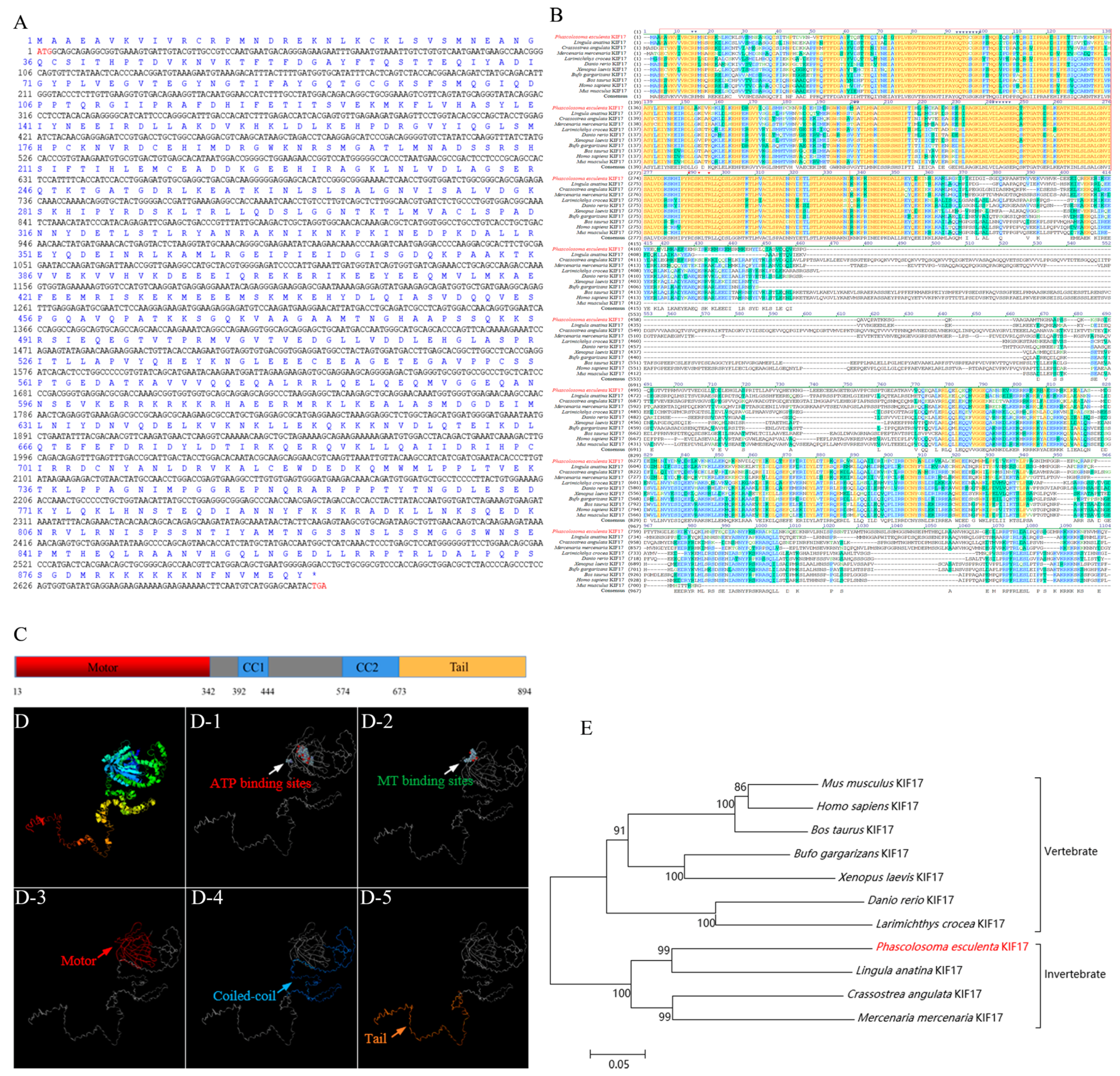
2.1. Bioinformatic Analysis of the Pe-kif17 Gene

Pe-kif17 is 2682 bp and encodes a protein of 894 aa with a molecular weight of 101.18 kDa and pI of 6.46 (Figure 1A). The predicted functional domain of the KIF17 protein was mainly composed of the N-terminal conserved motor domain (3–342 aa), the intermediate domain composed of two coiled-coil regions (392–444 aa/574–673 aa), and a C-terminal tail domain (673–894 aa) binding to cargo (Figure 1C). A predicted three-dimensional (3D) structural model of Pe-KIF17 was similar to the tertiary structure of KIF17 in other species (Figure 1D). We compared the homology of the predicted Pe-KIF17 aa sequence with that of other species (Figure 1B). The consensus and identity positions of the Pe-KIF17 aa sequence with its counterparts in Homo sapiens, Mus musculus, Bos taurus, Danio rerio, Larimichthys crocea, Xenopus laevis, Bufo gargarizans, Mercenaria mercenaria, Crassostrea angulata, and Lingula anatina were 52.6% and 41.7%, 53.4% and 42.4%, 52.2% and 40.4%, 59.5% and 46.4%, 57.9% and 45.7%, 59.7% and 49.3%, 62.0% and 50.6%, 60.3% and 50.7%, 54.8% and 45.3%, and 67.0% and 57.4%, respectively. The phylogenetic tree showed that Pe-KIF17 and L. anatina KIF17 were closely clustered and evolutionarily close, suggesting that their structures were conserved (Figure 1E).

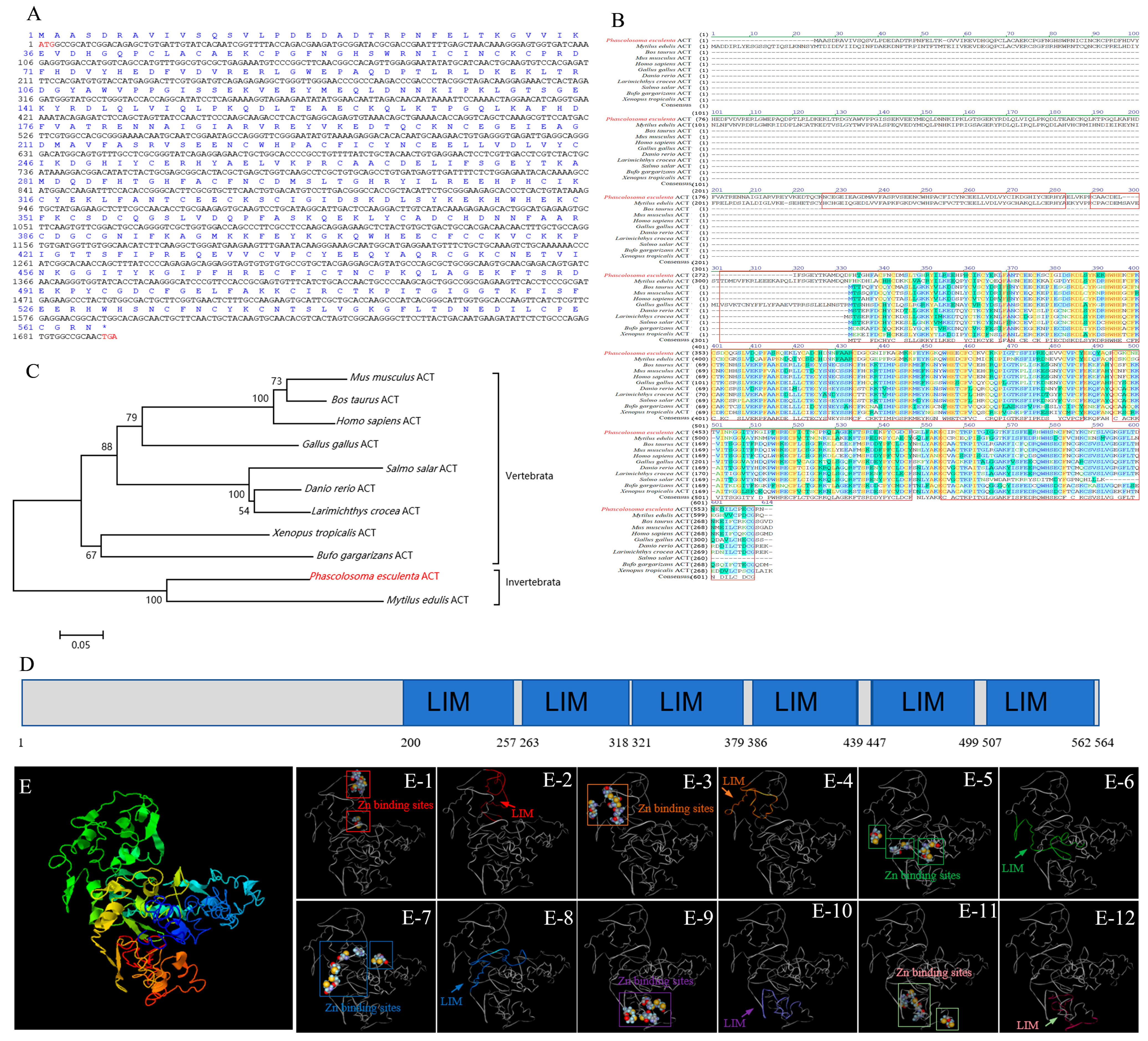
2.2. Bioinformatic Analysis of Pe-act Gene

The ORF of Pe-act contains 1695 bp, coding for a 564 aa protein with a molecular weight of 64.45 kDa and pI of 5.85 (Figure 2A). The functional domain of the ACT protein is mainly composed of six LIM domains (Figure 2D). Each LIM domain with two zinc finger structures, as well as the 3D structural model of Pe-ACT, was constructed (Figure 2E), indicating that it was evolutionarily conserved. The homology of Pe-ACT amino acid sequence was compared with that of other species (Figure 2B). The similarity and consistency of Pe-ACT aa sequence with H. sapiens, M. musculus, B. taurus, D. rerio, L. crocea, Salmo salar, Gallus gallus, Xenopus tropicalis, B. gargarizans, and Mytilus eduils were 32.0% and 23.9%, 30.9% and 23.6%, 31.5% and 24.1%, 32.6% and 26.2%, 32.7% and 25.5%, 27.7% and 20.7%, 34.2% and 25.4%, 32.1% and 25.6%, 32.6% and 25.4%, and 65.5% and 53.9%, respectively. The phylogenetic tree showed that Pe-ACT was closely related to the M. eduils ACT, suggesting that its structure may be conserved (Figure 2C).

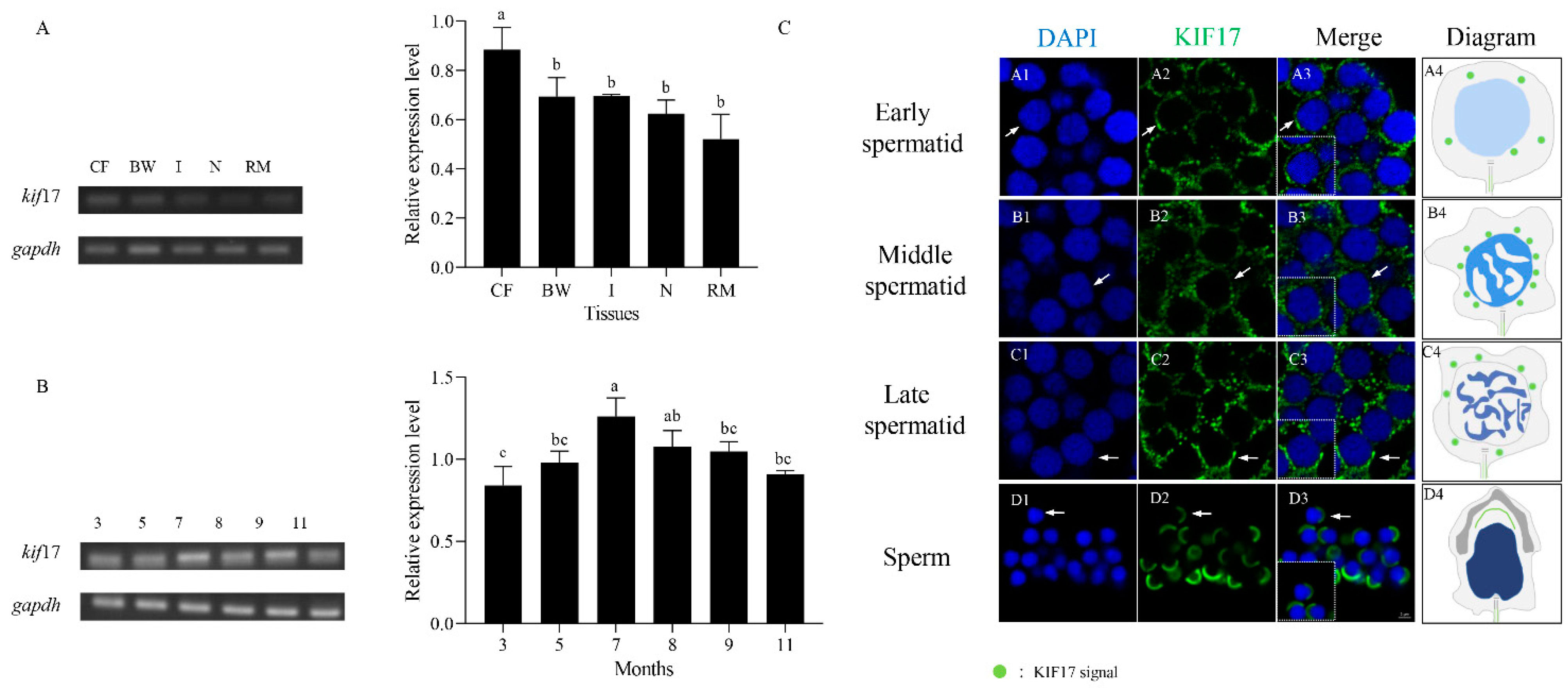
2.3. Expression Analysis of Pe-kif17 mRNA

We analyzed the relative expression levels of Pe-kif17 mRNA in the CF, body wall, intestine, nephridium, and retractor muscles of P. esculenta using semiquantitative RT-PCR. Pe-kif17 mRNA was expressed in all tissues. The expression level was highest in the CF compared with other tissues (Figure 3A). Similarly, we analyzed the relative expression of Pe-kif17 mRNA in the CF at different stages of sexual gland development (March, May, July, August, September, and November) using semiquantitative RT-PCR. The results showed that Pe-kif17 mRNA was expressed in the CF of P. esculenta in March, May, July, August, September, and November, with the highest expression observed in July (Figure 3B).
The spatiotemporal expression characteristics of Pe-kif17 mRNA during spermiogenesis were detected by FISH (Figure 3C). In early spermatids, Pe-kif17 mRNA were evenly distributed in the perinuclear cytoplasm (Figure 3C(A1–A4)). In middle spermatids, Pe-kif17 mRNA signals were enhanced and concentrated around the nucleus (Figure 3C(B1–B4)). In late spermatids, Pe-kif17 mRNA signals continued to be expressed around the nucleus (Figure 3C(C1–C4)). In mature sperm, Pe-kif17 mRNA signals mainly converged to one side of the nucleus (Figure 3C(D1–D4)).

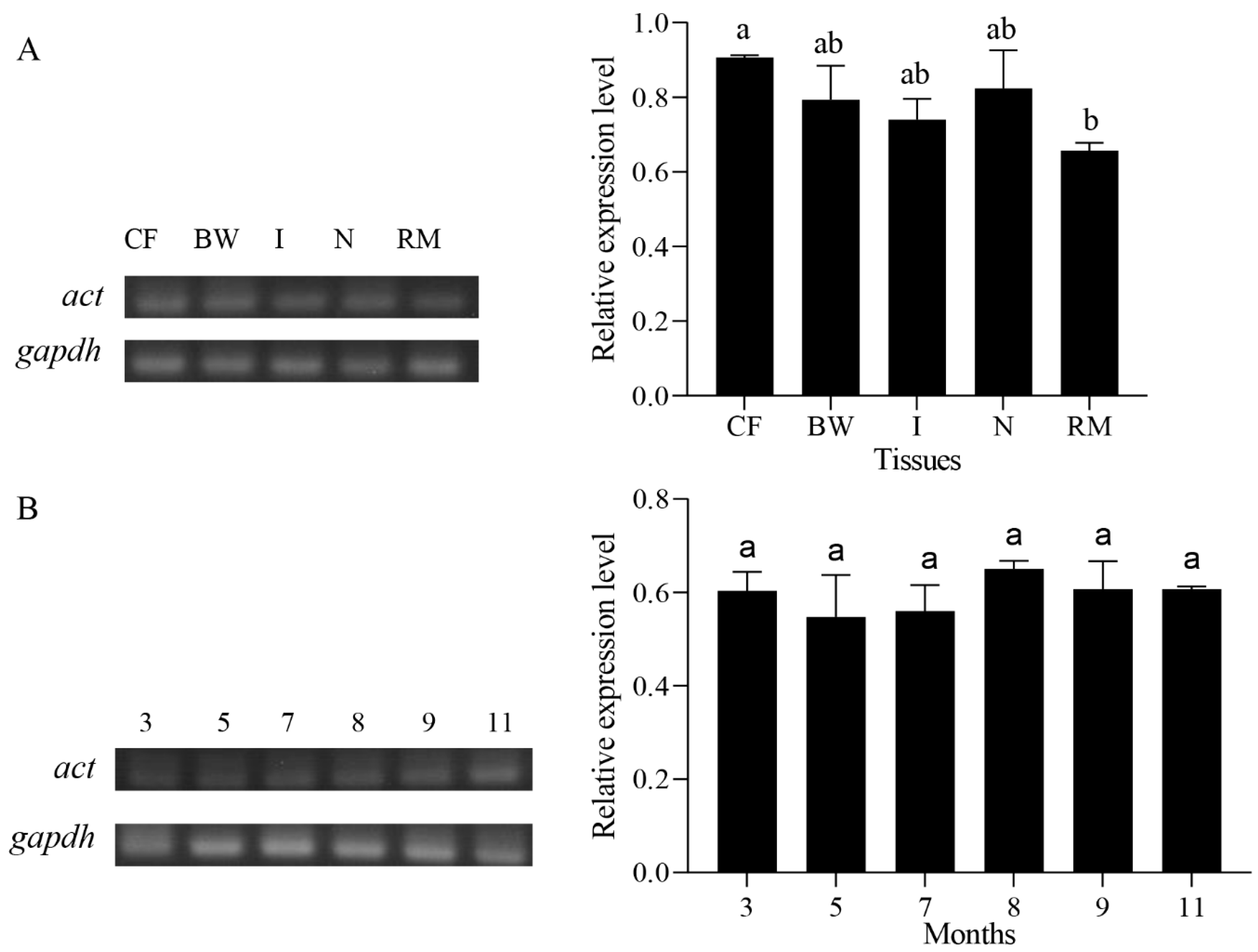
2.4. Expression Analysis of Pe-act mRNA

We analyzed the relative expression levels of Pe-act mRNA in the CF, body wall, intestine, nephridium, and retractor muscle of P. esculenta by semiquantitative RT-PCR. Pe-act mRNA was also expressed in all tissues (Figure 4A). The relative expression of Pe-act mRNA in the coelomic fluid in different stages of sexual gland development was determined (March, May, July, August, September, and November) by semiquantitative RT-PCR. Pe-act mRNA was expressed in the CF in March, May, July, August, September, and November (Figure 4B).

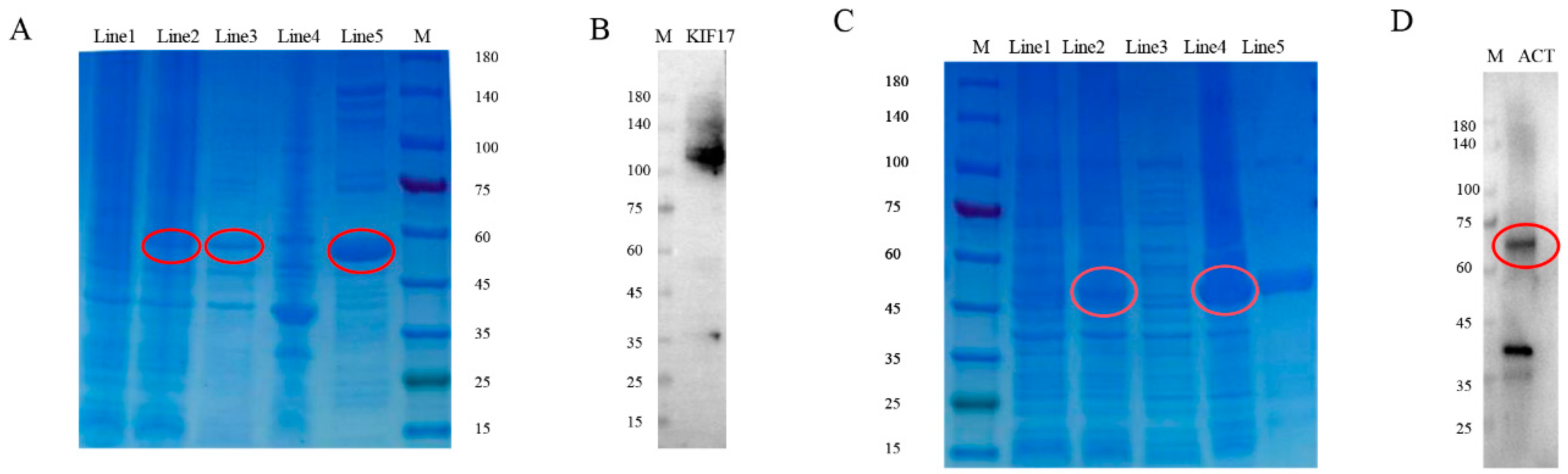
2.5. Preparation and Specificity of Pe-KIF17/ACT Antibodies

Pe-KIF17/ACT proteins were successfully expressed in E. coli Transetta (DE3) (Figure 5). SDS-PAGE clearly revealed bands with a molecular weight of 52.5 and 49.6 kDa in the induced cells, but not in the uninduced cells (Figure 5A,C). These two recombinant proteins were expressed in the supernatant and cell fractions. The purified recombinant proteins were each evident as a single band. Subsequently, the recombinant Pe-KIF17 and ACT protein was used to immunize rats and mice, respectively. Sera was collected to obtain rat polyclonal antibody against Pe-KIF17 and mouse polyclonal antibodies against Pe-ACT. Western blotting was performed to detect antibody specificity. The Pe-KIF17 antibody was detected as a single band of approximately 101.18kDa (Figure 5B). The Pe-ACT antibody was detected as two bands that matched the predicted molecular weight of the protein, at approximately 64.45 and 35–45 kDa (Figure 5D). These results suggest that rat anti-Pe-KIF17 and mouse anti-Pe-ACT polyclonal antibodies can be used for immunofluorescence assays.

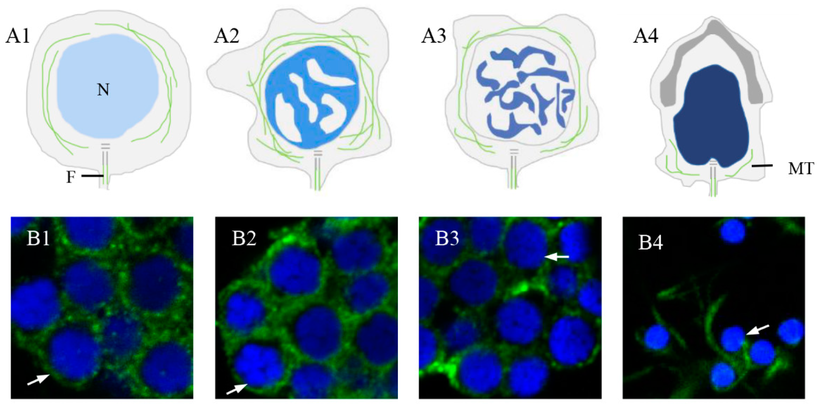
2.6. Expression and Colocalization Characteristics of Pe-KIF17 with Tubulin during Spermiogenesis

The distribution of α-tubulin during spermiogenesis in P. esculenta (Figure 6) and co-localization characteristics of Pe-KIF17 with tubulin during spermiogenesis was observed by IF (Figure 7). In early spermatids, Pe-KIF17 and tubulin signals were evenly distributed in the cytoplasm (Figure 6A1,B1 and Figure 7A1–A5). In middle spermatids, Pe-KIF17 and tubulin signals colocalized similarly to early spermatids and clustered around the nucleus (Figure 6A2,B2 and Figure 7B1–B5). In late spermatids, Pe-KIF17 and tubulin signals migrated to one side of the nucleus, where the sperm tail formed (Figure 6A3,B3 and Figure 7C1–C5). In mature sperms, Pe-KIF17 and tubulin signals were detected in the tail (Figure 6A4,B4 and Figure 7D1–D5).

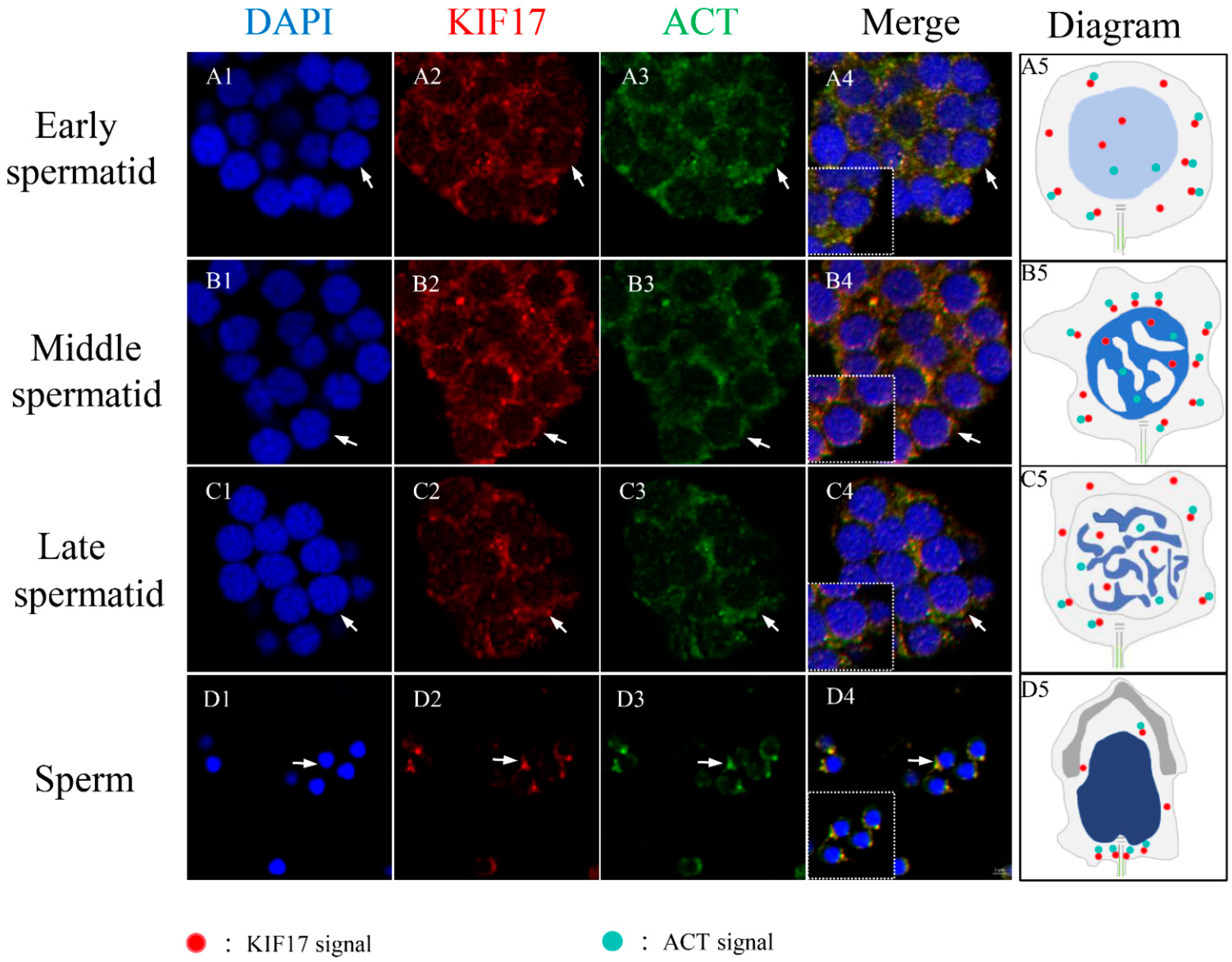
2.7. Colocalization between Pe-KIF17 and Pe-ACT in Spermiogenesis

Colocalization of Pe-KIF17 and Pe-ACT during spermiogenesis in P. esculenta was also observed by IF (Figure 8). In early spermatids, Pe-KIF17 and Pe-ACT signals were evenly distributed in the cytoplasm (Figure 8A1–A5). In middle spermatids, Pe-KIF17 and Pe-ACT signals were localized similarly to those in early spermatids and clustered around the nucleus (Figure 8B1–B5). In late spermatids, Pe-KIF17 and Pe-ACT signals migrated to one side of the nucleus where the sperm tail formed (Figure 8C1–C5). In mature sperm, Pe-KIF17 and Pe-ACT signals were detected in the tails of mature sperm (Figure 8D1–D5).

3. Discussion

3.1. Structural Features of KIF17 and ACT

KIF17 is a homologous dimer motor protein belonging to the kinesin-2 family. This protein consists of a microtubule-bound head motor domain, coiled-coil intermediate domain, and cargo-bound tail domain [14,15]. Its motor domain contains ATP hydrolysis sites and microtubule-binding sites, which can transport cargo to specific destinations in ATP hydrolysis-dependent manner [16]. Interestingly, the region where the KFI17b tail domain binds to the cargo does not bind to ACT. Its stem binds to ACT, enabling ACT transportation [27,31]. In ACT, the double zinc finger structure of the LIM domain is thought to mediate protein–protein interactions [25,26]. This particular interaction reflects the uniqueness of the KIF17b mediated transport of ACT [27]. In the present study, the predicted Pe-KIF17 protein had the same structural features as previously reported KIF17 [17,18]. Multiple sequence comparisons with KIF17 amino acid sequences from other species showed that their motor domains were highly conserved and contained microtubule-binding sites and ATP hydrolysis sites. These findings suggest that Pe-KIF17 might also use the energy generated by ATP hydrolysis to drive itself along microtubules through the motor domain. The structural conservation of Pe-KIF17 indicates that its function may have been evolutionarily conserved. Similarly, in this study, we found that the predicted Pe-ACT has six LIM domains, each of which has a structural motif consisting of two zinc fingers. The structurally conserved nature of both KIF17 and ACT suggests that the functions of both proteins may have been conserved during evolution.

3.2. Expression Characteristics of Pe-kif17/act mRNA

KIF17 is expressed at high levels in the gray matter of the brain, where it possibly plays a crucial role for neurons [14,31]. In mice, KIF17 is highly expressed in the brain and is involved in neuronal events required for learning and memory via the transport of the basic N-methyl-D-aspartate-type glutamate receptor (NR2B) [32,33]. In addition, KIF17 is highly expressed in the testes [20]. Saade et al. [19] reported that KIF17b and Spatial function in spermatid reshaping in mouse testes. This coincides with the high expression of kif17 mRNA in stage IV of L. polyactis and L. crocea spermiogenesis. The testes of both contain numerous spermatids [17,18,34], which suggests the involvement in spermiogenesis, due to the high expression of KIF17 in the testes of L. crocea and L. polyactis [17,18]. Corresponding findings regarding the expression of kif17 mRNA revealed the highest expression in the CF, where spermatids underwent morphological transformation into sperm throughout spermiogenesis, in contrast to other tissues, during July of the breeding season. Interestingly, spermatid masses, blood cells, and granular cells were also observed in the CF. Moreover, the proportions of these three cell types vary between the months. In the reproductive season (July–September), spermatid masses are the main components of the CF, whereas in the nonreproductive season (March, May, and November), spermatid masses are relatively small [28,35,36]. In addition, we studied the expression and distribution characteristics of kif17 mRNA during spermiogenesis. Pe-kif17 mRNA signals were continuously detected during spermiogenesis. The expression pattern of KIF17 provides an important basis for our research on KIF17 in P. esculenta.
ACT is expressed in the germ cells of male mice and may play an important role in sperm function [37]. Using FISH, we detected a strong act mRNA signal in human, mouse, and cynomolgus monkey round sperm cells. The findings indicate that act plays an important role in spermiogenesis [22]. In this study, semiquantitative RT-PCR analysis revealed that Pe-act mRNA was distributed in all tissues and was expressed in the CF in different months. These findings provided basic data for us to explore the association between KIF17 and ACT. Subsequently, we used IF to determine the colocalization of both.

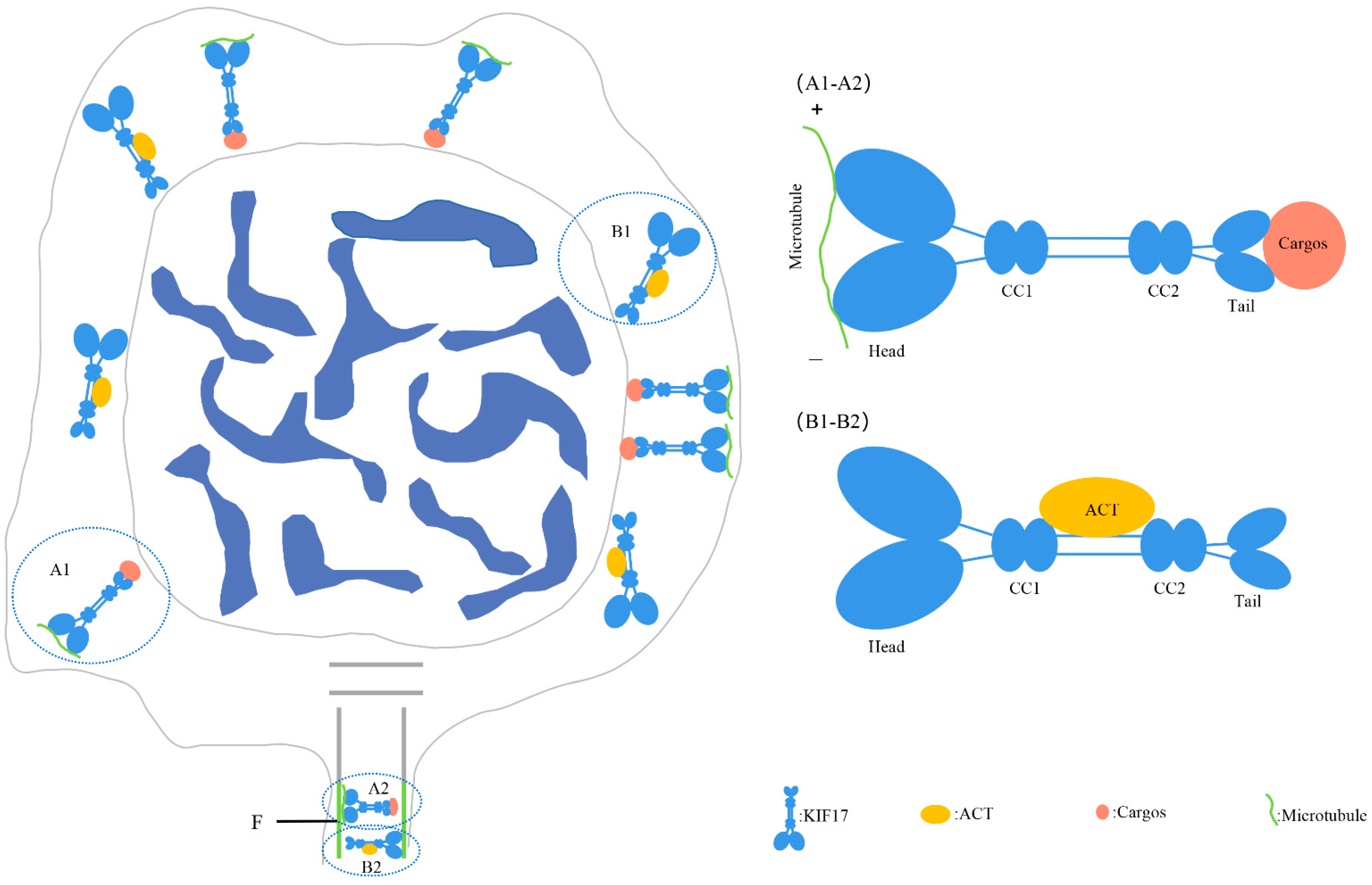
3.3. Pe-KIF17 May Be Involved in Nuclear Reshaping and Tail Formation in a Microtubule-Dependent Manner

The manchette plays an important role in the reshaping of the spermatid nucleus and is a type of cytoskeleton composed of microtubules [2,5]. In mice, Kotaja et al. [27] found that KIF17 signals were colocalized with α-tubulin, suggesting that KIF17 was related to the manchette. In the present study, we observed the distribution of α-tubulin during spermiogenesis in P. esculenta (Figure 6). Tubulin signals were randomly distributed in the perinuclear cytoplasm of the early spermatids (Figure 6A1,B1). Subsequently, tubulin signals were closely associated with signal intensification in the middle spermatids (Figure 6A2,B2). However, the tubulin signals gradually weakened in the final spermatids (Figure 6A3,B3). Ultimately, tubulin signals were mainly concentrated in the tail of mature sperm, suggesting that there may be microtubule structures similar to those of manchettes in spermatids (Figure 6A4,B4).
Moreover, we also observed that Pe-KIF17 co-localized with α-tubulin in the process of spermatid nucleus reshaping, and the protein signals gradually transferred from the perinuclear cytoplasm to one side of the sperm tail, with obvious changes in spermatid nucleus. These findings suggest that Pe-KIF17 may interact with microtubule structures similar to manchette to participate in spermatid remodeling (Figure 7). Previous studies have found that KIF17b and Spatial also exist in the manchette region and are continuously expressed in the sperm tails [19] and that KIF17b is expressed throughout spermatogenesis persists in the sperm tail [20,38]. Furthermore, in L. polyactis and L. crocea, KIF17 is involved in spermatid remodeling, including spermatid nucleus reshaping and sperm tail formation, through perinuclear microtubules during spermiogenesis [17,18]. Based on these findings, a model involving Pe-KIF17 in spermatid remodeling can be proposed (Figure 9A1,A2). Thus, it can be speculated that KIF17 may be involved in spermatid remodeling through a microtubule structure similar to the manchette.

3.4. Pe-KIF17 and Pe-ACT May Participate in Nuclear Reshaping and Tail Formation Independently of Microtubules

ACT (FHL5) belongs to the LIM-only protein family. The LIM domain, composed of approximately 50 amino acid residues, is rich in cysteine, forming two zinc finger structures. Its conserved cysteine and histidine residues bind to Zn2+ to form a stable tertiary protein structure, which is the main region mediating interactions with other proteins [25,26]. Studies in male mice have found that the sperm cell transcription mechanism is the transcriptional activation of CREM by binding to the activator ACT, without requiring phosphorylation at serine 117 as a transcriptional activator or association with CBP. These findings demonstrate the crucial role of ACT in male reproduction [21,22,23,24]. Moreover, in ACT−/− mice, a major abnormal phenomenon is the presence of a large number of tails with hairpin rings and sperm head deformity, indicating that ACT may play an important role in sperm tail formation and spermatid nucleus reshaping [38]. Studies in male mice have found that KIF17b, an isoform of KIF17, can also participate in spermatogenesis independent of microtubules and domains, by determining the subcellular localization of ACT (activator of CREM in testis) and participating in the transcription of related genes in spermiogenesis [21,22,23,27]. These findings indicate that KIF17b with ACT plays an important role in spermiogenesis.
We assessed the specificity of the Pe-KIF17 and ACT antibodies. Previous studies reported that antibody against KIF17 could also recognize KIF17b [20,27]. In the present study, the antibody specificity of Pe-ACT in this study was consistent with previous research [20,38], indicating the value of the Pe-KIF17 and Pe-ACT antibodies we generated for IF. These antibodies could be used for IF, which revealed signals of KIF17 and ACT that were randomly distributed in the perinuclear cytoplasm throughout the process from early spermiogenesis to mature sperm. The signals initially strengthened, gradually weakened, and finally became concentrated in the tail of mature sperm, especially in the middle piece of the sperm tail. These observations are similar to those of previous IF studies. In mice, KIF17b and ACT display coupled intracellular localization in male germ cells, where ACT is located in the nuclei of round spermatids, with KIF17b present in both the nucleus and cytoplasm [20]. In round spermatids, when CREM-dependent transcription stops and the nuclei of sperm begin to elongate, ACT migrates to the cytoplasm along with KIF17b and persists in the sperm tail [20,27,38]. Therefore, KIF17 and ACT may be involved in the formation of the sperm tail. Based on the above information, Pe-KIF17/ACT may be involved in sperm nuclear reshaping and tail formation during spermiogenesis in P. esculenta. A model involving Pe-KIF17 in spermatid remodeling is shown in Figure 9B1, B2. This model needs to be confirmed by techniques that include gene knockout and RNA interference.

4. Materials and Methods

4.1. Tissue Sampling

P. esculenta individuals weighing 2.9 to 4.8 g were obtained from Xiangshan (Zhejiang, China). The CF of male P. esculenta were obtained in March, May, July, August, September, and November. In addition to CF with mainly spermatid mass, body wall, intestine, retractor muscle, and nephridium were used as experimental materials in July of the reproductive season [35] for RNA and protein extraction. Simultaneously, a portion of the CF was handled in phosphate-buffered saline (PBS) containing 4% paraformaldehyde (PFA), and frozen sections were prepared for FISH and IF. Mature sperm were obtained by artificial induction [28], and the CF after successful induction treatment is described above.

4.2. RNA and Protein Extraction

Total RNA and protein were extracted from the CF, body wall, intestine, nephridium and retractor muscle using the TRIzol reagent (Invitrogen, Carlsbad, CA, USA) and RIPA lysis buffer (Beyotime, Shanghai, China) according to the manufactures’ instructions. The PrimeScript RT kit (TaKaRa Bio, Dalian, China) was used for reverse transcription. The complimentary DNA (cDNA) reverse transcription product was stored at −20 °C.

4.3. Cloning of kif17/act (ORF)

The intermediate segment sequence of kif17/act was obtained from the transcriptome sequence of P. esculenta. According to this sequence, specific primers were designed by Primer 5.0 software (Table 1). Touchdown PCR was performed using 94 °C for 5 min, 8 cycles of 94 °C for 30 s, 56 °C (each cycle reduced by 0.5 °C) and 72 °C for 1min; 27 cycles of 94 °C for 30 s, 53 °C for 30 s and 72 °C for 1 min; and a final extension at 72 °C for 10 min. Electrophoresis was performed on 1.0% agarose gel to resolve the DNA fragments, which were then extracted, purified, and ligated to the vector, and used to transform Escherichia. coli Trans-5α. The bacteria with the target band were selected and sent to Youkang Biology (Hangzhou, China) for sequencing.

4.4. Bioinformatics Analysis of KIF17/ACT

Pe-KIF17/ACT sequences were manipulated using the Sequence Manipulation Suite (http://www.bio-soft.net/sms/index, accessed on 21 April 2023). Multiple sequence alignments of Pe-KIF17/ACT amino acid sequences were performed using Vector NTI 10. Phylogenetic trees for KIF17/ACT were constructed using the adjacency method in MEGA 5.1. The ProtParam tool was used to calculate the molecular weights and isoelectric points (https://web.expasy.org/protparam/, accessed on 21 April 2023). Using the NCBI tool (https://www.ncbi.nlm.nih.gov/Structure/cdd/wrpsb.cgi, accessed on 21 April 2023) and online tools (https://zhanglab.ccmb.med.umich.edu/I-TASSER, accessed on 21 April 2023), we predicted the KIF17/ACT functional domain and tertiary structure of the KIF17/ACT proteins.

4.5. Semiquantitative RT-PCR Analysis of kif17/act mRNA Expression

The relative expression of kif17/act mRNA in the CF from different months (March, May, July, August, September, and November) and different tissues (CF, body wall, intestine, nephridium, and retractor muscle) was determined using semiquantitative RT-PCR. Pe-KIF17/ACT specific primers were designed using Primer 5 (Table 1) and were synthesized by Youkang Biology. The glyceraldehyde 3-phosphate dehydrogenase (gapdh) gene was used as the positive control. The relative expression of Pe-kif17 and Pe-act mRNAs was analyzed using Image J. The SPSS version 25.0 software (NIH, Bethesda, MD, USA) was used to determine the differences in expression levels of kif17/act mRNA in the CF of different months and different tissues of P. esculenta using means of one-way analysis of variance and Tukey’s multiple comparison. p < 0.05 indicated a significant difference.

4.6. FISH

As previously described by Zhao et al. [39], spermatids from the CF of P. esculenta were extracted and embedded in O.C.T compound. The samples were first stored at −20 °C for 1–2 h and then stored at −80 °C. The prepared samples were cut into 5 um thin slices using a freezing microtome (Thermo Fisher Scientific, Waltham, MA, USA). The sections were then attached to adhesion microscope sections and stored at −80 °C. The KIF17 probe (Table 1) was synthesized by GENEWIZ (Suzhou, China) and combined with fluorescein isothiocyanate (FITC). Its specificity was examined using the BLAST function of NCBI. The sections were examined by confocal laser scanning microscopy (CLSM) using the LSM880 microscope (Carl Zeiss, Jena, Germany) and imaged.

4.7. Antibodies

Pe-KIF17/ACT recombinant proteins were prepared and purified using a prokaryotic expression technique for animal immunization to obtain anti-Pe-KIF17 and anti-Pe-ACT antibodies. First, the full-length sequence of Pe-KIF17/ACT ORF (894aa) and (564aa) were obtained. The polypeptides were approximately 101.18 kDa and 64.45 kDa, respectively. The primers were as follows: anti-KIF17-F, GGCAACACAAAGACGCTCA; anti-KIF17-R, TTCTCCACCCACCATTTGT; anti-ACT-F, CCAGACGAAGATGCGGATAC; and anti-ACT-R, GAAAATCAACTCATCACAGGCT. The sequence was ligated to the pET-28a-SUMO vector and expressed in receptive E. coli Trans-5α. After PCR identification using T7 F/R primers, the bacterial suspension was sent to Youkang Biosciences for sequencing. The plasmid was extracted from the E. coli Trans-5α bacterial suspension carrying the correct sequence. The extracted plasmids were then introduced into Transetta (DE3) receptor cells (DE3-KIF17/ACT) for proliferation and induced by 0.1 M isopropyl β-D-1-thiogalactopyranoside (IPTG). Recombinant proteins were extracted and purified using a His-tag protein purification kit (Soluble Protein/Inclusion Body Protein; Beyotime, Shanghai, China) and identified using polyacrylamide gel electrophoresis. Finally, rats and mice were immunized with Pe-KIF17/ACT recombinant protein. Western blotting and immunofluorescence of extracted sera were performed as previously described [40]. In addition, we purchased commercial antibodies (all from Beyotime, Shanghai, China), which included rabbit anti α-tubulin antibody (Cat. No. AG0126), horseradish (HRP)-conjugated goat anti-rat IgG (H + L) (Cat. No. A0192), HRP-conjugated goat anti-mouse IgG (H + L) (Cat. No. A0216), Cy3-conjugated goat anti-rat IgG (H + L) (Cat. No. A0507), FITC-conjugated goat anti-mouse IgG (H + L) (Cat. No. A0568), and Alexa Fluor488 conjugated goat anti-rabbit IgG (H + L) (Cat. No. A0423).

4.8. Western Blotting

The specificity of Pe-KIF17/ACT antibodies was detected by Western blotting. First, total protein was extracted from the CF of P. esculenta using RIPA lysis buffer (Solarbio, Shanghai, China) and PMSF solution (Beyotime, Shanghai, China), and the protein concentration was determined using the BCA method (Kangweishiji, Jiangsu, China). The protein was diluted with 5× Sodium dodecyl sulfate-polyacrylamide gel electrophoresis (SDS-PAGE) (Beyotime, Shanghai, China) and boiled for 10 min for structural denaturation. The proteins were separated isolated on a 4–20% gel (ACE, Nanjing, China) and transferred to PVDF membranes (Bio-Rad, Hercules, CA, USA). After 2 h blocking with 5% skim milk solution at room temperature, the membranes were incubated with rat anti-Pe-KIF17 and mice anti-Pe-ACT antibodies at 4 °C overnight, and the antibodies were diluted at 1:200 by 0.1% Tris buffered saline containing Tween (TBST) solution. The membranes were washed four times with 0.1% TBST for 15 min each time. The membranes were then incubated with HRP-conjugated goat anti-rat IgG (H + L) and HRP-conjugated goat anti-mouse IgG (H + L) antibodies diluted 1:1500 in 0.1% TBST for 2 h at 37 °C. The membranes were washed four times with 0.1% TBST for 15 min each time. Finally, the membranes were imaged using a chemiluminescence imager (Tanon 5200, Shanghai, China).

4.9. IF

As previously described [41], 4% PFA-PBS was used to embed the CF of P. esculenta at 4 °C overnight. The fixed sample was transferred to 0.5 M sucrose in PBS (pH 7.4) at 4 °C for 2 h. Finally, the sample was embedded in O.C.T. compound, which was used for frozen sections as described in Section 4.6 and stored at −80 °C until required. At that time, the frozen sections were dried at room temperature for 10 min, permeabilized with 0.3% Tween in PBS (PBST) for 12 min and blocked with 5% bovine serum albumin (BSA) at room temperature for 2 h. The blocked sections were incubated with rat anti-Pe-KIF17 antibody diluted 1: 70 in 3% BSA) and rabbit anti α-tubulin antibodies (diluted at 1:100 by 3% BSA at 4 °C overnight followed by washing four times with 0.1% PBST for 10 min each time. The treated sections were incubated for 1 h with Cy3-conjugated goat anti-rat IgG (H + L) and Alexa Fluor488 conjugated goat anti-rabbit IgG (H + L) (both diluted 1: 500 in 3% BSA) and washed four times with 0.1% PBST for 10 min each time. Finally, the treated sections were incubated with 4′,6-diamidino-2-phenylindole (DAPI; Beyotime, Shanghai, China) for 7 min, mounted with Antifade Mounting Medium (Beyotime, Shanghai, China), and imaged by CLSM using the aforementioned LSM880 microscope.
In addition, as previously described [42], frozen sections retrieved from −80 °C storage were dried at room temperature for 10 min, permeated with 0.3% PBST for 12 min, then blocked with 5% BSA in 0.1% PBST for 2 h at 37 °C. The sections were then incubated with rat anti-Pe-KIF17 antibody diluted 1:70 using 3% BSA overnight at 4 °C. The sections were rewarmed at room temperature for 30 min then washed four times with PBS. The sections were then incubated with Cy3-conjugated goat anti-rat IgG (H + L) diluted 1:100 using 3% BSA at 37 °C for 1.5 h in a dark environment and washed five times with PBS. The treated sections were again blocked in the presence of 5% BSA at 37 °C for 2 h. These treated slices were incubated with mouse anti-ACT antibody diluted 1:70 with 3% BSA at 37 °C for 2 h and washed five times with PBS, incubated with FITC-conjugated goat anti-mouse IgG (H + L) diluted 1:100 with 3% BSA at 37 °C for 2 h, and washed five times with PBS. After incubation with DAPI (Beyotime, Shanghai, China) for 7 min, the treated sections were mounted with Antifade Mounting Medium (Beyotime, Shanghai, China) imaged by CLSM using the LSM880 microscope.

5. Conclusions

The ORFs of Pe-kif17 and act were cloned. The expression patterns and potential function of the Pe-KIF17 and ACT proteins during spermiogenesis were analyzed. Pe-kif17 and Pe-act mRNA were maximally expressed in the CF, with a large number of spermatids in the reproductive season, whose characteristics were perhaps essential for spermiogenesis. In addition, IF revealed the colocalization of Pe-KIF17 and tubulin and that the signal migrated from the perinuclear cytoplasm to the side of the sperm tail during spermiogenesis. Moreover, Pe-KIF17 with Pe-ACT colocalized during spermiogenesis. These results suggest that KIF17 may be involved in spermatid remodeling, including sperm nucleus reshaping and tail formation, either through interactions with a microtubule structure similar to that of the manchette or through its interaction with ACT. The detailed function of KIF17 in the spermiogenesis process of P. esculenta requires further study.

Author Contributions

Conceptualization: Y.P., J.W., C.H. and J.Z.; methodology: Y.P. and J.W.; formal analysis and investigation: Y.P. and J.W.; writing—original draft preparation: Y.P.; writing—review and editing: Y.P., J.W., X.G., D.T. and C.D.; funding acquisition: J.Z.; supervision: J.Z. All authors have read and agreed to the published version of the manuscript.

Funding

This research was funded by the Ningbo Science and Technology Plan Projects (2019B10016, 2016C10004), the Major Science and Technology Projects in Zhejiang Province (2011C12013), and the Collaborative Innovation Center for Zhejiang Marine High-efficiency and Healthy Aquaculture, the K.C. Wong Magna Fund in Ningbo University.

Institutional Review Board Statement

Animal experiment of immunizing mice and rats were approved by the Animal Ethics and Welfare Committee of Ningbo University (Approval number NBU20220155). The number of the animal ethics application project for this research is 11709.

Informed Consent Statement

Not applicable.

Data Availability Statement

The authors confirm that the data supporting the findings of this study are available within the article.

Conflicts of Interest

The authors declare no conflict of interest.

References

  1. Ma, D.D.; Wang, D.H.; Yang, W.X. Kinesins in Spermatogenesis. Biol. Reprod. 2017, 96, 267–276. [Google Scholar] [CrossRef] [PubMed]
  2. Mochida, K.; Tres, L.L.; Kierszenbaum, A.L. Structural and Biochemical Features of Fractionated Spermatid Manchettes and Sperm Axonemes of theAzh/Azh Mutant Mouse. Mol. Reprod. Dev. 1999, 52, 434–444. [Google Scholar] [CrossRef]
  3. Lehti, M.S.; Sironen, A. Formation and Function of the Manchette and Flagellum during Spermatogenesis. Reproduction 2016, 151, R43–R54. [Google Scholar] [CrossRef] [PubMed]
  4. Dooher, G.B.; Bennett, D. Spermiogenesis and Spermatozoa in Sterile Mice Carrying Different Lethal T/t Locus Haplotypes: A Transmission and Scanning Electron Microscopic Study. Biol. Reprod. 1977, 17, 269–288. [Google Scholar] [CrossRef] [PubMed]
  5. Hall, E.S.; Eveleth, J.; Jiang, C.; Redenbach, D.M.; Boekelheide, K. Distribution of the Microtubule-Dependent Motors Cytoplasmic Dynein and Kinesin in Rat Testis 1. Biol. Reprod. 1992, 46, 817–828. [Google Scholar] [CrossRef]
  6. Kierszenbaum, A.L. Spermatid Manchette: Plugging Proteins to Zero into the Sperm Tail. Mol. Reprod. Dev. 2001, 59, 347–349. [Google Scholar] [CrossRef]
  7. Russell, L.D.; Russell, J.A.; MacGregor, G.R.; Meistrich, M.L. Linkage of Manchette Microtubules to the Nuclear Envelope and Observations of the Role of the Manchette in Nuclear Shaping during Spermiogenesis in Rodents. Am. J. Anat. 1991, 192, 97–120. [Google Scholar] [CrossRef]
  8. Brady, S.T. A Novel Brain ATPase with Properties Expected for the Fast Axonal Transport Motor. Nature 1985, 317, 73–75. [Google Scholar] [CrossRef]
  9. Yang, W.-X.; Sperry, A.O. C-Terminal Kinesin Motor KIFC1 Participates in Acrosome Biogenesis and Vesicle Transport1. Biol. Reprod. 2003, 69, 1719–1729. [Google Scholar] [CrossRef]
  10. Kierszenbaum, A.L. Intramanchette Transport (IMT): Managing the Making of the Spermatid Head, Centrosome, and Tail. Mol. Reprod. Dev. 2002, 63, 1–4. [Google Scholar] [CrossRef]
  11. Navolanic, P.M.; Sperry, A.O. Identification of Isoforms of a Mitotic Motor in Mammalian Spermatogenesis1. Biol. Reprod. 2000, 62, 1360–1369. [Google Scholar] [CrossRef] [PubMed]
  12. Lehti, M.S.; Kotaja, N.; Sironen, A. KIF3A Is Essential for Sperm Tail Formation and Manchette Function. Mol. Cell. Endocrinol. 2013, 377, 44–55. [Google Scholar] [CrossRef] [PubMed]
  13. Gilbert, S.P.; Guzik-Lendrum, S.; Rayment, I. Kinesin-2 Motors: Kinetics and Biophysics. J. Biol. Chem. 2018, 293, 4510–4518. [Google Scholar] [CrossRef] [PubMed]
  14. Setou, M.; Nakagawa, T.; Seog, D.-H.; Hirokawa, N. Kinesin Superfamily Motor Protein KIF17 and mLin-10 in NMDA Receptor-Containing Vesicle Transport. Science 2000, 288, 1796–1802. [Google Scholar] [CrossRef] [PubMed]
  15. Yin, X.; Feng, X.; Takei, Y.; Hirokawa, N. Regulation of NMDA Receptor Transport: A KIF17–Cargo Binding/Releasing Underlies Synaptic Plasticity and Memory In Vivo. J. Neurosci. 2012, 32, 5486–5499. [Google Scholar] [CrossRef] [PubMed]
  16. Miki, H.; Okada, Y.; Hirokawa, N. Analysis of the Kinesin Superfamily: Insights into Structure and Function. Trends Cell Biol. 2005, 15, 467–476. [Google Scholar] [CrossRef] [PubMed]
  17. Wang, J.; Gao, X.; Zheng, X.; Du, C.; Hou, C.; Xie, Q.; Lou, B.; Liu, F.; Jin, S.; Zhu, J. Evidence for the Role of KIF17 in Fish Spermatid Reshaping: Expression Pattern of KIF17 in Larimichthys polyactis Spermiogenesis. J. Ocean. Limnol. 2021, 39, 2322–2335. [Google Scholar] [CrossRef]
  18. Wang, J.; Liu, Z.; Gao, X.; Du, C.; Hou, C.; Tang, D.; Lou, B.; Shen, W.; Zhu, J. The Potential Function of KIF17 in Large Yellow Croaker (Larimichthys crocea) Spermatid Remodeling: Molecular Characterization and Expression Pattern during Spermiogenesis. Fish Physiol. Biochem. 2022, 48, 603–616. [Google Scholar] [CrossRef]
  19. Saade, M.; Irla, M.; Govin, J.; Victorero, G.; Samson, M.; Nguyen, C. Dynamic Distribution of Spatial during Mouse Spermatogenesis and Its Interaction with the Kinesin KIF17b. Exp. Cell Res. 2007, 313, 614–626. [Google Scholar] [CrossRef]
  20. Macho, B.; Brancorsini, S.; Fimia, G.M.; Setou, M.; Hirokawa, N.; Sassone-Corsi, P. CREM-Dependent Transcription in Male Germ Cells Controlled by a Kinesin. Science 2002, 298, 2388–2390. [Google Scholar] [CrossRef]
  21. Krausz, C.; Sassone-Corsi, P. Genetic Control of Spermiogenesis: Insights from the CREM Gene and Implications for Human Infertility. Reprod. BioMed. Online 2005, 10, 64–71. [Google Scholar] [CrossRef] [PubMed]
  22. Steger, K. Expression of Activator of CREM in the Testis (ACT) during Normal and Impaired Spermatogenesis: Correlation with CREM Expression. Mol. Hum. Reprod. 2004, 10, 129–135. [Google Scholar] [CrossRef] [PubMed]
  23. Hogeveen, K.N.; Sassone-Corsi, P. Regulation of Gene Expression in Post-Meiotic Male Germ Cells: CREM-Signalling Pathways and Male Fertility. Hum. Fertil. 2006, 9, 73–79. [Google Scholar] [CrossRef] [PubMed]
  24. Fimia, G.M.; Cesare, D.D.; Sassone-Corsi, P. CBP-Independent Activation of CREM and CREB by the LIM-Only Protein ACT. Nature 1999, 398, 165–169. [Google Scholar] [CrossRef] [PubMed]
  25. Schmeichel, K. The LIM Domain Is a Modular Protein-Binding Interface. Cell 1994, 79, 211–219. [Google Scholar] [CrossRef] [PubMed]
  26. Dawid, I.B.; Breen, J.J.; Toyama, R. LIM Domains: Multiple Roles as Adapters and Functional Modifiers in Protein Interactions. Trends Genet. 1998, 14, 156–162. [Google Scholar] [CrossRef] [PubMed]
  27. Kotaja, N.; Macho, B.; Sassone-Corsi, P. Microtubule-Independent and Protein Kinase A-Mediated Function of Kinesin KIF17b Controls the Intracellular Transport of Activator of CREM in Testis (ACT). J. Biol. Chem. 2005, 280, 31739–31745. [Google Scholar] [CrossRef]
  28. Zhu, J.Q.; Wang, W.; Xu, S.J.; Zeng, H.X. Spermatogenesis and sperm morphology of Phascolosoma esculenta. Acta Zool. Sin. 2007, 53, 733–741. [Google Scholar] [CrossRef]
  29. Gao, X.M.; Mu, D.L.; Hou, C.C.; Zhu, J.Q.; Jin, S.; Wang, C.L. Expression and Putative Functions of KIFC1 for Nuclear Reshaping and Midpiece Formation during Spermiogenesis of Phascolosoma esculenta. Gene 2019, 683, 169–183. [Google Scholar] [CrossRef]
  30. Du, C.; Mu, D.; Gao, X.; Luo, S.; Wang, J.; Jin, S.; Zhu, J. Expression Characteristics and Putative Functions of KIF3A/KIF3B During Spermiogenesis of Phascolosoma esculenta. J. Ocean Univ. China 2022, 21, 998–1016. [Google Scholar] [CrossRef]
  31. Wong-Riley, M.T.T.; Besharse, J.C. The Kinesin Superfamily Protein KIF17: One Protein with Many Functions. BioMol. Concepts 2012, 3, 267–282. [Google Scholar] [CrossRef] [PubMed]
  32. Yin, X.; Takei, Y.; Kido, M.A.; Hirokawa, N. Molecular Motor KIF17 Is Fundamental for Memory and Learning via Differential Support of Synaptic NR2A/2B Levels. Neuron 2011, 70, 310–325. [Google Scholar] [CrossRef] [PubMed]
  33. Wong, R.W.-C.; Setou, M.; Teng, J.; Takei, Y.; Hirokawa, N. Overexpression of Motor Protein KIF17 Enhances Spatial and Working Memory in Transgenic Mice. Proc. Natl. Acad. Sci. USA 2002, 99, 14500–14505. [Google Scholar] [CrossRef] [PubMed]
  34. Kang, H.W.; Chung, E.Y.; Chung, J.S.; Lee, K.Y. Ultrastructural Studies of Spermatogenesis and the Functions of Leydig Cells and Sertoli Cells Associated with Spermatogenesis in Larimichthys polyactis (Teleostei, Perciformes, Sciaenidae). Anim. Cells Syst. 2013, 17, 250–258. [Google Scholar] [CrossRef]
  35. Chen, H.; Lin, G.W.; Chen, W.; Liu, Z.K.; Wang, X. Observation on the reproductive cycle of Phascolosoma esculenta. Mar. Fish. 2009, 31, 139–145. [Google Scholar] [CrossRef]
  36. Zhang, H.; Zeng, D.; Yan, H. Study on breeding biology of Phascolosoma esculenta. Pop. Sci. Technol. 2015, 17, 114–115. [Google Scholar] [CrossRef]
  37. Fimia, G.M.; De Cesare, D.; Sassone-Corsi, P. A Family of LIM-Only Transcriptional Coactivators: Tissue-Specific Expression and Selective Activation of CREB and CREM. Mol. Cell. Biol. 2000, 20, 8613–8622. [Google Scholar] [CrossRef] [PubMed]
  38. Kotaja, N.; De Cesare, D.; Macho, B.; Monaco, L.; Brancorsini, S.; Goossens, E.; Tournaye, H.; Gansmuller, A.; Sassone-Corsi, P. Abnormal Sperm in Mice with Targeted Deletion of the Act (Activator of cAMP-Responsive Element Modulator in Testis) Gene. Proc. Natl. Acad. Sci. USA 2004, 101, 10620–10625. [Google Scholar] [CrossRef]
  39. Zhao, Y.Q.; Mu, D.L.; Wang, D.; Han, Y.L.; Hou, C.-C.; Zhu, J.Q. Analysis of the Function of KIF3A and KIF3B in the Spermatogenesis in Boleophthalmus Pectinirostris. Fish Physiol. Biochem. 2018, 44, 769–788. [Google Scholar] [CrossRef]
  40. Zhang, H.L.; Wan, J.; Yan, F.; Zhang, X.S.; Cao, W.; Tao, W.; Huang, L.Q. The production and application of Ezrin polyclonal antibody. Biotechnol. Bull. 2013, 1, 130–133. [Google Scholar] [CrossRef]
  41. Hou, C.C.; Gao, X.M.; Ni, J.; Mu, D.L.; Yang, H.Y.; Liu, C.; Zhu, J.Q. The Expression Pattern and Potential Functions of PHB in the Spermiogenesis of Phascolosoma Esculenta. Gene 2018, 652, 25–38. [Google Scholar] [CrossRef] [PubMed]
  42. Chen, X.; Liu, Z.; Deng, M.; Feng, S. A feasibility study using primary antibodies from the same species in immunofluorescence double staining. Sichuan J. Anat. 2015, 23, 13–19. [Google Scholar] [CrossRef]
Figure 1. Analysis of Pe-kif17 ORF characteristics. (A) ORF of Pe-kif17 with the deduced amino acid sequence, and it contains 2682 bp and can encode 894 amino acids. The initiation codon is ATG and the termination codon is TGA (both marked in red). (B) Multiple sequence alignment of KIF17 amino acid sequence of P. esculenta and other species. The identical amino acids are represented in yellow regions; more than 50% similarity is represented in blue regions, and a lower similarity is represented in green regions. The motor domain is marked in a red box. Red triangles indicate microtubule-binding sites. Blue triangles indicate putative ATP hydrolysis sites. (C) Protein structure domain of Pe-KIF17. (D) Predicted tertiary structure of Pe-KIF17; (D-1) predicted ATP hydrolysis sites; (D-2) predicted microtubule-binding sites; (D-3) predicted N-terminal motor domain (red); (D-4) predicted intermediate domain (blue); and (D-5) predicted C-terminal tail domain (orange). Motor: N-terminal head motor domain; CC1: coiled-coil region 1; CC2: coiled-coil region 2; Tail: C-terminal tail domain. (E) Phylogenetic tree of Pe-KIF17 homologous proteins.
Ijms 25 00128 g001
Figure 2. Analysis of Pe-act ORF characteristics. (A) ORF of Pe-kif17 of 1695 bp encoding 564 amino acids. The initiation codon is ATG and the termination codon is TGA (both marked in red). (B) Multiple sequence alignment of ACT amino acid sequence of P. esculenta and other species. The LIM domain is marked in a red box. (C) Phylogenetic tree of Pe-KIF17 homologous proteins. (D) Protein structure domain of Pe-ACT with six LIM domains. (E) Predicted tertiary structure of Pe-ACT; (E-1,E-3,E-5,E-7,E-9,E-11) predicted the double zinc finger structure; (E-2,E-4,E-6,E-8,E-10,E-12) predicted the LIM domain.
Ijms 25 00128 g002
Figure 3. The relative expression level of Pe-kif17 mRNA. (A) The relative expression of Pe-kif17 mRNA in different tissues of P. esculenta, the highest expression level of kif17 mRNA was found in CF. Abbreviations: CF: coelomic fluid; BW: body wall; I: intestine; N: nephridium; and RM: retractor muscle. (B) The relative expression of Pe-kif17 mRNA in the CF of P. esculenta in different months. The highest expression of kif17 mRNA was found in July. Values are expressed as mean ± SD, with lower-case letters indicating significant differences between groups (p < 0.05). (C) Fluorescence in situ hybridization results of Pe-kif17 mRNA during spermiogenesis in P. esculenta. (A1A4) In the early spermatid, Pe-kif17 mRNA signals were distributed in the perinuclear cytoplasm. (B1B4) In the middle spermatid, Pe-kif17 mRNA signals were enhanced and distributed around the nucleus. (C1C4) In the late spermatid, Pe-kif17 mRNA signals continued to express around the nucleus. (D1D4) In the mature sperm, the signals were mainly concentrated on one side of the nucleus. Blue signal represents nuclei stained with 4′, 6-diamidino-2-phenylindole (DAPI). Pe-kif17 mRNA was stained with FITC labeled probe (green). The scale bar is 2 µm.
Ijms 25 00128 g003
Figure 4. The relative expression level of Pe-act mRNA. (A) The relative expression of Pe-act mRNA in different tissues of P. esculenta. The highest expression level of act mRNA was found in the CF. Abbreviations: CF: coelomic fluid; BW: body wall; I: intestine; N: nephridium; and RM: retractor muscle. (B) The relative expression of Pe-act mRNA in the CF of P. esculenta in different months. Values were expressed as mean ± SD. Lower case letters indicate significant differences between groups (p < 0.05).
Ijms 25 00128 g004
Figure 5. Expression and purification of Pe-KIF17 and ACT. (A) Lane 1: uninduced total protein; Lane 2: total protein expression of KIF17 induced by IPTG; Lane 3: soluble protein; Lane 4: inclusion body protein; Lane 5: purified recombinant protein with molecular weight of 52.53 kDa; and Lane 6: protein markers. (B) Specificity of anti-Pe-KIF17 antibody in rats as detected by Western blotting. Only a single band with a molecular weight of approximately 101.18 kDa is detected, which is consistent with the predicted molecular weight of Pe-KIF17, confirming the specificity of the antibody. (C) Lane 1: protein marker; Lane 2: uninduced total protein; Lane 3: total protein expression of ACT induced by IPTG; Lane 4: soluble protein; Lane 5: inclusion body protein; Line 6: purified recombinant protein with molecular weight of 49.6 kDa. (D) Specificity of anti-Pe-ACT antibody in mice as detected by Western blotting. A single band with a molecular weight of about 64.45 kDa is detected, which is consistent with the predicted molecular weight of Pe-ACT.
Ijms 25 00128 g005
Figure 6. Spermiogenesis in P. esculenta. (A1,B1) early spermatid; (A2,B2) middle spermatid; (A3,B3) late spermatid; (A4,B4) mature sperm. Abbreviations: N: nucleus; F: flagellum; MT: microtubule.
Ijms 25 00128 g006
Figure 7. Immunofluorescence localization of Pe-KIF17 with tubulin during spermiogenesis in P. esculenta. (A1A5) In early spermatid, Pe-KIF17 protein signals were randomly distributed in the perinuclear cytoplasm. (B1B5) In middle spermatid, Pe-KIF17 protein signals were enhanced and distributed around the nucleus. (C1C5) In late spermatid, Pe-KIF17 protein signals moved to one side of the nucleus where sperm tail formed. (D1D5) In mature sperm, the signal was mainly located in the sperm tail. Nuclei were stained with DAPI (blue). Pe-KIF17 was stained with Alexa Fluor Cy3 conjugated antibody (red). Tubulin was stained with AlexaFluor488 conjugated antibody (green). The scale bar is 2 µm.
Ijms 25 00128 g007
Figure 8. Immunofluorescence localization of Pe-KIF17 with Pe-ACT during spermiogenesis in P. esculenta. (A1A5) In early spermatid, Pe-KIF17 and Pe-ACT signals were randomly distributed in the perinuclear cytoplasm. (B1B5) In middle spermatid. Pe-KIF17 and Pe-ACT signals were localized similarly to those in early spermatids and clustered around the nucleus. (C1C5) In late spermatid, Pe-KIF17 and Pe-ACT signals moved to one side of the nucleus where sperm tail formed. (D1D5) In mature sperm, the signal was mainly located in the sperm tail. Nuclei were stained with DAPI (blue). Pe-KIF17 was stained with Alexa Fluor Cy3 conjugated antibody (red). Pe-ACT was stained with Alexa Fluor FITC conjugated antibody (green). The scale bar is 2 µm.
Ijms 25 00128 g008
Figure 9. (A1,A2) KIF17 may be involved in spermatid reshaping, including nuclear reshaping and tail formation, by interacting with microtubule structures similar to the manchette. (B1,B2) KIF17 with ACT may be involved in nuclear reshaping and tail formation independently of microtubules. Head: N-terminal head motor domain; CC1: coiled-coil region 1; CC2: coiled-coil region 2; Tail: C-terminal tail domain; F: flagellum.
Ijms 25 00128 g009
Table 1. The sequences of primers and probes.
Primers/ProbeSequences (5′-3′)Purpose
KIF17-F1GAATGGTAGGTGTGACGGPCR
KIF17-F2AGGAACAAATGGTGGGTGPCR
KIF17-R1TGGTCATAGCATAGATGGTGTPCR
KIF17-R2TTGCTCTGTGCTGTTGTGPCR
KIF17-RT-FAGGCCCTAAGGAGGCTACAASemiquantitative RT-PCR
KIF17-RT-RCCCATCCATGCTAGCCAGAGSemiquantitative RT-PCR
ACT-F1CGGGAAGAGCACCCTCACTGTAPCR
ACT-F2CAAGGCTGGGATGAAGAAGTTTPCR
ACT-R1CGGGATGCCCTTGTAGGTGATA PCR
ACT-R2TCCTCGAACGAGATGAACTTGGPCR
ACT-RT-FGGCTGGGATGAAGAAGTTTSemiquantitative RT-PCR
ACT-RT-RGTTGTGCCGATGGGTTTSemiquantitative RT-PCR
GAPDH-FCTGGTGAAGTTGGAGAAAAAGSemiquantitative RT-PCR
GAPDH-RGCTGAAGGAGCAGAGATGATSemiquantitative RT-PCR
KIF17 Probe sequenceGCTATATCTTGCTCTGTGCTGTTGTGTAGTTTFluorescence in situ hybridization
Disclaimer/Publisher’s Note: The statements, opinions and data contained in all publications are solely those of the individual author(s) and contributor(s) and not of MDPI and/or the editor(s). MDPI and/or the editor(s) disclaim responsibility for any injury to people or property resulting from any ideas, methods, instructions or products referred to in the content.

