Article

Changes in DNA Methylation and mRNA Expression in Lung Tissue after Long-Term Supplementation with an Increased Dose of Cholecalciferol

by
Alicja Wierzbicka
1,
Ewelina Semik-Gurgul
1,
Małgorzata Świątkiewicz
2,
Tomasz Szmatoła
1,3,
Anna Steg
1 and
Maria Oczkowicz
1,*
1
Department of Animal Molecular Biology, National Research Institute of Animal Production, ul. Krakowska 1, 32-083 Balice, Poland
2
Department of Animal Nutrition and Feed Science, National Research Institute of Animal Production, ul. Krakowska 1, 32-083 Balice, Poland
3
Center for Experimental and Innovative Medicine, The University of Agriculture in Kraków, Rędzina 1c, 30-248 Kraków, Poland
*
Author to whom correspondence should be addressed.
Int. J. Mol. Sci. 2024, 25(1), 464; https://doi.org/10.3390/ijms25010464
Submission received: 8 December 2023 / Revised: 22 December 2023 / Accepted: 26 December 2023 / Published: 29 December 2023
(This article belongs to the Section Molecular Genetics and Genomics)

Abstract

:
Maintaining an appropriate concentration of vitamin D is essential for the proper functioning of the body, regardless of age. Nowadays, there are more and more indications that vitamin D supplementation at higher than standard doses may show protective and therapeutic effects. Our study identified differences in the body’s response to long-term supplementation with cholecalciferol at an increased dose. Two groups of pigs were used in the experiment. The first group received a standard dose of cholecalciferol (grower, 2000 IU/kg feed, and finisher, 1500 IU/kg feed), and the second group received an increased dose (grower, 3000 IU/kg feed, and finisher, 2500 IU/kg feed). After slaughter, lung samples were collected and used for RRBS and mRNA sequencing. Analysis of the methylation results showed that 2349 CpG sites had significantly altered methylation patterns and 1116 (47.51%) identified DMSs (Differentially Methylated Sites) were related to genes and their regulatory sites. The mRNA sequencing results showed a significant change in the expression of 195 genes. The integrated analysis identified eleven genes with DNA methylation and mRNA expression differences between the analyzed groups. The results of this study suggested that an increased vitamin D intake may be helpful for the prevention of lung cancer and pulmonary fibrosis. These actions may stem from the influence of vitamin D on the expression of genes associated with collagen production, such as SHMT1, UGT1A6, and ITIH2.The anti-cancer properties of vitamin D are also supported by changes in KLHL3 and TTPA gene expression.

1. Introduction

Vitamin and mineral supplementation is becoming an increasingly integral part of both human and animal diets. Among the range of available agents, vitamin D is one of the most commonly recommended dietary supplements.
Vitamin D is unique because it can be synthesized by the body. However, skin contact with sun-emitted UV-B radiation (290–315 nm) is necessary for its synthesis. Moreover, several factors, including sex, skin color, and obesity, may influence the availability of this vitamin [1]. In humans, inadequate environmental conditions e.g., staying indoors, covering the skin, and using cosmetics with UV filters, are the main cause of vitamin D deficiencies. Similarly, in the case of animal husbandry, constant or seasonal habitation with no or only poor access to sunlight determines the need for vitamin D supplementation. Nowadays, vitamin D deficiencies are frequently diagnosed and cause multifaceted irregularities in the body. In humans, adequate vitamin D status in young individuals is one of the main elements determining their proper development and growth. Later in life, vitamin D continues to regulate the body’s calcium–phosphate balance; however, the significance of its action manifests itself evenly in other areas as well.
To date, many studies have investigated the effects of vitamin D on lung diseases of different etiologies such as pneumonia, chronic obstructive pulmonary disease, asthma, or COVID-19. However, reviews compiling this research have indicated ambiguous results [2,3,4]. A possible reason for these inconsistencies is the variety of doses and methods of vitamin D supplementation used in the experiments.
In farm animals, especially in pigs, lung diseases caused mainly by mycoplasma pneumonia infections and high ammonium concentrations in the air significantly impact the animals’ welfare and reduce breeders’ incomes. However, the maximal dose of vitamin D in pig nutrition is 2000 IU/kg, as recommended by the European Union (https://eur-lex.europa.eu/legal-content/EN/TXT/PDF/?uri=CELEX:32019R0849&rid=19, accessed on 1 May 2019). Therefore, similarly, in human nutrition, the question arises as to whether a higher dose should be used.
While the beneficial effects of vitamin D on the skeletal system are widely recognized, its effects on non-skeletal diseases are the subject to debate in the scientific community. Therefore, studies on changes caused by vitamin D supplementation at the molecular level, such as gene expression, proteins, and DNA methylation, can be important voices in this debate. Nevertheless, so far, there have been few studies describing the effect of vitamin D supplementation on gene expression in lungs on the scale of the whole transcriptome in vivo. It has been shown previously that maternal vitamin D deficiencies induce changes in 2233 transcripts in newborn rats’ lungs [5]. However, to our knowledge, the impact of the use of increased doses of vitamin D on the lung transcriptome or methylome in any model animal has not yet been investigated.
In recent years, the domestic pig has been increasingly used as a model animal in transcriptomic and epigenomic studies. Using the pig as a model may allow us to understand the changes at the molecular level that occur under the influence of vitamin D and assess its potential benefits on lung health. At the same time, this approach may contribute to the development of new recommendations in animal nutrition, contributing to improved health and well-being and reducing the amount of antibiotics used in animals on commercial farms.
The methodology used in this study enabled a bilateral analysis of supplementation-induced changes at both the epigenome and transcriptome levels. Our study—by combining changes in DNA methylation and gene expression in lung tissues—provides comprehensive knowledge on the effect of the increased long-term daily intake of chole-calciferol in healthy animals.

2. Results

2.1. Plasma Vitamin D Concentration

The average plasma 25(OH)D concentration in group 1 amounted to 39.67, and it was 63.96 ng/mL in group 2.

2.2. Methyl-Seq

The sequencing of RRBS libraries proceeded correctly for all 16 samples. The sequencing results are available in the Gene Expression Omnibus (GEO) database under access number GSE248607. The average number of raw reads per sample was 30.2 mln, and the average number of reads after filtering was 29.7 mln. The average number of uniquely mapped reads was 19.02 mln, which was an average of 64.16% for all the mapped reads. Detailed information on the individual samples is provided in Supplementary Table S1.
A total of 2349 CpG sides with statistically significant differences in methylation levels were identified. Of the DMSs (differentially methylated sites), 955 were hypomethylated (40.66%) and the remaining 1394 (59.34%) were hypermethylated. Most DMSs were identified on chromosomes 6 (n = 248) and 3 (n = 209). The smallest number of alterations were on the sex chromosomes (X, 45 and Y, 12) and chromosome 16 (n = 47). On all other chromosomes, the number of DMSs ranged from 70 to 158. A total of 1116 (47.51%) of the identified DMSs were related to genes and their regulatory sites (Supplementary Table S2), and 628 of these DMS were hypermethylated (56.27%) while 487 were hypomethylated (43.73%). Annotation of the DMSs according to the gene features revealed that most of the DMSs were in introns (47.17%) (n = 1108) and intergenic regions (34.40%) (n = 808). In contrast, the DMSs in the coding parts of the genes accounted for 5.24% (n = 123).

2.3. mRNA-Seq

Sequencing proceeded correctly for 9 of the 10 samples. One sample was rejected due to a significantly lower number of reads. The sequencing results are available in the GEO database under access number GSE242293. The average number of raw reads for the nine mRNA libraries was 11.3 million, with an average of 10 million uniquely mapped reads, constituting an average mapping rate of 88.3% for all the reads. Further details on the individual samples can be found in Supplementary Table S3.
A comparison of the transcriptomic profiles of the animals receiving the increased dose and those receiving the standard dose of cholecalciferol showed that the expression of 195 genes was significantly altered (q-value of <0.05) (Supplementary Table S4). Among the altered genes, 168 were downregulated (86.15%) while the remaining 27 were upregulated (13.85%) in the animals receiving increased doses of cholecalciferol (Supplementary Figure S1). The most highly altered genes (log2FoldChange > 3 or <−3) included EXTL1 (log2FoldChange = −4.04), ENSSSCG00000057577 (log2FoldChange = −3.619), ENSSSCG00000018197 (log2FoldChange = −3423), GLP2R (log2FoldChange = −3.36), NPC1L1 (log2FoldChange = −3.279), ENSSSCG00000042623 (log2FoldChange = −3.051), SCEL (log2FoldChange = 3.448), and SCPEP1 (log2FoldChange = −3.091). The remaining genes all showed log2Foldchange > 0.928 or <−1.02.

2.4. Integration of the Methyl-Seq and mRNA-Seq Results

A comparison of the sets of genes whose expressions and methylation profiles were significantly altered under the influence of the increasing cholecalciferol dosages identified 11 genes (Table 1). However, only the changes in the methylation of the ITIH2 gene (log2FoldChange = −2.164) were localized in the gene promoter.

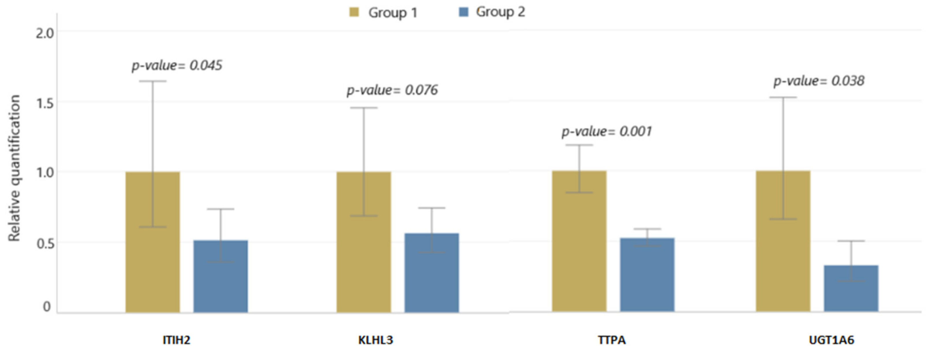
2.5. qPCR Validation

The ITIH2 gene, which exhibited a significant downregulation in expression and demonstrated hypermethylation in its promoter region, underwent qPCR analysis. This analysis confirmed the significant downregulation of the ITIH2 gene detected in the mRNA and RRBS sequencing results. Additionally, we selected three other genes (TTPA, UGT1A6, and KLKL3) whose expression as well as methylation levels changed under the influence of the increasing cholecalciferol dosages (Figure 1). The qPCR results confirmed significant reductions in the expression of TTPA (p-value = 0.001) and UGT1A6 (p-value = 0.038) genes. However, the decrease in the KLHL3 gene expression according to the qPCR results was not statistically significant (p-value = 0.076).

2.6. Functional Analysis

2.6.1. Methyl-Seq

Table 2 shows the top 10 (FDR < 0.0017 and strength > 0.3) functional effects identified by the RRBS sequencing. The analysis of a set of genes with altered methylation levels showed changes in 82 biological processes. According to the FDR values (<0.00075) and strength level (0.35), the actin filament-based process and actin cytoskeleton organization were most altered in this group. Potential effects on 39 biological functions were also observed. Within this group, according to the FDR values (<0.00031) and strength level (>0.35), the greatest effects of the increasing vitamin D intake were observed within the guanyl-nucleotide exchange factor activity, GTPase regulator activity, and actin binding. On the other hand, within the biological components, the results of the functional analysis showed effects on 26 areas. According to the FDR value (<0.00072) and strength level (>0.35), the greatest changes occurred in the actin cytoskeletons. All the results of the gene-set analysis are presented in Supplementary Table S5.
The separate analyses of the hypomethylated and hypermethylated DMSs were additionally carried out. The results of the analyses showed that hypomethylated DMSs enriched 16 biological process, 7 molecular functions, and 8 cellular components. According to the FDR and strength levels, the increasing vitamin D doses enriched the actin-binding (FDR = 0.016 and strength = 0.45) and cytoskeletal protein-binding (FDR < 0.001 and strength = 0.41) functions most potently. An analysis of the hypermethylated DMSs showed that 24 biological processes, 23 molecular functions, and 16 cellular components were enriched. According to the FDR and strength levels, the GTPase regulator activity was the most enriched (FDR < 0.001 and strength = 0.46). Interestingly, the analysis of the hypomethylated DMSs also showed a strong strength level of this molecular function (0.43; FDR = 0.02). On the other hand, according to the KEGG database, based on a set of hypermethylated DMSs, the phospholipase D signalling pathway was altered (FDR = 0.0153 and strength = 0.61).
The results of the hypomethylated/hypermethylated DMSs analysis are presented in Supplementary Table S7.

2.6.2. mRNA-Seq

Table 3 shows the top 10 strongest functional changes (5 from the GO database and 5 from KEEG) caused by the increasing cholecalciferol doses based on the mRNA sequencing results. The analysis showed that the applied changes in the diets of the pigs could significantly affect 103 biological processes. Based on the FDR (<0.0001) and strength (>1.2) values, we highlighted such processes as the negative regulation of blood coagulation, regulation of blood coagulation, retinoid metabolic process, and regulation of wound-healing. In turn, among the 19 significantly altered biological functions, we identified (FDR < 0.0001 and strength > 0.2) oxidoreductase activity and catalytic activity. Moreover, eight significant differences in the biological components were identified. In this area, the largest changes (FDR < 0.001 and strength > 0.49) occurred in the extracellular region and extracellular space. The results from the KEGG database indicated the effects of the increased vitamin D intake on 24 pathways, of which as many as 12 pathways with strength values of >1 and FDR values of <0.0001 could be distinguished. The results of the gene set analysis of the mRNA-seq results are presented in Supplementary Table S6.
The results of the analysis of the set of downregulated genes showed the enrichment of 106 biological processes, 28 molecular functions, and 8 cellular components. According to the FDR and strength levels, the most significantly enriched appeared to be the propionate metabolic process (FDR = 0.0007 and strength = 2.22). According to the KEGG database, the set of downregulated genes was most strongly involved (FDR < 0.001 and strength > 1.6) in the metabolism of the xenobiotics by the cytochrome P450 and chemical carcinogenesis pathways. The set of upregulated genes was not significantly involved in any pathway, nor did they significantly enrich any of the processes. The entire results of the analysis are presented in Supplementary Table S7.

2.6.3. Integration of the Methyl-Seq and mRNA-Seq Results

The analysis of a set of 11 genes, presented in Table 1, showed the significant affectation of 3 pathways (Table 4). The effects on the steroid hormone biosynthesis and retinol metabolism pathways were conditioned by the altered expression of CYP3A22 and HSD17B6. The glycine, serine, and threonine metabolism pathways, on the other hand, were found to be significantly altered due to the effects on the SHMT1 and BHMT genes.

3. Discussion

The present study showed that increasing doses of cholecalciferol caused significant changes in the transcriptomes and methylomes of swine lung tissues. We found that increased vitamin D intake significantly altered the expression of nearly 200 genes. Moreover, we identified 1116 differentially methylated sites related to genes and their regulatory sites. Among all the DMSs, only 11 were associated with genes with significant changes in expression levels (Table 1). A presumed reason for the poor correlation of the results (RRBS and mRNA) was the different number of samples used in each NGS study. However, based on the results of other researchers, it should be noted that relatively low correlations are common, especially in experiments where the effect of the studied factor is not exceptionally strong [6].
It has been assumed that, via the VDR, vitamin D regulates key mechanisms such as metabolism and cell proliferation [7,8]. Increased vitamin D intake causes activation of the VDR, which, in turn, intensifies gene transcription. However, our results indicated that more than 86% of the genes altered by the increased vitamin D intake were downregulated. Also, among the genes identified as those with altered methylation, there was little bias toward hypermethylation (56%).
A functional analysis of the methylome sequencing results showed that the increasing vitamin D intake affected the GTPase-, cytoskeleton-, and actin-related processes most significantly (Table 2). Interestingly, a recent finding showed that reduced SARS-CoV-2 lung disease severity was associated with methylation changes within processes related to GTPase and actin [9]. Moreover, it appeared that increasing the dose of cholecalciferol induced changes in the methylome associated with pulmonary fibrosis. Our results indicated methylation changes in the MeCP2 (methyl-CpG binding protein 2) gene, which is considered a key regulator of fibrosis (Supplementary Table S2) [10]. It has been shown, for example, that MeCP2 KO mice are resistant to pulmonary fibrosis [11]. Another study found that vitamin D could inhibit the TGFβ1 stimulation of α-smooth-muscle actin expression and polymerization and prevent the upregulation of fibronectin and collagen in fibroblasts in vitro. These results indicated that vitamin D may inhibit the pro-fibrotic phenotype of lung fibroblasts and epithelial cells [12]. Additionally, we identified four genes encoding collagen (COL4A1, COL5A1, COL6A3, and COL24A1) whose methylation levels were also changed (Supplementary Table S2). One of these, COL4A1 is considered a target of miR-29 in pulmonary fibrosis [10].
The functional analysis of the mRNA-seq results (Supplementary Table S6) identified genes related to complement and coagulation cascades (FDR < 0.0001) and chemical carcinogenesis (FDR < 0.0001). We detected the significant downregulation of the expression of genes encoding acute phase proteins (e.g., FBG, FGA, and FGG) and tumor-associated genes such as SERPINC1 and F2 (Supplementary Table S4). SERPINC1 is recognized as a key gene in the processes of cancer cell proliferation and migration. It has been shown that reducing SERPINC1 expression can be an effective treatment for lung cancer [13,14]. Furthermore, the F2-thrombin factor 2 gene may not only affect interactions between viral proteins and cytokine receptors [15] but also play an essential role in blood coagulation, angiogenesis, tissue repair, and vascular integrity.
Our study also showed that increasing vitamin D intake results in changes in the methylation and expression levels of genes linked to retinol and retinoid metabolism (Table 3 and Table 4). This was in line with the fact that the active form of vitamin D, combined with vitamin D receptor (VDR), forms a heterodimer with the retinoid X receptor (RXR). The heterodimer formed in this way can interact with the vitamin D gene’s response elements (VDRE) [16]. Interestingly, other researchers have shown that vitamin D supplementation can affect methylation within the RXR promoter [17].
The functional analysis of the results shared by the methylome and mRNA sequencing indicated the anti-tumor and anti-fibrotic effects of increasing the cholecalciferol dosage. We identified changes in the expression of the PSAT1 and SHMT1 genes, as well as methylation changes in the SHMT1 gene. The in vitro and in vivo studies on mice by Zhu et al. showed that PSAT1 is a strong promoter of pulmonary fibrosis and that VDR regulates the expression of this gene [18]. Moreover, an in vitro study on human cell lines showed that silencing the PSAT1 gene resulted in the inhibition of tumor proliferation and growth in non-small cell lung cancer [19]. The PSAT1 plays an important role in connecting pathways involving glycolysis and the biosynthesis of amino acids. Its decreased expression inhibits the synthesis of serine and, consequently, glycine. Glycine is a major component of collagen, which is the building material of connective tissue. Alterations in collagen production cause lung fibrosis and may regulate cell proliferation in lung tumors. Increasing the dose of cholecalciferol resulted in a significant decrease in PSAT1 expression and an effect on both the glycolysis and amino acid synthesis pathways, including glycine and serine. Therefore, our findings supported the suggestions that vitamin D may be a therapeutic agent in patients with pulmonary fibrosis and lung cancer, though not only through the regulation of PSAT1 but also the SHMT1 gene. SHMT1 encodes serine hydroxymethyltransferase 1, an enzyme essential for converting serine to glycine [18]. We identified the downregulation and hypermethylation of the SHMT1 gene under the influence of increasing cholecalciferol doses. Based on these results, it could be assumed that the effect of vitamin D in inhibiting collagen production in the lungs was bidirectional: first, through the inhibition of the synthesis of serine, the material necessary for the formation of collagen, and second, through the downregulation of the enzyme directly involved in collagen synthesis. Interestingly, the UGT1A6 gene, whose variations in expression and methylation were observed, is also significantly associated with the development of pulmonary fibrosis. There have been some indications that this gene is significantly upregulated in idiopathic pulmonary fibrosis patients compared to healthy controls [20]. Moreover, the increased expression of UGT1A6 is also characteristic of patients with cancers, including lung cancers [21,22]. UDP-glucuronosyltransferases are a group of enzymes associated with the catabolism of drugs and xenobiotics. Therefore, it is believed that altering the expression of UGT1A genes, including UGT1A6, can significantly modulate the response to the treatment, development, and progression of cancer [22]. A decrease in UGT1A6 gene expression under vitamin D (calcitriol) supplementation has already been observed by other researchers, though in rat liver tissues [23].
The effects of vitamin D on processes associated with pulmonary fibrosis and cancer were also suggested by changes within the ITIH2 (SHAP) gene. The ITIH2 gene in our study was the only one to have significantly altered mRNA expression via a change in methylation within the promoter. The ITIH2 gene, through the inter-alpha-trypsin inhibitor protein it encodes, is related to the serine inhibitor group. This gene may be involved in the control of inflammation and immune processes [24]. There have been indications that the ability to regulate angiogenesis may link ITIH2 to the process of pulmonary fibrosis [25]. A study by Garantziotis et al. showed that induced lung injury resulted in a greater than sixfold increase in ITIH2 expression in the liver and a decrease in its expression in the lungs [25]. Similarly, in lung cancers, ITIH2 shows significant downregulation in the altered tissues [24]. ITIH2 is a part of IaI protein, which is synthesized in the liver. IaI consists of a light chain and two heavy chains (ITIH1 and ITIH2). It is generally considered that IaI is a systemic factor that enhances angiogenesis. Histological studies have shown the strong colocalization of inter-α-trypsin inhibitor protein (IaI) in disease focus in patients with pulmonary fibrosis. In contrast, the lungs of healthy subjects were weakly stained for IaI [25]. It is noteworthy that angiogenesis in patients with pulmonary fibrosis and cancer may have a dual effect. In the early stages of the disease, it may promote tissue regeneration; however, in the exacerbated stages, it may accelerate the development of the disease. The results cited above have indicated that IaI shows higher levels in patients with fibrosis; however, the expression of the lung ITIH2 gene decreases. This result may suggest another role, unrelated to the IaI, for ITIH2 in lung tissues.
KLHL3 (kelch-like family member 3) is another gene that was downregulated by the increasing cholecalciferol intake in our study. There are many indications that the KLHL3 gene is linked to cancer development. This gene appears to be downregulated in the plasma samples of patients with lung cancers [26]. However, findings regarding the expression and regulation of this gene in lung tissues are lacking. Studies presenting the expression of this gene in tumor tissues have indicated that it can be both overexpressed and downregulated under the influence of disease [27]. Nevertheless, the role of KLHL3 in the pathogenesis of cancers formed through viral interaction appears to be well-proven [28]. Indeed, it appears that KLHL3 protein expression is significantly higher in cells expressing vIRF1 (viral interferon regulatory factor 1), as well as in cells infected with KSHV (Kaposi’s sarcoma-associated herpesvirus). Researchers have found that KLHL3 mediates the infection and replication of KSHV-induced tumorigenesis [28].
Another interesting result was the significant downregulation of the TTPA (alpha tocopherol transfer protein) gene, which we confirmed by the mRNA-seq (log2FoldChange = −2.297) and qPCR results. In addition, we observed significant methylation changes in the introns of this gene. TTPA binds the biologically active form of vitamin E (α-tocopherol) and plays an important role in regulating the levels of this vitamin in the body. Due to its strong antioxidant properties, numerous studies have been conducted to determine whether vitamin E supplementation can protect against cancer. The first clinical trial conducted on a large group of male tobacco smokers surprisingly showed that mortality was 2% higher in the group of those taking a daily dose of alpha-tocopherol compared to the group of those not supplemented with alpha-tocopherol [29]. The lack of protective effects of vitamin E supplementation in cancer and cardiovascular disease was also confirmed in another clinical trial conducted on a group of healthy women [30]. The change in the expression level of the TTPA gene we observed under the influence of vitamin D indicated a possible interaction between simultaneous dietary supplementation with vitamin D and E, and we recommend further studies in this area.
Altogether, our findings, along with those of other researchers, have suggested that increasing the intake of cholecalciferol in the daily diet may exhibit anti-cancer and anti-fibrotic effects (Figure 2). It is assumed that respiratory infections can cause chronic fibrotic reactions even up to several months after infection [31]. Therefore, our results appear to be important both for breeders of animals at risk of lung disease and from a human health point of view. It is important to highlight that our mRNA sequencing analysis was conducted on a limited number of samples exclusively from males. Nevertheless, the robustness of these findings was validated across a more extensive cohort encompassing both males and females through RRBS and qPCR testing. Nonetheless, further investigations are warranted to comprehensively explore the correlation between vitamin D and the development of pulmonary fibrosis and cancer formation.

4. Materials and Methods

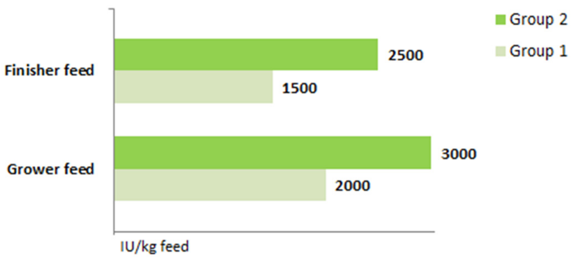
4.1. Animal, Diets, and 25(OH)D Blood Serum Concentration Measurements

All the procedures conducted on live animals had the approval of the local Ethical Committee for Experiments with Animals in Cracow (Resolution No. 427/2020 dated 22 July 2020). The animals were kept under the same conditions, in individual pens, at the Research Station of the State Research Institute of Animal Production in Grodziec Śląski. In the experiment, animals from two groups were utilized. Animals in the first group (group 1) received a standard dose, while those in the second group (group 2) received increased doses of cholecalciferol (Figure 3). Both groups contained 10 individuals, with 5 males and 5 females in each group. The animals received the same feed, though it differed in the levels of cholecalciferol. The feed covered all their current requirements (grower, 30–60 kg: metabolizable energy, 13.3 MJ and total protein, 172 g/kg; finisher, 60–110 kg: metabolizable energy, 13.3 MJ and total protein, 156 g/kg).
The feeding experiment ended when the animals reached individual weights of 110 kg (88 days). Immediately after the animals were slaughtered, samples from the middle parts of the upper lobes of the left lungs were collected. The samples were stored in a freezer (−85 °C) until further analysis.
The assays of total 25(OH)D concentrations in the animal plasma samples were carried out by the ANCHEM Laboratorium from Katowice in Poland, in line with the RIA method, using a DIAsource 25OH Vitamin D total-RIA-CT Kit (Rue de Bosquet 2, 1348 Louvain-La-Neuve, Belgium) and Multigamma 1260 multidetector instrument (LKB Wallac, Turku, Finland). The plasma vitamin D levels were measured using blood samples collected at slaughter.

4.2. DNA and RNA Isolations, Library Construction, and Sequencing

The total DNA from 16 lung samples (8 females and 8 males) was isolated using a Wizard® Genomic DNA Purification Kit (Promega, Madison, WI, USA). The concentrations of the genetic materials (DNA) were confirmed using NanoDrop™ 2000/2000c spectrophotometers (Thermo Scientific™, Waltham, MA, USA). Then, high-quality DNA samples were used to prepare the libraries. The libraries for the methyl-seq were prepared using an Ovation® RRBS Methyl-Seq System 1–16 kit (Tecan, San Jose, CA, USA). The RRBS libraries were sequenced in the United States by Medical Research Foundation NGS Core using Illumina NovaSeq 6000 device (Illumina, San Diego, CA, USA) as 150 bp-paired end reads and using PhiX control.
RNA was isolated from 10 lung samples (5 samples from group 1 and 5 samples from group 2, only females) using a PureLink™ RNA Mini Kit (Invitrogen, Waltham, MA, USA). The isolated genetic materials were quantitatively and qualitatively assessed using Tapestation 2200 (Agilent, Santa Clara, CA, USA). The high-quality RNA samples were used for the library preparation using a QuantSeq 3′mRNA-Seq Library Prep Kit FWD for Illumina (Lexogen, Vienna, Austria). The procedures were carried out in accordance with the manufacturers’ recommendations. Quantitative evaluation of the prepared libraries was performed using Qubit (Thermo Scientific™, Waltham, MA, USA), while a qualitative evaluation was assessed using Tapestation 2200 device (Agilent, Santa Clara, CA, USA). Sequencing of the mRNA pooled libraries (75 bp single read) was performed using a Nextseq 5500 device (Illumina, San Diego, CA, USA). The libraries were prepared for sequencing according to the standard normalization method from the NextSeq 500 and NextSeq 550 Sequencing Systems-Denature and Dilute Libraries Guide protocol. We used a 2 nM starting library concentration and a 10% PhiX addition.

4.3. qPCR Validation

Twenty samples were used for the qPCR analysis. The experiment began with RNA isolation from the 10 remaining samples. RNA from 20 lung samples was reverse transcribed. We performed qPCR on the KLHL3, TTPA, UGT1A6, and ITIH2 genes using RPS29 as an endogenous control. The genes were selected based on the results of the RRBS and mRNA sequencing, and 500 ng of RNA was reverse-transcribed to cDNA using a High-Capacity RNA-to-cDNA™ Kit (Applied Biosystems™, Waltham, MA, USA). The real-time PCR was performed using TaqMan™ Fast Advanced Master Mix for qPCR (Applied Biosystems™, Waltham, MA, USA) and TaqMan Real-Time PCR assays on a QuantStudio ™ 7 Flex Real-Time PCR System (Applied Biosystems™, Waltham, MA, USA). The relative quantity data were analyzed on a Thermo Fisher Cloud (Thermo Scientific™, Waltham, MA, USA). Analysis of the results was carried out using SAS 9.4 software (SAS Institute Inc., Cary, NC, USA).

4.4. Statistical Analysis

4.4.1. Methyl-Seq

The first step of the data analysis was the quality control of the raw sequencing reads using FastQC v. 0.12.1 software. Low-quality reads (quality level of <20 and read length of <36) and fragments containing adapter sequences were filtered using FlexBar v. 3.5.0 software. Matching to the swine reference genome (Sscrofa11.1) was performed using bisulfite mapping software-BSMAP v. 2.9.0, with the default options recommended for RRBS data specifying the enzyme cleavage site (MspI) and mapping to the two forward strands. The Methylation Caller software provided in the BSMAP v. 2.9.0 package was used to determine the percentage of methylation in the individual CpG sites with the coverage of more than 5 reads. The CpG methylation analysis included the distribution in the swine chromosomes and the distribution in the upstream regions, 5′-UTRs, 3′-UTRs, exons, introns, and intergenic regions. Next, the files were processed using R package (version 4.3) to obtain the input data for the Methylkit software. Methylkit v. 1.26.0 software was used to identify the differentially methylated sites (DMS) with cutoff values of at least 25% methylation differences between the two groups and q-values of <0.05. Gene annotations were obtained from the Sscrofa11.1 Ensembl GTF annotation.

4.4.2. mRNA-Seq

The demultiplexed fastq files downloaded from the sequencing server were quality-checked, trimmed from reads, and mapped from reads using FastQC 11.8, FLEXBAR 3.5.0, and TopHat 2.1.1, respectively. Samtools 1.9, RSeQC, HTSeq-count 0.11.1 software, and Gtf-Ensembl annotation 96 were used to assess the mapping statistics and read counts. Then, to perform the differential expression analysis, the R program and DESeq 2 software suites were used. Differentially expressed genes were regarded as genes with q-values <0.05 (FDR, false discovery rate), Benjamini–Hochberg (BH) adjustments, and no fold-change thresholds. Only the genes that showed base means > 20 were used for further analyses. The gene annotations were obtained from the Sscrofa11.1 Ensembl GTF annotation.

4.4.3. Integration of the Methyl-Seq and mRNA-Seq Results

We used the RRBS and RNA-seq datasets to relate the changes in the CpG site methylation to the changes in the expression levels of the associated genes. The Venny 2.1 program was used for this comparison.

4.4.4. Functional Analysis

An analysis of the methyl-seq results, mRNA-seq results, and the combined data was performed. The upregulated/hypermethylated and downregulated/hypomethylated genes were used for joint (gene set analysis (GSA)) and separate analyses.
The functional analyses were carried out using STRING software (version 12.0), with which Gene Ontology (GO) and Kyoto Encyclopedia of Genes and Genomes (KEGG) pathway enrichment analyses were performed. In addition, the BioMart (release 110) and Venny 2.1 programs were used during these analyses. The top 10 results from the functional enrichment analysis were selected based on the FDR (false discovery rate) and strength level values.

5. Conclusions

Our results showed the putative mechanism by which increasing vitamin D intake may reduce the risk of lung cancer and pulmonary fibrosis in healthy individuals. These actions may have been due to the effects of vitamin D on collagen production-related genes, such as SHMT1, UGT1A6, and ITIH2. The anticancer properties of vitamin D are further supported by changes in the expression levels of the KLHL3 and TTPA genes. Changes in TTPA gene expression also indicated that vitamin D can affect vitamin E action. The likely epigenetic mechanism regulating these processes is DNA methylation; however, further experimental studies are needed to confirm our hypothesis.

Supplementary Materials

The following supporting information can be downloaded at: https://www.mdpi.com/article/10.3390/ijms25010464/s1.

Author Contributions

Conceptualization, M.O. and M.Ś.; methodology, M.O., E.S.-G. and A.W.; software, T.S.; validation, A.W. and M.O.; formal analysis, A.W., A.S., M.O. and E.S.-G.; investigation, A.W. and M.O.; resources, M.O.; data curation, A.W.; writing—original draft preparation, A.W.; writing—review and editing, M.O. and E.S.-G.; visualization, A.W.; supervision, M.O. and E.S.-G.; project administration, A.W., M.O. and E.S.-G.; funding acquisition, M.O. and E.S.-G. All authors have read and agreed to the published version of the manuscript.

Funding

This research was funded by the National Research Institute of Animal Production under grant number 501-180-621.

Institutional Review Board Statement

The animal study protocol was approved by the Ethics Committee for Experiments with Animals in Cracow (resolution no. 427/2020 of 22 July 2020).

Informed Consent Statement

Not applicable.

Data Availability Statement

The sequencing results have been deposited in the NCBI GEO database under access numbers GSE248607 (RBBS data) and GSE242293 (mRNA data).

Conflicts of Interest

The authors declare no conflicts of interest.

References

  1. Wierzbicka, A.; Oczkowicz, M. Sex differences in vitamin D metabolism, serum levels and action. Br. J. Nutr. 2022, 128, 2115–2130. [Google Scholar] [CrossRef] [PubMed]
  2. Das, R.R.; Singh, M.; Naik, S.S. Vitamin D as an adjunct to antibiotics for the treatment of acute childhood pneumonia. Cochrane Database Syst. Rev. 2023, 1, CD011597. [Google Scholar] [CrossRef] [PubMed]
  3. Mullin, M.L.L.; Milne, S. Vitamin D deficiency in chronic obstructive pulmonary disease. Curr. Opin. Pulm. Med. 2023, 29, 96–103. [Google Scholar] [CrossRef] [PubMed]
  4. Ashique, S.; Gupta, K.; Gupta, G.; Mishra, N.; Singh, S.K.; Wadhwa, S.; Gulati, M.; Dureja, H.; Zacconi, F.; Oliver, B.G. Vitamin D-A prominent immunomodulator to prevent COVID-19 infection. Int. J. Rheum. Dis. 2023, 26, 13–30. [Google Scholar] [CrossRef] [PubMed]
  5. Mandell, E.; Ryan, S.; Seedorf, G.J.; Gonzalez, T.; Abman, S.H.; Fleet, J.C. Maternal vitamin D deficiency induces transcriptomic changes in newborn rat lungs. J. Steroid Biochem. Mol. Biol. 2020, 199, 105613. [Google Scholar] [CrossRef] [PubMed]
  6. Berger, T.C.; Vigeland, M.D.; Hjorthaug, H.S.; Etholm, L.; Nome, C.G.; Taubøll, E.; Heuser, K.; Selmer, K.K. Neuronal and glial DNA methylation and gene expression changes in early epileptogenesis. PLoS ONE 2019, 14, e0226575. [Google Scholar] [CrossRef] [PubMed]
  7. Saccone, D.; Asani, F.; Bornman, L. Regulation of the vitamin D receptor gene by environment, genetics and epigenetics. Gene 2015, 561, 171–180. [Google Scholar] [CrossRef] [PubMed]
  8. Hanel, A.; Carlberg, C. Vitamin D and evolution: Pharmacologic implications. Biochem. Pharmacol. 2020, 173, 113595. [Google Scholar] [CrossRef]
  9. Alkafaas, S.S.; Abdallah, A.M.; Ghosh, S.; Loutfy, S.A.; Elkafas, S.S.; Abdel Fattah, N.F.; Hessien, M. Insight into the role of clathrin-mediated endocytosis inhibitors in SARS-CoV-2 infection. Rev. Med. Virol. 2023, 33, e2403. [Google Scholar] [CrossRef]
  10. O’Reilly, S. Epigenetics in fibrosis. Mol. Asp. Med. 2017, 54, 89–102. [Google Scholar] [CrossRef]
  11. Biao, H.; Gharaee-Kermani, M.; Wu, Z.; Phan, S.H. Essential Role of MeCP2 in the Regulation of Myofibroblast Differentiation during Pulmonary Fibrosis. Am. J. Pathol. 2011, 178, 1500–1508. [Google Scholar] [CrossRef]
  12. Ramirez, A.M.; Wongtrakool, C.; Welch, T.; Steinmeyer, A.; Zügel, U.; Roman, J. Vitamin D inhibition of pro-fibrotic effects of transforming growth factor beta1 in lung fibroblasts and epithelial cells. J. Steroid Biochem. Mol. Biol. 2010, 118, 142–150. [Google Scholar] [CrossRef]
  13. Zhang, J.; Tang, Z.; Guo, X.; Wang, Y.; Zhou, Y.; Cai, W. Synergistic effects of nab-PTX and anti-PD-1 antibody combination against lung cancer by regulating the Pi3K/AKT pathway through the Serpinc1 gene. Front. Oncol. 2022, 12, 933646. [Google Scholar] [CrossRef] [PubMed]
  14. Wang, Y.; Sun, Y.; Feng, J.; Li, Z.; Yu, H.; Ding, X.; Yang, F.; Linghu, E. Glycopatterns and Glycoproteins Changes in MCN and SCN: A Prospective Cohort Study. Biomed. Res. Int. 2019, 2019, 2871289. [Google Scholar] [CrossRef] [PubMed]
  15. Feng, S.; Song, F.; Guo, W.; Tan, J.; Zhang, X.; Qiao, F.; Guo, J.; Zhang, L.; Jia, X. Potential Genes Associated with COVID-19 and Comorbidity. Int. J. Med. Sci. 2022, 19, 402–415. [Google Scholar] [CrossRef] [PubMed]
  16. Saponaro, F.; Saba, A.; Zucchi, R. An Update on Vitamin D Metabolism. Int. J. Mol. Sci. 2020, 21, 6573. [Google Scholar] [CrossRef]
  17. Curtis, E.M.; Krstic, N.; Cook, E.; D’Angelo, S.; Crozier, S.R.; Moon, R.J.; Murray, R.; Garratt, E.; Costello, P.; Cleal, J.; et al. Gestational Vitamin D Supplementation Leads to Reduced Perinatal RXRA DNA Methylation: Results from the MAVIDOS Trial. J. Bone Miner. Res. 2019, 34, 231–240. [Google Scholar] [CrossRef]
  18. Zhu, W.; Ding, Q.; Wang, L.; Xu, G.; Diao, Y.; Qu, S.; Chen, S.; Shi, Y. Vitamin D3 alleviates pulmonary fibrosis by regulating the MAPK pathway via targeting PSAT1 expression in vivo and in vitro. Int. Immunopharmacol. 2021, 101 Pt B, 108212. [Google Scholar] [CrossRef]
  19. Yang, Y.; Wu, J.; Cai, J.; He, Z.; Yuan, J.; Zhu, X.; Li, Y.; Li, M.; Guan, H. PSAT1 regulates cyclin D1 degradation and sustains proliferation of non-small cell lung cancer cells. Int. J. Cancer 2015, 136, E39–E50. [Google Scholar] [CrossRef]
  20. Qian, W.; Xia, S.; Yang, X.; Yu, J.; Guo, B.; Lin, Z.; Wei, R.; Mao, M.; Zhang, Z.; Zhao, G.; et al. Complex Involvement of the Extracellular Matrix, Immune Effect, and Lipid Metabolism in the Development of Idiopathic Pulmonary Fibrosis. Front. Mol. Biosci. 2022, 8, 800747. [Google Scholar] [CrossRef]
  21. Li, J.; Li, Q.; Su, Z.; Sun, Q.; Zhao, Y.; Feng, T.; Jiang, J.; Zhang, F.; Ma, H. Lipid metabolism gene-wide profile and survival signature of lung adenocarcinoma. Lipids Health Dis. 2020, 19, 222. [Google Scholar] [CrossRef]
  22. Cengiz, B.; Yumrutas, O.; Bozgeyik, E.; Borazan, E.; Igci, Y.Z.; Bozgeyik, I.; Oztuzcu, S. Differential expression of the UGT1A family of genes in stomach cancer tissues. Tumour Biol. 2015, 36, 5831–5837. [Google Scholar] [CrossRef] [PubMed]
  23. Doan, T.N.K.; Vo, D.K.; Kim, H.; Balla, A.; Lee, Y.; Yoon, I.S.; Maeng, H.J. Differential Effects of 1α,25-Dihydroxyvitamin D3 on the Expressions and Functions of Hepatic CYP and UGT Enzymes and Its Pharmacokinetic Consequences In Vivo. Pharmaceutics 2020, 12, 1129. [Google Scholar] [CrossRef]
  24. Hamm, A.; Veeck, J.; Bektas, N.; Wild, P.J.; Hartmann, A.; Heindrichs, U.; Kristiansen, G.; Werbowetski-Ogilvie, T.; Del Maestro, R.; Knuechel, R.; et al. Frequent expression loss of Inter-alpha-trypsin inhibitor heavy chain (ITIH) genes in multiple human solid tumors: A systematic expression analysis. BMC Cancer 2008, 8, 25. [Google Scholar] [CrossRef] [PubMed]
  25. Garantziotis, S.; Zudaire, E.; Trempus, C.S.; Hollingsworth, J.W.; Jiang, D.; Lancaster, L.H.; Richardson, E.; Zhuo, L.; Cuttitta, F.; Brown, K.K.; et al. Serum inter-alpha-trypsin inhibitor and matrix hyaluronan promote angiogenesis in fibrotic lung injury. Am. J. Respir. Crit. Care Med. 2008, 178, 939–947. [Google Scholar] [CrossRef] [PubMed]
  26. Zhang, W.; Zhang, Q.; Che, L.; Xie, Z.; Cai, X.; Gong, L.; Li, Z.; Liu, D.; Liu, S. Using biological information to analyze potential miRNA-mRNA regulatory networks in the plasma of patients with non-small cell lung cancer. BMC Cancer 2022, 22, 299. [Google Scholar] [CrossRef] [PubMed]
  27. Lin, Y.; Li, Q.; Jin, X. Kelch-like protein 3 in human disease and therapy. Mol. Biol. Rep. 2022, 49, 9813–9824. [Google Scholar] [CrossRef] [PubMed]
  28. Qi, X.; Yan, Q.; Shang, Y.; Zhao, R.; Ding, X.; Gao, S.J.; Li, W.; Lu, C. A viral interferon regulatory factor degrades RNA-binding protein hnRNP Q1 to enhance aerobic glycolysis via recruiting E3 ubiquitin ligase KLHL3 and decaying GDPD1 mRNA. Cell Death Differ. 2022, 29, 2233–2246. [Google Scholar] [CrossRef] [PubMed]
  29. Alpha-Tocopherol; Beta Carotene Cancer Prevention Study Group. The effect of vitamin E and beta carotene on the incidence of lung cancer and other cancers in male smokers. N. Engl. J. Med. 1994, 330, 1029–1035. [Google Scholar] [CrossRef]
  30. Lee, I.M.; Cook, N.R.; Gaziano, J.M.; Gordon, D.; Ridker, P.M.; Manson, J.E.; Hennekens, C.H.; Buring, J.E. Vitamin E in the primary prevention of cardiovascular disease and cancer: The Women’s Health Study: A randomized controlled trial. JAMA 2005, 294, 56–65. [Google Scholar] [CrossRef]
  31. Huang, S.; Goplen, N.P.; Zhu, B.; Cheon, I.S.; Son, Y.; Wang, Z.; Li, C.; Dai, Q.; Jiang, L.; Xiang, M.; et al. Macrophage PPAR-γ suppresses long-term lung fibrotic sequelae following acute influenza infection. PLoS ONE 2019, 14, e0223430. [Google Scholar] [CrossRef]
Figure 1. The qPCR results (RQ, relative quantification) for the groups of animals supplemented with different doses of vitamin D. The error bars represent the RQ min and RQ max values within the groups.
Ijms 25 00464 g001
Figure 2. Putative molecular mechanism of the effect of increasing vitamin D intake on lung cancer and pulmonary fibrosis risks. The red and blue dots illustrate the change in gene methylation, the red arrows show decreases in gene expression, the black arrows illustrate the potential inhibition of processes, and the green arrows show the likely final effects of the changes that occurred.
Ijms 25 00464 g002
Figure 3. The content of cholecalciferol in the grower and finisher feeds used in the groups of animals.
Ijms 25 00464 g003
Table 1. Similarities in the results of the mRNA-seq and methyl-seq data analyses.
GenemRNA-SeqMethyl-Seq
Log2FoldChangeBase MeanMeth. Diff.Consequence
ITIH2−2.1641543.60230.606upstream gene
HSD17B6−1.796318.769−32.363intron
CYP3A22−1.8841599.78−30.897intron
TTPA−2.297201.53−29.991intron
SHMT1−1.721007.481−28.281intron
MIPEP−1.021196.128−27.29intron
PSMA11.65841.83525.747intron
HDLBP−2.089291.19630.216intron
KLHL3−2.213227.07330.429intron
BHMT−2.2271523.9138.036intron
UGT1A6−1.603260.4150.914intron
Table 2. Most significant results from the functional analysis of the RRBS sequencing data from the lung tissues from pigs supplemented with increased doses of cholecalciferol.
BaseTerm DescriptionNo. of GenesStrengthFDR
GO molecular functionGTPase regulator activity550.453.3 × 10−7
GO molecular functionCytoskeletal protein binding840.311.34 × 10−6
GO molecular functionEnzyme regulator activity920.284.21 × 10−6
GO molecular functionKinase binding600.357.62 × 10−6
GO molecular functionGuanyl-nucleotide exchange factor activity280.470.00025
GO molecular functionActin binding430.360.00031
GO biological processesActin filament-based process510.350.00035
GO cellular componentActin cytoskeleton410.350.00072
GO biological processesActin cytoskeleton organization470.350.00075
Table 3. Most significant results from the functional analysis of the mRNA sequencing data for the lung tissues from pigs supplemented with increased doses of cholecalciferol.
BaseTerm DescriptionNo. of GenesStrengthFDR
KEGGChemical carcinogenesis121.551.66 × 10−12
KEGGMetabolism of xenobiotics by cytochrome P450111.579.57 × 10−12
KEGGRetinol metabolism101.522.77 × 10−10
KEGGDrug metabolism–cytochrome P45091.53.59 × 10−9
GO biological processNegative regulation of blood coagulation81.532.94 × 10−7
GO biological processRegulation of blood coagulation91.42.94 × 10−7
GO biological processRegulation of wound-healing101.218.14 × 10−7
GO biological processBlood coagulation101.170.0000017
GO biological processRetinoid metabolic process81.297.68 × 10−6
KEGGTyrosine metabolism51.340.00022
Table 4. Results of the functional analysis of the set of 11 genes identified in the RRBS and mRNA sequencing data from the lung tissues from pigs supplemented with increased doses of cholecalciferol.
BaseTerm DescriptionNo. of GenesStrengthFDR
KEGGSteroid hormone biosynthesis22.030.0387
KEGGGlycine, serine, and threonine metabolism22.110.0387
KEGGRetinol metabolism220.0387
Disclaimer/Publisher’s Note: The statements, opinions and data contained in all publications are solely those of the individual author(s) and contributor(s) and not of MDPI and/or the editor(s). MDPI and/or the editor(s) disclaim responsibility for any injury to people or property resulting from any ideas, methods, instructions or products referred to in the content.

