Article

1,25-Dihydroxyvitamin D3 Suppresses UV-Induced Poly(ADP-Ribose) Levels in Primary Human Keratinocytes, as Detected by a Novel Whole-Cell ELISA

by
Warusavithana Gunawardena Manori De Silva
1,
Vanessa Bernadette Sequeira
1,
Chen Yang
1,
Katie Marie Dixon
2,
Andrew J. A. Holland
3,
Rebecca Sara Mason
1,4,* and
Mark Stephen Rybchyn
1
1
Department of Physiology, School of Medical Sciences and Bosch Institute, The University of Sydney, Sydney, NSW 2006, Australia
2
Department of Anatomy and Histology and Bosch Institute, The University of Sydney, Sydney, NSW 2006, Australia
3
Douglas Cohen Department of Paediatric Surgery, The Children’s Hospital at Westmead Clinical School, The Faculty of Medicine and Health, The University of Sydney, Sydney, NSW 2006, Australia
4
School of Life and Environmental Sciences and Charles Perkins Centre, The University of Sydney, Sydney, NSW 2006, Australia
*
Author to whom correspondence should be addressed.
Int. J. Mol. Sci. 2024, 25(11), 5583; https://doi.org/10.3390/ijms25115583
Submission received: 5 April 2024 / Revised: 13 May 2024 / Accepted: 16 May 2024 / Published: 21 May 2024
(This article belongs to the Special Issue Irradiation and Anti-cancer Therapies: Future and Challenges)

Abstract

:
Photoprotective properties of 1,25-dihydroxyvitamin D3 (1,25(OH)2D3) to reduce UV-induced DNA damage have been established in several studies. UV-induced DNA damage in skin such as single or double strand breaks is known to initiate several cellular mechanisms including activation of poly(ADP-ribose) (pADPr) polymerase-1 (PARP-1). DNA damage from UV also increases extracellular signal-related kinase (ERK) phosphorylation, which further increases PARP activity. PARP-1 functions by using cellular nicotinamide adenine dinucleotide (NAD+) to synthesise pADPr moieties and attach these to target proteins involved in DNA repair. Excessive PARP-1 activation following cellular stress such as UV irradiation may result in excessive levels of cellular pADPr. This can also have deleterious effects on cellular energy levels due to depletion of NAD+ to suboptimal levels. Since our previous work indicated that 1,25(OH)2D3 reduced UV-induced DNA damage in part through increased repair via increased energy availability, the current study investigated the effect of 1,25(OH)2D3 on UV-induced PARP-1 activity using a novel whole-cell enzyme- linked immunosorbent assay (ELISA) which quantified levels of the enzymatic product of PARP-1, pADPr. This whole cell assay used around 5000 cells per replicate measurement, which represents a 200–400-fold decrease in cell requirement compared to current commercial assays that measure in vitro pADPr levels. Using our assay, we observed that UV exposure significantly increased pADPr levels in human keratinocytes, while 1,25(OH)2D3 significantly reduced levels of UV-induced pADPr in primary human keratinocytes to a similar extent as a known PARP-1 inhibitor, 3-aminobenzamide (3AB). Further, both 1,25(OH)2D3 and 3AB as well as a peptide inhibitor of ERK-phosphorylation significantly reduced DNA damage in UV-exposed keratinocytes. The current findings support the proposal that reduction in pADPr levels may be critical for the function of 1,25(OH)2D3 in skin to reduce UV-induced DNA damage.

1. Introduction

Poly(ADP-ribose) polymerase (PARP) is a ubiquitous nuclear enzyme that was reported as early as 1963 [1]. It is found in mammalian and some eukaryotic cells [2,3,4]. Eighteen different protein isoforms have been identified [5]. PARP-1 is the most prominent member of the PARP family, a 116 kDa zinc-finger nuclear protein that is involved in DNA repair due to environmental stress. PARP-1 binds to DNA single- or double- stranded breaks and then PARylates acceptor proteins (including itself) [6,7]. While DNA damage directly increases PARP-1 activity, there is evidence for further increased PARP activation after UV as a result of increased extracellular signal-regulated kinase (ERK) phosphorylation [8,9,10,11]. PARP has three highly conserved domains: an amino-terminal, DNA-binding domain (binds to the damaged DNA and causes conformational shift) [12,13,14,15]; a central auto-modification domain (releases proteins from DNA) [15,16,17]; and a carboxyl-terminal, catalytic domain [15,18,19]. PARP-1 is a substrate for cellular caspases [20], which facilitate apoptosis. PARP-1 is proteolysed by caspase-3 and -7 into an 89 kDa catalytic domain and a 24 kDa DNA-binding domain [12,21,22,23,24]. This cleavage suppresses PARP-1 activity [21,25] as binding of the 24 kDa fragment alone to the damaged DNA site inhibits repair due to the lack of the catalytic fragment [26].
Mild DNA damage initiates DNA repair pathways, but if the damage is severe, the cell will undergo apoptosis or necrosis. PARP-1 is activated upon DNA damage recognition, and activated PARP utilizes cellular nicotinamide adenine dinucleotide (NAD+) to form nicotinamide and ADP-ribosyl moieties (PARs) into protein acceptor molecules (histones, transcription factors) to form long-chain poly(ADP-ribose) (pADPr), a process known as PARylation. The removal of pADPr is mainly carried out by poly(ADP-ribose) glycohydrolase (PARG), without which there is uncontrolled pADPr accumulation and cytotoxicity [27]. Excessive PARylation depletes cellular NAD+ and energy levels [28,29,30,31,32,33] ultimately, if not controlled, leading to “parthanatos”, a distinct type of cellular death that occurs due to excessive PARP-1 activation [24]. It has also been reported that energy depletion by activated PARP-1 is due to inhibition of the rate-limiting enzyme, hexokinase, in glycolysis [34,35]. A combination of these factors and mitochondrial damage by UV, which increases oxidative stress, leads to depletion of cellular NAD+ and adenosine triphosphate (ATP) levels that may contribute to reduced energy levels which inhibit energy-demanding cellular repair processes and thereby may progress the cell to parthanatos, apoptosis, or necrosis. Although not the focus of this study, it should be noted that PARP-1 function is closely linked with poly(ADP-ribose) glycohydrolase (PARG) activity, an enzyme which catalyses the breakdown of PARs formed by PARP proteins. This process ultimately recycles energy back into the cellular system [36], and several lines of evidence suggest that PAR removal is required for efficient DNA repair [36,37,38].
The active vitamin D hormone, 1,25-dihydroxyvitamin D3 (1,25(OH)2D3), which is made in skin as a result of UVB exposure [39,40,41,42], has been shown to reduce UV-induced DNA damage, both cyclobutane pyrimidine dimers (CPD) [43] and oxidative DNA damage, measured as 8-hydroxy-2′-deoxyguanosine (8-OHdG) [44,45,46,47] in human keratinocytes [48,49,50], ex vivo in human skin explants [46] and in vivo in Skh:hr1 mice [43,48,49,50,51,52,53] and in human volunteers [54]. At least part of its mechanism of action is to increase DNA repair through an increase in glycolysis with concurrent suppression of energy consuming anabolic cellular signalling pathways ultimately leading to increased cellular ATP levels [45].
Given that UV exposure greatly increases PARP-1, which in turn inhibits glycolysis [34,35], we previously speculated that reduced accumulation of pADPr moieties was important in the mechanism of action of 1,25(OH)2D3, but had no way to test this given the large cell numbers required for commercial PARP assays available at the time. The current study investigated the effect of 1,25(OH)2D3 on UV-induced PARP in primary human keratinocytes using a novel whole-cell ELISA to quantify the enzymatic product, pADPr. Because UV also increases phosphorylation of ERK (ERK-p) [55], which increases PARP-1 activity [8,9,11], and we have reported that 1,25(OH)2D3 reduced UV-induced ERK-p [45], we examined whether the known PARP inhibitor, 3AB, as well as a peptide inhibitor of ERK-p would mimic the effects of 1,25(OH)2D3 on UV-induced DNA damage.

2. Results

2.1. UV Dose–Response Study to Detect Phosphorylated PARP Product, pADPr with a Novel Whole-Cell ELISA

Whole-cell enzyme linked immunosorbent assay (ELISA) or cell-based ELISA is an immunocytometric technique used to target and identify cellular proteins or post-translational modifications of target protein in cultured cells. It is a sensitive and a versatile technique [56]. For the novel assay, described in more detail in Methods, keratinocytes were fixed with formaldehyde 5 or 15 min after irradiation and then permeabilised. They were incubated with an antibody to pADPr or an isotype control. After overnight incubation at 4 °C, the cells were incubated with anti-mouse IgG horse radish peroxidase (HRP)-linked antibody, followed by the addition of HRP substrate. Absorbance was measured at 450 nm.
Whereas a solar-simulated UV dose equivalent to 4 min exposure to noon-day sun in October in Sydney, Australia (Dose 1) has generally been used on human keratinocytes for DNA damage studies by our group [45], this dose was insufficient to cause measurable UV-induced increases to the levels of pADPr compared to non-irradiated cells (Figure 1A). Increasing the UV irradiation dose three-fold (Dose 3) resulted in significant increases to UV-induced pADPr compared to non-irradiated cells from 0.05 ± 0.01 absorbance units (AU) to 0.12 ± 0.01 (AU) (Figure 1A, p < 0.01). Thus, Dose 3 of UV (equivalent to 12 min of noon-day sun in October in Sydney, Australia) was used for the ELISA experiments.

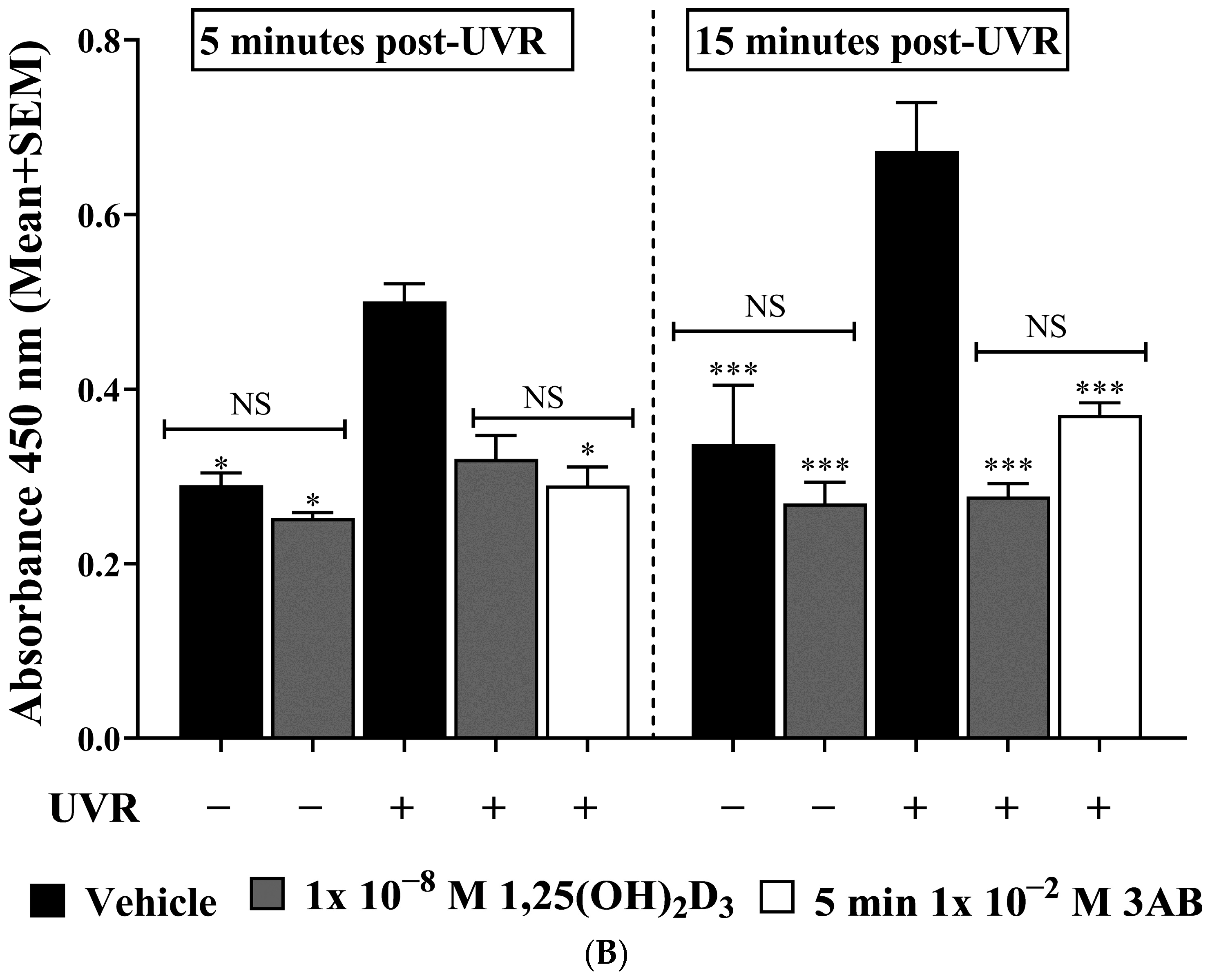
2.2. UV-Induced pADPr Levels Were Significantly Reduced by Treatment with 1,25(OH)2D3 or 3AB in Human Primary Keratinocytes

Significant increases in UV-induced pADPr were detected at 5 min from 0.3 ± 0.01 AU to 0.5 ± 0.02 AU (Figure 1B, p < 0.05) and 15 min post-UV from 0.3 ± 0.07 AU to 0.7 ± 0.06 AU (Figure 1B, p < 0.001) compared to non-irradiated cells. Treatment with 1,25(OH)2D3 resulted in significant suppression of pADPr levels 15 min post-irradiation from 0.7 ± 0.06 AU to 0.3 ± 0.01 AU (Figure 1B, p < 0.001) with a non-significant decrease at 5 min post-irradiation (Figure 1B, NS). The addition of the known PARP-1 inhibitor 3-aminobenzimide (3AB) [57] resulted in decreased pADPr levels after UV from 0.5 ± 0.02 AU to 0.3 ± 0.02 AU at 5 min (Figure 1B, p < 0.05) and from 0.7 ± 0.06 AU to 0.4 ± 0.01 AU 15 min post-UV (Figure 1B, p < 0.001), not significantly different from those in cells treated with 1,25(OH)2D3 (Figure 1B).

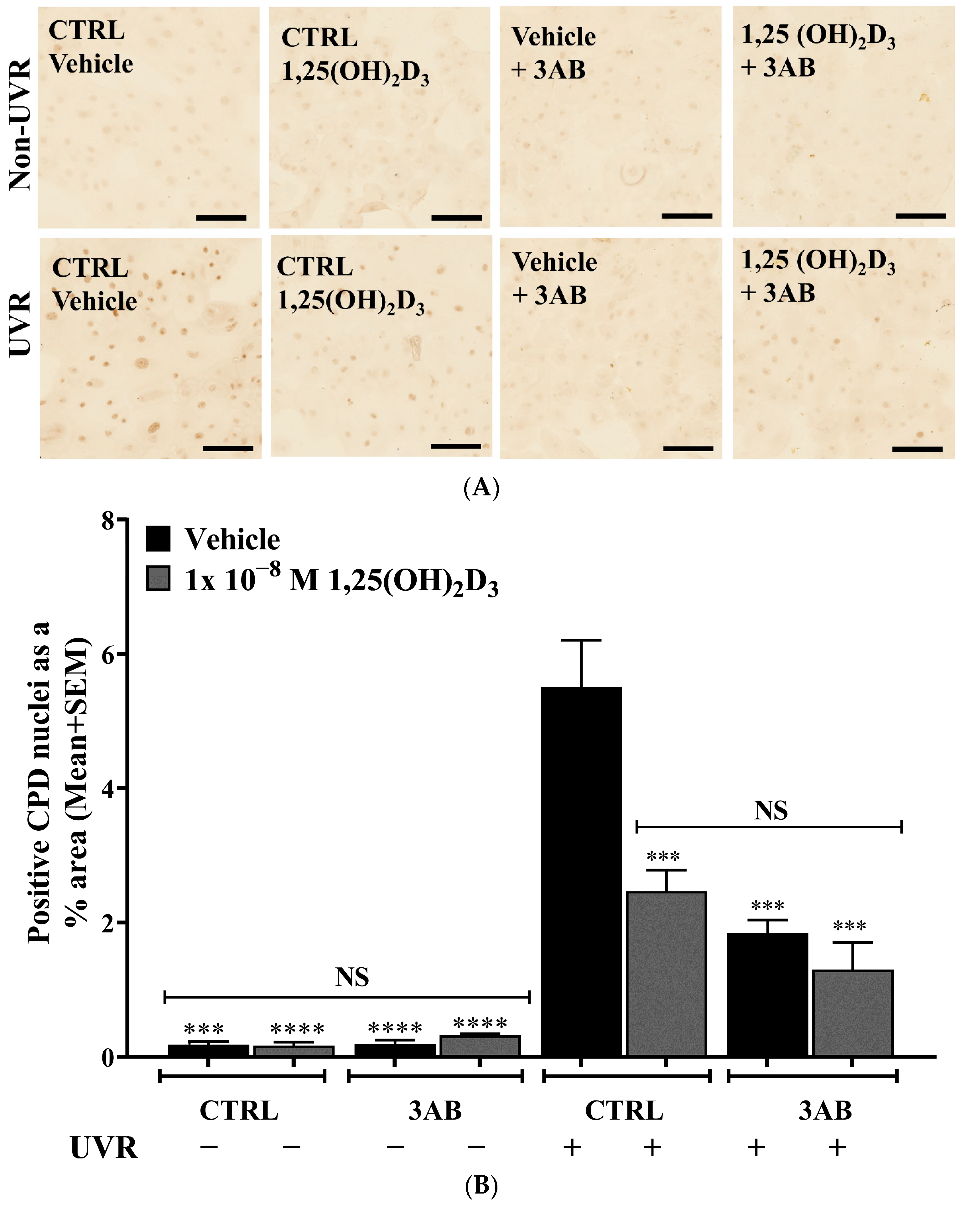
2.3. Treatment of UV-Irradiated Keratinocytes with 1,25(OH)2D3 or 3AB Resulted in a Significant Reduction in CPD Levels

UV irradiation of primary human keratinocytes resulted in a significant ~31-fold increase in detected CPD levels from 0.2 ± 0.05% area to 5.5 ± 0.7% area (Figure 2A,B, p < 0.0001), which is generally considered a marker of direct DNA damage by UV [58]. Treatment of keratinocytes with 1,25(OH)2D3 resulted in a significant ~50% reduction in detected CPD levels compared to vehicle-treated cells (Figure 2B, p < 0.001). Treatment of keratinocytes with 3AB also resulted in a similar significant ~60% reduction in detected CPD levels compared to vehicle-treated cells (Figure 2B, p < 0.001). The combination of 1,25(OH)2D3 and 3AB produced no further decrease in CPD.

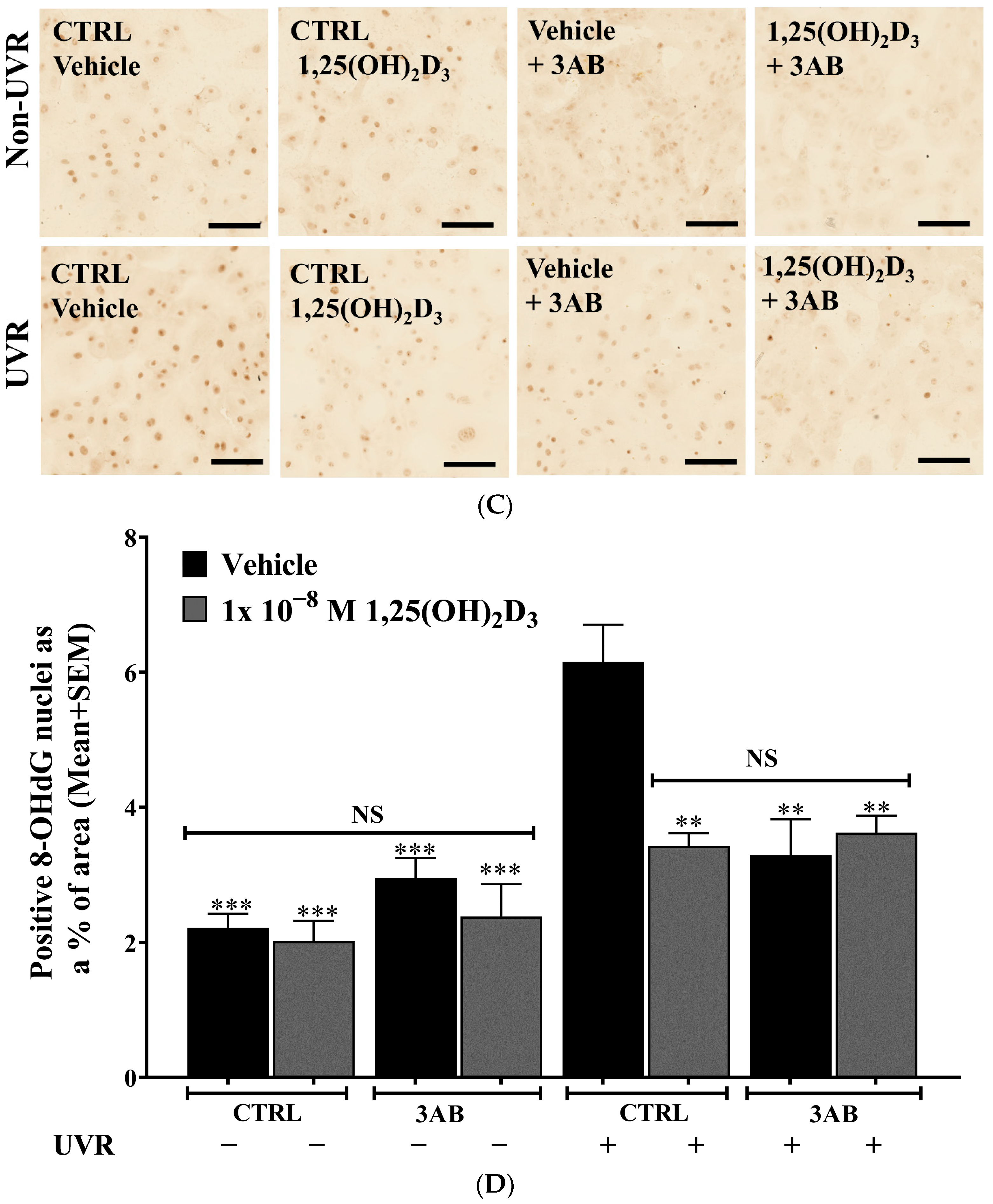
2.4. Treatment of UV-Irradiated Keratinocytes with 1,25(OH)2D3 or 3AB Resulted in a Significant Reduction in 8-OHdG Levels

UV irradiation of primary human keratinocytes resulted in a significant ~3-fold increase in detected 8-OHdG levels from 2.2 ± 0.2% area to 6.2 ± 0.5% area (Figure 2C,D, p < 0.001), a marker of oxidative DNA damage [59,60,61,62,63]. Treatment of keratinocytes with 1,25(OH)2D3 resulted in a significant ~50% reduction in detected 8-OHdG levels compared to vehicle-treated cells (Figure 2D, p < 0.01). Treatment of keratinocytes with 3AB also resulted in a similar significant ~50% reduction in detected 8-OHdG levels compared to vehicle-treated cells (Figure 2D, p < 0.01). Again, the combination of 1,25(OH)2D3 and 3AB produced no further decrease in 8-OHdG.

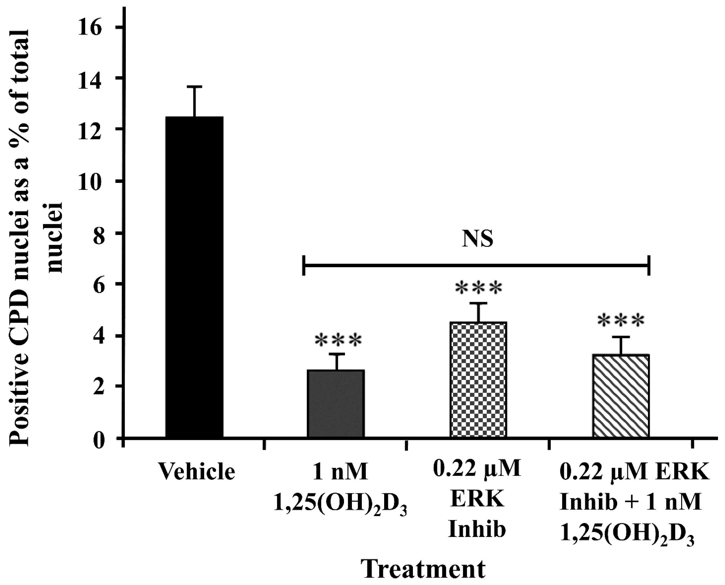
2.5. A Peptide ERK Inhibitor Reduced UV-Induced CPD Levels to a Similar Extent as 1,25(OH)2D3

PARP activity has previously been shown to be increased via direct interaction with phosphorylated ERK1/2 [10] as well as by direct DNA damage [6,7]. We previously reported that 1,25(OH)2D3 treatment of primary human keratinocytes reduced phosphorylation of ERK [45]. To provide a possible mechanistic explanation as to how 1,25(OH)2D3 suppresses PARP-1 activity in keratinocytes, we examined the effect of treatment of keratinocytes with ERK activation inhibitor peptide II (IC50 = 0.21 µM). This ERK inhibitor reduced UV-induced CPD from 12.5 ± 0.5% area to 4.7 ± 0.3% area, similar to the effect of 1,25(OH)2D3 to 2.7 ± 0.2% area (Figure 3), and there was no additional effect when the two agents were used together.

3. Discussion

Using a novel whole-cell ELISA, which required only ~5000 cells per replicate measurement, we were able to reliably quantify PARP activity as defined by the presence of the enzymatic product pADPr in the cells [64]. Commercially available PARP activity assays require 106–107 cells per treatment [65]. Due to the limited number of primary keratinocytes from a single donor and the limit of cell division cycles by keratinocytes in vitro [66] generating such large numbers of cells is not technically feasible. Furthermore, the formalin-based fixed cell assay used in this study did not require a lysis step, which may possibly allow specific (i.e., PARG) or non-specific breakdown of pADPr moieties prior to analysis. Our research group has previously used a similar type of assay to successfully measure UV-induced 3-nitrotyrosine levels in this cell phenotype [48].
Excessive activation of PARP-1 has been shown to result in depletion of cellular energy levels via NAD+ consumption [28,29,32], and due to inhibition of the rate-limiting enzyme, hexokinase, in glycolysis [34,35]. Suppressed PARP-1 activity by 1,25(OH)2D3 would in turn result in less UV-induced suppression of hexokinase activity, which would reduce UV-induced glycolytic blockade in the irradiated cell [34]. Reducing the depletion of NAD+ and conserving cellular energy pools [28,31,32] would contribute to increased repair of DNA damage. These results extend and complement earlier studies by our group showing that control of pathways and processes that regulate cellular energy is critical for repair of UV-induced DNA damage in skin cells [45]. UV-induced PARP-1 activity was shown to be reduced by treatment with 1,25(OH)2D3 in the immortalised keratinocyte HaCaT cell line [67,68]. Our group has previously shown that increased glycolysis and suppression of cellular growth pathways (e.g., mTOR) are observed following treatment with 1,25(OH)2D3 in primary human keratinocytes, ultimately leading to increased cellular ATP levels [45]. Such an energetically favorable outcome would be expected to be observed if UV-induced glycolytic blockade was alleviated in response to 1,25(OH)2D3 treatment. Therefore, the data presented here further reveal the role that 1,25(OH)2D3 plays in post-irradiation control of cell energy levels, via suppression of excessive accumulation of pADPr [69]. In vivo, it is likely that 1,25(OH)2D3, which is produced and accumulates in the skin after UV exposure [39,40], may in part contribute to increases in cellular energy levels and thus act as a defense mechanism against subsequent rounds of UV exposure by permitting the cell to more effectively repair UV-induced damage.
Actions of 1,25(OH)2D3 to reduce keratinocyte losses after UV and to inhibit oxidative stress, either measured as oxidative UV damage or other oxidative or nitrosative stress markers, have been reported in human keratinocytes and in mouse skin [45,51,70]. Indeed, patients with non-melanoma skin cancers and mostly low vitamin D status had higher markers of oxidative stress in their blood than non-skin cancer controls [71]. The combination of increased DNA repair, together with reduced oxidative stress in the presence of 1,25(OH)2D3, may explain the higher burden of non-melanoma skin cancers observed in mice with knockout of the vitamin D receptor after chemical or chronic UV treatments [72,73] and the reduced skin cancers reported in mice exposed to long-term UV but treated topically with 1,25(OH)2D3 [51]. It thus seems likely that 1,25(OH)2D3 and possibly other related compounds made in skin as a result of UV exposure [39,40,41,42] result in less skin damage than would otherwise be the case, although this is difficult to test in human populations.
Previously published studies have shown that PARP activity can be controlled by phosphorylated ERK [8,9,64,74,75,76]. As previously reported, 1,25(OH)2D3 reduced UV-induced phosphorylated ERK [45], thus providing a possible molecular mechanism for how 1,25(OH)2D3 may suppress PARP activity, since this inhibition of ERK phosphorylation would then result in decreased accumulation of pADPr moieties, as observed. In support of the proposal that increased PARP-1 activity leads to reduced energy for DNA repair, we observed that a peptide inhibitor of ERK phosphorylation and a direct inhibitor of PARP-1 [77], 3AB, both inhibited UV-induced DNA damage to a similar extent as 1,25(OH)2D3 and that there was no additive effect of 1,25(OH)2D3 with either agent.
In skin cells, 1,25(OH)2D3 is generally thought to function via two separate but probably complementary pathways: (1) the classical steroid receptor/genomic pathway, which involves VDR/RXR-dependent (vitamin D receptor-retinoid X receptors) gene transcription following translocation of the receptor complex from the nuclear membrane and (2) the rapid response “non–genomic” pathway, which is also thought to act via the VDR that is non-nuclear-associated [70]. The rapid suppression of ERK-p by 1,25(OH)2D3 might support a non-genomic mechanism. Other evidence also points to 1,25(OH)2D3-dependent suppression of PARP-1 activity in a variety of model systems via both the genomic and non-genomic pathways. In a diabetic rat model, increased parp-1 gene expression was significantly reduced in animals that were on a vitamin D3 diet [78]. In a separate diabetic rat model, direct oral administration of 1,25(OH)2D3 resulted in decreased PARP-1 quantified at the protein level [79]. In a study of Parkinson’s disease (PD) pathophysiology, 1,25(OH)2D3 relieved parthanatos in a neuroblastoma cellular model [80]. This study also showed a direct interaction between PARP-1 and the VDR, and overexpression of VDR in this cellular model reduced PARP-1 protein levels [80]. Another mechanistic possibility is that, given pADPr moieties need to be removed by PARG prior to DNA repair [36,69], 1,25(OH)2D3 may facilitate quicker removal of pADPr groups, thereby allowing the DNA repair machinery faster and better access to the DNA lesion for repair [27,81]. Our assay, which measures these pADPr moieties, does not distinguish between inhibited PARP activity or increased removal of pADPr. Other limitations of the study are that we needed three times the dose of solar-simulated UV to show a significant increase in pADPr compared to that used for demonstration of DNA damage. Although this is hardly excessive, being the equivalent of 12 min of noon day sun in Sydney, Australia in October, it suggests that the assay is not as sensitive as other immunohistochemical methods. Also limiting is that the studies, for practical reasons, were carried out in cell culture and not in whole skin.
Since PARP is known to contribute to the DNA repair process [11,27], why PARP inhibition would reduce UV-induced DNA lesions following a non-lethal dose of UV radiation is unclear. It is becoming increasingly accepted, however, that the balance between pADPr synthesis and removal is key to optimal repair [69]. It is also plausible that in the absence of PARP activation the cells may shift to other repair mechanisms, or energy-consuming functions, that would otherwise be suppressed. There is a wide range of examples in the literature where PARP inhibitors exert beneficial effects [82,83,84], but several other studies provided contradictory findings [85,86,87]. In UV-irradiated normal human epidermal keratinocytes treated with 3AB, reduced necrosis, apoptosis, and ROS production were reported [83]. PARP−/− mice were reported to be viable and fertile but with increased susceptibility to skin disease in older mice [88]. Further, the cells from PARP−/− mice maintained efficient repair of damaged DNA [88,89,90,91,92]. It is also possible that excessive PARP-1 activation may result in excessive accumulation of pADPr moieties which inhibit repair. Topical application of a PARP inhibitor, BPG-15M [O-(3-pyperidino-2-hydroxy-1-propyl) pyridine-3-carboxylic acid amidoxime monohydrochloride], to hairless mice before UV exposure reduced UV-induced DNA damage, immunosuppression, inflammation, and sunburn cells [84], indicating that reduced PARP activity has an acute photoprotective effect. The current data support this interpretation from these studies.
The current study used a novel whole-cell ELISA-based assay to allow measurement of pADPr levels in primary human keratinocytes after UV exposure. Optimal PARP activity relies on a balance between pADPr accumulation and removal [69]. The data presented here support the proposal that 1,25(OH)2D3 increases energy availability and thus increased DNA repair in keratinocytes after UV, at least in part through a reduction in phosphorylation of ERK and thus a reduction in excessive accumulation of UV-induced pADPr.

4. Materials and Methods

4.1. Cell Culture

Samples of skin removed at elective surgery were harvested after obtaining written informed consent from subjects or their parents/guardians, with approval from the University of Sydney Human Ethics Committee (Reference number 2015/063) and according to Declaration of Helsinki Principles. Keratinocytes were cultured from these skin explants as previously described [43,48]. Keratinocytes (passages 2–5) from at least three independent donors were used in all experiments.

4.2. Treatments

1,25(OH)2D3 (Sapphire Bioscience Pty Ltd., Sydney, Australia) was solubilised in spectroscopic ethanol (Merck, Darmstadt, Germany), and the vehicle was 0.1% (v/v) ethanol. The concentration of this compound was determined by a Nanodrop ND-1000 Spectrophotometer (Thermo Fisher Scientific, Wilmington, NC, USA). The 1,25(OH)2D3 was added immediately after UV exposure, as is our standard protocol [45,51]. 3-Aminobenzamide (3AB) was from Sigma Aldrich (St. Louis, MO, USA) and was used at a concentration of 1 × 10⁻³ M [57,83]. ERK activation inhibitor, peptide II (IC50 = 0.21 µM), which selectively binds to ERK2, preventing its interaction with MEK [93], was purchased from Calbiochem-EMD4 Biosciences (Gibbstown, NJ, USA). Both inhibitors were present during and after UV exposure.

4.3. UV Irradiation

UV irradiation (ssUV) was provided by an Oriel Sol1ATM 94042A 450W solar simulator (Newport Corporation, Irvine, CA, USA). The output of this lamp was around 10% UVB and 90% UVA as previously described [45]. Our standard protocol for irradiation used 400 mJ/cm2 UVB and 3600 mJ/cm2 UVA (4000 mJ/cm2 = Dose 1), measured by an IL1700 research radiometer (International Light Technologies, Peabody, MA, USA), equivalent to 4 min of noon-day sun in October in Sydney, Australia [43,51]. pADPr experiments were carried out at 3 times this level to test for significant increases in UV-induced pADPr levels.

4.4. PARP Measurements by Novel Whole-Cell ELISA

A UV-induced increase in PARP activity was used as positive control, as has been previously established to occur in skin cells [83]. 3-Aminobenzamide (3AB) was added to these cells to provide a negative control and was present during and after irradiation, as is standard protocol [57]. Cells were fixed with 10% (v/v) formaldehyde in PBS, pH 7.4, at 5 min and 15 min post-irradiation and treatment. Cell membranes were permeabilised with 0.1% (v/v) Triton X-100, and endogenous peroxidase activity was blocked with 1% (v/v) H2O2. Non-specific antibody binding was inhibited by 0.05% (w/v) heat-denatured casein (HDC) in PBS, pH 7.4, and pADPr was detected by incubation of mouse monoclonal IgG3 pADPr Antibody (10H): sc-56198 antibody (Santa Cruz Biotechnologies, Dallas, TX, USA) at 5 µg/mL in HDC overnight at 4 °C. Isotype mouse control antibody was used at a similar experimental concentration and under similar conditions. The following day, cells were incubated with anti-mouse IgG horse radish peroxidase (HRP)-linked antibody (Cell Signaling Technology, Danvers, MA, USA) at 0.4% (v/v) followed by addition of the HRP substrate, 3,3′,5,5′-tetramethylbenzidine (0.1 mg/mL in 0.05 M citrate 0.1 M phosphate buffer at pH 5.5) (Sigma-Aldrich, St. Louis, MO, USA) with 0.02% (v/v) H2O2. The colourimetric reaction was stopped by the addition of 50% (v/v) of 2 M H2SO4 (VWR international, Radnor, PA, USA). The absorbance was quantified at 450 nm. The absorbance generated by matched isotype wells was subtracted from test data to account for non-specific binding of the antibody.

4.5. CPD and 8-OHdG Immunohistochemistry

As an index of CPD, thymine dimers [94] and 8-OHdG were detected and measured as previously described [47,95]. In brief, primary human keratinocytes were cultured at 90% confluence on 5 mm poly-l-lysine-coated glass coverslips, exposed to UV and treatments as indicated and fixed at 1.5 or 3 h after UV (as stated). After rinses with phosphate buffered saline (PBS), cells were fixed with ice-cold 100% methanol (Sigma-Aldrich, St. Louis, MO, USA) at −20 °C for 10 min. Keratinocytes were then extensively washed with MilliQ water at pH 7.2 (Millipore SAS, Molsheim, France) and air dried at room temperature overnight. The next day immunohistochemistry was carried out as previously reported [43], with minor modifications. Treatment with peroxide (1% v/v—ThermoFisher Scientific, Waltham, MA, USA) in PBS for 5 min, followed by two MilliQ water washes at room temperature, was used to block endogenous peroxidase activity. Several steps were used for antigen retrieval. Sodium hydroxide (70 mM) (Sigma-Aldrich, St. Louis, MO, USA) in 70% ethanol for 2 min at room temperature was used to denature nuclear DNA. Protein digestion was carried out using proteinase K (Sigma-Aldrich, St. Louis, MO, USA) (1 μg/mL in 0.1 mM CaCl2) for 5 min for CPD detection and 10 min for 8-OHdG detection, followed by two MilliQ water washes. Treatment of cells with 50% (v/v) horse serum (Sigma-Aldrich, St. Louis, MO, USA) in PBS, pH 7.2 (blocker), for 1 hour at room temperature was used to inhibit non-specific antibody binding. Keratinocytes were incubated with primary antibody in blocker for 60 min at room temperature: mouse monoclonal anti-thymine dimer at 5 µg/mL (Sigma-Aldrich, St. Louis, MO, USA) was used for CPD or primary mouse monoclonal IgG2b anti-8-hydroxy-guanosine antibody (Santa Cruz Biotechnologies, Dallas, TX, USA) for 8-OHdG at 2.5 µg/mL or an equivalent concentration of isotype control, followed by three TBS-T washes at room temperature. Cells were incubated with biotinylated goat anti-mouse IgG secondary antibody (ThermoFisher Scientific, Waltham, MA, USA) diluted to 1:500 in Tris-buffered saline with 0.1% Tween 20 (TBS-T) for 15 min, followed by three TBS-T washes at room temperature. Streptavidin-conjugated horseradish peroxidase (HRP) (Invitrogen, Waltham, MA, USA) diluted 1:150 in TBS-T was added for 15 min, followed by three TBS-T washes to amplify the primary antibody signal. Insoluble HRP substrate, Diaminobenzidine (DAB) (Enhanced Liquid substrate System for Immunohistochemistry, Sigma-Aldrich, St. Louis, MO, USA), was then added for 5 min, followed by two MilliQ water washes following sufficient colour development. Isotype antibodies were used as controls for all experiments. Isotype controls resulted in minimal staining as previously reported [96]. Entellen rapid mounting medium (Merck, Darmstadt, Germany) was used to mount air-dried coverslips on glass slides for image analysis [43].
Bright-field images were acquired at 20× magnification on an Olympus stereo investigator scope (MBF Bioscience, Williston, VT, USA) or the Zeiss Axio Scan-Z1 slide scanner (Carl Zeiss Microscopy, Jena, Germany). Staining intensity was quantified using ImageJ software version 1.52u (released 17 March 2020) [47,95].

4.6. Statistical Analysis

All experiments were repeated a total of three times with triplicate measurement for each treatment group in each independent experiment. All experiments yielded similar results. Primary keratinocytes were sourced from different donors for replicate experiments. Generally, statistical analyses were carried out using GraphPad Prism version 8.3.1 released on 19 January 2020 (San Diego, CA, USA) by one-way ANOVA with Tukey’s post-test comparison unless otherwise stated.

Author Contributions

Conceptualization: W.G.M.D.S., M.S.R., R.S.M.; Methodology: M.S.R., R.S.M., W.G.M.D.S., V.B.S.; Data curation: W.G.M.D.S., V.B.S., C.Y., M.S.R.; Formal analysis: W.G.M.D.S., V.B.S., M.S.R.; Investigation, M.S.R., W.G.M.D.S., V.B.S., C.Y.; Project administration: M.S.R., W.G.M.D.S., R.S.M.; Resources: A.J.A.H., R.S.M.; Validation: M.S.R., K.M.D., R.S.M.; Writing—original draft preparation: W.G.M.D.S., M.S.R.; Writing, review and editing: W.G.M.D.S., M.S.R., A.J.A.H., R.S.M.; Visualization: W.G.M.D.S., V.B.S.; Supervision: M.S.R., R.S.M., K.M.D.; Funding acquisition: R.S.M. All authors have read and agreed to the published version of the manuscript.

Funding

The research was funded by the National Health and Medical Research Council of Australia (grant no. APP1070688), and by the Australian Research Council (ARC Linkage grant with Ultraceuticals Pty Ltd. LP100200680).

Institutional Review Board Statement

All human tissue studies were conducted in accordance with the Declaration of Helsinki and was approved by the Human Research Ethics Committee of the University of Sydney (protocol no. 2015/063).

Informed Consent Statement

Informed consent was obtained from all subjects or parents/guardians involved in the study. Skin tissue samples were collected under a protocol approved by the University of Sydney Human Ethics Committee (Reference number (2015/063) and were de-identified after collection.

Data Availability Statement

The data that support the findings of this study are available from the corresponding author upon reasonable request. Some data may not be made available because of privacy or ethical restrictions.

Acknowledgments

We would like to acknowledge expert assistance from the Bosch Advanced Microscopy Facility, and the Bosch Molecular Biology Facility at the University of Sydney. The authors acknowledge the technical assistance provided by the Sydney Informatics Hub, a Core Research Facility of the University of Sydney.

Conflicts of Interest

The authors declare that they have no conflicts.

References

  1. Chambon, P.; Weill, J.D.; Mandel, P. Nicotinamide mononucleotide activation of a new DNA-dependent polyadenylic acid synthesizing nuclear enzyme. Biochem. Biophys. Res. Commun. 1963, 11, 39–43. [Google Scholar] [CrossRef] [PubMed]
  2. Alkhatib, H.M.; Chen, D.F.; Cherney, B.; Bhatia, K.; Notario, V.; Giri, C.; Stein, G.; Slattery, E.; Roeder, R.G.; Smulson, M.E. Cloning and expression of cDNA for human poly(ADP-ribose) polymerase. Proc. Natl. Acad. Sci. USA 1987, 84, 1224–1228. [Google Scholar] [CrossRef]
  3. Uchida, K.; Morita, T.; Sato, T.; Ogura, T.; Yamashita, R.; Noguchi, S.; Suzuki, H.; Nyunoya, H.; Miwa, M.; Sugimura, T. Nucleotide sequence of a full-length cDNA for human fibroblast poly(ADP-ribose) polymerase. Biochem. Biophys. Res. Commun. 1987, 148, 617–622. [Google Scholar] [CrossRef]
  4. Ludwig, A.; Behnke, B.; Holtlund, J.; Hilz, H. Immunoquantitation and size determination of intrinsic poly(ADP-ribose) polymerase from acid precipitates. An analysis of the in vivo status in mammalian species and in lower eukaryotes. J. Biol. Chem. 1988, 263, 6993–6999. [Google Scholar] [CrossRef]
  5. Dantzer, F.; Giraud-Panis, M.-J.; Jaco, I.; Amé, J.-C.; Schultz, I.; Blasco, M.; Koering, C.-E.; Gilson, E.; Ménissier-de Murcia, J.; de Murcia, G.; et al. Functional interaction between poly(ADP-Ribose) polymerase 2 (PARP-2) and TRF2: PARP activity negatively regulates TRF2. Mol. Cell. Biol. 2004, 24, 1595–1607. [Google Scholar] [CrossRef] [PubMed]
  6. Javle, M.; Curtin, N.J. The role of PARP in DNA repair and its therapeutic exploitation. Br. J. Cancer 2011, 105, 1114–1122. [Google Scholar] [CrossRef]
  7. de Murcia, G.; de Murcia, J.M. Poly(ADP-ribose) polymerase: A molecular nick-sensor. Trends Biochem. Sci. 1994, 19, 172–176. [Google Scholar] [CrossRef]
  8. Kauppinen, T.M.; Chan, W.Y.; Suh, S.W.; Wiggins, A.K.; Huang, E.J.; Swanson, R.A. Direct phosphorylation and regulation of poly(ADP-ribose) polymerase-1 by extracellular signal-regulated kinases 1/2. Proc. Natl. Acad. Sci. USA 2006, 103, 7136–7141. [Google Scholar] [CrossRef] [PubMed]
  9. Cohen-Armon, M.; Visochek, L.; Rozensal, D.; Kalal, A.; Geistrikh, I.; Klein, R.; Bendetz-Nezer, S.; Yao, Z.; Seger, R. DNA-independent PARP-1 activation by phosphorylated ERK2 increases Elk1 activity: A link to histone acetylation. Mol. Cell 2007, 25, 297–308. [Google Scholar] [CrossRef]
  10. Cohen-Armon, M. PARP-1 activation in the ERK signaling pathway. Trends Pharmacol. Sci. 2007, 28, 556–560. [Google Scholar] [CrossRef]
  11. Boehi, F.; Manetsch, P.; Hottiger, M.O. Interplay between ADP-ribosyltransferases and essential cell signaling pathways controls cellular responses. Cell Discov. 2021, 7, 104. [Google Scholar] [CrossRef] [PubMed]
  12. Nicholson, D.W.; Ali, A.; Thornberry, N.A.; Vaillancourt, J.P.; Ding, C.K.; Gallant, M.; Gareau, Y.; Griffin, P.R.; Labelle, M.; Lazebnik, Y.A.; et al. Identification and inhibition of the ICE/CED-3 protease necessary for mammalian apoptosis. Nature 1995, 376, 37–43. [Google Scholar] [CrossRef]
  13. Gradwohl, G.; Ménissier de Murcia, J.M.; Molinete, M.; Simonin, F.; Koken, M.; Hoeijmakers, J.H.; de Murcia, G. The second zinc-finger domain of poly(ADP-ribose) polymerase determines specificity for single-stranded breaks in DNA. Proc. Natl. Acad. Sci. USA 1990, 87, 2990–2994. [Google Scholar] [CrossRef] [PubMed]
  14. Langelier, M.F.; Servent, K.M.; Rogers, E.E.; Pascal, J.M. A third zinc-binding domain of human poly(ADP-ribose) polymerase-1 coordinates DNA-dependent enzyme activation. J. Biol. Chem. 2008, 283, 4105–4114. [Google Scholar] [CrossRef] [PubMed]
  15. Langelier, M.F.; Planck, J.L.; Roy, S.; Pascal, J.M. Structural basis for DNA damage-dependent poly(ADP-ribosyl)ation by human PARP-1. Science 2012, 336, 728–732. [Google Scholar] [CrossRef] [PubMed]
  16. Loeffler, P.A.; Cuneo, M.J.; Mueller, G.A.; DeRose, E.F.; Gabel, S.A.; London, R.E. Structural studies of the PARP-1 BRCT domain. BMC Struct. Biol. 2011, 11, 37. [Google Scholar] [CrossRef] [PubMed]
  17. Langelier, M.F.; Eisemann, T.; Riccio, A.A.; Pascal, J.M. PARP family enzymes: Regulation and catalysis of the poly(ADP-ribose) posttranslational modification. Curr. Opin. Struct. Biol. 2018, 53, 187–198. [Google Scholar] [CrossRef] [PubMed]
  18. Simonin, F.; Ménissier-de Murcia, J.; Poch, O.; Muller, S.; Gradwohl, G.; Molinete, M.; Penning, C.; Keith, G.; de Murcia, G. Expression and site-directed mutagenesis of the catalytic domain of human poly(ADP-ribose)polymerase in Escherichia coli. Lysine 893 is critical for activity. J. Biol. Chem. 1990, 265, 19249–19256. [Google Scholar] [CrossRef]
  19. Ruf, A.; de Murcia, G.; Schulz, G.E. Inhibitor and NAD+ binding to poly(ADP-ribose) polymerase as derived from crystal structures and homology modeling. Biochemistry 1998, 37, 3893–3900. [Google Scholar] [CrossRef]
  20. Castri, P.; Lee, Y.J.; Ponzio, T.; Maric, D.; Spatz, M.; Bembry, J.; Hallenbeck, J. Poly(ADP-ribose) polymerase-1 and its cleavage products differentially modulate cellular protection through NF-kappaB-dependent signaling. Biochim. Biophys. Acta 2014, 1843, 640–651. [Google Scholar] [CrossRef]
  21. Lazebnik, Y.A.; Kaufmann, S.H.; Desnoyers, S.; Poirier, G.G.; Earnshaw, W.C. Cleavage of poly(ADP-ribose) polymerase by a proteinase with properties like ICE. Nature 1994, 371, 346–347. [Google Scholar] [CrossRef]
  22. Cohen, G.M. Caspases: The executioners of apoptosis. Biochem. J. 1997, 326 Pt 1, 1–16. [Google Scholar] [CrossRef] [PubMed]
  23. Tewari, M.; Quan, L.T.; O’Rourke, K.; Desnoyers, S.; Zeng, Z.; Beidler, D.R.; Poirier, G.G.; Salvesen, G.S.; Dixit, V.M. Yama/CPP32 beta, a mammalian homolog of CED-3, is a CrmA-inhibitable protease that cleaves the death substrate poly(ADP-ribose) polymerase. Cell 1995, 81, 801–809. [Google Scholar] [CrossRef] [PubMed]
  24. Mashimo, M.; Onishi, M.; Uno, A.; Tanimichi, A.; Nobeyama, A.; Mori, M.; Yamada, S.; Negi, S.; Bu, X.; Kato, J.; et al. The 89-kDa PARP1 cleavage fragment serves as a cytoplasmic PAR carrier to induce AIF-mediated apoptosis. J. Biol. Chem. 2021, 296, 100046. [Google Scholar] [CrossRef]
  25. Kaufmann, S.H.; Desnoyers, S.; Ottaviano, Y.; Davidson, N.E.; Poirier, G.G. Specific Proteolytic Cleavage of Poly(ADP-ribose) Polymerase: An Early Marker of Chemotherapy-induced Apoptosis. Cancer Res. 1993, 53, 3976–3985. [Google Scholar]
  26. Yung, T.M.; Satoh, M.S. Functional competition between poly(ADP-ribose) polymerase and its 24-kDa apoptotic fragment in DNA repair and transcription. J. Biol. Chem. 2001, 276, 11279–11286. [Google Scholar] [CrossRef]
  27. Nie, L.; Wang, C.; Huang, M.; Liu, X.; Feng, X.; Tang, M.; Li, S.; Hang, Q.; Teng, H.; Shen, X.; et al. DePARylation is critical for S phase progression and cell survival. bioRxiv 2024. [Google Scholar] [CrossRef] [PubMed]
  28. Goodwin, P.M.; Lewis, P.J.; Davies, M.I.; Skidmore, C.J.; Shall, S. The effect of gamma radiation and neocarzinostatin of NAD and ATP levels in mouse leukaemia cells. Biochim. Biophys. Acta (BBA)-Gen. Subj. 1978, 543, 576–582. [Google Scholar] [CrossRef]
  29. Skidmore, C.J.; Davis, M.I.; Goodwin, P.M.; Halldorsson, H.; Lewis, P.J.; Shall, S.; Zia’ee, A. The Involvement of Poly(ADP-ribose) Polymerase in the Degradation of NAD Caused by γ-Radiation and N-Methyl-N-Nitrosourea. Eur. J. Biochem. 1979, 101, 135–142. [Google Scholar] [CrossRef]
  30. Scaife, J.F. Effect of Ionizing Radiation on the Pyridine Nucleotides of Thymocytes. Can. J. Biochem. Physiol. 1963, 41, 1469–1481. [Google Scholar] [CrossRef]
  31. Jacobson, E.L.; Antol, K.M.; Juarez-Salinas, H.; Jacobson, M.K. Poly(ADP-ribose) metabolism in ultraviolet irradiated human fibroblasts. J. Biol. Chem. 1983, 258, 103–107. [Google Scholar] [CrossRef]
  32. Jacobson, E.L.; Giacomoni, P.U.; Roberts, M.J.; Wondrak, G.T.; Jacobson, M.K. Optimizing the energy status of skin cells during solar radiation. J. Photochem. Photobiol. B Biol. 2001, 63, 141–147. [Google Scholar] [CrossRef]
  33. Park, J.; Halliday, G.M.; Surjana, D.; Damian, D.L. Nicotinamide prevents ultraviolet radiation-induced cellular energy loss. Photochem. Photobiol. 2010, 86, 942–948. [Google Scholar] [CrossRef]
  34. Andrabi, S.A.; Umanah, G.K.E.; Chang, C.; Stevens, D.A.; Karuppagounder, S.S.; Gagné, J.-P.; Poirier, G.G.; Dawson, V.L.; Dawson, T.M. Poly(ADP-ribose) polymerase-dependent energy depletion occurs through inhibition of glycolysis. Proc. Natl. Acad. Sci. USA 2014, 111, 10209–10214. [Google Scholar] [CrossRef] [PubMed]
  35. Fouquerel, E.; Sobol, R.W. ARTD1 (PARP1) activation and NAD(+) in DNA repair and cell death. DNA Repair 2014, 23, 27–32. [Google Scholar] [CrossRef]
  36. Harrision, D.; Gravells, P.; Thompson, R.; Bryant, H.E. Poly(ADP-Ribose) Glycohydrolase (PARG) vs. Poly(ADP-Ribose) Polymerase (PARP)-Function in Genome Maintenance and Relevance of Inhibitors for Anti-cancer Therapy. Front. Mol. Biosci. 2020, 7, 191. [Google Scholar] [CrossRef]
  37. Wei, L.; Nakajima, S.; Hsieh, C.-L.; Kanno, S.; Masutani, M.; Levine, A.S.; Yasui, A.; Lan, L. Damage response of XRCC1 at sites of DNA single strand breaks is regulated by phosphorylation and ubiquitylation after degradation of poly(ADP-ribose). J. Cell Sci. 2013, 126, 4414–4423. [Google Scholar] [CrossRef]
  38. Gogola, E.; Duarte, A.A.; de Ruiter, J.R.; Wiegant, W.W.; Schmid, J.A.; de Bruijn, R.; James, D.I.; Guerrero Llobet, S.; Vis, D.J.; Annunziato, S.; et al. Selective Loss of PARG Restores PARylation and Counteracts PARP Inhibitor-Mediated Synthetic Lethality. Cancer Cell 2018, 33, 1078–1093.e1012. [Google Scholar] [CrossRef] [PubMed]
  39. Lehmann, B.; Sauter, W.; Knuschke, P.; Dreßler, S.; Meurer, M. Demonstration of UVB-induced synthesis of 1α,25-dihydroxyvitamin D3 (calcitriol) in human skin by microdialysis. Arch. Dermatol. Res. 2003, 295, 24–28. [Google Scholar] [CrossRef] [PubMed]
  40. Slominski, A.T.; Kim, T.-K.; Li, W.; Yi, A.-K.; Postlethwaite, A.; Tuckey, R.C. The role of CYP11A1 in the production of vitamin D metabolites and their role in the regulation of epidermal functions. J. Steroid Biochem. Mol. Biol. 2014, 144 Pt A, 28–39. [Google Scholar] [CrossRef]
  41. Bikle, D.D.; Nemanic, M.K.; Whitney, J.O.; Elias, P.W. Neonatal human foreskin keratinocytes produce 1,25-dihydroxyvitamin D3. Biochemistry 1986, 25, 1545–1548. [Google Scholar] [CrossRef]
  42. Bikle, D.D.; Nemanic, M.K.; Gee, E.; Elias, P. 1,25-Dihydroxyvitamin D3 production by human keratinocytes. Kinetics and regulation. J. Clin. Investig. 1986, 78, 557–566. [Google Scholar] [CrossRef] [PubMed]
  43. Gupta, R.; Dixon, K.M.; Deo, S.S.; Holliday, C.J.; Slater, M.; Halliday, G.M.; Reeve, V.E.; Mason, R.S. Photoprotection by 1,25 dihydroxyvitamin D3 is associated with an increase in p53 and a decrease in nitric oxide products. J. Investig. Dermatol. 2007, 127, 707–715. [Google Scholar] [CrossRef]
  44. Carter, S.E. Mechanisms of Photoprotection by 20-Hydroxyvitamin D3, a Naturally Occurring 1a25-Dihydroxyvitamin D3 Analogue. Master’s Thesis, University of Sydney, Sydney, Australia, 2014. [Google Scholar]
  45. Rybchyn, M.S.; De Silva, W.G.M.; Sequeira, V.B.; McCarthy, B.Y.; Dilley, A.V.; Dixon, K.M.; Halliday, G.M.; Mason, R.S. Enhanced repair of UV-induced DNA damage by 1,25-Dihydroxyvitamin D3 in skin is linked to pathways that control cellular energy. J. Investig. Dermatol. 2018, 138, 1146–1156. [Google Scholar] [CrossRef] [PubMed]
  46. Song, E.J.; Gordon-Thomson, C.; Cole, L.; Stern, H.; Halliday, G.M.; Damian, D.L.; Reeve, V.E.; Mason, R.S. 1α,25-Dihydroxyvitamin D3 reduces several types of UV-induced DNA damage and contributes to photoprotection. J. Steroid Biochem. Mol. Biol. 2013, 136, 131–138. [Google Scholar] [CrossRef]
  47. Gordon-Thomson, C.; Gupta, R.; Tongkao-on, W.; Ryan, A.; Halliday, G.M.; Mason, R.S. 1α,25 Dihydroxyvitamin D3 enhances cellular defences against UV-induced oxidative and other forms of DNA damage in skin. Photochem. Photobiol. Sci. 2012, 11, 1837–1847. [Google Scholar] [CrossRef] [PubMed]
  48. Sequeira, V.B.; Rybchyn, M.S.; Gordon-Thomson, C.; Tongkao-on, W.; Mizwicki, M.T.; Norman, A.W.; Reeve, V.E.; Halliday, G.M.; Mason, R.S. Opening of Chloride Channels by 1α,25-Dihydroxyvitamin D3 Contributes to Photoprotection against UVR-Induced Thymine Dimers in Keratinocytes. J. Investig. Dermatol. 2013, 133, 776–782. [Google Scholar] [CrossRef]
  49. Lee, J.-h.; Youn, J.I. The photoprotective effect of 1,25-dihydroxyvitamin D3 on ultraviolet light B-induced damage in keratinocyte and its mechanism of action. J. Dermatol. Sci. 1998, 18, 11–18. [Google Scholar] [CrossRef]
  50. De Haes, P.; Garmyn, M.; Verstuyf, A.; De Clercq, P.; Vandewalle, M.; Degreef, H.; Vantieghem, K.; Bouillon, R.; Segaert, S. 1,25-Dihydroxyvitamin D3 and analogues protect primary human keratinocytes against UVB-induced DNA damage. J. Photochem. Photobiol. B Biol. 2005, 78, 141–148. [Google Scholar] [CrossRef]
  51. Dixon, K.M.; Norman, A.W.; Sequeira, V.B.; Mohan, R.; Rybchyn, M.S.; Reeve, V.E.; Halliday, G.M.; Mason, R.S. 1α,25(OH)2-Vitamin D and a Nongenomic Vitamin D Analogue Inhibit Ultraviolet Radiation–Induced Skin Carcinogenesis. Cancer Prev. Res. 2011, 4, 1485–1494. [Google Scholar] [CrossRef]
  52. Dixon, K.M.; Deo, S.S.; Wong, G.; Slater, M.; Norman, A.W.; Bishop, J.E.; Posner, G.H.; Ishizuka, S.; Halliday, G.M.; Reeve, V.E.; et al. Skin cancer prevention: A possible role of 1,25dihydroxyvitamin D3 and its analogs. J. Steroid Biochem. Mol. Biol. 2005, 97, 137–143. [Google Scholar] [CrossRef] [PubMed]
  53. Kim, J.S.; Jung, M.; Yoo, J.; Choi, E.H.; Park, B.C.; Kim, M.H.; Hong, S.P. Protective Effect of Topical Vitamin D(3) against Photocarcinogenesis in a Murine Model. Ann. Dermatol. 2016, 28, 304–313. [Google Scholar] [CrossRef] [PubMed]
  54. Damian, D.L.; Kim, Y.J.; Dixon, K.M.; Halliday, G.M.; Javeri, A.; Mason, R.S. Topical calcitriol protects from UV-induced genetic damage but suppresses cutaneous immunity in humans. Exp. Dermatol. 2010, 19, e23–e30. [Google Scholar] [CrossRef] [PubMed]
  55. Ming, M.; Han, W.; Maddox, J.; Soltani, K.; Shea, C.R.; Freeman, D.M.; He, Y.Y. UVB-induced ERK/AKT-dependent PTEN suppression promotes survival of epidermal keratinocytes. Oncogene 2010, 29, 492–502. [Google Scholar] [CrossRef]
  56. Molnár, E. Cell-Based Enzyme-Linked Immunosorbent Assay (Cell-ELISA) Analysis of Native and Recombinant Glutamate Receptors. Methods Mol. Biol. 2019, 1941, 47–54. [Google Scholar] [CrossRef] [PubMed]
  57. Zingarelli, B.; Cuzzocrea, S.; Zsengellér, Z.; Salzman, A.L.; Szabó, C. Protection against myocardial ischemia and reperfusion injury by 3-aminobenzamide, an inhibitor of poly (ADP-ribose) synthetase. Cardiovasc. Res. 1997, 36, 205–215. [Google Scholar] [CrossRef] [PubMed]
  58. Douki, T.; Court, M.; Sauvaigo, S.; Odin, F.; Cadet, J. Formation of the Main UV-induced Thymine Dimeric Lesions within Isolated and Cellular DNA as Measured by High Performance Liquid Chromatography-Tandem Mass Spectrometry. J. Biol. Chem. 2000, 275, 11678–11685. [Google Scholar] [CrossRef] [PubMed]
  59. Agnez-Lima, L.F.; Melo, J.T.A.; Silva, A.E.; Oliveira, A.H.S.; Timoteo, A.R.S.; Lima-Bessa, K.M.; Martinez, G.R.; Medeiros, M.H.G.; Di Mascio, P.; Galhardo, R.S.; et al. DNA damage by singlet oxygen and cellular protective mechanisms. Mutat. Res./Rev. Mutat. Res. 2012, 751, 15–28. [Google Scholar] [CrossRef] [PubMed]
  60. Cadet, J.; Treoule, R. Comparative study of oxidation of nucleic acid components by hydroxyl radicals, singlet oxygen and superoxide anion radicals. Photochem. Photobiol. 1978, 28, 661–667. [Google Scholar] [CrossRef]
  61. Hallett, F.R.; Hallett, B.P.; Snipes, W. Reactions between Singlet Oxygen and the Constituents of Nucleic Acids: Importance of Reactions in Photodynamic Processes. Biophys. J. 1970, 10, 305–315. [Google Scholar] [CrossRef]
  62. Kasai, H.; Crain, P.F.; Kuchino, Y.; Nishimura, S.; Ootsuyama, A.; Tanooka, H. Formation of 8-hydroxyguanine moiety in cellular DNA by agents producing oxygen radicals and evidence for its repair. Carcinogenesis 1986, 7, 1849–1851. [Google Scholar] [CrossRef] [PubMed]
  63. Kasai, H.; Nishimura, S. Hydroxylation of deoxyguanosine at the C-8 position by ascorbic acid and other reducing agents. Nucleic Acids Res. 1984, 12, 2137–2145. [Google Scholar] [CrossRef]
  64. Bai, P. Biology of Poly(ADP-Ribose) Polymerases: The Factotums of Cell Maintenance. Mol. Cell 2015, 58, 947–958. [Google Scholar] [CrossRef] [PubMed]
  65. Silveira, S.C.; Buhagiar-Labarchède, G.; Onclercq-Delic, R.; Gemble, S.; Bou Samra, E.; Mameri, H.; Duchambon, P.; Machon, C.; Guitton, J.; Amor-Guéret, M. A decrease in NAMPT activity impairs basal PARP-1 activity in cytidine deaminase deficient-cells, independently of NAD+. Sci. Rep. 2020, 10, 13907. [Google Scholar] [CrossRef]
  66. Aasen, T.; Izpisua Belmonte, J.C. Isolation and cultivation of human keratinocytes from skin or plucked hair for the generation of induced pluripotent stem cells. Nat. Protoc. 2010, 5, 371–382. [Google Scholar] [CrossRef] [PubMed]
  67. Mabley, J.G.; Wallace, R.; Pacher, P.; Murphy, K.; SzabÓ, C. Inhibition of poly(adenosine diphosphate-ribose) polymerase by the active form of vitamin D. Int. J. Mol. Med. 2007, 19, 947–952. [Google Scholar] [CrossRef] [PubMed]
  68. Mabley, J.G.; Szabo, C. Inflammatory disease and sunlight: The vitamin D-poly (ADP-ribose) polymerase connection. Future Rheumatol. 2008, 3, 169. [Google Scholar] [CrossRef]
  69. Schuhwerk, H.; Atteya, R.; Siniuk, K.; Wang, Z.Q. PARPing for balance in the homeostasis of poly(ADP-ribosyl)ation. Semin. Cell Dev. Biol. 2017, 63, 81–91. [Google Scholar] [CrossRef]
  70. De Silva, W.G.M.; Han, J.Z.R.; Yang, C.; Tongkao-On, W.; McCarthy, B.Y.; Ince, F.A.; Holland, A.J.A.; Tuckey, R.C.; Slominski, A.T.; Abboud, M.; et al. Evidence for Involvement of Nonclassical Pathways in the Protection From UV-Induced DNA Damage by Vitamin D-Related Compounds. J. Bone Miner. Res. Plus 2021, 5, e10555. [Google Scholar] [CrossRef]
  71. Karampinis, E.; Aloizou, A.M.; Zafiriou, E.; Bargiota, A.; Skaperda, Z.; Kouretas, D.; Roussaki-Schulze, A.V. Non-Melanoma Skin Cancer and Vitamin D: The “Lost Sunlight” Paradox and the Oxidative Stress Explanation. Antioxidants 2023, 12, 1107. [Google Scholar] [CrossRef]
  72. Zinser, G.M.; Sundberg, J.P.; Welsh, J. Vitamin D3 receptor ablation sensitizes skin to chemically induced tumorigenesis. Carcinogenesis 2002, 23, 2103–2109. [Google Scholar] [CrossRef] [PubMed]
  73. Ellison, T.I.; Smith, M.K.; Gilliam, A.C.; MacDonald, P.N. Inactivation of the Vitamin D Receptor Enhances Susceptibility of Murine Skin to UV-induced Tumorigenesis. J. Investig. Dermatol. 2008, 128, 2508–2517. [Google Scholar] [CrossRef] [PubMed]
  74. Visochek, L.; Cohen-Armon, M. PARP1-Erk synergism in proliferating cells. Oncotarget 2018, 9, 29140–29145. [Google Scholar] [CrossRef] [PubMed]
  75. Cohen-Armon, M.; Yeheskel, A.; Pascal, J.M. Signal-induced PARP1-Erk synergism mediates IEG expression. Signal Transduct. Target. Ther. 2019, 4, 8. [Google Scholar] [CrossRef] [PubMed]
  76. Visochek, L.; Grigoryan, G.; Kalal, A.; Milshtein-Parush, H.; Gazit, N.; Slutsky, I.; Yeheskel, A.; Shainberg, A.; Castiel, A.; Seger, R.; et al. A PARP1-ERK2 synergism is required for the induction of LTP. Sci. Rep. 2016, 6, 24950. [Google Scholar] [CrossRef] [PubMed]
  77. Li, S.; Cui, Z.; Meng, X. Knockdown of PARP-1 Inhibits Proliferation and ERK Signals, Increasing Drug Sensitivity in Osteosarcoma U2OS Cells. Oncol. Res. 2016, 24, 279–286. [Google Scholar] [CrossRef]
  78. Kuchmerovska, T.; Guzyk, M.; Tykhonenko, T.; Yanitska, L.; Pryvrotska, I.; Diakun, K. The parp-1 and bax genes as potential targets for treatment of the heart functioning impairments induced by type 1 diabetes mellitus. Endocr. Regul. 2021, 55, 61–71. [Google Scholar] [CrossRef] [PubMed]
  79. Wang, D.; Li, Y.; Wang, N.; Luo, G.; Wang, J.; Luo, C.; Yu, W.; Hao, L. 1α,25-Dihydroxyvitamin D3 prevents renal oxidative damage via the PARP1/SIRT1/NOX4 pathway in Zucker diabetic fatty rats. Am. J. Physiol.-Endocrinol. Metab. 2020, 318, E343–E356. [Google Scholar] [CrossRef] [PubMed]
  80. Hu, J.; Wu, J.; Wan, F.; Kou, L.; Yin, S.; Sun, Y.; Li, Y.; Zhou, Q.; Wang, T. Calcitriol Alleviates MPP(+)- and MPTP-Induced Parthanatos Through the VDR/PARP1 Pathway in the Model of Parkinson’s Disease. Front. Aging Neurosci. 2021, 13, 657095. [Google Scholar] [CrossRef]
  81. Rizvi, A.; Naseem, I. Causing DNA damage and stopping DNA repair–Vitamin D supplementation with Poly(ADP-ribose) polymerase 1 (PARP1) inhibitors may cause selective cell death of cancer cells: A novel therapeutic paradigm utilizing elevated copper levels within the tumour. Med. Hypotheses 2020, 144, 110278. [Google Scholar] [CrossRef]
  82. Lee, Y.W.; Ha, M.S.; Kim, Y.K. H2O2-Induced Cell Death in Human Glioma Cells: Role of Lipid Peroxidation and PARP Activation. Neurochem. Res. 2001, 26, 337–343. [Google Scholar] [CrossRef] [PubMed]
  83. Lakatos, P.; Szabó, É.; Hegedűs, C.; Haskó, G.; Gergely, P.; Bai, P.; Virág, L. 3-Aminobenzamide protects primary human keratinocytes from UV-induced cell death by a poly(ADP-ribosyl)ation independent mechanism. Biochim. Biophys. Acta (BBA)-Mol. Cell Res. 2013, 1833, 743–751. [Google Scholar] [CrossRef] [PubMed]
  84. Farkas, B.; Magyarlaki, M.; Csete, B.; Nemeth, J.; Rabloczky, G.; Bernath, S.; Literáti Nagy, P.; Sümegi, B. Reduction of acute photodamage in skin by topical application of a novel PARP inhibitor. Biochem. Pharmacol. 2002, 63, 921–932. [Google Scholar] [CrossRef] [PubMed]
  85. Robu, M.; Shah, R.G.; Petitclerc, N.; Brind’Amour, J.; Kandan-Kulangara, F.; Shah, G.M. Role of poly(ADP-ribose) polymerase-1 in the removal of UV-induced DNA lesions by nucleotide excision repair. Proc. Natl. Acad. Sci. USA 2013, 110, 1658–1663. [Google Scholar] [CrossRef] [PubMed]
  86. Flohr, C.; Bürkle, A.; Radicella, J.P.; Epe, B. Poly(ADP-ribosyl)ation accelerates DNA repair in a pathway dependent on Cockayne syndrome B protein. Nucleic Acids Res. 2003, 31, 5332–5337. [Google Scholar] [CrossRef] [PubMed]
  87. Hunting, D.J.; Gowans, B.J. Inhibition of repair patch ligation by an inhibitor of poly(ADP-ribose) synthesis in normal human fibroblasts damaged with ultraviolet radiation. Mol. Pharmacol. 1988, 33, 358–362. [Google Scholar] [PubMed]
  88. Wang, Z.Q.; Auer, B.; Stingl, L.; Berghammer, H.; Haidacher, D.; Schweiger, M.; Wagner, E.F. Mice lacking ADPRT and poly(ADP-ribosyl)ation develop normally but are susceptible to skin disease. Genes Dev. 1995, 9, 509–520. [Google Scholar] [CrossRef]
  89. Satoh, M.S.; Lindahl, T. Role of poly(ADP-ribose) formation in DNA repair. Nature 1992, 356, 356–358. [Google Scholar] [CrossRef] [PubMed]
  90. Satoh, M.S.; Poirier, G.G.; Lindahl, T. NAD(+)-dependent repair of damaged DNA by human cell extracts. J. Biol. Chem. 1993, 268, 5480–5487. [Google Scholar] [CrossRef]
  91. Smulson, M.; Istock, N.; Ding, R.; Cherney, B. Deletion Mutants of Poly(ADP-Ribose) Polymerase A Support a Model of Cyclic Association and Dissociation of Enzyme from DNA Ends During DNA Repair. Biochemistry 1994, 33, 6186–6191. [Google Scholar] [CrossRef]
  92. Lindahl, T.; Satoh, M.S.; Poirier, G.G.; Klungland, A. Post-translational modification of poly(ADP-ribose) polymerase induced by DNA strand breaks. Trends Biochem. Sci. 1995, 20, 405–411. [Google Scholar] [CrossRef] [PubMed]
  93. Kelemen, B.R.; Hsiao, K.; Goueli, S.A. Selective in Vivo Inhibition of Mitogen-activated Protein Kinase Activation Using Cell-permeable Peptides*210. J. Biol. Chem. 2002, 277, 8741–8748. [Google Scholar] [CrossRef] [PubMed]
  94. Douki, T.; Cadet, J. Individual determination of the yield of the main UV-induced dimeric pyrimidine photoproducts in DNA suggests a high mutagenicity of CC photolesions. Biochemistry 2001, 40, 2495–2501. [Google Scholar] [CrossRef] [PubMed]
  95. De Silva, W.G.M.; McCarthy, B.Y.; Han, J.; Yang, C.; Holland, A.J.A.; Stern, H.; Dixon, K.M.; Tang, E.K.Y.; Tuckey, R.C.; Rybchyn, M.S.; et al. The Over-Irradiation Metabolite Derivative, 24-Hydroxylumister-ol(3), Reduces UV-Induced Damage in Skin. Metabolites 2023, 13, 775. [Google Scholar] [CrossRef]
  96. Wong, G.; Gupta, R.; Dixon, K.M.; Deo, S.S.; Choong, S.M.; Halliday, G.M.; Bishop, J.E.; Ishizuka, S.; Norman, A.W.; Posner, G.H.; et al. 1,25-Dihydroxyvitamin D and three low-calcemic analogs decrease UV-induced DNA damage via the rapid response pathway. J. Steroid Biochem. Mol. Biol. 2004, 89–90, 567–570. [Google Scholar] [CrossRef]
Figure 1. (A) UV dose response for ssUV-induced pADPr in human primary keratinocytes. Human primary keratinocytes were exposed to solar-simulated UV as described in Methods. Dose 1 = 400 mJ/cm2 UVB and 3600 mJ/cm2 UVA, equivalent to 4 min of noon-day sun in Sydney, Australia in October (Doses 2 and 3 were 2-fold and 3-fold dose 1 respectively). pADPr were measured by a whole-cell ELISA by absorbance at 450 nm. Significant increases in UV-induced pADPr compared to non-UV were not detected at UV doses 1 and 2, but at dose 3 (** p < 0.01), 15 min post-UV. Results from a single experiment, performed in triplicate, representative of 4 independent experiments at the highest UV dose, each with similar results. Means + SEM; ** p < 0.01 when compared to UV vehicle; NS—not significant between data sets. (B) ssUV-induced pADPr levels after 5 or 15 min and reduction in pADPr activity by treatment with 1,25(OH)2D3 or PARP inhibitor 3-aminobenzidine (3AB). UV-exposed keratinocytes treated with vehicle [0.1% (v/v) Etoh], 1 × 10⁻⁸ M 1,25(OH)2D3 or 1 × 10⁻³ M 3AB concentrations, fixed 5 or 15 min after UV. Results are from a single experiment performed in triplicate, representative of 4 separate experiments with similar results. Means + SEM; * p < 0.05, when compared to 5 min UV vehicle; *** p < 0.001 when compared to 15 min UV vehicle; NS—not significant between datasets.
Ijms 25 05583 g001aIjms 25 05583 g001b
Figure 2. Both 1,25(OH)2D3 and 3AB reduced UV-induced DNA damage. Example bright field confocal IHC photomicrographs of (A) UV-induced CPD or (C) UV-induced 8-OHdG in keratinocytes treated with vehicle [0.1% (v/v) Etoh], 1 × 10⁻⁸ M 1,25(OH)2D3 or 1 × 10⁻³ M 3AB or a combination of these at the aforementioned concentrations, fixed 1.5 h after UV for immunohistochemical staining (scale bar = 100 µm). (B) Densitometry presented as CPD-positive nuclei as a % area. (D) 8-OHdG positive nuclei as a % area. Results are from a single experiment performed in triplicate, representative of 3 separate experiments with similar results. Means + SEM; ** p < 0.01; *** p < 0.001; **** p < 0.0001, when compared to UV vehicle; NS—not significant between data sets.
Ijms 25 05583 g002aIjms 25 05583 g002b
Figure 3. UV-induced DNA damage was reduced by both 1,25(OH)2D3 and an ERK peptide inhibitor in human keratinocytes. UV-induced CPD detected by immunohistochemistry 3 h after UV was quantified by image analysis and expressed as positively stained nuclei as a proportion of total nuclei. Keratinocytes were treated with vehicle [0.1% (v/v) Etoh], 0.22 µM ERK peptide inhibitor II or 1 nM 1,25(OH)2D or a combination of 1,25(OH)2D and the ERK peptide inhibitor at the aforementioned concentrations. Pooled data from a minimum of three independent experiments; means + SEM. Significantly different from vehicle, *** p < 0.001. NS—not significant between data sets.
Ijms 25 05583 g003
Disclaimer/Publisher’s Note: The statements, opinions and data contained in all publications are solely those of the individual author(s) and contributor(s) and not of MDPI and/or the editor(s). MDPI and/or the editor(s) disclaim responsibility for any injury to people or property resulting from any ideas, methods, instructions or products referred to in the content.

