
Ruthenium Antivenom Inhibits the Defibrinogenating Activity of Crotalus adamanteus Venom in Rabbits


Abstract
1. Introduction
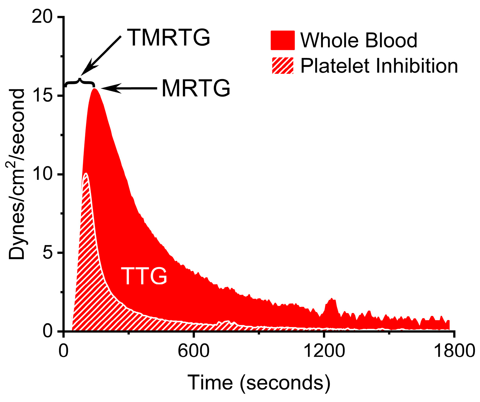
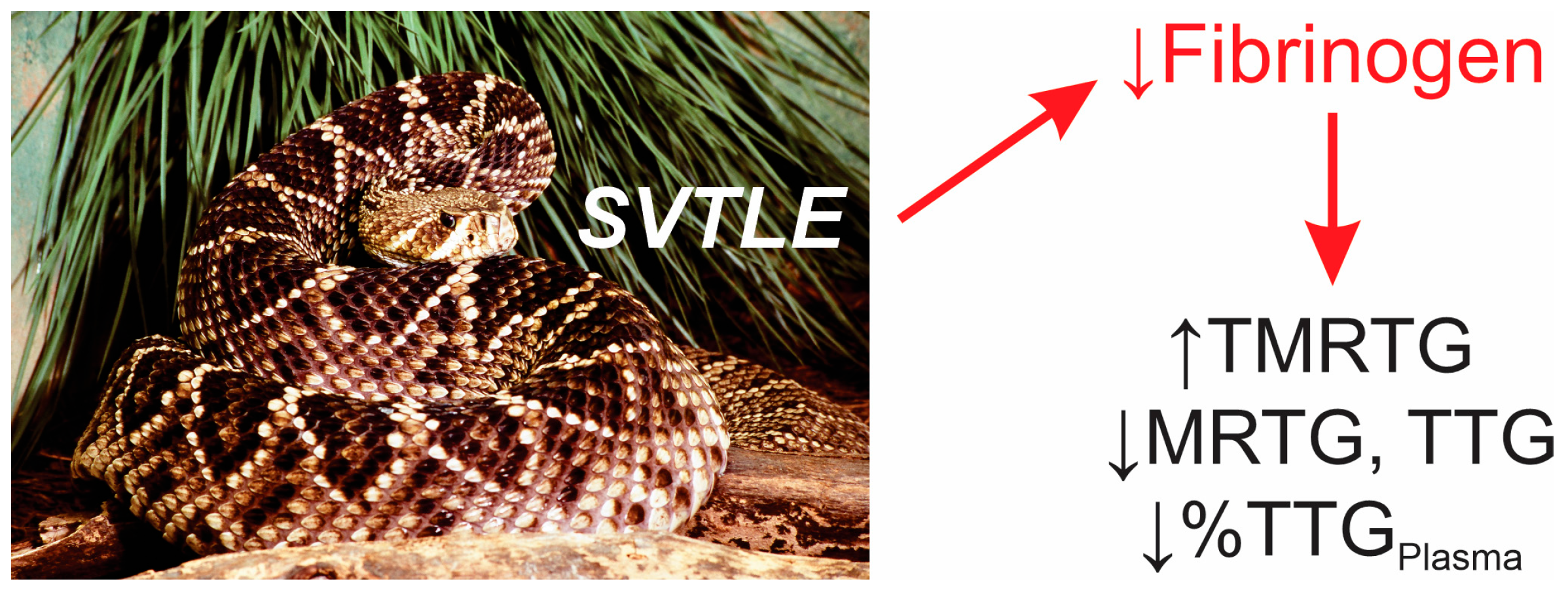
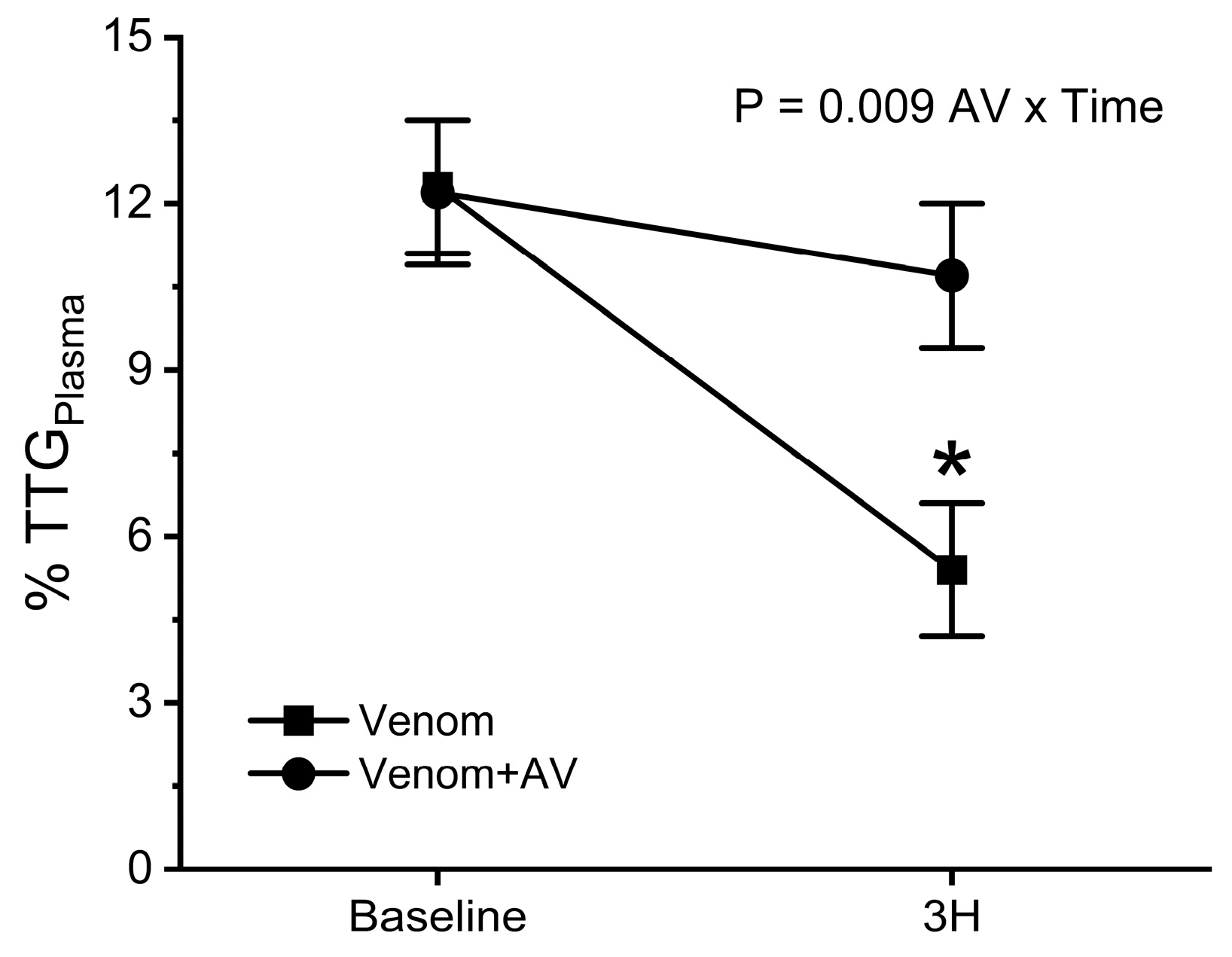
2. Results
Effects of C. adamanteus Venom on Circulating Coagulation Function in Rabbits
3. Discussion
4. Materials and Methods
4.1. Chemicals and Venoms
4.2. Rabbit Model of Envenomation
4.3. Coagulation Monitoring
4.4. Statistical Analyses
Funding
Institutional Review Board Statement
Informed Consent Statement
Data Availability Statement
Acknowledgments
Conflicts of Interest
References
- Weiss, H.J.; Allan, S.; Davidson, E.; Kochwa, S. Afibrinogenemia in man following the bite of a rattlesnake (Crotalus adamanteus). Am. J. Med. 1969, 47, 625–634. [Google Scholar] [CrossRef]
- Damus, P.S.; Markland, F.S.; Davidson, T.M.; Shanley, J.D. A purified procoagulant enzyme from the venom of the eastern diamondback rattlesnake (Crotalus adamanteus): In vivo and in vitro studies. J. Lab. Clin. Med. 1972, 79, 906–923. [Google Scholar] [PubMed]
- Kitchens, C.S.; Van Mierop, L.H. Mechanism of defibrination in humans after envenomation by the Eastern diamondback rattlesnake. Am. J. Hematol. 1983, 14, 345–353. [Google Scholar] [CrossRef] [PubMed]
- Margres, M.J.; McGivern, J.J.; Wray, K.P.; Seavy, M.; Calvin, K.; Rokyta, D.R. Linking the transcriptome and proteome to characterize the venom of the eastern diamondback rattlesnake (Crotalus adamanteus). J. Proteom. 2014, 96, 145–158. [Google Scholar] [CrossRef] [PubMed]
- Rushton, W.F.; Rivera, J.V.; Brown, J.; Kurz, M.C.; Arnold, J. Utilization of thromboelastograms in management of Crotalus adamanteus envenomation. Clin. Toxicol. 2021, 59, 256–259. [Google Scholar] [CrossRef] [PubMed]
- Fazelat, J.; Teperman, S.H.; Touger, M. Recurrent hemorrhage after western diamondback rattlesnake envenomation treated with crotalidae polyvalent immune fab (ovine). Clin. Toxicol. 2008, 46, 823–826. [Google Scholar] [CrossRef] [PubMed]
- Budzynski, A.Z.; Pandya, B.V.; Rubin, R.N.; Brizuela, B.S.; Soszka, T.; Stewart, G.J. Fibrinogenolytic afibrinogenemia after envenomation by western diamondback rattlesnake (Crotalus atrox). Blood 1984, 63, 1–14. [Google Scholar] [CrossRef] [PubMed]
- Trautman, W.; Pizon, A. Severe, persistent thrombocytopenia in Crotalus horridus envenomation despite antivenom: A retrospective review. Toxicon 2023, 224, 107029. [Google Scholar] [CrossRef] [PubMed]
- Bond, R.G.; Burkhart, K.K. Thrombocytopenia following timber rattlesnake envenomation. Ann. Emerg. Med. 1997, 30, 40–44. [Google Scholar] [CrossRef]
- Williams, K.L.; Woslager, M.; Garland, S.L.; Barton, R.P.; Banner, W. Use of polyvalent equine anti-viper serum to treat delayed coagulopathy due to suspected Sistrurus miliarius streckeri envenomation in two children. Clin. Toxicol. 2017, 55, 326–331. [Google Scholar] [CrossRef] [PubMed]
- Hardy, D.L.; Jeter, M.; Corrigan, J.J., Jr. Envenomation by the northern blacktail rattlesnake (Crotalus molossus molossus): Report of two cases and the in vitro effects of the venom on fibrinolysis and platelet aggregation. Toxicon 1982, 20, 487–493. [Google Scholar] [CrossRef] [PubMed]
- Schaeffer, R.C., Jr.; Briston, C.; Chilton, S.M.; Carlson, R.W. Failure of electric shock treatment for rattlesnake envenomation. J. Pharmacol. Exp. Ther. 1984, 230, 393–398. [Google Scholar]
- Nielsen, V.G. Iron and carbon monoxide prevent degradation of plasmatic coagulation by thrombin-like activity in rattlesnake venom. Hum. Exp. Toxicol. 2016, 35, 1116–1122. [Google Scholar] [CrossRef] [PubMed]
- Aitchison, J.M. Boomslang bite--diagnosis and management. A report of 2 cases. S. Afr. Med. J. 1990, 78, 39–42. [Google Scholar] [PubMed]
- Faulkner, J.D.; Carballo, C.J.; Acquista, E.; Baughman, S.D., Jr.; Powers, W.F., 4th; Novosel, T.; Yon, J.R. Thromboelastography Use to Guide Resuscitation and Antivenom Administration after Gaboon Viper Bite. Am. Surg. 2023, 89, 3968–3970. [Google Scholar] [CrossRef] [PubMed]
- Rucavado, A.; Chacón, M.; Villalobos, D.; Argüello, I.; Campos, M.; Guerrero, G.; Méndez, M.L.; Escalante, T.; Gutiérrez, J.M. Coagulopathy induced by viperid snake venoms in a murine model: Comparison of standard coagulation tests and rotational thromboelastimetry. Toxicon 2022, 214, 121–129. [Google Scholar] [CrossRef] [PubMed]
- Dang, X.T.; Nguyen, T.X.; Nguyen, H.T.T.; Ha, H.T. Correlations between rotational thromboelastometry (ROTEM) and standard coagulation tests following viper snakebites. J. Int. Med. Res. 2022, 50, 3000605211067321. [Google Scholar] [CrossRef]
- Tacon, C.L.; Munas, A.; Little, M. Case Report: Rotational Thromboelastometry in Taipan Envenomation. Am. J. Trop. Med. Hyg. 2021, 106, 746–749. [Google Scholar] [CrossRef] [PubMed]
- Tambwe, M.J.; Lalloo, V.; Engelbrecht, A.; Pelle, P. A case report of detecting subclinical coagulopathy in a patient with boomslang (Dipholidus typus) bite. S. Afr. Fam. Pract. 2021, 63, e1–e5. [Google Scholar] [CrossRef] [PubMed]
- Gooneratne, L.V.; Dharmasena, I.; Premawardana, N.; Wimalachandra, M.; Arya, R.; Gnanathasan, A. Comparison of rotational thromboelastometry parameters with 20-minute whole blood clotting test as a predictor of envenoming in Russell’s viper bite patients. Trans. R. Soc. Trop. Med. Hyg. 2021, 115, 561–565. [Google Scholar] [CrossRef] [PubMed]
- Atamna, R.; Kelmer, E.; Aroch, I.; Klainbart, S. Echis coloratus envenomation in a dog: Clinical, hemostatic and thromboelastometric findings and treatment. Clin. Toxicol. 2021, 59, 639–643. [Google Scholar] [CrossRef] [PubMed]
- Kopke, M.A.; Botha, W.J. Thromboelastographic evaluation of 2 dogs with boomslang (Dispholidus typus) envenomation. J. Vet. Emerg. Crit. Care 2020, 30, 712–717. [Google Scholar] [CrossRef] [PubMed]
- Park, E.J.; Choi, S.; Kim, H.H.; Jung, Y.S. Novel Treatment Strategy for Patients with Venom-Induced Consumptive Coagulopathy from a Pit Viper Bite. Toxins 2020, 12, 295. [Google Scholar] [CrossRef]
- Mullins, M.E.; Freeman, W.E. Thromboelastometry (ROTEM) and thromboelastography (TEG) in copperhead snakebites: A case series. Clin. Toxicol. 2020, 58, 931–934. [Google Scholar] [CrossRef]
- Coggins, A.; Symes, E.; Cheeseman, C.; Salter, M. Thromboelastography for the detection of acute anticoagulant coagulopathy associated with Black Snake envenomation. Emerg. Med. Australas. 2019, 31, 900–902. [Google Scholar] [CrossRef]
- Kang, A.M.; Fisher, E.S. Thromboelastography with Platelet Studies (TEG® with PlateletMapping®) after Rattlesnake Envenomation in the Southwestern United States Demonstrates Inhibition of ADP-Induced Platelet Activation as Well as Clot Lysis. J. Med. Toxicol. 2020, 16, 24–32. [Google Scholar] [CrossRef] [PubMed]
- Leffers, P.; Ferreira, J.; Sollee, D.; Schauben, J. Thromboelastography in the management of snakebite-induced coagulopathy: A case series and literature review. Blood Coagul. Fibrinolysis 2018, 29, 656–660. [Google Scholar] [CrossRef] [PubMed]
- Larréché, S.; Jean, F.X.; Benois, A.; Mayet, A.; Bousquet, A.; Vedy, S.; Clapson, P.; Dehan, C.; Rapp, C.; Kaiser, E.; et al. Thromboelastographic study of the snakebite-related coagulopathy in Djibouti. Blood Coagul. Fibrinolysis 2018, 29, 196–204. [Google Scholar] [CrossRef] [PubMed]
- McBride, K.M.; Bromberg, W.; Dunne, J. Thromboelastography Utilization in Delayed Recurrent Coagulopathy after Severe Eastern Diamondback Rattlesnake Envenomation. Am. Surg. 2017, 83, 332–336. [Google Scholar] [CrossRef]
- Roszko, P.J.; Kavanaugh, M.J.; Boese, M.L.; Longwell, J.J.; Earley, A.S. Rotational thromboelastometry (ROTEM) guided treatment of an Afghanistan viper envenomation at a NATO military hospital. Clin. Toxicol. 2017, 55, 229–230. [Google Scholar] [CrossRef] [PubMed]
- Hadley, G.P.; McGarr, P.; Mars, M. The role of thromboelastography in the management of children with snake-bite in southern Africa. Trans. R. Soc. Trop. Med. Hyg. 1999, 93, 177–179. [Google Scholar] [CrossRef] [PubMed]
- Nielsen, V.G. Ruthenium-based antivenom attenuates Crotalus atrox venom mediated coagulopathy in rabbits. Blood Coagul. Fibrinolysis 2024, 35, 167–172. [Google Scholar] [CrossRef] [PubMed]
- Nielsen, V.G. Novel Toxicodynamic Model of Subcutaneous Envenomation to Characterize Snake Venom Coagulopathies and Assess the Efficacy of Site-Directed Inorganic Antivenoms. Int. J. Mol. Sci. 2023, 24, 13939. [Google Scholar] [CrossRef] [PubMed]
- Nielsen, V.G. Crotalus atrox Venom Exposed to Carbon Monoxide Has Decreased Fibrinogenolytic Activity In Vivo in Rabbits. Basic Clin. Pharmacol. Toxicol. 2018, 122, 82–86. [Google Scholar] [CrossRef]
- Wells, M.; Raja, M.; Rahman, S. Point-of-care viscoelastic testing. BJA Educ. 2022, 22, 416–423. [Google Scholar] [CrossRef] [PubMed]
- Nielsen, V.G.; Geary, B.T.; Baird, M.S. Evaluation of the contribution of platelets to clot strength by thromboelastography in rabbits: The role of tissue factor and cytochalasin D. Anesth. Analg. 2000, 91, 35–39. [Google Scholar] [CrossRef] [PubMed]
- Kreutz, R.P.; Owens, J.; Lu, D.; Nystrom, P.; Jin, Y.; Kreutz, Y.; Desta, Z.; Flockhart, D.A. Platelet factor XIIIa release during platelet aggregation and plasma clot strength measured by thrombelastography in patients with coronary artery disease treated with clopidogrel. Platelets 2015, 26, 358–363. [Google Scholar] [CrossRef] [PubMed]
- Nielsen, V.G.; Cohen, B.M.; Cohen, E. Effects of coagulation factor deficiency on plasma coagulation kinetics determined via thrombelastography: Critical roles of fibrinogen and factors II, VII, X and XII. Acta Anaesthesiol. Scand 2005, 49, 222–231. [Google Scholar] [CrossRef] [PubMed]
- Roberts, H.R.; Hoffman, M.; Monroe, D.M. A cell-based model of thrombin generation. Semin. Thromb. Hemost. 2006, 32 (Suppl. S1), 32–38. [Google Scholar] [CrossRef] [PubMed]
- Arnesen, H. Fibrinogen/fibrin degradation products (FDP/fdp). Characterization, anticoagulant activity, and influence on laboratory assays. J. Oslo. City Hosp. 1977, 27, 69–80. [Google Scholar] [PubMed]
- Nielsen, V.G. Ruthenium, Not Carbon Monoxide, Inhibits the Procoagulant Activity of Atheris, Echis, and Pseudonaja Venoms. Int. J. Mol. Sci. 2020, 21, 2970. [Google Scholar] [CrossRef]


Disclaimer/Publisher’s Note: The statements, opinions and data contained in all publications are solely those of the individual author(s) and contributor(s) and not of MDPI and/or the editor(s). MDPI and/or the editor(s) disclaim responsibility for any injury to people or property resulting from any ideas, methods, instructions or products referred to in the content. |
© 2024 by the author. Licensee MDPI, Basel, Switzerland. This article is an open access article distributed under the terms and conditions of the Creative Commons Attribution (CC BY) license (https://creativecommons.org/licenses/by/4.0/).
Share and Cite
Nielsen, V.G. Ruthenium Antivenom Inhibits the Defibrinogenating Activity of Crotalus adamanteus Venom in Rabbits. Int. J. Mol. Sci. 2024, 25, 6334. https://doi.org/10.3390/ijms25126334
Nielsen VG. Ruthenium Antivenom Inhibits the Defibrinogenating Activity of Crotalus adamanteus Venom in Rabbits. International Journal of Molecular Sciences. 2024; 25(12):6334. https://doi.org/10.3390/ijms25126334
Chicago/Turabian StyleNielsen, Vance G. 2024. "Ruthenium Antivenom Inhibits the Defibrinogenating Activity of Crotalus adamanteus Venom in Rabbits" International Journal of Molecular Sciences 25, no. 12: 6334. https://doi.org/10.3390/ijms25126334
APA StyleNielsen, V. G. (2024). Ruthenium Antivenom Inhibits the Defibrinogenating Activity of Crotalus adamanteus Venom in Rabbits. International Journal of Molecular Sciences, 25(12), 6334. https://doi.org/10.3390/ijms25126334