Abstract
Ovarian mature teratomas (OMTs) originate from post-meiotic germ cells. Malignant transformation occurs in approximately 1–2% of OMTs; however, sebaceous carcinoma arising from OMTs is rare. This is the first report of a detailed genomic analysis of sebaceous carcinoma arising from an OMT. A 36-year-old woman underwent evaluation for abdominal tumors and subsequent hysterectomy and salpingo-oophorectomy. Pathologically, a diagnosis of stage IA sebaceous carcinoma arising from an OMT was established. Eight months post-surgery, the patient was alive without recurrence. Immunohistochemically, the tumor was negative for mismatch repair proteins. A nonsense mutation in TP53 (p.R306*) and a deletion in PIK3R1 were identified. Single nucleotide polymorphisms across all chromosomes displayed a high degree of homozygosity, suggestive of uniparental disomy. Herein, the OMT resulting from the endoreduplication of oocytes underwent a malignant transformation to sebaceous carcinoma via TP53 as an early event and PIK3R1 as a late event.
1. Introduction
Ovarian mature teratomas (OMTs) originate from post-meiotic germ cells and are associated with genome-wide uniparental disomy (UPD) patterns [1,2]. As OMTs contain all three germ cell layers, and they often display multiple differentiated tissue types, including teeth, bone, and hair. Malignant transformation occurs in approximately 1–2% of OMTs, and the prognosis is poor at advanced stages [3]. Up to 80% of transformed teratomas contain squamous cell carcinoma, whereas, the remaining 20% contain adenocarcinoma, thyroid carcinoma, carcinoid tumors, and melanoma [3,4]. The malignant transformation of OMTs is characterized by difficulty in diagnosis and histological diversity [3,4,5,6].
Sebaceous carcinoma is a rare tumor with an incidence of 2.4 cases per million persons per year [7]. TP53 (~70%), RB1, ZNF750, and NOTCH1 have been identified as common genetic mutations [7]. Sebaceous carcinoma arising from an OMT is extremely rare, with only 14 previously reported cases [8,9,10,11,12,13,14,15,16,17,18,19,20]. Previous studies have reported the association between sebaceous carcinoma arising from OMTs and abnormalities in mismatch repair proteins/genes. However, genomic information is currently lacking. As sebaceous carcinomas originate from OMTs, it may have a unique genetic pattern related to meiosis [1,2]. Herein, we report a case of sebaceous carcinoma arising from an OMT and provide a detailed genomic analysis of the tumor.
2. Case Presentation
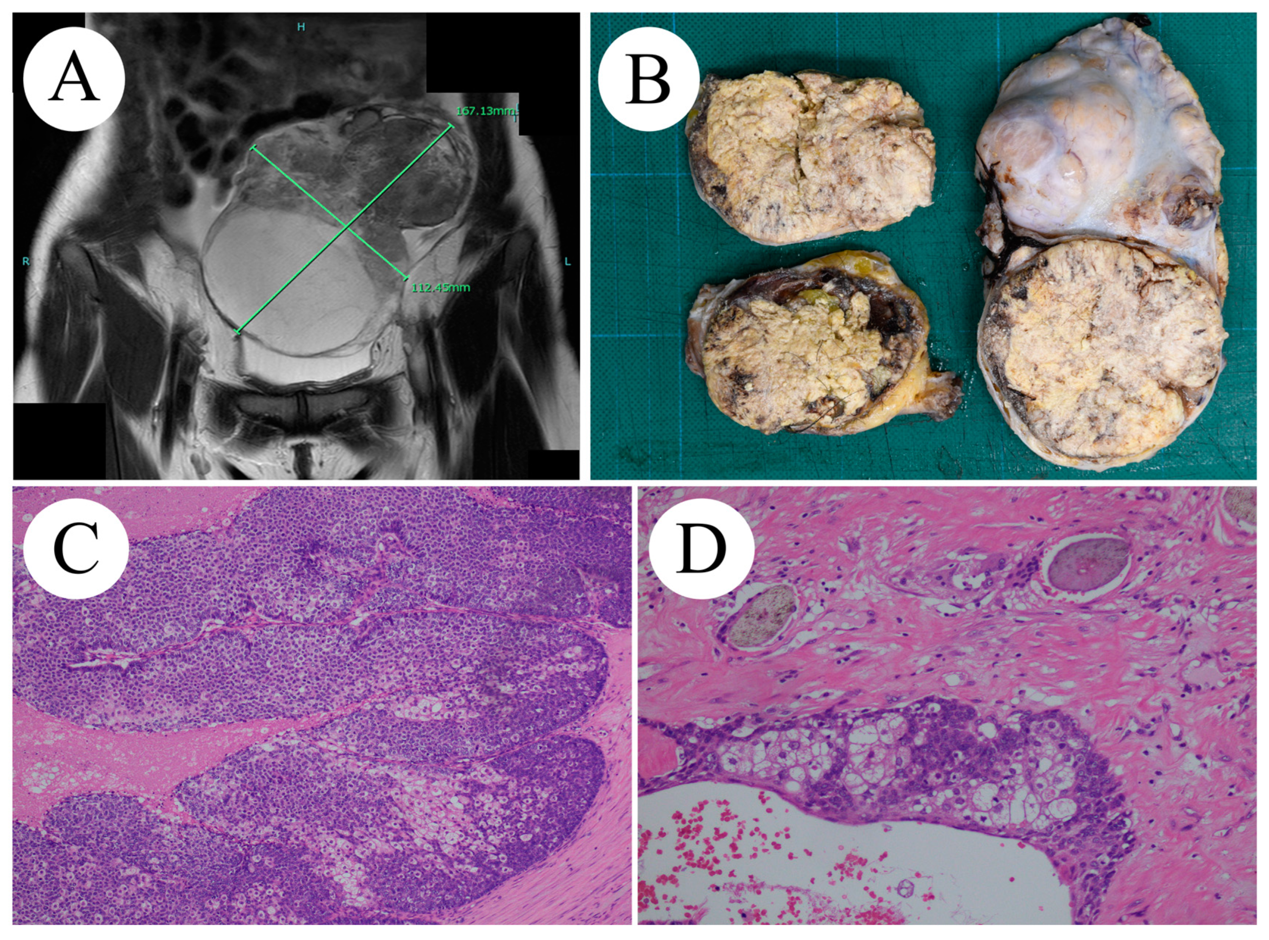
A 36-year-old woman (gravida 1, para 1) was referred to our hospital for an evaluation of abdominal tumors. Subsequently, she became pregnant. At the age of 27, she simultaneously underwent a right oophorectomy for an OMT and a cesarean section. She had no other relevant medical or family history. Her primary complaint was persistent abdominal swelling for 1 month. Systemic enhanced computed tomography and T2-weighted images from pelvic enhanced magnetic resonance imaging showed a 167 × 112 × 110 mm mass occupying the pelvic cavity, with suspected omental disseminations (Figure 1A). The preoperative diagnosis was stage IIIC left ovarian immature teratoma (cT3c cN0 M0). An intraoperative pathological diagnosis of sebaceous carcinoma was performed using frozen sections. The patient underwent total abdominal hysterectomy, left salpingo-oophorectomy, right salpingectomy, subtotal omentectomy, pelvic lymphadenectomy, and para-aortic lymphadenectomy. Complete resection was achieved. Macroscopically, the right ovarian tumor appeared as a well-circumscribed, yellowish-white mass with hair and partial necrosis (Figure 1B). Microscopically, the tumor exhibited sheets or lobules separated by a fibrovascular stroma of basophilic and atypical cells with central comedo-type necrosis (Figure 1C,D).

Figure 1.
(A) MRI image findings: A 167 × 112 × 110 mm mass extending from the midline of the pelvis to the left with dissemination into the large omentum. (B) Macroscopic findings: solid proliferation mainly composed of fat components. (C,D) Histological findings (H&E staining): (C, ×10) proliferation of tumor cells with differentiation potential into sebaceous glands cells on the background of hairs, (D, ×40) proliferation of basophilic and mildly atypical cells with mitotic figure on the background of fibrosis and necrosis.
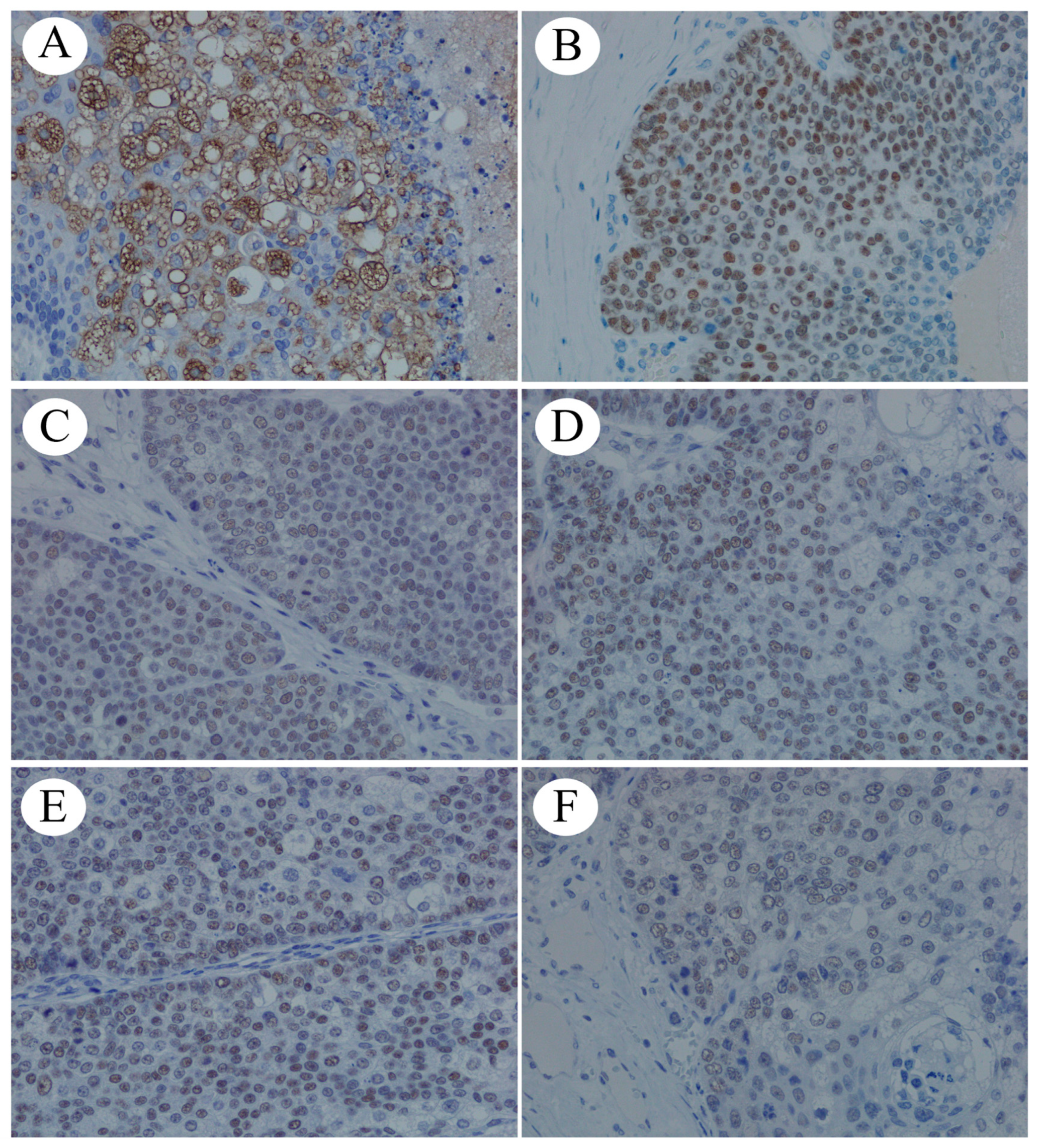
The tumor exhibited an epidermis, hair follicles, and sebaceous glands. No dissemination to the greater omentum or lymph node metastases was observed. Immunohistochemical staining revealed tumor cells positive for androgen receptors (Figure 2A), GATA-binding protein 3, adipophilin (Figure 2B), and mismatch repair proteins MLH1 (Figure 2C), MSH2 (Figure 2D), MSH6 (Figure 2E), and PMS2 (Figure 2F).

Figure 2.
Immunohistochemical staining (×40): (A) androgen receptor, (B) adipophilin, (C) MLH1, (D) MSH2, (E) MSH6, and (F) PMS2.
Accordingly, a diagnosis of stage IA (pT1a, pN0, M0) sebaceous carcinoma arising from an OMT was established. No post-operative adjuvant therapy was administered. The patient was alive without recurrence 8 months post-operation.
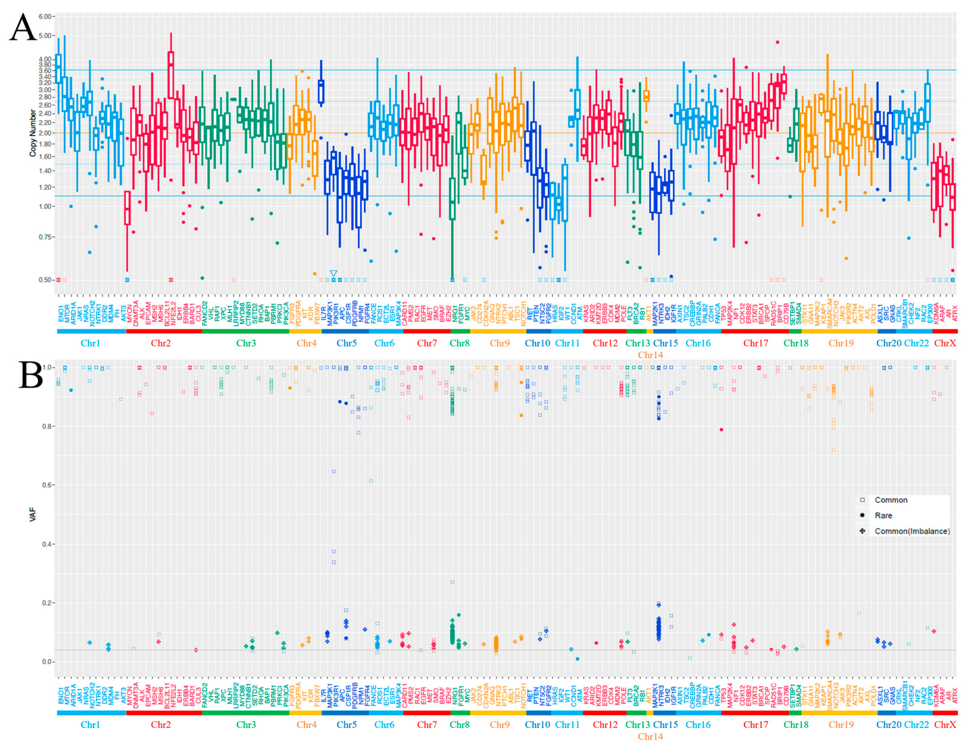
Genomic DNA was obtained from a sample classified as a sebaceous carcinoma but not an OMT—which was considered a separate precursor lesion—because of limited sample volume. Cancer gene profiling was performed using the PleSSision system as previously described [21]. The average sequencing depth was 619.4× for sebaceous carcinoma. Histologically, the average tumor cellularity was 60%. In an analysis using a cancer gene panel of 160 genes in a sebaceous carcinoma sample, several actionable gene alterations were observed (Table S1). Nonsense mutation in TP53 (p.R306*) and deletions in PIK3R1, ATRX, and APC were identified. In summary, a DNA quality check using the Agilent 2000 TapeStation (Agilent Technologies, Santa Clara, CA, USA), targeted amplicon exome sequencing using the Illumina MiSeq sequencing platform (Illumina, San Diego, CA, USA), and sequencing data analysis using the GenomeJack bioinformatics pipeline (version 1.0, Mitsubishi Space Software, Tokyo, Japan) were performed. The copy number loss/amplification cutoff was set to 1.1/3.6, which is statistically > 2σ. Single nucleotide polymorphisms across all chromosomes displayed a high degree of homozygosity, with allelic frequencies near 100% in nearly all of the genes examined (Figure 3 and Table S2). This pervasive pattern was evident across the entire genome. The tumor was characterized as microsatellite stable, and the tumor mutation burden was measured at 5.5 single nucleotide variants per megabase.

Figure 3.
(A) Copy number alteration and (B) allelic frequencies of single nucleotide polymorphisms (SNPs) across all examined genes in a sebaceous carcinoma sample. The horizontal axis corresponds to each examined gene, and the vertical axis corresponds to the (A) copy number or (B) VAF. Common means >1% genetic alteration in HGVD (https://www.hgvd.genome.med.kyoto-u.ac.jp/ accessed on 14 September 2023), ToMMo (https://www.megabank.tohoku.ac.jp/english/ accessed on 14 September 2023), or gnomAD (https://gnomad.broadinstitute.org/ accessed on 14 September 2023), and rare means anything else.
3. Discussion
To the best of our knowledge, this is the first report of a detailed genomic analysis of sebaceous carcinoma arising from an OMT. The 14 previously reported cases are summarized in Table 1 [8,9,10,11,12,13,14,15,16,17,18,19,20]. In the four reports that investigated mismatch repair abnormality, protein/gene abnormality was identified in all four cases [8,9,11,14]. However, since this case did not exhibit mismatch repair/gene abnormality and microsatellite instability, alternate pathogeneses had to be considered. Nonsense mutation of TP53 (p.R306*) and a deletion in PIK3R1 were detected in the targeted next-generation sequencing. Cooke et al. [22] reported that the most frequently altered genes were TP53 (80%), PIK3CA (52%), and CDKN2A (44%), and that TP53 mutation is an early event in squamous cell carcinoma arising from OMTs. Strikingly, 40% of the TP53 mutations were biallelic, which may be associated with improved outcomes [22]. KRAS amplification and a deletion of PTEN and RB1 were detected in malignant melanoma arising from OMT [21]. No previous studies have reported cases of OMT or sebaceous carcinoma with PIK3R1 mutations. Rubinstein et al. [23] reported the safety and efficacy of the dual PI3K/mTOR inhibitor in patients with advanced endometrial cancer and activating mutations in the PI3K pathway, including PIK3R1. Single nucleotide polymorphisms across all chromosomes displayed a high degree of homozygosity, except PIK3R1A. PIK3R1 mutations had a relatively low VAF because they were present in some, but not all, tumors. This PIK3R1 mutation is considered a late event that occurs in a subclone after the malignant transformation of an OMT. The observed extensive homozygosity and the uniform allelic frequencies close to 100% suggest the presence of UPD across all chromosomes. Such widespread UPD is unusual and highlights a significant alteration from the expected heterozygous genetic landscape typical of diploid cells. The implications of this finding are profound, as UPD can lead to disruptions in gene expression, loss of heterozygosity, and potential impacts on tumor suppressor genes and oncogenes. This genetic uniformity may contribute to the oncogenic process by altering the cellular landscape, potentially leading to an enhanced tumorigenic capacity. The mechanisms driving such extensive UPD and its role in the progression of the tumor warrant further investigation to understand its contribution to cancer biology and therapy.

Table 1.
Summary of sebaceous carcinomas arising from mature ovarian teratomas.
OMTs are classified into five types (I-V) based on their cytogenetic features [24]. Type I OMTs result from errors in meiosis I, type II OMTs result from meiosis II failure, type III OMTs occur via endoreduplication of a haploid ovum, type IV arises from oogonia, and type V OMTs are considered to originate from the fusion of two normal haploid ovaries [1,2,24]. Notably, the sebaceous carcinoma lacked heterozygous pleomorphism in the whole genome, suggesting that the sebaceous carcinoma arose from a type III OMT via endoreduplication of a haploid ovum [1,2]. Using short tandem repeat polymorphism analysis of centromeric and distal markers, Usui et al. reported that all OMTs were of post-meiotic origin rather than of pre-meiotic origin. Finally, the developmental process of this tumor is explained as follows. Type III OMTs resulted from the endoreduplication of oocytes after meioses I and II. As an early event, a TP53 mutation occurred, leading to the development of sebaceous gland carcinoma. Subsequently, a subclone acquired a PIK3R1 mutation as a late event. However, the present study is a case report, and future case studies are required to confirm the reproducibility of our findings.
In conclusion, an OMT resulting from the endoreduplication of oocytes underwent malignant transformation to sebaceous carcinoma via TP53 as an early event and PIK3R1 as a late event in the present case. These mutations may also be useful for prognosis prediction and targeted therapy. Genetic analysis is important to elucidate the pathogenesis of this rare tumor, and further case collection is required.
Supplementary Materials
The following supporting information can be downloaded at: https://www.mdpi.com/article/10.3390/ijms25126351/s1.
Author Contributions
Conceptualization: S.Z. and M.Y. Data curation: S.Z., Y.A., K.N. and M.Y. Formal analysis: S.Z., M.Y., K.N. and E.K. Funding acquisition: M.Y. Investigation, Methodology, Project administration, Resources, and Software: S.Z., Y.A., H.N., M.Y., K.N. and E.K. Supervision: M.Y., K.N. and E.K. Validation: Y.A., H.N. and M.Y. Visualization: S.Z., Y.A., H.N., M.Y. and K.N. Writing of original draft: S.Z. and M.Y. Writing—review and editing: K.N. and E.K. All authors have read and agreed to the published version of the manuscript.
Funding
This research was supported by Grants-in-Aid for Scientific Research from the Ministry of Education, Culture, Sports, Science and Technology (grant number 22K15409).
Institutional Review Board Statement
This study was approved by The Ethics Committee of the Faculty of Medicine, Oita University, (approval Code: 2586, approval Date: 4 August 2023).
Informed Consent Statement
Informed consent was obtained from the patient and the patient’s family for her anonymized information to be published in the present case report.
Data Availability Statement
The datasets used and/or analyzed during the current study are available from the corresponding author upon reasonable request.
Conflicts of Interest
The authors declare no conflict of interest.
References
- Usui, H.; Nakabayashi, K.; Kaku, H.; Maehara, K.; Hata, K.; Shozu, M. Elucidation of the developmental mechanism of ovarian mature cystics teratomas using B allele-frequency plots of single nucleotide polymorphism array data. Genes Chromosomes Cancer 2018, 57, 409–419. [Google Scholar] [CrossRef] [PubMed]
- Kaku, H.; Usui, H.; Qu, J.; Shozu, M. Mature cystic teratomas arise from meiotic oocytes, but not from pre-meiotic oogonia. Genes Chromosomes Cancer 2016, 55, 355–364. [Google Scholar] [CrossRef] [PubMed]
- Hackethal, A.; Brueggmann, D.; Bohlmann, M.K.; Franke, F.E.; Tinneberg, H.R.; Münstedt, K. Squamous-cell carcinoma in mature cystic teratoma of the ovary: Systematic review and analysis of published data. Lancet Oncol. 2008, 9, 1173–1180. [Google Scholar] [CrossRef] [PubMed]
- Yano, M.; Nasu, K.; Yasuda, M.; Katoh, T.; Kagabu, M.; Kobara, H.; Matsuura, M.; Tokuyama, O.; Yamawaki, T.; Wakahashi, S.; et al. Clinicopathological features and programmed death-ligand 1 immunohistochemical expression in a multicenter cohort of uterine and ovarian melanomas: A retrospective study in Japan (KCOG-G1701s). Melanoma Res. 2022, 32, 150–158. [Google Scholar] [CrossRef] [PubMed]
- Yano, M.; Katoh, T.; Hamaguchi, T.; Kozawa, E.; Hamada, M.; Nagata, K.; Yasuda, M. Tumor-to-tumor metastasis from appendiceal adenocarcinoma to an ovarian mature teratoma, mimicking malignant transformation of a teratoma: A case report. Diagn. Pathol. 2019, 14, 88. [Google Scholar] [CrossRef] [PubMed]
- Sato, Y.; Yano, M.; Eto, S.; Takano, K.; Nasu, K. Metastasis from follicular lymphoma to an ovarian mature teratoma: A case report of tumor-to-tumor metastasis. J. Ovarian Res. 2023, 16, 106. [Google Scholar] [CrossRef] [PubMed]
- Sargen, M.R.; Starrett, G.J.; Engels, E.A.; Cahoon, E.K.; Tucker, M.A.; Goldstein, A.M. Sebaceous carcinoma epidemiology and genetics: Emerging concepts and clinical implications for screening, prevention, and treatment. Clin. Cancer Res. 2021, 27, 389–393. [Google Scholar] [CrossRef]
- Mohammed, M.; Keating, A.; McIlwaine, P. Rare Case: Sebaceous Carcinoma Arising within an Ovarian Dermoid Cyst. 2023. Available online: https://www.medicalandresearch.com/current_issue/1823 (accessed on 14 September 2023).
- Murray, J.; McIlwaine, P.; Morrison, P.J.; McCluggage, W.G. Sebaceous carcinoma arising in ovarian teratoma: First report associated with germline mismatch repair gene mutation. Int. J. Gynecol. Pathol. 2022, 41, 608–614. [Google Scholar] [CrossRef] [PubMed]
- de Lima, R.B.; Jung, J.E.; Ioshii, S.O.; Kami, R.M. Sebaceous carcinoma in a mature teratoma of the ovary. Autops. Case Rep. 2018, 8, e2018060. [Google Scholar]
- Wield, A.; Hodeib, M.; Khan, M.; Gubernick, L.; Li, A.J.; Kandukuri, S. Sebaceous carcinoma arising within an ovarian mature cystic teratoma: A case report with discussion of clinical management and genetic evaluation. Gynecol. Oncol. Rep. 2018, 26, 37–40. [Google Scholar] [CrossRef]
- Moghaddam, Y.; Lindsay, R.; Tolhurst, J.; Millan, D.; Siddiqui, N. A case of sebaceous carcinoma arising in a benign cystic teratoma of the ovary and review of the literature. Scott. Med. J. 2013, 58, e18–e22. [Google Scholar] [CrossRef] [PubMed]
- An, H.J.; Jung, Y.H.; Yoon, H.K.; Jung, S.J. Sebaceous carcinoma arising in mature cystic teratoma of ovary. Korean J. Pathol. 2013, 47, 383–387. [Google Scholar] [CrossRef] [PubMed]
- Smith, J.; Crowe, K.; McGaughran, J.; Robertson, T. Sebaceous adenoma arising within an ovarian mature cystic teratoma in Muir-Torre syndrome. Ann. Diagn. Pathol. 2012, 16, 485–488. [Google Scholar] [CrossRef]
- Venizelos, I.D.; Tatsiou, Z.A.; Roussos, D.; Karagiannis, V. A case of sebaceous carcinoma arising within a benign ovarian cystic teratoma. Onkologie 2009, 32, 353–355. [Google Scholar] [CrossRef]
- Ribeiro-Silva, A.; Chang, D.; Bisson, F.W.; Ré, L.O. Clinicopathological and immunohistochemical features of a sebaceous carcinoma arising within a benign dermoid cyst of the ovary. Virchows Arch. Int. J. Pathol. 2003, 443, 574–578. [Google Scholar] [CrossRef] [PubMed]
- Vartanian, R.K.; McRae, B.; Hessler, R.B. Sebaceous carcinoma arising in a mature cystic teratoma of the ovary. Int. J. Gynecol. Pathol. 2002, 21, 418–421. [Google Scholar] [CrossRef]
- Changchien, C.C.; Chen, L.; Eng, H.L. Sebaceous carcinoma arising in a benign dermoid cyst of the ovary. Acta Obstet. Gynecol. Scand. 1994, 73, 355–358. [Google Scholar] [CrossRef]
- Chumas, J.C.; Scully, R.E. Sebaceous tumors arising in ovarian dermoid cysts. Int. J. Gynecol. Pathol. 1991, 10, 356–363. [Google Scholar] [CrossRef]
- Betta, P.G.; Cosimi, M.F. Sebaceous carcinoma arising in benign cystic teratoma of the ovary. Case report. Eur. J. Gynaecol. Oncol. 1984, 5, 146–149. [Google Scholar]
- Nakamura, K.; Aimono, E.; Takamatsu, R.; Tanishima, S.; Tohyama, T.; Sasano, K.; Sakuma, H.; Nishihara, H. Genetic profiling of malignant melanoma arising from an ovarian mature cystic teratoma: A case report. Int. J. Mol. Sci. 2021, 22, 2436. [Google Scholar] [CrossRef]
- Cooke, S.L.; Ennis, D.; Evers, L.; Dowson, S.; Chan, M.Y.; Paul, J.; Hirschowitz, L.; Glasspool, R.M.; Singh, N.; Bell, S.; et al. The driver mutational landscape of ovarian squamous cell carcinomas arising in mature cystic teratoma. Clin. Cancer Res. 2017, 23, 7633–7640. [Google Scholar] [CrossRef] [PubMed]
- Rubinstein, M.M.; Hyman, D.M.; Caird, I.; Won, H.; Soldan, K.; Seier, K.; Iasonos, A.; Tew, W.P.; O’Cearbhaill, R.E.; Grisham, R.N.; et al. Phase 2 study of LY3023414 in patients with advanced endometrial cancer harboring activating mutations in the PI3K pathway. Cancer 2020, 126, 1274–1282. [Google Scholar] [CrossRef] [PubMed]
- Surti, U.; Hoffner, L.; Chakravarti, A.; Ferrell, R.E. Genetics and biology of human ovarian teratomas. I. Cytogenetic analysis and mechanism of origin. Am. J. Hum. Genet. 1990, 47, 635–643. [Google Scholar] [PubMed]
Disclaimer/Publisher’s Note: The statements, opinions and data contained in all publications are solely those of the individual author(s) and contributor(s) and not of MDPI and/or the editor(s). MDPI and/or the editor(s) disclaim responsibility for any injury to people or property resulting from any ideas, methods, instructions or products referred to in the content. |
© 2024 by the authors. Licensee MDPI, Basel, Switzerland. This article is an open access article distributed under the terms and conditions of the Creative Commons Attribution (CC BY) license (https://creativecommons.org/licenses/by/4.0/).