Article

The Role of Z Chromosome Localization Gene psmd9 in Spermatogenesis of Cynoglossus semilaevis

1
State Key Laboratory of Mariculture Biobreeding and Sustainable Goods, Yellow Sea Fisheries Research Institute, Chinese Academy of Fishery Sciences, Qingdao 266071, China
2
Laboratory for Marine Fisheries Science and Food Production Processes, Qingdao Marine Science and Technology Center, Qingdao 266237, China
*
Authors to whom correspondence should be addressed.
Int. J. Mol. Sci. 2024, 25(12), 6372; https://doi.org/10.3390/ijms25126372
Submission received: 19 March 2024 / Revised: 28 May 2024 / Accepted: 3 June 2024 / Published: 9 June 2024
(This article belongs to the Section Molecular Genetics and Genomics)

Abstract

:
Proteasome 26S Subunit, Non-ATPase 9 (psmd9) plays an important role in the balance of protamine and the stability of the nucleolar structure during spermatogenesis. In this study, we cloned the psmd9 of Cynoglossus semilaevis and analyzed its expression pattern. psmd9 was identified on the Z chromosome of C. semilaevis, which is considered an interesting candidate gene for spermatogenesis. qRT-PCR and FISH experiments showed that the psmd9 gene was significantly highly expressed in the testes. It is worth noting that the expression level of psmd9 in male fish testes is significantly higher than that in pseudomales. In order to further explore the role of psmd9 in spermatogenesis, a male testicular cell line was used as the experimental material. The results of the psmd9-RNAi and overexpression experiments showed that psmd9 had a synergistic effect with spermatogenesis-related genes dnd1, cfap69, dnah3 and dnajb13, but had an antagonistic effect with ccne2. Our findings offer a scientific foundation for comprehending the role of psmd9 in the spermatogenesis regulatory network of C. semilaevis.

1. Introduction

Spermatogenesis is a dynamic, continuous and complex process, which involves key biological events, such as spermatogonial proliferation, the meiosis of spermatocytes, and sperm cell deformation and differentiation into mature sperm [1,2]. Similarly, during fish spermatogenesis, spermatogenic cells and somatic cells undergo highly specialized cell differentiation under precise molecular controls to produce functional sperm [3]. These processes of spermatogenesis are essential for continuous sperm production, which is dependent upon numerous factors, both intrinsic (Sertoli and germ cells) and extrinsic (androgens and retinoic acids) [4]. Studying the mechanism of spermatogenesis can not only deepen understanding of the reproductive biology of fish, but also provide a scientific basis for breaking through precision breeding technologies, such as sex control and artificial reproduction of fish [5,6], thus realizing efficient breeding and cultivating excellent varieties of sustainable aquaculture.
It is of great significance to analyze the regulation mechanism of cell fate in the process of spermatogenesis for realizing sperm production in vitro and accurate breeding with sex control. Cynoglossus semilaevis is an important economic fish in China, and there is a great sexual size dimorphism between males and females, with females being 2–4 times larger than males [7]. Genetic females may be transformed into phenotypic males, while the transformation from genetic male to phenotypic female is irreversible, which means that sex control breeding has great practical significance [7]. According to the statistical data of Li et al., in a natural state, the proportion of males is always higher than that of females [8]. The acquisition of a high female population is of great significance, so there is an urgent need to prepare superfemale individuals. Ideally, a pseudomale crossed with a normal female could produce a superfemale (WW), and then a superfemale crossed with a normal male (ZZ) could produce all-female offspring. Unfortunately, in the published literature, there are only rare records of the existence of W sperm in pseudomales [9,10]. Additional research into the molecular mechanism of spermatogenesis in C. semilaevis will offer valuable understanding of this intriguing biological process.
With the development of modern genetic science research, the acquisition of genome, transcriptome and protein sequencing may provide ideas for exploring the molecular mechanism of spermatogenesis. The construction of the genome of C. semilaevis provides help for the study of gene function [10]. Several genes located on the Z chromosome are thought to be related to the processes of spermatogenesis. For example, neuralized E3 ubiquitin protein ligase 3 (neurl3) plays a role in the spermatogenesis of C. semilaevis by regulating testis protein ubiquitination in a dosage-dependent manner [11]. In addition, the testis-specific protein kinase 1 gene (tesk1) is found mainly expressed in the sperm cells and mature sperm of C. semilaevis, which may play a role in the stages of sperm formation, and the spermatid perinuclear RNA-binding protein (strbp) is found mainly expressed at the stage of sperm cell metamorphosis, and may play a role in this stage [12,13]. Through the analysis of the gonad transcriptome and proteome of adult pseudomale fish and male fish, it was shown that the extracellular environment, such as growth factors and steroid hormones, are not the cause of sperm loss, and there may be many gene regulation defects during sperm maturation, which may lead to sperm maturation being blocked [14]. On the other hand, high-resolution single-cell RNA-seq maps from the testes of genotypic male and pseudomale fish revealed the dynamic changes in gene expression patterns during spermatogenesis in C. semilaevis [15]. In particular, the psmd9 gene was significantly differentially expressed between pseudomales and males [15], and it was located on the Z chromosome [9], which indicates that psmd9 might be related to spermatogenesis.
Proteasome 26S Subunit, Non-ATPase 9 (psmd9) is an active hub gene that links numerous pathways that are important for organelle structure, cell signaling, growth, survival and therapy resistance [16]. PSMD family members and their interacting molecules participate in the regulation of biological processes, such as the cell cycle, cell invasion and migration. In addition, knocking out psmd9 inhibits cell proliferation, invasion and migration and induces G2/M cell cycle arrest [17]. It has been verified in an oligozoospermia model of mice that once the G2/M phase is successfully passed by adding follicle stimulating hormone (FSH) and estradiol (E2), haploid spermatogenic cells are increased and spermatogenesis is promoted [18]. However, the role of psmd9 in the spermatogenesis of C. semilaevis remains unknown.
In this paper, the coding sequence (CDS) region of the C. semilaevis psmd9 gene was cloned, and the expression level of the target gene in different tissues and different stages of the gonad was detected. In addition, overexpression and RNAi experiments were carried out on the testicular cell line of C. semilaevis to detect the relationship between psmd9 and spermatogenesis-related genes. The results show that psmd9 may affect the process of spermatogenesis by regulating the expression of multiple genes.

2. Results

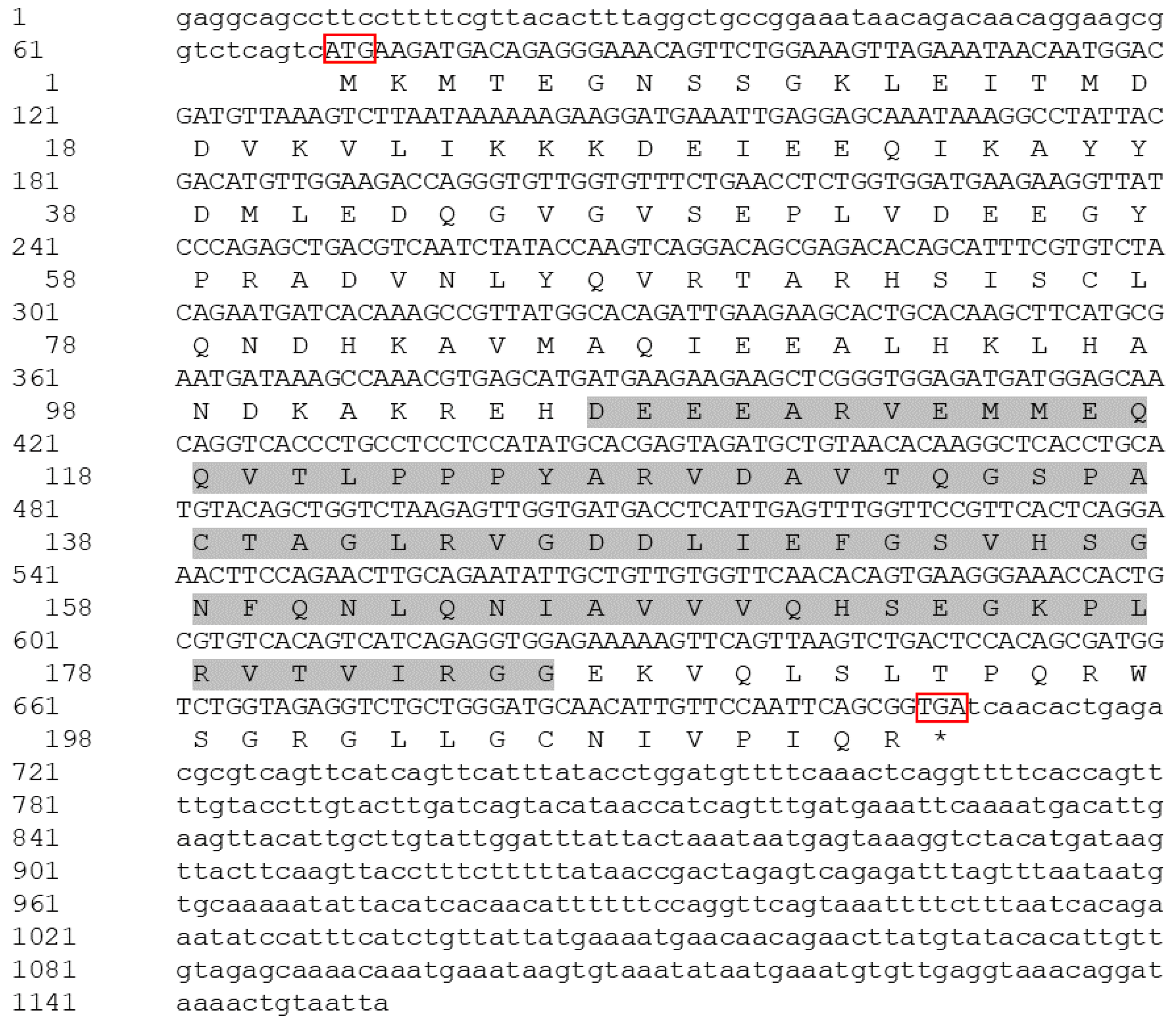
2.1. Cloning and Sequence Analysis of psmd9

The total length of the psmd9 sequence is 1153 bp, including a 69 bp 5′UTR, 445 bp 3′UTR and 639 bp open reading frame. The sequence contains five introns and six exons encoding a total of 212 amino acids (Figure 1). The predicted protein has a relative molecular weight of 23.62 kDa and a theoretical isoelectric point (pI) of 5.41. The protein sequence analysis revealed that 106–185 aa have a PDZ domain.

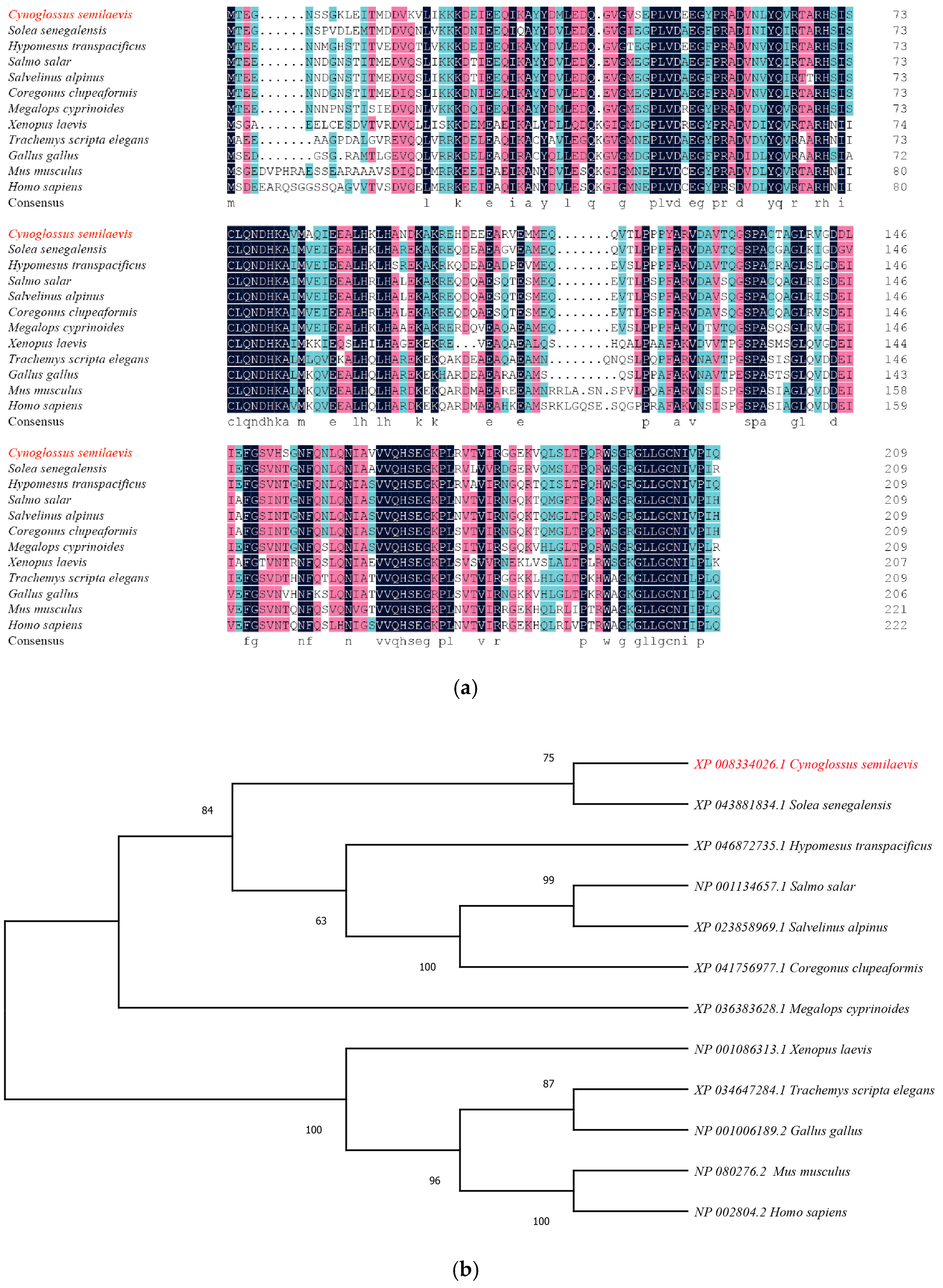
2.2. Multisequence Alignment and Molecular Phylogenetic Analysis

To further understand the structure and function of psmd9, we compared the protein sequences of psmd9 with those of other species. The results of multiple sequence alignments show that the amino acid sequence of psmd9 has a 58.3~79.72% identity with that of other animals (Figure 2a). A phylogenetic tree was constructed based on the amino acid sequences, and the results show that C. semilaevis is clustered with Solea senegalensis (Figure 2b).

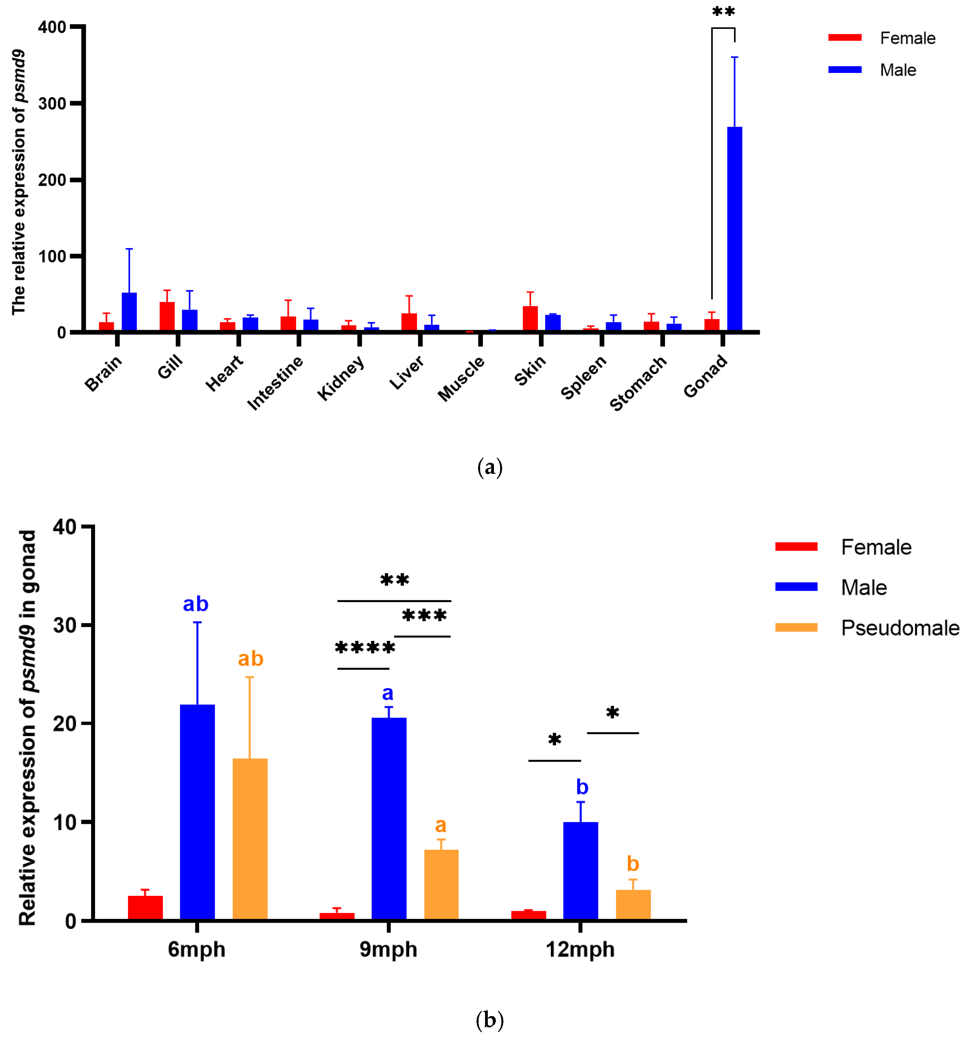
2.3. Expression Pattern of psmd9

In adult C. semilaevis, we measured the expression levels of psmd9 in different tissues by qRT-PCR. The expression of psmd9 was much higher in the testes than that in the ovaries. It also showed expression in the brain, gill, heart, intestine, kidney, liver, skin, spleen and stomach, and low expression only in the muscle (Figure 3a).
We further assessed psmd9 expression patterns for different stages of the gonad. Overall, the expression level of psmd9 in male testes was significantly higher than that in pseudomales, and both were higher than that in female ovaries at 9 mph (months post hatching) and 12 mph. The expression level of psmd9 was highest at 6 mph, with no significant difference between the male and pseudomale testes. After that, psmd9 expression decreased gradually from 6 mph to 9 mph to 12 mph, while the level of expression in males was always higher than that in pseudomales (Figure 3b).

2.4. FISH of psmd9 in Gonads

We studied the location of psmd9 in the gonads at 9 mph by fluorescence in situ hybridization (FISH). In 9 mph testes, mature sperm begins to be produced. psmd9 has fluorescence signals in both the somatic cells and germ cells in the testes (Figure 4). Notably, the fluorescence signal is concentrated in the center of the testes of male fish (Figure 4c). Interestingly, the signal strength is significantly higher in males than in pseudomales. This is consistent with previous qRT-PCR results.

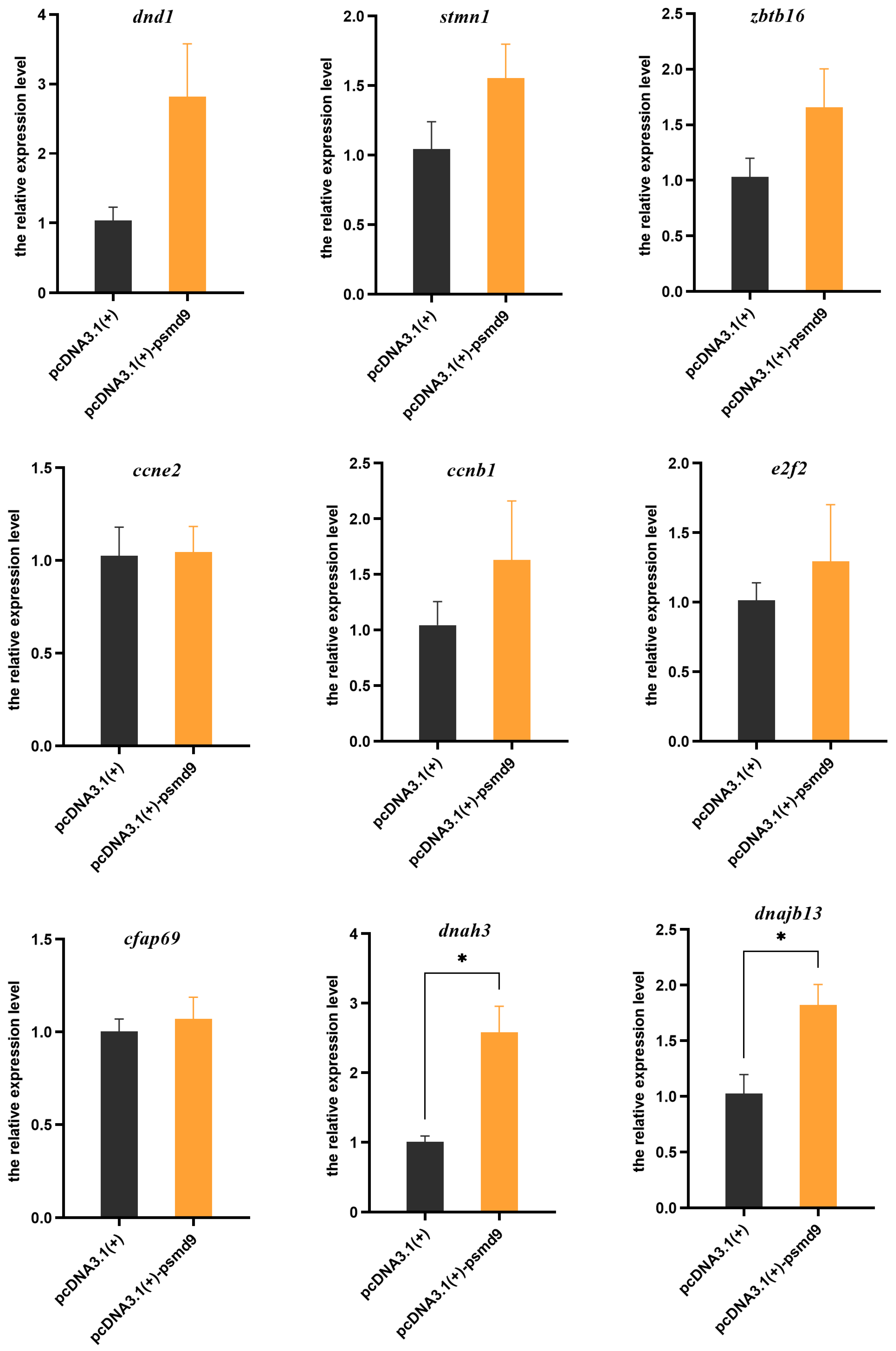
2.5. In Vitro Overexpression of psmd9 and its Effect on Other Genes

To evaluate the role of psmd9 in spermatogenesis, psmd9 was overexpressed in the male testicular cell line of C. semilaevis, and its effect on other germ cell marker genes was detected. The results showed that compared with the control group, overexpressed psmd9 significantly elevated the expression levels of spermatid/spermatozoa markers dnajb13 and dnah3 (p < 0.05). The expression levels of spermatogonia markers dnd1, stmn1 and zbtb16; spermatocytes markers ccne2, ccnb1 and e2f2; and spermatid/spermatozoa marker cfap69 were not significantly affected (p > 0.05), even though there was an elevated trend (Figure 5).

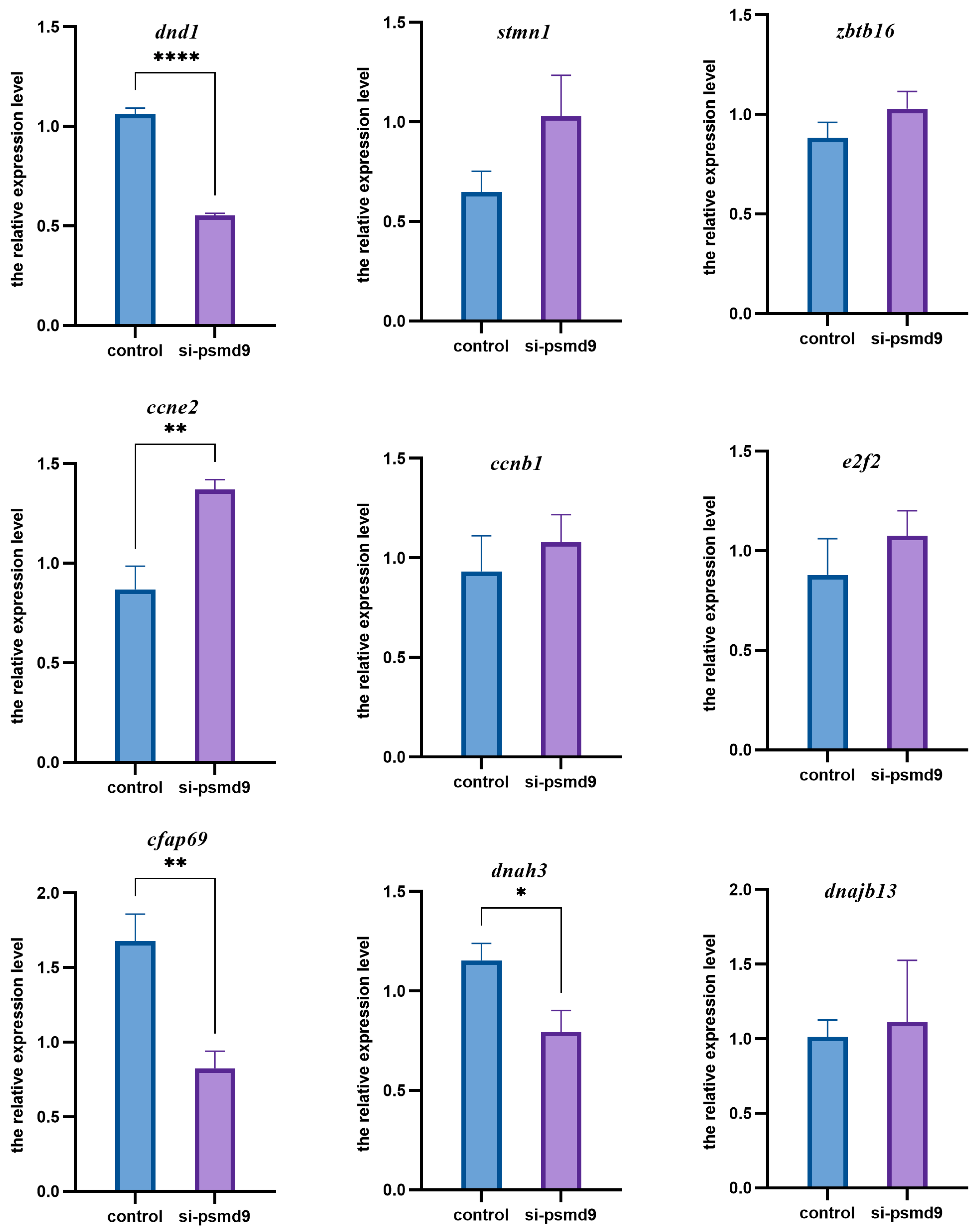
2.6. psmd9 Knockdown and Its Effect on Other Genes

We further performed a knockdown of psmd9 by RNAi. The pre-experimental results showed that the efficiency of three mixed siRNAs had the best knockdown effect. In the following experiment, the transfection reagent group was set as the control group, and the transfection reagent plus siRNAs group was set as the experimental group. In the experimental group, spermatogonia marker dnd1 and spermatid/spermatozoa markers cfap69 and dnah3 were significantly decreased (p < 0.05), while spermatocytes marker ccne2 was significantly increased (p < 0.05). There was no significant change in spermatogonia markers stmn1 and zbtb16, spermatocytes markers ccnb1 and e2f2, or spermatid/spermatozoa marker dnajb13 (Figure 6).

3. Discussion

In this study, the CDS sequence of the C. semilaevis psmd9 gene is verified. psmd9 encodes 212 amino acids, including five introns and six exons. It contains a PDZ conserved protein domain. The PDZ domain plays a role in transcription regulation, mRNA processing and editing, hormone and receptor activity and protein translation through its interaction with protein C-terminal residues [19]. A comparison of psmd9 and similar proteins from different species shows a high degree of identity. In the phylogenetic tree, the predicted target gene proteins are clustered together in other teleost fishes. These results indicate that the PSMD9 protein is highly conserved in structure.
We further analyzed the expression pattern of the psmd9 gene. The whole-tissue quantitative results showed that psmd9 was highly expressed in the testis. It is worth considering that the significant difference between male and female gonads suggests that psmd9 may play an important role in spermatogenesis. The results of the qRT-PCR and FISH on the C. semilaevis gonad showed that psmd9 was highly expressed in the testis, especially in the male testis, and was significantly higher than that in the pseudomale testis. The fluorescence intensity showed that psmd9 is expressed in both the somatic cells and germ cells of the testis, especially near the efferent duct. However, the expression of psmd9 was extremely low in ZW pseudomales, which may be due to the fact that they have only one set of Z chromosomes.
In order to further understand the function of psmd9, we analyzed the expression of psmd9 at different stages after gonad differentiation. The testis differentiation stage of C. semilaevis is slightly later than that of the ovary, and the efferent duct appears at 100 dph (days post hatching), and the formation of the seminal lobules at 150 dph marks the formation of the testis [20]. Because of the difference in water temperatures and seasons, there may be subtle differences in time [21]. At 6 mph, in the testis of C. semilaevis, there were more spermatocytes in the seminal lobule cavity, while spermatogonia were mainly at the edge of the seminal lobule [22]. At 9 mph, the seminal lobules in the testis are closely arranged, and the seminal vesicles in the seminal lobules contain spermatogonia, spermatocytes and sperm cells with synchronous development. At 12 mph, most of the sperm in the testis have matured, and the male fish enters the late stage of sperm release [23,24]. The qRT-PCR results after gonadal differentiation showed that psmd9 was highly expressed at 6 mph and 9 mph, which indicates that psmd9 may play an important role in spermatogenesis.
Sperm maturation involves the recombination of chromatin into highly dense nuclei, thus enhancing hydrodynamic ability and ensuring the integrity of the paternal genome [1]. This process is mediated by the replacement of histone by sperm nuclear basic protein (also known as protamine, PRM). Furthermore, the specific balance of protamine plays an important role in sperm DNA packaging [25]. In humans, small changes in the ratio of PRM1 to PRM2 can also lead to infertility [26]. The nucleolin modulo can mediate the transformation from histone to protamine during spermatogenesis in Drosophila [27]. PSMD9 is a companion to protease assembly. A cell knockout experiment confirmed that psmd9 plays a significant role in maintaining protein balance [16]. In addition, PSMD9 interacts with several free ribosomes subunits and plays a role in the transport of ribosomal proteins to the nucleolus, which helps to maintain the structure and morphology of the nucleolar membrane-free compartment [28]. Therefore, psmd9 may play a role in the balance of protamine during sperm maturation.
In order to further understand the role of psmd9 in the spermatogenesis of C. semilaevis, overexpression and gene knockdown (RNAi) experiments were carried out on the testicular cell line of C. semilaevis, and the markers of germ cells at various development stages were detected. The types of germ cells were classified as follows: spermatogonia (dnd1, stmn1 and zbtb16), spermatocytes (ccne2, ccnb1 and e2f2) and spermatid/spermatozoa (cfap69, dnah3 and dnajb13) [15]. In the experiment on the overexpression of psmd9, the expression level of spermatid/spermatozoa marker genes dnah3 and dnajb13 increased significantly. DNAH3 and DNAJB13, as dynein and flagella structural proteins, are considered to play an important role in sperm flagella movement [29,30]. DNAH3 is considered to be a dynein of flagella, and has ATPase activity. It produces a force towards the minus end of microtubules, depending on the release of ADP [29]. Thus, dnah3 is related to sperm flagella assembly and participates in sperm motility [31]. The protein encoded by dnajb13 plays an important role during the development of sperm flagella, through direct interaction with SEPTIN4 in the assembly and positioning of sperm annulus [30]. It is speculated that psmd9 may play a synergistic role with dnah3 and dnajb13 in sperm flagella formation and sperm movement.
In the psmd9-RNAi experiment, the spermatid/spermatozoa marker gene dnah3 was, correspondingly, significantly decreased. In addition, the spermatogonia marker gene dnd1 and spermatid/spermatozoa marker gene cfap69 decreased significantly, while the spermatocyte marker gene ccne2 increased significantly. This may be due to the complexity of the spermatogenesis regulatory network, or the fact that other genes have similar functions as the target genes, which may have compensatory effects. dnd1 encodes a protein that binds to the microRNA targeting sequence of mRNA, and positively regulates gene expression by inhibiting microRNA-mediated inhibition [32]. dnd1 may play a role in the survival of primordial germ cells (PGC) [33]. The protein encoded by ccne2 belongs to a highly conserved family of cyclin, which plays a role in the G1/S transformation of the cell cycle [34]. cfap69 acts on the biogenesis of the sperm tail and the assembly of the central pair complex, which is located at the midpiece of the flagellum [35]. As a flagella-related protein, CFAP69 is essential for the assembly and stability of sperm flagella [36]. The knockdown of psmd9 may promote spermatogonia to differentiate into spermatocytes. Therefore, psmd9, as a subunit of protease, cooperates with dnd1, cfap69 and dnah3 in the process of spermatogonia maintenance and sperm maturation, but antagonizes ccne2 in the process of spermatogenesis.
It can be seen that psmd9 mainly plays a role in PGC survival and sperm maturation. It has been reported that in mammals, psmd9 silencing leads to the disintegration of the nucleolar structure, the accumulation of p53, and the slow growth of cells [28]. This phenomenon is similar to the pathophysiology of sperm damage after a vasectomy. A spermatogenesis disorder after a vasectomy is related to an increase in p53 and a decrease in proliferating cell nuclear antigen expression. Also, the P53-Bax pathway has been found to affect the apoptosis of some seminiferous tubules in the delayed period after a vasectomy [37]. Thus, the lack of psmd9 may affect nucleolar instability, which may cause the accumulation of p53 protein and apoptosis in sperm. The specific mechanism of psmd9 in apoptosis and spermatogenesis needs further study.

4. Materials and Methods

4.1. Fish and Tissue Collection

All the C. semilaevis used in this experiment were obtained from Huanghai Aquiculture, Ltd. (Yantai, China). After approval by the Institutional Animal Care and Use Committee (IACUC) of the Yellow Sea Fisheries Research Institute (CAFS) (Qingdao, China), the fish were collected and handled in accordance with the “Guidelines for Experimental Animals” of the Ministry of Science and Technology (Beijing, China). We dissected tissue samples from adult fish (12 mph), including the spleen, kidney, heart, liver, intestine, gill, muscle, skin, brain, and gonads, and immediately froze them with liquid nitrogen and stored them at −80 °C. The anatomical acquisition of tongue soles gonads at different stages of development was achieved, including at 6 months and 9 months and 12 months post hatching. After acquisition, one half of each sample was placed in liquid nitrogen and then stored at −80 °C for RNA extraction, and the other part was stored in 4% paraformaldehyde fixative for fluorescence in situ hybridization (FISH). The tailfins were cut and stored in ethanol for DNA extraction and genetic sex determination.

4.2. CDS Cloning of psmd9

The total RNA was extracted from the tissue samples according to the instructions for the TRIzol reagent (Invitrogen, Carlsbad, CA, USA). The RNA concentration and purity were determined by a Nano Drop 2000 spectrophotometer (Thermo, Waltham, MA, USA), and the integrity of the RNA was assessed with 1% agarose gel electrophoresis. The qualified RNA was stored at −80 °C. Then, the first-strand cDNA was synthesized using a HiScript III first-strand cDNA synthesis kit (+gDNA wiper) (Vazyme, Nanjing, China). A phenol–chloroform extraction method was used to extract the genomic DNA from the tailfin, and the genetic sex was identified by PCR using published methods and primers (sex F and sex R in Table 1) [38].
Based on the predicted sequences in the NCBI database (XM_008335804.2), we designed and synthesized primers (Table 1) to amplify psmd9 from gonadal cDNA with PCR. After detection by 1% agarose gel electrophoresis, the product was purified using a FastPure® Gel DNA Extraction Mini Kit (Vazyme, Nanjing, China). Then, the purified target fragment was ligated into a pEASY-T1 cloning vector (TransGene, Beijing, China), and the positive clones were selected for transformation into DH5α competent cells (TaKaRa, Shiga, Japan) for sequencing.

4.3. Real-Time Quantitative PCR

A Prime-Script RT Reagent Kit with gDNA Eraser (Takara, Dalian, China) was used to generate the cDNA templates from the total RNA. The primer sequences were designed using the target gene sequences as templates, and β-actin was used as an internal reference gene [39]. All the primers were synthesized by Ruibo, Ltd. (Beijing, China). According to the operation instructions for the QuantiNova SYBR Green PCR Kit (Qiagen, Hilden, Germany), qRT-PCR was performed with a LightCycler® 480 PCR instrument (Roche, Basel, Switzerland). The reaction volume was 10 µL, including 5 µL 2 × SYBR Green PCR Master Mix, 0.4 µL primers (Table 1), and 1 µL of cDNA. The amplification conditions were as follows: 95 °C for 2 min, 40 cycles at 95 °C for 5 s, and 60 °C for 10 s. The melting curve reaction conditions were 95 °C for 15 s, 65 °C for 1 min, +0.11 °C/s to 95 °C, and 40 °C for 10 s. Three biological and three technical repetitions were tested. The 2−∆∆Ct method was used for relative gene expression analysis [40].

4.4. Fluorescence In Situ Hybridization of psmd9

The testes at 9 mph were fixed with 4% PFA, embedded in paraffin and sliced into 3 µm sections. psmd9-specific fluorescent in situ hybridization probes labeled with 5′Cy3 were designed and synthesized. Fluorescence in situ hybridization was performed according to the instructions for the RNA FISH kit (Gene Pharma, Shanghai, China). The images were captured using a laser confocal microscope (Olympus, Tokyo, Japan) and analyzed.

4.5. Culture and Transfection of C. semilaevis Testicular Cell Line

The psmd9 overexpression vector was constructed to verify the role of psmd9. The primers psmd9-EcoR1-F and psmd9-Xho1-R (Table 1) were used to amplify the target fragment for homologous recombination. The pcDNA3.1(+) vectors (Invitrogen, Carlsbad, CA, USA) were digested with EcoRI and XhoI. The pcDNA3.1(+)-psmd9 plasmid was ligated using a ClonExpress Ultra One Step Cloning Kit (Vazyme, Nanjing, China). The recombinant plasmid was transformed into Escherichia coli and plated on a medium containing ampicillin. After 12 h, single colonies were selected for colony PCR, and the positive clones were sent for sequencing to verify whether the recombinant plasmid was correctly connected. The recombinant plasmid was extracted using an EndoFree Mini Plasmid Kit II (Tiangen Biotech, Beijing, China) for transfection.
The testicular cell line of C. semilaevis originated from the researchers’ laboratory and was cultured in L15 compound medium at 24 °C [41]. Taking an empty plasmid as the control group, Lipofectamine 3000 Reagent (Thermo Fisher, Invitrogen, Carlsbad, CA, USA) was used to transfect the recombinant plasmid into 6-well plates. Cells were collected 48 h after transfection, and the total RNA was extracted. The expression of the spermatogenesis-related genes was detected using qRT-PCR.

4.6. siRNA-Mediated Knockdown of psmd9 in Testicular Cell Line

Based on the sequence of psmd9, three specific siRNAs were designed and synthesized (Gene Pharma, Shanghai, China). The siRNA with the best knockdown effect was selected for further study. The testicular cell line of C. semilaevis was prepared according to the method mentioned above. In the control group, only the transfection reagents were treated without adding siRNAs. According to the previously established experimental protocol, the psmd9 siRNA transfection and control transfection were performed in triplicate. After transfection for 48 h, the transfection efficiency was observed under a fluorescent inverted microscope and the cells were collected. The RNA was extracted according to the previous method and the expression of related genes was detected.

4.7. Statistical Analysis

All the experimental data were presented as means ± SEM, and all the experiments were performed in triplicate. Prism 6.0 (GraphPad Software, San Diego, CA, USA) was utilized for data processing, and t-tests and one-way ANOVA followed by Tukey multiple comparisons were conducted for the analysis. p-values less than 0.05 were considered statistically significant (* p < 0.05; ** p < 0.01; *** p < 0.001; **** p < 0.0001).

Author Contributions

Conceptualization, C.S., Q.W., H.W. and Y.Z.; methodology, Y.Z., Q.L. and Q.W.; software, Y.W. and Y.Z.; validation, Q.L. and Y.Z.; investigation, Y.W. and Y.Z.; writing—original draft preparation, Y.Z., Q.W. and C.S.; writing—review and editing, Y.Z., Q.W. and C.S.; visualization, Y.Z.; supervision, Q.W. and C.S. All authors have read and agreed to the published version of the manuscript.

Funding

This research was funded by the National Key R&D Program of China (grant number 2022YFD2400100); the National Nature Science Foundation of China (grant numbers 31722058, 31472269 and 32002371); the AoShan Talents Cultivation Program Supported by Qingdao National Laboratory for Marine Science and Technology (grant number 2017ASTCP-ES06); the Taishan Scholars Program (NO. tstp20221149), with a grant to C.S.; the National Ten-Thousands Talents Special Support Program, with a grant to C.S.; the Central Public-interest Scientific Institution Basal Research Fund, CAFS (grant number 2023TD19); the China Agriculture Research System (grant number CARS-47-G03); and a project by the National Marine Genetic Resource Center.

Institutional Review Board Statement

The animal study protocol was approved by the Institutional Animal Care and Use Committee (IACUC) of the Yellow Sea Fisheries Research Institute (CAFS) (Qingdao, China) (Approval No.: YSFRI-2024045).

Informed Consent Statement

Not applicable.

Data Availability Statement

Data are contained within the article.

Conflicts of Interest

The authors declare no conflicts of interest.

References

  1. Sharma, R.; Agarwal, A. Spermatogenesis: An Overview. In Sperm Chromatin: Biological and Clinical Applications in Male Infertility and Assisted Reproduction; Zini, A., Agarwal, A., Eds.; Springer: New York, NY, USA, 2011; pp. 19–44. [Google Scholar]
  2. Renkawitz-Pohl, R.; Hempel, L.; Hollmann, M.; Schäfer, M.A. 1.4—Spermatogenesis. In Comprehensive Molecular Insect Science; Gilbert, L.I., Ed.; Elsevier: Amsterdam, The Netherlands, 2005; pp. 157–177. [Google Scholar]
  3. Schulz, R.W.; de França, L.R.; Lareyre, J.-J.; LeGac, F.; Chiarini-Garcia, H.; Nobrega, R.H.; Miura, T. Spermatogenesis in fish. Gen. Comp. Endocrinol. 2010, 165, 390–411. [Google Scholar] [CrossRef]
  4. Hess, R.A.; de Franca, L.R. Spermatogenesis and Cycle of the Seminiferous Epithelium. In Molecular Mechanisms in Spermatogenesis; Cheng, C.Y., Ed.; Springer: New York, NY, USA, 2008; pp. 1–15. [Google Scholar]
  5. Li, X.-Y.; Mei, J.; Ge, C.-T.; Liu, X.-L.; Gui, J.-F. Sex determination mechanisms and sex control approaches in aquaculture animals. Sci. China Life Sci. 2022, 65, 1091–1122. [Google Scholar] [CrossRef] [PubMed]
  6. Wang, H.-P.; Shen, Z.-G. Sex Control in Aquaculture. In Sex Control in Aquaculture; John Wiley & Sons Ltd.: Hoboken, NJ, USA, 2018; pp. 1–34. [Google Scholar]
  7. Chen, S.-L.; Xu, W.-T. Sex Identification and Control in Half-Smooth Tongue Sole. In Sex Control in Aquaculture; John Wiley & Sons Ltd.: Hoboken, NJ, USA, 2018; pp. 547–563. [Google Scholar]
  8. Hu, L.; Siqing, C.; Haiqing, L.; Hongbo, J.; Meiyu, W. Study on sex ratio and comparison of morphological variation between genders of cultured half-smooth tongue sole(Cynoglossus semilaevis). J. Fish. China 2012, 36, 1331–1336. [Google Scholar]
  9. Shao, C.; Li, Q.; Chen, S.; Zhang, P.; Lian, J.; Hu, Q.; Sun, B.; Jin, L.; Liu, S.; Wang, Z.; et al. Epigenetic modification and inheritance in sexual reversal of fish. Genome Res. 2014, 24, 604–615. [Google Scholar] [CrossRef] [PubMed]
  10. Chen, S.; Zhang, G.; Shao, C.; Huang, Q.; Liu, G.; Zhang, P.; Song, W.; An, N.; Chalopin, D.; Volff, J.N.; et al. Whole-genome sequence of a flatfish provides insights into ZW sex chromosome evolution and adaptation to a benthic lifestyle. Nat. Genet. 2014, 46, 253–260. [Google Scholar] [CrossRef] [PubMed]
  11. Xu, W.; Li, H.; Dong, Z.; Cui, Z.; Zhang, N.; Meng, L.; Zhu, Y.; Liu, Y.; Li, Y.; Guo, H.; et al. Ubiquitin ligase gene neurl3 plays a role in spermatogenesis of half-smooth tongue sole (Cynoglossus semilaevis) by regulating testis protein ubiquitination. Gene 2016, 592, 215–220. [Google Scholar] [CrossRef] [PubMed]
  12. Meng, L.; Zhu, Y.; Zhang, N.; Liu, W.; Liu, Y.; Shao, C.; Wang, N.; Chen, S. Cloning and characterization of tesk1, a novel spermatogenesis-related gene, in the tongue sole (Cynoglossus semilaevis). PLoS ONE 2014, 9, e107922. [Google Scholar] [CrossRef]
  13. Meng, L.; Xu, W.; Zhu, Y.; Zhang, N.; Shao, C.; Liu, Y.; Chen, S. Molecular characterization and expression analysis of strbp in Chinese tongue sole (Cynoglossus semilaevis). Theriogenology 2018, 118, 225–232. [Google Scholar] [CrossRef] [PubMed]
  14. Wang, L.-P. Discriminant Analysis of Sexual Morphology Juvenile and Integrative Analysis of Omics Reveals the Clues Why Pseudomales Are Not Able to Produce W Sperms in Cynoglossus semilaevis. Master’s Thesis, Shanghai Ocean University, Shanghai, China, 2022. [Google Scholar]
  15. Wang, H.Y.; Liu, X.; Chen, J.Y.; Huang, Y.; Lu, Y.; Tan, F.; Liu, Q.; Yang, M.; Li, S.; Zhang, X.; et al. Single-cell-resolution transcriptome map revealed novel genes involved in testicular germ cell progression and somatic cells specification in Chinese tongue sole with sex reversal. Sci. China Life Sci. 2023, 66, 1151–1169. [Google Scholar] [CrossRef]
  16. Christie, J.; Anthony, C.M.; Harish, M.; Mudartha, D.; Ud Din Farooqee, S.B.; Venkatraman, P. The interaction network of the proteasome assembly chaperone PSMD9 regulates proteostasis. FEBS J. 2023, 290, 5581–5604. [Google Scholar] [CrossRef]
  17. Li, Y.; Liu, X.; Zhao, F.; Zhao, Z.; Li, X.; Wang, J.; Huang, B.; Chen, A. Comprehensive analysis of PSMD family members and validation of PSMD9 as a potential therapeutic target in human glioblastoma. CNS Neurosci. Ther. 2023, 30, e14366. [Google Scholar] [CrossRef]
  18. Jafarian, A.; Sadeghi, M.R.; Pejhan, N.; Salehkhou, S.; Lakpour, N.; Akhondi, M.M. Regeneration of spermatogenesis in a mouse model of azoospermia by follicle-stimulating hormone and oestradiol. Andrologia 2014, 46, 1098–1106. [Google Scholar] [CrossRef] [PubMed]
  19. Sangith, N.; Srinivasaraghavan, K.; Sahu, I.; Desai, A.; Medipally, S.; Somavarappu, A.K.; Verma, C.; Venkatraman, P. Discovery of novel interacting partners of PSMD9, a proteasomal chaperone: Role of an Atypical and versatile PDZ-domain motif interaction and identification of putative functional modules. FEBS Open Bio 2014, 4, 571–583. [Google Scholar] [CrossRef] [PubMed]
  20. Ma, X.K.; Liu, X.Z.; Wen, H.S.; Xu, Y.; Zhang, L.J. Histological observation on gonadal sex differentiation in Cynoglossus semilaevis Günther. Mar. Fish. Res. 2006, 27, 55–61. [Google Scholar]
  21. Liang, Z.; Chen, S.; Zhang, J.; Song, W.; Liu, S. Gonadal development process observation of half-smooth tongue sole in rearing population. J. South. Agric. 2012, 43, 2074–2078. [Google Scholar]
  22. Wang, Y. Spermatogenesis and Fertilization Process of Cynoglossus semilaevis Günther. Ph.D. Thesis, Ocean University of China, Qingdao, China, 2008. [Google Scholar]
  23. Kang, X.; Liang, C.; Guo, M.; Ge, S.; Mu, S.; Su, W.; Liu, X. Ultrastructure of Spermatogenesis and Spermiogenesis in Half-smooth tongue sole Cynoglossus semilaevis. Curr. Zool. 2008, 54, 356–365. [Google Scholar]
  24. Song, H.; Weng, Y.; Liu, Z.; Fang, Y. A Historical Study on Spermatogenesis in Cynoglossus semilaevis. J. Appl. Oceanogr. 2009, 28, 19–24. [Google Scholar]
  25. Akmal, M.; Widodo, M.A.; Sumitro, S.B.; Purnomo, B.B. The important role of protamine in spermatogenesis and quality of sperm: A mini review. Asian Pac. J. Reprod. 2016, 5, 357–360. [Google Scholar] [CrossRef]
  26. Schmid, C.; Heng, H.H.; Rubin, C.; Ye, C.J.; Krawetz, S.A. Sperm nuclear matrix association of the PRM1→PRM2→TNP2 domain is independent of Alu methylation. Mol. Hum. Reprod. 2001, 7, 903–911. [Google Scholar] [CrossRef]
  27. Park, J.I.; Bell, G.W.; Yamashita, Y.M. Derepression of Y-linked multicopy protamine-like genes interferes with sperm nuclear compaction in D. melanogaster. Proc. Natl. Acad. Sci. USA 2023, 120, e2220576120. [Google Scholar] [CrossRef]
  28. Ud Din Farooqee, S.B.; Christie, J.; Venkatraman, P. PSMD9 ribosomal protein network maintains nucleolar architecture and WT p53 levels. Biochem. Biophys. Res. Commun. 2021, 563, 105–112. [Google Scholar] [CrossRef] [PubMed]
  29. Meng, G.-Q.; Wang, Y.; Luo, C.; Tan, Y.-M.; Li, Y.; Tan, C.; Tu, C.; Zhang, Q.-J.; Hu, L.; Zhang, H.; et al. Bi-allelic variants in DNAH3 cause male infertility with asthenoteratozoospermia in humans and mice. Hum. Reprod. Open 2024, 2024, hoae003. [Google Scholar] [CrossRef] [PubMed]
  30. Guan, J.; Kinoshita, M.; Yuan, L. Spatiotemporal association of DNAJB13 with the annulus during mouse sperm flagellum development. BMC Dev. Biol. 2009, 9, 23. [Google Scholar] [CrossRef] [PubMed]
  31. Sironen, A.; Shoemark, A.; Patel, M.; Loebinger, M.R.; Mitchison, H.M. Sperm defects in primary ciliary dyskinesia and related causes of male infertility. Cell. Mol. Life Sci. 2020, 77, 2029–2048. [Google Scholar] [CrossRef] [PubMed]
  32. Kedde, M.; Strasser, M.J.; Boldajipour, B.; Vrielink, J.A.F.O.; Slanchev, K.; le Sage, C.; Nagel, R.; Voorhoeve, P.M.; van Duijse, J.; Ørom, U.A.; et al. RNA-Binding Protein Dnd1 Inhibits MicroRNA Access to Target mRNA. Cell 2007, 131, 1273–1286. [Google Scholar] [CrossRef]
  33. Imai, A.; Matsuda, K.; Niimi, Y.; Suzuki, A. Loss of Dead end1 induces testicular teratomas from primordial germ cells that failed to undergo sexual differentiation in embryonic testes. Sci. Rep. 2023, 13, 6398. [Google Scholar] [CrossRef] [PubMed]
  34. Sonntag, R.; Giebeler, N.; Nevzorova, Y.A.; Bangen, J.-M.; Fahrenkamp, D.; Lambertz, D.; Haas, U.; Hu, W.; Gassler, N.; Cubero, F.J.; et al. Cyclin E1 and cyclin-dependent kinase 2 are critical for initiation, but not for progression of hepatocellular carcinoma. Proc. Natl. Acad. Sci. USA 2018, 115, 9282–9287. [Google Scholar] [CrossRef] [PubMed]
  35. Dong, F.N.; Amiri-Yekta, A.; Martinez, G.; Saut, A.; Tek, J.; Stouvenel, L.; Lorès, P.; Karaouzène, T.; Thierry-Mieg, N.; Satre, V.; et al. Absence of CFAP69 Causes Male Infertility due to Multiple Morphological Abnormalities of the Flagella in Human and Mouse. Am. J. Hum. Genet. 2018, 102, 636–648. [Google Scholar] [CrossRef] [PubMed]
  36. McClintock, T.S.; Glasser, C.E.; Bose, S.C.; Bergman, D.A. Tissue expression patterns identify mouse cilia genes. Physiol. Genomics 2008, 32, 198–206. [Google Scholar] [CrossRef]
  37. Shiraishi, K.; Naito, K.; Yoshida, K.-I. Vasectomy impairs spermatogenesis through germ cell apoptosis mediated by the p53-Bax pathway in rats. J. Urol. 2001, 166, 1565–1571. [Google Scholar] [CrossRef]
  38. Liu, Y.; Chen, S.; Gao, F.; Liang, M.M.; Hu, Q.; Song, W.; Shao, C.; Lv, W. SCAR-transformation of sex-specific SSR marker and its application in half-smooth tongue sole (Cynoglossus semiliaevis). J. Agric. Biotechnol. 2014, 22, 787–792. [Google Scholar]
  39. Li, Z.; Yang, L.; Wang, J.; Shi, W.; Pawar, R.A.; Liu, Y.; Xu, C.; Cong, W.; Hu, Q.; Lu, T.; et al. β-Actin is a useful internal control for tissue-specific gene expression studies using quantitative real-time PCR in the half-smooth tongue sole Cynoglossus semilaevis challenged with LPS or Vibrio anguillarum. Fish Shellfish Immunol. 2010, 29, 89–93. [Google Scholar] [CrossRef] [PubMed]
  40. Livak, K.J.; Schmittgen, T.D. Analysis of relative gene expression data using real-time quantitative PCR and the 2−ΔΔCT Method. Methods 2001, 25, 402–408. [Google Scholar] [CrossRef]
  41. Liu, Q.; Wang, Y.; Tan, L.; Ma, W.; Zhao, X.; Shao, C.; Wang, Q. The Role of the Heat Shock Cognate Protein 70 Genes in Sex Determination and Differentiation of Chinese Tongue Sole (Cynoglossus semilaevis). Int. J. Mol. Sci. 2023, 24, 3761. [Google Scholar] [CrossRef] [PubMed]
Figure 1. The cDNA sequence and deduced amino acid sequence of the psmd9 gene. The red box represents the start codon and stop codon of psmd9, and the stop codon is indicated by an asterisk (*). The grey shadows represent the predicted protein domain.
Ijms 25 06372 g001
Figure 2. Multiple sequence alignments and phylogenetic tree of psmd9 proteins. (a) DNAMAN was used to perform the multisequence alignment of the psmd9 amino acid sequences. The blue boxes indicate 100% sequence identity between amino acid sequences, the pink boxes indicate more than 75% similarity between amino acid sequences, and the cyan boxes indicate 50% to 75% similarity between amino acid sequences. Note: C. semilaevis (Sequence ID: XP_008334026.1), Solea senegalensis (Sequence ID: XP_043881834.1), Hypomesus transpacificus (Sequence ID: XP_046872735.1), Salmo salar (Sequence ID: NP_001134657.1), Salvelinus alpinus (Sequence ID: XP_023858969.1), Coregonus clupeaformis (Sequence ID: XP_041756977.1), Megalops cyprinoides (Sequence ID: XP_036383628.1), Xenopus laevis (Sequence ID: NP_001086313.1), Trachemys scripta elegans (Sequence ID: XP_034647284.1), Gallus gallus (Sequence ID: NP_001006189.2), Mus musculus (Sequence ID: NP_080276.2) and Homo sapiens (Sequence ID: NP_002804.2). (b) Phylogenetic tree based on the psmd9 amino acid sequence, using the neighbor-joining method. The number displayed on the branch node represents the guiding value (%). C. semilaevis psmd9 is in red color.
Ijms 25 06372 g002
Figure 3. Expression of psmd9 in C. semilaevis. (a) Expression of psmd9 genes in adult C. semilaevis tissues. (b) Expression of psmd9 from post-gonadal differentiation to maturation. Means ± SEM from three independent individuals (n = 3) are shown. β-actin was used as reference gene. Different letters indicate statistically significant differences (p < 0.05) in each sex among different stages. Asterisks indicate statistically significant differences (* p < 0.05; ** p < 0.01; *** p < 0.001; **** p < 0.0001) in each stage among different sexes.
Ijms 25 06372 g003
Figure 4. psmd9 localization in the gonads of C. semilaevis at 9 mph. (a) Localization of psmd9 detected using 5′Cy3-labeled fluorescent probe in male testes. (b) Spermatogenic cell nucleus and somatic cell nucleus were stained with DAPI in male testes. (d) Localization of psmd9 detected using 5′Cy3-labeled fluorescent probe in pseudomale testes. (e) Spermatogenic cell nucleus and somatic cell nucleus were stained with DAPI in pseudomale testes. (g) Somatic nuclei were stained with DAPI in ovaries. (h) Localization of psmd9 detected using 5′Cy3-labeled fluorescent probe in ovaries. (c,f,i) Combination of psmd9 and nucleus localization. The blue signals represent nuclei and the red signals represent psmd9 mRNAs, respectively.
Ijms 25 06372 g004
Figure 5. Relative expression level of germ cell marker genes in C. semilaevis testicular cell lines with overexpression of psmd9. Means ± SEM from three independent individuals (n = 3) are shown. β-actin was used as the reference gene. Asterisks indicate statistically significant differences (* p < 0.05).
Ijms 25 06372 g005
Figure 6. Relative expression level of germ cell marker genes in C. semilaevis testicular cell line with psmd9 knockdown. Means ± SEM from three independent individuals (n = 3) are shown. β-actin was used as reference gene. Asterisks indicate statistically significant differences (* p < 0.05; ** p < 0.01; **** p < 0.0001).
Ijms 25 06372 g006
Table 1. Primers used for cloning and gene expression analysis.
Primer NameSequence (5′–3′)Purpose
psmd9-FAACAGACAACAGGAAGCGPartial fragment amplification
psmd9-RCAGTTTTATCCTGTTTACCTC
psmd9-EcoR1-FtagtccagtgtggtggaattcATGAAGATGACAGAGGGAAACAGTTConstruction of overexpression vector
psmd9-Xho1-RaacgggccctctagactcgagTCACCGCTGAATTGGAACAAT
psmd9-qFACCAGGGTGTTGGTGTTTCTGRT-qPCR
psmd9-qRCGTTTGGCTTTATCATTCGCA
zbtb16-qFGCCGAAGTCAGTCAGATGGG
zbtb16-qRGCGTCCGATTTGTGATTGATA
dnd1-qFGCTGTGTAAGTTGCTTGATGTTGT
dnd1-qRGCCTGTGAAGGGTGCGG
stmn1-qFTTCCCTTGTCTGCCCCC
stmn1-qRCTTCTTTTTCATGTTCACGCTTC
ccne2-qFGAGCAGGAAAACAGTGGTGAAG
ccne2-qRGAATGTGTGGAAGTGACAGAAGG
ccnb1-qFACCGCTTTCTTCAGGACCAC
ccnb1-qRTCTCCTCAAACTTAGACGCCAG
e2f2-qFCAGTGGCTGGTGGGAGATG
e2f2-qRCAGGGATTTCTCCGCTCG
cfap69-qFTGTTGAACCTGTTGGTGTTGATG
cfap69-qRGTGGTGGATTTGGGCGG
dnah3-qFTCTTTTGGGAGGTTAGGCAAG
dnah3-qRGTGGGATTTAGATGGGGTCG
dnajb13-qFTCTGGAGATGGCTCTGACTGG
dnajb13-qRGCACAATGTCGTTGATGGGTAT
β-actin-qFGAGTAGCCACGCTCTGTC
β-actin-qRGCTGTGCTGTCCCTGTA
sex-FCCTAAATGATGGATGTAGATTCTGTCSex identification
sex-RGATCCAGAGAAAA-TAAACCCAGG
Disclaimer/Publisher’s Note: The statements, opinions and data contained in all publications are solely those of the individual author(s) and contributor(s) and not of MDPI and/or the editor(s). MDPI and/or the editor(s) disclaim responsibility for any injury to people or property resulting from any ideas, methods, instructions or products referred to in the content.

