Article

Inhibition of Protein Synthesis Attenuates Formation of Traumatic Memory and Normalizes Fear-Induced c-Fos Expression in a Mouse Model of Posttraumatic Stress Disorder

by
Tatyana A. Zamorina
1,2,†,
Olga I. Ivashkina
1,3,†,
Ksenia A. Toropova
1,3,† and
Konstantin V. Anokhin
1,3,*
1
Institute for Advanced Brain Studies, Lomonosov Moscow State University, 119991 Moscow, Russia
2
Faculty of Biology, Department of Higher Nervous Activity, Lomonosov Moscow State University, 119234 Moscow, Russia
3
Laboratory of Neuronal Intelligence, Lomonosov Moscow State University, 119991 Moscow, Russia
*
Author to whom correspondence should be addressed.
These authors contributed equally to this work.
Int. J. Mol. Sci. 2024, 25(12), 6544; https://doi.org/10.3390/ijms25126544
Submission received: 16 April 2024 / Revised: 24 May 2024 / Accepted: 11 June 2024 / Published: 13 June 2024
(This article belongs to the Collection Feature Papers in Molecular Neurobiology)

Abstract

:
Posttraumatic stress disorder (PTSD) is a debilitating psychosomatic condition characterized by impairment of brain fear circuits and persistence of exceptionally strong associative memories resistant to extinction. In this study, we investigated the neural and behavioral consequences of inhibiting protein synthesis, a process known to suppress the formation of conventional aversive memories, in an established PTSD animal model based on contextual fear conditioning in mice. Control animals were subjected to the conventional fear conditioning task. Utilizing c-Fos neural activity mapping, we found that the retrieval of PTSD and normal aversive memories produced activation of an overlapping set of brain structures. However, several specific areas, such as the infralimbic cortex and the paraventricular thalamic nucleus, showed an increase in the PTSD group compared to the normal aversive memory group. Administration of protein synthesis inhibitor before PTSD induction disrupted the formation of traumatic memories, resulting in behavior that matched the behavior of mice with usual aversive memory. Concomitant with this behavioral shift was a normalization of brain c-Fos activation pattern matching the one observed in usual fear memory. Our findings demonstrate that inhibiting protein synthesis during traumatic experiences significantly impairs the development of PTSD in a mouse model. These data provide insights into the neural underpinnings of protein synthesis-dependent traumatic memory formation and open prospects for the development of new therapeutic strategies for PTSD prevention.

1. Introduction

Posttraumatic stress disorder (PTSD) is an incapacitating condition characterized by compromised emotional reactions resulting from acute psychological trauma. The disorder is marked by the impairment of information processing in fear memory brain circuits and the establishment of strong memories associated with the traumatic episode [1,2,3]. Memory of a traumatic event is implicated in many symptoms of PTSD, including intrusive thoughts and dreams, obsessive memories, flashbacks, and abnormal avoidance behavior [4,5,6]. Obsessive memories in PTSD patients are unusually strong, with reliving experiences comparable in intensity to the original trauma (flashbacks) [4]. However, PTSD is distinguished not only by extraordinarily vivid memories of certain aspects of a traumatic event (hypermnesia) but also by partial retrograde amnesia for other aspects [7]. This evidence has led to the hypothesis of aberrant aversive memory formation following a traumatic event. Support for this hypothesis is found in the impaired extinction of traumatic memories in PTSD [8,9,10], unlike normal aversive memories formed through fear conditioning, which can be extinguished [11]. fMRI studies have also demonstrated altered activation of fear memory neural circuits in PTSD patients [12,13,14,15,16,17,18], including the amygdala [12,13], hippocampus [14,15], cingulate [16,17], and prefrontal cortices [17,18].
To explain PTSD aberrant memories, Siegmund and Wotjak [19] proposed a “dual-branch hypothesis,” which suggests that traumatic experiences simultaneously lead to associative memories (involving classical conditioning) and non-associative memories (involving sensitization processes) [19,20]. Building on this hypothesis, authors developed a mouse PTSD model that replicates key features of the traumatic event leading to PTSD in humans: unpredictability and uncontrollability [20]. The model exhibits a good alignment of animal behavioral phenotypes with human PTSD symptoms, including prolonged PTSD manifestations and impaired traumatic memory extinction [19,20]. The developed mouse model, based on the presentation of a robust and unavoidable footshock, allows experimental study of both associative and sensitized PTSD components. While the associative component undergoes typical consolidation stages mirroring normal aversive memory, the sensitization processes unfold more slowly, requiring an incubation period for its expression [20]. Distinct synaptic mechanisms underlie the two components: associative memory is N-Methyl-D-Aspartate (NMDA)-dependent, whereas sensitized components are not [21]. However, the brain activity in this PTSD model has not been studied, leaving its validation for human PTSD symptoms and mechanisms incomplete. Therefore, in the present study, we applied brain-wide c-Fos imaging [22,23] to compare brain activity patterns after retrieval of traumatic and normal fear memory in this experimental model.
We also explored the effects of inhibition of protein synthesis (PSI) on the formation of associative and non-associative components of traumatic memory and concomitant patterns of brain activity. Protein synthesis inhibitors (PSIs) administered before or shortly after the traumatic event are known to prevent the development of PTSD-like behavioral symptoms [24,25,26]. These data suggest a dependency of PTSD on molecular mechanisms involved in the consolidation of normal aversive memory [27,28]. Still, it remains unclear whether such relief of behavioral PTSD-like symptoms is accompanied by normalization of brain activity in fear circuits. By administering the PSI cycloheximide before traumatic events, we investigated its effects on associative (conditioned fear) and non-associative (behavioral sensitization) PTSD-like symptoms and patterns of brain activity related to the retrieval of intact or impaired fear and traumatic memory.

2. Results

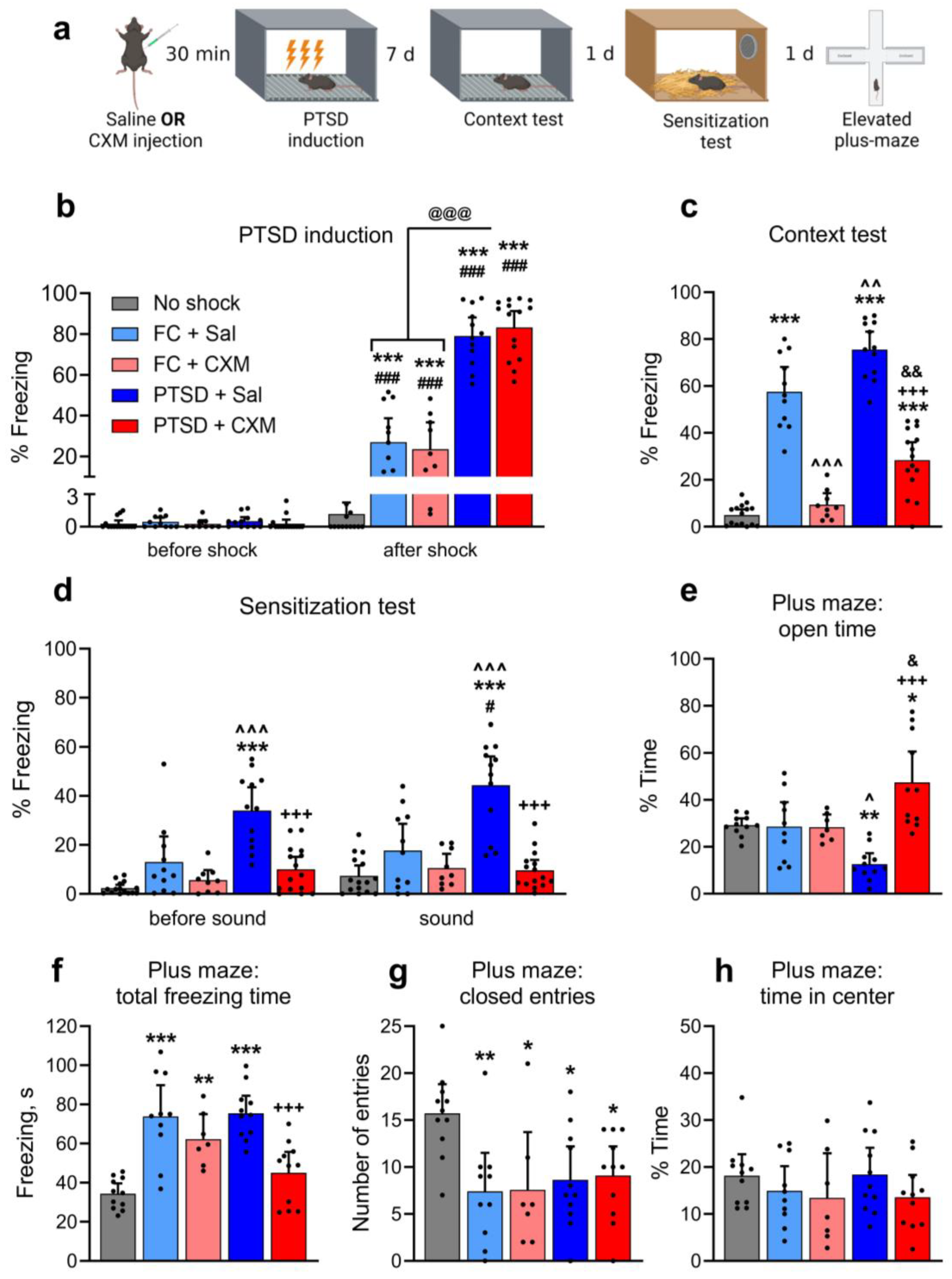
2.1. Effects of Protein Synthesis Inhibition on PTSD-Induced Fear and Anxiety

In this experiment, we studied the effect of PSI cycloheximide (CXM, 100 mg/kg) on PTSD formation in mice in a robust footshock model [20,29]. We used five groups of animals: mice trained in a standard context fear conditioning (FC) paradigm with CXM (FC + CXM, n = 9) or saline (FC + Sal, n = 11) injection; PTSD mice with CXM (PTSD + CXM, n = 15) or saline injection (PTSD + Sal, n = 12) and an active control group with saline injection (No shock, n = 15). For FC training, mice were placed in a novel context, allowed to explore for a while, and then given a 1 mA footshock for 2 s. It is known that such a moderate footshock induces normal aversive fear memory in mice [29]. For PTSD induction, mice were placed in the same context, allowed to explore, and then given three strong footshocks of 1.5 mA, each lasting 10 s. We previously demonstrated that such a procedure induces a PTSD-like phenotype in mice [29]. Control No shock mice were only exposed to the context without receiving footshock. All animals were then returned to the shocked context forassociative fear memory test. All animals underwent the following experimental stages: (1) FC training/PTSD induction/context exposure; (2) conditioned (associative) fear test; tests for non-associative consequences of the traumatic event—(3) behavioral sensitization and (4) anxiety in the elevated plus-maze (Figure 1a).
Freezing behavior was nearly absent in all animals during the novel context exploration before the footshock (Figure 1b). All animals that received the footshock demonstrated significantly more freezing behavior immediately after the footshock compared to the episode before footshock application (two-way ANOVA, post hoc Sidak’s multiple comparisons test, factor “Group”: F (4, 57) = 95.75, p < 0.0001; factor “Time interval”: F (1, 51) = 50.85, p < 0.0001; interaction of factors: F(4, 57) = 97.55, p < 0.0001; pairwise comparison of intervals before and after footshock: p < 0.0001 for FC + Sal, FC + CXM, PTSD + Sal, PTSD + CXM groups). All animals that received footshock also demonstrated significantly more freezing behavior immediately after footshock than the control group animals (Tukey’s multiple comparisons test, p < 0.0001). Finally, the freezing level after the footshock application in the PTSD + CXM and PTSD + Sal groups was significantly higher compared to the FC + Sal and FC + CXM groups (p < 0.0001). Thus, administering saline or CXM did not affect the level of fear in naive mice in a novel context. Footshock in this context caused the formation of short-term fear memory for both moderate and robust aversive stimuli. At the same time, the strong footshock used for PTSD induction produced a higher level of freezing than the moderate footshock used in FC training.
In the associative memory retrieval test seven days after the training, the animals of the FC + Sal and PTSD + Sal groups showed a significantly higher level of freezing than the control animals (one-way ANOVA: F (4, 57) = 87.81, p < 0.0001, post hoc Tukey’s multiple comparisons test: p < 0.0001). The freezing level in the PTSD + Sal group was significantly higher than in FC + Sal mice (p = 0.0032) (Figure 1c). The CXM injection before the FC training or PTSD induction resulted in impairment of associative memory, both normal and traumatic. Thus, the animals of the FC + CXM group showed significantly less freezing behavior than the mice that received a saline injection in the FC + Sal group (p < 0.0001) and did not differ in the level of freezing from the control animals (p = 0.8880). At the same time, animals of the PTSD + CXM group showed significantly less freezing behavior than PTSD + Sal group mice (p < 0.0001) and even less than animals with normal memory (PTSD + Sal vs. FC + Sal: p = 0.0018), but significantly more than the control animals (p < 0.0001).
PTSD animals exhibited higher levels of freezing during the memory retrieval compared to the animals with normal aversive memory. Injection of CXM before memory formation led to the complete erasure of the contextual fear in the FC paradigm. However, CXM injection before the PTSD induction did not cause a complete disruption of the fear memory, though it significantly reduced freezing levels compared to the saline group. Notably, the level of fear in animals exposed to CXM before the PTSD induction was relatively high, although significantly lower than in groups of animals with normal aversive memory. Thus, the protein synthesis inhibition before the traumatic exposure prevented the formation of a robust associative fear in animals with PTSD but did not lead to the complete elimination of the contextual fear memory, as in animals that were exposed to usual contextual fear conditioning.
Next, we performed a behavioral sensitization test (Figure 1d). In a novel context (before sound presentation), the level of freezing in the groups with normal aversive memory did not differ from that in the control animals (two-way ANOVA, post hoc Tukey’s multiple comparisons test, factor “Group”: F (4, 57) = 25.62, p < 0.0001; factor “Time interval”: F (1, 57) = 9.260, p = 0.0035; interaction of factors: F (4, 57) = 5.224, p = 0.0311; FC + Sal or FC + CXM vs. No shock: p = 0.1344). Thus, there was no generalization of aversive memory to the new context in trained mice. The PTSD + Sal group had a significantly higher level of freezing in the new context compared to the control group and the FC + Sal group (when comparing PTSD + Sal and No shock groups: p < 0.0001, when comparing PTSD + Sal and FC + Sal groups: p = 0.0002). At the same time, CXM administration led to a reduced level of freezing in the new context in the PTSD animals (p < 0.0001, compared to the PTSD + Sal group), and the level of freezing in the new environment did not differ in the PTSD + CXM and No shock groups (p = 0.3582).
The sensitized fear component test showed that fear conditioning did not affect freezing to a novel sound compared to a control group (p = 0.1593), i.e., FC + Sal animals did not demonstrate behavioral sensitization. The freezing level in the PTSD + Sal group was higher than in the other groups during the presentation of a novel sound (PTSD + Sal vs. No shock: p < 0.0001; PTSD + Sal vs. FC + Sal: p < 0.0001; PTSD + Sal vs. PTSD + CXM: p < 0.0001) (Figure 1d), indicating that sensitization developed in mice with PTSD. When comparing the freezing level before and after the sound presentation, a significant increase in freezing behavior was observed only in the PTSD + Sal group (Sidak’s multiple comparisons test, freezing level in PTSD + Sal mice before and during a sound presentation: p = 0.03). CXM administration led to a significant decrease in the freezing level in the PTSD animals (p < 0.0001), and the freezing level of the PTSD + CXM group during novel sound presentation did not differ from that of the control animals (p = 0.9840). These results indicate that protein synthesis inhibition during the induction of PTSD prevented the development of a sensitized fear.
To evaluate the level of anxiety, we analyzed animal behavior in the elevated plus-maze. Animals in the FC + Sal and FC + CXM groups did not differ from the control group in time spent in the open arms (one-way ANOVA, post hoc Tukey’s multiple comparisons test: F (4, 45) = 11.38, p < 0.0001; FC + Sal or FC + CXM vs. No shock: p = 0.9999), i.e., they did not exhibit elevated levels of anxiety (Figure 1e). At the same time, mice of the PTSD + Sal group spent less time in open arms compared with animals of the No shock and FC + Sal groups (PTSD + Sal vs. No shock: p = 0.0202; PTSD + Sal vs. FC + Sal: p = 0.032). Animals in the PTSD + CXM group spent more time in open arms compared to the animals in the No shock (p = 0.0081), FC + Sal (p = 0.0172), and PTSD + Sal groups (p < 0.0001).
The total freezing time in the FC + Sal (p < 0.0001), FC + CXM (p = 0.0042), and PTSD + Sal (p < 0.0001) groups was significantly higher compared to the control animals (F (4, 45) = 14.79, p < 0.0001, Figure 1f). At the same time, animals in the PTSD + CXM group demonstrated less freezing behavior compared to the PTSD + Sal (p = 0.0003) and FC + Sal (p = 0.0009) groups and did not differ in their level of freezing from the control group (p = 0.4872).
All groups of animals that received footshock had a lower number of entries into the closed arms compared with the control group (F (4, 45) = 4. 426, p = 0.0042; when compared with the FC + Sal group: p = 0.0069, FC + CXM: p = 0.0214, PTSD + Sal: p = 0.0241, PTSD + CXM: p = 0.0401, Figure 1g). We found no significant differences between the groups for the percentage of time in the center (F (4, 45) = 0.9539, p = 0.4420). So, protein synthesis inhibition during PTSD induction prevented increased anxiety in animals.

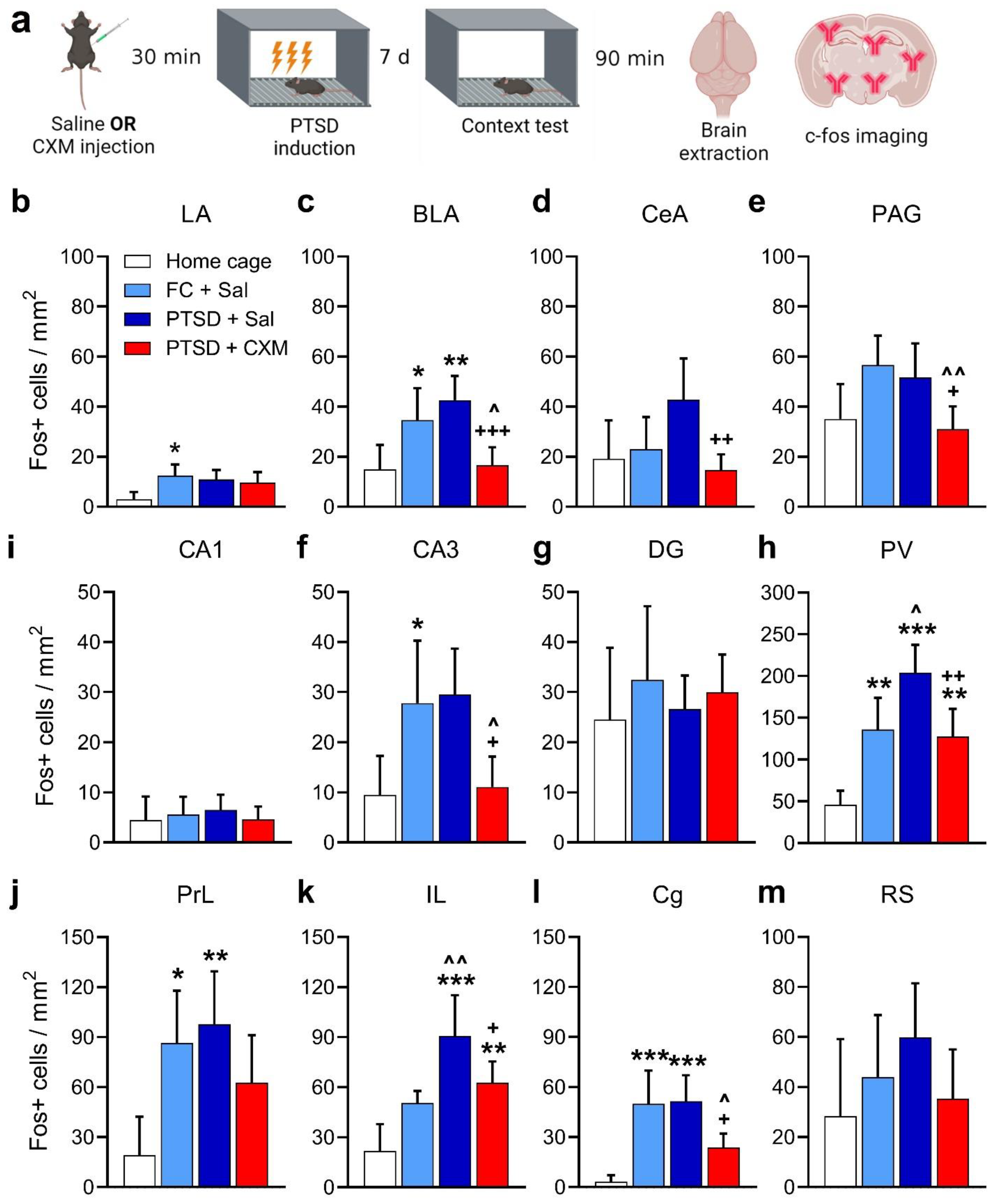
2.2. Patterns of c-Fos Brain Activity following Fear and Traumatic Memory Retrieval and the Effects of Protein Synthesis Inhibition

Next, we compared patterns of brain activity in animals during the retrieval of normal fear or traumatic memories, both disrupted and undamaged by CXM injection.
The formation of traumatic memory was preceded by the administration of CXM, while the control group received an injection of saline. There were four groups in the experiment: FC + Sal (n = 13), PTSD + Sal (n = 13), PTSD + CXM (n = 14), and Home cage (animals sacrificed from the home cage, n = 8). Seven days after PTSD induction or FC training, we retrieved normal or traumatic memory, and then brain samples of the animals were taken—Figure 2a.
We found a higher activation level in the lateral (p = 0.0201, Figure 2b) [LA] and basolateral (p = 0.0492, Figure 2c) [BLA] amygdala nuclei, prelimbic (p = 0.0225, Figure 2j) [PrL] and cingulate (p = 0.006, Figure 2l) [Cg] cortices and paraventricular nucleus of the thalamus (p = 0.0028, Figure 2i) [PV] in FC + Sal group compared to Home cage group—one-way ANOVA, post hoc Tukey’s multiple comparisons test: LA: F (3, 42) = 3.261, p = 0.0307; BLA: F (3, 41) = 8.584, p = 0.0002; PrL: F (3, 42) = 4.647, p = 0.0068; Cg: F (3, 43) = 9.255, p < 0.0001; PV: F (3, 40) = 16.03, p < 0.0001.
We found a significant increase in the number of c-Fos-positive cells in the PTSD + Sal group after the traumatic memory retrieval compared to the Home cage group in the infralimbic (p < 0.0001, Figure 2k) [IL], PrL (p = 0.006, Figure 2j) and Cg cortices (p = 0.0004, Figure 2l); in the CA3 field of the hippocampus (p = 0.0307, Figure 2g); BLA (p = 0.0028, Figure 2c); and PV (p < 0.0001, Figure 2i)—one-way ANOVA, post hoc Tukey’s multiple comparisons test: BLA: F (3, 41) = 8.584, p = 0.0002; PL: F (3, 42) = 4.647, p = 0.0068; IL: F (3, 37) = 12.46, p < 0.0001; Cg: F (3, 43) = 9.255, p < 0.0001; CA3: F (3, 38) = 5.802, p = 0.0023; PV: F (3, 40) = 16.03, p < 0.0001. We also showed a significantly higher level of activation of the IL (p = 0.0036) and PV (p = 0.015) in the PTSD + Sal group compared to the FC + Sal group, indicating enhanced activation of these areas, specifically during traumatic but not normal aversive memory retrieval.
Injection of CXM before the traumatic experience resulted in the decreased number of c-Fos-positive cells after the retrieval of memory in PTSD + CXM animals compared to the intact traumatic memory retrieval in PTSD + Sal group in the Cg (p = 0.0188, Figure 2l); the CA3 (p = 0.0114, Figure 2g); BLA (p = 0.0006, Figure 2c); central amygdala nucleus (p = 0.0034, Figure 2d) [CeA]; PV (p = 0.0022, Figure 2i) and periaqueductal gray matter (p = 0.0395, Figure 2e) [PAG]—one-way ANOVA, post hoc Tukey’s multiple comparisons test: Cg: F (3, 43) = 9.255, p < 0.0001; CA3: F (3, 38) = 5.802, p = 0.0023; BLA: F (3, 41) = 8.584, p = 0.0002; CeA: F (3, 41) = 4.936, p = 0.0051; PV: F (3, 40) = 16.03, p < 0.0001; PAG: F (3, 40) = 5.345, p = 0.0034. Furthermore, animals that received an injection of CXM before PTSD induction did not show differences from the Home cage group in the vast majority of structures examined, except for the IL (p = 0.0071, Figure 2k) and PV (p = 0.0038, Figure 2I)—one-way ANOVA, post hoc Tukey’s multiple comparisons test: IL: F (3, 37) = 12.46, p < 0.0001; PV: F (3, 40) = 16.03, p < 0.0001. A lower number of c-Fos-positive cells compared to the FC + Sal group was observed in animals of the PTSD + CXM group in the Cg (p = 0.03, Figure 2i); CA3 (p = 0.0337, Figure 2g); BLA (p = 0.0239, Figure 2c); and PAG (p = 0.0059, Figure 2k)—one-way ANOVA, post hoc Tukey’s multiple comparisons test: Cg: F (3, 43) = 9.255, p < 0.0001; CA3: F (3, 38) = 5.802, p = 0.0023; BLA: F (3, 41) = 8.584, p = 0.0002; PAG: F (3, 40) = 5.345, p = 0.0034. Thus, preventing the development of traumatic memory through PSI injection resulted in the normalization of behavior and restoration of normal patterns of brain c-Fos activity.

3. Discussion

In the first experiment, we investigated the effect of protein synthesis inhibition during traumatic experience on subsequent PTSD-like behavioral manifestations. Although protein synthesis inhibitors can produce acute effects on neural activity and behavior [30,31,32], administering CXM 30 min before the footshock did not affect the subsequent freezing behavior of mice in the conditioning chamber. This result suggests that the observed action of CXM is not due to its nonspecific effects on mice reactivity during PTSD acquisition.
Moderate and strong footshock both led to the formation of contextual fear memory. Higher-intensity footshock during PTSD induction resulted in increased fear levels compared to animals with normal aversive memory. Protein synthesis inhibition by CXM entirely prevented normal fear memory. The animals showed no memory of the aversive experience seven days after training, with fear levels similar to the control group without footshock. The effect of CXM on traumatic memory was less pronounced. In mice receiving CXM before the traumatic event, the levels of contextual fear were reduced compared to animals with traumatic and normal aversive memory. However, the contextual fear memory was not entirely eliminated since freezing did not decrease to the level of the control group. Thus, exposure to a traumatic event in an animal model of PTSD leads to the formation of a persistent memory, with the associative fear memory component proving resistant to such strong disruptive influences as protein synthesis inhibition, a phenomenon not previously reported [24,25,26].
When exposed to novel contexts or sound stimuli, mice with PTSD exhibited elevated fear levels, a characteristic of the sensitization of the fear response seen in PTSD. Remarkably, protein synthesis inhibition before the footshock completely disrupted these PTSD-like behavioral manifestations when assessed seven days after training.
Further analysis of anxiety-related behaviors and overall activity in the elevated plus maze revealed significant differences between PTSD animals and those with impaired traumatic memory. PTSD mice displayed increased freezing behavior, reduced time spent in open arms, and diminished exploratory behavior. These changes indicated substantial alterations in fear and defensive behavior in potentially threatening environments, consistent with the development of PTSD symptoms. However, protein synthesis inhibition before traumatic memory formation led to the normalization of elevated plus-maze behavior. Intriguingly, animals with impaired traumatic memory spent even more time in the maze’s open arms than those without any footshock experience, suggesting that traumatic memory impairment by protein synthesis inhibition may induce complex changes in stress response systems due to prolonged CXM effects.
Contextual fear conditioning also influenced the anxiety levels of animals. Those with normal aversive memory exhibited a slight increase in anxiety, reflected in increased freezing duration and reduced exploratory activity in the elevated plus maze, indicating an increase in anxious behavior. Importantly, protein synthesis inhibition before the formation of normal aversive memory had no impact on anxiety or exploratory activity, with animals having impaired and preserved normal fear memory displaying similar elevated plus maze performance.
In summary, these results demonstrate that strong, inescapable footshock leads to the formation of robust contextual associative memory and significant reorganization of fear and defensive behavior in mice, resulting in the manifestation of PTSD-like behaviors. Protein synthesis inhibition before regular contextual fear conditioning results in the complete prevention of long-term memory consolidation, while administration before the strong footshock impairs but does not entirely prevent the development of PTSD symptoms. Administration of CXM during PTSD induction almost completely eliminates symptoms of sensitization and anxiety without erasing the association between the context and the footshock.
We therefore hypothesize that there is an early protein-dependent stage in the development of PTSD, akin to the consolidation stage of long-term memory formation [24,25,33,34]. This stage involves the formation of long-term associative traumatic memory and protein-dependent non-associative plasticity within neuronal fear and defense circuits. It may explain the need for an incubation period to develop hyperreactivity symptoms [21], as initial non-associative plasticity processes may be necessary but insufficient for full PTSD manifestation. Subsequent, slow development and modification of non-associative plasticity processes may involve new neuronal populations analogous to processes of distant memory consolidation [35,36,37,38]. This process could be triggered by intrusive memories in humans or circumstantial reminders in rodents [39].
Previous studies have explored the impact of protein synthesis inhibition on PTSD development, primarily using the rat model of acute predator stress and without investigating associative memory formation [24,25,33,40]. Our findings align with prior research, indicating that protein synthesis inhibition during traumatic experiences can suppress PTSD symptoms. However, our data suggest that reorganization in fear and defense neuronal systems during PTSD development is not unidirectional. Following a traumatic experience, profound protein-dependent reorganization occurs at the neuronal level, disrupting typical defensive behavioral patterns. These changes result in a reduced anxiety level compared to animals without any traumatic experience. Our data suggest the possibility that old, unreactivated memories may be impaired during the consolidation of new memories [41]. Moreover, it raises the question of whether the innate behavior could be impaired at its realization.
Using a PTSD induction model based on the footshock, a series of animal experiments were conducted to investigate the involvement of molecular mechanisms of aversive memory in the development of the disorder [42,43]. It was shown that at the moment of trauma, molecular cascades involved in the development of NMDA-dependent long-term potentiation are activated in the hippocampus and amygdala [44,45,46]. An increase in the phosphorylation level of CREB (calcium-response element binding protein) during trauma was also demonstrated [47,48,49], along with the activation of gene expression related to memory consolidation [26,50,51]. Meanwhile, administering kinase inhibitors involved in memory consolidation at the moment of trauma reduces the likelihood of developing PTSD in animals [2,52]. Furthermore, in another PTSD model involving predator exposure, it was shown that inhibition of protein synthesis disrupts the development of PTSD-like behavioral disorders [24,25,26] within the same period as memory formation [34] and leads to a decrease in corticosterone levels in the animals’ blood [33]. These findings support the hypothesis that the development of PTSD and memory consolidation in classical conditioning depend on similar molecular mechanisms.
Lastly, our examination of c-Fos expression patterns revealed significant activation of specific brain regions during the development of PTSD-like behavior, including the prefrontal cortex (PrL, IL, Cg), amygdala (BLA), and PV. This activation pattern is consistent with data on the involvement of these regions in PTSD in humans and animals [53,54,55,56]. The high BLA activity in PTSD aligns with extensive evidence linking amygdala functioning to PTSD symptoms in both humans [57,58,59] and animals [60,61]. For instance, an NMDA receptor antagonist in the amygdala reduced anxiety symptoms in a predator-exposure-based PTSD model [61]. Cg activity is associated with fear expression in the contextual FC paradigm [62]. Abnormal Cg activity may reflect impaired emotional regulation, increased reactivity to aversive stimuli, and fear generalization in PTSD [63,64,65]. IL dysfunction is linked to PTSD, as deficits in memory extinction accompany the disorder [66,67]. IL is central to fear memory extinction [68,69,70,71]. Increased c-Fos expression in IL during traumatic memory retrieval may indicate abnormal plasticity associated with impaired extinction. Elevated PV activity in mice with PTSD aligns with its role in regulating fear behavior [72,73] and heightened sensitized fear components.
Protein synthesis inhibition before traumatic experience significantly impacted brain activation patterns after memory retrieval. Reduced c-Fos-positive cell numbers in BLA, CeA, PAG, CA3, PV, IL, and Cg in the PTSD + CXM group indicated decreased activity in these structures. Lower BLA activity may reflect reduced hyperreactivity to dangerous stimuli and reduced anxiety [74]. In addition, the reduced activity of this structure may be reflected in reduced anxiety in mice compared to the group with intact traumatic memory [12,75]. Reduced CeA activation suggests a less pronounced fear response to trauma context, as CeA mediates emotional responses and fear extinction [57,76,77]. PAG, linked to fight or flight responses, showed abnormal activity in PTSD, leading to increased fear expression [5,78]. Thus, decreased PAG activity in the PTSD + CXM group may signify normalized fear and anxiety reactions. Lower CA3 activity suggests more accurate context memory encoding, which was observed as improved discrimination between dangerous and safe contexts in animals with impaired traumatic memory. Normalized Cg activity may indicate reduced generalization in mice with impaired PTSD [76], contributing to behavior more similar to control animals without traumatic experience.
At the molecular level PTSD is characterized by a complex interplay of various signaling pathways that contribute to its induction and development. Molecular mechanisms involved in PTSD are diverse and encompass changes in neurotransmitters, receptors, neurotrophic factors, and intracellular signaling cascades [52,79]. Signaling pathways involved in PTSD include those mediated by cyclic AMP response element-binding protein (CREB) and mitogen-activated protein kinases (MAPKs) [79]. One or several of these described molecular mechanisms may be responsible for the normalization of brain activity patterns during memory retrieval.
Altogether, the c-Fos activity patterns obtained in our study are consistent with the existing literature linking the discussed brain regions to PTSD in both humans and animals. However, c-Fos imaging is a correlative approach. Further exploration of the neural substrates involved in PTSD and protein synthesis-dependent consolidation using approaches for the causal investigation such as optogenetic manipulation will be critical to understanding this complex disorder and potential therapeutic strategies.

4. Materials and Methods

4.1. Animals

Male C57Bl/6J mice (3–4 months old) were used. Mice were group-housed 3–5 per cage with the ad libitum food and water access. Animals were kept under a 12 h light/dark cycle. Experiments were performed during the light phase of the cycle. All methods for animal care and all experiments were approved by the Commission on Bioethics of Lomonosov Moscow State University (Application No. 178-a, approved during the Bioethics Commission meeting No. 162-d held on 16 May 2024) and were in accordance with the Russian Federation Order Requirements N 267 M3 and the National Institutes of Health Guide for the Care and Use of Laboratory Animals.

4.2. Behavior

During FC training, mice were placed into the fear conditioning chamber (MED Associates Inc., Fairfax, VT, USA) for a 230 s exploration of context A. Then, the footshock (2 s, 1 mA—moderate footshock) was applied, and animals remained in context A for 128 s before being returned to their home cages. PTSD induction was performed according to the modified protocol by Siegmund and Wotjak [20,29]. For PTSD induction, mice were placed in context A for 170 s, then three consecutive footshocks were applied (10 s, 1.5 mA—strong footshock) v2.5.5.0, and animals remained in the chamber for 60 s. The interval between the footshocks was 50 s. Animals of the No shock group were placed in context A for 300 s without footshock.
Seven days later, animals were placed in context A for context memory retrieval (180 s). One week is a necessary interval for the development of posttraumatic behavioral symptoms [20]. The following day (the eighth day after the FC training/PTSD induction), animals were placed in context B for a memory sensitization test [20]. After 180 s of novel context exploration, a neutral tone (80 dB, 9 kHz) was presented for 180 s.
Context A was a white light (87 lux) and IR light illuminated plastic box (30 cm × 23 cm × 21 cm) with a grid floor. To change the context for the memory sensitization test, we placed the black plastic A-shaped insert into the chamber and covered the grid floor with a plastic sheet and wood sawdust (context B). Context B was illuminated with an IR light. Context A and B were cleaned before and after each session with 70% ethanol or 53% ethanol solution of peppermint oil, respectively. Freezing behavior was scored automatically using Video Freeze software version No. 2.5.5.0 (MED Associates Inc., Fairfax, VT, USA).
On the ninth day after the FC training/PTSD induction, animals were tested in the elevated plus-maze to address the anxiety level [80,81,82]. Animal behavior in the elevated plus-maze was video recorded and analyzed using the EthoVision XT software version No. 8.5 (Noldus, Wageningen, The Netherlands). The following behavioral parameters were analyzed: (i) time spent in the open arms and (ii) freezing duration (as measures of anxiety [83,84,85]), (iii) time spent on the central platform (as a measure of risk assessment and decision making [86,87,88]), (iv) the number of entries to the closed arms (as a measure of exploratory behavior [84]).

4.3. Cycloheximide Injection

Animals received a single i.p. injection of CXM (100 mg/kg, Sigma-Aldrich, St. Louis, MO, USA) 30 min before the FC training/PTSD induction. At this dose, CXM suppresses protein synthesis in the mouse brain at 95% level 10–40 min after administration [89,90]. Control groups of animals received a single i.p. injection of saline 30 min before the FC training/PTSD induction.

4.4. Immunohistochemistry

To analyze the brain’s activity, mice were sacrificed by cervical translocation 90 min after context memory retrieval. Home cage control animals with no novel experience were sacrificed at the same time. Brains were removed and immediately frozen in liquid nitrogen vapors. The 20 µm coronal sections were cut in a cryostat (Leica CM1950, Leica Biosystems, Wetzlar, Germany) and fixed in 4% paraformaldehyde. Brain sections were taken at various distances from the Bregma [91]: (i) +1.7 mm for the infralimbic (IL) and the prelimbic (PrL) cortices; (ii) +0.98 mm for the cingulate (Cg) cortex; (iii) −1.46 mm for the retrosplenial (RS) cortex, hippocampal CA1, CA3, and dentate gyrus (DG), the basolateral (BLA), lateral (LA) and central (CeA) amygdalar nuclei and for the paraventricular (PV) thalamic nucleus; (iv) −3.15 for the periaqueductal gray matter (PAG). For c-Fos immunostaining, primary rabbit polyclonal antibodies against the c-Fos protein (226 003, Synaptic Systems, Goettingen, Germany, dilution 1:1000) and donkey secondary antibodies against rabbit conjugated with Alexa 488 (A-21206, Thermo Fisher, Waltham, MA, USA, dilution 1:500) were used. Whole section images were acquired with the fluorescence microscope scanner (Olympus VS110, Waltham, MA, USA) at the 10×.
The c-Fos-positive cell count was performed using the Image-Pro Plus 3.0 software (Media Cybernetics, Rockville, MD, USA). The density of cells was calculated as the ratio of the number of positive cells to the structure area (in mm2). Three slices from each brain were analyzed for each structure.

4.5. Statistical Analysis

Statistical analysis was performed using the Prism 7 software (GraphPad, Boston, MA, USA). We used one-way and two-way ANOVA followed by multiple comparisons with Tukey’s post hoc for unpaired samples and Sidak’s post hoc for paired samples, with a significance level set to 0.05. Graphs were plotted in Prism, and schemes of experiments were created with BioRender.com, accessed on 3 October 2023.

Author Contributions

Conceptualization O.I.I., K.A.T. and K.V.A.; methodology, O.I.I., K.A.T. and K.V.A.; formal analysis, T.A.Z., O.I.I. and K.A.T.; investigation, T.A.Z., O.I.I. and K.A.T.; writing—original draft preparation, T.A.Z. and O.I.I.; writing—review and editing, O.I.I., K.A.T. and K.V.A.; visualization, T.A.Z., O.I.I. and K.A.T.; supervision, K.V.A.; project administration, O.I.I. and K.A.T.; funding acquisition, K.V.A. All authors have read and agreed to the published version of the manuscript.

Funding

This research was funded by Non-Commercial Foundation for Support of Science and Education “INTELLECT”.

Institutional Review Board Statement

The study was conducted according to the guidelines of the Declaration of Helsinki and approved by the Commission on Bioethics of Lomonosov Moscow State University (Application No. 178-a, approved during the Bioethics Commission meeting No. 162-d held on 16 May 2024) and were in accordance with the Russian Federation Order Requirements N 267 M3 and the National Institutes of Health Guide for the Care and Use of Laboratory Animals.

Informed Consent Statement

Not applicable.

Data Availability Statement

The raw data supporting the conclusions of this article will be made available by the authors on request.

Conflicts of Interest

The authors declare no conflicts of interest. The funders had no role in the design of the study; in the collection, analyses, or interpretation of data; in the writing of the manuscript, or in the decision to publish the results.

References

  1. Elzinga, B.M.; Bremner, J.D. Are the Neural Substrates of Memory the Final Common Pathway in Posttraumatic Stress Disorder (PTSD)? J. Affect. Disord. 2002, 70, 1–17. [Google Scholar] [CrossRef]
  2. Cohen, H.; Kozlovsky, N.; Matar, M.A.; Kaplan, Z.; Zohar, J. Mapping the Brain Pathways of Traumatic Memory: Inactivation of Protein Kinase M Zeta in Different Brain Regions Disrupts Traumatic Memory Processes and Attenuates Traumatic Stress Responses in Rats. Eur. Neuropsychopharmacol. 2010, 20, 253–271. [Google Scholar] [CrossRef]
  3. Johnson, L.R.; McGuire, J.; Lazarus, R.; Palmer, A.A. Pavlovian Fear Memory Circuits and Phenotype Models of PTSD. Neuropharmacology 2012, 62, 638–646. [Google Scholar] [CrossRef]
  4. Van der Kolk, B.A. The Body Keeps the Score: Memory and the Evolving Psychobiology of Posttraumatic Stress. Harv. Rev. Psychiatry 1994, 1, 253–265. [Google Scholar] [CrossRef]
  5. Lis, S.; Thome, J.; Kleindienst, N.; Mueller-Engelmann, M.; Steil, R.; Priebe, K.; Schmahl, C.; Hermans, D.; Bohus, M. Generalization of Fear in Post-Traumatic Stress Disorder. Psychophysiology 2020, 57, e13422. [Google Scholar] [CrossRef]
  6. Thakur, A.; Choudhary, D.; Kumar, B.; Chaudhary, A. A Review on Post-Traumatic Stress Disorder (PTSD): Symptoms, Therapies and Recent Case Studies. Curr. Mol. Pharmacol. 2022, 15, 502–516. [Google Scholar] [CrossRef]
  7. Ehlers, A.; Hackmann, A.; Michael, T. Intrusive Re-experiencing in Post-traumatic Stress Disorder: Phenomenology, Theory, and Therapy. Memory 2004, 12, 403–415. [Google Scholar] [CrossRef]
  8. Rothbaum, B.O.; Davis, M. Applying Learning Principles to the Treatment of Post-trauma Reactions. Ann. N. Y. Acad. Sci. 2003, 1008, 112–121. [Google Scholar] [CrossRef]
  9. Germain, A.; Buysse, D.J.; Nofzinger, E. Sleep-Specific Mechanisms Underlying Posttraumatic Stress Disorder: Integrative Review and Neurobiological Hypotheses. Sleep Med. Rev. 2008, 12, 185–195. [Google Scholar] [CrossRef]
  10. Milad, M.R.; Orr, S.P.; Lasko, N.B.; Chang, Y.; Rauch, S.L.; Pitman, R.K. Presence and Acquired Origin of Reduced Recall for Fear Extinction in PTSD: Results of a Twin Study. J. Psychiatr. Res. 2008, 42, 515–520. [Google Scholar] [CrossRef]
  11. Quirk, G.J.; Garcia, R.; González-Lima, F. Prefrontal Mechanisms in Extinction of Conditioned Fear. Biol. Psychiatry 2006, 60, 337–343. [Google Scholar] [CrossRef] [PubMed]
  12. Rauch, S.L.; Whalen, P.J.; Shin, L.M.; McInerney, S.C.; Macklin, M.L.; Lasko, N.B.; Orr, S.P.; Pitman, R.K. Exaggerated Amygdala Response to Masked Facial Stimuli in Posttraumatic Stress Disorder: A Functional MRI Study. Biol. Psychiatry 2000, 47, 769–776. [Google Scholar] [CrossRef] [PubMed]
  13. Bryant, R.A.; Kemp, A.H.; Felmingham, K.L.; Liddell, B.; Olivieri, G.; Peduto, A.; Gordon, E.; Williams, L.M. Enhanced Amygdala and Medial Prefrontal Activation during Nonconscious Processing of Fear in Posttraumatic Stress Disorder: An FMRI Study. Hum. Brain Mapp. 2008, 29, 517–523. [Google Scholar] [CrossRef] [PubMed]
  14. Carrion, V.G.; Haas, B.W.; Garrett, A.; Song, S.; Reiss, A.L. Reduced Hippocampal Activity in Youth with Posttraumatic Stress Symptoms: An FMRI Study. J. Pediatr. Psychol. 2010, 35, 559–569. [Google Scholar] [CrossRef] [PubMed]
  15. Werner, N.S.; Meindl, T.; Engel, R.R.; Rosner, R.; Riedel, M.; Reiser, M.; Fast, K. Hippocampal Function during Associative Learning in Patients with Posttraumatic Stress Disorder. J. Psychiatr. Res. 2009, 43, 309–318. [Google Scholar] [CrossRef] [PubMed]
  16. Shin, L.M.; Whalen, P.J.; Pitman, R.K.; Bush, G.; Macklin, M.L.; Lasko, N.B.; Orr, S.P.; McInerney, S.C.; Rauch, S.L. An FMRI Study of Anterior Cingulate Function in Posttraumatic Stress Disorder. Biol. Psychiatry 2001, 50, 932–942. [Google Scholar] [CrossRef]
  17. Rougemont-Bücking, A.; Linnman, C.; Zeffiro, T.A.; Zeidan, M.A.; Lebron-Milad, K.; Rodriguez-Romaguera, J.; Rauch, S.L.; Pitman, R.K.; Milad, M.R. Altered Processing of Contextual Information during Fear Extinction in PTSD: An FMRI Study. CNS Neurosci. Ther. 2011, 17, 227–236. [Google Scholar] [CrossRef] [PubMed]
  18. Dickie, E.W.; Brunet, A.; Akerib, V.; Armony, J.L. An FMRI Investigation of Memory Encoding in PTSD: Influence of Symptom Severity. Neuropsychologia 2008, 46, 1522–1531. [Google Scholar] [CrossRef] [PubMed]
  19. Siegmund, A.; Wotjak, C.T. Toward an Animal Model of Posttraumatic Stress Disorder. Ann. N. Y. Acad. Sci. 2006, 1071, 324–334. [Google Scholar] [CrossRef]
  20. Siegmund, A.; Wotjak, C.T. A Mouse Model of Posttraumatic Stress Disorder That Distinguishes between Conditioned and Sensitised Fear. J. Psychiatr. Res. 2007, 41, 848–860. [Google Scholar] [CrossRef]
  21. Siegmund, A.; Wotjak, C.T. Hyperarousal Does not Depend on Trauma-Related Contextual Memory in an Animal Model of Posttraumatic Stress Disorder. Physiol. Behav. 2007, 90, 103–107. [Google Scholar] [CrossRef] [PubMed]
  22. Anokhin, K.V.; Sudakov, K. V Genome of Brain Neurons in Organization of Systemic Mechanisms of Behavior. Bull. Exp. Biol. Med. 2003, 135, 107–113. [Google Scholar] [CrossRef] [PubMed]
  23. Miyashita, T.; Kikuchi, E.; Horiuchi, J.; Saitoe, M. Long-Term Memory Engram Cells Are Established by c-Fos/CREB Transcriptional Cycling. Cell Rep. 2018, 25, 2716–2728. [Google Scholar] [CrossRef] [PubMed]
  24. Adamec, R.; Strasser, K.; Blundell, J.; Burton, P.; McKay, D.W. Protein Synthesis and the Mechanisms of Lasting Change in Anxiety Induced by Severe Stress. Behav. Brain Res. 2006, 167, 270–286. [Google Scholar] [CrossRef] [PubMed]
  25. Cohen, H.; Kaplan, Z.; Matar, M.A.; Loewenthal, U.; Kozlovsky, N.; Zohar, J. Anisomycin, a Protein Synthesis Inhibitor, Disrupts Traumatic Memory Consolidation and Attenuates Posttraumatic Stress Response in Rats. Biol. Psychiatry 2006, 60, 767–776. [Google Scholar] [CrossRef] [PubMed]
  26. Kozlovsky, N.; Kaplan, Z.; Zohar, J.; Matar, M.A.; Shimon, H.; Cohen, H. Protein Synthesis Inhibition before or after Stress Exposure Results in Divergent Endocrine and BDNF Responses Disassociated from Behavioral Responses. Depress. Anxiety 2008, 25, E24–E34. [Google Scholar] [CrossRef]
  27. Johansen, J.P.; Cain, C.K.; Ostroff, L.E.; LeDoux, J.E. Molecular Mechanisms of Fear Learning and Memory. Cell 2011, 147, 509–524. [Google Scholar] [CrossRef]
  28. Schafe, G.E.; Nader, K.; Blair, H.T.; LeDoux, J.E. Memory Consolidation of Pavlovian Fear Conditioning: A Cellular and Molecular Perspective. Trends Neurosci. 2001, 24, 540–546. [Google Scholar] [CrossRef]
  29. Toropova, K.A.; Anokhin, K.V. Modeling of Post-Traumatic Stress Disorder in Mice: Nonlinear Relationship with the Strength of the Traumatic Event. Neurosci. Behav. Physiol. 2019, 49, 875–886. [Google Scholar] [CrossRef]
  30. Sharma, A.V.; Nargang, F.E.; Dickson, C.T. Neurosilence: Profound Suppression of Neural Activity Following Intracerebral Administration of the Protein Synthesis Inhibitor Anisomycin. J. Neurosci. 2012, 32, 2377–2387. [Google Scholar] [CrossRef]
  31. Squire, L.R.; Smith, G.A.; Barondes, S.H. Cycloheximide Affects Memory within Minutes after the Onset of Training. Nature 1973, 242, 201–202. [Google Scholar] [CrossRef] [PubMed]
  32. Gutwein, B.M.; Quartermain, D.; McEwen, B.S. Dissociation of Cycloheximide’s Effects on Activity from Its Effects on Memory. Pharmacol. Biochem. Behav. 1974, 2, 753–756. [Google Scholar] [CrossRef] [PubMed]
  33. Kozlovsky, N.; Matar, M.A.; Kaplan, Z.; Kotler, M.; Zohar, J.; Cohen, H. The Immediate Early Gene Arc Is Associated with Behavioral Resilience to Stress Exposure in an Animal Model of Posttraumatic Stress Disorder. Eur. Neuropsychopharmacol. 2008, 18, 107–116. [Google Scholar] [CrossRef] [PubMed]
  34. Gold, P.E. Protein Synthesis Inhibition and Memory: Formation vs Amnesia. Neurobiol. Learn. Mem. 2008, 89, 201–211. [Google Scholar] [CrossRef]
  35. Frankland, P.W.; Bontempi, B. The Organization of Recent and Remote Memories. Nat. Rev. Neurosci. 2005, 6, 119–130. [Google Scholar] [CrossRef] [PubMed]
  36. Diekelmann, S.; Born, J. One Memory, Two Ways to Consolidate? Nat. Neurosci. 2007, 10, 1085–1086. [Google Scholar] [CrossRef] [PubMed]
  37. Born, J.; Wilhelm, I. System Consolidation of Memory during Sleep. Psychol. Res. 2012, 76, 192–203. [Google Scholar] [CrossRef]
  38. Takehara-Nishiuchi, K. Neurobiology of Systems Memory Consolidation. Eur. J. Neurosci. 2021, 54, 6850–6863. [Google Scholar] [CrossRef] [PubMed]
  39. Pynoos, R.S.; Ritzmann, R.F.; Steinberg, A.M.; Goenjian, A.; Prisecaru, I. A Behavioral Animal Model of Posttraumatic Stress Disorder Featuring Repeated Exposure to Situational Reminders. Biol. Psychiatry 1996, 39, 129–134. [Google Scholar] [CrossRef]
  40. Hymel, K.A.; Eans, S.O.; Sitchenko, K.L.; Gomes, S.M.; Lukowsky, A.L.; Medina, J.M.; Sypek, E.I.; Carey, A.N.; McLaughlin, J.P. Stress-Induced Increases in Depression-like and Cocaine Place-Conditioned Behaviors Are Reversed by Disruption of Memories during Reconsolidation. Behav. Pharmacol. 2014, 25, 599–608. [Google Scholar] [CrossRef]
  41. Debiec, J.; LeDoux, J.E. Contribution of Noradrenergic Transmission to Memory Reconsolidation in Animals and Humans: Implications for PTSD. In Biological Psychiatry; Dennis, S., Charney, M.D., Eds.; Elsevier: New York, NY, USA, 2006; Volume 59, pp. 14s–15s. [Google Scholar]
  42. Dunsmoor, J.E.; Cisler, J.M.; Fonzo, G.A.; Creech, S.K.; Nemeroff, C.B. Laboratory Models of Post-Traumatic Stress Disorder: The Elusive Bridge to Translation. Neuron 2022, 110, 1754–1776. [Google Scholar] [CrossRef]
  43. Nadel, L.; Hupbach, A.; Gomez, R.; Newman-Smith, K. Memory Formation, Consolidation and Transformation. Neurosci. Biobehav. Rev. 2012, 36, 1640–1645. [Google Scholar] [CrossRef] [PubMed]
  44. Nordman, J.; Ma, X.; Li, Z. Traumatic Stress Induces Prolonged Aggression Increase through Synaptic Potentiation in the Medial Amygdala Circuits. eNeuro 2020, 7, 1–14. [Google Scholar] [CrossRef]
  45. Adamec, R.; Kent, P.; Anisman, H.; Shallow, T.; Merali, Z. Neural Plasticity, Neuropeptides and Anxiety in Animals—Implications for Understanding and Treating Affective Disorder Following Traumatic Stress in Humans. Neurosci. Biobehav. Rev. 1998, 23, 301–318. [Google Scholar] [CrossRef] [PubMed]
  46. Dahlhoff, M.; Siegmund, A.; Golub, Y.; Wolf, E.; Holsboer, F.; Wotjak, C.T. AKT/GSK-3β/β-Catenin Signalling within Hippocampus and Amygdala Reflects Genetically Determined Differences in Posttraumatic Stress Disorder like Symptoms. Neuroscience 2010, 169, 1216–1226. [Google Scholar] [CrossRef]
  47. Azevedo, M.; Martinho, R.; Oliveira, A.; Correia-de-Sá, P.; Moreira-Rodrigues, M. Molecular Pathways Underlying Sympathetic Autonomic Overshooting Leading to Fear and Traumatic Memories: Looking for Alternative Therapeutic Options for Post-Traumatic Stress Disorder. Front. Mol. Neurosci. 2024, 16, 1332348. [Google Scholar] [CrossRef]
  48. Adamec, R.E.; Blundell, J.; Burton, P. Phosphorylated Cyclic AMP Response Element Binding Protein Expression Induced in the Periaqueductal Gray by Predator Stress: Its Relationship to the Stress Experience, Behavior and Limbic Neural Plasticity. Prog. Neuro-Psychopharmacol. Biol. Psychiatry 2003, 27, 1243–1267. [Google Scholar] [CrossRef]
  49. Blundell, J.; Adamec, R. Elevated PCREB in the PAG after Exposure to the Elevated plus Maze in Rats Previously Exposed to a Cat. Behav. Brain Res. 2006, 175, 285–295. [Google Scholar] [CrossRef]
  50. Chang, S.-H.; Chang, Y.-M.; Chen, H.-Y.; Shaw, F.-Z.; Shyu, B.-C. Time-Course Analysis of Frontal Gene Expression Profiles in the Rat Model of Posttraumatic Stress Disorder and a Comparison with the Conditioned Fear Model. Neurobiol. Stress 2023, 27, 100569. [Google Scholar] [CrossRef] [PubMed]
  51. Zhang, L.; Zhou, R.; Xing, G.; Hough, C.J.; Li, X.; Li, H. Identification of Gene Markers Based on Well Validated and Subcategorized Stressed Animals for Potential Clinical Applications in PTSD. Med. Hypotheses 2006, 66, 309–314. [Google Scholar] [CrossRef]
  52. Girgenti, M.J.; Hare, B.D.; Ghosal, S.; Duman, R.S. Molecular and Cellular Effects of Traumatic Stress: Implications for PTSD. Curr. Psychiatry Rep. 2017, 19, 85. [Google Scholar] [CrossRef]
  53. Fenster, R.J.; Lebois, L.A.M.; Ressler, K.J.; Suh, J. Brain Circuit Dysfunction in Post-Traumatic Stress Disorder: From Mouse to Man. Nat. Rev. Neurosci. 2018, 19, 535–551. [Google Scholar] [CrossRef] [PubMed]
  54. Kunimatsu, A.; Yasaka, K.; Akai, H.; Kunimatsu, N.; Abe, O. MRI Findings in Posttraumatic Stress Disorder. J. Magn. Reson. Imaging 2020, 52, 380–396. [Google Scholar] [CrossRef] [PubMed]
  55. Zilcha-Mano, S.; Zhu, X.; Lazarov, A.; Suarez-Jimenez, B.; Helpman, L.; Kim, Y.; Maitlin, C.; Neria, Y.; Rutherford, B.R. Structural Brain Features Signaling Trauma, PTSD, or Resilience? A Systematic Exploration. Depress. Anxiety 2022, 39, 695–705. [Google Scholar] [CrossRef] [PubMed]
  56. Leite, L.; Esper, N.B.; Junior, J.R.M.L.; Lara, D.R.; Buchweitz, A. An Exploratory Study of Resting-State Functional Connectivity of Amygdala Subregions in Posttraumatic Stress Disorder Following Trauma in Adulthood. Sci. Rep. 2022, 12, 9558. [Google Scholar] [CrossRef] [PubMed]
  57. Morey, R.A.; Clarke, E.K.; Haswell, C.C.; Phillips, R.D.; Clausen, A.N.; Mufford, M.S.; Saygin, Z.; Wagner, H.R.; LaBar, K.S. Amygdala Nuclei Volume and Shape in Military Veterans with Posttraumatic Stress Disorder. Biol. Psychiatry. Cogn. Neurosci. Neuroimaging 2020, 5, 281–290. [Google Scholar] [CrossRef] [PubMed]
  58. Kredlow, A.M.; Fenster, R.J.; Laurent, E.S.; Ressler, K.J.; Phelps, E.A. Prefrontal Cortex, Amygdala, and Threat Processing: Implications for PTSD. Neuropsychopharmacology 2022, 47, 247–259. [Google Scholar] [CrossRef] [PubMed]
  59. Del Casale, A.; Ferracuti, S.; Barbetti, A.S.; Bargagna, P.; Zega, P.; Iannuccelli, A.; Caggese, F.; Zoppi, T.; De Luca, G.P.; Parmigiani, G. Grey Matter Volume Reductions of the Left Hippocampus and Amygdala in PTSD: A Coordinate-Based Meta-Analysis of Magnetic Resonance Imaging Studies. Neuropsychobiology 2022, 81, 257–264. [Google Scholar] [CrossRef] [PubMed]
  60. Sharp, B.M. Basolateral Amygdala and Stress-Induced Hyperexcitability Affect Motivated Behaviors and Addiction. Transl. Psychiatry 2017, 7, e1194. [Google Scholar] [CrossRef]
  61. Adamec, R.E.; Burton, P.; Shallow, T.; Budgell, J. NMDA Receptors Mediate Lasting Increases in Anxiety-Like Behavior Produced by the Stress of Predator Exposure—Implications for Anxiety Associated with Posttraumatic Stress Disorder. Physiol. Behav. 1998, 65, 723–737. [Google Scholar] [CrossRef]
  62. Pitman, R.K.; Rasmusson, A.M.; Koenen, K.C.; Shin, L.M.; Orr, S.P.; Gilbertson, M.W.; Milad, M.R.; Liberzon, I. Biological Studies of Post-Traumatic Stress Disorder. Nat. Rev. Neurosci. 2012, 13, 769–787. [Google Scholar] [CrossRef] [PubMed]
  63. Ehring, T.; Quack, D. Emotion Regulation Difficulties in Trauma Survivors: The Role of Trauma Type and PTSD Symptom Severity. Behav. Ther. 2010, 41, 587–598. [Google Scholar] [CrossRef] [PubMed]
  64. Felmingham, K.L.; Rennie, C.; Manor, B.; Bryant, R.A. Eye Tracking and Physiological Reactivity to Threatening Stimuli in Posttraumatic Stress Disorder. J. Anxiety Disord. 2011, 25, 668–673. [Google Scholar] [CrossRef] [PubMed]
  65. Bian, X.-L.; Qin, C.; Cai, C.-Y.; Zhou, Y.; Tao, Y.; Lin, Y.-H.; Wu, H.-Y.; Chang, L.; Luo, C.-X.; Zhu, D.-Y. Anterior Cingulate Cortex to Ventral Hippocampus Circuit Mediates Contextual Fear Generalization. J. Neurosci. 2019, 39, 5728–5739. [Google Scholar] [CrossRef] [PubMed]
  66. Milad, M.R.; Pitman, R.K.; Ellis, C.B.; Gold, A.L.; Shin, L.M.; Lasko, N.B.; Zeidan, M.A.; Handwerger, K.; Orr, S.P.; Rauch, S.L. Neurobiological Basis of Failure to Recall Extinction Memory in Posttraumatic Stress Disorder. Biol. Psychiatry 2009, 66, 1075–1082. [Google Scholar] [CrossRef] [PubMed]
  67. Uniyal, A.; Singh, R.; Akhtar, A.; Dhaliwal, J.; Kuhad, A.; Sah, S.P. Pharmacological Rewriting of Fear Memories: A Beacon for Post-Traumatic Stress Disorder. Eur. J. Pharmacol. 2020, 870, 172824. [Google Scholar] [CrossRef] [PubMed]
  68. Bukalo, O.; Nonaka, M.; Weinholtz, C.A.; Mendez, A.; Taylor, W.W.; Holmes, A. Effects of Optogenetic Photoexcitation of Infralimbic Cortex Inputs to the Basolateral Amygdala on Conditioned Fear and Extinction. Behav. Brain Res. 2021, 396, 112913. [Google Scholar] [CrossRef] [PubMed]
  69. Do-Monte, F.H.; Manzano-Nieves, G.; Quiñones-Laracuente, K.; Ramos-Medina, L.; Quirk, G.J. Revisiting the Role of Infralimbic Cortex in Fear Extinction with Optogenetics. J. Neurosci. 2015, 35, 3607–3615. [Google Scholar] [CrossRef] [PubMed]
  70. Bloodgood, D.W.; Sugam, J.A.; Holmes, A.; Kash, T.L. Fear Extinction Requires Infralimbic Cortex Projections to the Basolateral Amygdala. Transl. Psychiatry 2018, 8, 60. [Google Scholar] [CrossRef]
  71. Qin, C.; Bian, X.-L.; Wu, H.-Y.; Xian, J.-Y.; Cai, C.-Y.; Lin, Y.-H.; Zhou, Y.; Kou, X.-L.; Chang, L.; Luo, C.-X. Dorsal Hippocampus to Infralimbic Cortex Circuit Is Essential for the Recall of Extinction Memory. Cereb. Cortex 2021, 31, 1707–1718. [Google Scholar] [CrossRef]
  72. Kirouac, G.J. The Paraventricular Nucleus of the Thalamus as an Integrating and Relay Node in the Brain Anxiety Network. Front. Behav. Neurosci. 2021, 15, 627633. [Google Scholar] [CrossRef] [PubMed]
  73. Penzo, M.A.; Gao, C. The Paraventricular Nucleus of the Thalamus: An Integrative Node Underlying Homeostatic Behavior. Trends Neurosci. 2021, 44, 538–549. [Google Scholar] [CrossRef] [PubMed]
  74. Dirven, B.C.J.; Botan, A.; van der Geugten, D.; Kraakman, B.; van Melis, L.; Merjenburgh, S.; van Rijn, R.; Waajen, L.; Homberg, J.R.; Kozicz, T. Longitudinal Assessment of Amygdala Activity in Mice Susceptible to Trauma. Psychoneuroendocrinology 2022, 145, 105912. [Google Scholar] [CrossRef] [PubMed]
  75. Shin, L.M.; Orr, S.P.; Carson, M.A.; Rauch, S.L.; Macklin, M.L.; Lasko, N.B.; Peters, P.M.; Metzger, L.J.; Dougherty, D.D.; Cannistraro, P.A. Regional Cerebral Blood Flow in the Amygdala and Medial Prefrontalcortex during Traumatic Imagery in Male and Female Vietnam Veterans with Ptsd. Arch. Gen. Psychiatry 2004, 61, 168–176. [Google Scholar] [CrossRef] [PubMed]
  76. Kalin, N.H.; Shelton, S.E.; Davidson, R.J. The Role of the Central Nucleus of the Amygdala in Mediating Fear and Anxiety in the Primate. J. Neurosci. 2004, 24, 5506–5515. [Google Scholar] [CrossRef] [PubMed]
  77. Whittle, N.; Fadok, J.; MacPherson, K.P.; Nguyen, R.; Botta, P.; Wolff, S.B.E.; Müller, C.; Herry, C.; Tovote, P.; Holmes, A.; et al. Central Amygdala Micro-Circuits Mediate Fear Extinction. Nat. Commun. 2021, 12, 4156. [Google Scholar] [CrossRef] [PubMed]
  78. Aspesi, D.; Pinna, G. Animal Models of Post-Traumatic Stress Disorder and Novel Treatment Targets. Behav. Pharmacol. 2019, 30, 130–150. [Google Scholar] [CrossRef] [PubMed]
  79. Iqbal, J.; Huang, G.-D.; Xue, Y.-X.; Jia, X.-J. The neural circuits and molecular mechanisms underlying fear dysregulation in posttraumatic stress disorder. Front. Neurosci. 2023, 17, 1281401. [Google Scholar] [CrossRef] [PubMed]
  80. Cohen, H.; Zohar, J.; Matar, M.A.; Zeev, K.; Loewenthal, U.; Richter-Levin, G. Setting Apart the Affected: The Use of Behavioral Criteria in Animal Models of Post Traumatic Stress Disorder. Neuropsychopharmacology 2004, 29, 1962–1970. [Google Scholar] [CrossRef]
  81. Shinba, T.; Shinozaki, T.; Mugishima, G. Clonidine Immediately after Immobilization Stress Prevents Long-Lasting Locomotion Reduction in the Rat. Prog. Neuropsychopharmacol. Biol. Psychiatry 2001, 25, 1629–1640. [Google Scholar] [CrossRef]
  82. Cohen, H.; Zohar, J. An Animal Model of Posttraumatic Stress Disorder: The Use of Cut-Off Behavioral Criteria. Ann. N. Y. Acad. Sci. 2004, 1032, 167–178. [Google Scholar] [CrossRef] [PubMed]
  83. Lister, R.G. The Use of a Plus-Maze to Measure Anxiety in the Mouse. Psychopharmacology 1987, 92, 180–185. [Google Scholar] [CrossRef] [PubMed]
  84. Rodgers, R.J.; Johnson, N.J.T. Factor Analysis of Spatiotemporal and Ethological Measures in the Murine Elevated Plus-Maze Test of Anxiety. Pharmacol. Biochem. Behav. 1995, 52, 297–303. [Google Scholar] [CrossRef] [PubMed]
  85. Walf, A.A.; Frye, C.A. The Use of the Elevated plus Maze as an Assay of Anxiety-Related Behavior in Rodents. Nat. Protoc. 2007, 2, 322–328. [Google Scholar] [CrossRef] [PubMed]
  86. Rodgers, R.J.; Lee, C.; Shepherd, J.K. Effects of Diazepam on Behavioural and Antinociceptive Responses to the Elevated Plus-Maze in Male Mice Depend upon Treatment Regimen and Prior Maze Experience. Psychopharmacology 1992, 106, 102–110. [Google Scholar] [CrossRef] [PubMed]
  87. Trullas, R.; Skolnick, P. Differences in Fear Motivated Behaviors among Inbred Mouse Strains. Psychopharmacology 1993, 111, 323–331. [Google Scholar] [CrossRef] [PubMed]
  88. Cruz, A.P.d.M.; Frei, F.; Graeff, F.G. Ethopharmacological Analysis of Rat Behavior on the Elevated Plus-Maze. Pharmacol. Biochem. Behav. 1994, 49, 171–176. [Google Scholar] [CrossRef]
  89. Randt, C.T.; Barnett, B.M.; McEwen, B.S.; Quartermain, D. Amnesic Effects of Cycloheximide on Two Strains of Mice with Different Memory Characteristics. Exp. Neurol. 1971, 30, 467–474. [Google Scholar] [CrossRef] [PubMed]
  90. Gotthard, G.H.; Bashford, A.R.; Bsales, D.A.; Golbitz, J.-A.; Shear, R. Effects of Cycloheximide on Recent and Remote Appetitive Odor Discrimination Memory in Rats. Learn. Motiv. 2020, 72, 101670. [Google Scholar] [CrossRef]
  91. Vogt, B.A.; Paxinos, G. Cytoarchitecture of Mouse and Rat Cingulate Cortex with Human Homologies. Brain Struct. Funct. 2014, 219, 185–192. [Google Scholar] [CrossRef]
Figure 1. (a) Experimental design. Freezing level during (b) PTSD induction (3 × 1.5 mA, 10 s footshock in context A) or normal fear memory formation (1 × 1 mA, 2 s footshock in Context A), (c) context memory retrieval (3 min in context A) and (d) fear sensitization test (3 min in context B + 3 min sound 75 dB, 80 kHz), mean, 95% CI. Elevated plus-maze results: (e) time spent in open arms, (f) total freezing time in elevated plus-maze, (g) number of entries to the closed arms, (h) time spent in the center of elevated plus-maze, mean, 95% CI. *—p < 0.05, **—p < 0.01, ***—p < 0.0001, compared to the No shock group (n = 15); ^—p < 0.05, ^^—p < 0.01, ^^^—p < 0.0001, compared to the FC + Sal group (n = 11); &—p < 0.05, &&—p < 0.01, compared to the FC + CXM group (n = 9); +++—p < 0.0001, compared to the PTSD + Sal group (n = 12); #—p < 0.05, ###—p < 0.0001 compared to the freezing level in the same group in different time interval, @@@—p < 0.0001. FC + CXM group—n = 15. One-way or two-way ANOVA followed by multiple comparisons with Tukey’s post hoc for unpaired samples and Sidak’s post hoc for paired samples, with a significance level set to 0.05.
Ijms 25 06544 g001
Figure 2. (a) Experimental design. (bm) c-Fos brain activation after the intact or impaired memory retrieval (mean, 95% CI). BLA—basolateral nucleus of amygdala; CA1—hippocampal CA1; CA3—hippocampal CA3; CeA—central nucleus of amygdala; Cg—cingulate cortex; DG—dentate gyrus; IL—infralimbic cortex; LA—lateral nucleus of amygdala; PAG—periaqueductal gray matter; PrL—prelimbic cortex; PV—paraventricular thalamic nucleus; RS—retrosplenial cortex. *—p < 0.05, **—p < 0.01, ***—p < 0.001, compared to the Home cage group (n = 8); ^—p < 0.05, ^^—p < 0.01, compared to the FC + Sal group (n = 13); +—p < 0.05, ++—p < 0.01, +++—p < 0.001, compared to the PTSD + Sal group (n = 13). PTSD + CXM group—n = 14. One-way ANOVA followed by multiple comparisons with Tukey’s post hoc for unpaired samples, with a significance level set to 0.05.
Ijms 25 06544 g002
Disclaimer/Publisher’s Note: The statements, opinions and data contained in all publications are solely those of the individual author(s) and contributor(s) and not of MDPI and/or the editor(s). MDPI and/or the editor(s) disclaim responsibility for any injury to people or property resulting from any ideas, methods, instructions or products referred to in the content.

