Review

The Role of the PAX Genes in Renal Cell Carcinoma

by
Lei Li
1,
Sultana Mehbuba Hossain
1,2 and
Michael R. Eccles
1,2,*
1
Department of Pathology, Dunedin School of Medicine, University of Otago, Dunedin 9016, New Zealand
2
Maurice Wilkins Centre for Molecular Biodiscovery, Level 2, 3A Symonds Street, Auckland 1010, New Zealand
*
Author to whom correspondence should be addressed.
Int. J. Mol. Sci. 2024, 25(12), 6730; https://doi.org/10.3390/ijms25126730
Submission received: 29 April 2024 / Revised: 10 June 2024 / Accepted: 16 June 2024 / Published: 19 June 2024
(This article belongs to the Special Issue A Molecular Perspective on the Genetics of Kidney Diseases)

Abstract

:
Renal cell carcinoma (RCC) is a significant oncological challenge due to its heterogeneous nature and limited treatment options. The PAX developmental gene family encodes nine highly conserved transcription factors that play crucial roles in embryonic development and organogenesis, which have been implicated in the occurrence and development of RCC. This review explores the molecular landscape of RCC, with a specific focus on the role of the PAX gene family in RCC tumorigenesis and disease progression. Of the various RCC subtypes, clear cell renal cell carcinoma (ccRCC) is the most prevalent, characterized by the loss of the von Hippel–Lindau (VHL) tumor suppressor gene. Here, we review the published literature on the expression patterns and functional implications of PAX genes, particularly PAX2 and PAX8, in the three most common RCC subtypes, including ccRCC, papillary RCC (PRCC), and chromophobe RCC (ChRCC). Further, we review the interactions and potential biological mechanisms involving PAX genes and VHL loss in driving the pathogenesis of RCC, including the key signaling pathways mediated by VHL in ccRCC and associated mechanisms implicating PAX. Lastly, concurrent with our update regarding PAX gene research in RCC, we review and comment on the targeting of PAX towards the development of novel RCC therapies.

1. Introduction

Renal cell carcinoma (RCC) is the predominant form of kidney cancer, representing approximately 80–90% of kidney cancers, and it primarily originates from the renal tubular epithelium or renal cortex [1,2]. According to the 2020 Globocan Registry, RCC ranks as the 12th most common cancer globally, with 1.2 million cases reported over the past five years [3]. In 2020, the global age-standardized incidence rate (ASR) for RCC was 4.6 per 100,000 individuals. The ASR for men (6.1 per 100,000) was nearly double that for women (3.2 per 100,000) [3]. The incidence of RCC varies significantly by region, with higher rates generally observed in Europe and North America [4]. The gradual increase in the risk of RCC over the past decade is noteworthy and warrants greater attention.
With its diverse histological and molecular characteristics, RCC is classified into three main subtypes: clear cell renal cell carcinoma (ccRCC), papillary renal cell carcinoma (PRCC), and chromophobe renal cell carcinoma (ChRCC). Among these, ccRCC is the most prevalent, accounting for approximately 70–90% of cases, followed by PRCC with 10–15% of cases, and ChRCC with 3–5% of cases [5,6]. Additionally, rare types of RCC account for less than 1% of incidence each. Approximately 90% of sporadic ccRCC cases exhibit an alteration of the Von Hippel–Lindau tumor-suppressor gene (VHL), located on chromosome 3 [7].
Normal kidney development is a highly orchestrated process involving a multitude of genes that regulate the rapid proliferation and differentiation of embryonic tissues. PAX proteins are vital for organ development, but their expression typically diminishes post-development [8]. The PAX genes (PAX1–PAX9) regulate cell processes, including proliferation, differentiation, apoptosis, migration, and stem cell maintenance [8,9]. Dysregulation of the PAX genes has been linked to a variety of diseases, including cancer [8,9]. PAX2 and PAX8 are frequently expressed during kidney development, whereas other PAX genes either show low expression or are not expressed [10,11]. As the nascent nephron matures, PAX2 expression is downregulated in the pedunculated progenitor cells of the developing glomerulus and subsequently in all proximal and distal tubules of the nephron [12,13]. PAX8 is still persistently expressed in epithelial cells of fully developed nephrons [14]. However, it is noteworthy that PAX2 was found to be reactivated in renal cell carcinoma and that around 90% of renal cell carcinoma patients have high PAX2 and PAX8 expression levels [5,15].
PAX genes are thought to originate during evolution from a proto-PAX ancestor. The highly conserved PD (N-terminal paired domain) motif, which contains 128 amino acids, is present in all PAX proteins [16]. The genetic and functional homology observed in PAX genes across species highlights their evolutionary stability, suggesting strong evolutionary pressure to preserve these motifs due to their critical roles in development and cellular function. The PAX gene family is categorized into subgroups I–IV based on the presence or absence of an octapeptide region and the presence, absence, or truncation of a homeodomain. Of these, both the PD and homeodomain (HD) motifs show DNA binding ability [16], although only a partial HD is observed in the PAX2 and PAX8 genes. The DNA binding motifs enable PAX proteins to function as transcription factors and play crucial roles in embryonic development. During embryogenesis, mutations in subgroup I members (PAX1 or PAX9) have been associated with congenital vertebral malformations, Klippel–Feil syndrome, and oligodontia [17,18]. Mutations in the PAX2 gene cause coloboma syndrome [19]. Interestingly, PAX2 is not normally expressed in healthy adult kidneys, although it becomes reactivated in RCC [20,21]. In our previous investigations, we found that the knockdown of PAX2 sensitized RCC cells to cisplatin-induced apoptosis [22]. PAX3 has been associated with the survival of melanoma cells and is considered essential for the viability of melanoma cell lines [23]. PAX4 predominantly exhibits expression in the pancreas during embryonic development, and mutations in the PAX4 gene have been implicated in the development of diabetes mellitus [24]. PAX5 serves as a key driver in B-cell malignancies [25]. Mutations in PAX6 lead to conditions like “small eye” in mice and aniridia, congenital cataracts, or Peter’s anomaly in humans [26]. PAX8 was found to be frequently expressed in RCC patients, and validation from a large-scale functional genomic screen confirmed that PAX8 silencing leads to reduced proliferation of RCC cell lines [27]. Moreover, disturbances in PAX function are linked to cancerous growth in humans, such as rhabdomyosarcoma (involving PAX3 and PAX7), non-Hodgkin lymphoma (involving PAX5), follicular thyroid carcinoma (involving PAX8), and RCC (involving PAX2 and PAX8) further implicating the functional significance of PAX genes (Table 1) [28,29,30].
Several members of the PAX gene family have been found to be expressed in RCC, and their expression was found to be diagnostic and potentially a therapeutic target for RCC [20,27,31]. In this review, we discuss the expression characteristics and potential roles of PAX genes in RCC. In addition, we discuss the molecular characteristics of different RCC subtypes, especially regarding the identified relationship between VHL mutations and the potential role of PAX genes in RCC.

2. Recruitment of PAX Gene Expression Enables Renal Cell Carcinomas to Survive, Proliferate, and Metastasize

The PAX family of proteins comprises developmental regulatory proteins crucial for normal tissue development [8,9]. These proteins possess DNA binding domains that encode partially or fully functional transcription factor-associated domains to regulate the expression of a range of downstream genes [9,32]. PAX gene expression undergoes tight regulation during development. PAX2 and PAX8 are expressed during the early stages of urogenital development [33,34], playing a crucial role in the differentiation of renal precursor cells in the intermediate mesoderm and the transformation of metanephric mesenchymal cells into epithelial cells within developing kidney nephrons [34,35]. In general, PAX genes are not expressed in adult proximal and distal tubules. However, the PAX2 gene is selectively re-expressed to modulate and enhance kidney regeneration, such as after renal tissue injury [36]. While PAX genes are transiently expressed during kidney development, their deregulation has been associated with certain kidney cell abnormalities in humans [8,35]. The recruitment of PAX gene expression, especially PAX2 and PAX8, plays an important regulatory role in the survival, proliferation, and metastasis of RCC (Figure 1).

2.1. PAX Genes in Renal Cell Carcinoma Survival and Proliferation

Cancer entails a multi-step process characterized by cumulative alterations in both positive and negative regulators of cell proliferation and survival. The frequent expression of PAX genes in cancer is deemed essential for cancer cell growth and survival [33]. Distinct survival dependencies on PAX2 are observed in ovarian and bladder cancer cell lines [33]. Additionally, overexpression of PAX2 in Kaposi’s sarcoma cells was found to enhance their resistance to apoptotic signals [37]. Other PAX genes are also involved in cell survival. In melanoma cell lines, for example, the inhibition of PAX3 induces apoptosis and markedly impedes cell survival [33]. Additionally, PAX4 functions as a survival gene in INS-1E insulinoma cells [38]. Notably, PAX8 is recognized as the prototypical lineage survival oncogene in epithelial ovarian cancer, and dysregulation of PAX8 promotes the progression of ovarian cancer [39].
In RCC, PAX2 and PAX8 exhibit heightened expression levels compared to other PAX genes [5]. PAX2 is among the earliest genes expressed in the kidney lineage and plays a crucial role in the survival, proliferation, and functional differentiation of kidney lineage cells [19]. Reactivation of PAX2 expression in RCC correlates with the anti-apoptotic capacity of RCC cells, while its inhibition helps to promote RCC cell apoptosis and sensitization to chemotherapy drugs such as cisplatin [22,31]. During epithelial-to-mesenchyme (EMT) transition of RCC cells, silencing of PAX2 expression reduces ADAM10 expression, which induces L1-CAM-dependent activation of the PI3K/Akt pathway, which is crucial for RCC cell survival [40].
Similarly, PAX8 plays a pivotal role in the survival and proliferation of epithelial cells [41]. The silencing of PAX8 leads to decreased proliferation in RCC cell lines [27], inducing apoptosis through a p53-dependent pathway, which involves caspase-3 activation and poly(ADP)ribose polymerase cleavage [41]. Notably, within the metabolic dysregulation hallmark of renal cell carcinoma, PAX8 is implicated in activating metabolic genes via enhancer elements [27]. Interaction of the PAX8 protein with hypoxia inducible factor 2 subunit alpha (HIF2α) recruits the latter to transcriptional enhancers, promoting downstream oncogenic signaling pathways and supporting the expression of typical oncogenes like CCND1 and MYC [42,43].
Moreover, PAX2, PAX8, and the paralogous PAX5 have been demonstrated to inhibit tumor suppressor gene TP53 expression by directly binding to its first exon, thereby suppressing apoptosis and promoting tumor progression [44]. This inappropriate expression of PAX genes not only impedes TP53 tumor suppressor function but also activates specific target genes that facilitate tumorigenesis [44,45].

2.2. PAX Genes in Renal Cell Carcinoma Metastasis

EMT involves a cellular mechanism wherein epithelial cells undergo a phenotypic change, adopting a mesenchymal phenotype characterized by enhanced motility and invasiveness [46]. Local and distant dissemination constitutes one of the hallmarks of tumor progression, with EMT significantly contributing to the metastatic potential of tumors. The expression profile and biological properties of EMT underscore its potential as a therapeutic target in cancer, including RCC. In RCC, EMT signifies a transient shift of tumor cells from an epithelial to a mesenchymal state, facilitating tumor cell motility, invasion, and metastasis in advanced stages. Conversely, mesenchymal–epithelial transition (MET) halts cell migration and promotes colonization in distant organs [46]. Loss of, or suppression of E-cadherin expression emerges as a pivotal event in EMT, associated with heightened expression of transcriptional repressors such as ZEB1, ZEB2, Slug, Snail, and Twist [47], some of which are known to repress E-cadherin expression. Additionally, EMT has been associated with an elevated risk of recurrence and poorer overall survival (OS) in RCC patients [47]. Targeting dysregulatory factors that drive EMT may improve the prognosis of patients with RCC.
The expression of both PAX2 and PAX8 is positive in most patients with metastatic RCC [20]. The reactivation of PAX2 in the early stages of RCC is fundamental to the development of the RCC phenotype. PAX2 is involved in a complex interplay with ADAM10 and the TGF-β1 signaling pathway, exerting regulatory control over RCC progression and metastasis [40,48]. ADAM10, which is a member of a family of transmembrane and secreted proteins, plays an important role in the progression and metastasis of various cancers [40]. PAX2 binds to the promoter of the ADAM10 gene in RCC and regulates ADAM10 protein expression in RCC cells [40]. Moreover, silencing PAX2 expression results in decreased ADAM10 expression, leading to a more scattered cell phenotype accompanied by induction of SNAI2 (Slug) expression and loss of CDH1 expression (E-cadherin), which is observed during EMT. Furthermore, downregulation of ADAM10 reduces RCC cell proliferation, while PAX2 knockdown results in increased L1 cell adhesion molecule (L1CAM) expression, which then induces endothelial cell proliferation and RCC cell migration. Some studies have noted a decline in PAX2 expression levels in high-grade RCC [49,50]. It is thought this reduction in PAX2 expression plays a contributory role in driving RCC towards a more aggressive phenotype.
PAX8 is recognized as a promising diagnostic marker for metastatic RCC, demonstrating higher sensitivity compared to PAX2 [20]. Several groups have identified key target genes regulated by PAX8, among them being GATA binding protein 3 (GATA3), LIM homeobox protein 1 (LHX1), and Wilms tumor 1 (WT1) transcription factor [51,52]. These transcription factors, along with PAX2 and PAX8, collectively form a core transcription factor circuit that plays a crucial role in driving proximal tubular epithelial fates. PAX8 has been implicated in regulating the tumor microenvironment by enhancing fibronectin and collagen expression while increasing TGF-β1 secretion [53]. PBRM1 deletion was found to be the second most prevalent genetic alteration in RCC [54], and loss of PBRM1 coactivator in RCCs is thought to lead to an imbalance in coregulator content within a PAX8-associated master transcription factor hub for the kidney lineage. This imbalance favors the over-representation of co-repressors, resulting in the repression of the terminal proximal tubule epithelial program [55].

3. Different Renal Cell Carcinoma Subtypes Are Distinguished by Molecular Features with Various Roles

Determining the source/origin of metastatic cancer is important, as it greatly influences the choice of local treatment strategy and systemic therapy. Different levels of PAX gene expression are observed in different RCC subtypes. Gupta et al. conducted a study involving 200 renal tumors and found that almost all ccRCC were immunoreactive for PAX2 (28/30, 93%), whereas more than half (53%) of PRCC expressed PAX2, while most of ChRCC (94%) were non-immunoreactive for PAX2 [15]. We previously also found high expression of PAX2 and PAX8 mRNA in ccRCC and PRCC cell lines, whereas these genes were expressed at low levels or not at all in ChRCC cell lines [5,32].

3.1. Molecular Features of Different Renal Cell Carcinoma Subtypes

Classical PRCC is often associated with concurrent gains of chromosomes 7 and 17, alongside infrequent gains of chromosomes 2, 3, 12, 16, or 20 [56,57]. Chromosome 7 contains several potential oncogenes, including MET, EGFR, and BRAF [57]. Somatic activating mutations of MET have been identified in a subset of classical PRCC cases, while hereditary PRCC stems from germline activating mutations of MET [57]. An analysis conducted by TCGA revealed somatic or germline MET mutations in 18.6% of classical PRCC cases, accompanied by occurrences of somatically acquired alternative MET RNA transcripts and MET gene fusions [58]. These alterations, along with the frequent gain of chromosome 7, were found to collectively contribute to MET modifications in classical PRCC. Genetic studies on metastatic ChRCC have revealed high-risk features associated with unfavorable survival outcomes, such as TP53 mutations, PTEN mutations, DNA hypermethylation, and imbalanced chromosome duplication [59]. Most ChRCCs exhibit complete losses of several chromosomes (1, 2, 6, 10, 13, 17, and 21) [60,61].
ccRCC is characterized by a near-total deletion of the short arm of chromosome 3, resulting in the loss of multiple tumor suppressor genes [62]. Additionally, numerous genomic alterations have been identified in ccRCC, primarily involving epigenetic reprogramming and modifications in oncogenic metabolism pathways [63]. Frequently mutated genes include those regulating the cellular oxygen pathway (e.g., VHL) and maintaining chromatin structure integrity (e.g., PBRM1 and BAP1) [64,65]. BAP1 and PBRM1 are situated near the VHL region on chromosome 3p [66,67]. The VHL protein (pVHL) is a crucial component of the oxygen-sensing system responsible for hypoxia inducible factor (HIF) degradation. Mutations or loss of function in the VHL gene result in the stabilization of HIF-1/2-α subunits, activating HIF target genes [68,69]. This pathway is implicated in various processes such as tumor angiogenesis, invasion, cancer metabolic reprogramming, and metastasis. Although VHL inactivation is nearly ubiquitous in ccRCC, it alone is insufficient to cause the disease. Preneoplastic cysts have been observed with VHL loss, and even mice with VHL disruption in the kidneys do not develop ccRCC, even when both alleles of VHL are inactivated in the appropriate kidney compartment [70,71]. These findings suggest that ccRCC tumorigenesis requires additional driver mutations to cooperate.

3.2. The Role of VHL Loss in Clear Cell Renal Cell Carcinoma

VHL functions as a subunit in a larger protein complex that recognizes specific protein substrates, initiating their ubiquitination and subsequent degradation by the proteasome [72,73]. This enables VHL to regulate cellular responses to fluctuating oxygen levels, ensuring appropriate cellular adaptations to hypoxic environments (Figure 2). Loss of VHL can disrupt hypoxia signaling pathways in ccRCC, impairing HIF regulation and leading to HIFα accumulation and constitutive expression of HIF target genes. Elevated HIFα levels then interact with hypoxia-responsive components, resulting in abnormal upregulation of genes like VEGF, platelet-derived growth factor (PDGF), CA-IX, TGFα, and PAX2 which contribute to ccRCC development [74,75].
Following VHL loss, overexpression of HIF-2α is necessary and sufficient for tumor growth [76]. In addition, VHL deficiency can lead to mitochondrial dysfunction [77]. Perrotta et al. found that VHL defects affect energy metabolism and impair mitochondrial function [77]. Mitochondrial dysfunction encompasses structural damage, defects in the respiratory chain, genetic abnormalities, reduced mitochondrial number, and altered oxidative protein activity [78]. The kidney is one of the body’s highest energy-demanding organs, second only to the heart in oxygen consumption and mitochondrial abundance [79]. Aerobic respiration is the primary mechanism for production of adenosine triphosphate (ATP) [80]. Renal tubular epithelial cells are highly dependent on ATP production via oxidative phosphorylation (OXPHOS) for epithelial transport functions [81]. The persistence of mitochondrial dysfunction underscores the protraction of tubular injury, thereby potentially impeding the process of renal recuperation following an episode of acute kidney injury (AKI). Moreover, this unrelenting dysfunction serves as a catalyst for the advancement of chronic kidney disease (CKD) [82].
Mitochondrial dysfunction occupies a pivotal role in the developmental trajectory of RCC [42,83]. Most cancer cells, instead of relying on OXPHOS for ATP production even when sufficient oxygen is available, primarily derive ATP from glycolysis [84]. This phenomenon, termed “aerobic glycolysis”, was identified by Warburg many years ago [85]. In ccRCC, this shift to aerobic glycolysis is attributed to hypoxia-induced stabilization of the transcription factor HIF, which is further facilitated by VHL inactivation [86]. Mitochondrial transcription factor A (TFAM) plays a crucial role in activating mitochondrial transcription and replication [87]. EGLN3 hydroxylates TFAM, and hydroxylated TFAM then binds to pVHL. This binding with pVHL stabilizes TFAM, protecting it from mitochondrial protein hydrolysis [87]. Furthermore, a deficiency of TFAM was shown to trigger mitochondrial dysfunction in HK2 (Human Kidney-2) cells [87]. Metabolic perturbations are characteristic of many tumor types, especially RCC [88]. Mitochondrial dysfunction engenders augmented reliance on substrate-level phosphorylation for energy production [89]. These VHL tumors have been found to display reduced mitochondrial content, indicating that dysfunctional mitochondrial biogenesis might be linked to VHL tumorigenesis.

4. Signaling Pathways with Important Roles in Renal Cell Carcinoma and Their Relationship to PAX

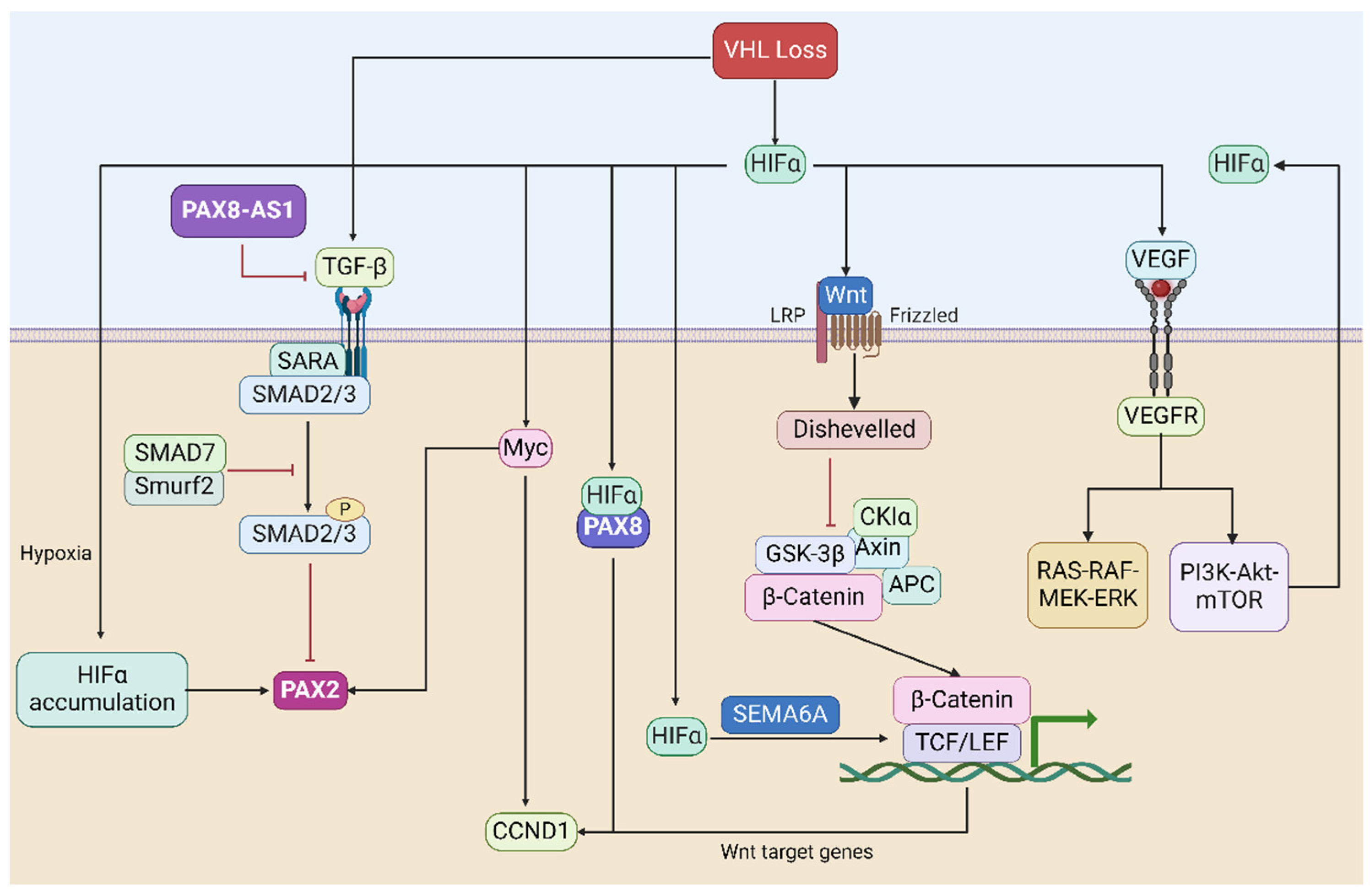
The occurrence and progression of cancer are frequently linked to the dysregulation of signaling pathways. Within RCC, a diverse range of histological subtypes exists, each characterized not only by distinct molecular signatures but also increasingly by unique molecular pathways that undergo deregulation in each subtype [90]. Loss of VHL triggers the accumulation and nuclear translocation of HIFα, subsequently activating crucial oncogenic signaling pathways in RCC. The absence of VHL seems to serve as an early foundation for RCC development, with the PAX genes also contributing and being upregulated by the VHL-mediated hypoxia signaling pathway (Figure 3).

4.1. The HIF-Based Hypoxia Signaling Pathway

Hypoxia (a state of oxygen deficiency) is a prominent characteristic within numerous solid malignancies, primarily attributed to inadequate perfusion and/or the development of poorly or non-functional blood vessels within neoplasms. This phenomenon is particularly pronounced in rapidly proliferating tumors. In most ccRCC, hypoxia-inducible factors (HIFs), which are global transcriptional regulators of the hypoxic response, are persistently activated through the inactivation of a component of the E3 ubiquitin ligase complex, the VHL tumor-suppressor protein [91].
The VHL tumor-suppressor gene mediates and participates in the transduction of the hypoxia signaling pathway through HIFα [91] (see Figure 3). Luu et al. showed that PAX2 reactivation in ccRCC is induced by hypoxia and depends on the functional integrity of pVHL and that PAX2 reactivation after pVHL loss is driven by HIF-dependent mechanisms [92]. HIF is a heterodimer complex comprising a labile α subunit and a stable β subunit [93]. The α subunit is the major regulatory subunit of HIF. Its level is mainly regulated by intracellular oxygen concentration. Under normoxic conditions, hydroxylation of the HIFα subunit enables its recognition and ubiquitination by the pVHL complex [94], thereby keeping intracellular HIFα levels low. Conversely, during hypoxia, HIFα remains unhydroxylated, resulting in its non-interaction with VHL proteins and consequent lack of degradation. Isono et al. found that HIF2α is overexpressed in VHL-deficient RCC cells, leading to higher cell viability in tumor cells [95]. Additionally, in a ccRCC xenograft model, HIF-2α overexpression in RCC cells contributes to increased tumor burden [76]. When HIF-1α was overexpressed, a reduction in tumor size was observed, whereas the knockdown of HIF-1α increased RCC cell proliferation [76]. Therefore, HIF-1α may exert a tumor suppressor function in the context of RCC.

4.2. The TGF-β Signaling Pathway

In RCC, the TGF-β pathway is often dysregulated, leading to tumor promotion [96]. TGF-β acts as a tumor suppressor in the early stages of RCC, inhibiting cell proliferation and promoting differentiation. However, in later stages, TGF-β may promote tumor progression by inducing EMT. TGF-β1 can exert its functions through both canonical (Smad-dependent) and non-canonical (Smad-independent) signaling pathways [97]. Canonical TGF-β1 signaling (Smad-based) can promote RCC progression by promoting invasion and metastasis and inducing EMT [98]. TGF-β signaling has been found to interact with HIF to promote renal fibrosis [99]. Inhibition of PAX2 transcription in ccRCC cells through classical TGF-β signaling has been reported [48] (see Figure 3). LncRNA-PAX8-AS1 was found to be located in chromosome 2q13 upstream of the PAX8 gene and has a negative regulatory effect on PAX8 [100]. PAX8-AS1 can bind to miR-96-5p to inhibit the malignant phenotype of papillary thyroid carcinoma cells [101]. In renal peduncle cells, PAX8-AS1 reduces renal peduncle apoptosis and inhibits the expression of VEGF/TGF-β1 [102]. Shang et al. showed that TGF-β1 may be a direct target of VHL rather than regulating TGF-β1 expression through the HIF pathway [103]. Furthermore, the inactivation of VHL in ccRCC may enhance the metastatic ability of RCC cells by increasing TGF-β1 expression [103].

4.3. The VEGF-Related Signaling Pathway

VEGF is a cytokine that plays a central role in promoting the formation of angiogenesis [104]. In cancer, increased VEGF expression and angiogenesis are crucial for tumor growth and metastasis [104]. VEGF was found to be a potential HIFα target in ccRCC, and knockdown of HIFα decreased the expression level of VEGF [105]. Elevated VEGF expression is frequently linked to poor prognosis in RCC patients [106]. VEGF, upon binding to its corresponding cell surface receptors such as VEGFR, induces tyrosine phosphorylation of the receptor, initiating downstream signaling through various kinase-dependent pathways, including the PI3K-Akt-mTOR pathway and the RAS-RAF-MEK-ERK pathway [107,108] (see Figure 3). PAX2 has been reported to be related to angiogenesis in RCC [109]. Research by Fonsato et al. shows that PAX2 may participate in the Akt pathway and affect angiogenesis by regulating PTEN [109]. PTEN is a phosphatase that regulates PI3K-dependent activation of Akt. PAX8 was found to promote angiogenesis in ovarian cancer by interacting with SOX17, but whether it is related to angiogenesis in RCC remains unclear [110]. It is important to highlight that mTOR is among the downstream effectors of PI3K/AKT, and its activation results in increased expression levels of HIFα [107]. Notably, an aberrant VHL function can establish a harmful positive feedback loop. The absence of VHL leads to impaired degradation of HIFα, causing its accumulation at high levels. Subsequently, elevated HIFα levels further stimulate the abnormal expression of VEGF, which binds to VEGFR, activating the PI3K-Akt pathway. Consequently, mTOR is also activated through this pathway, leading to even higher HIFα levels. This vicious positive feedback loop exacerbates the defects caused by aberrant VHL function, thereby promoting the onset and progression of ccRCC.

4.4. The MYC-Related Signaling Pathway

MYC is a family of regulatory and proto-oncogenes encoding transcription factors (-MYC (MYC), L-MYC (MYCL), and N-MYC (MYCN)) [111,112]. MYC regulates various biological processes, including cell differentiation, cell growth, apoptosis, and stem cell self-renewal [111,112]. Hwang et al. showed that pVHL can bind to the C-MYC promoter along with C-MYC and then transcriptionally repress the C-MYC gene [113]. In metanephric mesenchyme cells, N-MYC acting downstream of PAX2 may restore PAX2 expression at the transcriptional level and thus regulate cell proliferation and apoptosis [114]. HIF-2α can promote C-MYC activity and induce cell cycle progression in ccRCC cells [115]. On the other hand, HIF-1α acts as an inhibitor of C-MYC activity, leading to reduced levels of E2F and cyclin D2 while concurrently enhancing the expression of cell cycle progression inhibitors p21 and p27 [115,116]. Another significant discovery is that HIF-1 inhibits mitochondrial biogenesis and cellular respiration in VHL-deficient renal cell carcinoma by impeding C-MYC activity [117]. C-MYC can transactivate CCND1 expression [118] (see Figure 3). CCND1 plays a tumor-promoting role in RCC, and its knockdown has been shown to attenuate xenograft growth in vivo [119]. Additionally, CCND1 is considered a potential downstream gene of HIF-2α. Its expression decreases upon HIF-2α inhibition and increases upon re-expression of HIF-2α in vitro [76]. Notably, HIF-2α and PAX8 interact with each other at the level of chromatin, and they may be synergistically involved in the regulation of CCND1 expression [120].

4.5. The Wnt/β-Catenin Signaling Pathway

Wnts represent a family of secreted glycoproteins responsible for regulating essential cellular processes, including cell proliferation, differentiation, and migration [121]. Multiple lines of evidence suggest the involvement of the Wnt/β-catenin signaling pathway in RCC [122,123] (see Figure 3). Blockade of the WNT signaling pathway inhibits the migration and invasion ability of PAX2-positive cells [124]. Wnt1 was found to be significantly overexpressed in ccRCC, and high levels of Wnt1 are associated with more aggressive RCC cells and worse prognosis in patients [123]. Among the intracellular signal transducers, β-catenin plays a crucial role as a participant in the canonical Wnt signaling pathway [125,126]. Unphosphorylated β-catenin can interact with members of the LEF and TCF families, thereby further promoting the transcription of Wnt target genes (such as C-MYC and CCND1) [127,128]. While β-catenin-activating point mutations are not common in RCC, studies have demonstrated that inducing overexpression of β-catenin leads to the development of renal tumors in mice [129]. Ji et al. showed that activation of the Wnt/β-catenin pathway correlates with the expression of SEMA6A in ccRCC [122]. The VHL-HIF-2α axis can stabilize β-catenin and promote ccRCC progression by inducing SEMA6A upregulation [122].

5. The Development of PAX-Related Therapeutic Strategies in Renal Cell Carcinoma

In recent years, advancements in medicine have continuously updated and optimized the treatment of RCC. Surgery remains the primary treatment for RCC, especially for localized and locally advanced renal cancer. However, the emergence of immune checkpoint inhibitors (ICIs) has dramatically transformed the treatment prospects for RCC. Currently, the standard first-line treatment for advanced ccRCC includes ICI combinations. The dual immune checkpoint blockade with ipilimumab and nivolumab, targeting cytotoxic T lymphocyte-associated protein 4 (CTLA-4) and programmed cell death protein 1 (PD-1), respectively, is the first ICI combination approved by the US Food and Drug Administration (FDA) under the CheckMate 214 trial [130,131]. ICI combination therapy has brought overall survival benefits and improved prognostic characteristics to some patients. Nevertheless, a significant number of patients exhibit primary resistance to ICI combinations or develop acquired resistance, ultimately succumbing to the disease [131]. Therefore, the development of new treatment options and the identification of novel therapeutic targets to enhance the prognosis of RCC remain the primary focus of current RCC research.
Genetic loss-of-function studies suggest that PAX genes, particularly PAX2 and PAX8, which are highly expressed in RCC, are crucial for the continued proliferation and/or survival of RCC cells. Consequently, targeting PAX2 or PAX8 may represent a potentially effective therapeutic approach for the treatment of RCC (Table 2). Previous studies have utilized small interfering RNA (siRNA) to knock down PAX2, demonstrating that PAX2 knockdown sensitized RCC cells to cisplatin-induced apoptosis, resulting in the death of 50–60% of cisplatin-resistant ACHN and CAKI-1 cells [22]. Further animal experiments have corroborated these findings, indicating that silencing PAX2 expression could partially overcome the resistance of RCC to chemotherapy in vivo [31]. Additionally, large-scale functional genomic screening validated that PAX8 silencing led to reduced proliferation of RCC cell lines [27]. Grimley et al. have screened large chemical libraries and identified a compound, EG1, that can effectively block PAX2 activity and DNA binding through the PAX2 paired domain [132]. Their findings revealed that EG1 can significantly inhibit the proliferation of PAX2-positive RCC cells while having minimal impact on PAX2-negative RCC cells. Recently, Bradford et al. [133] discovered three triazolo pyrimidine derivatives that inhibit PAX-mediated reporter gene transcriptional activation. Analysis of cultured renal epithelial cells demonstrated that these small molecules specifically inhibit PAX2 and PAX8 function by preventing the recruitment of the adaptor protein PTIP and the assembly of histone methylation complexes at target promoters. Furthermore, these compounds can specifically slow the proliferation of PAX2-positive RCC cells [133]. Combining PAX targeting with immunotherapy may represent a promising therapeutic direction. However, progress in developing therapeutic strategies targeting PAX proteins remains inadequate, particularly regarding the development of PAX-related inhibitors, which face significant obstacles. One major challenge arises from the inherently disordered structure of PAX proteins, rendering direct targeting approaches complex. Additionally, the predominant nuclear localization of PAX proteins poses significant hurdles to effective drug delivery. Addressing these challenges demands concerted efforts to explore novel methodologies and deepen our understanding of PAX genes, thereby facilitating the development of more efficacious therapeutic interventions.

6. Conclusions

The PAX gene family plays an important role in the occurrence and development of RCC [8,9]. PAX2 undergoes reactivation during the early stages of ccRCC [12,13]. Approximately 90% of RCC tumors exhibit high PAX2 and PAX8 expression levels, while other PAX genes have low or no expression in RCC [5,15]. Moreover, it is noteworthy that PAX2 and PAX8 exhibit heightened expression frequencies in both ccRCC and PRCC, while their expression remains either low or entirely absent in ChRCC. The upregulation of PAX2 and PAX8 within the context of RCC may be intricately linked to the maintenance of the epithelial phenotype during the process of EMT. Several lines of evidence implicate PAX2 and PAX8 as promising, emerging potential therapeutic targets for the treatment of RCC.
The most frequently mutated gene in ccRCC is VHL, where approximately 90% of sporadic ccRCC cases show alterations in the VHL gene located on chromosome 3 [7]. Notably, the loss of VHL function results in the upregulation of PAX2 expression in ccRCC. Loss of VHL alone is insufficient to induce ccRCC, which requires additional epigenetic events to occur. When the VHL gene mutates or loses its function, the HIF-1/2-α subunit becomes stable, leading to the activation of HIF target genes and their downstream signaling pathways [72,73,92]. HIF-2α is preferentially recruited to PAX8-bound transcriptional enhancers, which together coordinate the expression of the oncogene CCND1 [120]. In conclusion, the loss of VHL in ccRCC may be involved in the occurrence and development of ccRCC in conjunction with PAX gene expression through HIF-mediated downstream target genes and signaling pathways.
Targeting PAX2 or PAX8 may be a potentially valuable treatment approach for RCC [12,31]. Small molecule compounds that target PAX have been developed and employed in targeted cancer therapy (non-clinical), which yielded promising in vitro results [132,133]. However, it is important to note that the expression levels of PAX2 undergo dynamic changes during RCC development, reflecting a phenotypic shift in RCC. PAX2 is reactivated in early ccRCC, but its expression levels begin to decline in high-grade ccRCC. This coincides with ccRCC transitioning towards a more aggressive phenotype and favoring glycolysis for energy production. Consequently, in patients with high-grade ccRCC, regardless of whether PAX2 is targeted or not, its expression has decreased. The potential reasons for this phenomenon could include metabolic reprogramming of RCC and alterations in the tumor microenvironment, although the specific mechanisms remain unclear. Therefore, the clinical application of PAX gene inhibitors will require a further understanding of the role of PAX genes in cancer.
The generation of small molecule compounds that directly target PAX proteins is at a relatively early stage in terms of PAX inhibitor drug development because in vivo delivery of PAX-targeting small molecules is a significant barrier. Similarly, in vivo stability and delivery of siRNAs against PAX mRNAs are also significant barriers. Consequently, there remain significant gaps in the development of targeted therapies for PAX in RCC. Additionally, there continues to be ongoing debate regarding the use of PAX2 expression levels as a metastatic marker for RCC. One potential reason for this debate may be the discrepancies in methods used to quantify gene expression, whether through the detection of RNA or protein markers. Although mRNA levels are early indicators of gene expression, they do not necessarily correlate directly with protein levels due to the influence of various regulatory factors such as translation efficiency and protein degradation rates. Furthermore, the expression of PAX genes varies in different regions of RCC at different times.
Future investigations could prioritize the elucidation of the potential mechanisms associated with PAX involvement in RCC and advancing therapeutic strategies targeting PAX. Presently, the precise mechanisms underlying PAX-mediated RCC onset and progression remain elusive. Notably, PAX genes are frequently expressed during developmental processes and are implicated in tissue regeneration and repair. Emerging research suggests a correlation between PAX expression and signaling pathways associated with stemness, indicating that the reactivation of PAX genes in RCC may contribute to the manifestation of cancer stemness characteristics [134,135]. Moreover, combination therapies targeting multiple pathways hold promise as a burgeoning approach. Identified potential targets upregulated in RCC, such as MUC1, AXL, EGFR, C-MET, PD-L1, VEGF, HIF, etc., could be targeted in combination with targeting PAX, offering valuable support in the development of potential therapeutic strategies for RCC [134,135,136,137,138].

Author Contributions

Conceptualization, L.L., S.M.H. and M.R.E.; writing—original draft preparation, L.L.; writing—review and editing, S.M.H. and M.R.E. All authors have read and agreed to the published version of the manuscript.

Funding

L.L. is supported by a PhD scholarship from the Chinese Scholarship Council–New Zealand-China Research Collaboration Centres (CSC-NZ CRCC) joint funding program and the New Zealand-China Non-Communicable Diseases Research Collaboration Centre (NCD CRCC). S.M.H. is supported by a Postdoctoral Fellowship from the New Zealand Institute for Cancer Research Trust. Additionally, the authors received funding support from the Maurice Wilkins Centre for Molecular Biodiscovery, and Dunedin School of Medicine, University of Otago.

Acknowledgments

Figure 1, Figure 2 and Figure 3 were produced using Biorender software (URL accessed on 19 April 2024; www.biorender.com).

Conflicts of Interest

The authors declare no conflicts of interest.

References

  1. Rini, B.I.; Campbell, S.C.; Escudier, B. Renal cell carcinoma. Lancet 2009, 373, 1119–1132. [Google Scholar] [CrossRef] [PubMed]
  2. Chen, Y.W.; Wang, L.; Panian, J.; Dhanji, S.; Derweesh, I.; Rose, B.; Bagrodia, A.; McKay, R.R. Treatment Landscape of Renal Cell Carcinoma. Curr. Treat. Options Oncol. 2023, 24, 1889–1916. [Google Scholar] [CrossRef] [PubMed] [PubMed Central]
  3. Cirillo, L.; Innocenti, S.; Becherucci, F. Global epidemiology of kidney cancer. Nephrol. Dial. Transplant. 2024, 39, 920–928. [Google Scholar] [CrossRef] [PubMed]
  4. Bukavina, L.; Bensalah, K.; Bray, F.; Carlo, M.; Challacombe, B.; Karam, J.A.; Kassouf, W.; Mitchell, T.; Montironi, R.; O’Brien, T.; et al. Epidemiology of Renal Cell Carcinoma: 2022 Update. Eur. Urol. 2022, 82, 529–542. [Google Scholar] [CrossRef] [PubMed]
  5. Li, L.; Li, C.G.; Almomani, S.N.; Hossain, S.M.; Eccles, M.R. Co-Expression of Multiple PAX Genes in Renal Cell Carcinoma (RCC) and Correlation of High PAX Expression with Favorable Clinical Outcome in RCC Patients. Int. J. Mol. Sci. 2023, 24, 11432. [Google Scholar] [CrossRef] [PubMed] [PubMed Central]
  6. Linehan, W.M.; Ricketts, C.J. The Cancer Genome Atlas of renal cell carcinoma: Findings and clinical implications. Nat. Rev. Urol. 2019, 16, 539–552. [Google Scholar] [CrossRef] [PubMed]
  7. Hu, J.; Tan, P.; Ishihara, M.; Bayley, N.A.; Schokrpur, S.; Reynoso, J.G.; Zhang, Y.; Lim, R.J.; Dumitras, C.; Yang, L.; et al. Tumor heterogeneity in VHL drives metastasis in clear cell renal cell carcinoma. Signal Transduct. Target. Ther. 2023, 8, 155. [Google Scholar] [CrossRef] [PubMed] [PubMed Central]
  8. Thompson, B.; Davidson, E.A.; Liu, W.; Nebert, D.W.; Bruford, E.A.; Zhao, H.; Dermitzakis, E.T.; Thompson, D.C.; Vasiliou, V. Overview of PAX gene family: Analysis of human tissue-specific variant expression and involvement in human disease. Hum. Genet. 2021, 140, 381–400. [Google Scholar] [CrossRef] [PubMed] [PubMed Central]
  9. Li, C.G.; Eccles, M.R. PAX Genes in Cancer; Friends or Foes? Front. Genet. 2012, 3, 6. [Google Scholar] [CrossRef] [PubMed] [PubMed Central]
  10. Bouchard, M.; Souabni, A.; Mandler, M.; Neubüser, A.; Busslinger, M. Nephric lineage specification by Pax2 and Pax8. Genes Dev. 2002, 16, 2958–2970. [Google Scholar] [CrossRef] [PubMed] [PubMed Central]
  11. Narlis, M.; Grote, D.; Gaitan, Y.; Boualia, S.K.; Bouchard, M. Pax2 and pax8 regulate branching morphogenesis and nephron differentiation in the developing kidney. J. Am. Soc. Nephrol. 2007, 18, 1121–1129. [Google Scholar] [CrossRef] [PubMed]
  12. Grimley, E.; Dressler, G.R. Are Pax proteins potential therapeutic targets in kidney disease and cancer? Kidney Int. 2018, 94, 259–267. [Google Scholar] [CrossRef] [PubMed] [PubMed Central]
  13. Patel, S.R.; Dressler, G.R. The genetics and epigenetics of kidney development. Semin. Nephrol. 2013, 33, 314–326. [Google Scholar] [CrossRef] [PubMed] [PubMed Central]
  14. Kakun, R.R.; Melamed, Z.; Perets, R. PAX8 in the Junction between Development and Tumorigenesis. Int. J. Mol. Sci. 2022, 23, 7410. [Google Scholar] [CrossRef] [PubMed] [PubMed Central]
  15. Gupta, R.; Balzer, B.; Picken, M.; Osunkoya, A.O.; Shet, T.; Alsabeh, R.; Luthringer, D.; Paner, G.P.; Amin, M.B. Diagnostic implications of transcription factor Pax 2 protein and transmembrane enzyme complex carbonic anhydrase IX immunoreactivity in adult renal epithelial neoplasms. Am. J. Surg. Pathol. 2009, 33, 241–247. [Google Scholar] [CrossRef] [PubMed]
  16. Shaw, T.; Barr, F.G.; Üren, A. The PAX Genes: Roles in Development, Cancer, and Other Diseases. Cancers 2024, 16, 1022. [Google Scholar] [CrossRef] [PubMed] [PubMed Central]
  17. Peters, H.; Wilm, B.; Sakai, N.; Imai, K.; Maas, R.; Balling, R. Pax1 and Pax9 synergistically regulate vertebral column development. Development 1999, 126, 5399–5408. [Google Scholar] [CrossRef] [PubMed]
  18. Ogasawara, M.; Shigetani, Y.; Hirano, S.; Satoh, N.; Kuratani, S. Pax1/Pax9-Related genes in an agnathan vertebrate, Lampetra japonica: Expression pattern of LjPax9 implies sequential evolutionary events toward the gnathostome body plan. Dev. Biol. 2000, 223, 399–410. [Google Scholar] [CrossRef] [PubMed]
  19. Amiel, J.; Audollent, S.; Joly, D.; Dureau, P.; Salomon, R.; Tellier, A.L.; Augé, J.; Bouissou, F.; Antignac, C.; Gubler, M.C.; et al. PAX2 mutations in renal-coloboma syndrome: Mutational hotspot and germline mosaicism. Eur. J. Hum. Genet. 2000, 8, 820–826. [Google Scholar] [CrossRef] [PubMed]
  20. Ozcan, A.; de la Roza, G.; Ro, J.Y.; Shen, S.S.; Truong, L.D. PAX2 and PAX8 expression in primary and metastatic renal tumors: A comprehensive comparison. Arch. Pathol. Lab. Med. 2012, 136, 1541–1551. [Google Scholar] [CrossRef] [PubMed]
  21. Knoepp, S.M.; Kunju, L.P.; Roh, M.H. Utility of PAX8 and PAX2 immunohistochemistry in the identification of renal cell carcinoma in diagnostic cytology. Diagn. Cytopathol. 2012, 40, 667–672. [Google Scholar] [CrossRef] [PubMed]
  22. Hueber, P.A.; Waters, P.; Clark, P.; Eccles, M.; Goodyer, P. PAX2 inactivation enhances cisplatin-induced apoptosis in renal carcinoma cells. Kidney Int. 2006, 69, 1139–1145, Erratum in Kidney Int. 2006, 70, 1666. [Google Scholar] [CrossRef] [PubMed]
  23. Liu, Y.; Cui, S.; Li, W.; Zhao, Y.; Yan, X.; Xu, J. PAX3 is a biomarker and prognostic factor in melanoma: Database mining. Oncol. Lett. 2019, 17, 4985–4993. [Google Scholar] [CrossRef] [PubMed] [PubMed Central]
  24. Ko, J.; Fonseca, V.A.; Wu, H. Pax4 in Health and Diabetes. Int. J. Mol. Sci. 2023, 24, 8283. [Google Scholar] [CrossRef] [PubMed] [PubMed Central]
  25. Nasri Nasrabadi, P.; Martin, D.; Gharib, E.; Robichaud, G.A. The Pleiotropy of PAX5 Gene Products and Function. Int. J. Mol. Sci. 2022, 23, 10095. [Google Scholar] [CrossRef] [PubMed] [PubMed Central]
  26. Azuma, N.; Yamaguchi, Y.; Handa, H.; Tadokoro, K.; Asaka, A.; Kawase, E.; Yamada, M. Mutations of the PAX6 gene detected in patients with a variety of optic-nerve malformations. Am. J. Hum. Genet. 2003, 72, 1565–1570. [Google Scholar] [CrossRef] [PubMed] [PubMed Central]
  27. Bleu, M.; Gaulis, S.; Lopes, R.; Sprouffske, K.; Apfel, V.; Holwerda, S.; Pregnolato, M.; Yildiz, U.; Cordo’, V.; Dost, A.F.M.; et al. PAX8 activates metabolic genes via enhancer elements in Renal Cell Carcinoma. Nat. Commun. 2019, 10, 3739. [Google Scholar] [CrossRef] [PubMed] [PubMed Central]
  28. Wachtel, M.; Schäfer, B.W. Unpeaceful roles of mutant PAX proteins in cancer. Semin. Cell Dev. Biol. 2015, 44, 126–134. [Google Scholar] [CrossRef] [PubMed]
  29. Dumont, S.N.; Lazar, A.J.; Bridge, J.A.; Benjamin, R.S.; Trent, J.C. PAX3/7-FOXO1 fusion status in older rhabdomyosarcoma patient population by fluorescent in situ hybridization. J. Cancer Res. Clin. Oncol. 2012, 138, 213–220. [Google Scholar] [CrossRef] [PubMed] [PubMed Central]
  30. Sorensen, P.H.; Lynch, J.C.; Qualman, S.J.; Tirabosco, R.; Lim, J.F.; Maurer, H.M.; Bridge, J.A.; Crist, W.M.; Triche, T.J.; Barr, F.G. PAX3-FKHR and PAX7-FKHR gene fusions are prognostic indicators in alveolar rhabdomyosarcoma: A report from the children’s oncology group. J. Clin. Oncol. 2002, 20, 2672–2679. [Google Scholar] [CrossRef] [PubMed]
  31. Hueber, P.A.; Iglesias, D.; Chu, L.L.; Eccles, M.; Goodyer, P. In vivo validation of PAX2 as a target for renal cancer therapy. Cancer Lett. 2008, 265, 148–155. [Google Scholar] [CrossRef] [PubMed]
  32. Robson, E.J.; He, S.J.; Eccles, M.R. A PANorama of PAX genes in cancer and development. Nat. Rev. Cancer 2006, 6, 52–62. [Google Scholar] [CrossRef] [PubMed]
  33. Muratovska, A.; Zhou, C.; He, S.; Goodyer, P.; Eccles, M.R. Paired-Box genes are frequently expressed in cancer and often required for cancer cell survival. Oncogene 2003, 22, 7989–7997. [Google Scholar] [CrossRef] [PubMed]
  34. Patel, S.R.; Ranghini, E.; Dressler, G.R. Mechanisms of gene activation and repression by Pax proteins in the developing kidney. Pediatr. Nephrol. 2014, 29, 589–595. [Google Scholar] [CrossRef] [PubMed] [PubMed Central]
  35. Sharma, R.; Sanchez-Ferras, O.; Bouchard, M. Pax genes in renal development, disease and regeneration. Semin. Cell Dev. Biol. 2015, 44, 97–106. [Google Scholar] [CrossRef] [PubMed]
  36. Jiang, Y.S.; Jiang, T.; Huang, B.; Chen, P.S.; Ouyang, J. Epithelial-mesenchymal transition of renal tubules: Divergent processes of repairing in acute or chronic injury? Med. Hypotheses 2013, 81, 73–75. [Google Scholar] [CrossRef] [PubMed]
  37. Buttiglieri, S.; Deregibus, M.C.; Bravo, S.; Cassoni, P.; Chiarle, R.; Bussolati, B.; Camussi, G. Role of Pax2 in apoptosis resistance and proinvasive phenotype of Kaposi’s sarcoma cells. J. Biol. Chem. 2004, 279, 4136–4143. [Google Scholar] [CrossRef] [PubMed]
  38. Brun, T.; Duhamel, D.L.; Hu He, K.H.; Wollheim, C.B.; Gauthier, B.R. The transcription factor PAX4 acts as a survival gene in INS-1E insulinoma cells. Oncogene 2007, 26, 4261–4271. [Google Scholar] [CrossRef] [PubMed]
  39. Shi, K.; Yin, X.; Cai, M.C.; Yan, Y.; Jia, C.; Ma, P.; Zhang, S.; Zhang, Z.; Gu, Z.; Zhang, M.; et al. PAX8 regulon in human ovarian cancer links lineage dependency with epigenetic vulnerability to HDAC inhibitors. eLife 2019, 8, e44306. [Google Scholar] [CrossRef] [PubMed] [PubMed Central]
  40. Doberstein, K.; Pfeilschifter, J.; Gutwein, P. The transcription factor PAX2 regulates ADAM10 expression in renal cell carcinoma. Carcinogenesis 2011, 32, 1713–1723. [Google Scholar] [CrossRef] [PubMed]
  41. Di Palma, T.; Filippone, M.G.; Pierantoni, G.M.; Fusco, A.; Soddu, S.; Zannini, M. Pax8 has a critical role in epithelial cell survival and proliferation. Cell Death Dis. 2013, 4, e729. [Google Scholar] [CrossRef] [PubMed] [PubMed Central]
  42. Wang, J.; Zhao, X.; Qi, J.; Yang, C.; Cheng, H.; Ren, Y.; Huang, L. Eight proteins play critical roles in RCC with bone metastasis via mitochondrial dysfunction. Clin. Exp. Metastasis 2015, 32, 605–622. [Google Scholar] [CrossRef] [PubMed] [PubMed Central]
  43. Hoefflin, R.; Harlander, S.; Schäfer, S.; Metzger, P.; Kuo, F.; Schönenberger, D.; Adlesic, M.; Peighambari, A.; Seidel, P.; Chen, C.Y.; et al. HIF-1α and HIF-2α differently regulate tumour development and inflammation of clear cell renal cell carcinoma in mice. Nat. Commun. 2020, 11, 4111. [Google Scholar] [CrossRef] [PubMed] [PubMed Central]
  44. Stuart, E.T.; Haffner, R.; Oren, M.; Gruss, P. Loss of p53 function through PAX-mediated transcriptional repression. EMBO J. 1995, 14, 5638–5645. [Google Scholar] [CrossRef] [PubMed] [PubMed Central]
  45. Frost, V.; Grocott, T.; Eccles, M.R.; Chantry, A. Self-regulated Pax gene expression and modulation by the TGFbeta superfamily. Crit. Rev. Biochem. Mol. Biol. 2008, 43, 371–391. [Google Scholar] [CrossRef] [PubMed]
  46. Pei, D.; Shu, X.; Gassama-Diagne, A.; Thiery, J.P. Mesenchymal-epithelial transition in development and reprogramming. Nat. Cell Biol. 2019, 21, 44–53. [Google Scholar] [CrossRef] [PubMed]
  47. Harada, K.; Miyake, H.; Kusuda, Y.; Fujisawa, M. Expression of epithelial-mesenchymal transition markers in renal cell carcinoma: Impact on prognostic outcomes in patients undergoing radical nephrectomy. BJU Int. 2012, 110 Pt C, E1131–E1137. [Google Scholar] [CrossRef] [PubMed]
  48. Kaur, G.; Li, C.G.; Chantry, A.; Stayner, C.; Horsfield, J.; Eccles, M.R. SMAD proteins directly suppress PAX2 transcription downstream of transforming growth factor-beta 1 (TGF-β1) signalling in renal cell carcinoma. Oncotarget 2018, 9, 26852–26867. [Google Scholar] [CrossRef] [PubMed] [PubMed Central]
  49. Mazal, P.R.; Stichenwirth, M.; Koller, A.; Blach, S.; Haitel, A.; Susani, M. Expression of aquaporins and PAX-2 compared to CD10 and cytokeratin 7 in renal neoplasms: A tissue microarray study. Mod. Pathol. 2005, 18, 535–540. [Google Scholar] [CrossRef] [PubMed]
  50. Truong, L.D.; Shen, S.S. Immunohistochemical diagnosis of renal neoplasms. Arch. Pathol. Lab. Med. 2011, 135, 92–109. [Google Scholar] [CrossRef] [PubMed]
  51. Grote, D.; Souabni, A.; Busslinger, M.; Bouchard, M. Pax 2/8-regulated Gata 3 expression is necessary for morphogenesis and guidance of the nephric duct in the developing kidney. Development 2006, 133, 53–61. [Google Scholar] [CrossRef] [PubMed]
  52. Boualia, S.K.; Gaitan, Y.; Tremblay, M.; Sharma, R.; Cardin, J.; Kania, A.; Bouchard, M. A core transcriptional network composed of Pax2/8, Gata3 and Lim1 regulates key players of pro/mesonephros morphogenesis. Dev. Biol. 2013, 382, 555–566. [Google Scholar] [CrossRef] [PubMed]
  53. Salvi, A.; Hardy, L.R.; Heath, K.N.; Watry, S.; Pergande, M.R.; Cologna, S.M.; Burdette, J.E. PAX8 modulates the tumor microenvironment of high grade serous ovarian cancer through changes in the secretome. Neoplasia 2023, 36, 100866. [Google Scholar] [CrossRef] [PubMed] [PubMed Central]
  54. Aili, A.; Wen, J.; Xue, L.; Wang, J. Mutational Analysis of PBRM1 and Significance of PBRM1 Mutation in Anti-PD-1 Immunotherapy of Clear Cell Renal Cell Carcinoma. Front. Oncol. 2021, 11, 712765. [Google Scholar] [CrossRef] [PubMed] [PubMed Central]
  55. Gu, X.; Enane, F.; Tohme, R.; Schuerger, C.; Radivoyevitch, T.; Parker, Y.; Zuberi, E.; Przychodzen, B.; Jha, B.K.; Lindner, D.; et al. PBRM1 loss in kidney cancer unbalances the proximal tubule master transcription factor hub to repress proximal tubule differentiation. Cell Rep. 2021, 36, 109747. [Google Scholar] [CrossRef] [PubMed] [PubMed Central]
  56. Jiang, F.; Richter, J.; Schraml, P.; Bubendorf, L.; Gasser, T.; Sauter, G.; Mihatsch, M.J.; Moch, H. Chromosomal imbalances in papillary renal cell carcinoma: Genetic differences between histological subtypes. Am. J. Pathol. 1998, 153, 1467–1473. [Google Scholar] [CrossRef] [PubMed] [PubMed Central]
  57. Durinck, S.; Stawiski, E.W.; Pavía-Jiménez, A.; Modrusan, Z.; Kapur, P.; Jaiswal, B.S.; Zhang, N.; Toffessi-Tcheuyap, V.; Nguyen, T.T.; Pahuja, K.B.; et al. Spectrum of diverse genomic alterations define non-clear cell renal carcinoma subtypes. Nat. Genet. 2015, 47, 13–21. [Google Scholar] [CrossRef] [PubMed] [PubMed Central]
  58. Cancer Genome Atlas Research Network; Linehan, W.M.; Spellman, P.T.; Ricketts, C.J.; Creighton, C.J.; Fei, S.S.; Davis, C.; Wheeler, D.A.; Murray, B.A.; Schmidt, L.; et al. Comprehensive Molecular Characterization of Papillary Renal-Cell Carcinoma. N. Engl. J. Med. 2016, 374, 135–145. [Google Scholar] [CrossRef] [PubMed]
  59. Casuscelli, J.; Weinhold, N.; Gundem, G.; Wang, L.; Zabor, E.C.; Drill, E.; Wang, P.I.; Nanjangud, G.J.; Redzematovic, A.; Nargund, A.M.; et al. Genomic landscape and evolution of metastatic chromophobe renal cell carcinoma. JCI Insight 2017, 2, e92688. [Google Scholar] [CrossRef] [PubMed] [PubMed Central]
  60. Speicher, M.R.; Schoell, B.; du Manoir, S.; Schröck, E.; Ried, T.; Cremer, T.; Störkel, S.; Kovacs, A.; Kovacs, G. Specific loss of chromosomes 1, 2, 6, 10, 13, 17, and 21 in chromophobe renal cell carcinomas revealed by comparative genomic hybridization. Am. J. Pathol. 1994, 145, 356–364. [Google Scholar] [PubMed] [PubMed Central]
  61. Davis, C.F.; Ricketts, C.J.; Wang, M.; Yang, L.; Cherniack, A.D.; Shen, H.; Buhay, C.; Kang, H.; Kim, S.C.; Fahey, C.C.; et al. The somatic genomic landscape of chromophobe renal cell carcinoma. Cancer Cell 2014, 26, 319–330. [Google Scholar] [CrossRef] [PubMed] [PubMed Central]
  62. Mitchell, T.J.; Turajlic, S.; Rowan, A.; Nicol, D.; Farmery, J.H.R.; O’Brien, T.; Martincorena, I.; Tarpey, P.; Angelopoulos, N.; Yates, L.R.; et al. TRACERx Renal Consortium. Timing the Landmark Events in the Evolution of Clear Cell Renal Cell Cancer: TRACERx Renal. Cell 2018, 173, 611–623.e17. [Google Scholar] [CrossRef] [PubMed] [PubMed Central]
  63. El-Mokadem, I.; Fitzpatrick, J.; Bondad, J.; Rauchhaus, P.; Cunningham, J.; Pratt, N.; Fleming, S.; Nabi, G. Chromosome 9p deletion in clear cell renal cell carcinoma predicts recurrence and survival following surgery. Br. J. Cancer 2014, 111, 1381–1390. [Google Scholar] [CrossRef] [PubMed] [PubMed Central]
  64. Dalgliesh, G.L.; Furge, K.; Greenman, C.; Chen, L.; Bignell, G.; Butler, A.; Davies, H.; Edkins, S.; Hardy, C.; Latimer, C.; et al. Systematic sequencing of renal carcinoma reveals inactivation of histone modifying genes. Nature 2010, 463, 360–363. [Google Scholar] [CrossRef] [PubMed] [PubMed Central]
  65. Brannon, A.R.; Reddy, A.; Seiler, M.; Arreola, A.; Moore, D.T.; Pruthi, R.S.; Wallen, E.M.; Nielsen, M.E.; Liu, H.; Nathanson, K.L.; et al. Molecular Stratification of Clear Cell Renal Cell Carcinoma by Consensus Clustering Reveals Distinct Subtypes and Survival Patterns. Genes Cancer 2010, 1, 152–163. [Google Scholar] [CrossRef] [PubMed] [PubMed Central]
  66. Bihr, S.; Ohashi, R.; Moore, A.L.; Rüschoff, J.H.; Beisel, C.; Hermanns, T.; Mischo, A.; Corrò, C.; Beyer, J.; Beerenwinkel, N.; et al. Expression and Mutation Patterns of PBRM1, BAP1 and SETD2 Mirror Specific Evolutionary Subtypes in Clear Cell Renal Cell Carcinoma. Neoplasia 2019, 21, 247–256. [Google Scholar] [CrossRef] [PubMed] [PubMed Central]
  67. Brugarolas, J. PBRM1 and BAP1 as novel targets for renal cell carcinoma. Cancer J. 2013, 19, 324–332. [Google Scholar] [CrossRef] [PubMed] [PubMed Central]
  68. Mazumder, S.; Higgins, P.J.; Samarakoon, R. Downstream Targets of VHL/HIF-α Signaling in Renal Clear Cell Carcinoma Progression: Mechanisms and Therapeutic Relevance. Cancers 2023, 15, 1316. [Google Scholar] [CrossRef] [PubMed] [PubMed Central]
  69. Zhang, J.; Zhang, Q. VHL and Hypoxia Signaling: Beyond HIF in Cancer. Biomedicines 2018, 6, 35. [Google Scholar] [CrossRef] [PubMed] [PubMed Central]
  70. Kaelin, W.G., Jr. The von Hippel-Lindau tumor suppressor protein and clear cell renal carcinoma. Clin. Cancer Res. 2007, 13 Pt 2, 680s–684s. [Google Scholar] [CrossRef] [PubMed]
  71. Lonser, R.R.; Glenn, G.M.; Walther, M.; Chew, E.Y.; Libutti, S.K.; Linehan, W.M.; Oldfield, E.H. von Hippel-Lindau disease. Lancet 2003, 361, 2059–2067. [Google Scholar] [CrossRef] [PubMed]
  72. Iwai, K.; Yamanaka, K.; Kamura, T.; Minato, N.; Conaway, R.C.; Conaway, J.W.; Klausner, R.D.; Pause, A. Identification of the von Hippel-lindau tumor-suppressor protein as part of an active E3 ubiquitin ligase complex. Proc. Natl. Acad. Sci. USA 1999, 96, 12436–12441. [Google Scholar] [CrossRef] [PubMed] [PubMed Central]
  73. Siemeister, G.; Weindel, K.; Mohrs, K.; Barleon, B.; Martiny-Baron, G.; Marmé, D. Reversion of deregulated expression of vascular endothelial growth factor in human renal carcinoma cells by von Hippel-Lindau tumor suppressor protein. Cancer Res. 1996, 56, 2299–2301. [Google Scholar] [PubMed]
  74. Gudas, L.J.; Fu, L.; Minton, D.R.; Mongan, N.P.; Nanus, D.M. The role of HIF1α in renal cell carcinoma tumorigenesis. J. Mol. Med. 2014, 92, 825–836. [Google Scholar] [CrossRef] [PubMed] [PubMed Central]
  75. Kamura, T.; Sato, S.; Iwai, K.; Czyzyk-Krzeska, M.; Conaway, R.C.; Conaway, J.W. Activation of HIF1alpha ubiquitination by a reconstituted von Hippel-Lindau (VHL) tumor suppressor complex. Proc. Natl. Acad. Sci. USA 2000, 97, 10430–10435. [Google Scholar] [CrossRef] [PubMed] [PubMed Central]
  76. Raval, R.R.; Lau, K.W.; Tran, M.G.; Sowter, H.M.; Mandriota, S.J.; Li, J.L.; Pugh, C.W.; Maxwell, P.H.; Harris, A.L.; Ratcliffe, P.J. Contrasting properties of hypoxia-inducible factor 1 (HIF-1) and HIF-2 in von Hippel-Lindau-associated renal cell carcinoma. Mol. Cell Biol. 2005, 25, 5675–5686. [Google Scholar] [CrossRef] [PubMed] [PubMed Central]
  77. Perrotta, S.; Roberti, D.; Bencivenga, D.; Corsetto, P.; O’Brien, K.A.; Caiazza, M.; Stampone, E.; Allison, L.; Fleck, R.A.; Scianguetta, S.; et al. Effects of Germline VHL Deficiency on Growth, Metabolism, and Mitochondria. N. Engl. J. Med. 2020, 382, 835–844. [Google Scholar] [CrossRef] [PubMed]
  78. Brand, M.D.; Nicholls, D.G. Assessing mitochondrial dysfunction in cells. Biochem. J. 2011, 435, 297–312, Erratum in Biochem. J. 2011, 437, 575. [Google Scholar] [CrossRef] [PubMed] [PubMed Central]
  79. Wang, Z.; Ying, Z.; Bosy-Westphal, A.; Zhang, J.; Schautz, B.; Later, W.; Heymsfield, S.B.; Müller, M.J. Specific metabolic rates of major organs and tissues across adulthood: Evaluation by mechanistic model of resting energy expenditure. Am. J. Clin. Nutr. 2010, 92, 1369–1377. [Google Scholar] [CrossRef] [PubMed] [PubMed Central]
  80. Lunt, S.Y.; Vander Heiden, M.G. Aerobic glycolysis: Meeting the metabolic requirements of cell proliferation. Annu. Rev. Cell Dev. Biol. 2011, 27, 441–464. [Google Scholar] [CrossRef] [PubMed]
  81. Natarajan, B.; Arige, V.; Khan, A.A.; Reddy, S.S.; Barthwal, M.K.; Mahapatra, N.R. Hypoxia-mediated regulation of mitochondrial transcription factors in renal epithelial cells: Implications for hypertensive renal physiology. Hypertens. Res. 2021, 44, 154–167. [Google Scholar] [CrossRef] [PubMed]
  82. Szeto, H.H.; Liu, S.; Soong, Y.; Seshan, S.V.; Cohen-Gould, L.; Manichev, V.; Feldman, L.C.; Gustafsson, T. Mitochondria Protection after Acute Ischemia Prevents Prolonged Upregulation of IL-1β and IL-18 and Arrests CKD. J. Am. Soc. Nephrol. 2017, 28, 1437–1449. [Google Scholar] [CrossRef] [PubMed] [PubMed Central]
  83. Yang, O.C.; Maxwell, P.H.; Pollard, P.J. Renal cell carcinoma: Translational aspects of metabolism and therapeutic consequences. Kidney Int. 2013, 84, 667–681. [Google Scholar] [CrossRef] [PubMed]
  84. Fukushi, A.; Kim, H.D.; Chang, Y.C.; Kim, C.H. Revisited Metabolic Control and Reprogramming Cancers by Means of the Warburg Effect in Tumor Cells. Int. J. Mol. Sci. 2022, 23, 10037. [Google Scholar] [CrossRef] [PubMed] [PubMed Central]
  85. Koppenol, W.H.; Bounds, P.L.; Dang, C.V. Otto Warburg’s contributions to current concepts of cancer metabolism. Nat. Rev. Cancer 2011, 11, 325–337, Erratum in Nat. Rev. Cancer 2011, 11, 618. [Google Scholar] [CrossRef] [PubMed]
  86. Hervouet, E.; Simonnet, H.; Godinot, C. Mitochondria and reactive oxygen species in renal cancer. Biochimie 2007, 89, 1080–1088. [Google Scholar] [CrossRef] [PubMed]
  87. Li, S.; Li, W.; Yuan, J.; Bullova, P.; Wu, J.; Zhang, X.; Liu, Y.; Plescher, M.; Rodriguez, J.; Bedoya-Reina, O.C.; et al. Impaired oxygen-sensitive regulation of mitochondrial biogenesis within the von Hippel-Lindau syndrome. Nat. Metab. 2022, 4, 739–758, Erratum in Nat. Metab. 2022, 4, 1421. [Google Scholar] [CrossRef] [PubMed] [PubMed Central]
  88. Lu, J.; Tan, M.; Cai, Q. The Warburg effect in tumor progression: Mitochondrial oxidative metabolism as an anti-metastasis mechanism. Cancer Lett. 2015, 356 Pt A, 156–164. [Google Scholar] [CrossRef] [PubMed] [PubMed Central]
  89. Chinopoulos, C.; Seyfried, T.N. Mitochondrial Substrate-Level Phosphorylation as Energy Source for Glioblastoma: Review and Hypothesis. ASN Neuro 2018, 10, 1759091418818261. [Google Scholar] [CrossRef] [PubMed] [PubMed Central]
  90. Banumathy, G.; Cairns, P. Signaling pathways in renal cell carcinoma. Cancer Biol. Ther. 2010, 10, 658–664. [Google Scholar] [CrossRef] [PubMed] [PubMed Central]
  91. Schödel, J.; Grampp, S.; Maher, E.R.; Moch, H.; Ratcliffe, P.J.; Russo, P.; Mole, D.R. Hypoxia, Hypoxia-inducible Transcription Factors, and Renal Cancer. Eur. Urol. 2016, 69, 646–657. [Google Scholar] [CrossRef] [PubMed] [PubMed Central]
  92. Luu, V.D.; Boysen, G.; Struckmann, K.; Casagrande, S.; von Teichman, A.; Wild, P.J.; Sulser, T.; Schraml, P.; Moch, H. Loss of VHL and hypoxia provokes PAX2 up-regulation in clear cell renal cell carcinoma. Clin. Cancer Res. 2009, 15, 3297–3304. [Google Scholar] [CrossRef] [PubMed]
  93. Maxwell, P.H.; Wiesener, M.S.; Chang, G.W.; Clifford, S.C.; Vaux, E.C.; Cockman, M.E.; Wykoff, C.C.; Pugh, C.W.; Maher, E.R.; Ratcliffe, P.J. The tumour suppressor protein VHL targets hypoxia-inducible factors for oxygen-dependent proteolysis. Nature 1999, 399, 271–275. [Google Scholar] [CrossRef] [PubMed]
  94. Ivan, M.; Kondo, K.; Yang, H.; Kim, W.; Valiando, J.; Ohh, M.; Salic, A.; Asara, J.M.; Lane, W.S.; Kaelin, W.G., Jr. HIFalpha targeted for VHL-mediated destruction by proline hydroxylation: Implications for O2 sensing. Science 2001, 292, 464–468. [Google Scholar] [CrossRef] [PubMed]
  95. Isono, T.; Chano, T.; Yoshida, T.; Kageyama, S.; Kawauchi, A.; Suzaki, M.; Yuasa, T. Hydroxyl-HIF2-alpha is potential therapeutic target for renal cell carcinomas. Am. J. Cancer Res. 2016, 6, 2263–2276. [Google Scholar] [PubMed] [PubMed Central]
  96. Gao, S.L.; Yin, R.; Zhang, L.F.; Wang, S.M.; Chen, J.S.; Wu, X.Y.; Yue, C.; Zuo, L.; Tang, M. The oncogenic role of MUC12 in RCC progression depends on c-Jun/TGF-β signalling. J. Cell Mol. Med. 2020, 24, 8789–8802. [Google Scholar] [CrossRef] [PubMed] [PubMed Central]
  97. Peng, D.; Fu, M.; Wang, M.; Wei, Y.; Wei, X. Targeting TGF-β signal transduction for fibrosis and cancer therapy. Mol. Cancer 2022, 21, 104. [Google Scholar] [CrossRef] [PubMed] [PubMed Central]
  98. Tretbar, S.; Krausbeck, P.; Müller, A.; Friedrich, M.; Vaxevanis, C.; Bukur, J.; Jasinski-Bergner, S.; Seliger, B. TGF-β inducible epithelial-to-mesenchymal transition in renal cell carcinoma. Oncotarget 2019, 10, 1507–1524. [Google Scholar] [CrossRef] [PubMed] [PubMed Central]
  99. Hanna, C.; Hubchak, S.C.; Liang, X.; Rozen-Zvi, B.; Schumacker, P.T.; Hayashida, T.; Schnaper, H.W. Hypoxia-inducible factor-2α and TGF-β signaling interact to promote normoxic glomerular fibrogenesis. Am. J. Physiol. Renal Physiol. 2013, 305, F1323–F1331. [Google Scholar] [CrossRef] [PubMed] [PubMed Central]
  100. Han, J.; Zhou, W.; Jia, M.; Wen, J.; Jiang, J.; Shi, J.; Zhang, K.; Ma, H.; Liu, J.; Ren, J.; et al. Expression quantitative trait loci in long non-coding RNA PAX8-AS1 are associated with decreased risk of cervical cancer. Mol. Genet. Genom. 2016, 291, 1743–1748. [Google Scholar] [CrossRef] [PubMed]
  101. Zhou, P.; Xu, T.; Hu, H.; Hua, F. Overexpression of PAX8-AS1 Inhibits Malignant Phenotypes of Papillary Thyroid Carcinoma Cells via miR-96-5p/PKN2 Axis. Int. J. Endocrinol. 2021, 2021, 5499963. [Google Scholar] [CrossRef] [PubMed] [PubMed Central]
  102. Shen, Y.; Tong, Z.W.; Zhou, Y.; Sun, Y.; Xie, Y.; Li, R.; Liu, H. Inhibition of lncRNA-PAX8-AS1-N directly associated with VEGF/TGF-β1/8-OhdG enhances podocyte apoptosis in diabetic nephropathy. Eur. Rev. Med. Pharmacol. Sci. 2020, 24, 6864–6872. [Google Scholar] [CrossRef] [PubMed]
  103. Shang, D.; Liu, Y.; Yang, P.; Chen, Y.; Tian, Y. TGFBI-promoted adhesion, migration and invasion of human renal cell carcinoma depends on inactivation of von Hippel-Lindau tumor suppressor. Urology 2012, 79, 966.e1–966.e7. [Google Scholar] [CrossRef] [PubMed]
  104. Siveen, K.S.; Prabhu, K.; Krishnankutty, R.; Kuttikrishnan, S.; Tsakou, M.; Alali, F.Q.; Dermime, S.; Mohammad, R.M.; Uddin, S. Vascular Endothelial Growth Factor (VEGF) Signaling in Tumour Vascularization: Potential and Challenges. Curr. Vasc. Pharmacol. 2017, 15, 339–351. [Google Scholar] [CrossRef] [PubMed]
  105. Cho, H.; Du, X.; Rizzi, J.P.; Liberzon, E.; Chakraborty, A.A.; Gao, W.; Carvo, I.; Signoretti, S.; Bruick, R.K.; Josey, J.A.; et al. On-target efficacy of a HIF-2α antagonist in preclinical kidney cancer models. Nature 2016, 539, 107–111. [Google Scholar] [CrossRef] [PubMed] [PubMed Central]
  106. Mei, X.; Shu, J.; Huang, R.; Chu, X.; Tian, Y. Expression of VEGF, CD73 and their relationship with clinical pathology, microvessel density, and prognosis in renal cell carcinoma. Transl. Androl. Urol. 2020, 9, 1366–1373. [Google Scholar] [CrossRef] [PubMed] [PubMed Central]
  107. Wang, Y.; Lu, Y.H.; Tang, C.; Xue, M.; Li, X.Y.; Chang, Y.P.; Cheng, Y.; Li, T.; Yu, X.C.; Sun, B.; et al. Calcium Dobesilate Restores Autophagy by Inhibiting the VEGF/PI3K/AKT/mTOR Signaling Pathway. Front. Pharmacol. 2019, 10, 886. [Google Scholar] [CrossRef] [PubMed] [PubMed Central]
  108. Su, C.C. Tanshinone IIA inhibits gastric carcinoma AGS cells by decreasing the protein expression of VEGFR and blocking Ras/Raf/MEK/ERK pathway. Int. J. Mol. Med. 2018, 41, 2389–2396. [Google Scholar] [CrossRef] [PubMed]
  109. Fonsato, V.; Buttiglieri, S.; Deregibus, M.C.; Puntorieri, V.; Bussolati, B.; Camussi, G. Expression of Pax2 in human renal tumor-derived endothelial cells sustains apoptosis resistance and angiogenesis. Am. J. Pathol. 2006, 168, 706–713. [Google Scholar] [CrossRef] [PubMed] [PubMed Central]
  110. Chaves-Moreira, D.; Mitchell, M.A.; Arruza, C.; Rawat, P.; Sidoli, S.; Nameki, R.; Reddy, J.; Corona, R.I.; Afeyan, L.K.; Klein, I.A.; et al. The transcription factor PAX8 promotes angiogenesis in ovarian cancer through interaction with SOX17. Sci. Signal. 2022, 15, eabm2496. [Google Scholar] [CrossRef] [PubMed] [PubMed Central]
  111. Baluapuri, A.; Wolf, E.; Eilers, M. Target gene-independent functions of MYC oncoproteins. Nat. Rev. Mol. Cell Biol. 2020, 21, 255–267. [Google Scholar] [CrossRef] [PubMed] [PubMed Central]
  112. Solé, X.; Hernández, P.; de Heredia, M.L.; Armengol, L.; Rodríguez-Santiago, B.; Gómez, L.; Maxwell, C.A.; Aguiló, F.; Condom, E.; Abril, J.; et al. Genetic and genomic analysis modeling of germline c-MYC overexpression and cancer susceptibility. BMC Genom. 2008, 9, 12. [Google Scholar] [CrossRef] [PubMed] [PubMed Central]
  113. Hwang, I.Y.; Roe, J.S.; Seol, J.H.; Kim, H.R.; Cho, E.J.; Youn, H.D. pVHL-mediated transcriptional repression of c-Myc by recruitment of histone deacetylases. Mol. Cells. 2012, 33, 195–201. [Google Scholar] [CrossRef] [PubMed] [PubMed Central]
  114. Zhang, S.L.; Chen, Y.W.; Tran, S.; Liu, F.; Nestoridi, E.; Hébert, M.J.; Ingelfinger, J.R. Pax-2 and N-myc regulate epithelial cell proliferation and apoptosis in a positive autocrine feedback loop. Pediatr. Nephrol. 2007, 22, 813–824. [Google Scholar] [CrossRef] [PubMed]
  115. Gordan, J.D.; Lal, P.; Dondeti, V.R.; Letrero, R.; Parekh, K.N.; Oquendo, C.E.; Greenberg, R.A.; Flaherty, K.T.; Rathmell, W.K.; Keith, B.; et al. HIF-alpha effects on c-Myc distinguish two subtypes of sporadic VHL-deficient clear cell renal carcinoma. Cancer Cell 2008, 14, 435–446. [Google Scholar] [CrossRef] [PubMed] [PubMed Central]
  116. Gordan, J.D.; Thompson, C.B.; Simon, M.C. HIF and c-Myc: Sibling rivals for control of cancer cell metabolism and proliferation. Cancer Cell 2007, 12, 108–113. [Google Scholar] [CrossRef] [PubMed] [PubMed Central]
  117. Zhang, H.; Gao, P.; Fukuda, R.; Kumar, G.; Krishnamachary, B.; Zeller, K.I.; Dang, C.V.; Semenza, G.L. HIF-1 inhibits mitochondrial biogenesis and cellular respiration in VHL-deficient renal cell carcinoma by repression of C-MYC activity. Cancer Cell 2007, 11, 407–420. [Google Scholar] [CrossRef] [PubMed]
  118. Yang, Z.; Xu, T.; Xie, T.; Yang, L.; Wang, G.; Gao, Y.; Xi, G.; Zhang, X. CDC42EP3 promotes glioma progression via regulation of CCND1. Cell Death Dis. 2022, 13, 290. [Google Scholar] [CrossRef] [PubMed] [PubMed Central]
  119. Zhang, T.; Niu, X.; Liao, L.; Cho, E.A.; Yang, H. The contributions of HIF-target genes to tumor growth in RCC. PLoS ONE 2013, 8, e80544. [Google Scholar] [CrossRef] [PubMed] [PubMed Central]
  120. Patel, S.A.; Hirosue, S.; Rodrigues, P.; Vojtasova, E.; Richardson, E.K.; Ge, J.; Syafruddin, S.E.; Speed, A.; Papachristou, E.K.; Baker, D.; et al. The renal lineage factor PAX8 controls oncogenic signalling in kidney cancer. Nature 2022, 606, 999–1006. [Google Scholar] [CrossRef] [PubMed] [PubMed Central]
  121. Miller, J.R. The Wnts. Genome Biol. 2002, 3, REVIEWS3001. [Google Scholar] [CrossRef] [PubMed] [PubMed Central]
  122. Ji, J.; Xu, Y.; Xie, M.; He, X.; Ren, D.; Qiu, T.; Liu, W.; Chen, Z.; Shi, W.; Zhang, Z.; et al. VHL-HIF-2α axis-induced SEMA6A upregulation stabilized β-catenin to drive clear cell renal cell carcinoma progression. Cell Death Dis. 2023, 14, 83. [Google Scholar] [CrossRef] [PubMed] [PubMed Central]
  123. Kruck, S.; Eyrich, C.; Scharpf, M.; Sievert, K.D.; Fend, F.; Stenzl, A.; Bedke, J. Impact of an altered Wnt1/β-catenin expression on clinicopathology and prognosis in clear cell renal cell carcinoma. Int. J. Mol. Sci. 2013, 14, 10944–10957. [Google Scholar] [CrossRef] [PubMed] [PubMed Central]
  124. Li, L.; Gong, L. Impact of WNT Pathway Blockade on the Biological Characteristics of Renal Tubular Epithelial Cells Stably Transfected with PAX2. Ann. Clin. Lab. Sci. 2021, 51, 368–375. [Google Scholar] [PubMed]
  125. Nusse, R.; Clevers, H. Wnt/β-Catenin Signaling, Disease, and Emerging Therapeutic Modalities. Cell 2017, 169, 985–999. [Google Scholar] [CrossRef] [PubMed]
  126. Piotrowska, Ż.; Niezgoda, M.; Młynarczyk, G.; Acewicz, M.; Kasacka, I. Comparative Assessment of the WNT/β-Catenin Pathway, CacyBP/SIP, and the Immunoproteasome Subunit LMP7 in Various Histological Types of Renal Cell Carcinoma. Front. Oncol. 2020, 10, 566637. [Google Scholar] [CrossRef] [PubMed] [PubMed Central]
  127. Yang, C.M.; Ji, S.; Li, Y.; Fu, L.Y.; Jiang, T.; Meng, F.D. β-Catenin promotes cell proliferation, migration, and invasion but induces apoptosis in renal cell carcinoma. OncoTargets Ther. 2017, 10, 711–724. [Google Scholar] [CrossRef] [PubMed] [PubMed Central]
  128. Ueno, K.; Hirata, H.; Majid, S.; Tabatabai, Z.L.; Hinoda, Y.; Dahiya, R. IGFBP-4 activates the Wnt/beta-catenin signaling pathway and induces M-CAM expression in human renal cell carcinoma. Int. J. Cancer 2011, 129, 2360–2369. [Google Scholar] [CrossRef] [PubMed]
  129. Saadi-Kheddouci, S.; Berrebi, D.; Romagnolo, B.; Cluzeaud, F.; Peuchmaur, M.; Kahn, A.; Vandewalle, A.; Perret, C. Early development of polycystic kidney disease in transgenic mice expressing an activated mutant of the beta-catenin gene. Oncogene 2001, 20, 5972–5981. [Google Scholar] [CrossRef] [PubMed]
  130. Atkins, M.B.; Clark, J.I.; Quinn, D.I. Immune checkpoint inhibitors in advanced renal cell carcinoma: Experience to date and future directions. Ann. Oncol. 2017, 28, 1484–1494. [Google Scholar] [CrossRef] [PubMed]
  131. Gebrael, G.; Sahu, K.K.; Agarwal, N.; Maughan, B.L. Update on combined immunotherapy for the treatment of advanced renal cell carcinoma. Hum. Vaccines Immunother. 2023, 19, 2193528. [Google Scholar] [CrossRef] [PubMed] [PubMed Central]
  132. Grimley, E.; Liao, C.; Ranghini, E.J.; Nikolovska-Coleska, Z.; Dressler, G.R. Inhibition of Pax2 Transcription Activation with a Small Molecule that Targets the DNA Binding Domain. ACS Chem. Biol. 2017, 12, 724–734. [Google Scholar] [CrossRef] [PubMed] [PubMed Central]
  133. Bradford, S.T.J.; Grimley, E.; Laszczyk, A.M.; Lee, P.H.; Patel, S.R.; Dressler, G.R. Identification of Pax protein inhibitors that suppress target gene expression and cancer cell proliferation. Cell Chem. Biol. 2022, 29, 412–422.e4. [Google Scholar] [CrossRef] [PubMed] [PubMed Central]
  134. Lasorsa, F.; Rutigliano, M.; Milella, M.; Ferro, M.; Pandolfo, S.D.; Crocetto, F.; Autorino, R.; Battaglia, M.; Ditonno, P.; Lucarelli, G. Cancer Stem Cells in Renal Cell Carcinoma: Origins and Biomarkers. Int. J. Mol. Sci. 2023, 24, 13179. [Google Scholar] [CrossRef] [PubMed] [PubMed Central]
  135. Swetha, G.; Chandra, V.; Phadnis, S.; Bhonde, R. Glomerular parietal epithelial cells of adult murine kidney undergo EMT to generate cells with traits of renal progenitors. J. Cell. Mol. Med. 2011, 15, 396–413. [Google Scholar] [CrossRef]
  136. Milella, M.; Rutigliano, M.; Lasorsa, F.; Ferro, M.; Bianchi, R.; Fallara, G.; Crocetto, F.; Pandolfo, S.D.; Barone, B.; d’Amati, A.; et al. The Role of MUC1 in Renal Cell Carcinoma. Biomolecules 2024, 14, 315. [Google Scholar] [CrossRef] [PubMed] [PubMed Central]
  137. Barata, P.C.; Rini, B.I. Treatment of renal cell carcinoma: Current status and future directions. CA Cancer J. Clin. 2017, 67, 507–524. [Google Scholar] [CrossRef]
  138. Jin, J.; Xie, Y.; Zhang, J.S.; Wang, J.Q.; Dai, S.J.; He, W.F.; Li, S.Y.; Ashby, C.R., Jr.; Chen, Z.S.; He, Q. Sunitinib resistance in renal cell carcinoma: From molecular mechanisms to predictive biomarkers. Drug Resist. Updates 2023, 67, 100929. [Google Scholar] [CrossRef]
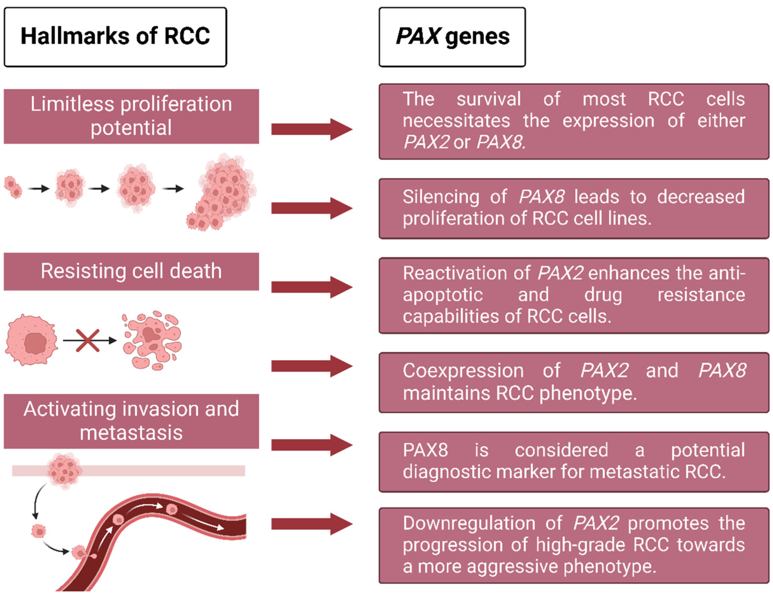
Figure 1. The hallmarks of RCC: exploring the potential role of PAX genes. PAX genes primarily participate in three hallmarks of RCC: cell survival, proliferation, and metastasis. Among these hallmarks, PAX2 and PAX8 are implicated in the transformation of RCC phenotype, either individually or cooperatively.
Ijms 25 06730 g001
Figure 2. VHL signaling participates in the regulation of cellular adaptation to hypoxia and contributes to reactivated PAX2 expression in ccRCC. During normoxic conditions, HIF-α undergoes hydroxylation by prolyl hydroxylases (PHD) and subsequently becomes recognized by the VHL protein. Upon binding of VHL to HIF-α, the latter undergoes ubiquitination and subsequent degradation through the proteasome machinery. Conversely, under hypoxic conditions, HIF-α remains un-hydroxylated, leading to its accumulation within the cytosol. This accumulated HIF-α then translocates into the nucleus, where it forms a heterodimer with its counterpart, HIF-β. The resultant complex drives the transcription of genes possessing a hypoxia-responsive element (HRE) within their promoters and further promotes the expression of their potential target genes (including VEGF, PDGF, CA-IX, TGFα, and PAX2). Furthermore, it is noteworthy that in ccRCC, PAX2 reactivation is driven by HIF-dependent mechanisms following pVHL loss [36].
Ijms 25 06730 g002
Figure 3. VHL-mediated signaling/regulatory pathways in ccRCC with respect to PAX involvement. The loss of VHL leads to HIFα accumulation and translocation into the nucleus, which subsequently activates the transcription of HIF target genes that are involved in critical oncogenic pathways (such as HIF-based hypoxia signaling pathway, TGF-β signaling pathway, VEGF-related signaling pathways, MYC-related signaling pathways, and Wnt/β-catenin signal pathway). Loss of VHL promotes the progression and metastasis of RCC, wherein PAX genes also exert a positive contribution.
Ijms 25 06730 g003
Table 1. PAX family members.
Groups* PAX GenesHLStructuralEAERRCReferences
IPAX120p11PD+OCTSkeleton, thymusNo or low expressionNetural or favourable[8,17,18]
PAX914q12PD+OCTSkeleton, craniofacial, toothNo or low expressionNetural or favourable[12,17,18]
IIPAX210q24PD+OCT+partial HDCNS, kidney, eye, earHigh expressionTumor-promoting[19,20,21,22]
PAX59p13PD+OCT+partial HDCNS, B cells, testisNo or low expressionNetural or favourable[8,12,25]
PAX82q12PD+OCT+partial HDCNS, kidney, thyroidHigh expressionTumor-promoting[8,10,27]
IIIPAX32q35PD+OCT+HDCNS, neural crest, skeletal muscleNo or low expressionNetural or favourable[8,12,23]
PAX71p36PD+OCT+HDCNS, craniofacial, skeletal muscleNo or low expressionNetural or favourable[8,12]
IVPAX47q32PD+HDCNS, PancreasNo or low expressionNetural or favourable[8,12,24]
PAX611p13PD+HDCNS, eye, pancreasNo or low expressionNetural or favourable[8,26]
* The PAX family may be categorized into four subgroups (Groups I–IV) based on the distinctive assembly of three structural motifs: the amino (N)-terminal paired domain (PD), the octapeptide (OCT), and the homeodomain (HD). Both the PD and HD motifs demonstrate DNA binding capabilities. The highly conserved PD motif comprises 128 amino acids and is ubiquitous among all PAX proteins. Group I (PAX1 and PAX9) entirely lack HD, while group IV members (PAX4 and PAX6) have a complete HD but lack an OCT domain. Group III members (PAX3 and PAX7) feature a complete HD and an OCT domain, whereas Group II members (PAX2, PAX5, and PAX8) possess a partial HD. HL: human chromosome location; EA: expression area; CNS: central nervous system; ER: expression levels in RCC; RC: RCC contribution.
Table 2. Strategies used for PAX-related therapeutic approaches in RCC.
PAXMethodsModelMain OutcomesAuthors, Year [Reference]
PAX2SiRNA inactivation of PAX2RCC cell linesPAX2 inactivation enhances cisplatin-induced apoptosis in renal carcinoma cellsHueber et al., 2006 [22]
PAX2shRNA targeting PAX2RCC cell lines and nude miceSubcutaneous ACHN/shPAX2 xenografts in nude mice respond better to cisplatin therapy than control ACHN tumorsHueber et al., 2008 [31]
PAX2Virtual screening and experimental validationRCC cell linesA small molecule inhibitor (EG1) targeting the DNA binding domain of PAX2Grimley et al., 2017 [132]
PAX8Large-scale functional genomic screens (ChIP-seq, RNA-seq, and ATAC-seq)RCC cell linesPAX8 silencing results in decreased proliferation of RCC cell linesBleu et al., 2019 [27]
PAX2Unbiased cell-based high-throughput screening assayRCC cell linesThree triazolo pyrimidine derivatives were identified that inhibited PAX-mediated transcriptional activation of reporter genes and suppressed proliferation of PAX2-positive RCC cells.Bradford et al., 2022 [133]
Disclaimer/Publisher’s Note: The statements, opinions and data contained in all publications are solely those of the individual author(s) and contributor(s) and not of MDPI and/or the editor(s). MDPI and/or the editor(s) disclaim responsibility for any injury to people or property resulting from any ideas, methods, instructions or products referred to in the content.