Review

The Role of the JAK–STAT Pathway in Childhood B-Cell Acute Lymphoblastic Leukemia

by
Karolina Joanna Ziętara
1,
Kinga Wróblewska
1,
Monika Zajączkowska
1,
Joanna Taczała
2 and
Monika Lejman
3,*
1
Student Scientific Society, Independent Laboratory of Genetic Diagnostics, Medical University of Lublin, 20-093 Lublin, Poland
2
Faculty of Medicine, Medical University of Warsaw, 02-091 Warszawa, Poland
3
Independent Laboratory of Genetic Diagnostics, Medical University of Lublin, 20-059 Lublin, Poland
*
Author to whom correspondence should be addressed.
Int. J. Mol. Sci. 2024, 25(13), 6844; https://doi.org/10.3390/ijms25136844
Submission received: 14 May 2024 / Revised: 16 June 2024 / Accepted: 17 June 2024 / Published: 21 June 2024
(This article belongs to the Section Molecular Immunology)

Abstract

:
B-cell lymphoblastic leukemia is a hematologic neoplasm that poses a serious health concern in childhood. Genetic aberrations, such as mutations in the genes IL-7, IL7R, JAK1, JAK2, TLSP, CRLF2, and KTM2A or gene fusions involving BCR::ABL1, ETV6::RUNX1, and PAX5::JAK2, often correlate with the onset of this disease. These aberrations can lead to malfunction of the JAK–STAT signaling pathway, which is implicated in various important biological processes, including those related to immunology. Understanding the mechanisms underlying the malfunction of the JAK–STAT pathway holds potential for research on drugs targeting its components. Available drugs that interfere with the JAK–STAT pathway include fludarabine, ruxolitinib, and fedratinib.

1. Introduction

Cancer is one of the major causes of death among children worldwide. According to the WHO, in 2022, leukemias accounted for 28.8% of all cancers in children under the age of 19, followed by brain tumors and non-Hodgkin’s lymphomas [1,2]. Acute lymphoblastic leukemia (ALL), which derives from precursor B (more than 80% of ALL) and T lymphocyte lineages, is the most common childhood leukemia [3,4]. The foundation for the development of this type of leukemia often involves genetic alterations including chromosomal rearrangements, hyperdiploidy, hypoploidy, sequence changes, and DNA copy number changes. Some of the most significant subtypes found in B-cell acute lymphoblastic leukemia (B-ALL) are MLL (KMT2A) rearrangements, ETV6::RUNX1 translocation, ETV6::RUNX1-like ALL, BCR::ABL1 Philadelphia chromosome ALL, Philadelphia chromosome-like ALL, ZNF384-rearranged ALL, PAX5-rearranged ALL and CRLF2-rearranged ALL [5,6,7]. Several of the genetic changes directly or indirectly affect the Janus kinase–signal transducer and transcription activator (JAK–STAT) pathway, the proper functioning of which determines the maintenance of stem cells, the process of differentiation and proliferation of immune cells, hematopoiesis, and the pro-inflammatory response. The various B-ALL genetic subtypes associated with the functioning of the JAK–STAT signaling pathway represent research potential regarding new therapeutic targets and methods in childhood leukemia [8,9]. The following article describes abnormalities in the mechanism of action of the JAK–STAT pathway implicated in selected genetic alterations associated with childhood B-ALL, alongside available and experimental therapeutic options.

2. Background

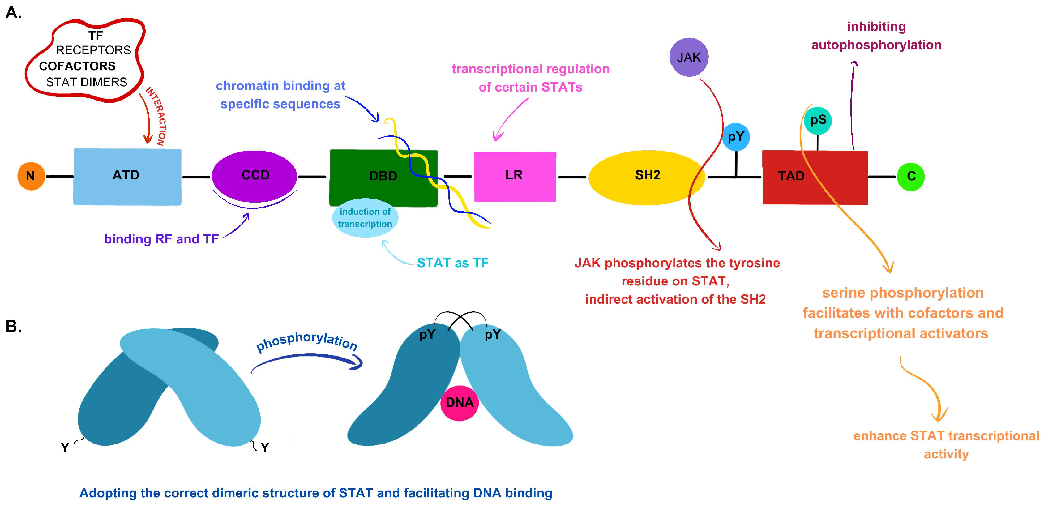
2.1. JAK–STAT Signaling Pathway Components and Their Primary Functions

The JAK–STAT signaling pathway holds significant importance in numerous biological processes within the human body, thus assuming a pivotal role in the pathogenesis of various diseases, notably including those affecting the immune system. This pathway comprises three principal components: ligand–receptor complexes, Janus kinases (JAKs), and signal transducers and activators of transcription (STATs) [10,11].
Either cytokines or growth factors as ligands can engage tyrosine kinase-associated receptors, leading to ligand–receptor complexes. The binding of a ligand to its receptor activates JAK, facilitated by ligand-mediated receptor multimerization. The proximity of JAKs enables trans-phosphorylation. The activated JAKs then phosphorylate specific tyrosine residues on the receptor’s intracellular domain. Subsequently, STAT proteins are recruited, activated, and phosphorylated on tyrosine residues by the JAKs [11,12,13]. In mammals, four kinases belong to the Jak family: Jak1, Jak2, Jak3, and Tyrosine kinase 2 (Tyk2). The genes encoding these kinases in humans are located on different chromosomes. Specifically, the JAK1 gene is located on chromosome 1p.31.3, and JAK2 is on 9p24.1, while Jak3 and TYK2 are clustered on chromosome 19p13.11 and chromosome 19p13.2, respectively. These kinases are generally ubiquitously expressed, except for JAK3, which is mainly expressed in hematopoietic cells [14,15,16,17,18,19]. JAKs consist of four domains in the following order: N-terminal FERM (band 4.1, Ezrin, Radixin, Moesin) domain, SH2 domain, pseudokinase domain (JAK Homology 2, JH2), and C-terminal kinase domain (JH1). The first two domains, FERM and Src-homology 2 (SH2), are tightly associated and form a cohesive unit linked to the peptide receptor and its Box1 and Box2 motifs. Their primary function is to bind JAKs to the cytokine receptor. The subsequent domain, the pseudokinase, exhibits catalytic inactivity, while it binds ATP and assumes a pivotal role in modulating the activity of the C-terminal kinase domain. The last domain is active and has catalytic activity, being responsible for substrate phosphorylation of the cytokine receptor, thereby activating STAT transcription factors [20,21,22,23].
The third component of the JAK/STAT pathway consists of inactive protein dimers that are located in the cytoplasm and belong to the STAT family. Seven STAT subtypes have been identified in mammals: STAT1, STAT2, STAT3, STAT4, STAT5a, STAT5b, and STAT6. Upon the initiation of signaling by various ligands, such as cytokines, growth factors, interleukins, and hormones, these proteins become activated. Their role involves transmitting signals from the ligands and promoting the transcription of specific genes. [20,24]. Each STAT is composed of distinct domains arranged in the following order: the N-terminal domain, the coiled-coil domain (CCD), the DNA binding domain (DBD), the linker region, the SH2 domain, and the C-terminal region containing the transactivation domain (TAD) [20,25,26]. The N-terminal domain is responsible for interacting with transcription factors, cofactors, receptors, and STAT dimers by binding to the DNA regions where the binding sites of STAT proteins are located [20,25]. The adjacent CCD domain contains a surface to which proteins, among other regulatory factors, attach [26]. The DBD is responsible for chromatin binding and induction of transcription, with STAT proteins acting as transcription factors [20,26]. The region connecting this domain and the SH2 domain is known as the linker region, serving as the bridge between them and participating in the transcriptional regulation of certain STAT proteins [20,25,26]. JAK phosphorylates the tyrosine residue (pY) on STAT molecules, thereby indirectly activating the SH2 domain through the recognition of phosphotyrosine motifs within cytokine receptors. The active SH2 domain serves as a dimerization site for two STAT proteins. Following homo- or heterodimerization, they are translocated to the cell nucleus, where they initiate gene transcription by binding to gamma-activated sequences (GASs). Within the TAD, serine phosphorylation occurs, facilitating interactions with cofactors and transcriptional activators, thereby enhancing STAT transcriptional activity. Moreover, the TAD is responsible for regulating protein stability by inhibiting autophosphorylation [26,27].
Figure 1 shows all STAT domains and their functions.

2.2. The Regulation of the JAK–STAT Signaling Pathway

Regulation of the JAK–STAT signaling pathway relies on efficient activation following cytokine stimulation and inhibition of subsequent signaling in its absence [22].

2.2.1. Signaling Activation via Cytokines

The activation of the JAK–STAT signaling pathway involves molecules such as cytokines, interferons, or growth factors interacting with JAKs. The binding of these substances induces multimerization or conformational changes within the receptor complex, leading to the activation of JAKs. These processes initiate a cascade of events where the phosphorylation of STAT proteins by active JAKs assumes a pivotal role. Subsequently, the phosphorylated STAT proteins dimerize and translocate into the nucleus [12,22].
Various cytokines activate different JAKs and STAT proteins. Table 1 illustrates the specific cytokine types interacting with distinct JAKs and STAT proteins, outlining their major functions in B-cell lineage development.

2.2.2. Negative Regulation of the JAK–STAT Pathway

The negative regulation of the JAK–STAT signaling pathway involves three main groups of inhibitory factors, namely, the suppressor of cytokine signaling (SOCS), protein inhibitor of activated STAT (PIAS), and protein tyrosine phosphatase (PTP) groups [12].
The first group comprises eight proteins: cytokine-inducible SH2-containing protein (CIS), SOCS1, SOCS2, SOCS3, SOCS4, SOCS5, SOCS6, and SOCS7. These proteins negatively regulate the JAK–STAT signaling pathway through several mechanisms. Firstly, they prevent the recruitment of the SH2-STAT domain to the cytokine receptor by binding to the phosphotyrosine residue on this receptor. Additionally, they directly inhibit JAK activity by either binding to JAK itself or recruiting the SH2 domain to the receptor [37,38,39,40].
Another group, the PIAS group, consist of four proteins: PIAS1, PIAS2 (PIASx), PIAS3, and PIAS4 (PIASy), which interact with STAT dimers following JAK activation. PIAS proteins employ several specific mechanisms, including blocking STAT proteins and their DNA-binding activity, promoting SUMOylation (Small Ubiquitin-related Modifier), chelating transcription factors, and recruiting other cofactors to prevent STAT from binding to DNA [9,41,42].
The third group of negative regulators of the JAK–STAT pathway are PTP enzymes, which act by dephosphorylating tyrosine residues. Their targets for dephosphorylation may include active STAT or JAK proteins. By mediating the dephosphorylation and subsequent inactivation of JAK, these enzymes can also interact with the ligand–receptor complex [9,43].

3. Selected Genes Associated with Disorders of the JAK/STAT Pathway
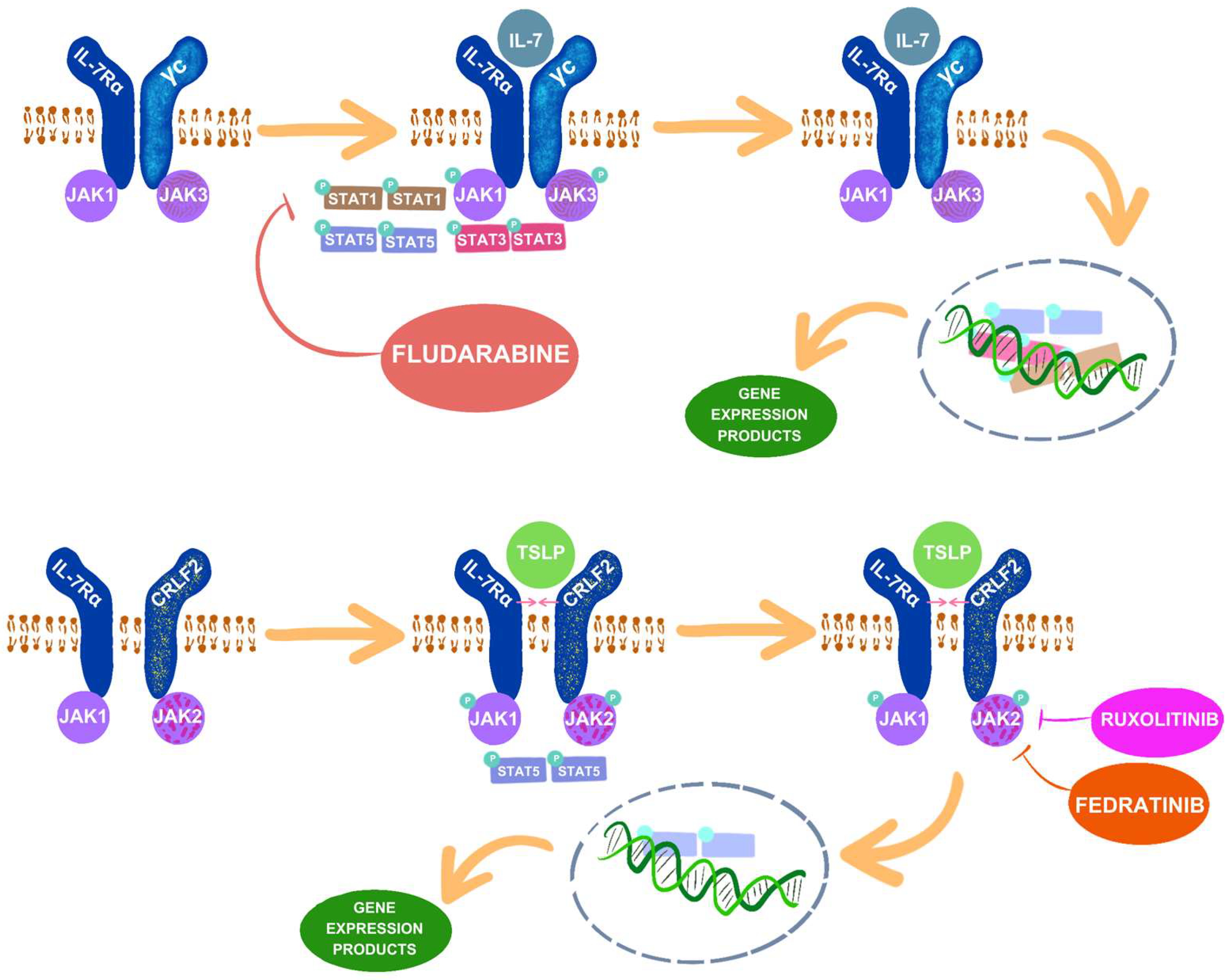
3.1. IL-7 and IL-7R

The maturation of B lymphocytes is facilitated by multiple cytokines that transmit signals to their precursors through specific receptors. Errors and imbalances in signaling during early B-cell development at various stages can disrupt the sequential progression and ultimately promote the development of ALL from B precursor cells [44]. Among these cytokines, Interleukin-7 (IL-7) plays a crucial role in hematopoiesis. Its receptor comprises the γc chain, shared by all lymphopoietic cytokines, and the interleukin-7 alpha receptor subunit (IL-7Rα), which is encoded by the IL-7R gene located on the short arm of chromosome 5 (5p13.2) [45,46,47,48].
IL-7Rα, unlike its counterpart with which it forms a receptor complex, exhibits greater specific expression and is present, among others, on the surfaces of cells ranging from common lymphoid progenitors (CLPs) through pro-B to large pre-B cells and their T-cell counterparts. Positioned in this manner, IL-7 plays a pivotal role in the transition of pro-B cells to pre-B cells, and animal studies suggest that inactivation of one of the downstream cytokine signaling effectors may lead to the retention of B-cells in the pre-pro-B stage. Within hematopoietic cells, IL-7 primarily transmits information through three pathways: the JAK–STAT pathway, the phosphatidylinositol 3-kinase (PI3K)–Akt pathway, and the RAS–mitogen-activated protein kinase (MAPK) pathway [45,48].
The binding of IL-7 to its receptor induces heterodimerization and conformational changes in the constituent chains of IL-7R (IL-7Rα, γc). Consequently, constitutively bound tyrosine kinases JAK1 and JAK3 are activated, leading to their mutual phosphorylation. Subsequently, these activated JAK proteins catalyze the phosphorylation of a tyrosine residue within the cytoplasmic domain of IL-7Rα, creating a docking site for STAT1, STAT3, and STAT5. Among these, STAT5 plays a pivotal role in lymphopoiesis. Upon binding to IL-7Rα, STAT5 undergoes phosphorylation and homodimerization, translocating to the nucleus [45,47,49,50].
One of the consequential effects is the activation of the myeloid leukemia 1 (Mcl1) gene, which serves as a crucial regulator of the survival of B lymphocytes in early developmental stages, thereby inhibiting their apoptosis [51]. Furthermore, STAT5 is implicated in coordinating immunoglobulin gene rearrangements, thereby influencing Igk recombination in pro-B cells [45,51].
Therefore, IL-7’s influence through the JAK–STAT pathway on the early stages of lymphopoiesis profoundly shapes the subsequent development of B-cells. This process is further modulated by the E2A and EBF1 transcription factors [45,52].
Mutations in the IL-7 receptor gene can significantly disrupt lymphocyte development due to the pivotal roles played by IL-7. These mutations typically possess activating properties; by causing homodimerization of IL-7Rα, they augment its effects and consequently enhance JAK–STAT5 signaling. Aberrations involving the substitution of serine with cysteine at the 185th amino acid position in the extracellular domain or insertions or deletions in the transmembrane domain of IL-7R account for approximately 12% of cases of Ph-like ALL and 2–3% of B-ALL cases [47,53]. Frequently, these mutations coexist with loss-of-function mutations in the negative regulator SH2B adapter protein 3 (SH2B3), also known as lymphocyte adapter protein (LNK), or alterations in the DNA-binding protein Ikaros, also known as Ikaros family zinc finger protein 1 (IKZF2) [54,55].
According to a study by Ifat Geron et al., mutated IL-7Rα in human hematopoietic cells serves as the initiator of a pre-leukemic state, as observed in mice with transplanted cells harboring mutated IL-7Rα. This mutation contributes to the silencing of cyclin-dependent kinase inhibitor 2A (CDKN2A) and the aberrant expression of cytokine receptor-like factor 2 (CRLF2) in the development of BCP-ALL [53,56].

3.2. TLSP and CRLF2

Thymic stromal lymphopoietin (TSLP) is a type I cytokine belonging to the IL-2 family, similar to IL-7 in its effects and signaling mechanisms [57]. Similar to its counterpart, TSLP regulates the immune response and influences B-cell development by augmenting the proliferation of their fetal precursors. TSLP signals through cytokine receptor-like factor 2 (CRLF2), located in the pseudoautosomal region (PAR1) of the X and Y chromosomes, and the IL-7Rα subunit, which it shares with IL-7. Consequently, both cytokines activate identical effectors through a shared signaling pathway. Analogous to the IL-7 receptor, the TSLP receptor is made of components that are constitutively associated with members of the Janus kinase family: IL-7Rα with JAK1 and CRLF2 with JAK2. The binding of TSLP to both receptor subunits induces their heterodimerization, subsequently activating JAK1 and JAK2, which then phosphorylate the transcriptional regulator STAT5 [45,57,58,59,60,61,62,63,64].
Abnormalities that cause changes in signaling and significantly impact B-ALL development may affect each of the pathway components mentioned. They can be categorized into five main subgroups: gain-of-function mutations in CRLF2; mutations conferring JAK1 function (typically associated with T-ALL); mutations conferring JAK2 function; mutations activating the extracellular domains of IL-7Rα, leading to heterodimer formation with CRLF2; and mutations in the transmembrane domain of IL-7Rα, resulting in ligand-independent homodimerization [65].
Figure 2 shows the effect of IL-7 and TLSP on the activation of the JAK–STAT pathway and selected drugs that inhibit components of the pathway.

3.2.1. Enhancement of CRLF2 Function

Mutations in this subgroup often coincide with JAK2 abnormalities, ultimately leading to the constitutive activation of the crucial transcriptional regulator STAT5, which exists in two related isoforms, STAT5A and STAT5B, each with distinct functions. Alternations involving CRLF2 and their impact on STAT5 can induce cytokine-independent survival and proliferation of early hematopoietic cells, potentially contributing to the development of B-cell precursor acute lymphoblastic leukemia (BCP-ALL) [45].
The most prevalent causes of CRLF2 overexpression include rearrangements involving the immunoglobulin heavy chain (IGH) and P2Y receptor family member 8 (P2RY8) genes. Other changes associated with CRLF2 overexpression include deletions in the Ikaros gene, activating mutations in JAK2, point mutations such as Phe232Cys, additional copies of the X or Y chromosome or the CRLF2 gene, and rare fusions such as CSF2RA–CRLF2. CSF2RA is located in the pseudoautosomal region of the X and Y chromosomes and encodes a protein that serves as the alpha subunit of colony-stimulating factor 2, a cytokine that controls the production, differentiation, and function of granulocytes and macrophages [66,67].
Translocation between IGH on chromosome 14 and the pseudoautosomal region of the sex chromosome, where the CRLF2 gene resides, results in the formation of an IGH–CRLF2 fusion. The P2RY8–CRLF2 fusion arises from the deletion of the promoter region of P2RY8. Both genes are located in the same chromosomal region on the X or Y chromosome, making CRLF2 expression reliant on the P2RY8 promoter, which confers oncogenic potential [58,68,69]. Conversely, diminished CRLF2 expression due to deletion of colony-stimulating factor 2 receptor alpha (CSF2RA) leads to the gene being controlled by CSF2RA, a low-affinity enhancer [66,67]. The Phe232Cys substitution mutation in the extracellular domain of CRLF2 enhances its function, leading to constitutive homodimerization of CRLF2 and activating components of the JAK–STAT pathway [61,65,70].

3.2.2. Clinical Significance of CRLF2 Mutations

CRLF2 overexpression resulting from the described genetic abnormalities is detected in 15% of children with high-risk B-ALL and 47% of pediatric patients with Ph-like B-ALL [55,71]. Therefore, high expression is considered one of the characteristic features of this subtype of ALL and, according to some researchers, has the potential to be a prognostic factor when making decisions regarding treatment selection [70,72]. There is a correlation between impaired gene expression and the ethnic origin of patients. For example, in families with Latin roots, abnormalities in the CRLF2 gene are detected five times more often than in other patients [58]. The prognosis of B-ALL in children depends on mutations in leukemic cell genes. The overexpression of CRLF2, which leads to constitutive activation of JAK2–STAT5, correlates with high-risk BCP-ALL and promotes poorer recurrence-free survival rates in pediatric patients [68,73,74]. Moreover, Naglaa M. Hassan and colleagues reported that patients with high CRLF2 expression are characterized by a greater number of blasts in peripheral blood and severe organomegaly [68,75]. However, mutations causing the activation of CRLF2 may also be receptor-independent prognostic factors in leukemia. For example, the combination of P2RY8–CRLF2 fusion and the associated deletion in the Ikaros gene (IKZF1) may increase the risk of leukemia relapse [76].

3.3. JAK1 and JAK2

The JAK2 gene is located on the short arm of chromosome 9 (9p24.1) and encodes a non-receptor tyrosine kinase pivotal in cytokine signaling by binding to both type I and type II cytokine receptors. Upon ligand binding, both classes of receptors undergo dimerization, facilitating transphosphorylation and activation of JAK2. Activated JAK2, via autophosphorylation of tyrosine residues on the cytoplasmic receptor, creates anchoring sites for proteins with an SH2 domain, including STAT [17,77,78].
JAK2 activation, induced by various cytokines, is crucial for proper cell function, especially in hematopoiesis [77]. JAK2 plays a fundamental role in maintaining and regulating hematopoietic stem cell (HSC) function. It associates with the thrombopoietin receptor (TPOR), governing megakaryocyte and platelet production, as well as with EPOR, GM-CSFR, G-CSFR, IL-3R, and IL-5R, making it vital for myeloid-lineage cell differentiation. Additionally, the erythropoietin receptor (EPOR) and growth hormone receptor (GHR) are homodimeric and bind solely to JAK2 [23].
JAK2 gain-of-function mutations, commonly found in ALL, typically occur in exon 16, predominantly around amino acid R683, which is altered to glycine or serine [23,78]. These mutations within the ATP-binding site of the JAK2 pseudokinase domain destabilize the inhibitory JH2–JH1 interface, disrupting autoinhibition mediated by JH2. This exposure of an interface facilitates JAK2-related cytokine receptor dimerization and subsequent activation [77,78,79].
Notably, JAK2 mutations alone, disrupting JH2-mediated autoinhibition, may not suffice for sustained cytokine-independent JAK2 activation [22,77,79]. The lymphoid transformation is proposed to be driven by the high association of JAK1/2 and CRLF2 mutations [77,80].

Clinical Significance of JAK2 Mutations

All cases of B-ALL with changes in JAK2 are accompanied by CRLF2 overexpression, facilitating the cytokine-receptor scaffolding necessary for this signaling [81]. However, it is important to note that there are various causes of CRLF2 overexpression, and they may not always coincide with JAK abnormalities. JAK mutations, particularly JAK2, are identified in around 10% of BCP-ALL cases. This percentage increases to over 20% in patients with Down syndrome who also have ALL [23,82,83]. It is noteworthy that JAK2 mutations occur more frequently in patients diagnosed with Ph-like ALL, a high-risk subtype characterized by gene expression patterns resembling Ph+ ALL, despite the absence of the chromosomal translocation t(9;22)(q34;q11) [58,59]. Among subjects showing CRLF2 changes simultaneously with JAK2 variants, poorer treatment outcomes for ALL are observed [84,85].

3.4. BCR::ABL1 Fusion

The BCR::ABL1 oncoprotein associated with Ph+ ALL directly and independently activates the JAK2–STAT pathway. The BCR gene, located on the long arm of chromosome 22 (22q11.2), encodes the breakpoint cluster region protein, regulating cellular signaling processes. Conversely, the ABL1 gene, a proto-oncogene situated on the long arm of chromosome 9 (9q34.1), encodes a protein tyrosine kinase that influences cell division, adhesion, differentiation, and stress responses [86,87].
A reciprocal translocation of chromosomes 9 and 22, t(9;22)(q34;q11), results in the Philadelphia chromosome (Ph), observed in the transformed HSCs, and the formation of the BCR::ABL fusion gene [86,87]. Depending on the site of the break, three protein isoforms are distinguished: a variant with a molecular weight of 185–190 kDa (p190), commonly found in ALL; a variant with a mass of 210 kDa (p210), prevalent in chronic myeloid leukemia (CML); and the rarest variant, with the highest molecular weight of 230 kDa (p230) [87]. The constitutively activated ABL1 catalytic domain, due to fusion with BCR, drives BCR–ABL1 activity, enabling direct phosphorylation of cytokine receptors by the kinase protein, thereby activating the JAK2/STAT pathway [86,88,89]. Gene fusion alters JAK2–STAT pathway components’ activity by directly affecting STAT5 or mediating JAK2 phosphorylation independently. Both actions ultimately enhance signaling, promoting cell proliferation and increased survival [90,91]. Moreover, studies by Run Qina et al., using mouse models, revealed altered functions of STAT6 in the context of the described translocation. The activated cell cycle regulator enhances c-Myc transcription, intensifying the pathogenicity of ALL cells and worsening the prognosis. Their research also distinguished Ph+ ALL from Ph+ CML based on the subtypes of the BCR::ABL fusion protein, P190 and P210, respectively. They concluded that Ph+ ALL exhibits more pronounced activation of the JAK2–STAT6 pathway than Ph+ CML [92].

3.5. ETV6::RUNX Fusion

The t(12;21)(p13;q22) translocation results in the fusion of two genes: ETV6 (also known as TEL) and RUNX1 (also known as AML1) [93]. ETV6 encodes an ETS family transcription factor crucial for hematopoiesis and vascular network development [94]. In contrast, RUNX1 encodes a protein essential for normal hematopoiesis [95]. This fusion represents the most common genetic alteration in ALL and has various functional effects, including repression of RUNX1-dependent transcription, disruption of wild-type ETV6 activity, induction of EPOR expression, and activation of STAT3 through phosphorylation [93,96,97,98,99]. It disrupts the JAK–STAT pathway by overactivation of STAT3, typically phosphorylated by JAK2 [99]. In leukemias, the visible overactivation of STAT3 accelerates leukemia cell proliferation, blocks leukemia cell differentiation, and inhibits apoptosis of leukemia cells [100,101].
As mentioned before, the fusion of these genes is often present in leukemias; however, the presence of the fusion alone does not lead to the occurrence of these diseases [102]. The genetic alternation occurring in B-ALL with this fusion is EPOR mutation, which has a crucial effect on the development of leukemia. This gene encodes the receptor responsible for binding erythropoietin but also for the activation of JAK2 tyrosine kinase [103,104]. Overexpression of ETV6::RUNX fusion and the presence of EPO simultaneously in cells result in the activation of STAT5. Additionally, this process is JAK2-dependent and cannot happen in the presence of a JAK2 inhibitor [102].

3.6. KMT2A

KMT2A (also known as MLL), located on human chromosome 11q23, encodes a lysine methyltransferase serving as a transcriptional coactivator essential in regulating gene expression during early organism growth, formation, and hematopoiesis [105,106]. Mutation of KMT2A involves translocations involving certain genes, particularly AFF1, MLLT3, and MLLT1, leading to the formation of oncogenic chimeric proteins [107]. KMT2A mutations occur in 5% of ALL cases, resulting in mixed-lineage leukemia rearrangement (MLL-r), cases of which exhibit worse outcomes than non-MLL cases [108,109]. These differences include increased white blood cell counts at onset, insensitivity to traditional chemotherapeutic agents, low complete remission rates, a relatively high incidence of central nervous system (CNS) involvement, and shortened survival. MLL-r ALL depends on JAK/STAT-mediated inflammatory signals in leukemia development [107,108,109].

3.7. PAX5::JAK2

PAX5, located on chromosome 9p13, is a transcription factor belonging to the paired box (PAX) family, critical in early organism development. Changes in its gene expression are implicated in cancer development. Specifically, this gene encodes a protein activating the B-cell lineage and is active in early B-cell development stages, not in later ones [110].
Rearrangement of PAX5 is observed in 2.5% of B-cell precursor ALL, with particular emphasis on the PAX5::JAK2 fusion, which plays an important role in tumorigenesis [111,112]. This fusion produces a protein that includes the PAX5 DNA-binding region and the constitutively active JAK2 kinase domain. It disrupts the normal transcriptional program regulated by PAX5 while also initiating the JAK–STAT signaling pathway by enhancing STAT5 expression. This dual effect on crucial pathways suggests a potential mechanism facilitating leukemia development [113].

4. JAK/STAT Inhibitors as Therapeutic Targets

JAK–STAT signaling is one of the key mediators of information transfer, tasked with activating transcription proteins engaged in performing various roles, including those related to lymphopoiesis. Given the significant link between abnormal JAK–STAT signaling and B-ALL development, ongoing research is being conducted on the use of transducer components as therapeutic targets. Targeting JAK proteins, which initiate the activation of transcription regulators, holds promise for achieving improved therapeutic outcomes and potentially reducing treatment-related side effects in patients. The effect exerted by JAK inhibitors on cells is to block the active sites of kinases, which prevents proper signal transduction and abolishes the effects caused by cytokines [114].
The therapeutic potential of JAK inhibitors has been substantiated by ex vivo studies on cell lines, demonstrating that inhibiting JAK and thereby preventing STAT activation leads to restricted cell development and increased apoptosis [114,115].
JAK inhibitors can be divided based on the selectivity of action (selective or non-selective) as well as the method of binding and type of interaction with amino acids in the Janus kinase. The second classification method includes reversible inhibitors, called competitive inhibitors (divided into allosteric JAK inhibitors and ATP-competitive inhibitors) and irreversible, or covalent, inhibitors (their target is JAK3, which is associated with T-ALL and therefore will not be discussed). Notable examples of competitive inhibitors are ruxolitinib and the newer-generation drug fedratinib, both well known for their therapeutic potential in ALL [116,117].

4.1. Ruxolitinib

Ruxolitinib is a tyrosine kinase inhibitor (TKI) that selectively targets subtypes JAK1 and JAK2 by competing with ATP in the catalytic sites of the aforementioned kinases [117]. It is administered orally and is approved for various treatments, including myelofibrosis, polycythemia vera, and steroid-refractory graft-versus-host disease in the setting of allogeneic stem-cell transplantation [118]. Ruxolitinib acts by attenuation of the inflammatory state caused by constitutive JAK/STAT activation, and it also induces nonspecific myelosuppression [119]. Its role as a TKI has shown potential as a treatment option for various types of leukemias characterized by JAK–STAT pathway aberrations [120].
A study conducted by Downes et al. presents results from the treatment of 12 patients with Ph-like ALL with ruxolitinib. In all the patients, there was a gene mutation leading to activation of the JAK–STAT pathway (e.g., gene fusion affecting JAK2, overexpression of CRLF1, etc.). The majority of them (n = 9) achieved completed or partial remission [121].
Ruxolitinib has also been used in treatment of MLL-r, specifically in cases with mutation of the KMT2A gene [109]. This type of leukemia is associated with poor prognosis, and few treatment options are available [109]. Laboratory data confirm the positive effects of the use of ruxolitinib in MLL-r leukemia, but further clinical research must be provided before it is used generally in clinical practice [109].
Despite promising results from numerous studies, in vitro models suggest that administering ruxolitinib as monotherapy may lead to the development of mutations conferring resistance to the drug or cross-resistance to multiple JAK-inhibitor therapies. Data from murine models suggested that cells acquired JAK2 kinase domain mutations that were resistant to multiple type I JAK inhibitors after targeted monotherapy. Therefore, ruxolitinib should be considered only as an adjunct to standard ALL treatment [121]. Nevertheless, the use of ruxolitinib as an additional drug may result in an enhanced effect in the treatment of Ph-like ALL because of its synergistic interaction with certain anti-leukemic drugs. A study performed by Böhm et al. in 2021 [85] revealed that the best effect was attainable with glucocorticoids, topoisomerase I and II inhibitors, microtubule-targeting agents, and antimetabolites.

4.2. Fedratinib

Fedratinib, an oral selective JAK2 inhibitor targeting FMS-like tyrosine kinase-3 (FLT-3), was approved by the Food and Drug Administration (FDA) in 2019 for the treatment of primary or secondary myelofibrosis, eight years after the introduction of ruxolitinib [116,122]. The use of fedratinib, this time in B-ALL, was tested in combination with venetoclax in cell lines that were then injected into mice. Venetoclax is a selective B-cell lymphoma 2 (Bcl-2) protein inhibitor with anti-apoptotic properties, which play an important role in high-risk B-ALL, especially with KMT2A mutations [123]. The combination of these two drugs has made it possible to achieve better effects, with the exception of cell lines that do not express the Flt3 tyrosine kinase, responsible for promoting growth and inhibiting apoptosis in the early stages of hematopoiesis. The conducted research proved the validity of combining the two inhibitors in order to achieve enhanced anti-leukemic effects and showed the correlation between the effect of the drugs and Flt3 overexpression, which often occurs in B-ALL with poor prognosis [123,124]. Given that the phosphorylation of STATs and their movement from the cytosol to the nucleus triggers the transcriptional activation of specific genes, there has been interest in inhibiting this mechanism. For this reason, research has begun on STAT inhibitors that may target the SH2 domain, mRNA-binding domain or DNA-binding domain. Currently, the only FDA-approved agent for the leukemia treatment is a STAT1 inhibitor, a purine analogue called fludarabine [114,125]. This drug is administered intravenously in the form of a monophosphate prodrug (F-ara-AMP), which is quickly converted into an intracellularly distributed metabolite: F-ara-A. In the next step, intracellular phosphorylation occurs, leading to the formation of the active metabolite fludarabine triphosphate, which is incorporated into DNA and RNA, thus inhibiting the synthesis of these nucleic acids [126]. The precise mechanism and effect of fludarabine on STAT1 are not fully elucidated; however, it has been shown to reduce STAT1 phosphorylation, thereby inhibiting its activity [127,128].
Additionally, fludarabine was combined with cyclophosphamide in a lymphodepletion regimen prior to chimeric CD19 antigen receptor (CAR) T-cell therapy. Research has demonstrated that the combination of these drugs resulted in significant improvements in outcomes in patients with relapsed or refractory B-ALL [128,129,130].

5. Conclusions

Over the years, there has been notable progress in the development of both diagnostic modalities and therapeutic protocols for pediatric B-ALL, resulting in considerable achievements. This advancement has manifested in an impressive overall survival rate of 90% among affected children. However, despite these strides, substantial challenges persist within this domain, and the trajectory of medical innovation remains uninterrupted. This is exemplified by ongoing endeavors to conceptualize novel therapeutic interventions targeting the JAK–STAT signaling pathway, which is unequivocally implicated in the pathogenesis of B-ALL.
Independent mutations affecting constituents of this pathway, alongside those implicating its associated genes, engender dysregulation of gene expression and cellular processes. Such perturbations contribute significantly to the initiation, progression, and prognostication of leukemia. Consequently, JAK–STAT transducers and effectors, ubiquitous across multiple signaling cascades, emerge as auspicious targets for inhibitors such as ruxolitinib, fedratinib, and fludarabine. These agents hold considerable promise for incorporation into the therapeutic arsenal against B-ALL characterized by the aforementioned genetic alterations. Continued research and clinical trials will be crucial in harnessing their full potential and improving outcomes for pediatric patients affected by this challenging disease.

Author Contributions

Conceptualization, K.J.Z. and M.L.; writing—original draft preparation and editing, K.J.Z., K.W., M.Z. and J.T.; visualization, K.J.Z. and M.L.; supervision, M.L. All authors have read and agreed to the published version of the manuscript.

Funding

This research received no external funding.

Conflicts of Interest

The authors declare no conflicts of interest.

References

  1. Huang, J.; Chan, S.C.; Ngai, C.H.; Lok, V.; Zhang, L.; Lucero-Prisno, D.E., III; Xu, W.; Zheng, Z.-J.; Elcarte, E.; Withers, M.; et al. Global Incidence, Mortality and Temporal Trends of Cancer in Children: A Joinpoint Regression Analysis. Cancer Med. 2023, 12, 1903–1911. [Google Scholar] [CrossRef] [PubMed]
  2. Estimated Number of Prevalent Cases (5-Year), Both Sexes, Age [0–19], in 2022. Available online: https://gco.iarc.fr/today/en/dataviz/pie-prevalence?populations=900&mode=cancer&prev_time=5&types=2&group_populations=1&multiple_populations=0&age_end=3 (accessed on 4 June 2024).
  3. Onyije, F.M.; Olsson, A.; Baaken, D.; Erdmann, F.; Stanulla, M.; Wollschläger, D.; Schüz, J. Environmental Risk Factors for Childhood Acute Lymphoblastic Leukemia: An Umbrella Review. Cancers 2022, 14, 382. [Google Scholar] [CrossRef] [PubMed]
  4. Al Shamahy, H.A. Prevalence of Different Types of Leukemia and Associated Factors among Children with Leukemia in Children’s Cancer Units at Al-Kuwait Hospital, Sana’a City: A Cross-Sectional Study. Glob. J. Pediatr. Neonatal. Care 2021, 3, 000569. [Google Scholar] [CrossRef]
  5. Lejman, M.; Chałupnik, A.; Chilimoniuk, Z.; Dobosz, M. Genetic Biomarkers and Their Clinical Implications in B-Cell Acute Lymphoblastic Leukemia in Children. Int. J. Mol. Sci. 2022, 23, 2755. [Google Scholar] [CrossRef] [PubMed]
  6. Tebbi, C.K. Etiology of Acute Leukemia: A Review. Cancers 2021, 13, 2256. [Google Scholar] [CrossRef] [PubMed]
  7. Malard, F.; Mohty, M. Acute Lymphoblastic Leukaemia. Lancet 2020, 395, 1146–1162. [Google Scholar] [CrossRef] [PubMed]
  8. Agashe, R.P.; Lippman, S.M.; Kurzrock, R. JAK: Not Just Another Kinase. Mol. Cancer Ther. 2022, 21, 1757–1764. [Google Scholar] [CrossRef] [PubMed]
  9. Hu, X.; Li, J.; Fu, M.; Zhao, X.; Wang, W. The JAK/STAT Signaling Pathway: From Bench to Clinic. Signal Transduct. Target. Ther. 2021, 6, 402. [Google Scholar] [CrossRef] [PubMed]
  10. Valle-Mendiola, A.; Gutiérrez-Hoya, A.; Soto-Cruz, I. JAK/STAT Signaling and Cervical Cancer: From the Cell Surface to the Nucleus. Genes 2023, 14, 1141. [Google Scholar] [CrossRef]
  11. Imada, K.; Leonard, W.J. The Jak-STAT Pathway. Mol. Immunol. 2000, 37, 1–11. [Google Scholar] [CrossRef]
  12. Xin, P.; Xu, X.; Deng, C.; Liu, S.; Wang, Y.; Zhou, X.; Ma, H.; Wei, D.; Sun, S. The Role of JAK/STAT Signaling Pathway and Its Inhibitors in Diseases. Int. Immunopharmacol. 2020, 80, 106210. [Google Scholar] [CrossRef] [PubMed]
  13. Liongue, C.; O’Sullivan, L.A.; Trengove, M.C.; Ward, A.C. Evolution of JAK-STAT Pathway Components: Mechanisms and Role in Immune System Development. PLoS ONE 2012, 7, e0032777. [Google Scholar] [CrossRef] [PubMed]
  14. Yamaoka, K.; Saharinen, P.; Pesu, M.; Holt, V.E.T.; Silvennoinen, O.; O’Shea, J.J. The Janus Kinases (Jaks). Genome Biol. 2004, 5, 253. [Google Scholar] [CrossRef] [PubMed]
  15. Rane, S.G.; Reddy, E.P. Janus Kinases: Components of Multiple Signaling Pathways. Oncogene 2000, 19, 5662–5679. [Google Scholar] [CrossRef] [PubMed]
  16. JAK1 Janus Kinase 1 [Homo Sapiens (Human)]. Available online: https://www.ncbi.nlm.nih.gov/gene/3716 (accessed on 4 June 2024).
  17. JAK2 Janus Kinase 2 [Homo Sapiens (Human)]. Available online: https://www.ncbi.nlm.nih.gov/gene/3717 (accessed on 4 June 2024).
  18. JAK3 Janus Kinase 3 [Homo Sapiens (Human)]. Available online: https://www.ncbi.nlm.nih.gov/gene/3718 (accessed on 4 June 2024).
  19. TYK2 Tyrosine Kinase 2 [Homo Sapiens (Human)]. Available online: https://www.ncbi.nlm.nih.gov/gene/7297 (accessed on 4 June 2024).
  20. Morris, R.; Kershaw, N.J.; Babon, J.J. The Molecular Details of Cytokine Signaling via the JAK/STAT Pathway. Protein Sci. 2018, 27, 1984–2009. [Google Scholar] [CrossRef]
  21. Ferrao, R.; Lupardus, P.J. The Janus Kinase (JAK) FERM and SH2 Domains: Bringing Specificity to JAK–Receptor Interactions. Front. Endocrinol. 2017, 8, 71. [Google Scholar] [CrossRef] [PubMed]
  22. Hammarén, H.M.; Virtanen, A.T.; Raivola, J.; Silvennoinen, O. The Regulation of JAKs in Cytokine Signaling and Its Breakdown in Disease. Cytokine 2019, 118, 48–63. [Google Scholar] [CrossRef]
  23. Raivola, J.; Haikarainen, T.; Abraham, B.G.; Silvennoinen, O. Janus Kinases in Leukemia. Cancers 2021, 13, 800. [Google Scholar] [CrossRef]
  24. Rah, B.; Rather, R.A.; Bhat, G.R.; Baba, A.B.; Mushtaq, I.; Farooq, M.; Yousuf, T.; Dar, S.B.; Parveen, S.; Hassan, R.; et al. JAK/STAT Signaling: Molecular Targets, Therapeutic Opportunities, and Limitations of Targeted Inhibitions in Solid Malignancies. Front. Pharmacol. 2022, 13, 821344. [Google Scholar] [CrossRef]
  25. Standing, D.; Feess, E.; Kodiyalam, S.; Kuehn, M.; Hamel, Z.; Johnson, J.; Thomas, S.M.; Anant, S. The Role of STATs in Ovarian Cancer: Exploring Their Potential for Therapy. Cancers 2023, 15, 2485. [Google Scholar] [CrossRef]
  26. Liu, J.; Wang, F.; Luo, F. The Role of JAK/STAT Pathway in Fibrotic Diseases: Molecular and Cellular Mechanisms. Biomolecules 2023, 13, 119. [Google Scholar] [CrossRef] [PubMed]
  27. Verhoeven, Y.; Tilborghs, S.; Jacobs, J.; De Waele, J.; Quatannens, D.; Deben, C.; Prenen, H.; Pauwels, P.; Trinh, X.B.; Wouters, A.; et al. The Potential and Controversy of Targeting STAT Family Members in Cancer. Semin. Cancer Biol. 2020, 60, 41–56. [Google Scholar] [CrossRef] [PubMed]
  28. Leonard, W.J. Role of Jak Kinases and STATs in Cytokine Signal Transduction. Int. J. Hematol. 2001, 73, 271–277. [Google Scholar] [CrossRef] [PubMed]
  29. Chou, W.-C.; Levy, D.E.; Lee, C.-K. STAT3 Positively Regulates an Early Step in B-Cell Development. Blood 2006, 108, 3005–3011. [Google Scholar] [CrossRef] [PubMed]
  30. Corfe, S.A.; Paige, C.J. The Many Roles of IL-7 in B Cell Development; Mediator of Survival, Proliferation and Differentiation. Semin. Immunol. 2012, 24, 198–208. [Google Scholar] [CrossRef]
  31. Chen, D.; Tang, T.-X.; Deng, H.; Yang, X.-P.; Tang, Z.-H. Interleukin-7 Biology and Its Effects on Immune Cells: Mediator of Generation, Differentiation, Survival, and Homeostasis. Front. Immunol. 2021, 12, 747324. [Google Scholar] [CrossRef] [PubMed]
  32. Chen, N.; Wang, X. Role of IL-9 and STATs in Hematological Malignancies (Review). Oncol. Lett. 2014, 7, 602–610. [Google Scholar] [CrossRef] [PubMed]
  33. Wu, Y.; van Besouw, N.M.; Shi, Y.; Hoogduijn, M.J.; Wang, L.; Baan, C.C. The Biological Effects of IL-21 Signaling on B-Cell-Mediated Responses in Organ Transplantation. Front. Immunol. 2016, 7, 319. [Google Scholar] [CrossRef]
  34. Yu, X.; Li, H.; Ren, X. Signaling Cascades Initiated by TSLP-Mediated Signals in Different Cell Types. Cell Immunol. 2012, 279, 174–179. [Google Scholar] [CrossRef]
  35. Lu, H.; Wu, X.; Peng, Y.; Sun, R.; Nie, Y.; Li, J.; Wang, M.; Luo, Y.; Peng, L.; Fei, Y.; et al. TSLP Promoting B Cell Proliferation and Polarizing Follicular Helper T Cell as a Therapeutic Target in IgG4-Related Disease. J. Transl. Med. 2022, 20, 414. [Google Scholar] [CrossRef]
  36. Horikawa, K.; Takatsu, K. Interleukin-5 Regulates Genes Involved in B-cell Terminal Maturation. Immunology 2006, 118, 497–508. [Google Scholar] [CrossRef] [PubMed]
  37. Kopalli, S.R.; Annamneedi, V.P.; Koppula, S. Potential Natural Biomolecules Targeting JAK/STAT/SOCS Signaling in the Management of Atopic Dermatitis. Molecules 2022, 27, 4660. [Google Scholar] [CrossRef]
  38. Liongue, C.; Ward, A.C. Evolution of the JAK-STAT Pathway. Jak-Stat 2013, 2, e22756. [Google Scholar] [CrossRef]
  39. Croker, B.A.; Kiu, H.; Nicholson, S.E. SOCS Regulation of the JAK/STAT Signalling Pathway. Semin. Cell Dev. Biol. 2008, 19, 414–422. [Google Scholar] [CrossRef] [PubMed]
  40. Cooney, R.N. Suppressors of Cytokine Signaling (SOCS): Inhibitors of the JAK/STAT Pathway. Shock 2002, 17, 83–90. [Google Scholar] [CrossRef]
  41. Seif, F.; Khoshmirsafa, M.; Aazami, H.; Mohsenzadegan, M.; Sedighi, G.; Bahar, M. The Role of JAK-STAT Signaling Pathway and Its Regulators in the Fate of T Helper Cells. Cell Commun. Signal. 2017, 15, 23. [Google Scholar] [CrossRef] [PubMed]
  42. Bang, I.S. JAK/STAT Signaling in Insect Innate Immunity. Entomol. Res. 2019, 49, 339–353. [Google Scholar] [CrossRef]
  43. Behl, T.; Gupta, A.; Sehgal, A.; Albarrati, A.; Albratty, M.; Meraya, A.M.; Najmi, A.; Bhatia, S.; Bungau, S. Exploring Protein Tyrosine Phosphatases (PTP) and PTP-1B Inhibitors in Management of Diabetes Mellitus. Biomed. Pharmacother. 2022, 153, 113405. [Google Scholar] [CrossRef]
  44. Welner, R.S.; Pelayo, R.; Kincade, P.W. Evolving views on the genealogy of B cells. Nat. Rev. Immunol. 2008, 8, 95–106. [Google Scholar] [CrossRef]
  45. Malin, S.; McManus, S.; Busslinger, M. STAT5 in B cell development and leukemia. Curr. Opin. Immunol. 2010, 22, 168–176. [Google Scholar] [CrossRef]
  46. Mackeh, R.; El Bsat, Y.; Elmi, A.; Bibawi, H.; Karim, M.Y.; Hassan, A.; Lo, B. Novel Synonymous Variant in IL7R Causes Preferential Expression of the Soluble Isoform. J. Clin. Immunol. 2024, 44, 96. [Google Scholar] [CrossRef] [PubMed]
  47. Campos, L.W.; Pissinato, L.G.; Yunes, J.A. Deleterious and Oncogenic Mutations in the IL7RA. Cancers 2019, 11, 1952. [Google Scholar] [CrossRef]
  48. Kovanen, P.E.; Leonard, W.J. Cytokines and immunodeficiency diseases: Critical roles of the gamma(c)-dependent cytokines interleukins 2, 4, 7, 9, 15, and 21, and their signaling pathways. Immunol. Rev. 2004, 202, 67–83. [Google Scholar] [CrossRef] [PubMed]
  49. Lodewijckx, I.; Cools, J. Deregulation of the Interleukin-7 Signaling Pathway in Lymphoid Malignancies. Pharmaceuticals 2021, 14, 443. [Google Scholar] [CrossRef] [PubMed]
  50. Hennighausen, L.; Robinson, G.W. Interpretation of cytokine signaling through the transcription factors STAT5A and STAT5B. Genes. Dev. 2008, 22, 711–721. [Google Scholar] [CrossRef]
  51. Wang, C.; Kong, L.; Kim, S.; Lee, S.; Oh, S.; Jo, S.; Jang, I.; Kim, T.D. The Role of IL-7 and IL-7R in Cancer Pathophysiology and Immunotherapy. Int. J. Mol. Sci. 2022, 23, 10412. [Google Scholar] [CrossRef]
  52. Nutt, S.L.; Kee, B.L. The transcriptional regulation of B cell lineage commitment. Immunity 2007, 26, 715–725. [Google Scholar] [CrossRef] [PubMed]
  53. Shochat, C.; Tal, N.; Bandapalli, O.R.; Palmi, C.; Ganmore, I.; te Kronnie, G.; Cario, G.; Cazzaniga, G.; Kulozik, A.E.; Stanulla, M.; et al. Gain-of-function mutations in interleukin-7 receptor-α (IL7R) in childhood acute lymphoblastic leukemias. J. Exp. Med. 2011, 208, 901–908. [Google Scholar] [CrossRef]
  54. Thomas, K.R.; Allenspach, E.J.; Camp, N.D.; Wray-Dutra, M.N.; Khim, S.; Zielinska-Kwiatkowska, A.; Timms, A.E.; Loftus, J.P.; Liggitt, H.D.; Georgopoulos, K.; et al. Activated interleukin-7 receptor signaling drives B-cell acute lymphoblastic leukemia in mice. Leukemia 2022, 36, 42–57. [Google Scholar] [CrossRef]
  55. Roberts, K.G.; Li, Y.; Payne-Turner, D.; Harvey, R.C.; Yang, Y.L.; Pei, D.; McCastlain, K.; Ding, L.; Lu, C.; Song, G.; et al. Targetable kinase-activating lesions in Ph-like acute lymphoblastic leukemia. N. Engl. J. Med. 2014, 371, 1005–1015. [Google Scholar] [CrossRef]
  56. Geron, I.; Savino, A.M.; Fishman, H.; Tal, N.; Brown, J.; Turati, V.A.; James, C.; Sarno, J.; Hameiri-Grossman, M.; Lee, Y.N.; et al. An instructive role for Interleukin-7 receptor α in the development of human B-cell precursor leukemia. Nat. Commun. 2022, 13, 659. [Google Scholar] [CrossRef] [PubMed]
  57. Zhong, J.; Sharma, J.; Raju, R.; Palapetta, S.M.; Prasad, T.S.; Huang, T.C.; Yoda, A.; Tyner, J.W.; van Bodegom, D.; Weinstock, D.M.; et al. TSLP signaling pathway map: A platform for analysis of TSLP-mediated signaling. Database 2014, 2014, bau007. [Google Scholar] [CrossRef] [PubMed]
  58. Harvey, R.C.; Mullighan, C.G.; Chen, I.M.; Wharton, W.; Mikhail, F.M.; Carroll, A.J.; Kang, H.; Liu, W.; Dobbin, K.K.; Smith, M.A.; et al. Rearrangement of CRLF2 is associated with mutation of JAK kinases, alteration of IKZF1, Hispanic/Latino ethnicity, and a poor outcome in pediatric B-progenitor acute lymphoblastic leukemia. Blood 2010, 115, 5312–5321. [Google Scholar] [CrossRef] [PubMed]
  59. Isaksen, D.E.; Baumann, H.; Trobridge, P.A.; Farr, A.G.; Levin, S.D.; Ziegler, S.F. Requirement for stat5 in thymic stromal lymphopoietin-mediated signal transduction. J. Immunol. 1999, 163, 5971–5977. [Google Scholar] [CrossRef] [PubMed]
  60. Wohlmann, A.; Sebastian, K.; Borowski, A.; Krause, S.; Friedrich, K. Signal transduction by the atopy-associated human thymic stromal lymphopoietin (TSLP) receptor depends on Janus kinase function. Biol. Chem. 2010, 391, 181–186. [Google Scholar] [CrossRef] [PubMed]
  61. Dou, H.; Chen, X.; Huang, Y.; Su, Y.; Lu, L.; Yu, J.; Yin, Y.; Bao, L. Prognostic significance of P2RY8-CRLF2 and CRLF2 overexpression may vary across risk subgroups of childhood B-cell acute lymphoblastic leukemia. Genes. Chromosomes Cancer 2017, 56, 135–146. [Google Scholar] [CrossRef] [PubMed]
  62. Levin, S.D.; Koelling, R.M.; Friend, S.L.; Isaksen, D.E.; Ziegler, S.F.; Perlmutter, R.M.; Farr, A.G. Thymic stromal lymphopoietin: A cytokine that promotes the development of IgM+ B cells in vitro and signals via a novel mechanism. J. Immunol. 1999, 162, 677–683. [Google Scholar] [CrossRef] [PubMed]
  63. Astrakhan, A.; Omori, M.; Nguyen, T.; Becker-Herman, S.; Iseki, M.; Aye, T.; Hudkins, K.; Dooley, J.; Farr, A.; Alpers, C.E.; et al. Local increase in thymic stromal lymphopoietin induces systemic alterations in B cell development. Nat. Immunol. 2007, 8, 522–531. [Google Scholar] [CrossRef] [PubMed]
  64. Russell, L.J.; Capasso, M.; Vater, I.; Akasaka, T.; Bernard, O.A.; Calasanz, M.J.; Chandrasekaran, T.; Chapiro, E.; Gesk, S.; Griffiths, M.; et al. Deregulated expression of cytokine receptor gene, CRLF2, is involved in lymphoid transformation in B-cell precursor acute lymphoblastic leukemia. Blood 2009, 114, 2688–2698. [Google Scholar] [CrossRef]
  65. Scott, L.M. Lymphoid malignancies: Another face to the Janus kinases. Blood Rev. 2013, 27, 63–70. [Google Scholar] [CrossRef]
  66. Schmäh, J.; Fedders, B.; Panzer-Grümayer, R.; Fischer, S.; Zimmermann, M.; Dagdan, E.; Bens, S.; Schewe, D.; Moericke, A.; Alten, J.; et al. Molecular characterization of acute lymphoblastic leukemia with high CRLF2 gene expression in childhood. Pediatr. Blood Cancer 2017, 64, e26359. [Google Scholar] [CrossRef]
  67. Yano, M.; Imamura, T.; Asai, D.; Moriya-Saito, A.; Suenobu, S.; Hasegawa, D.; Deguchi, T.; Hashii, Y.; Kawasaki, H.; Hori, H.; et al. An overall characterization of pediatric acute lymphoblastic leukemia with CRLF2 overexpression. Genes. Chromosomes Cancer 2014, 53, 815–823. [Google Scholar] [CrossRef] [PubMed]
  68. Hassan, N.M.; Abdellateif, M.S.; Radwan, E.M.; Hameed, S.A.; Desouky, E.D.E.; Kamel, M.M.; Gameel, A.M. Prognostic significance of CRLF2 overexpression and JAK2 mutation in Egyptian pediatric patients with B-precursor acute lymphoblastic leukemia. Clin. Lymphoma Myeloma Leuk. 2022, 22, e376–e385. [Google Scholar] [CrossRef] [PubMed]
  69. Tasian, S.K.; Doral, M.Y.; Borowitz, M.J.; Wood, B.L.; Chen, I.M.; Harvey, R.C.; Gastier-Foster, J.M.; Willman, C.L.; Hunger, S.P.; Mullighan, C.G.; et al. Aberrant STAT5 and PI3K/mTOR pathway signaling occurs in human CRLF2-rearranged B-precursor acute lymphoblastic leukemia. Blood 2012, 120, 833–842. [Google Scholar] [CrossRef] [PubMed]
  70. Juárez-Velázquez, M.D.R.; Moreno-Lorenzana, D.L.; Martínez Anaya, D.A.; Hernández Monterde, E.A.; Aguilar-Hernández, M.M.; Reyes-León, A.; Chávez-González, M.A.; López Santiago, N.; Zapata Tarrés, M.; Juárez Villegas, L.; et al. High occurrence of CRLF2 abnormalities in Mexican children with B-cell acute lymphoblastic leukemia. Cytokine 2022, 155, 155896. [Google Scholar] [CrossRef]
  71. Ghazavi, F.; Lammens, T.; Van Roy, N.; Poppe, B.; Speleman, F.; Benoit, Y.; Van Vlierberghe, P.; De Moerloose, B. Molecular basis and clinical significance of genetic aberrations in B-cell precursor acute lymphoblastic leukemia. Exp. Hematol. 2015, 43, 640–653. [Google Scholar] [CrossRef]
  72. Hunger, S.P.; Mullighan, C.G. Acute Lymphoblastic Leukemia in Children. N. Engl. J. Med. 2015, 373, 1541–1552. [Google Scholar] [CrossRef]
  73. Cui, L.; Gao, C.; Wang, C.J.; Zhao, X.X.; Li, W.J.; Li, Z.G.; Zheng, H.Y.; Wang, T.Y.; Zhang, R.D. Combined analysis of IKZF1 deletions and CRLF2 expression on prognostic impact in pediatric B-cell precursor acute lymphoblastic leukemia. Leuk. Lymphoma 2021, 62, 410–418. [Google Scholar] [CrossRef]
  74. Chen, I.M.; Harvey, R.C.; Mullighan, C.G.; Gastier-Foster, J.; Wharton, W.; Kang, H.; Borowitz, M.J.; Camitta, B.M.; Carroll, A.J.; Devidas, M.; et al. Outcome modeling with CRLF2, IKZF1, JAK, and minimal residual disease in pediatric acute lymphoblastic leukemia: A Children’s Oncology Group study. Blood 2012, 119, 3512–3522. [Google Scholar] [CrossRef]
  75. Palmi, C.; Vendramini, E.; Silvestri, D.; Longinotti, G.; Frison, D.; Cario, G.; Shochat, C.; Stanulla, M.; Rossi, V.; Di Meglio, A.M.; et al. Poor prognosis for P2RY8-CRLF2 fusion but not for CRLF2 over-expression in children with intermediate risk B-cell precursor acute lymphoblastic leukemia. Leukemia 2012, 26, 2245–2253. [Google Scholar] [CrossRef]
  76. Pastorczak, A.; Sedek, L.; Braun, M.; Madzio, J.; Sonsala, A.; Twardoch, M.; Fendler, W.; Nebral, K.; Taha, J.; Bielska, M.; et al. Surface expression of Cytokine Receptor-Like Factor 2 increases risk of relapse in pediatric acute lymphoblastic leukemia patients harboring IKZF1 deletions. Oncotarget 2018, 9, 25971–25982. [Google Scholar] [CrossRef]
  77. Downes, C.E.; McClure, B.J.; McDougal, D.P.; Heatley, S.L.; Bruning, J.B.; Thomas, D.; Yeung, D.T.; White, D.L. JAK2 Alterations in Acute Lymphoblastic Leukemia: Molecular Insights for Superior Precision Medicine Strategies. Front. Cell Dev. Biol. 2022, 10, 942053. [Google Scholar] [CrossRef]
  78. Hubbard, S.R. Mechanistic Insights into Regulation of JAK2 Tyrosine Kinase. Front. Endocrinol. 2018, 8, 361. [Google Scholar] [CrossRef] [PubMed]
  79. Hammarén, H.M.; Virtanen, A.T.; Abraham, B.G.; Peussa, H.; Hubbard, S.R.; Silvennoinen, O. Janus kinase 2 activation mechanisms revealed by analysis of suppressing mutations. J. Allergy Clin. Immunol. 2019, 143, 1549–1559. [Google Scholar] [CrossRef] [PubMed]
  80. Kim, S.K.; Knight, D.A.; Jones, L.R.; Vervoort, S.; Ng, A.P.; Seymour, J.F.; Bradner, J.E.; Waibel, M.; Kats, L.; Johnstone, R.W. JAK2 is dispensable for maintenance of JAK2 mutant B-cell acute lymphoblastic leukemias. Genes Dev. 2018, 32, 849–864. [Google Scholar] [CrossRef]
  81. Francis, O.L.; Milford, T.A.; Martinez, S.R.; Baez, I.; Coats, J.S.; Mayagoitia, K.; Concepcion, K.R.; Ginelli, E.; Beldiman, C.; Benitez, A.; et al. A novel xenograft model to study the role of TSLP-induced CRLF2 signals in normal and malignant human B lymphopoiesis. Haematologica 2016, 101, 417–426. [Google Scholar] [CrossRef]
  82. Akin-Bali, D.F.; Doganay Erdogan, B.; Aslar Oner, D.; Mahmud, A.; Tasdelen, S.; Kurekci, E.; Akar, N.; Ozdag Sevgili, H. Genetic Profiling of Pediatric Patients with B-Cell Precursor Acute Lymphoblastic Leukemia. J. Pediatr. Genet. 2022, 12, 288–300. [Google Scholar] [CrossRef] [PubMed]
  83. Peroni, E.; Gottardi, M.; D’Antona, L.; Randi, M.L.; Rosato, A.; Coltro, G. Hematologic Neoplasms Associated with Down Syndrome: Cellular and Molecular Heterogeneity of the Diseases. Int. J. Mol. Sci. 2023, 24, 15325. [Google Scholar] [CrossRef] [PubMed]
  84. Gil, J.V.; Miralles, A.; de Las Heras, S.; Such, E.; Avetisyan, G.; Díaz-González, Á.; Santiago, M.; Fuentes, C.; Fernández, J.M.; Lloret, P.; et al. Comprehensive detection of CRLF2 alterations in acute lymphoblastic leukemia: A rapid and accurate novel approach. Front. Mol. Biosci. 2024, 11, 1362081. [Google Scholar] [CrossRef]
  85. Böhm, J.W.; Sia, K.C.S.; Jones, C.; Evans, K.; Mariana, A.; Pang, I.; Failes, T.; Zhong, L.; Mayoh, C.; Landman, R.; et al. Combination efficacy of ruxolitinib with standard-of-care drugs in CRLF2-rearranged Ph-like acute lymphoblastic leukemia. Leukemia 2021, 35, 3101–3112. [Google Scholar] [CrossRef] [PubMed]
  86. Al Hamad, M. Contribution of BCR-ABL molecular variants and leukemic stem cells in response and resistance to tyrosine kinase inhibitors: A review. F1000Research 2021, 10, 1288. [Google Scholar] [CrossRef] [PubMed]
  87. Komorowski, L.; Fidyt, K.; Patkowska, E.; Firczuk, M. Philadelphia Chromosome-Positive Leukemia in the Lymphoid Lineage-Similarities and Differences with the Myeloid Lineage and Specific Vulnerabilities. Int. J. Mol. Sci. 2020, 21, 5776. [Google Scholar] [CrossRef] [PubMed]
  88. NCBI National Center for Biotechnology Information. ABL1 ABL Proto-Oncogene 1, Non-Receptor Tyrosine Kinase [Homo Sapiens (Human)]—Gene. Available online: https://www.ncbi.nlm.nih.gov/gene/25 (accessed on 4 June 2024).
  89. NCBI National Center for Biotechnology Information. BCR Aktywator BCR RhoGEF i GTPazy [Homo Sapiens (Człowiek)]. Available online: https://www.ncbi.nlm.nih.gov/gene/613 (accessed on 5 May 2024).
  90. Wang, W.; Wang, L.; Zha, B. The roles of STAT6 in regulating B cell fate, activation, and function. Immunol. Lett. 2021, 233, 87–91. [Google Scholar] [CrossRef] [PubMed]
  91. Cuesta-Domínguez, Á.; Ortega, M.; Ormazábal, C.; Santos-Roncero, M.; Galán-Díez, M.; Steegmann, J.L.; Figuera, Á.; Arranz, E.; Vizmanos, J.L.; Bueren, J.A.; et al. Transforming and tumorigenic activity of JAK2 by fusion to BCR: Molecular mechanisms of action of a novel BCR-JAK2 tyrosine-kinase. PLoS ONE 2012, 7, e32451. [Google Scholar] [CrossRef]
  92. Qin, R.; Wang, T.; He, W.; Wei, W.; Liu, S.; Gao, M.; Huang, Z. Jak2/STAT6/c-Myc pathway is vital to the pathogenicity of Philadelphia-positive acute lymphoblastic leukemia caused by P190BCR-ABL. Cell Commun. Signal. 2023, 21, 27. [Google Scholar] [CrossRef]
  93. Sundaresh, A.; Williams, O. Mechanism of ETV6-runx1 leukemia. In Advances in Experimental Medicine and Biology; Springer: Berlin/Heidelberg, Germany, 2017; pp. 201–216. [Google Scholar] [CrossRef]
  94. NCBI National Center for Biotechnology Information. ETV6 ETS Variant Transcription Factor 6 [Homo Sapiens (Human)]—Gene. Available online: https://www.ncbi.nlm.nih.gov/gene/2120 (accessed on 5 June 2024).
  95. NCBI National Center for Biotechnology Information. Runx1 RUNX Family Transcription Factor 1 [Homo Sapiens (Human)]—Gene. Available online: https://www.ncbi.nlm.nih.gov/gene/861 (accessed on 5 June 2024).
  96. Hiebert, S.W.; Sun, W.; Nathan Davis, J.; Golub, T.; Shurtleff, S.; Buijs, A.; Downing, J.R.; Grosveld, G.; Roussel, M.F.; Gary Gilliland, D.; et al. The T(12;21) Translocation Converts AML-1B from an Activator to a Repressor of Transcription. Mol. Cell. Biol. 1996, 16, 1349–1355. [Google Scholar] [CrossRef] [PubMed]
  97. McLean, T.; Ringold, S.; Neuberg, D.; Stegmaier, K.; Tantravahi, R.; Ritz, J.; Koeffler, H.; Takeuchi, S.; Janssen, J.; Seriu, T.; et al. Tel/AML-1 Dimerizes and Is Associated with a Favorable Outcome in Childhood Acute Lymphoblastic Leukemia. Blood 1996, 88, 4252–4258. [Google Scholar] [CrossRef]
  98. Inthal, A.; Krapf, G.; Beck, D.; Joas, R.; Kauer, M.O.; Orel, L.; Fuka, G.; Mann, G.; Panzer-Grümayer, E.R. Role of the Erythropoietin Receptor in ETV6/RUNX1-Positive Acute Lymphoblastic Leukemia. Clin. Cancer Res. 2008, 14, 7196–7204. [Google Scholar] [CrossRef]
  99. Mangolini, M.; de Boer, J.; Walf-Vorderwülbecke, V.; Pieters, R.; den Boer, M.L.; Williams, O. STAT3 Mediates Oncogenic Addiction to Tel-AML1 in T(12;21) Acute Lymphoblastic Leukemia. Blood 2013, 122, 542–549. [Google Scholar] [CrossRef]
  100. Shi, Y.; Zhang, Z.; Qu, X.; Zhu, X.; Zhao, L.; Wei, R.; Guo, Q.; Sun, L.; Yin, X.; Zhang, Y.; et al. Roles of STAT3 in Leukemia (Review). Int. J. Oncol. 2018, 53, 7–20. [Google Scholar] [CrossRef]
  101. Ma, L.; Zhou, M.; Wen, S.; Ni, C.; Jiang, L.; Fan, J.; Xia, L. Effects of STAT3 Silencing on Fate of Chronic Myelogenous Leukemia K562 Cells. Leuk. Lymphoma 2010, 51, 1326–1336. [Google Scholar] [CrossRef]
  102. Torrano, V.; Procter, J.; Cardus, P.; Greaves, M.; Ford, A.M. ETV6-RUNX1 Promotes Survival of Early B Lineage Progenitor Cells via a Dysregulated Erythropoietin Receptor. Blood 2011, 118, 4910–4918. [Google Scholar] [CrossRef] [PubMed]
  103. Tasian, S.K.; Loh, M.L.; Hunger, S.P. Philadelphia Chromosome–like Acute Lymphoblastic Leukemia. Blood 2017, 130, 2064–2072. [Google Scholar] [CrossRef] [PubMed]
  104. NCBI National Center for Biotechnology Information. EPOR Erythropoietin Receptor [Homo Sapiens (Human)]—Gene. Available online: https://www.ncbi.nlm.nih.gov/gene/2057 (accessed on 5 June 2024).
  105. NCBI National Center for Biotechnology Information. KMT2A Lysine Methyltransferase 2A [Homo Sapiens (Human)]—Gene. Available online: https://www.ncbi.nlm.nih.gov/gene/4297 (accessed on 5 June 2024).
  106. Castiglioni, S.; Di Fede, E.; Bernardelli, C.; Lettieri, A.; Parodi, C.; Grazioli, P.; Colombo, E.A.; Ancona, S.; Milani, D.; Ottaviano, E.; et al. KMT2A: Umbrella Gene for Multiple Diseases. Genes 2022, 13, 514. [Google Scholar] [CrossRef] [PubMed]
  107. El Chaer, F.; Keng, M.; Ballen, K.K. MLL-Rearranged Acute Lymphoblastic Leukemia. Curr. Hematol. Malig. Rep. 2020, 15, 83–89. [Google Scholar] [CrossRef] [PubMed]
  108. Qiu, K.; Zhou, D.; Liao, X.; Huang, K.; Li, Y.; Xu, H.; Weng, W.; Xu, L.; Fang, J. Prognostic Value and Outcome for Acute Lymphocytic Leukemia in Children with MLL Rearrangement: A Case-Control Study. BMC Cancer 2022, 22, 1257. [Google Scholar] [CrossRef] [PubMed]
  109. Liao, X.-Y.; Zhou, D.-H.; Fang, J.-P.; Qiu, K.-Y. Ruxolitinib Inhibits the Proliferation and Induces the Apoptosis of MLL-R All Cells through Inactivating JAK/Stat Signaling Pathway. Transl. Pediatr. 2023, 12, 1088–1097. [Google Scholar] [CrossRef]
  110. NCBI National Center for Biotechnology Information. Pax5 Paired Box 5 [Homo Sapiens (Human)]—Gene. Available online: https://www.ncbi.nlm.nih.gov/gene/5079 (accessed on 5 June 2024).
  111. Vainchenker, W.; Constantinescu, S.N. JAK/STAT Signaling in Hematological Malignancies. Oncogene 2012, 32, 2601–2613. [Google Scholar] [CrossRef] [PubMed]
  112. Nebral, K.; Denk, D.; Attarbaschi, A.; König, M.; Mann, G.; Haas, O.A.; Strehl, S. Incidence and Diversity of PAX5 Fusion Genes in Childhood Acute Lymphoblastic Leukemia. Leukemia 2008, 23, 134–143. [Google Scholar] [CrossRef]
  113. Schinnerl, D.; Fortschegger, K.; Kauer, M.; Marchante, J.R.; Kofler, R.; Den Boer, M.L.; Strehl, S. The Role of the Janus-Faced Transcription Factor Pax5-JAK2 in Acute Lymphoblastic Leukemia. Blood 2015, 125, 1282–1291. [Google Scholar] [CrossRef]
  114. Liang, D.; Wang, Q.; Zhang, W.; Tang, H.; Song, C.; Yan, Z.; Liang, Y.; Wang, H. Jak/STAT in Leukemia: A Clinical Update. Mol. Cancer 2024, 23, 25. [Google Scholar] [CrossRef] [PubMed]
  115. Faderl, S.; Ferrajoli, A.; Harris, D.; Van, Q.; Kantarjian, H.M.; Estrov, Z. ATIPRIMOD Blocks Phosphorylation of Jak-STAT and Inhibits Proliferation of Acute Myeloid Leukemia (AML) Cells. Leuk. Res. 2007, 31, 91–95. [Google Scholar] [CrossRef]
  116. Shawky, A.M.; Almalki, F.A.; Abdalla, A.N.; Abdelazeem, A.H.; Gouda, A.M. A Comprehensive Overview of Globally Approved JAK Inhibitors. Pharmaceutics 2022, 14, 1001. [Google Scholar] [CrossRef] [PubMed]
  117. Becker, H.; Engelhardt, M.; von Bubnoff, N.; Wäsch, R. Ruxolitinib. Recent. Results Cancer Res. 2014, 201, 249–257. [Google Scholar] [PubMed]
  118. Appeldoorn, T.Y.; Munnink, T.H.; Morsink, L.M.; Hooge, M.N.; Touw, D.J. Pharmacokinetics and Pharmacodynamics of Ruxolitinib: A Review. Clin. Pharmacokinet. 2023, 62, 559–571. [Google Scholar] [CrossRef]
  119. Ajayi, S.; Becker, H.; Reinhardt, H.; Engelhardt, M.; Zeiser, R.; von Bubnoff, N.; Wäsch, R. Ruxolitinib. Small Mol. Hematol. 2018, 119–132. [Google Scholar] [CrossRef] [PubMed]
  120. Kołodrubiec, J.; Kozłowska, M.; Irga-Jaworska, N.; Sędek, Ł.; Pastorczak, A.; Trelińska, J.; Młynarski, W. Efficacy of Ruxolitinib in Acute Lymphoblastic Leukemia: A Systematic Review. Leuk. Res. 2022, 121, 106925. [Google Scholar] [CrossRef] [PubMed]
  121. Downes, C.E.; McClure, B.J.; Bruning, J.B.; Page, E.; Breen, J.; Rehn, J.; Yeung, D.T.; White, D.L. Acquired JAK2 Mutations Confer Resistance to JAK Inhibitors in Cell Models of Acute Lymphoblastic Leukemia. NPJ Precis. Oncol. 2021, 5, 75. [Google Scholar] [CrossRef] [PubMed]
  122. Patel, A.A.; Odenike, O. The next Generation of JAK Inhibitors: An Update on Fedratinib, Momelotonib, and Pacritinib. Curr. Hematol. Malig. Rep. 2020, 15, 409–418. [Google Scholar] [CrossRef]
  123. Rinella, S.P.; Bell, H.C.; Turicek, D.P.; Shi, L.; Hoang, N.-M.; Rui, L.; Hess, N.J.; Capitini, C.M. Combinatorial fedratinib and Venetoclax treatment is effective on human B cell acute lymphoblastic leukemia with high FLT3 expression. bioRxiv 2023. [Google Scholar] [CrossRef]
  124. Poubel, C.P.; Mansur, M.B.; Boroni, M.; Emerenciano, M. FLT3 Overexpression in Acute Leukaemias: New Insights into the Search for Molecular Mechanisms. Biochim. Biophys. Acta (BBA)—Rev. Cancer 2019, 1872, 80–88. [Google Scholar] [CrossRef]
  125. Wallen, H.; Thompson, J.A.; Reilly, J.Z.; Rodmyre, R.M.; Cao, J.; Yee, C. Fludarabine Modulates Immune Response and Extends in Vivo Survival of Adoptively Transferred CD8 T Cells in Patients with Metastatic Melanoma. PLoS ONE 2009, 4, e4794. [Google Scholar] [CrossRef]
  126. Langenhorst, J.B.; Dorlo, T.P.; van Maarseveen, E.M.; Nierkens, S.; Kuball, J.; Boelens, J.J.; van Kesteren, C.; Huitema, A.D. Population Pharmacokinetics of Fludarabine in Children and Adults during Conditioning Prior to Allogeneic Hematopoietic Cell Transplantation. Clin. Pharmacokinet. 2018, 58, 627–637. [Google Scholar] [CrossRef]
  127. Hui, Z.; Tretiakova, M.; Zhang, Z.; Li, Y.; Wang, X.; Zhu, J.X.; Gao, Y.; Mai, W.; Furge, K.; Qian, C.-N.; et al. Radiosensitization by Inhibiting STAT1 in Renal Cell Carcinoma. Int. J. Radiat. Oncol. Biol. Phys. 2009, 73, 288–295. [Google Scholar] [CrossRef]
  128. Feng, Z.; Zheng, W.; Tang, Q.; Cheng, L.; Li, H.; Ni, W.; Pan, X. FLUDARABINE Inhibits STAT1-Mediated up-Regulation of Caspase-3 Expression in Dexamethasone-Induced Osteoblasts Apoptosis and Slows the Progression of Steroid-Induced Avascular Necrosis of the Femoral Head in Rats. Apoptosis 2017, 22, 1001–1012. [Google Scholar] [CrossRef]
  129. Dekker, L.; Calkoen, F.G.; Jiang, Y.; Blok, H.; Veldkamp, S.R.; De Koning, C.; Spoon, M.; Admiraal, R.; Hoogerbrugge, P.; Vormoor, B.; et al. Fludarabine Exposure Predicts Outcome after CD19 Car T-Cell Therapy in Children and Young Adults with Acute Leukemia. Blood Advances 2022, 6, 1969–1976. [Google Scholar] [CrossRef]
  130. Inaba, H.; Pui, C.-H. Immunotherapy in Pediatric Acute Lymphoblastic Leukemia. J. Cancer Immunol. 2020, 2. [Google Scholar] [CrossRef]
Figure 1. STAT domains and their functions. (A) From the left, the following domains of STAT are shown: the amino terminal domain (ATD), the coiled-coil domain (CCD), the DNA binding domain (DBD), the linker region (LR), the SH2 domain, and the transactivation domain (TAD) on the C-terminal end. Next to each domain, the particular functions have been presented. Abbreviations: TF—transcriptional factors, RF—regulatory factors, pY—tyrosine residue, and pS—serine residue. (B) The formation of the proper dimeric structure of STAT, which is capable of binding DNA, is shown graphically. The SH2 domain, tyrosine residue, and JAK proteins are involved in this process. Phosphorylation of the tyrosine residue by JAK indirectly activates the SH2 domain. A number of these actions induce dimeric changes in STAT.
Ijms 25 06844 g001
Figure 2. Effects of IL-7 and TLSP on the JAK–STAT signaling pathway and the action of selected inhibitors of pathway components: IL-7 leads to the activation of Janus kinases JAK1 and JAK3, as well as STAT1, STAT3, and STAT5, which are translocated to the cell nucleus, regulating gene expression. TSLP, through heterodimerization of IL-7α with CRLF2, activates JAK1 and JAK2, as well as the effector STAT5, which translocate to the cell nucleus and influences gene expression. Fludarabine is an inhibitor of STAT1, while ruxolitinib and fedratinib are JAK2 inhibitors. By preventing the activation of their target molecules, these drugs exert inhibitory effects on the JAK–STAT pathway.
Ijms 25 06844 g002
Table 1. Janus kinases and signal transducer and transcription activators activated by selected cytokines and their meaning in B-cell lineage development.
CytokineJAKsSTATsFunctionsReferences
IL-7JAK1, JAK3STAT1, STAT3, STAT5a, STAT5bB-cell progenitor growth, differentiation, survival, and proliferation[9,15,20,28,29,30,31,32,33,34,35,36]
IL-9JAK1, JAK3STAT1, STAT3, STAT5a, STAT5bDevelopment and stimulation of B-cells
IL-21JAK1, JAK3STAT3, STAT5a, STAT5bMaturation and development of B-cells;
production of memory B-cells from naive precursors
IL-5JAK1, JAK2STAT1, STAT3, STAT5a, STAT5bB-cell growth, differentiation, and survival
TLSPJAK1, JAK2STAT1, STAT3, STAT5a, STAT5bB-cell proliferation and stimulation
Disclaimer/Publisher’s Note: The statements, opinions and data contained in all publications are solely those of the individual author(s) and contributor(s) and not of MDPI and/or the editor(s). MDPI and/or the editor(s) disclaim responsibility for any injury to people or property resulting from any ideas, methods, instructions or products referred to in the content.

