Brief Report

Mutational Landscape of Alzheimer’s Disease and Frontotemporal Dementia: Regional Variances in Northern, Central, and Southern Italy

1
Molecular Markers Laboratory, IRCCS Istituto Centro San Giovanni di Dio Fatebenefratelli, 25125 Brescia, Italy
2
Department of Primary Care, Regional Neurogenetic Centre (CRN), ASP Catanzaro, 88046 Lamezia Terme, Italy
3
Service of Statistics, IRCCS Istituto Centro San Giovanni di Dio Fatebenefratelli, 25125 Brescia, Italy
4
Department of Neuroscience, Psychology, Drug Research and Child Health, University of Florence, 50139 Florence, Italy
5
Vita-Salute San Raffaele University, 20132 Milan, Italy
6
IRCCS Policlinico San Donato, 20097 San Donato Milanese, Italy
7
MAC-Memory Clinic and Molecular Markers, IRCCS Istituto Centro San Giovanni di Dio Fatebenefratelli, 25125 Brescia, Italy
8
Department of Biology, Ecology and Earth Sciences, University of Calabria, 87036 Rende, Italy
9
IRCCS Fondazione Don Carlo Gnocchi, 50143 Florence, Italy
*
Author to whom correspondence should be addressed.
These authors contributed equally to this work.
Int. J. Mol. Sci. 2024, 25(13), 7035; https://doi.org/10.3390/ijms25137035
Submission received: 7 May 2024 / Revised: 24 June 2024 / Accepted: 25 June 2024 / Published: 27 June 2024
(This article belongs to the Special Issue The Role of Genetics in Dementia)

Abstract

:
Alzheimer’s Disease (AD) and Frontotemporal Dementia (FTD) are the two major neurodegenerative diseases with distinct clinical and neuropathological profiles. The aim of this report is to conduct a population-based investigation in well-characterized APP, PSEN1, PSEN2, MAPT, GRN, and C9orf72 mutation carriers/pedigrees from the north, the center, and the south of Italy. We retrospectively analyzed the data of 467 Italian individuals. We identified 21 different GRN mutations, 20 PSEN1, 11 MAPT, 9 PSEN2, and 4 APP. Moreover, we observed geographical variability in mutation frequencies by looking at each cohort of participants, and we observed a significant difference in age at onset among the genetic groups. Our study provides evidence that age at onset is influenced by the genetic group. Further work in identifying both genetic and environmental factors that modify the phenotypes in all groups is needed. Our study reveals Italian regional differences among the most relevant AD/FTD causative genes and emphasizes how the collaborative studies in rare diseases can provide new insights to expand knowledge on genetic/epigenetic modulators of age at onset.

1. Introduction

Alzheimer’s Disease (AD) and Frontotemporal Dementia (FTD) are the two major neurodegenerative diseases with distinct clinical and neuropathological profiles that ultimately result in dementia, characterized by substantial synaptic and neuronal loss, leading to brain atrophy [1].
AD is the most common neurodegenerative cause of dementia in the elderly, and currently affects around 50 million patients worldwide [2,3,4]. It is characterized by progressive decline in cognitive domains, encompassing memory loss, behavioral changes, and loss of functional abilities [5,6]. The modification in the brain that causes these alterations is thought to begin many years before the symptoms’ onset [7,8,9,10]. Although the exact etiology of AD remains unknown, two components have been identified thus far as key players in the disease: amyloid-β (Aβ) plaques, formed by the aggregation of intra- and extra-cellular Aβ, and intracellular neurofibrillary tangles (NFT) composed of hyperphosphorylated tau protein accumulation [11,12,13,14]. The majority of AD appears to be sporadic, with patients who exhibit Late-Onset AD (LOAD), defined as AD with an onset later than 65 years [15], while a small percentage of all AD cases are linked to rare and highly penetrant mutations in one of three principal genes: amyloid precursor protein (APP) [16,17,18,19], presenilin 1 (PSEN1) [20,21], and presenilin 2 (PSEN2) [22,23]. Inherited in an autosomal dominant mode, these mutations are linked to an Early-Onset AD (EOAD) before the age of 65, and might cause an alteration of Aβ production, leading to the apoptosis of the neurons and dementia [24,25,26,27]. APP encodes for a protein called amyloid precursor protein, whose cleavage by the subsequent action of two enzymes, β- and γ-secretase, leads to the production of the neurotoxic fragment Aβ 1-42. PSEN1 and PSEN2, encoding for presenilin-1 and presenilin-2, are the catalytic subunits of γ-secretase [28,29].
FTD is the second most common form of early-onset dementia, with clinical presentations in individuals under 65 years old [30,31]. It involves the degeneration of the frontal and temporal brain regions and it is marked by abnormalities in personality, language, and executive function [32,33]. FTD encompasses different phenotypes, namely the behavioral variant of FTD (bvFTD) and the agrammatic or the semantic variant of primary progressive aphasia (avPPA, svPPA, respectively) [34,35]. Protein aggregation, glia hyperproliferation and inflammation, lysosomal alteration, and neuronal death are the primary pathogenic features of FTD. Specifically, the most prevalent neuropathological hallmarks are intracellular ubiquitin, TAR DNA-binding protein (TDP)-43-positive inclusions, microtubule-associated protein tau (MAPT), and fused in sarcoma (FUS) protein deposition, present in both hereditary and sporadic FTD [33,36].
In general, up to 40% of FTD patients report a family history of dementia, although only 10% show an autosomal dominant trait [37,38,39]. The most common causative genes are: MAPT [40,41], granulin (GRN) [42,43], and the chromosome 9 open reading frame 72 (C9orf72) [44,45]. MAPT, encoding for tau protein, was the first gene found to have a role in families affected by FTD. Mutations in MAPT alter the physiological balance of the tau isoform, increasing or decreasing its interactions with microtubules and consequently altering the microtubules’ structural stability [40,46]. GRN mutations are the most frequent genetic determinant of familial dementia in Northern Italy [47], and account for around 5% of all FTD cases and up to 25% of familial ones [48]. A decrease in circulating progranulin protein is accounted for by the majority of GRN stop codons mutations. Also, alterations in the secretion or processing of progranulin is caused by some missense mutations, resulting in a reduced protein functionality [49]. A total of 25% of familial FTD [36,44,50,51,52] and 6% of sporadic cases [53] are associated with a pathological expansion of the hexanucleotide GGGGCC (>30) in the first intron/promoter of C9orf72 [36,44,50,51,52,53]. Alleles with up to 25 repeats have been associated with a normal phenotype in a healthy Italian population [54]. The intermediate expansion (12–30) has a risk effect in familial/sporadic FTD, and its repeat unit number influences C9orf72 expression and disease phenotype in terms of age at onset and associated clinical subtype [44,52,55].
Mutations in APP, PSEN1, PSEN2, MAPT, GRN, and C9orf72 are well described and updated in countless gene mutation databases. The most specific for AD and FTD are Alzforum (https://www.alzforum.org/mutations, accessed on 24 June 2024) and The AD&FTD Mutation Database (www.molgen.ua.ac.be/ADMutations or https://uantwerpen.vib.be/mutations, accessed on 24 June 2024). All these mutations share autosomal dominant inheritance in familial cases with early onset dementia, and although Mendelian forms represent a small fraction of occurrences, studies conducted on the implicated genes can reveal the underlying mechanisms of these disorders. Broad phenotypic expression variability between or within pedigrees bearing the same mutation characterizes many monogenic conditions. Evidence from cohort studies and individual case series suggested that the age at onset, age at death, and disease duration are highly variable across the genes implicated in FTD, in particular in GRN/C9orf72 pedigrees [56]. Age-related penetrance was described in individuals with GRN and C9orf72 mutations, with MAPT mutations usually being fully penetrant. Missense mutations in the PSEN2 gene may show incomplete penetrance [57], as also reported in a pair of mutated monozygotic twins [58]. Thus, the identification of a mutation is not a certain predictor of disease or age at onset. Substantial variation remains within many AD/FTD families and mutation types, suggesting the existence of genetic or environmental modifiers, both of which could act through epigenetic changes, such as DNA methylation at specific CpG sites [57,58,59,60,61,62,63]. The purpose of the present study, which is based on the collaboration of researchers from the north, the center, and the south of Italy as part of the GARDENIA Consortium, is to conduct a population-based investigation in well-characterized cohorts in Italy, a country with a rich history of cultural influences.
Therefore, in this Italian retrospective cohort study, we aimed to analyze the phenotypic characteristics of the main forms of genetic Alzheimer’s Disease and Frontotemporal Dementia, including age at onset, as well as examining the effect of mutation type (APP, PSEN1, PSEN2, MAPT, GRN, and C9orf72 genes).

2. Results and Discussion

Our combined dataset comprised a total of 467 individuals, 349 patients and 118 asymptomatic subjects from 218 pedigrees who had data available for age at onset, sex, mutation, and clinical diagnosis (Table 1): a total of 144 individuals with GRN mutations (from 79 families), 125 individuals with PSEN1 mutations (from 32 families), 79 individuals with C9orf72 expansions (from 57 families), 58 individuals with APP mutations (from 21 families), 34 individuals with PSEN2 mutations (from 19 families), and 29 individuals with MAPT mutations (from 16 families). Interestingly, two individuals carried a double mutation, in PSEN2/MAPT and PSEN2/GRN, respectively, and a subject carried two different mutations in the GRN gene.
In total, 21 different GRN mutations, 20 PSEN1, 11 MAPT, 9 PSEN2, and 4 APP mutations, were described. The most common mutations are as follows: GRN gene, Leu271LeufsX10 (rs63749877; 105 individuals [104 from Northern cohort, 1 from Central cohort], 47 families); PSEN1 gene, Met146Leu (rs63750306; 58 individuals [52 from Southern cohort, 4 from Northern cohort, and 2 from Central cohort] 3 families); APP gene, Ala713Thr (rs63750066; 36 individuals [35 from Southern cohort, 1 from Central cohort], 14 families); PSEN2 gene, Met239Val (rs28936379; 10 individuals, [9 from Central cohort, 1 from Southern cohort] 3 families); MAPT gene, Pro301Leu (rs63751273; 9 individuals [5 from Northern cohort, 4 from Southern cohort], 3 families).
Overall, the most prevalent genetic group was that comprising GRN mutation carriers (144 [30.8%] of 467 individuals), followed by PSEN1 mutation carriers (125 [26.8%]), followed by individuals carrying C9orf72 expansion (79 [16.9%]), APP (58 [12.4%]), PSEN2 (34 [7.3%]), followed by the least common group with mutations in MAPT (29 [6.20%]) (Figure 1a). Moreover, we observed geographical variability in mutation frequencies by looking at each cohort of participants (Figure 1b–d).
In the Northern cohort we found a total of 169 subjects from 82 families: 115 with GRN mutations (from 55 families [67.1%] of 82), 27 with C9orf72 expansion (15 families [18.3%]), 10 with MAPT mutations (5 families [6.1%]), 9 with PSEN1 mutations (4 families [4.9%]), 7 with PSEN2 (2 families [2.4%]), and 1 with APP mutation (1 family [1.2%]). In GRN, a total of 10 mutations were identified and the most common mutation is Leu271LeufsX10 (104 individuals from 46 families). The Leu271LeufsX10 mutation in exon 7 of GRN was first described in Northern cohorts [64,65], and is one of the most common GRN mutations worldwide [48]. The Leu271LeufsX10 mutation in exon 7 of GRN was then identified in a number of families belonging to the north of Italy, in particular the Lombardy region, suggesting a founder effect from a common ancestor. Performing a haplotype sharing analysis (on 32 families, residents of Lombardy), we previously demonstrated that almost all families can be traced to a single founder; moreover, we estimated the age of this mutation using different methods and population growth rates, both for Italy and Lombardy, and we dated the origin of this mutation to the Middle Ages, at the turn of the first millennium [66]. In MAPT we observed 4 mutations, with Pro301Leu as the most frequent (5 individuals from 2 families). For PSEN1 and PSEN2 we identified 3 mutations each, with Met146Leu (4 individuals in the same family) and Met239Ile (rs63749884; 4 individuals in the same family) as the most represented, respectively. The only mutation identified for APP is Thr719Pro (rs2146237857, 1 individual from 1 family). Interestingly, in this cohort, a patient affected by FTD is a carrier of two distinct mutations in GRN gene, Leu271LeufsX10 and Ala505Gly (rs780159686).
In the Central cohort we found a total of 112 subjects from 58 families: 50 with PSEN1 mutations (from 20 families [34.5%] of 58), 29 with C9orf72 expansion (27 families [46.6%]), 21 with APP mutations (6 families [10.3%]), 9 with a mutation in PSEN2 (2 families [3.4%]), 2 with MAPT mutations (2 families [3.4%]), and 1 with a GRN mutation (1 family [1.7%]). For PSEN1 we identified 12 mutations with Cys92Ser as the most frequent (rs63751141; 14 individuals, 5 families), and 3 mutations in APP with Val717Ile as the most represented (rs63750264; 19 individuals, 3 families). Met239Val was the only mutation identified in PSEN2. For GRN, the mutation identified was Leu271LeufsX10, and the mutations identified for MAPT were Val755Ile and Ser712Phe (rs63750869; rs63750635).
In the Southern cohort we found a total of 186 subjects from 84 families: 66 with PSEN1 mutations (from 8 families [12.5%] of 84), 36 with APP mutations (14 families [16.7%]), 28 with GRN mutations (23 families [27.4%]), 23 with C9orf72 expansion (15 families [17.9%]), 18 with PSEN2 mutations (15 families [17.9%]), and 17 with MAPT mutations (9 families [10.7%]). We found 7 mutations in PSEN1, with the pathogenic mutation Met146Leu carried by 52 individuals belonging to the same family (“N family” already described in [67], 2 mutations in APP with Ala713Thr as the most frequent (35 individuals, 13 families), 12 mutations for GRN with Thr382fs as the most common (rs63750805; 8 individuals, 5 families), 7 mutations for PSEN2 with the most frequent being Arg62His (rs58973334; 7 individuals, 6 families), and 9 mutations in MAPT with Pro301Leu as the most represented. The Met146Leu in PSEN1 gene was considered as a private mutation, with a founder in the Calabrian population, dated around the year 1000 [68] and shared among several AD patients dispersed across centuries and continents due to emigration flow [67,68]. Interestingly, in the Southern cohort, a subject carried a double mutation in PSEN2 (Arg62His)-MAPT (Gly335Ser, rs63750095). Moreover, in another family, two siblings, a male and a female, carried two distinct mutations, respectively, a MAPT mutation Val75Ala and a PSEN2 mutation Arg62Hys (already described in [69]).
As previously reported, in the Northern cohort, the frequency of individuals with GRN mutations was higher than those of other groups (115 [68%] of 169 individuals); whereas, individuals with PSEN1 mutation were found more frequently in the Central (58 [44.6%] of 112) and in the Southern cohort (66 [35.5%] of 186). Interestingly, individuals with APP mutations share a similar and higher frequency in the Central and in the Southern cohorts (21 [18.8%] of 112 and 36 [19.4%] of 186, respectively) compared to the Northern cohort (1 [0.6%] of 169).
No significant differences in the number of men and women were shown among the genetic groups in the total Italian cohort. Regarding the age at onset in the different genetic groups, the lowest mean value was for the PSEN1 gene (range, 23–73 years), with a significant decrease compared to APP (41–82 years), PSEN2 (22–84 years), GRN (40–82 years), and C9orf72 (40–80 years) (p < 0.0001 for each comparison, one-way ANOVA test with Bonferroni post hoc correction) (Figure 2). MAPT was the second group with the lowest age at onset, with a significant decrease compared to APP (p = 0.0004), PSEN2 (p = 0.0024), GRN (p < 0.0001), and C9orf72 (p = 0.0049, one-way ANOVA test with Bonferroni post hoc correction). The only common mutation identified among the three different cohorts was Met146Leu, observed in PSEN1 group. This mutation did not show a significant effect in terms of age at onset. Significant differences between the six genetic groups were also maintained, including sex, Italian origin (north, center, south), and family membership as covariate in the statistical model, used to evaluate the age at onset in the three cohorts. Interestingly, neither the origin nor the sex, but only the genetic group, were associated with age at onset. We confirmed a significant difference in age at onset between the PSEN1 group and APP (p = 0.0002), PSEN2 (p = 0.0005), GRN (p < 0.0001), C9orf72 (p = 0.0002), and between MAPT and GRN (p = 0.0009) (Linear Mixed Model adjusted for sex, origin, and family). We observed a similar range of age at onset (about 40 to 82) for APP, GRN, and C9orf72 genetic group. The largest range of age at onset was observed in PSEN2 genetic group (22–84), while PSEN1, PSEN2, and MAPT were the genetic groups with the youngest age at onset (22 and 23 years).
Moreover, to identify the presence of variation in age at onset between mutations of the same gene, we selected the most represented mutations (n ≥ 5) for each genetic group. We found a significant difference in APP group, in particular an earlier age at onset for Val717Ile (n = 11, mean ± st. dev., 53.82 ± 5.72) compared to Ala713Thr (n = 25, 63.66 ± 11.19) (p = 0.0008, t test), and in the PSEN1 group, in particular an earlier age at onset for Leu392Val (rs63751416, n = 10, 44 ± 12) compared to Cys92Ser (n = 7, 57.63 ± 4.66) (p = 0.0003) and to Ile143Val (rs63750322, n = 6, 58.17 ± 3.76) (p = 0.0002), and an earlier age at onset for Met146Leu (n = 45, 41.1 ±4.63) compared to Cys92Ser (n = 7, 57.63 ± 4.66) (p < 0.0001) and to Ile143Val (rs63750322, n = 6, 58.17 ± 3.76) (p < 0.0001) (one-way ANOVA test with Bonferroni post hoc correction). Significant differences between the mutations previously described in APP and PSEN1 were also maintained, including sex and family membership as covariate in the statistical model, used to evaluate variation in age at onset between mutations in each gene (APP, Val717Ile vs. Ala713Thr, p = 0.0496; PSEN1, Leu392Val vs. Cys92Ser, p = 0.0004 and Leu392Val vs. Ile143Val, p = 0.0003; Met146Leu vs. Cys92Ser, p < 0.0001 and Met146Leu vs. Ile143Val, p < 0.0001) (Linear Mixed Model adjusted for sex and family). We did not include the Italian origin in the model due to the cohort peculiarity of several mutations.

3. Materials and Methods

3.1. Study Design and Participants

In this study, we collected the data of AD/FTD patients and asymptomatic subjects (recruited over the last 30 years) belonging to pedigrees from the north, the center, and the south of Italy, and whose biological samples were already stored at the institutional biobank/biorepositories of three Italian centers that are part of the GARDENIA Consortium, in the context of the project GARDENIA “Genetic and epigenetic modulAtors in Rare neurodegenerative disease with DEmentia: a National study on autosomal dominant Alzheimer disease and genetic frontotemporal degeneration with dementia” funded by the European Union—Next Generation EU (PNRR-MR1-2022-12375654). The aim of the Consortium is to generate the first national collection of clinical and deep sequencing data on monogenic AD/FTD. We included participants carrying mutations in APP, PSEN1, PSEN2, MAPT, GRN, and C9orf72 genes. Data were obtained from (i) Northern cohort: IRCCS Centro San Giovanni di Dio Fatebenefratelli BioBank Brescia, Italy (bbmri-eric ID: IT_138442378660827 and Orphanet Biobank) (n = 169 individuals); (ii) Central cohort: Azienda Ospedaliero Universitaria Careggi in Florence, Tuscany (n = 112); (iii) Southern cohort: Azienda Sanitaria Provinciale di Catanzaro, Calabria (n = 186). The clinical diagnosis of AD and FTD were made in accordance with international guidelines [5,35,70,71]. Data collected from the centers contain genetic group, individual mutation, sex, clinical diagnosis, and age at onset.

3.2. Statistical Analysis

We categorized participants into the APP, PSEN1, PSEN2, MAPT, GRN, or C9orf72 group according to the mutations present. We calculated the numbers and percentages of individuals within each genetic group by geographic location. We calculated the means and standard deviation for age at symptom onset in each genetic group and for the most represented mutations (defined as those with five or more carriers). The normality assumption of age at onset was assessed using the Shapiro–Wilk test. To examine the relationship between categorical variables and genetic groups, we employed the Chi-square test. We used the independent samples t-test or the ANOVA test followed by post hoc pairwise t-tests with Bonferroni adjustment to identify significant differences in age at onset among genetic groups or mutations. Subsequently, we applied a Linear Mixed Model to test differences in age at onset among genetic groups, adjusting for the fixed effects of sex and origin (north, center, or south Italy), and accounting for the random effects of subject code and family code to control for individual variability and familial clustering. We also used linear mixed effects modeling to test differences in age at onset among the most represented mutations within the same gene, adjusting for the fixed effect of sex and considering family code as a random effect. The origin was not included as a fixed effect factor, due to the cohort peculiarity of several mutations, resulting in a very high association between mutation and origin. Post hoc pairwise comparisons between genetic groups or mutations were conducted, and p-values were adjusted using Tukey’s method (the adjusted p-values are reported). All statistical tests were two-tailed, with statistical significance set at p < 0.05. These analyses were performed using Rstudio (R version: 4.3.2).

3.3. Ethics Committee

All participants provided written informed consent. The study protocol was approved by the local ethics committee (Prot. N. 63/2022; date of approval: 7 December 2022).

4. Conclusions

In this study, we aimed to complement previous regional phenotypic studies by conducting an Italian national study of age at symptom onset in individuals with mutations in AD/FTD related genes (i.e., APP, PSEN1, PSEN2, MAPT, GRN, and C9orf72).
Italy is a country with a rich history of cultural influences: the Italian peninsula has been shaped by waves of conquest and settlement by different peoples for ages, and the country became unified only in the 19th century. Indeed, we observed a geographical variability in the frequency of mutations of AD/FTD genes. We showed that the most prevalent genetic group in the Northern cohort was the GRN one, due to the high number of individuals carrying the Leu271LeufsX10 mutation that was in fact first described in northern Italy, suggesting a founder effect from a common ancestor in the Middle Ages. The high number of Met146Leu carriers in PSEN1, a private mutation with a common ancestor in a Calabrian family dated around the year 1000, may explain the higher distribution of PSEN1 genetic group in the Southern and Central cohorts.
The analysis of phenotypic characteristics on the entire Italian cohort confirms previous studies regarding the high variability of age at onset across the genes implicated in AD and FTD. This variation is present not only at the gene level, but also between specific mutations in APP and PSEN1 genes. We observed a similar range of age at onset (about 40 to 82) for APP, GRN, and the C9orf72 genetic group. The largest range of age at onset was observed in the PSEN2 genetic group (22–84), while PSEN1, PSEN2, and MAPT were the genetic groups with the youngest age at onset (22 and 23 years). Interestingly, neither the origin nor the sex, but only the genetic group, were associated with age at onset.
Further study in identifying both the genetic and environmental factors that modify the phenotypes in all groups is needed. Our study reveals Italian regional differences among the most relevant AD/FTD causative genes, and emphasizes how the collaborative studies in rare diseases can provide new insights to expand the knowledge on the genetic/epigenetic modulators of age at onset.

Author Contributions

Conceptualization, R.M., B.N. and R.G.; methodology, C.S., L.P. and A.G.; formal analysis, C.S., L.P. and A.G.; investigation, C.S., L.P., V.L., S.B. (Silvia Bagnoli), A.I., S.M., S.F. and G.B.; resources, G.B., R.M., B.N. and R.G.; data curation, C.S., L.P., V.L., B.N. and R.G.; writing—original draft preparation, C.S., L.P. and R.G.; writing—review and editing, V.L., A.G., S.B. (Silvia Bagnoli), A.I., S.M., A.L., S.F., S.B. (Sonia Bellini), A.M., G.B., R.M. and B.N.; visualization, C.S. and L.P.; supervision, R.M., B.N. and R.G.; project administration, R.G.; funding acquisition, R.G., B.N., R.M. and A.M. All authors have read and agreed to the published version of the manuscript.

Funding

This research was funded by the European Union—Next Generation EU—NRRP M6C2—Investment 2.1 Enhancement and strengthening of biomedical research in the NHS—PNRR-MR1-2022-12375654—Cup Code: C83C22001300001. Title: Genetic and epigenetic modulAtors in Rare neurodegenerative diseases with DEmentia: a National study on autosomal dominant Alzheimer disease and genetic frontotemporal degeneration with dementIA (GARDENIA).

Institutional Review Board Statement

The study was conducted in accordance with the Declaration of Helsinki, and approved by the local Ethics Committee “Comitato Etico IRCCS San Giovanni di Dio Fatebenefratelli” of the IRCCS Centro San Giovanni di Dio Fatebenefratelli, Brescia (Prot. N. 63/2022; date of approval: 7 December 2022).

Informed Consent Statement

Informed consent was obtained from all subjects involved in the study.

Data Availability Statement

The data presented in this study are available in the Zenodo Data Repository at https://doi.org/10.5281/zenodo.11125535 [72].

Acknowledgments

The Molecular Markers Lab researchers and all the authors mourn the sudden loss of Luisa Benussi. Luisa Benussi passed away on 26 June 2023, at the age of 51. She was the PI of the project and we would like to dedicate this study to her. Dear friend and colleague, you are always with us, beside us.

Conflicts of Interest

The authors declare no conflicts of interest. The funders had no role in the design of the study; in the collection, analyses, or interpretation of data; in the writing of the manuscript; or in the decision to publish the results.

References

  1. Rascovsky, K.; Salmon, D.P.; Lipton, A.M.; Leverenz, J.B.; DeCarli, C.; Jagust, W.J.; Clark, C.M.; Mendez, M.F.; Tang-Wai, D.F.; Graff-Radford, N.R.; et al. Rate of Progression Differs in Frontotemporal Dementia and Alzheimer Disease. Neurology 2005, 65, 397–403. [Google Scholar] [CrossRef] [PubMed]
  2. Bird, T.D. Alzheimer Disease Overview. In GeneReviews(®); Adam, M.P., Feldman, J., Mirzaa, G.M., Pagon, R.A., Wallace, S.E., Bean, L.J.H., Gripp, K.W., Amemiya, A., Eds.; GeneReviews is a Registered Trademark of the University of Washington, Seattle; All Rights Reserved; University of Washington: SeattleSeattle, WA, USA, 1993. [Google Scholar]
  3. 2021 Alzheimer’s Disease Facts and Figures. Alzheimer’s Dement. 2021, 17, 327–406. [CrossRef] [PubMed]
  4. 2024 Alzheimer’s Disease Facts and Figures. Alzheimer’s Dement. 2024, 20, 3708–3821. [CrossRef] [PubMed]
  5. McKhann, G.; Drachman, D.; Folstein, M.; Katzman, R.; Price, D.; Stadlan, E.M. Clinical Diagnosis of Alzheimer’s Disease: Report of the NINCDS-ADRDA Work Group Under the Auspices of Department of Health and Human Services Task Force on Alzheimer’s Disease. Neurology 1984, 34, 939–944. [Google Scholar] [CrossRef] [PubMed]
  6. Lynch, C. World Alzheimer Report 2019: Attitudes to Dementia, a Global Survey. Alzheimer’s Dement. 2020, 16, e038255. [Google Scholar] [CrossRef]
  7. Villemagne, V.L.; Burnham, S.; Bourgeat, P.; Brown, B.; Ellis, K.A.; Salvado, O.; Szoeke, C.; Macaulay, S.L.; Martins, R.; Maruff, P.; et al. Amyloid Β Deposition, Neurodegeneration, and Cognitive Decline in Sporadic Alzheimer’s Disease: A Prospective Cohort Study. Lancet Neurol. 2013, 12, 357–367. [Google Scholar] [CrossRef] [PubMed]
  8. Bateman, R.J.; Xiong, C.; Benzinger, T.L.S.; Fagan, A.M.; Goate, A.; Fox, N.C.; Marcus, D.S.; Cairns, N.J.; Xie, X.; Blazey, T.M.; et al. Clinical and Biomarker Changes in Dominantly Inherited Alzheimer’s Disease. N. Engl. J. Med. 2012, 367, 795–804. [Google Scholar] [CrossRef] [PubMed]
  9. Barthélemy, N.R.; Li, Y.; Joseph-Mathurin, N.; Gordon, B.A.; Hassenstab, J.; Benzinger, T.L.S.; Buckles, V.; Fagan, A.M.; Perrin, R.J.; Goate, A.M.; et al. A Soluble Phosphorylated Tau Signature Links Tau, Amyloid and the Evolution of Stages of Dominantly Inherited Alzheimer’s Disease. Nat. Med. 2020, 26, 398–407. [Google Scholar] [CrossRef] [PubMed]
  10. Braak, H.; Thal, D.R.; Ghebremedhin, E.; Del Tredici, K. Stages of the Pathologic Process in Alzheimer Disease: Age Categories from 1 to 100 Years. J. Neuropathol. Exp. Neurol. 2011, 70, 960–969. [Google Scholar] [CrossRef]
  11. Gouras, G.K.; Tsai, J.; Naslund, J.; Vincent, B.; Edgar, M.; Checler, F.; Greenfield, J.P.; Haroutunian, V.; Buxbaum, J.D.; Xu, H.; et al. Intraneuronal Abeta42 Accumulation in Human Brain. Am. J. Pathol. 2000, 156, 15–20. [Google Scholar] [CrossRef]
  12. Lee, V.M.; Goedert, M.; Trojanowski, J.Q. Neurodegenerative Tauopathies. Annu. Rev. Neurosci. 2001, 24, 1121–1159. [Google Scholar] [CrossRef] [PubMed]
  13. Brion, J.P. Neurofibrillary Tangles and Alzheimer’s Disease. Eur. Neurol. 1998, 40, 130–140. [Google Scholar] [CrossRef] [PubMed]
  14. Querfurth, H.W.; LaFerla, F.M. Alzheimer’s Disease. N. Engl. J. Med. 2010, 362, 329–344. [Google Scholar] [CrossRef] [PubMed]
  15. Harman, D. Alzheimer’s Disease Pathogenesis: Role of Aging. Ann. N. Y. Acad. Sci. 2006, 1067, 454–460. [Google Scholar] [CrossRef] [PubMed]
  16. Prasher, V.P.; Farrer, M.J.; Kessling, A.M.; Fisher, E.M.; West, R.J.; Barber, P.C.; Butler, A.C. Molecular Mapping of Alzheimer-Type Dementia in Down’s Syndrome. Ann. Neurol. 1998, 43, 380–383. [Google Scholar] [CrossRef] [PubMed]
  17. Tanzi, R.E.; Gusella, J.F.; Watkins, P.C.; Bruns, G.A.; St George-Hyslop, P.; Van Keuren, M.L.; Patterson, D.; Pagan, S.; Kurnit, D.M.; Neve, R.L. Amyloid Beta Protein Gene: cDNA, mRNA Distribution, and Genetic Linkage Near the Alzheimer Locus. Science 1987, 235, 880–884. [Google Scholar] [CrossRef] [PubMed]
  18. Goate, A.; Chartier-Harlin, M.C.; Mullan, M.; Brown, J.; Crawford, F.; Fidani, L.; Giuffra, L.; Haynes, A.; Irving, N.; James, L. Segregation of a Missense Mutation in the Amyloid Precursor Protein Gene with Familial Alzheimer’s Disease. Nature 1991, 349, 704–706. [Google Scholar] [CrossRef]
  19. Pilotto, A.; Padovani, A.; Borroni, B. Clinical, Biological, and Imaging Features of Monogenic Alzheimer’s Disease. Biomed. Res. Int. 2013, 2013, 689591. [Google Scholar] [CrossRef] [PubMed]
  20. Sherrington, R.; Rogaev, E.I.; Liang, Y.; Rogaeva, E.A.; Levesque, G.; Ikeda, M.; Chi, H.; Lin, C.; Li, G.; Holman, K.; et al. Cloning of a Gene Bearing Missense Mutations in Early-Onset Familial Alzheimer’s Disease. Nature 1995, 375, 754–760. [Google Scholar] [CrossRef]
  21. Van Broeckhoven, C.; Backhovens, H.; Cruts, M.; De Winter, G.; Bruyland, M.; Cras, P.; Martin, J.J. Mapping of a Gene Predisposing to Early-Onset Alzheimer’s Disease to Chromosome 14q24.3. Nat. Genet. 1992, 2, 335–339. [Google Scholar] [CrossRef]
  22. Levy-Lahad, E.; Wasco, W.; Poorkaj, P.; Romano, D.M.; Oshima, J.; Pettingell, W.H.; Yu, C.E.; Jondro, P.D.; Schmidt, S.D.; Wang, K. Candidate Gene for the Chromosome 1 Familial Alzheimer’s Disease Locus. Science 1995, 269, 973–977. [Google Scholar] [CrossRef] [PubMed]
  23. Jayadev, S.; Leverenz, J.B.; Steinbart, E.; Stahl, J.; Klunk, W.; Yu, C.; Bird, T.D. Alzheimer’s Disease Phenotypes and Genotypes Associated with Mutations in Presenilin 2. Brain 2010, 133, 1143–1154. [Google Scholar] [CrossRef] [PubMed]
  24. Schellenberg, G.D.; Anderson, L.; O’dahl, S.; Wisjman, E.M.; Sadovnick, A.D.; Ball, M.J.; Larson, E.B.; Kukull, W.A.; Martin, G.M.; Roses, A.D. APP717, APP693, and PRIP Gene Mutations are Rare in Alzheimer Disease. Am. J. Hum. Genet. 1991, 49, 511–517. [Google Scholar] [PubMed]
  25. Tanzi, R.E.; Vaula, G.; Romano, D.M.; Mortilla, M.; Huang, T.L.; Tupler, R.G.; Wasco, W.; Hyman, B.T.; Haines, J.L.; Jenkins, B.J. Assessment of Amyloid Beta-Protein Precursor Gene Mutations in a Large Set of Familial and Sporadic Alzheimer Disease Cases. Am. J. Hum. Genet. 1992, 51, 273–282. [Google Scholar] [PubMed]
  26. Bertram, L.; Tanzi, R.E. The Genetic Epidemiology of Neurodegenerative Disease. J. Clin. Investig. 2005, 115, 1449–1457. [Google Scholar] [CrossRef] [PubMed]
  27. Rademakers, R.; Cruts, M.; Van Broeckhoven, C. Genetics of Early-Onset Alzheimer Dementia. Sci. World J. 2003, 3, 497–519. [Google Scholar] [CrossRef] [PubMed]
  28. Haass, C.; Kaether, C.; Thinakaran, G.; Sisodia, S. Trafficking and Proteolytic Processing of APP. Cold Spring Harb Perspect. Med. 2012, 2, a006270. [Google Scholar] [CrossRef] [PubMed]
  29. Cole, S.L.; Vassar, R. The Role of Amyloid Precursor Protein Processing by BACE1, the Beta-Secretase, in Alzheimer Disease Pathophysiology. J. Biol. Chem. 2008, 283, 29621–29625. [Google Scholar] [CrossRef] [PubMed]
  30. Bang, J.; Spina, S.; Miller, B.L. Frontotemporal Dementia. Lancet 2015, 386, 1672–1682. [Google Scholar] [CrossRef] [PubMed]
  31. Ratnavalli, E.; Brayne, C.; Dawson, K.; Hodges, J.R. The Prevalence of Frontotemporal Dementia. Neurology 2002, 58, 1615–1621. [Google Scholar] [CrossRef]
  32. Neumann, M.; Mackenzie, I.R.A. Review: Neuropathology of Non-Tau Frontotemporal Lobar Degeneration. Neuropathol. Appl. Neurobiol. 2019, 45, 19–40. [Google Scholar] [CrossRef] [PubMed]
  33. Mackenzie, I.R.A.; Neumann, M. Molecular Neuropathology of Frontotemporal Dementia: Insights into Disease Mechanisms from Postmortem Studies. J. Neurochem. 2016, 138 (Suppl. S1), 54–70. [Google Scholar] [CrossRef] [PubMed]
  34. Gorno-Tempini, M.L.; Hillis, A.E.; Weintraub, S.; Kertesz, A.; Mendez, M.; Cappa, S.F.; Ogar, J.M.; Rohrer, J.D.; Black, S.; Boeve, B.F.; et al. Classification of Primary Progressive Aphasia and its Variants. Neurology 2011, 76, 1006–1014. [Google Scholar] [CrossRef] [PubMed]
  35. Rascovsky, K.; Hodges, J.R.; Knopman, D.; Mendez, M.F.; Kramer, J.H.; Neuhaus, J.; van Swieten, J.C.; Seelaar, H.; Dopper, E.G.P.; Onyike, C.U.; et al. Sensitivity of Revised Diagnostic Criteria for the Behavioural Variant of Frontotemporal Dementia. Brain 2011, 134, 2456–2477. [Google Scholar] [CrossRef] [PubMed]
  36. Ferrari, R.; Manzoni, C.; Hardy, J. Genetics and Molecular Mechanisms of Frontotemporal Lobar Degeneration: An Update and Future Avenues. Neurobiol. Aging 2019, 78, 98–110. [Google Scholar] [CrossRef] [PubMed]
  37. Rohrer, J.D.; Guerreiro, R.; Vandrovcova, J.; Uphill, J.; Reiman, D.; Beck, J.; Isaacs, A.M.; Authier, A.; Ferrari, R.; Fox, N.C.; et al. The Heritability and Genetics of Frontotemporal Lobar Degeneration. Neurology 2009, 73, 1451–1456. [Google Scholar] [CrossRef] [PubMed]
  38. Wood, E.M.; Falcone, D.; Suh, E.; Irwin, D.J.; Chen-Plotkin, A.S.; Lee, E.B.; Xie, S.X.; Van Deerlin, V.M.; Grossman, M. Development and Validation of Pedigree Classification Criteria for Frontotemporal Lobar Degeneration. JAMA Neurol. 2013, 70, 1411–1417. [Google Scholar] [CrossRef]
  39. Fostinelli, S.; Ciani, M.; Zanardini, R.; Zanetti, O.; Binetti, G.; Ghidoni, R.; Benussi, L. The Heritability of Frontotemporal Lobar Degeneration: Validation of Pedigree Classification Criteria in a Northern Italy Cohort. J. Alzheimer’s Dis. 2018, 61, 753–760. [Google Scholar] [CrossRef] [PubMed]
  40. Hutton, M.; Lendon, C.L.; Rizzu, P.; Baker, M.; Froelich, S.; Houlden, H.; Pickering-Brown, S.; Chakraverty, S.; Isaacs, A.; Grover, A.; et al. Association of Missense and 5′-Splice-Site Mutations in Tau with the Inherited Dementia FTDP-17. Nature 1998, 393, 702–705. [Google Scholar] [CrossRef]
  41. Poorkaj, P.; Bird, T.D.; Wijsman, E.; Nemens, E.; Garruto, R.M.; Anderson, L.; Andreadis, A.; Wiederholt, W.C.; Raskind, M.; Schellenberg, G.D. Tau is a Candidate Gene for Chromosome 17 Frontotemporal Dementia. Ann. Neurol. 1998, 43, 815–825. [Google Scholar] [CrossRef]
  42. Baker, M.; Mackenzie, I.R.; Pickering-Brown, S.M.; Gass, J.; Rademakers, R.; Lindholm, C.; Snowden, J.; Adamson, J.; Sadovnick, A.D.; Rollinson, S.; et al. Mutations in Progranulin Cause Tau-Negative Frontotemporal Dementia Linked to Chromosome 17. Nature 2006, 442, 916–919. [Google Scholar] [CrossRef] [PubMed]
  43. Cruts, M.; Gijselinck, I.; van der Zee, J.; Engelborghs, S.; Wils, H.; Pirici, D.; Rademakers, R.; Vandenberghe, R.; Dermaut, B.; Martin, J.; et al. Null Mutations in Progranulin Cause Ubiquitin-Positive Frontotemporal Dementia Linked to Chromosome 17q21. Nature 2006, 442, 920–924. [Google Scholar] [CrossRef] [PubMed]
  44. DeJesus-Hernandez, M.; Mackenzie, I.R.; Boeve, B.F.; Boxer, A.L.; Baker, M.; Rutherford, N.J.; Nicholson, A.M.; Finch, N.A.; Flynn, H.; Adamson, J.; et al. Expanded GGGGCC Hexanucleotide Repeat in Noncoding Region of C9ORF72 Causes Chromosome 9p-Linked FTD and ALS. Neuron 2011, 72, 245–256. [Google Scholar] [CrossRef] [PubMed]
  45. Renton, A.E.; Majounie, E.; Waite, A.; Simón-Sánchez, J.; Rollinson, S.; Gibbs, J.R.; Schymick, J.C.; Laaksovirta, H.; van Swieten, J.C.; Myllykangas, L.; et al. A Hexanucleotide Repeat Expansion in C9ORF72 is the Cause of Chromosome 9p21-Linked ALS-FTD. Neuron 2011, 72, 257–268. [Google Scholar] [CrossRef] [PubMed]
  46. Spillantini, M.G.; Goedert, M. Tau Pathology and Neurodegeneration. Lancet Neurol. 2013, 12, 609–622. [Google Scholar] [CrossRef] [PubMed]
  47. Benussi, L.; Ghidoni, R.; Binetti, G. Progranulin Mutations are a Common Cause of FTLD in Northern Italy. Alzheimer Dis. Assoc. Disord. 2010, 24, 308–309. [Google Scholar] [CrossRef] [PubMed]
  48. Benussi, L.; Ghidoni, R.; Pegoiani, E.; Moretti, D.V.; Zanetti, O.; Binetti, G. Progranulin Leu271LeufsX10 is One of the most Common FTLD and CBS Associated Mutations Worldwide. Neurobiol. Dis. 2009, 33, 379–385. [Google Scholar] [CrossRef] [PubMed]
  49. Wang, J.; Van Damme, P.; Cruchaga, C.; Gitcho, M.A.; Vidal, J.M.; Seijo-Martínez, M.; Wang, L.; Wu, J.Y.; Robberecht, W.; Goate, A. Pathogenic Cysteine Mutations Affect Progranulin Function and Production of Mature Granulins. J. Neurochem. 2010, 112, 1305–1315. [Google Scholar] [CrossRef]
  50. Pottier, C.; Ravenscroft, T.A.; Sanchez-Contreras, M.; Rademakers, R. Genetics of FTLD: Overview and what Else we can Expect from Genetic Studies. J. Neurochem. 2016, 138 (Suppl. S1), 32–53. [Google Scholar] [CrossRef]
  51. Seelaar, H.; Rohrer, J.D.; Pijnenburg, Y.A.L.; Fox, N.C.; van Swieten, J.C. Clinical, Genetic and Pathological Heterogeneity of Frontotemporal Dementia: A Review. J. Neurol. Neurosurg. Psychiatry 2011, 82, 476–486. [Google Scholar] [CrossRef]
  52. van der Zee, J.; Gijselinck, I.; Dillen, L.; Van Langenhove, T.; Theuns, J.; Engelborghs, S.; Philtjens, S.; Vandenbulcke, M.; Sleegers, K.; Sieben, A.; et al. A Pan-European Study of the C9orf72 Repeat Associated with FTLD: Geographic Prevalence, Genomic Instability, and Intermediate Repeats. Hum. Mutat. 2013, 34, 363–373. [Google Scholar] [CrossRef] [PubMed]
  53. Takada, L.T. The Genetics of Monogenic Frontotemporal Dementia. Dement. Neuropsychol. 2015, 9, 219–229. [Google Scholar] [CrossRef] [PubMed]
  54. Giardina, E.; Mandich, P.; Ghidoni, R.; Ticozzi, N.; Rossi, G.; Fenoglio, C.; Tiziano, F.D.; Esposito, F.; Capellari, S.; Nacmias, B.; et al. Distribution of the C9orf72 Hexanucleotide Repeat Expansion in Healthy Subjects: A Multicenter Study Promoted by the Italian IRCCS Network of Neuroscience and Neurorehabilitation. Front. Neurol. 2024, 15, 1284459. [Google Scholar] [CrossRef] [PubMed]
  55. Benussi, L.; Rossi, G.; Glionna, M.; Tonoli, E.; Piccoli, E.; Fostinelli, S.; Paterlini, A.; Flocco, R.; Albani, D.; Pantieri, R.; et al. C9ORF72 Hexanucleotide Repeat Number in Frontotemporal Lobar Degeneration: A Genotype-Phenotype Correlation Study. J. Alzheimer’s Dis. 2014, 38, 799–808. [Google Scholar] [CrossRef] [PubMed]
  56. Moore, K.M.; Nicholas, J.; Grossman, M.; McMillan, C.T.; Irwin, D.J.; Massimo, L.; Van Deerlin, V.M.; Warren, J.D.; Fox, N.C.; Rossor, M.N.; et al. Age at Symptom Onset and Death and Disease Duration in Genetic Frontotemporal Dementia: An International Retrospective Cohort Study. Lancet Neurol. 2020, 19, 145–156. [Google Scholar] [CrossRef] [PubMed]
  57. Sherrington, R.; Froelich, S.; Sorbi, S.; Campion, D.; Chi, H.; Rogaeva, E.A.; Levesque, G.; Rogaev, E.I.; Lin, C.; Liang, Y.; et al. Alzheimer’s Disease Associated with Mutations in Presenilin 2 is Rare and Variably Penetrant. Hum. Mol. Genet. 1996, 5, 985–988. [Google Scholar] [CrossRef] [PubMed]
  58. Binetti, G.; Signorini, S.; Squitti, R.; Alberici, A.; Benussi, L.; Cassetta, E.; Frisoni, G.B.; Barbiero, L.; Feudatari, E.; Nicosia, F.; et al. Atypical Dementia Associated with a Novel Presenilin-2 Mutation. Ann. Neurol. 2003, 54, 832–836. [Google Scholar] [CrossRef] [PubMed]
  59. Grossman, M. The Non-Fluent/Agrammatic Variant of Primary Progressive Aphasia. Lancet Neurol. 2012, 11, 545–555. [Google Scholar] [CrossRef]
  60. Barbier, M.; Camuzat, A.; Hachimi, K.E.; Guegan, J.; Rinaldi, D.; Lattante, S.; Houot, M.; Sánchez-Valle, R.; Sabatelli, M.; Antonell, A.; et al. SLITRK2, an X-Linked Modifier of the Age at Onset in C9orf72 Frontotemporal Lobar Degeneration. Brain 2021, 144, 2798–2811. [Google Scholar] [CrossRef]
  61. Zhang, M.; Xi, Z.; Ghani, M.; Jia, P.; Pal, M.; Werynska, K.; Moreno, D.; Sato, C.; Liang, Y.; Robertson, J.; et al. Genetic and Epigenetic Study of ALS-Discordant Identical Twins with Double Mutations in SOD1 and ARHGEF28. J. Neurol. Neurosurg. Psychiatry. 2016, 87, 1268–1270. [Google Scholar] [CrossRef]
  62. Chouliaras, L.; Rutten, B.P.F.; Kenis, G.; Peerbooms, O.; Visser, P.J.; Verhey, F.; van Os, J.; Steinbusch, H.W.M.; van den Hove, D.L.A. Epigenetic Regulation in the Pathophysiology of Alzheimer’s Disease. Prog. Neurobiol. 2010, 90, 498–510. [Google Scholar] [CrossRef] [PubMed]
  63. Bradley-Whitman, M.A.; Lovell, M.A. Epigenetic Changes in the Progression of Alzheimer’s Disease. Mech. Ageing Dev. 2013, 134, 486–495. [Google Scholar] [CrossRef] [PubMed]
  64. Benussi, L.; Binetti, G.; Sina, E.; Gigola, L.; Bettecken, T.; Meitinger, T.; Ghidoni, R. A Novel Deletion in Progranulin Gene is Associated with FTDP-17 and CBS. Neurobiol. Aging 2008, 29, 427–435. [Google Scholar] [CrossRef] [PubMed]
  65. Tremolizzo, L.; Gelosa, G.; Galbussera, A.; Isella, V.; Arosio, C.; Bertola, F.; Casati, G.; Piperno, A.; Ferrarese, C.; Appollonio, I. Higher than Expected Progranulin Mutation Rate in a Case Series of Italian FTLD Patients. Alzheimer Dis. Assoc. Disord. 2009, 23, 301. [Google Scholar] [CrossRef] [PubMed]
  66. Benussi, L.; Rademakers, R.; Rutherford, N.J.; Wojtas, A.; Glionna, M.; Paterlini, A.; Albertini, V.; Bettecken, T.; Binetti, G.; Ghidoni, R. Estimating the Age of the most Common Italian GRN Mutation: Walking Back to Canossa Times. J. Alzheimer’s Dis. 2013, 33, 69–76. [Google Scholar] [CrossRef] [PubMed]
  67. Bruni, A.C.; Bernardi, L.; Colao, R.; Rubino, E.; Smirne, N.; Frangipane, F.; Terni, B.; Curcio, S.A.M.; Mirabelli, M.; Clodomiro, A.; et al. Worldwide Distribution of PSEN1 Met146Leu Mutation: A Large Variability for a Founder Mutation. Neurology 2010, 74, 798–806. [Google Scholar] [CrossRef] [PubMed]
  68. Bruno, F.; Laganà, V.; Di Lorenzo, R.; Bruni, A.C.; Maletta, R. Calabria as a Genetic Isolate: A Model for the Study of Neurodegenerative Diseases. Biomedicines 2022, 10, 2288. [Google Scholar] [CrossRef] [PubMed]
  69. Gallo, M.; Tomaino, C.; Puccio, G.; Frangipane, F.; Curcio, S.A.M.; Bernardi, L.; Geracitano, S.; Anfossi, M.; Mirabelli, M.; Colao, R.; et al. Novel MAPT Val75Ala Mutation and PSEN2 Arg62Hys in Two Siblings with Frontotemporal Dementia. Neurol. Sci. 2010, 31, 65–70. [Google Scholar] [CrossRef] [PubMed]
  70. McKhann, G.M.; Knopman, D.S.; Chertkow, H.; Hyman, B.T.; Jack, C.R.J.; Kawas, C.H.; Klunk, W.E.; Koroshetz, W.J.; Manly, J.J.; Mayeux, R.; et al. The Diagnosis of Dementia due to Alzheimer’s Disease: Recommendations from the National Institute on Aging-Alzheimer’s Association Workgroups on Diagnostic Guidelines for Alzheimer’s Disease. Alzheimer’s Dement. 2011, 7, 263–269. [Google Scholar] [CrossRef]
  71. Neary, D.; Snowden, J.S.; Gustafson, L.; Passant, U.; Stuss, D.; Black, S.; Freedman, M.; Kertesz, A.; Robert, P.H.; Albert, M.; et al. Frontotemporal Lobar Degeneration: A Consensus on Clinical Diagnostic Criteria. Neurology 1998, 51, 1546–1554. [Google Scholar] [CrossRef]
  72. Ghidoni, R. Rawdata AD_FTD Italian Cohorts; [Dataset]; Zenodo: Geneve, Switzerland, 2024. [Google Scholar] [CrossRef]
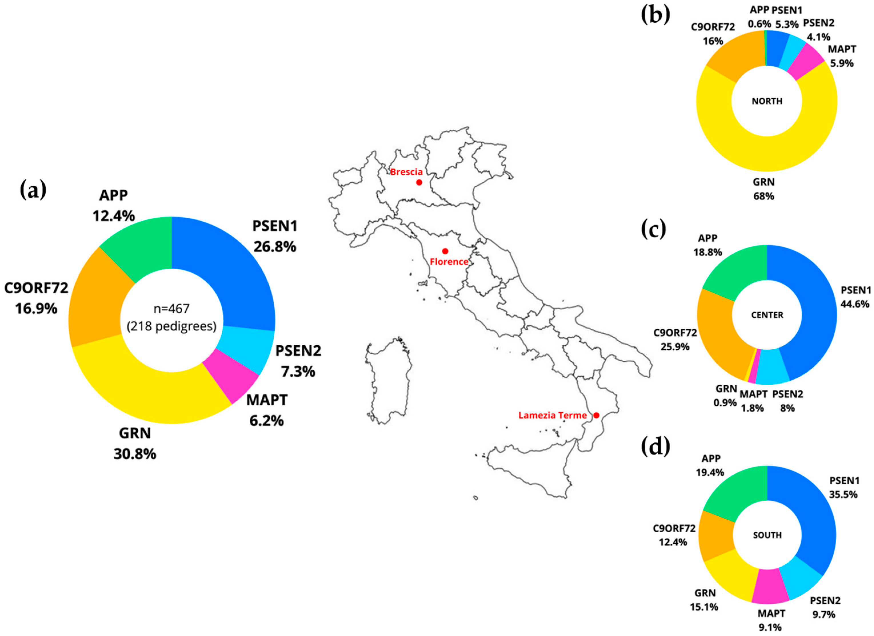
Figure 1. Frequency of each of the six genetic groups APP, PSEN1, PSEN2, MAPT, GRN, and C9orf72 in the combined dataset of this study (a), in the Northern (b), in the Central (c), and in the Southern cohort (d) (p < 0.0001, Chi-square test).
Ijms 25 07035 g001
Figure 2. Violin plots of age at onset for the six genetic groups APP, PSEN1, PSEN2, MAPT, GRN, and C9orf72 (p < 0.0001, Linear Mixed Model).
Ijms 25 07035 g002
Table 1. Characteristics of the Italian cohort.
APP
(n = 58)
PSEN1
(n = 125)
PSEN2
(n = 34)
MAPT
(n = 29)
GRN
(n = 144)
C9orf72
(n = 79)
p-Value
Number of families213219167957
Sex (% female)34.552.850.051.747.253.20.2676 a
Age at onset59.9 ± 10.7 44.9 ± 9.759.3 ± 15.248.0 ± 11.061.4 ± 8.957.4 ± 8.7<0.0001 b
APP, APP mutation carriers; PSEN1, PSEN1 mutation carriers; PSEN2, PSEN2 mutation carriers; MAPT, MAPT mutation carriers; GRN, GRN mutation carriers; C9orf72, C9orf72 mutation carriers. a Chi-square; b One-way ANOVA test with Bonferroni post hoc correction. Means ± standard deviation.
Disclaimer/Publisher’s Note: The statements, opinions and data contained in all publications are solely those of the individual author(s) and contributor(s) and not of MDPI and/or the editor(s). MDPI and/or the editor(s) disclaim responsibility for any injury to people or property resulting from any ideas, methods, instructions or products referred to in the content.