Article

Neuroprotection by Anethum graveolens (Dill) Seeds and Its Phytocompounds in SH-SY5Y Neuroblastoma Cell Lines and Acellular Assays

Department of Bionano Technology, Gachon Bionano Research Institute, Gachon University, 1342 Seongnam-daero, Sujung-gu, Seongnam-si 461-701, Gyeonggi-do, Republic of Korea
*
Authors to whom correspondence should be addressed.
Int. J. Mol. Sci. 2024, 25(13), 7104; https://doi.org/10.3390/ijms25137104
Submission received: 15 May 2024 / Revised: 25 June 2024 / Accepted: 25 June 2024 / Published: 28 June 2024
(This article belongs to the Special Issue Medicinal Plants and Bioactive Compounds in Health and Disease)

Abstract

:
Neurodegeneration diseases (NDs) are a group of complex diseases primarily characterized by progressive loss of neurons affecting mental function and movement. Oxidative stress is one of the factors contributing to the pathogenesis of NDs, including Alzheimer’s disease (AD). These reactive species disturb mitochondrial function and accelerate other undesirable conditions including tau phosphorylation, inflammation, and cell death. Therefore, preventing oxidative stress is one of the imperative methods in the treatment of NDs. To accomplish this, we prepared hexane and ethyl acetate extracts of Anethum graveolens (dill) and identified the major phyto-components (apiol, carvone, and dihydrocarvone) by GC-MS. The extracts and major bioactives were assessed for neuroprotective potential and mechanism in hydrogen peroxide-induced oxidative stress in the SH-SY5Y neuroblastoma cell model and other biochemical assays. The dill (extracts and bioactives) provided statistically significant neuroprotection from 0.1 to 30 µg/mL by mitigating ROS levels, restoring mitochondrial membrane potential, reducing lipid peroxidation, and reviving the glutathione ratio. They moderately inhibited acetylcholine esterase (IC50 dill extracts 400–500 µg/mL; carvone 275.7 µg/mL; apiole 388.3 µg/mL), displayed mild anti-Aβ1–42 fibrilization (DHC 26.6%) and good anti-oligomerization activity (>40% by dill-EA, carvone, and apiole). Such multifactorial neuroprotective displayed by dill and bioactives would help develop a safe, low-cost, and small-molecule drug for NDs.

1. Introduction

Neurodegenerative diseases (NDs) are a group of diseases that result from advancing deterioration in the structure and function of neurons. The worldwide statistical data speculated that over 152 million people will be affected by ND-related issues by 2050 [1]. NDs include Alzheimer’s disease (AD), Parkinson’s disease (PD), Huntington’s disease (HD), amyotrophic lateral sclerosis (ALS), prion diseases, etc. In NDs, different regions in the brain are affected but some characteristics like proteinopathy and induced cell death are common [2,3]. Hence, therapeutic interventions against one ND can improve symptoms of other ND as well. Additionally, oxidative stress and neuroinflammation are identified as major contributors to the disease [4,5]. The cause of oxidative stress is an abundance of reactive oxygen species (ROS) due to overproduction or decreased elimination in the system. Increased ROS reacts and damages cellular components hastening inflammation, tau phosphorylation, and apoptosis in neurons [6]. Thus, protecting the cells from oxidative stress would be a significant approach to treating NDs. To date, no remedy effectively cures complex NDs, and available symptomatic medications entail significant side effects. Therefore, a plant-based potential multi-targeted therapy would be valuable in treating NDs. Plants are a rich source of antioxidants and other bioactive components that can target complex pathological pathways of neurodegeneration in various ways.
Anethum graveolens L. (dill; Family Apiaceae) is an aromatic herb cultivated worldwide. The dill seeds have an intense fragrance and are used as a spice for flavoring the food. On crushing, the leaves and fruits release a strong fragrance due to aromatic compounds in the essential oils rich in α-phellandrene, limonene, dill apiole, carvacrol, carvone, and p-cymene [7,8]. Both the leaves and fruits have therapeutic properties like antioxidant, anti-cancer [9], anti-inflammatory [10], antinociceptive [11], anti-microbial [12], anti-parasitic [13], carminative and diuretic properties [14]. It is also effective in treating irritable bowel syndrome [15], hyperlipidemia [16], diabetes [17], etc. Furthermore, dill essence [18] and combined extract [19,20] reportedly improved cognition in animal models. In the central nervous system (CNS), cholinergic transmission (acetylcholine; ACh) has a vital role in maintaining neuronal plasticity, and cell survival, thus compromised cholinergic signaling results in memory deficits [21].
To gain insight into the neuroprotective mechanisms of dill, we evaluated the effects of dill extract (hexane and ethyl acetate) and its bioactive components on hydrogen peroxide-induced oxidative stress in human neuroblastoma SH-SY5Y cells. These cells are widely used in neuroscience as in vitro models, particularly for studying neuroprotective mechanisms. The hydrogen peroxide (H2O2) treatment was employed to induce oxidative stress in the SH-SY5Y cells, mimicking conditions observed in NDs. Additionally, acetylcholine esterase enzyme (AChE) inhibition, anti-oligomerization, and anti-fibrillation activity were also studied to identify various other mechanisms for neuroprotection.

2. Results

2.1. Phytochemical Estimation and Antioxidant Potential of Dill Extract

Total phenolic (TPC) and flavonoids (TFC) were estimated in the extracts using colorimetric assays. The content of phenolics was calculated from the regression equation of the calibration curve (R2 = 0.995, y = 0.046x + 0.0059), expressed in gallic acid equivalent (GAE) as milligrams per gram of the extract or fraction (mg GAE/g extract). TPC of the extracts was estimated to be 16.20 ± 2.88 mg GAE/g and 43.55 ± 3.42 mg GAE/g in dill-H (hexane) and dill-EA (ethyl acetate), respectively. The flavonoid content was calculated from the regression equation of the calibration curve (R2 = 0.994, y = 0.0432x + 0.0051), expressed in quercetin equivalent (QE) as milligrams per gram of the extract or fraction (mg QE/g extract). TFC in the dill-EA (18.94 ± 3.45 mg QE/g) was higher than the dill-H (8.69 ± 2.05 mg QE/g). The antioxidant potential of the extracts was evaluated using DPPH, ABTS, and FRAP assays. In the DPPH assay, the percent radical scavenging activity observed was 37.11 ± 0.23% and 53 ± 0.71% for dill-H and dill-EA, respectively. A similar activity like dill-H was observed for the pure compounds (apiole: 29.73 ± 2.27%; carvone: 37.05 ± 0.25%; DHC: 36.85 ± 1.51%) in the assay. Ascorbic (10 μg/mL) displayed 85.43 ± 1.02% activity as a positive control in the assay. In the ABTS assay, dill-EA (45.72 ± 0.64%) displayed better radical scavenging activity than dill-H (37.17 ± 0.14%). The pure compounds (apiole: 20.71 ± 0.26%; carvone: 20.02 ± 0.18%; DHC: 17.53 ± 0.47%) exhibited lower activity as compared to the extracts. Positive control, quercetin (10 μg/mL) displayed 97.55 ± 0.05% activity in the assay. In the FRAP assay reduction of Fe3+ to Fe2+ in the presence of antioxidants is measured. Here also dill-EA exhibited a better FRAP value (103 ± 1.03 µM Fe2+/g) compared to dill-H (68.48 ± 1.2 µM Fe2+/g). The FRAP activity of pure compounds was lower than that of the extracts (ranging from 42.41 to 45.22 µM Fe2+/g). Ascorbic acid positive control (10 μg/mL) displayed 142.83 ± 0.4 1% activity in the assay. The results were summarized in Supplementary Table S1.

2.2. GC–MS Analysis

The GC–MS chromatogram of hexane and ethyl acetate extracts of dill identified three peaks each. The peaks were identified by comparing retention time, peak area (%), height (%), and mass spectral fragmentation to that of the known compounds in the NIST library (Supplementary Figure S1). The main components identified were 19.9% cyclohexanone,2-methyl-5-(1-methylethenyl)- [syn. DHC; dihydrocarvone], 26.55% D-carvone, and 52.46% apiole in dill-H. Meanwhile, 9.26% cyclohexanone,2-methyl-5-(1-methylethenyl)-, 14.9% D-carvone, and 53.61% apiole were identified in dill-EA (Figure 1).

2.3. In Vitro Acetylcholinesterase Inhibitory Activity

Acetylcholinesterase (AChE; E.C.3.1.1.7) is a cholinergic enzyme generally present at the postsynaptic neuromuscular junctions and hydrolyses acetylcholine (ACh), an important neurotransmitter. In AD patients, the level of ACh declines in the synaptic junction hence, inhibition of AChE is desirable to maintain the normal ACh levels. Therefore, the extracts and the phytocompounds were screened for anti-AChE activity and the IC50 values (half maximal inhibitory concentration) were calculated using galantamine hydrobromide as inhibitor control. The IC50 value of dill-H (470.62 ± 37.40 μg/mL) was lower than the dill-EA (504.10 ± 33.79 μg/mL) extract. Among the phytocompounds, D-carvone displayed better inhibitory activity (IC50: 275.70 ± 5.37 μg/mL or 1.83 mM) compared to apiole (IC50: 388.35 ± 4.73 μg/mL or 1.74 mM), and DHC (IC50 > 1197.67 ± 101.29 μg/mL or 7.86 mM) (Figure 2). The IC50 value of galantamine was 3.12 ± 0.71 μg/mL (8.47 μM), similar to the previously reported value of 4.31 μg/mL.
The Km and Vmax values (Table 1) were calculated using a non-linear fit (Michaelis–Menten equation) and the inhibition curves (with and without extract/phytocompound) were plotted using the linear regression (Lineweaver–Burk plot) on GraphPad Prism 10. We have not conducted kinetic analysis on DHC as it was a weak AChE inhibitor (IC50 > 1 mg/mL). The competitive inhibition pattern observed by the extracts and the phytocompounds for the enzyme (Supplementary Figure S2) suggested that they compete with the substrate for binding to the enzyme’s active site. The Ki value is the dissociation constant describing the binding affinity between the inhibitor and the enzyme. Ki (inhibitor constant) value for the competitive inhibition was calculated from the formula IC50 = Ki (1 + [S]/Km) [22]. From the Ki values (Table 1), carvone had the lowest Ki value (270 μg/mL or 1.79 mM) compared to others suggesting that carvone has the strongest binding affinity for the enzyme, followed by apiol (382 μg/mL or 1.71 mM) and dill-H (464 μg/mL).

2.4. Aβ -Fibrilization and Oligomerization Inhibition by Dill

The multimeric detection system (MDS) was used to investigate the effect of dill on Aβ oligomerization. The results were calculated based on the zero h value at which all samples and control signals were set at 1.0. The oligomerization reduction at 2 h was statistically significant in dill-H (* p < 0.05), and apiole and carvone (**** p < 0.0001). A non-significant oligomerization reduction was observed in dill-EA and DHC. After 4 h of incubation, a statistically significant oligomerization reduction (*** p < 0.0001) was observed in the case of dill-H, apiole, and carvone except for dill-EA (* p < 0.01) (Figure 3A). The compounds may inhibit Aβ oligomerization at different times due to structural differences affecting their interaction with the protein. Additionally, the compounds may degrade with time [23].
Figure 3B displays the percent oligomerization inhibition by dill. The statistical significance was evaluated compared to the negative control. The extracts showed weak inhibition in the assay (dill-H: 7.53 ± 3.27%, non-significant, and dill-EA: 12.82 ± 1.74%; * p < 0.05). Among the bioactives; DHC (26.62 ± 8.62%; **** p < 0.0001) showed more potent oligomerization inhibition as compared to apiole (13.53 ± 4.4%; ** p < 0.01) and carvone (8.12 ± 2.68%, non-significant).
Inhibition of Aβ fibrilization by the extracts and the phytocompounds was analyzed by ThT assay. The binding of ThT to the β-sheet of amyloid fibrils increases the fluorescence thus it is used to monitor fibril formation [6]. The samples (500 μg/mL) were screened for anti-fibrilization potential using phenol red as the positive control. The results were statistically significant (#### p < 0.0001) compared to the negative control (Buffer + Aβ). Dill-H exhibited 28.77 ± 1.83% inhibitory activity while dill-EA (42.05 ± 0.14%), carvone (42.37 ± 0.76%), apiole (41.85 ± 1.73%), and DHC (33.44 ± 3.43%) showed potent inhibitory activity. The inhibition exhibited by phenol red (60.3 ± 4.29%) was similar to a previously reported value [24] Figure 3C.

2.5. Cytotoxic Effect of Dill Extracts and Its Bioactive Compounds

The cellular viability was estimated in the SH-SY5Y neuroblastoma cell line after 24 h of treatment with different concentrations of the extracts/ phytocompounds (1, 10, 30 µg/mL) using WST-8 dye. As no cytotoxicity was observed up to 30 µg/mL in both cases (Supplementary Figure S3) the subsequent cell culture experiments were conducted using this as maximum concentration.

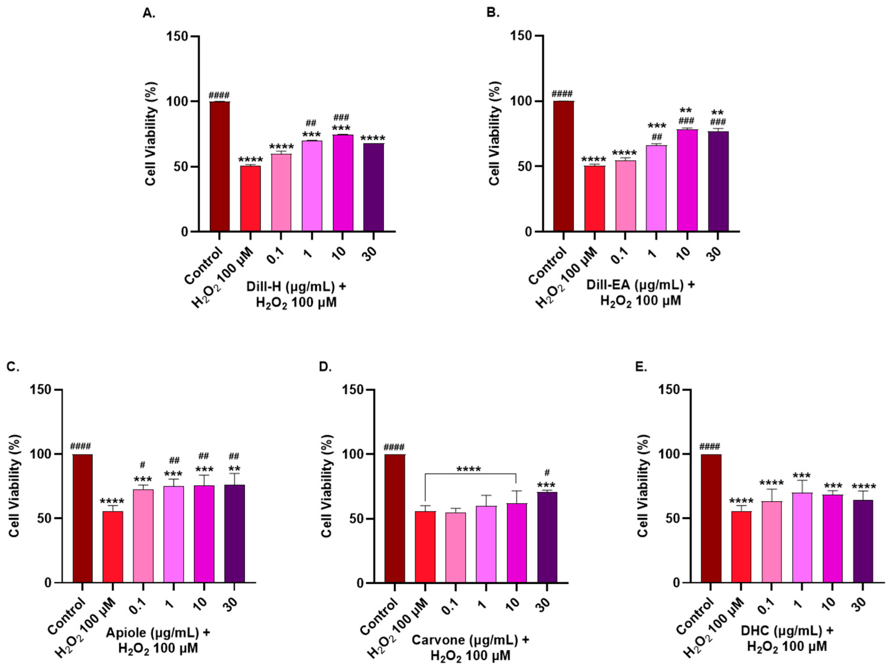
2.6. Protective Effect of Dill Extracts and Its Bioactive Compounds against H2O2-Induced Oxidative Stress

The neuroprotective effects of the extracts/phytocompounds were assessed by H2O2-induced oxidative stress in the SH-SY5Y cells. Around 50% cell survival after 1 h treatment with H2O2 (100 µM) was observed in our preliminary experiment. Therefore, this H2O2 concentration was used to induce stress in SH-SY5Y cells pre-treated with the extracts for 12 h. Dill-H displayed a significant increase in cell viability (70.26 ± 0.01%) at 1 µg/mL (# p < 0.05) and 10 µg/mL (74.74 ± 0.04%; ## p < 0.01), as compared to the H2O2 control. At 30 µg/mL, the cell viability decreased (68.09 ± 0.00%) compared to 10 µg/mL. Dill-EA provided better neuroprotection than dill-H at 1 µg/mL (66.44 ± 1.04%; # p < 0.01) and 10–30 µg/mL (78.43 ± 1.00% and 77.01 ± 2.06%; ### p < 0.001) (Figure 4). Apiole exerted better neuroprotective activity than the other two pure compounds and increased significantly (72.82 ± 3.13%; # p < 0.05) at 0.1 µg/mL. The increase in cell viability was statistically significant (## p < 0.01) and almost constant (~75%) at higher concentrations (1–30 µg/mL), as compared to the H2O2 control (Supplementary Figure S4). Carvone displayed significant (70.50 ± 1.67%; # p < 0.05) neuroprotection at 30 µg/mL only while DHC non-significantly increased the cell viability at all concentrations (0.1–30 µg/mL).

2.7. Dill and Its Phytocompounds Mitigated H2O2-Induced ROS Generation

To access the intracellular reactive oxygen species (ROS) scavenging activity of the extracts, the SH-SY5Y cells were pre-treated with the extracts for 12 h, followed by 2 h exposure to H2O2 (100 µM). The ROS production in the cells was monitored by a fluorescent dye (H2DCFDA), which is oxidized to DCF by ROS. The SH-SY5Y cells treated with H2O2 generated 156.96 ± 5.07% ROS compared to the untreated cells (100%). Pre-treatment of the cells with extracts resulted in a significant dose-dependent decrease in ROS production in dill-H (134.84 ± 4.15% at 1 µg/mL, ## p < 0.01; 127.86 ± 6.04% at 10 µg/mL, ### p < 0.001; and 115.13 ± 9.26% at 30 µg/mL, #### p < 0.0001) (Figure 5A) and dill-EA (129.20 ± 9.02% at 1 µg/mL, # p < 0.05; 114.95 ± 7.84% at 10 µg/mL, ## p < 0.01; and 110.9 ± 3.3% at 30 µg/mL, ## p < 0.01) (Figure 5B).
Among the pure compounds, apiole and DHC displayed similar results with significant dose-dependent reduction in ROS [10 µg/mL (113.63 ± 6.25%; ## p < 0.01), and 30 µg/mL (102.92 ± 9.11%; #### p < 0.0001) for apiole and 10 µg/mL (107.37 ± 8.26%; ## p < 0.01), and 30 µg/mL (96.73 ± 9.11%; ### p < 0.001) for DHC] (Figure 5C,E). Carvone performed better as it significantly (#### p < 0.0001) reduced ROS production at 1 µg/mL (122.82 ± 6.63%), 10 µg/mL (110.70 ± 4.90%), and 30 µg/mL (107.11 ± 8.34%) (Figure 5D).

2.8. Dill Extract and Phytocompounds Improved Mitochondrial Membrane Potential

Oxidative stress and mitochondrial dysfunction have been implicated in the pathogenesis of several neurodegenerative diseases. The overproduction of reactive oxygen species (oxidative stress) can damage the mitochondrial respiratory chain, alter membrane potential (MMP; ΔΨm), and influence Ca2+ homeostasis.
We used H2O2 to induce oxidative stress in SH-SY5Y cells and the ΔΨm was monitored using tetramethylrhodamine, an ethyl ester (TMRE) fluorescent dye that has an affinity for active mitochondria. The ΔΨm decreased in the depolarized membrane due to the inability to sequester the dye properly. In our study, a 50% reduction in the ΔΨm was observed in the untreated cells at 200 μM H2O2 hence this concentration was used for further examination. The cells were pre-treated with the extract/bioactives for 12 h followed by 200 μM H2O2 treatment for 2 h. The extracts behaved similarly by increasing MMP significantly (# p < 0.05) at the highest dose (~75–78% at 30 µg/mL) whereas the lower concentrations (1 and 10 µg/mL) had no significant effect on MMP (Figure 6A,B).
The pure compounds displayed a significant dose-dependent increase in MMP from 0.1 µg/mL and the results for carvone and DHC were statistically more significant than apiole (Figure 6C–E). Apiole significantly (# p < 0.05) increased the MMP at 0.1 µg/mL (71.53 ± 0.37%) after which it changed slightly from 75 to 77% (## p < 0.001) at higher concentrations (1–30 µg/mL) (Figure 6C). Carvone pre-treatment significantly dose-dependently increased the MMP at 0.1 µg/mL (71.61 ± 0.70%; ## p < 0.01), 1 µg/mL (80.80 ± 1.20%; ### p < 0.0001), 10 µg/mL (85.18 ± 4.96%; #### p < 0.0001), and 30 µg/mL (87.19 ± 6.26%; #### p < 0.0001) (Figure 6D). DHC had a similar effect as carvone in increasing MMP with 74.94 ± 1.20% (## p < 0.01) at 0.1 µg/mL, 79.26 ± 0.56% (### p < 0.001) at 1 µg/mL, 85.21 ± 0.19% (#### p < 0.0001) at 10 µg/mL, and 86.91 ± 5.47% (#### p < 0.0001) at 30 µg/mL (Figure 6E).

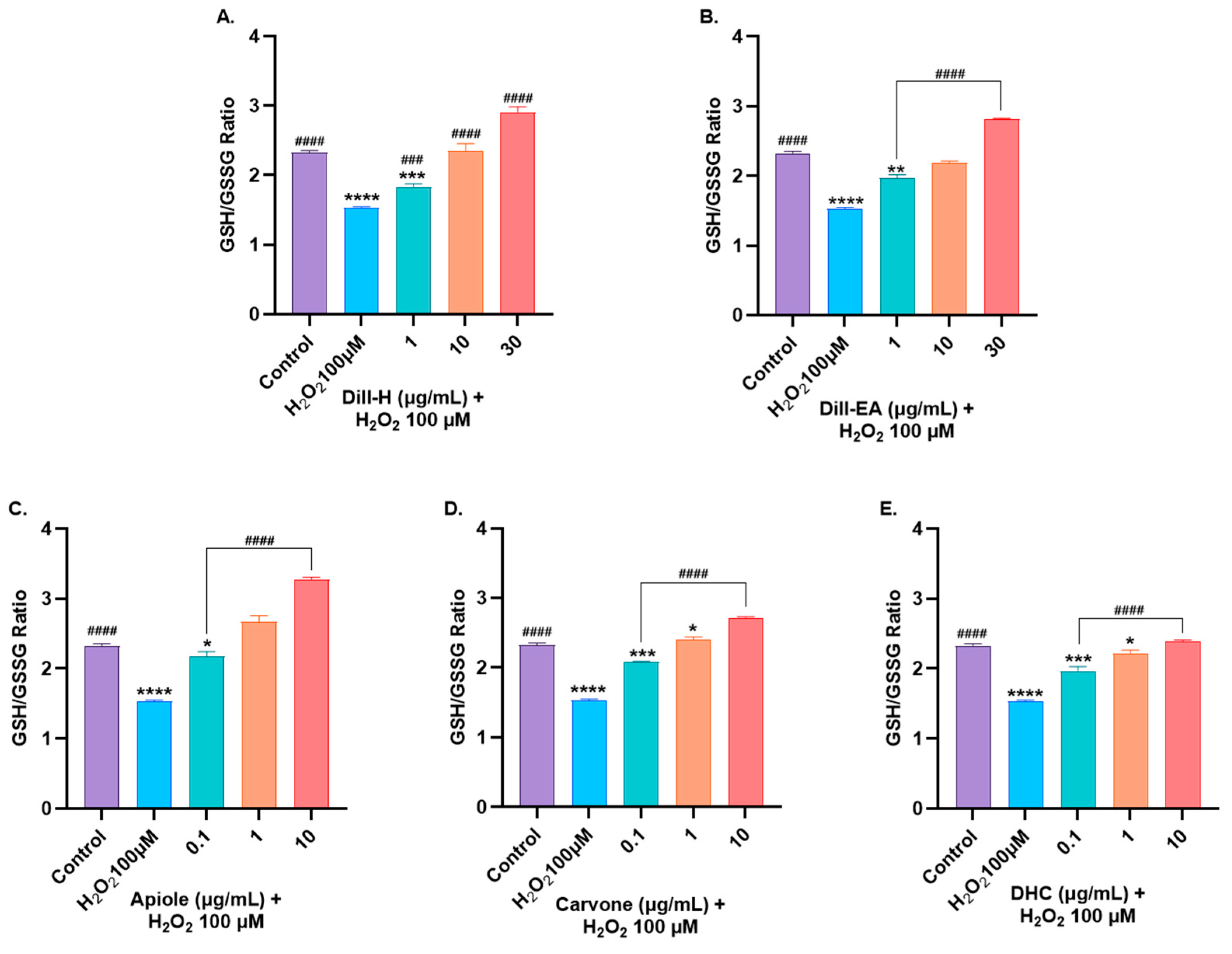
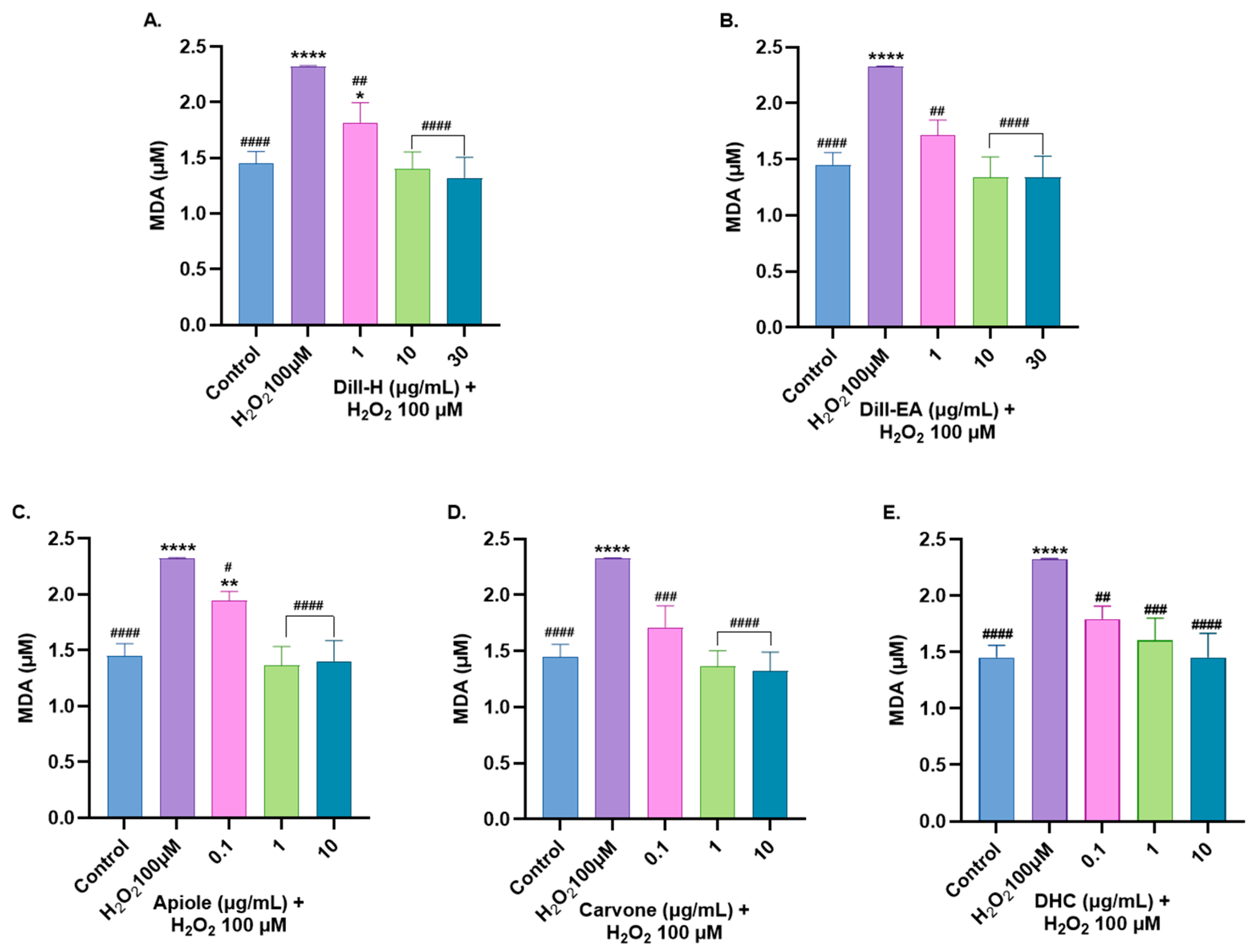
2.9. Dill Extract and Bioactive Compounds Restored Oxidative Stress Markers Altered by H2O2-Induced Oxidative Stress

Subsequently, the effect of pre-treatment with dill extracts and the major bioactives (apiole, carvone, and DHC) was studied on the H2O2-induced oxidative stress-affected parameters (lipid peroxidation, glutathione) in the SH-SY5Y cells.

2.9.1. Restoration of Glutathione Levels

The pre-treatment of SH-SY5Y cells with varying concentrations of dill extract (1, 10, and 30 μg/mL) and bioactives (0.1, 1, and 10 μg/mL) for 24 h before 6 h incubation with H2O2 (100 μM) resulted in a significant dose-dependent increase in GSH level (Supplementary Figure S5). Dill-EA performed significantly better at 1 µg/mL (#### p < 0.0001) compared to dill-H at the same concentration. At 10 and 30 µg/mL both the extracts behaved similarly (#### p < 0.0001) in restoring GSH level. All three bioactives showed similar dose-dependent responses at all concentrations (#### p < 0.0001). The oxidized glutathione (GSSG) levels were also measured (Supplementary Figure S6). The H2O2-induced stress raised intracellular GSSG levels. In our study, dill extracts significantly (#### p < 0.0001) reduced GSSG levels at 10 and 30 μg/mL. In the case of apiole and DHC, a significant decrease in GSSG was observed at 1 μg/mL and 10 μg/mL. However, DHC showed substantial reduction only at 10 μg/mL (# p < 0.05).
Reduced glutathione (GSH) is one of the most significant ROS scavengers, and its ratio with oxidized glutathione (GSSG) is a crucial indicator of cell health. We observed a significant dose-dependent increase in the GSH: GSSG ratio towards the normal state at all the tested concentrations of extracts and pure compounds (#### p < 0.0001) except for dill-H at 1 μg/mL (### p < 0.001) (Figure 7). Slightly better significant activity displayed by dill-EA than dill-H might be due to the higher ratio of bioactives in it.

2.9.2. Attenuation of MDA Level by Dill Extract

MDA is the most common biomarker for lipid peroxidation and is directly proportional to the oxidative injury. Lipid peroxidation refers to the degradation of lipids resulting from oxidative damage.
In the present study, dill extracts decreased MDA content in a statistically significant dose-dependent manner with complete attenuation at 10 and 30 µg/mL (~1.35 µM; #### p < 0.0001) compared to the control (Figure 8). Carvone displayed more significant results compared to apiole and DHC. It decreased MDA levels significantly (1.71 µM; ### p < 0.001) at 0.1 µg/mL followed by a complete reduction in MDA levels at 1 and 10 µg/mL (~1.33 µM; #### p < 0.0001). DHC decreased MDA levels in a significant dose-dependent manner; 0.1 µg/mL (1.79 µM; ## p < 0.01), 1 µg/mL (1.60 µM; ### p < 0.001), and 10 µg/mL (1.45 µM; #### p < 0.0001). Apiole reduced MDA level significantly at 0.1 µg/mL (1.94 µM; # p < 0.05) and the complete reduction was observed at 1 and 10 µg/mL (~1.38 µM; #### p < 0.0001).

3. Discussion

The present study described the neuroprotective potential of dill extract and its key phytocompounds in the oxidative stress model using human neuroblastoma cell lines (SH-SY5Y). In the phytochemical analysis and antioxidant activity measurements, dill-EA performed better than dill-H due to higher TPC and TFC. The phenols and flavonoids are known to scavenge free radicals [25] and protect against oxidative stress. A positive correlation between phenolic content and antioxidant potential has been reported [23]. A similar antioxidant potential was observed for the dill tablet [26]. This property is commonly associated with reductones that provide hydrogen atoms, leading to the breaking of the free radical chain [27]. Previously, TPC and TFC in the methanolic extract were reported as 69.87 mg GAE/100 g dw and 49.10 mg CE/100 g dw, respectively, with 81.52% DPPH activity [28]. The difference in the obtained values can be related to variable varieties and extraction solvents.
GC-MS identified apiole, carvone, and DHC as the major phytocompounds. Dill herb oil has been approved as GRAS by the US FDA for its use in the food industry as a flavoring and seasoning agent [29]. DHC is a colorless, oily monoterpenoid with p-menthane consisting of the cyclohexane ring with a methyl group at position 1 and a 2-methyl-propyl group at position 4. D-carvone is a monoterpene ketone present as a volatile component of essential oils and exhibits numerous pharmacologic properties like antimicrobial, antidiabetic, antioxidant, anti-inflammatory, anticancer, and neurological activities [30]. Its LD50 has been reported as 1640 mg/kg in rodent models [31]. Carvone can cross BBB and exhibit drug-like properties according to Lipinski’s rule of five [32]. On the other hand, apiole is a benzodioxole and has acaricidal [33], anti-inflammatory [34], gastroprotective [35], and anticancer [36] activities. However, it lacks significant BBB permeability [32]. Chemically, apiole is a benzodioxole while carvone and DHC are monoterpenoids with potent antioxidant activity [37,38]. The presence of an unsaturated hydroxyl group and a conjugated double bond in carvone provides greater free radical scavenging activity [38,39]. Apiol has two electron-donating methoxy groups which increases the stability of the benzene ring and hence increases radical scavenging activity [40]. The synergistic action of these phytocompounds provided antioxidant properties to the dill extracts. The higher apiole content in dill-EA might contribute to its improved antioxidant activity.
The anti-AChE potential of the dill extracts and the key phytocompounds were investigated in vitro using AChE from E. electricus. The plant extracts with an anti-AChE activity have been classified as potent (>50% inhibition), moderate (30–50% inhibition), and low activity (<30% inhibition) at 100 μg/mL [41]. In the preliminary screening, dill extracts and bioactives at 100 μg/mL moderately inhibited AChE. From the experimental results, dill-EA (504.1 ± 33.79 μg/mL) had a slightly higher IC50 value than dill-H (470.62 ± 37.40 μg/mL). On the other hand, D-carvone reported the lowest IC50 value (275.7 ± 5.37 μg/mL) compared to apiole (388.35 ± 4.73 μg/mL), and DHC (>1 mg/mL). Previously, IC50 > 200 μg/mL was reported for the ethanolic dill extract [42]. The IC50 value for carvone obtained in our experiment was lower than previously obtained values of 835.2 μg/mL (5.56 mM) [43] but was similar to 277.9 μg/mL (1.85 mM) [44]. The conjugated double bonds present in carvone are responsible for better AChE inhibition [45]. It can be suggested that the anti-AChE activity of the extracts is the result of the synergistic action of several compounds present. Previously, essential oils from dill showed 100% inhibition, while carvone and DHC displayed only 23.6% and 31.1% AChE inhibition at 1 mg/mL [46]. Conversely, no AChE inhibition was observed in the aerial parts of the dill plant grown under organic and conventional agricultural conditions [47].
In silico studies predicted the binding energy of −5.6 and −5.9 kcal/mol for carvone and apiole, respectively [48]. In another study, carvone displayed a similar binding energy value (−7.7 kcal/mol) to galantamine, the positive inhibitor control for AChE [31]. Carvone was also reported as a competitive inhibitor of AChE (bovine erythrocyte) [44]. The molecular docking study revealed that carvone binds to the most important region of the AChE active site, interacting with esteratic site residues (Ser203, His447), and anionic site amino acids (Trp86, Tyr133, Tyr337, Phe338) responsible for catalytic action and substrate binding, respectively. It exhibited additional hydrogen bond interactions with Tyr337 to create an anionic subsite [49]. Conversely, the aromatic moiety of apiole interacted with Tyr341 and Trp286 at the active site of human AChE forming a π-bond. Additionally, the methylenedioxy group of apiole formed a hydrogen bond with the phenolic hydroxyl group of Tyr124 while the 2-methoxy group of apiole interacted with the peptide bond between Val294–Phe295 of the enzyme [50]. The above reports support our results for the competitive inhibition observed for dill, carvone, and apiole. In our study, Vmax (the maximal reaction velocity when the enzyme is saturated with its substrate) remains unchanged (2.975 μmole/min/mg), but Km (the concentration of the substrate that permits the enzyme to achieve half Vmax) increased in the presence of the inhibitor, indicating a competitive inhibition. An increased Km value in the presence of an inhibitor reduces the binding affinity of the enzyme for the substrate requiring a higher concentration of the substrate to achieve Vmax. Since the competitive inhibitors can only bind to the free enzyme (E) and not to the enzyme–substrate (ES) complex, they cannot disturb ES catalysis, thus Vmax is unaffected. Ki (inhibitor constant) determines the potential of the inhibitor and is described as the concentration required to produce half maximum inhibition. A smaller value of Ki indicates a stronger binding. In our study, the Ki value for carvone (270 μg/mL or 1.79 mM) was the lowest suggesting its strong binding to the enzyme. Previously, a lower Ki value (0.68 mM) was reported for carvone by Grundy et al. [51].
Formation and accumulation of amyloid plaques are serious pathological features of several diseases, including neurodegenerative diseases like AD and PD. Previously, aqueous [52], methanolic dill leaf extract [53], and dill tablet [26] markedly inhibited protein aggregation, which can generate amyloid cross-β structure and consequently affect protein structure and stability [53]. In an earlier report, hot water extract of dill inhibited amyloid-like fibril formation by approximately 10% [54]. Inhibitor binding can also alter the secondary structure of amyloid fibrils to less ordered or non-beta sheet structures without changing the morphology of amyloid aggregates or inhibiting fibril formation [55]. Additionally, compounds interacting with the central domain of Aβ inhibit oligomerization [56]. In our study, dill extracts and pure bioactives displayed a lower, but significant, Aβ fibrilization inhibition compared to the control. In the ThT assay, all pure compounds and dill-EA displayed similar activity while dill-H displayed lower inhibition. This difference might be due to the lower ratio of these bioactives (DHC: carvone: apiole:: 1: 1.3: 2.6) compared to dill-EA (DHC: carvone: apiole:: 1: 1.4: 5.4). In MDS assay, DHC was more effective in inhibiting oligomerization compared to others. DHC is the degradation product of carvone produced by hydrogenation. The better inhibition exhibited by DHC indicates the importance of hydrogenation in inhibition. The lower concentration of bioactives in the extracts might be responsible for the lower inhibition exerted by the extracts. The aromatic compounds interact with diphenylalanine’s π-stacking to exercise anti-amyloidogenic activity [57]. Consequently, it is reasoned that aromatic compounds in the extract stabilize the protein β structure through π-stacking or hydrophobic interaction [6]. According to the classification of compounds that inhibit Aβ assembly [58], our compounds are Class II inhibitors that stabilize Aβ conformation and do not promote oligomer of fibril formation.
The imbalance between the generation and detoxification of free radicals generates oxidative stress in the system, which is associated with the pathogenesis of various diseases including NDs. At low concentrations, ROS serves as a secondary messenger in cell signaling but at higher concentrations, it harms cellular components. Therefore, alleviating ROS provides a therapeutic approach to the treatment of diseases. In this regard, several plants have been identified that can reduce oxidative stress [59]. In the present study, dill extracts and bioactives displayed dose-dependent neuroprotection by reducing the ROS, protecting MMP, and restoring GSH/GSSG balance, most likely by antioxidant activity of the phytocompounds. The better neuroprotective effect exerted by dill extract might be due to the synergistic effect of major bioactive components. Additionally, the better neuroprotection displayed by dill-EA could be due to a higher content of apiole. In ROS assay, apiole, and DHC displayed significant ROS reduction at higher concentrations (10 and 30 μg/mL), while carvone also reduced ROS at lower (1 μg/mL) concentrations. Better ROS reduction by carvone could be due to its antioxidant [60,61,62] and anti-inflammatory actions involving the NF-kB signaling pathway [63]. In restoring MMP, the pure compounds performed better than the extracts, which might be due to the lower concentration of these bioactives in the extracts.
The slightly better significant activity displayed by dill-EA than dill-H in restoring glutathione levels might be due to a better profile of bioactives in it. The pretreatment of cells with dill restored levels of GSH/GSSG by promoting homeostatic redox control. A higher GSH: GSSG value than the control could be due to the de novo synthesis of GSH [64]. Moreover, sometimes flawed protection of free-SH groups of GSH during sample preparation might result in non-specific interactions, affecting the total concentration of free GSH [65]. Carvone displayed more significant results than apiole and DHC in reducing lipid peroxidation, which is again attributed to its antioxidant and anti-inflammatory nature [30,66].
Our results are supported by previous studies where ethanolic [67], aqueous [68], and hydroalcoholic [19] dill extracts improved cognition by inhibiting AChE, reducing MDA, and restoring the levels of protective enzymes in the antioxidant system [67,68]. In another study, a mixture of dill and Ocimum extract improved cognition by reducing stress and Aβ levels in the hippocampus [69] while methanolic extract provided neuroprotection in Aβ-induced PC12 cells [70].
An essential characteristic for a suitable neuroprotective agent to reach the target in CNS is its BBB permeability. Another pathway is the gut–microbiota–brain axis, by which orally administered phytocompounds may alter brain activity. While it is unclear if brain tissues have receptors or transporters for polyphenols or other phytochemicals, drugs targeting multiple sites are promising as a potential treatment for diseases with multifactorial etiology. Additionally, the possibility of secondary binding of phytocompounds to the targets cannot be denied [71]. Such extra-CNS actions have a slower effect than the direct interactions with the targets in CNS.

4. Materials and Methods

4.1. Chemicals

Acetylcholinesterase (Electrophorus electricus, Type VI-S), apiole, ascorbic acid, 2,2′-azinobis-(3-ethylbenzothiazoline-6-sulfonic acid) (ABTS), acetyl thiocholine chloride, 2,2-diphenyl-1-picrylhydrazyl (DPPH), 5,5′-Dithiobis(2-nitrobenzoic acid) (DTNB), Dihydrocarvone, 2′,7′-dichlorofluorescin diacetate (DCFDA), Folin–Ciocalteu reagent (FCR), galantamine, gallic acid, hydrogen peroxide, RIPA (Radio-Immunoprecipitation Assay) Buffer, Thioflavin T (ThT), 2,4,6-tripyridyl-s-triazine (TPTZ), tetramethylrhodamine, ethyl ester (TMRE) were bought from Sigma-Aldrich (St. Louis, MO, USA). Carvone was purchased from Santa Cruz Biotechnology (Dallas, TX, USA), and Aβ1–42 (Aggresure™) was bought from AnaSpec (Fremont, CA, USA). The WST-8 kit was purchased from Roche Diagnostics GmbH (Mannheim, Germany). Aβ1–42 for MDS (GenicBio Inc., Shanghai, China), purified anti-Aβ1–16 antibody (Biolegend, San Diego, CA, USA), and the horseradish peroxidase (HRP)-conjugated W0–2 monoclonal antibody (Peoplebio Inc., Seongnam, Republic of Korea). Thermo Fisher Scientific (Waltham, MA, USA) was the source for 3,3′,5,5′-Tetramethylbenzidine solution (TMB), fetal bovine serum (FBS), kanamycin, penicillin, and phosphate-buffered saline (PBST). Dulbecco’s modified Eagle’s medium (DMEM) was supplied by Gibco (Thermo Fisher, Seoul, Republic of Korea). All organic solvents of HPLC grade were purchased from Sigma-Aldrich (St. Louis, MO, USA).

4.2. Plant Material and Extraction

The pre-weighted (25 g) dried seeds of dill (Expat Mart, Seoul, South Korea) were ground using a pestle mortar and sequentially extracted by increasing polarity in the following order: (i) hexane and (ii) ethyl acetate. To a conical flask containing the sample, hexane (polarity = 0.009) was added and subjected to mild shaking for 8 h. They were first filtered with muslin cloth and then through Whatman No. 1 filter paper. The residue was further extracted twice using the same fresh solvent and all the filtrates were pooled together. The resulting residue was air-dried and further extracted with ethyl acetate (polarity = 0.228) using the same procedure. Finally, the solvent was removed using a rotatory evaporator (EYELA, Japan) under reduced pressure and low temperature. The fractions were weighed and kept at 4 °C until additional experiments.

4.3. Gas Chromatography-Mass Spectrometry (GC-MS) Method

The sample was separated on a fused silica capillary column (DB-5ms UI, 30 m × 0.25 mm i.d., film thickness 0.25 μm, Agilent, Santa Clara, CA, USA) installed on GCMS-QP2020 (Shimadzu, Kyoto, Japan). The oven temperature was programmed as isothermic at 60 °C for 2 min, 100 °C at 4 °C/min, 290 °C at 10 °C/min, and finally isothermic for 10 min. The split injection mode (1:10) was used. The carrier gas was helium at a constant flow rate (1 mL/min). The injection port, ion source, and interface temperatures were 280, 280, and 150 °C, respectively. The ionization energy was 70 eV. The mass spectra were obtained in full scan mode (40–700 AMU). The sample (1 μL, 1 mg/mL) was auto-injected into the GC-MS. The unknown compounds were identified by matching known compounds in the National Institute of Standards and Technology (NIST) library.

4.4. Determination of Total Phenolic and Flavonoid Content

The total phenol content (TPC) of extracts (1 mg/mL) was determined colorimetrically using the Folin–Ciocalteu reagent with slight modification [23] in the original method [72]. Gallic acid standard (10–100 μg/L) was used for calibration. Total phenolic content was expressed as mg gallic acid equivalents (GAE) per g of plant extract.
The total flavonoid content of extracts (1 mg/mL) was determined by aluminum chloride colorimetric assay with slight modification [23] in a previously reported method [72]. Quercetine standard (10–100 μg/mL) was used for calibration. Total flavonoid contents were expressed as mg quercetin equivalents (QE) per g of plant extract.

4.5. Determination of Antioxidant Capacity

(i)
2,2′-Azino-Bis (3-Ethylbenzothiazoline-6-Sulfonic Acid) [ABTS] Radical Scavenging Assay
The free radical scavenging activity of the extracts and phytocompounds at 1 mg/mL was measured spectrophotometrically in a plate reader (Synergy-H1 BioTek, Agilent, Santa Clara, CA, USA) by modifying a previous method [73] to suit the 96-well plate format [23]. Quercetin served as a positive control, methanol as a negative control, and extract without ABTS as blank. A standard curve for quercetin (1–30 μg/mL) was prepared for calibration and the results were expressed in milligram equivalents of quercetin per milligram of dry weight. The percentage of inhibition of ABTS+ was calculated as:
% RSA = (Ab Ae/Ab) × 100
where Ab = absorbance of the blank and Ae = absorbance of the extract.
(ii)
Free Radical Scavenging by 2,2-Diphenyl-1-Picrylhydrazylhydrate (DPPH) Radical Assay
DPPH radical scavenging capacity of the extracts and phytocompounds at 1 mg/mL was measured with slight modification [74]. The absorbance was monitored at 515 nm (Multimode reader, Synergy-H1 BioTek, Agilent, Santa Clara, CA, USA) using ascorbic acid (0.1–10 μg/mL) as a standard. Radical scavenging activity (RSA) was calculated as:
% RSA = (Ab Ae/Ab) × 100
where Ab = absorbance of the blank and Ae = absorbance of the extract.
(iii)
Ferric Reducing Antioxidant Potential (FRAP) Assay
FRAP assay was used to assess the metal-chelating ability of the extracts and phytocompounds with slight modification [23] in a previously reported method [75]. For the assay, the extract (1 mg/mL) was incubated with 200 μL of FRAP reagent, and the reduction of ferric tripyridyltriazine was monitored at 593 nm (Multimode reader, Synergy-H1 BioTek, Agilent, USA). Ascorbic acid (2 μg/mL) was used as a positive control. FRAP values of the extracts and phytocompounds were calculated from the standard curve of FeSO4 (15–250 μM) and expressed as μM Fe2+/g.

4.6. Acetylcholinesterase Inhibitory Activity

The AChE activity was examined by slight modifications [23] in Ellman’s method [76]. The extracts and phytocompounds were incubated for 15 min with AChE and 10 mM ATCC at 37 °C. The reaction was stopped by DTNB (15 mM), and the absorbance was measured at 412 nm (Multi-mode plate reader, Synergy-H1 BioTek, Agilent, USA). Galantamine was used as the positive control. The percent inhibition was calculated as:
Percent Inhibition (I%) = [(Ao Ac) (Bi Bc)]/(Ao Ac) × 100
where Ao is the absorbance without inhibitor; Ac is the negative control without inhibitor; Bi is the absorbance with inhibitor; and Bc is the negative control with inhibitor. The IC50 values were calculated using GraphPad Prism 10. Lineweaver–Burk plot was used to prepare the inhibition curves (with and without extract/phytocompound) using GraphPad Prism 10. The kinetic parameters were calculated from a non-linear fit (Michaelis–Menten equation) in GraphPad Prism 10.

4.7. Anti-Aβ1–42 Oligomerization and Fibrilization Activity

The anti-Aβ1–42 oligomerization and fibrilization activity of the extracts/bioactives were measured by Multiple Detection System (MDS) and ThT assay, respectively [6]. Briefly, the extracts and phytocompounds were incubated with Aβ1–42 at RT for different time points (0 h, 2 h, and 4 h). The samples were incubated on an anti-β-amyloid pre-coated plate for 1 h at RT. An HRP-conjugated W0–2 monoclonal antibody was added, and the plate was kept at RT for 30 min. Later, TMB was added, and the plate was incubated for 15 min at RT. The absorbance was read at 450 nm using a microplate reader (Victor3, PerkinElmer, Shelton, CT, USA)
The anti-Aβ1–42 fibrilization activity of the extracts/bioactives was monitored using a ThT assay [6]. The samples were incubated in the presence/absence of Aβ1–42 at 37 °C for 24 h. The samples were incubated with 100 μM ThT at 37 °C for 15 min. The fluorescence was monitored at Ex 450 nm/Ems 490 nm (Synergy-H1 BioTek, Agilent, Santa Clara, CA, USA). For the control, phenol red (50 μM) was used. The Aβ1–42 aggregation inhibition was calculated as follows:
Percent Inhibition (%) = [(1 − Fi/Fc) × 100]
where Fi and Fc are the fluorescence intensity with and without the inhibitors, respectively.

4.8. Cell Culture

Human neuroblastoma SH-SY5Y cells (ATCC CRL-2266, Manassas, VA, USA) were maintained in Dulbecco’s modified Eagle’s medium (DMEM, Gibco) supplemented with 10% fetal bovine serum (FBS), 1% kanamycin, and 1% penicillin (Thermo Fisher Scientific, Waltham, MA, USA) at 37 °C with 5% CO2, and a 95% humidified atmosphere in the incubator. The experiments were executed at 80–90% cell confluency.

4.8.1. Cell Viability Assay

The cells were seeded (1 × 10⁴ cells/well) in 96-well sterile plates and pre-treated with various extract concentrations (1, 10, 30 µg/mL) for 24 h. The extracts and phytocompounds were removed and incubated for 2 h with 10% WST-8 reagent (Roche, Grenzach-Wyhlen, Germany) as described previously [23]. The absorbance was determined at 450 nm in a multi-plate reader (Synergy-H1 BioTek, Agilent, USA). The percent cytotoxicity was calculated as:
Cytotoxicity % = (Ac − At)/(Ac) × 100
where Ac = absorbance of the control cells, At = absorbance of the treated cells.
The plot of percent cytotoxicity versus sample concentration was used to calculate the extract concentration that killed 50% of the cells (IC50).

4.8.2. Neuroprotection Assay

The neuroprotective effect of extracts and phytocompounds on H2O2-induced oxidative stress in SH-SY5Y by a previously described method [23]. The cells (1 × 10⁴ cells/well) were seeded in a 96-well sterile plate. After stabilization, the cells were pre-treated with the extracts and phytocompounds for 24 h. The extracts and phytocompounds were removed and treated with H2O2 (100 μM) for 6 h. A solvent control, H2O2 alone, and extract alone treatments were also included. After incubation, the % cell viability was measured using WST-8 reagent in triplicate experiments.

4.8.3. Measurement of Intracellular Reactive Oxygen Species (ROS)

The cells (1 × 10⁴ cells/well) were seeded in a 96-well sterile plate, after which they were pre-treated with the extract for 12 h. The extracts and phytocompounds were removed, followed by a 4 h treatment with H2O2 (100 μM) and H2DCFDA (25 μM) for another 2 h in the dark at 37 °C [23]. The fluorescence intensity (Ex 495 nm, Ems 520 nm) was measured by a microplate reader (Synergy-H1 BioTek, Agilent, USA). The ROS was calculated as a percentage of the untreated control cells (100%) in triplicate measurements.

4.8.4. Mitochondrial Membrane Potential (ΔΨm) Assay

The mitochondrial membrane potential was measured using the tetramethylrhodamine, methyl ester (TMRE) staining method [23]. The cells (1 × 10⁴ cells/well) were seeded in a 96-well sterile plate; after which, they were pre-treated with the extract for 12 h. The extracts and phytocompounds were removed, followed by a 2 h treatment with H2O2 (200 μM). A 1 μM amount of TMRE was added to the cells and incubated for 1 h at 37 °C. The fluorescence was assessed (Ex 549 nm, Ems 575 nm) using a microplate reader (Synergy-H1 BioTek, Agilent, USA). The ΔΨm was calculated as a percentage of the untreated control cells (100%) in triplicate measurements.

4.8.5. Antioxidant Parameters in Cell Lysate

The cells (5 × 10⁴ cells/well) were seeded in a 6-well sterile plate and incubated for 18–24 h. After stabilization, cells were pre-treated with the extracts and phytocompounds for 24 h before 6 h incubation with H2O2 (100 μM). The culture media was removed, and the cells were washed with cold PBS (1X). The cells were placed on ice and incubated with pre-chilled RIPA buffer for 10 min. Transfer the lysate in microfuge tubes and centrifuge (Labogene 1730R, BMS, Paju-si, Republic of Korea) for 10 min at 20,000× g, 4 °C. The supernatant was collected and stored at −80 °C.

Protein Estimation

The protein concentration in the samples was measured using the BCA protein estimation kit (Thermo Scientific, Waltham, MA, USA). The BSA standard (10–1000 μg/mL) was used to calculate protein concentration in the unknown samples.

Estimation of Glutathione

The concentrations of GSH (reduced) and GSSG (oxidized) in the lysate were measured fluorometrically using a previously described [23] and the fluorescence was recorded at 350/420 (Ex/Ems) in a microplate reader (Synergy-H1 BioTek, Agilent, Santa Clara, CA, USA).

Estimation of Malondialdehyde (MDA)

MDA levels were measured using TBA: TCA reagent as described previously [23]. In the reaction, MDA reacts with two molecules of thiobarbituric acid (TBA) to give a pink pigment that absorbs at 532 nm. The standard curve of MDA (1–100 μM) was used to calculate lipid peroxidation in the lysate.

4.9. Data and Statistical Analysis

Statistical analysis was established by a one-way ANOVA followed by Dunnett’s post hoc test. Data are registered as the mean ± SEM of three sets of experiments. The symbols ####, **** represents p < 0.0001, ###, *** represents p < 0.001, ##, ** represents p < 0.01, and #, * represents p < 0.05. The symbol # indicates significance compared to the H2O2 control while * indicates significance compared to the untreated control. The IC50 values were determined using non-linear regression. The Vmax and Km were calculated from a Michaelis–Menten plot drawn using a non-linear plot (GraphPad Prism 10). Lineweaver–Burk plots were drawn using linear regression analysis (GraphPad Prism 10). The Ki values were calculated from the formula IC50 = Ki (1 + [S]/Km).

5. Conclusions

The study provides a phytochemical basis for some of the effects of dill extracts and their main phytocompounds on neuroprotection. In summary, we evaluated the neuroprotective potential of dill extracts and main bioactive compounds in H2O2-induced oxidative stress in human neuroblastoma SH-SY5Y cell lines and biochemical studies. The dill extract and phytocompounds significantly provided neuroprotection by reducing oxidative stress, restoring MMP, and re-establishing redox homeostasis in the cells, indicating their antioxidant potential at varying concentrations (0.1–30 μg/mL). The antioxidant potential of dill is the most reasonable explanation for the neuroprotective effect in the cells. In addition, they exerted moderate anti-AChE activity and competitively inhibited the enzyme. The anti-AChE activity of pure compounds was relatively lower than the extracts, except for DHC. They also showed mild anti-Aβ oligomerization and good anti-Aβ fibrilization activity, stabilizing Aβ conformation to prevent oligomer/fibril development. The structural modifications in these lead compounds will be helpful in further refinement of activities. This attractive alternative multitargeted neuroprotective approach would be beneficial in the development of cost-effective drugs for multifactorial ailments like NDs, especially AD. However, despite promising results, additional research is needed to explore their role in neuroprotection especially in human subjects.

Supplementary Materials

The following supporting information can be downloaded at: https://www.mdpi.com/article/10.3390/ijms25137104/s1.

Author Contributions

Conceptualization, N.S. and S.S.A.A.; experimentation and data analysis, H.S., H.Y. and N.S.; writing—original draft preparation, N.S.; writing—review and editing, N.S., H.S. and S.S.A.A.; funding acquisition, S.S.A.A. All authors have read and agreed to the published version of the manuscript.

Funding

This research was supported by the Basic Science Research Program through the National Research Foundation of Korea (NRF) funded by the Ministry of Education (Grant number RS-2023-00251396 and 2021R1A6A1A03038996).

Institutional Review Board Statement

Not applicable.

Informed Consent Statement

Not applicable.

Data Availability Statement

Data are contained within the article. Additional information is provided as Supplementary Materials.

Conflicts of Interest

The authors declare no conflicts of interest.

References

  1. GBD 2019 Dementia Forecasting Collaborators. Estimation of the global prevalence of dementia in 2019 and forecasted prevalence in 2050: An analysis for the Global Burden of Disease Study 2019. Lancet Public Health 2022, 7, e105–e125. [Google Scholar] [CrossRef] [PubMed]
  2. Bredesen, D.E.; Rao, R.V.; Mehlen, P. Cell death in the nervous system. Nature 2006, 443, 796–802. [Google Scholar] [CrossRef] [PubMed]
  3. Rubinsztein, D.C. The roles of intracellular protein-degradation pathways in neurodegeneration. Nature 2006, 443, 780–786. [Google Scholar] [CrossRef] [PubMed]
  4. Guzman-Martinez, L.; Maccioni, R.B.; Andrade, V.; Navarrete, L.P.; Pastor, M.G.; Ramos-Escobar, N. Neuroinflammation as a common feature of neurodegenerative disorders. Front. Pharmacol. 2019, 10, 1008. [Google Scholar] [CrossRef]
  5. Singh, A.; Kukreti, R.; Saso, L.; Kukreti, S. Oxidative stress: A key modulator in neurodegenerative diseases. Molecules 2019, 24, 1583. [Google Scholar] [CrossRef] [PubMed]
  6. Sharma, H.; Yang, H.; Sharma, N.; An, S.S. Trachyspermum ammi Bioactives Promote Neuroprotection by Inhibiting Acetylcholinesterase, Aβ-Oligomerization/Fibrilization, and Mitigating Oxidative Stress In Vitro. Antioxidants 2024, 13, 9. [Google Scholar] [CrossRef] [PubMed]
  7. Vera, R.R.; Chane-Ming, J. Chemical Composition of Essential Oil of Dill (Anethum graveolens L.) Growing in Reunion Island. J. Essent. Oil Res. 1998, 10, 539–542. [Google Scholar] [CrossRef]
  8. Dimov, M.; Dobreva, K.; Stoyanova, A. Chemical composition of the dill essential oils (Anethum graveolens L.) from Bulgaria. Bulg. Chem. Commun. 2019, 51, 214–216. [Google Scholar]
  9. Al-Oqail, M.M.; Farshori, N.N. Antioxidant and Anticancer Efficacies of Anethum graveolens against Human Breast Carcinoma Cells through Oxidative Stress and Caspase Dependency. Biomed Res. Int. 2021, 2021, 5535570. [Google Scholar] [CrossRef]
  10. Nam, H.-H.; Nan, L.; Choo, B.-K. Anti-Inflammation and Protective Effects of Anethum graveolens L. (Dill Seeds) on Esophageal Mucosa Damages in Reflux Esophagitis-Induced Rats. Foods 2021, 10, 2500. [Google Scholar] [CrossRef]
  11. Rezaee-Asl, M.; Nikoui, A.B.V.; Bakhtiarian, A.; Nikoui, V.; Sabour, M.; Ostadhadi, S.; Giorgi, M.-S.Y.-N.A.M. Antinociceptive properties of hydro alcoholic extracts of Anethum graveolens L. (dill) seed and aerial parts in mice. Clin. Exp. Pharmacol. 2013, 3, 122. [Google Scholar] [CrossRef]
  12. Derakhshan, S.; Navidinia, M.; Ahmadi, A. Antibacterial activity of Dill (Anethum graveolens) essential oil and antibiofilm activity of Cumin (Cuminum cyminum) alcoholic extract. Infect. Epidemiol. Microbiol. 2017, 3, 122–126. [Google Scholar]
  13. Abas, A.-S.M.; Elagib, S.M. Antiparasitic activity of aqueous extract of Anethum graveolens against Entamoeba histolytica: In vitro and in vivo study. Biocatal. Agric. Biotechnol. 2021, 34, 102026. [Google Scholar] [CrossRef]
  14. Jana, S.; Shekhawat, G.S. Anethum graveolens: An Indian traditional medicinal herb and spice. Pharmacogn. Rev. 2010, 4, 179–184. [Google Scholar] [PubMed]
  15. Mohammad, I.H. Use of Anethum graveolens in the management of patients with irritable bowel syndrome. Mustansiriya Med. J. 2012, 11, 94–98. [Google Scholar]
  16. Sadeghi, M.; Kabiri, S.; Amerizadeh, A.; Heshmat-Ghahdarijani, K.; Masoumi, G.; Teimouri-Jervekani, Z.; Amirpour, A. Anethum graveolens L. (Dill) Effect on Human Lipid Profile: An Updated Systematic Review. Curr. Probl. Cardiol. 2022, 47, 101072. [Google Scholar] [CrossRef] [PubMed]
  17. Haidari, F.; Zakerkish, M.; Borazjani, F.; Angali, K.A.; Foroushani, G.A. The effects of Anethum graveolens (dill) powder supplementation on clinical and metabolic status in patients with type 2 diabetes. Trials 2020, 21, 483. [Google Scholar] [CrossRef] [PubMed]
  18. Mesripour, A.; Rafieian-Kopaei, M.; Bahrami, B. The effects of Anethum graveolens essence on scopolamine-induced memory impairment in mice. Res. Pharm. Sci. 2016, 11, 145. [Google Scholar] [PubMed]
  19. Thukham-Mee, W.; Wattanathorn, J. Evaluation of safety and protective effect of combined extract of Cissampelos pareira and Anethum graveolens (PM52) against age-related cognitive impairment. Evid.-Based Complement. Altern. Med. 2012, 2012, 674101. [Google Scholar] [CrossRef]
  20. Ohnon, W.; Wattanathorn, J.; Thukham-Mee, W.; Muchimapura, S.; Wannanon, P.; Tong-Un, T. The Combined Extract of Black Sticky Rice and Dill Improves Poststroke Cognitive Impairment in Metabolic Syndrome Condition. Oxid. Med. Cell. Longev. 2019, 2019, 9089035. [Google Scholar] [CrossRef]
  21. Li, Y.; Fan, H.; Sun, J.; Ni, M.; Zhang, L.; Chen, C.; Hong, X.; Fang, F.; Zhang, W.; Ma, P. Circular RNA expression profile of Alzheimer’s disease and its clinical significance as biomarkers for the disease risk and progression. Int. J. Biochem. Cell Biol. 2020, 123, 105747. [Google Scholar] [CrossRef] [PubMed]
  22. Burlingham, B.T.; Widlanski, T.S. An intuitive look at the relationship of Ki and IC50: A more general use for the Dixon plot. J. Chem. Educ. 2003, 80, 214. [Google Scholar] [CrossRef]
  23. Sharma, H.; Kim, D.Y.; Shim, K.H.; Sharma, N.; An, S.S.A. Multi-Targeting Neuroprotective Effects of Syzygium aromaticum Bud Extracts and Their Key Phytocompounds against Neurodegenerative Diseases. Int. J. Mol. Sci. 2023, 24, 8148. [Google Scholar] [CrossRef] [PubMed]
  24. Tan, M.A.; Lagamayo, M.W.; Alejandro, G.J.; An, S.S. Neuroblastoma SH-SY5Y cytotoxicity, anti-amyloidogenic activity and cyclooxygenase inhibition of Lasianthus trichophlebus (Rubiaceae). 3 Biotech 2020, 10, 152. [Google Scholar] [CrossRef] [PubMed]
  25. Martínez, S.; Fuentes, C.; Carballo, J. Antioxidant Activity, Total Phenolic Content and Total Flavonoid Content in Sweet Chestnut (Castanea sativa Mill.) Cultivars Grown in Northwest Spain under Different Environmental Conditions. Foods 2022, 11, 3519. [Google Scholar] [CrossRef] [PubMed]
  26. Oshaghi, E.A.; Tavilani, H.; Khodadadi, I.; Goodarzi, M.T. Dill tablet: A potential antioxidant and anti-diabetic medicine. Asian Pac. J. Trop. Biomed. 2015, 5, 720–727. [Google Scholar] [CrossRef]
  27. Salmanian, S.; Mahoonak, A.R.S.; Alami, M.; Ghorbani, M. Phenolic content, antiradical, antioxidant, and antibacterial properties of hawthorn (Crataegus elbursensis) seed and pulp extract. J. Agric. Sci. Technol. 2014, 16, 343–354. [Google Scholar]
  28. Christova-Bagdassarian, V.L.; Bagdassarian, K.S.; Atanassova, M. Phenolic profile, antioxidant and antimicrobial activities from the Apiaceae family (dry seeds). Mintage J. Pharm. Med. Sci. 2013, 2, 26–31. [Google Scholar]
  29. FDA. Substances Added to Food; U.S. Food and Drug Administration: Washington, DC, USA, 2022.
  30. Bouyahya, A.; Mechchate, H.; Benali, T.; Ghchime, R.; Charfi, S.; Balahbib, A.; Burkov, P.; Shariati, M.A.; Lorenzo, J.M.; El Omari, N. Health Benefits and Pharmacological Properties of Carvone. Biomolecules 2021, 11, 1803. [Google Scholar] [CrossRef]
  31. Amat-Ur-Rasool, H.; Ahmed, M.; Hasnain, S.; Ahmed, A.; Carter, W.G. In Silico Design of Dual-Binding Site Anti-Cholinesterase Phytochemical Heterodimers as Treatment Options for Alzheimer’s Disease. Curr. Issues Mol. Biol. 2022, 44, 152–175. [Google Scholar] [CrossRef]
  32. İstİflİ, E.S.; Tepe, A.; Sarikürkcü, C.; Tepe, B. Interaction of certain monoterpenoid hydrocarbons with the receptor binding domain of 2019 novel coronavirus (2019-nCoV), transmembrane serine protease 2 (TMPRSS2), cathepsin B, and cathepsin L (CatB/L) and their pharmacokinetic properties. Turk. J. Biol. 2020, 44, 242–264. [Google Scholar] [CrossRef] [PubMed]
  33. Song, H.Y.; Yang, J.Y.; Suh, J.W.; Lee, H.S. Acaricidal activities of apiol and its derivatives from Petroselinum sativum seeds against Dermatophagoides pteronyssinus, Dermatophagoides farinae, and Tyrophagus putrescentiae. J. Agric. Food Chem. 2011, 59, 7759–7764. [Google Scholar] [CrossRef] [PubMed]
  34. Parise-Filho, R.; Pastrello, M.; Camerlingo, C.E.P.; Silva, G.J.; Agostinho, L.A.; de Souza, T.; Magri, F.M.M.; Ribeiro, R.R.; Brandt, C.A.; Polli, M.C. The anti-inflammatory activity of dillapiole and some semisynthetic analogues. Pharm. Biol. 2011, 49, 1173–1179. [Google Scholar] [CrossRef] [PubMed]
  35. Rojas-Martínez, R.; Arrieta, J.; Cruz-Antonio, L.; Arrieta-Baez, D.; Velázquez-Méndez, A.M.; Sánchez-Mendoza, M.E. Dillapiole, Isolated from Peperomia pellucida, Shows Gastroprotector Activity against Ethanol-Induced Gastric Lesions in Wistar Rats. Molecules 2013, 18, 11327–11337. [Google Scholar] [CrossRef] [PubMed]
  36. Ferreira, A.K.; Damião, M.C.F.C.B.; De-Sá-Júnior, P.L.; Pasqualoto, K.F.M.; de Azevedo, R.A.; Câmara, D.A.D.; Costa, A.S.; Figueiredo, C.R.; Matsuo, A.L.; Massaoka, M.H.; et al. Cytotoxic effects of dillapiole on MDA-MB-231 cells involve the induction of apoptosis through the mitochondrial pathway by inducing an oxidative stress while altering the cytoskeleton network. Biochimie 2014, 99, 195–207. [Google Scholar] [CrossRef] [PubMed]
  37. Benissa, Z.; Dumas, F.; Zouari, S. Chemical Variability and Antioxidant Activity of Stems and Seeds Essential Oils of Pituranthos chloranthus Benth. and Hook Collected from Tunisia. J. Essent. Oil Bear. Plants 2022, 25, 369–379. [Google Scholar] [CrossRef]
  38. Moller, A.C.; Parra, C.; Said, B.; Werner, E.; Flores, S.; Villena, J.; Russo, A.; Caro, N.; Montenegro, I.; Madrid, A. Antioxidant and anti-proliferative activity of essential oil and main components from leaves of Aloysia polystachya harvested in Central Chile. Molecules 2020, 26, 131. [Google Scholar] [CrossRef] [PubMed]
  39. Milenković, L.; Ilić, Z.S.; Stanojević, L.; Danilović, B.; Šunić, L.; Kevrešan, Ž.; Stanojević, J.; Cvetković, D. Chemical Composition and Bioactivity of Dill Seed (Anethum graveolens L.) Essential Oil from Plants Grown under Shading. Plants 2024, 13, 886. [Google Scholar] [CrossRef] [PubMed]
  40. Zhang, H.; Chen, F.; Wang, X.; Yao, H.-Y. Evaluation of antioxidant activity of parsley (Petroselinum crispum) essential oil and identification of its antioxidant constituents. Food Res. Int. 2006, 39, 833–839. [Google Scholar] [CrossRef]
  41. Vinutha, B.; Prashanth, D.; Salma, K.; Sreeja, S.; Pratiti, D.; Padmaja, R.; Radhika, S.; Amit, A.; Venkateshwarlu, K.; Deepak, M. Screening of selected Indian medicinal plants for acetylcholinesterase inhibitory activity. J. Ethnopharmacol. 2007, 109, 359–363. [Google Scholar] [CrossRef]
  42. Akkapinya, P.; Sattaponpan, C.; Itharat, A. Anti-cholinesterase activity of 5-Theins recipes potentially used for Alzheimer’s disease. Planta Medica 2013, 79, PN3. [Google Scholar] [CrossRef]
  43. López, M.D.; Pascual-Villalobos, M.J. Are monoterpenoids and phenylpropanoids efficient inhibitors of acetylcholinesterase from stored product insect strains? Flavour Fragr. J. 2015, 30, 108–112. [Google Scholar] [CrossRef]
  44. Miyazawa, M.; Watanabe, H.; Kameoka, H. Inhibition of acetylcholinesterase activity by monoterpenoids with ap-menthane skeleton. J. Agric. Food Chem. 1997, 45, 677–679. [Google Scholar] [CrossRef]
  45. López, M.D.; Pascual-Villalobos, M.J. Mode of inhibition of acetylcholinesterase by monoterpenoids and implications for pest control. Ind. Crops Prod. 2010, 31, 284–288. [Google Scholar] [CrossRef]
  46. Orhan, I.; Kartal, M.; Kan, Y.; Şener, B. Activity of essential oils and individual components against acetyland butyrylcholinesterase. Z. Naturforschung C 2008, 63, 547–553. [Google Scholar] [CrossRef] [PubMed]
  47. Erdogan Orhan, I.; Senol, F.S.; Ozturk, N.; Celik, S.A.; Pulur, A.; Kan, Y. Phytochemical contents and enzyme inhibitory and antioxidant properties of Anethum graveolens L. (dill) samples cultivated under organic and conventional agricultural conditions. Food Chem. Toxicol. 2013, 59, 96–103. [Google Scholar] [CrossRef] [PubMed]
  48. Bouzekri, O.; Elgamouz, S.; El Khatabi, K.; Amechrouq, A.; Ajana, M.A.; Bouachrine, M.; Lakhlifi, T.; El Idrissi, M.; Choukrad, M. Chemical composition and in silico acetylcholinesterase inhibitory activity of essential oils of six apiaceae species from South-East Morocco. Biointerface Res. Appl. Chem. 2023, 13, 36. [Google Scholar]
  49. Wojtunik-Kulesza, K.A.; Targowska-Duda, K.; Klimek, K.; Ginalska, G.; Jóźwiak, K.; Waksmundzka-Hajnos, M.; Cieśla, Ł. Volatile terpenoids as potential drug leads in Alzheimer’s disease. Open Chem. 2017, 15, 332–343. [Google Scholar] [CrossRef]
  50. da Silva, J.K.R.; Silva, N.N.S.; Santana, J.F.S.; Andrade, E.H.A.; Maia, J.G.S.; Setzer, W.N. Phenylpropanoid-rich essential oils of Piper species from the Amazon and their antifungal and anti-cholinesterase activities. Nat. Prod. Commun. 2016, 11, 1934578X1601101233. [Google Scholar] [CrossRef]
  51. Grundy, D.L.; Still, C.C. Inhibition of acetylcholinesterases by pulegone-1, 2-epoxide. Pestic. Biochem. Physiol. 1985, 23, 383–388. [Google Scholar] [CrossRef]
  52. Oshaghi, E.A.; Khodadadi, I.; Tavilani, H.; Goodarzi, M.T. Aqueous Extract of Anethum graveolens L. has Potential Antioxidant and Antiglycation Effects. Iran. J. Med. Sci. 2016, 41, 328–333. [Google Scholar] [PubMed]
  53. Oshaghi, E.A.; Khodadadi, I.; Mirzaei, F.; Khazaei, M.; Tavilani, H.; Goodarzi, M.T. Methanolic Extract of Dill Leaves Inhibits AGEs Formation and Shows Potential Hepatoprotective Effects in CCl4 Induced Liver Toxicity in Rat. J. Pharm. 2017, 2017, 6081374. [Google Scholar]
  54. Kotormán, M.; Varga, A.; Kasi, P.B.; Nemcsók, J. Inhibition of the formation of amyloid-like fibrils with spices, especially cloves. Acta Biol. Hung. 2018, 69, 385–394. [Google Scholar] [CrossRef]
  55. Banerjee, S.; Baghel, D.; Pacheco de Oliveira, A.; Ghosh, A. β-Carotene, a Potent Amyloid Aggregation Inhibitor, Promotes Disordered Aβ Fibrillar Structure. Int. J. Mol. Sci. 2023, 24, 5175. [Google Scholar] [CrossRef]
  56. Im, D.; Kim, S.; Yoon, G.; Hyun, D.G.; Eom, Y.G.; Lee, Y.E.; Sohn, C.H.; Choi, J.M.; Kim, H.I. Decoding the Roles of Amyloid-β (1–42)’s Key Oligomerization Domains toward Designing Epitope-Specific Aggregation Inhibitors. JACS Au 2023, 3, 1065–1075. [Google Scholar] [CrossRef] [PubMed]
  57. Gour, N.; Koshti, B.; Kshtriya, V.S. A chemical perspective to the anti-amyloid action of compounds and a nanoparticle based assay for screening amyloid inhibitors. ChemRxiv 2019. [Google Scholar] [CrossRef]
  58. Necula, M.; Kayed, R.; Milton, S.; Glabe, C.G. Small molecule inhibitors of aggregation indicate that amyloid β oligomerization and fibrillization pathways are independent and distinct. J. Biol. Chem. 2007, 282, 10311–10324. [Google Scholar] [CrossRef] [PubMed]
  59. Heckmann, M.; Stadlbauer, V.; Drotarova, I.; Gramatte, T.; Feichtinger, M.; Arnaut, V.; Atzmüller, S.; Schwarzinger, B.; Röhrl, C.; Blank-Landeshammer, B.; et al. Identification of Oxidative-Stress-Reducing Plant Extracts from a Novel Extract Library—Comparative Analysis of Cell-Free and Cell-Based In Vitro Assays to Quantitate Antioxidant Activity. Antioxidants 2024, 13, 297. [Google Scholar] [CrossRef]
  60. Asle-Rousta, M.; Amini, R.; Aghazadeh, S. Carvone suppresses oxidative stress and inflammation in the liver of immobilised rats. Arch. Physiol. Biochem. 2023, 129, 597–602. [Google Scholar] [CrossRef]
  61. Ogaly, H.A.; Aldulmani, S.A.A.; Al-Zahrani, F.A.M.; Abd-Elsalam, R.M. D-Carvone Attenuates CCl(4)-Induced Liver Fibrosis in Rats by Inhibiting Oxidative Stress and TGF-ß 1/SMAD3 Signaling Pathway. Biology 2022, 11, 739. [Google Scholar] [CrossRef]
  62. Rajeshwari, T.; Raja, B. D-carvone, a monoterpene reverses alterations in heart rate, nitric oxide, aortic lipids and enzymatic antioxidant status in nitric oxide deficient hypertensive rats. Int. Lett. Nat. Sci. 2015, 5, 18–30. [Google Scholar] [CrossRef]
  63. Sharma, N.; Tan, M.A.; An, S.S.A. Mechanistic aspects of Apiaceae family spices in ameliorating Alzheimer’s disease. Antioxidants 2021, 10, 1571. [Google Scholar] [CrossRef] [PubMed]
  64. Vásquez-Garzón, V.R.; Arellanes-Robledo, J.; García-Román, R.; Aparicio-Rautista, D.I.; Villa-Treviño, S. Inhibition of reactive oxygen species and pre-neoplastic lesions by quercetin through an antioxidant defense mechanism. Free. Radic. Res. 2009, 43, 128–137. [Google Scholar] [CrossRef]
  65. Zitka, O.; Skalickova, S.; Gumulec, J.; Masarik, M.; Adam, V.; Hubalek, J.; Trnkova, L.; Kruseova, J.; Eckschlager, T.; Kizek, R. Redox status expressed as GSH:GSSG ratio as a marker for oxidative stress in paediatric tumour patients. Oncol. Lett. 2012, 4, 1247–1253. [Google Scholar] [CrossRef] [PubMed]
  66. Mohamed, M.E.; Younis, N.S. Ameliorative effect of D-carvone against hepatic ischemia-reperfusion-induced injury in rats. Life 2022, 12, 1502. [Google Scholar] [CrossRef] [PubMed]
  67. Kumar, N.; Dhiman, C.; Kothiyal, P. Evaluation of Anethum graveolens extract on memory impaired mice. Indo Am. J. Pharm. Sci. 2017, 4, 1965–1975. [Google Scholar]
  68. Koppula, S.; Choi, D.K. Anethum Graveolens linn (Umbelliferae) extract attenuates stress-induced urinary biochemical changes and improves cognition in scopolamineinduced amnesic rats. Trop. J. Pharm. Res. 2011, 10, 1. [Google Scholar] [CrossRef]
  69. Mohammadali, S.; Heshami, N.; Komaki, A.; Tayebinia, H.; Oshaghi, E.A.; Karimi, J.; Hashemnia, M.; Khodadadi, I. Dill tablet and Ocimum basilicum aqueous extract: Promising therapeutic agents for improving cognitive deficit in hypercholesterolemic rats. J. Food Biochem. 2020, 44, e13485. [Google Scholar] [CrossRef] [PubMed]
  70. Park, S.-Y.; Kim, H.-S.; Hong, S.S.; Sul, D.; Hwang, K.W.; Lee, D. The neuroprotective effects of traditional oriental herbal medicines against β-amyloid-induced toxicity. Pharm. Biol. 2009, 47, 976–981. [Google Scholar] [CrossRef]
  71. Leclerc, M.; Dudonné, S.; Calon, F. Can Natural Products Exert Neuroprotection without Crossing the Blood–Brain Barrier? Int. J. Mol. Sci. 2021, 22, 3356. [Google Scholar] [CrossRef]
  72. Sembiring, E.N.; Elya, B.; Sauriasari, R. Phytochemical screening, total flavonoid and total phenolic content and antioxidant activity of different parts of Caesalpinia bonduc (L.) Roxb. Pharmacogn. J. 2018, 10, 123–127. [Google Scholar] [CrossRef]
  73. Re, R.; Pellegrini, N.; Proteggente, A.; Pannala, A.; Yang, M.; Rice-Evans, C. Antioxidant activity applying an improved ABTS radical cation decolorization assay. Free Radic. Biol. Med. 1999, 26, 1231–1237. [Google Scholar] [CrossRef] [PubMed]
  74. Sharma, H.; Sharma, N.; An, S.S.A. Black Pepper (Piper nigrum) Alleviates Oxidative Stress, Exerts Potential Anti-Glycation and Anti-AChE Activity: A Multitargeting Neuroprotective Agent against Neurodegenerative Diseases. Antioxidants 2023, 12, 1089. [Google Scholar] [CrossRef] [PubMed]
  75. Aktumsek, A.; Zengin, G.; Guler, G.O.; Cakmak, Y.S.; Duran, A. Antioxidant potentials and anticholinesterase activities of methanolic and aqueous extracts of three endemic Centaurea L. species. Food Chem. Toxicol. 2013, 55, 290–296. [Google Scholar] [CrossRef]
  76. Ellman, G.L.; Courtney, K.D.; Andres, V., Jr.; Featherstone, R.M. A new and rapid colorimetric determination of acetylcholinesterase activity. Biochem. Pharmacol. 1961, 7, 88–95. [Google Scholar] [CrossRef]
Figure 1. The main phytocompounds identified in the dill extract.
Ijms 25 07104 g001
Figure 2. IC50 curves of dill extracts, apiole, carvone, and DHC with inhibitor control (galantamine) against AChE (Electrophorus). The IC50 values were calculated using GraphPad Prism 10. The values were expressed as a mean of three experiments ± SEM.
Ijms 25 07104 g002
Figure 3. Aβ oligomerization and fibrilization inhibition by dill extracts, apiole, carvone, and DHC. (A) The relative amount of Aβ oligomers at 0, 2, and 4 h. (B) Aβ oligomerization inhibition. (C) ThT anti-fibrilization assay. Phenol red (50 μM) was used as a positive control. All data are expressed as mean ± SEM (n = 3). A significant difference * (p < 0.05), ** (p < 0.01), and **** (p < 0.0001) using the two-way ANOVA (A) and one-way ANOVA (B,C) followed by Dunnett’s post hoc was observed in the percent oligomerization reduction vs. the negative control (Buffer + Aβ).
Ijms 25 07104 g003
Figure 4. Neuroprotective effects of (A) dill-H, (B) dill-EA, (C) apiole, (D) carvone, and (E) DHC in H2O2-induced neuroblastoma SH-SY5Y cells at 0.1, 1, 10, and 30 µg/mL. The SH-SY5Y cells were preincubated with the extracts for 12 h, followed by 1 h of H2O2 (100 µM) treatment. The results indicate % cell viability vs. the control cells mean ± SEM (n = 3). A significant difference # (p < 0.05), **/## (p < 0.01), ***/### (p < 0.001) and ****/#### (p < 0.0001), using one-way ANOVA followed by Dunnett’s test, was observed in the % of cell viability vs. untreated cells (*) and H2O2 treated cells (#).
Ijms 25 07104 g004
Figure 5. Effect of dill extracts, and phytocompounds on H2O2-induced ROS production in SH-SY5Y cells. The SH-SY5Y cells were preincubated with the (A) dill-H, (B) dill-EA, (C) apiole, (D) carvone, and (E) DHC at different concentrations (1, 10, and 30 µg/mL) for 12 h, followed by 2 h of H2O2 (100 µM) treatment. The results indicate the ROS level (%) in the control (untreated cells) and treated cells. Values are mean ± SEM (n = 3). The data were analyzed by one-way ANOVA followed by Dunnett’s test. A significant difference * (p < 0.05), **/## (p < 0.01), ***/### (p < 0.001), and ****/#### (p < 0.0001) was observed in the % ROS vs. untreated cells (*) and H2O2 treated cells (#).
Ijms 25 07104 g005
Figure 6. Mitochondrial membrane potential in SH-SY5Y cells pre-treated with (A) dill-H, (B) dill-EA, (C) apiole, (D) carvone, and (E) DHC followed by 200 μM H2O2 treatment for 2 h. The results indicate % ∆Ψm vs. the control cells (untreated cells). Values are mean ± SEM (n = 3). The data were analyzed by one-way ANOVA followed by Dunnett’s test. A significant difference */# (p < 0.05), **/## (p < 0.01), ***/### (p < 0.001), and ****/#### (p < 0.0001) was observed in the % cell viability vs. untreated cells (*) and H2O2 treated cells (#).
Ijms 25 07104 g006
Figure 7. GSH/GSSG ratio in SH-SY5Y cell lysate exposed to 100 μM H2O2 for 6 h after 24 h of pre-treatment with (A) dill-H, (B) dill-EA, (C) apiole, (D) carvone, and (E) DHC. The results indicate the GSH/GSSG ratio in treated and control cells (untreated cells). Values are mean ± SEM (n = 3). The data were analyzed by one-way ANOVA followed by Dunnett’s test. A significant difference of * (p < 0.05), ** (p < 0.01), ***/### (p < 0.001), and ****/#### (p < 0.0001), was observed in comparison to untreated cells (*) and H2O2 treated cells (#).
Ijms 25 07104 g007
Figure 8. Malondialdehyde (MDA) content in SH-SY5Y cells lysate exposed to 100 μM H2O2 for 6 h after 24 h pre-treatment with (A) dill-H, (B) dill-EA, (C) apiole, (D) carvone, and (E) DHC. The results indicate MDA (μM) in treated and control cells (untreated cells). Values are mean ± SEM (n = 3). The data were analyzed by one-way ANOVA followed by Dunnett’s test. A significant difference */# (p < 0.05, **/## (p < 0.01), ### (p < 0.001), and ****/#### (p < 0.0001) was observed in comparison to untreated cells (*) and H2O2 treated (#) cells.
Ijms 25 07104 g008
Table 1. Kinetic parameters for AChE inhibition by dill and its phytocompounds.
Vmax (μmole/min/mg)Km
(mM)
Ki
(µg/mL)
Type of Inhibition
No inhibitor2.97523.43
Dill-H (100 μg/mL)3.47231.94463.36Competitive
Dill-H (200 μg/mL)3.71039.14464.68
Dill-EA (100 μg/mL)2.92127.39495.05Competitive
Dill-EA (200 μg/mL)3.51238.21497.58
Apiole (100 μg/mL)3.55131.02382.18Competitive
Apiole (200 μg/mL)3.71234.59382.81
Carvone (100 μg/mL)2.90025.12270.31Competitive
Carvone (200 μg/mL)3.14125.28270.34
Disclaimer/Publisher’s Note: The statements, opinions and data contained in all publications are solely those of the individual author(s) and contributor(s) and not of MDPI and/or the editor(s). MDPI and/or the editor(s) disclaim responsibility for any injury to people or property resulting from any ideas, methods, instructions or products referred to in the content.

