Article

Expression and Functional Analysis of the Metallothionein and Metal-Responsive Transcription Factor 1 in Phascolosoma esculenta under Zn Stress

1
Key Laboratory of Aquacultural Biotechnology, Ministry of Education, Ningbo University, Ningbo 315211, China
2
Key Laboratory of Marine Biotechnology of Zhejiang Province, College of Marine Sciences, Ningbo University, Ningbo 315211, China
3
College of Ecology, Lishui University, Lishui 323000, China
*
Authors to whom correspondence should be addressed.
Int. J. Mol. Sci. 2024, 25(13), 7368; https://doi.org/10.3390/ijms25137368
Submission received: 9 June 2024 / Revised: 1 July 2024 / Accepted: 3 July 2024 / Published: 5 July 2024
(This article belongs to the Section Molecular Toxicology)

Abstract

:
Metallothioneins (MTs) are non-enzymatic metal-binding proteins widely found in animals, plants, and microorganisms and are regulated by metal-responsive transcription factor 1 (MTF1). MT and MTF1 play crucial roles in detoxification, antioxidation, and anti-apoptosis. Therefore, they are key factors allowing organisms to endure the toxicity of heavy metal pollution. Phascolosoma esculenta is a marine invertebrate that inhabits intertidal zones and has a high tolerance to heavy metal stress. In this study, we cloned and identified MT and MTF1 genes from P. esculenta (designated as PeMT and PeMTF1). PeMT and PeMTF1 were widely expressed in all tissues and highly expressed in the intestine. When exposed to 16.8, 33.6, and 84 mg/L of zinc ions, the expression levels of PeMT and PeMTF1 in the intestine increased first and then decreased, peaking at 12 and 6 h, respectively, indicating that both PeMT and PeMTF1 rapidly responded to Zn stress. The recombinant pGEX-6p-1-MT protein enhanced the Zn tolerance of Escherichia coli and showed a dose-dependent ABTS free radical scavenging ability. After RNA interference (RNAi) with PeMT and 24 h of Zn stress, the oxidative stress indices (MDA content, SOD activity, and GSH content) and the apoptosis indices (Caspase 3, Caspase 8, and Caspase 9 activities) were significantly increased, implying that PeMT plays an important role in Zn detoxification, antioxidation, and anti-apoptosis. Moreover, the expression level of PeMT in the intestine was significantly decreased after RNAi with PeMTF1 and 24 h of Zn stress, which preliminarily proved that PeMTF1 has a regulatory effect on PeMT. Our data suggest that PeMT and PeMTF1 play important roles in the resistance of P. esculenta to Zn stress and are the key factors allowing P. esculenta to endure the toxicity of Zn.

1. Introduction

Heavy metal pollution is a global environmental problem [1]. Human activities have increased the concentrations of heavy metals in water [2,3], which then accumulate in aquatic organisms through respiration, feeding, and skin penetration [4]. Among heavy metals, zinc (Zn) is an essential trace element for organisms that participates in DNA replication, transcription, cell proliferation, and a variety of metabolic processes [5]. However, excessive levels of Zn are harmful to aquatic organisms. Zn affects the stability of the lipid bilayer, thereby destroying the structure of enzymes and affecting the active transportation of cell membranes [6]. In addition, it inhibits the absorption of other trace elements, resulting in anemia and enzyme inactivation [7]. Moreover, studies have shown that excessive Zn causes oxidative stress, resulting in an imbalance of reactive oxygen species [8], and influences apoptosis by acting on several molecular regulators of programmed cell death, including caspases and proteins from the Bcl and Bax families [9].
Metallothioneins (MTs) are non-enzymatic metal-binding proteins widely found in animals, plants, and microorganisms. They are the first line allowing organisms to endure the toxicity of heavy metals [10,11] and have the characteristics of low molecular weight and high cysteine (Cys) content [12]. Their Cys residues can strongly chelate heavy metals and aid in detoxification [13,14], and the sulfhydryl group exists in a reduced state, allowing them to react with free radicals and act as antioxidants [15]. Additionally, MTs play important roles in anti-apoptosis; high expression levels of MTs can enhance the anti-apoptosis ability of organisms, whereas low expression levels of MTs may promote apoptosis [16]. The expression of MTs is regulated by metal-responsive transcription factor 1 (MTF1). As a zinc-induced transcription factor, MTF1 is activated by Zn2+ and regulates MT by binding to metal-responsive elements (MREs) in the promoter of MT [17]. MT and MTF1 play important roles in the regulation of Zn homeostasis, which protects the body from external stimuli and helps maintain normal physiological functions. Studies have shown that vertebrate MT and MTF1 homologs are highly conserved [18], whereas invertebrate MT and MTF1 homologs have low homology, and their functions require further exploration [19].
Phascolosoma esculenta is a valuable marine invertebrate belonging to the phylum Sipuncula [20]. It inhabits intertidal zones and feeds on organic debris and benthic diatoms [21], which makes it vulnerable to heavy metal pollution. Previous studies have shown that P. esculenta has a high tolerance to heavy metal stress [22]; however, there is still a lack of research on the adaptation mechanism of P. esculenta in response to heavy metals, and whether PeMT and PeMTF1 are involved in the resistance to heavy metals has not been reported.
Therefore, in this study, we cloned and identified the PeMT and PeMTF1 genes and detected the tissue-specific expression of PeMT and PeMTF1 mRNA, and their temporal expression profiles under Zn stress. We then assessed the Zn tolerance of E. coli overexpressing pGEX-6p-1-MT and the in vitro antioxidant activity of the recombinant pGEX-6p-1-MT protein. Additionally, RNA interference (RNAi) and Zn stress were performed at the individual level, and changes in intestinal oxidative stress and apoptosis indices after interference with PeMT, as well as changes in intestinal PeMT mRNA expression after interference with PeMTF1, were detected. This study fills the gap in functional research on MT and MTF1 in Sipuncula and provides basic data for studying the molecular toxicology of P. esculenta.

2. Results

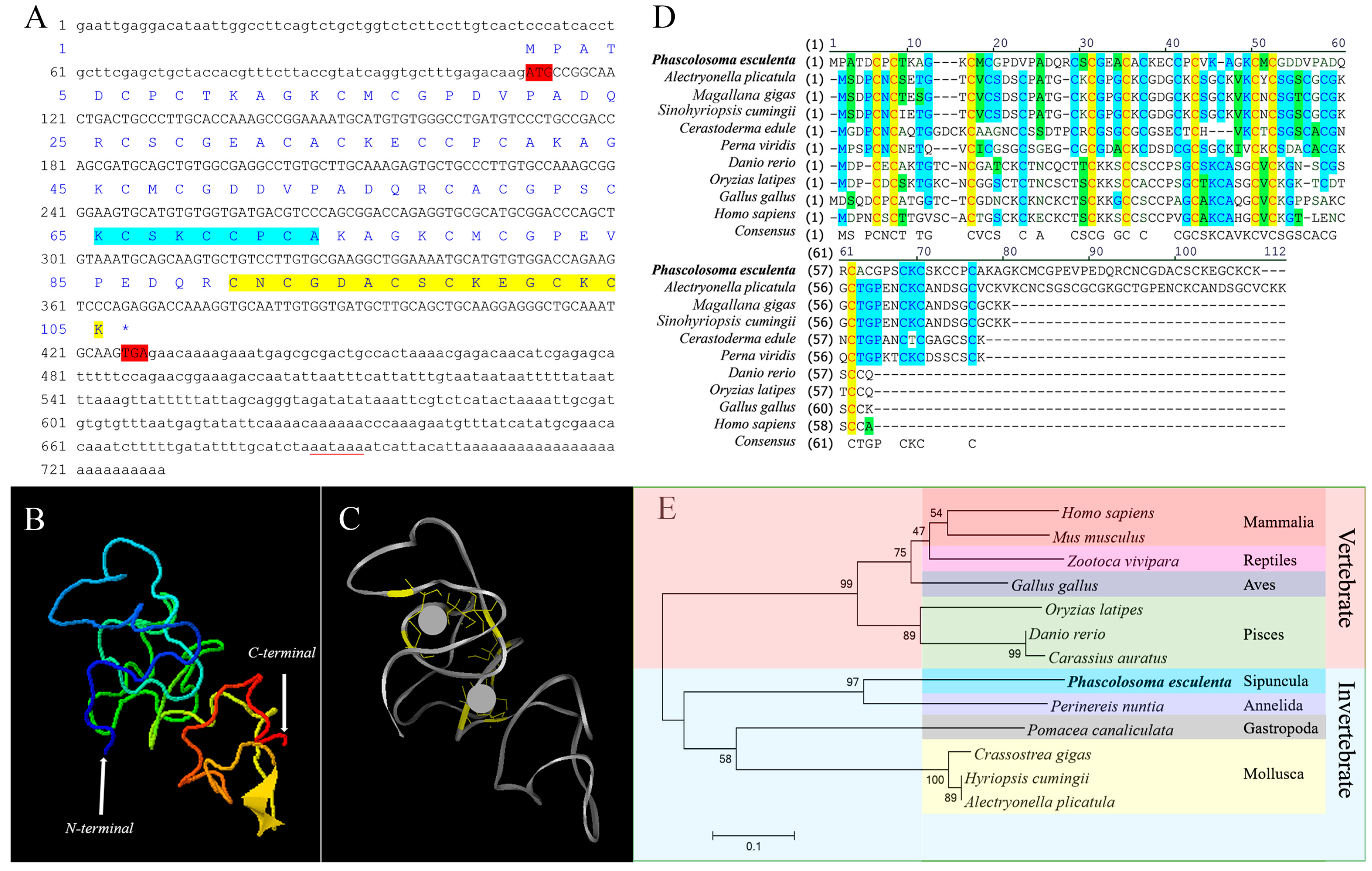
2.1. Sequence Features and Bioinformatics Analysis of PeMT

The full-length cDNA of PeMT was 730 bp in length (GenBank Accession No. QQ848618.1), containing a 110 bp 5′-untranslated region (UTR), a 318 bp open reading frame (ORF) that encoded 105 amino acids, and a 302 bp 3′-UTR with a conserved polyadenylation signal (AATAAA). The predicted molecular weight was 10.7 kDa, and the isoelectric point was 7.63. The “CKCSKCCPCA” was consistent with the conserved motif “CKCXXXCXCX” of invertebrate MTs and the “CNCGDACSCKEGCKCK” in the C-terminus was similar to the conserved motif “CXCXXXCTGXXXCXCXXXCXCK” of Mollusca MTs. This protein contains 28 Cys and 12 lysine (Lys) residues, and no histidine (His) or aromatic amino acids (Phe, Trp, Tyr). The 28 Cys residues were mainly presented in the forms of Cys-Cys, Cys-X-Cys, Cys-X-X-Cys, and Cys-X-X-X-Cys (Figure 1A). Among them, nine conserved Cys residues were predicted to bind to two Zn2+ units (Figure 1B,C). The similarities between PeMT and its homologs in Alectryonella plicatula, Magallana gigas, Sinohyriopsis cumingii, Cerastoderma edule, Perna viridis, Gallus gallus, Oryzias latipes, Danio rerio, and Homo sapiens were 37.1%, 36.8%, 33.3%, 35.2%, 32.4%, 25.7%, 20.0%, 17.1%, and 16.2%, respectively (Figure 1D), indicating that PeMT has low homology with other homologs. Phylogenetic tree analysis revealed that the MT homologs were divided into invertebrate and vertebrate branches. PeMT clustered in the invertebrate branch and had the closest relationship with MT from Perinereis nuntia among the selected species (Figure 1E).

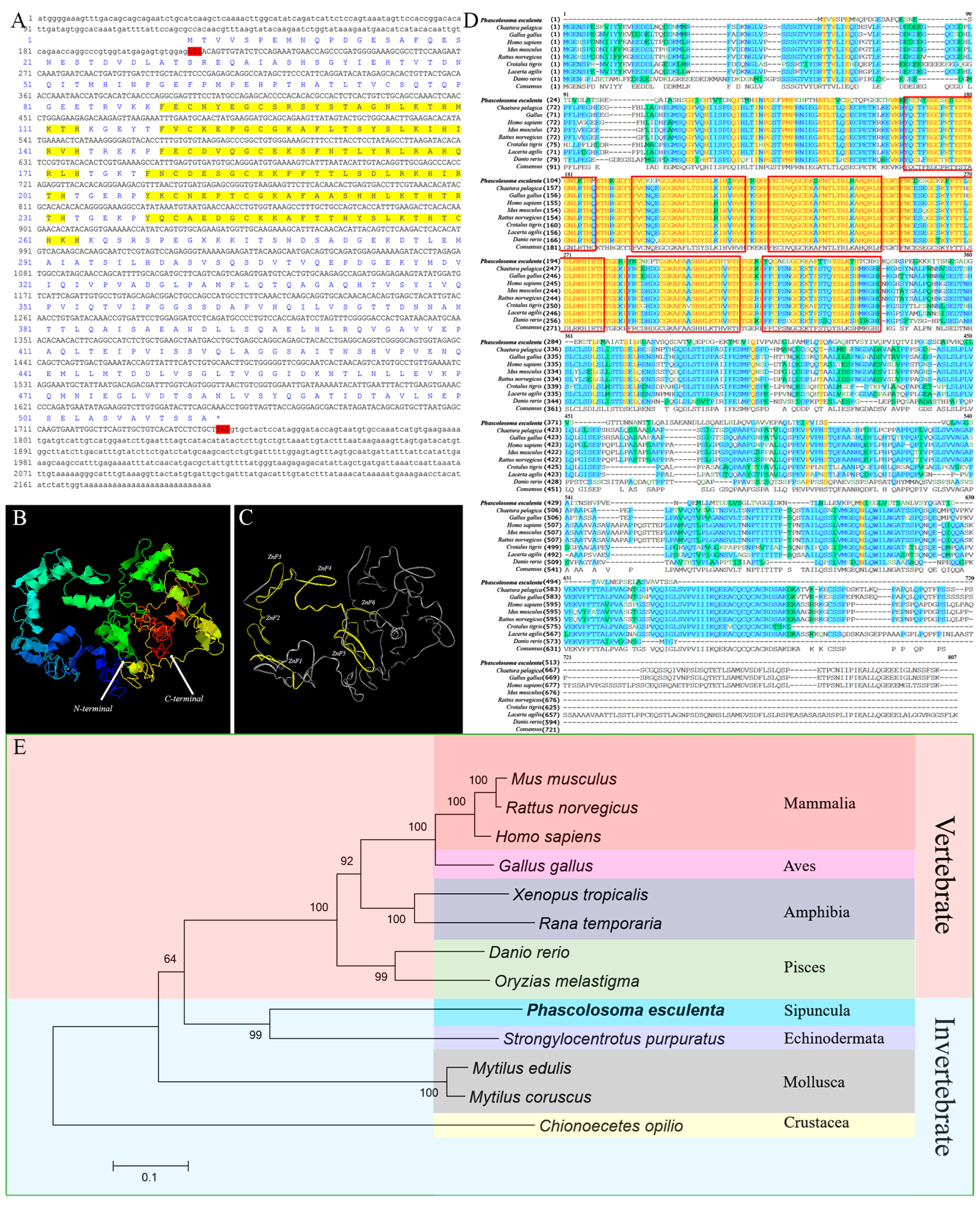
2.2. Sequence Features and Bioinformatics Analysis of PeMTF1

The full-length cDNA of PeMTF1 was 2197 bp in length (GenBank Accession No. QQ848619.1), containing a 212 bp 5′-UTR, a 1539 bp ORF that encoded 512 amino acids, and a 446 bp 3′-UTR (Figure 2A). The predicted molecular weight was 55.74 kDa, and the isoelectric point was 5.83. Moreover, PeMTF1 was predicted to be localized in the cytoplasm and nucleus. The N-terminus of PeMTF1 contains six conserved C2H2-type zinc finger domains (Figure 2B,C). The similarities between PeMTF1 and its homologs in Chaetura pelagica, G. gallus, H. sapiens, Mus musculus, Rattus norvegicus, Crotalus tigris, Lacerta agilis, and D. rerio were 36.4%, 37.0%, 34.4%, 37.0%, 38.7%, 39.9%, 36.5%, and 43.0%, respectively. However, the similarities between their zinc finger domains were 84.0%, 84.0%, 84.6%, 84.6%, 84.6%, 84.6%, 84.6%, and 85.7%, respectively (Figure 2D), indicating that PeMTF1 has low homology with the other homologs, but the zinc finger domains are highly conserved. Phylogenetic tree analysis showed that the MTF1 homologs were divided into invertebrate and vertebrate branches. PeMTF1 clustered in the invertebrate branch and had the closest relationship with MTF1 from Strongylocentrotus purpuratus among the selected species (Figure 2E).

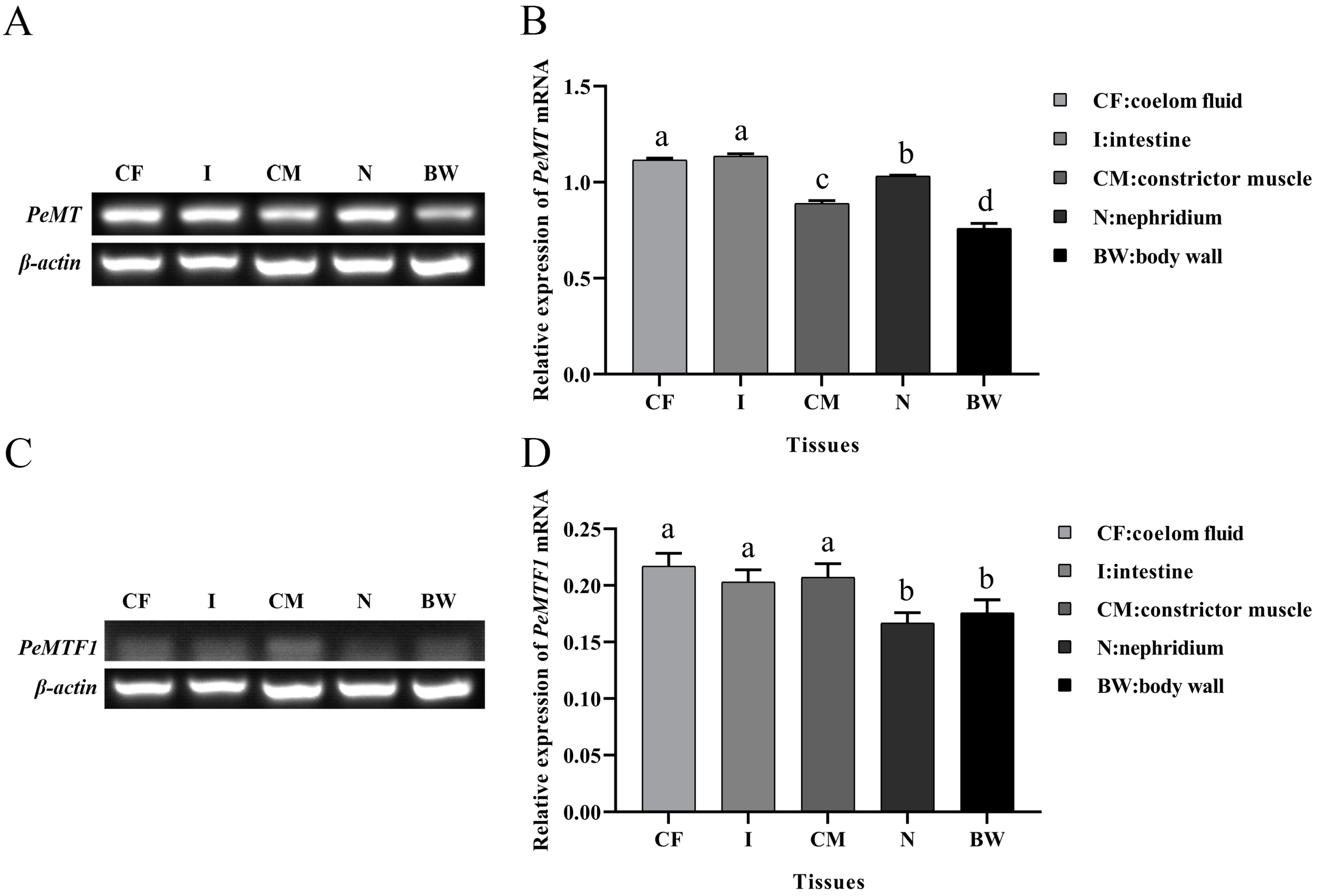
2.3. Tissue-Specific Expression of PeMT and PeMTF1 mRNA

The relative expression levels of PeMT and PeMTF1 mRNA in the coelom fluid, intestine, constrictor muscle, nephridium, and body wall were detected using RT-PCR. The results showed that PeMT and PeMTF1 mRNA were widely expressed in all tissues. PeMT mRNA had the highest expression level in the intestine, followed by the coelom fluid, nephridium, constrictor muscle, and body wall (Figure 3A,B), whereas PeMTF1 mRNA showed the higher expression levels in the coelom fluid, constrictor muscle, and intestine than in the body wall and nephridium (Figure 3C,D).

2.4. Expression Changes of PeMT and PeMTF1 mRNA in the Intestine under Zn Stress

The temporal expression profiles of PeMT and PeMTF1 mRNA in the intestine of P. esculenta, following exposure to different concentrations of Zn, are shown in Figure 4. No significant differences were observed in the control group (p > 0.05). However, they were upregulated first and then downregulated under Zn stress and reached the maximum at 12 and 6 h, respectively. When stressed for 24 and 48 h, the expression level of PeMT mRNA in the 84 mg/L group was significantly higher than that in the other groups (p < 0.05). After 96 h of Zn stress, the expression level of PeMT mRNA in the Zn stress groups was significantly lower than that in the control group (p < 0.05). Additionally, the expression level of PeMTF1 mRNA in the Zn stress groups was higher than that in the control group at all time points (except at 96 h of exposure to 84 mg/L Zn2+).

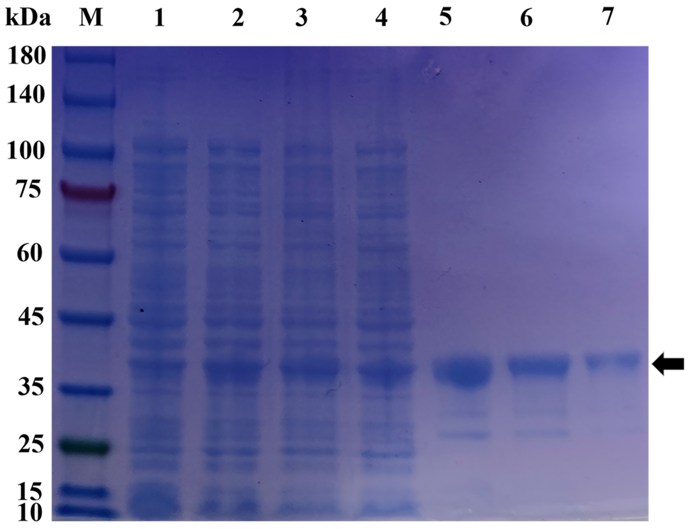
2.5. Prokaryotic Expression and Protein Purification of pGEX-6p-1-MT

The recombinant pGEX-6p-1-MT protein was obtained using a prokaryotic expression system. SDS-PAGE (sodium dodecyl sulfate–polyacrylamide gel electrophoresis) analysis showed that it was expressed in the supernatant and precipitate of broken E. coli cells. Then, the supernatant was purified, and a single band with a molecular weight of 36.7 kDa was obtained, which was consistent with the theoretical value predicted by the Expasy ProtParam tool (Figure 5). The concentration of the purified pGEX-6p-1-MT protein was 1.44 mg/mL.

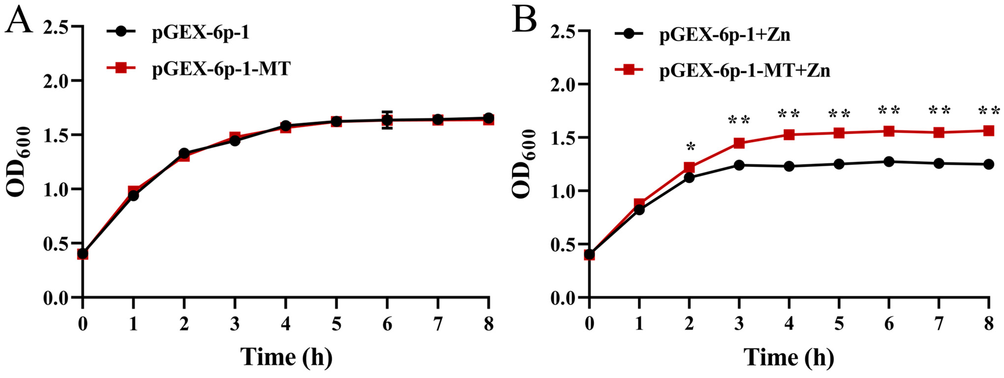
2.6. Zn Tolerance of the Recombinant E. coli

As shown in Figure 6, the growth rates of the bacteria that transformed pGEX-6p-1-MT and pGEX-6p-1 were almost the same in the control group, whereas they were inhibited by 0.3 mM Zn stress. The OD600 value of the recombinant strain was significantly higher than that of the control strain after 2 h of Zn stress (p < 0.05) and reached an extremely significant level after 3 h of Zn stress (p < 0.01).

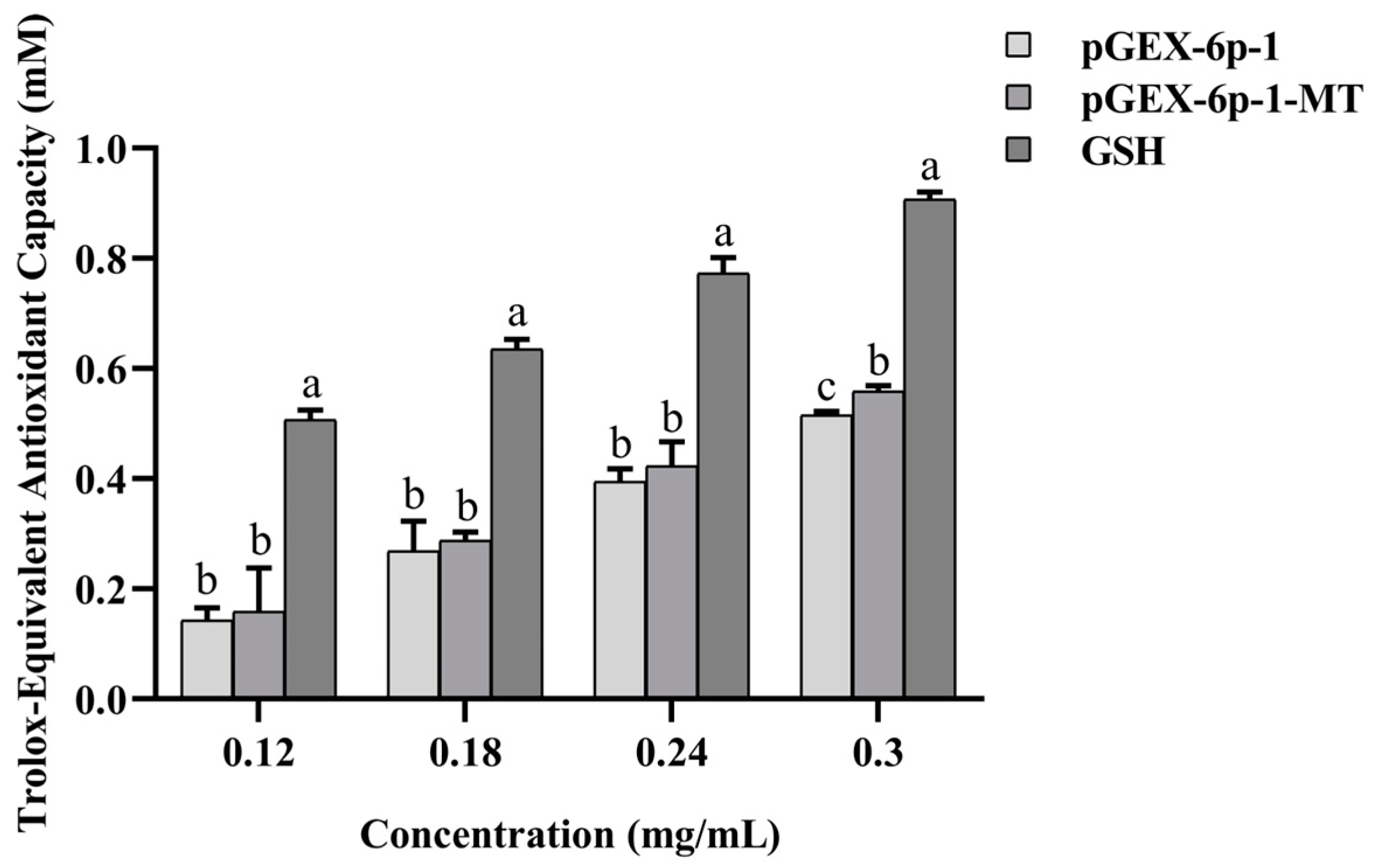
2.7. Antioxidant Capacity of the Recombinant pGEX-6p-1-MT

The ABTS radical scavenging abilities of GSH, pGEX-6p-1-MT, and pGEX-6p-1 were positively correlated with their concentrations and were in the following order: GSH > pGEX-6p-1-MT > pGEX-6p-1. At concentrations of 0.12, 0.18, and 0.24 mg/mL, no significant differences were observed between pGEX-6p-1-MT and pGEX-6p-1 (p > 0.05). However, at 0.3 mg/L, the ABTS radical scavenging ability of pGEX-6p-1-MT was significantly higher than that of pGEX-6p-1 (Figure 7).

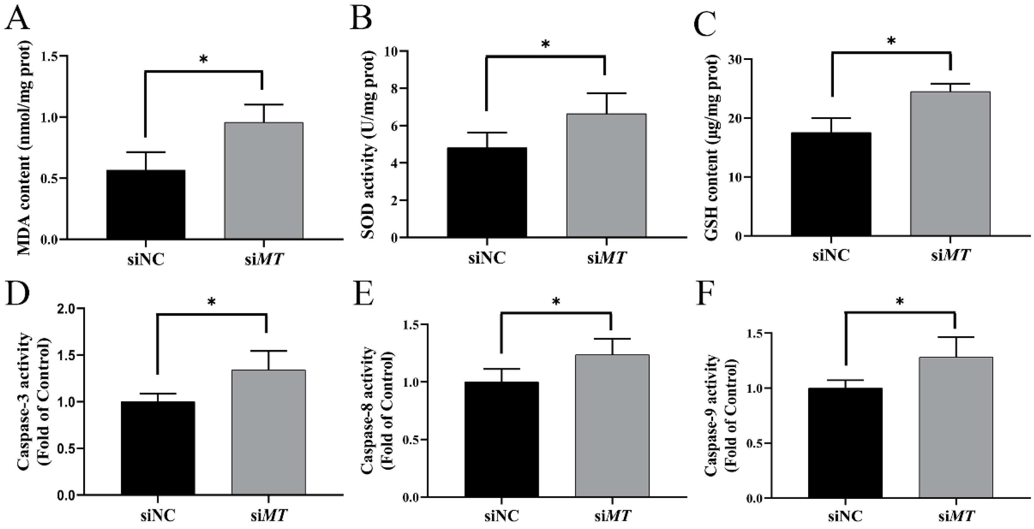
2.8. Changes in Intestinal Oxidative Stress and Apoptosis Indices after RNAi with PeMT and Zn Stress

Compared to the siNC group, the expression level of intestinal PeMT mRNA in the siMT group was significantly decreased by 46.89% after RNAi and 24 h of Zn stress (p < 0.05) (Figure 8). At the same time, the malondialdehyde (MDA) content, superoxide dismutase (SOD) activity, and glutathione (GSH) content were significantly increased to 169%, 137.76%, and 139.62% of the siNC group, respectively (p < 0.05) (Figure 9A–C). Moreover, the Caspase 3, Caspase 8, and Caspase 9 activities were significantly increased to 134.14%, 123.64%, and 128.13% of the siNC group, respectively (p < 0.05) (Figure 9D–F).

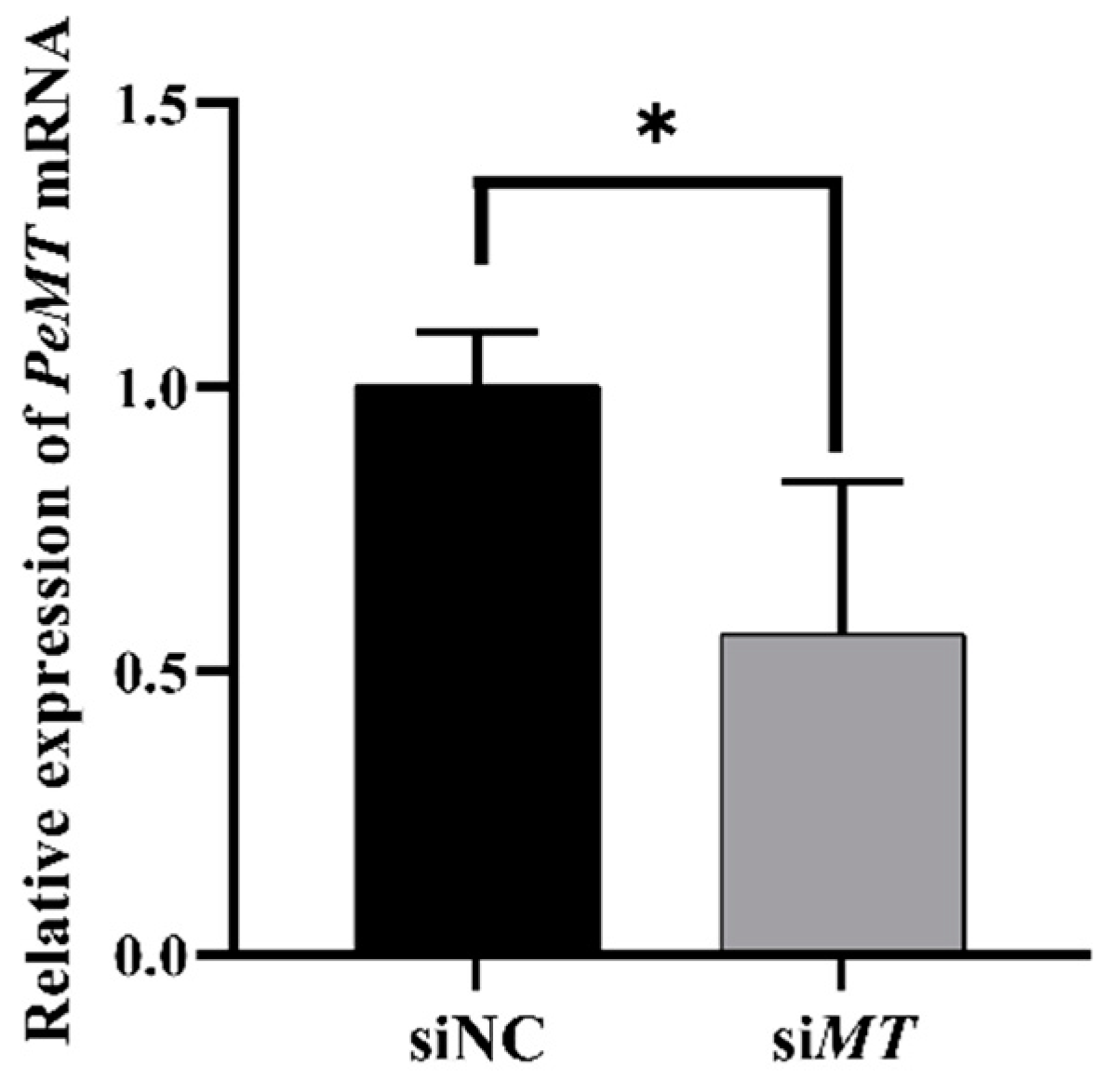
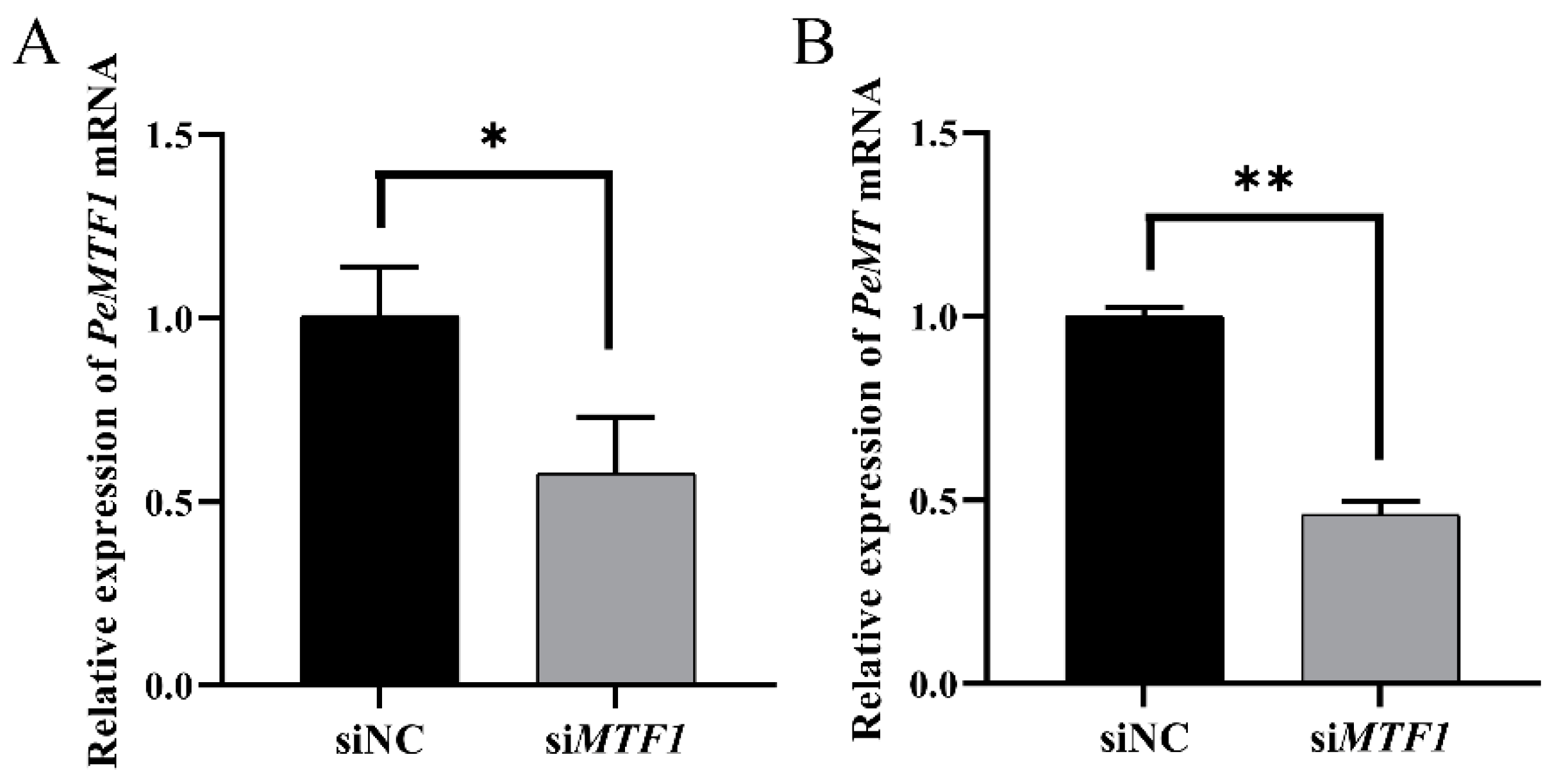
2.9. Changes in Intestinal PeMT mRNA Expression Level after RNAi with PeMTF1 and Zn Stress

Compared to the siNC group, the expression level of intestinal PeMTF1 mRNA in the siMTF1 group was significantly decreased by 42.69% (p < 0.05) (Figure 10A), and the expression level of intestinal PeMT mRNA decreased by 53.95% (p < 0.01) (Figure 10B).

3. Discussion

3.1. Sequence Features and Protein Structure of PeMT and PeMTF1

The full-length PeMT cDNA was cloned from P. esculenta. The “CKCSKCCPCA” was in line with the conserved motif “CKCXXXCXCX” of invertebrate MTs, and the “CNCGDACSCKEGCKCK” in the C-terminus was similar to the conserved motif “CXCXXXCTGXXXCXCXXXCXCK” of Mollusca MTs [23]. Moreover, the conserved polyadenylation signal “AATAAA” was observed in the 3′-UTR, which is crucial for mRNA localization and translation [24] and has been found in the MTs of Mollusca [25], Crustacea [26], and Pisces [27]. PeMT showed low similarity to other homologs, and similar results have been found in invertebrate MTs of Hermetia illucens [28], Oxya chinensis [29], and Apostichopus japonicus [30]. PeMT contains 28 Cys and 12 Lys residues. These Cys residues exist in the motifs of Cys-Cys, Cys-X-Cys, Cys-X-X-Cys, and Cys-X-X-X-Cys, which show great affinity for binding heavy metals such as Zn, Cu, and Cd, and participate in divalent detoxification and maintain the stability of metal binding clusters together with Lys [25,31,32]. Studies have shown that the tertiary structure of vertebrate MTs is conserved, consisting of an α domain and a β domain, which is dumbbell-shaped [33]. However, it varies among invertebrate MTs. For example, Charybdis japonica and Portunus trituberculatus [31,32] MTs consist of two β domains. The Crassostrea gigas MT1 consists of an α domain and a β domain, and the C. gigas MT2 consists of an α domain and two β domains [34]. Moreover, the Drosophila melanogaster MT has only one domain [35]. In this study, PeMT was also composed of one domain, further proving that MTs convergently evolved.
Additionally, the full-length PeMTF1 cDNA was cloned. PeMTF1 was predicted to be located in the cytoplasm and nucleus. Previous studies have shown that MTF1 is located in the cytoplasm and nucleus under normal conditions. However, when cells are stimulated by heavy metals, hypoxia, and other external stimuli. MTF1 may translocate to the nucleus and bind to serval target genes [17]. Moreover, sequence analysis showed that the N-terminus of PeMTF1 has six conserved C2H2-type zinc finger domains that have high homology with other homologs. The zinc finger domains play important roles in metal regulation and DNA binding [36], and their high conservation indicates that they may have conserved functions. However, in other regions, PeMTF1 showed low similarity to other homologs and lacked transactivation domains. In a study by Ren et al. [37], MTF1 homologs of Cerapachys biroi, Hymenoptera, and Oxya chinesis showed similar characteristics. In addition, Ferencz et al. [38] found that the splicer MTF1-1.1a of Cyprinus carpio has six zinc finger domains and a truncated acidic domain but lacks proline and serine/threonine domains. Based on these results, we identified PeMTF1 as a new member of the MTF1 superfamily.

3.2. Tissue-Specific Expression of PeMT and PeMTF1 and Their Expression Changes under Zn Stress

Extensive studies have reported that MT is widely expressed in all vertebrate and invertebrate tissues [39], and tissues with high expression levels of MT were determined to be different among species. For example, Meretrix meretrix [40], Haliotis discus hannai [41], and Litopenaeus vannamei [42] showed the highest MT expression levels in the liver, while MTs of liverless species such as A. japonicus [30], H. illucens [28], and O. chinensis [29] were highly expressed in the intestine. Consistent with these findings, PeMT was widely expressed in the tested tissues, with the intestine showing the highest expression. The intestine is one of the main organs involved in nutrient absorption and immune defense [43,44], and high expression of PeMT indicates that PeMT plays an important role in the intestine of P. esculenta. Therefore, we further analyzed the temporal expression profile of intestinal PeMT under four concentrations (0, 16.8, 33.6, and 84 mg/L) of Zn2+. The expression pattern of PeMT showed an inverted “U” shape as the stress time increased, which is consistent with previous studies [45]. For instance, when exposed to Cd and Zn, the expression level of MT in the gills of Septifer virgatus increased significantly and reached a maximum after 5 days [46]. In C. japonica, the expression level of MT significantly increased within 12 h of Pb stress and then slowly decreased [47]. Additionally, A. japonicus MT displayed the same trend under Cd and Zn stress [30]. The increase in MTs in the early stages indicates that MTs rapidly respond to heavy metal stress [46], whereas the decrease in the later stages may be related to peroxidation stress. These findings confirm our speculation that PeMT is a functional gene involved in Zn detoxification in P. esculenta and is expected to be a biomarker for monitoring Zn pollution.
PeMTF1 was also expressed in the tested tissues, and its expression levels in the coelom fluid, constrictor muscle, and intestine were higher than those in the body wall and nephridium. Under Zn stress, it showed the same trend as PeMT and reached the maximum at 6 h. In C. carpio, the expression of MTF1-1.1a was first upregulated and then downregulated under Cd stress, reaching a maximum at 48 h [38]. In the liver of Pelteobagrus fulvidraco, the expression levels of MTF1 and MT significantly increased within 48 h of Zn stress [17]. In C. gigas, the expression levels of MTF1 and MT were induced under 0.1 ppm Cd, and the response of MTF1 was faster than that of MT [48]. These findings indicate that MTF1 plays an important role in the response to oxidative stress induced by heavy metals, and its response is faster than that of MT, which may be involved in the regulation of MT.

3.3. Detoxification, Antioxidant, and Anti-Apoptosis Functions of PeMT

MTs play an important role in heavy metal detoxification and increase metal tolerance in organisms [49]. Wang et al. [50] inserted Sinopotamon MT into the pGEX-4t-1 vector and exposed it to varying concentrations of Cd, Cu, and Zn. It was found that the recombinant strain showed stronger metal tolerance than the control group. Yang et al. [40] constructed the recombinant pET32a-MnMT and found that it could bind to Cu2+ and Cd2+, making the recombinant strain more resistant to toxic heavy metals. Li et al. [51] constructed the recombinant pET28a-NtMT2F and found that the growth rate of the recombinant strain was faster than that of the control group when exposed to Cd. In the present study, we induced the expression of PeMT in E. coli and exposed the cells to Zn2+. The results showed that Zn2+ inhibited the growth of E. coli, but the growth of the recombinant strain was better than that of the control. Based on these results, we believe that PeMT exerts detoxifying effects and enhances Zn tolerance in E. coli.
The sulfhydryl group of MTs exists in a reduced state and can react with free radicals to exert an antioxidant function [52,53]. Recombinant human MT-III has been reported to reduce the oxidative damage caused by Cu and Cd stress [54]. Purified Channa punctate MT has been shown to have in vitro activities; it plays an important role in scavenging superoxide radicals (O2−), ABTS radicals, and DPPH radicals and can protect the body from oxidative damage induced by Fe-NTA [55]. In this study, the recombinant pGEX-6p-1-MT protein was used to conduct an in vitro experiment. The results showed that PeMT exhibited ABTS radical scavenging ability in a dose-dependent manner. These findings indicated that PeMT has antioxidant functions and plays an important role in resisting Zn-induced oxidative stress.
To further investigate the functions of PeMT, we performed RNAi at the individual level. After RNAi with PeMT and 24 h of Zn stress, the expression level of PeMT was significantly decreased by 46.89%, and the oxidative indices (MDA, SOD, and GSH) and apoptosis indices (Caspase 3, Caspase 8, and Caspase 9) were significantly increased. MDA is the product of lipid peroxidation, and its content is related to the degree of oxidative damage [56]. SOD is the first barrier of organisms’ antioxidant defense, and it can disproportionate the superoxide anion (O2−) to H2O2 and O2 [57]. GSH acts as the substrate of GSH-Px and is involved in the removal of H2O2 [58]. Additionally, when oxidative stress exceeds physiological tolerance, Caspase 8 and Caspase 9 can bind to apoptotic signals, thus activating Caspase 3 and causing apoptosis [59]. In this study, the oxidative stress and apoptosis indices of the siMT group were significantly increased, indicating that the degree of oxidative damage and apoptosis was more severe in the siMT group, which further proved that PeMT plays an important role in antioxidation and anti-apoptosis.

3.4. Regulation Effect of PeMTF1 on PeMT

To explore the regulatory effects of PeMTF1 on PeMT, we performed RNAi experiments at the individual level. After RNAi with PeMTF1 and 24 h of Zn stress, the expression level of PeMT significantly decreased (p < 0.05). Similarly, Meng et al. [60] found that the expression levels of CgMT1 and CgMT4 in the gills of C. gigas were significantly decreased after RNAi with CgMTF1 and 48 h of Zn stress. Ren et al. [37] found that the expression level of OcMT in O. chinensis significantly decreased after RNAi with OcMTF1. Troadec et al. [61] knocked down MTF1 in NIH 3T3 cells and found that the expression level of MT was significantly decreased. Moreover, Atanesyan et al. [62] found that the MTnE of Drosophila was almost unexpressed after RNAi with MTF1 and was highly expressed after the overexpression of MTF1. These findings suggest that PeMTF1 has a conserved function and participates in the regulation of PeMT. PeMT and PeMTF1 are key factors allowing P. esculenta to endure the toxicity of heavy metal pollution.

4. Materials and Methods

4.1. Animals

P. esculenta individuals (3.9 ± 0.5) g were collected from Xizhou of Ningbo, Zhejiang, China. They were then kept in filtered seawater at a temperature of 20.5 ± 0.5 °C, salinity of 25.5 ± 0.5‰, and pH of 7.6 ± 0.2 for four days.

4.2. Treatments and Sampling

Preliminary experiments showed that the 96 h median lethal concentration (LC50) of Zn2+ for P. esculenta was 167.909 mg/L. Based on this value, four Zn2+ concentrations (0, 16.8, 33.6, and 84 mg/L) were set for 96 h of stress. Three tanks were used for each concentration, and 30 individuals were placed in each tank. The seawater was changed daily, and the Zn concentrations were kept constant.
Samples of the coelom fluid, intestine, constrictor muscle, nephridium, and body wall were collected from six individuals in the control group at 0 h, and the intestines of six individuals at each concentration were sampled after 6, 12, 24, 48, and 96 h of Zn stress. At each time point, two individuals were randomly selected from each tank, they were then sampled and mixed into one sample. The coelom fluid was extracted with a 1 mL syringe. Then, P. esculenta individuals were placed on the ice, and the intestines, constrictor muscles, nephridia, and body walls were collected and washed with distilled water. Finally, the samples were immediately frozen in liquid nitrogen and then stored at −80 °C. All experimental procedures were approved by the Animal Care and Use Committee of Ningbo University (Ningbo, China).

4.3. RNA Extraction and cDNA Synthesis

The RNA-Solv Reagent (Omega, GA, USA) was used to extract total RNA from the tissues of P. esculenta. Some of the obtained RNA was reverse transcribed by PrimeScript® RT Kit (Takara, Dalian, China) for intermediate fragment cloning and tissue expression analysis, and the remaining was reverse transcribed by SMARTer RACE 5′/3′ reagent (Takara, Dalian, China) for 5′ and 3′ RACE (rapid amplification of cDNA ends).

4.4. Full-Length cDNA Cloning of PeMT and PeMTF1

We obtained the ORFs of PeMT and PeMTF1 from the transcriptome data of P. esculenta (GenBank Accession No. OL757513) and designed specific primers using Primer Premier 5.0 for verification. Based on the verification results, specific primers for 5′ and 3′ RACE were designed, and full-length cloning was carried out. The primers used are listed in Table S1.
The PCR procedures involved the following steps: 94 °C 5 min; 8 cycles of 94 °C 30 s, 57 °C 30 s (with a decrease of 0.5 °C/cycle), and 72 °C 30 s; 27 cycles of 94 °C 30 s; 53 °C 30 s, and 72 °C 30 s; 72 °C 10 min. The product was separated by 1% agarose gel electrophoresis and purified by DNA Gel Extraction Kit (BioTeke, Beijing, China). Next, the purified product was ligated to the pMD-19T vector (Takara, Dalian, China) and transformed into Trans5α (TransGen Biotech, Beijing, China). Finally, positive clones were identified and sequenced by Zhejiang Youkang Biotechnology Co., Ltd. (Youkang, Zhejiang, China).

4.5. Bioinformatics Analysis of PeMT and PeMTF1

The ORFs of PeMT and PeMTF1 were predicted by NCBI’s ORFfinder tool (https://www.ncbi.nlm.nih.gov/orffinder/, accessed on 16 June 2023). The sequences in the ORFs of PeMT and PeMTF1 were translated into amino acids using the Bioxm 2.6 software. The molecular weights and isoelectric points of PeMT and PeMTF1 were predicted by the Expasy ProtParam tool (https://web.expasy.org/protparam/, accessed on 8 July 2023). The intracellular localization of PeMTF1 was predicted by Cell-PLoc 2.0 (http://www.csbio.sjtu.edu.cn/bioinf/Cell-PLoc-2/, accessed on 20 June 2024). The DNA-binding domain of PeMTF1 was predicted by Smart (http://smart.embl-heidelberg.de/, accessed on 20 June 2024). The tertiary structures of PeMT and PeMTF1 were predicted by the I-TASSER (https://zhanglab.ccmb.med.umich.edu/I-TASSER/, accessed on 8 July 2023). The multiple sequence alignment and phylogenetic tree analysis of the MT and MTF1 homologs were performed by Vector NTI 11.5 and MEGA 5.1 software. The species and their GenBank Accession numbers used in the multiple sequence alignment and phylogenetic tree analysis are listed in Tables S2 and S3.

4.6. Tissue-Specific Expression of PeMT and PeMTF1 mRNA and Its Expression Changes under Zn Stress

Tissue-specific expression of PeMT and PeMTF1 mRNA in the coelom fluid, intestine, constrictor muscle, nephridium, and body wall was monitored by RT-PCR, with the β-actin serving as an internal reference. The PCR procedures were as follows: 94 °C 5 min; 30 cycles of 94 °C 15 s, 55 °C 15 s, and 72 °C 15 s; 72 °C 5 min. The PCR products were separated by 1% agarose gel electrophoresis, visualized by a gel image analysis system (FR, Shanghai, China), and then analyzed by Image J software. All data are presented as mean ± SD (n = 3).
RT-qPCR was conducted to study the temporal expression profiles of intestinal PeMT and PeMTF1 mRNA under Zn stress. The PCR procedures were as follows: 95 °C 5 min; 40 cycles of 95 °C 30 s, 55 °C 30 s, and 72 °C 30 s; 72 °C 5 min. Finally, the relative expression level of PeMT and PeMTF1 mRNA was calculated by the 2−ΔΔCT method. All data are presented as mean ± SD (n = 3). The primers used are listed in Table S1.

4.7. Expression and Purification of the Recombinant pGEX-6p-1-MT

Primers with restriction sites (BamHI and XhoI) were used to amplify the ORF of PeMT. The PCR product and pGEX-6p-1 vector were double digested with BamHI and XhoI restriction enzymes (Thermo Fisher Scientific, Waltham, MA, USA) and then ligated by T4 ligase (Takara, Dalian, China). The recombinant plasmid was transformed into Trans5α (TransGen Biotech, Beijing, China) and cultured overnight on a solid LB medium with 100 μg/mL of Ampicillin. The following day, positive clones were screened, and plasmids were extracted by the Plasmid Extraction Mini Kit (Solarbio, Beijing, China).
After sequencing, the correct recombinant plasmid was transformed into Transetta (DE3) (TransGen Biotech, Beijing, China) and cultured in the liquid LB medium with 100 μg/mL Ampicillin at 37 °C and 180 rpm. When the OD600 reached a density of 0.5–0.7, 1 mM of IPTG was added to induce the expression of the recombinant protein. Based on the SDS-PAGE analysis, the supernatant was purified by the GST-tag Protein Purification Kit (Beyotime, Shanghai, China).

4.8. Zn Tolerance of the Recombinant E. coli

The recombinant E. coli strains that transformed pGEX-6p-1-MT and pGEX-6p-1 were cultured in liquid LB medium with 100 μg/mL Ampicillin until the OD600 reached 0.4, and then 1 mM of IPTG was added. They were then divided into control and 0.3 mM Zn stress groups based on our pre-experiment (Figure S1), and their OD600 was measured in the following 8 h. The results are presented as mean ± SD (n = 3).

4.9. Antioxidant Capacity of the Recombinant pGEX-6p-1-MT

The pGEX-6p-1 protein was purified as described previously. We determined the concentrations of pGEX-6p-1 and pGEX-6p-1-MT using the Bradford Protein Concentration Assay Kit (Beyotime, Shanghai, China) and prepared 1 mg/mL of GSH. Then, they were diluted to 0.12, 0.18, 0.24, and 0.3 mg/mL, and the ABTS free radical scavenging ability was determined by the Total Antioxidant Capacity Assay Kit (Beyotime, Shanghai, China).

4.10. Detection of Intestinal Oxidative Stress and Apoptosis Indices after RNAi with PeMT and Zn Stress

Interference primers were designed and synthesized by Shanghai GenePharma Technology Co., Ltd. (GenePharma, Shanghai, China) (Table S1). Eight P. esculenta individuals (3.9 ± 0.5 g) were divided into siNC and siMT groups. Lipo6000TM (Beyotime, Shanghai, China) and siRNA were diluted with 1 × PBS at a volume ratio of 17:3 and mixed at a volume ratio of 1:1. After mixing, the mixture was stored at room temperature for 5 min and injected at a dose of 1 μg/g. Then, P. esculenta individuals were exposed to 33.6 mg/L Zn for 24 h. The intestines were then collected and stored at −80 °C.
The interference efficiency of PeMT mRNA was determined by RT-qPCR using the method described above. Intestines were accurately weighed and mixed with extract solution in the ratio of weight (g)/volume (mL) = 1:10. The mixture was then homogenized in an ice bath and centrifuged at 10,000× g for 8 min at 4 °C. The oxidative stress indices (MDA, SOD, and GSH) and apoptosis indices (Caspase 3, Caspase 8, and Caspase 9) were measured by assay kits according to the instructions (Solarbio, Beijing, China).

4.11. Detection of Intestinal PeMT mRNA Expression Level after RNAi with PeMTF1 and Zn Stress

The RNAi method for PeMTF1 was similar to that for PeMT. After sampling, the interference efficiency of PeMTF1 mRNA and changes in the expression of PeMT mRNA were detected by RT-qPCR, as described above.

4.12. Statistical Analysis

SPSS 26.0, Excel 2016, and GraphPad Prism 8.0 were used for statistical analysis, and significance was tested using one-way ANOVA followed by Duncan’s test and independent samples t-test. Different letters simultaneously indicate a significant difference among the groups (p < 0.05).

5. Conclusions

PeMT and PeMTF1 were cloned and identified from P. esculenta. Their expression patterns indicate that they respond rapidly to Zn stress. Prokaryotic expression, biochemical detection, and RNAi experiments provided evidence for the functions of PeMT and PeMTF1, suggesting that PeMT plays an important role in Zn detoxification, antioxidation, and anti-apoptosis, and its expression is regulated by PeMTF1, proving that PeMT and PeMTF1 are key factors allowing P. esculenta to endure the toxicity of Zn. This study made up for the gap in the functional research of MT and MTF1 in Sipuncula and accumulated basic data for studying the molecular toxicology of P. esculenta.

Supplementary Materials

The following supporting information can be downloaded at: https://www.mdpi.com/article/10.3390/ijms25137368/s1.

Author Contributions

Conceptualization, J.W. and L.S.; data curation, S.G.; formal analysis, S.G., X.G., X.Z., Y.C. and L.S.; funding acquisition, J.Z.; investigation, J.W. and Y.C.; methodology, S.G., J.W., Y.L. and L.S.; project administration, J.Z.; resources, X.Z., L.S. and J.Z.; software, J.W., Y.C. and L.S.; supervision, J.W., X.G., X.Z., Y.L. and L.S.; validation, S.G., X.G. and Y.C.; visualization, Y.L.; writing—original draft, S.G. and Y.C.; writing—review and editing, S.G., X.Z., Y.C., L.S. and J.Z. All authors have read and agreed to the published version of the manuscript.

Funding

This research was funded by the Ningbo Science and Technology Plan Projects (2019B10016, 2016C10004), the Major Science and Technology Projects in Zhejiang Province (2011C12013), and the Collaborative Innovation Center for Zhejiang Marine High-efficiency and Healthy Aquaculture, the K.C. Wong Magna Fund of Ningbo University.

Institutional Review Board Statement

The experimental animal of this study is P. esculenta, a species of invertebrate belonging to Sipuncula. In China, this species does not require ethical approval for experiments. All experiments comply with the requirements of the governing regulation for the use of experimental animals in Zhejiang Province (Zhejiang provincial government order No. 263, released on 17 August 2009, effective from 1 October 2010) and the Animal Care and Use Committee of Ningbo University.

Informed Consent Statement

Not applicable.

Data Availability Statement

Data are contained within the article and Supplementary Materials.

Conflicts of Interest

The authors declare no conflict of interest.

References

  1. Tchounwou, P.B.; Yedjou, C.G.; Patlolla, A.K.; Sutton, D.J. Heavy Metal Toxicity and the Environment. Exp. Suppl. 2012, 101, 133–164. [Google Scholar] [PubMed]
  2. Cui, Y.B.; Bai, L.; Li, C.H.; He, Z.J.; Liu, X.R. Assessment of heavy metal contamination levels and health risks in environmental media in the northeast region. Sustain. Cities Soc. 2022, 80, 103796. [Google Scholar] [CrossRef]
  3. Wu, J.; Lu, J.; Zhang, C.; Zhang, Y.X.; Lin, Y.C.; Xu, J. Pollution, sources, and risks of heavy metals in coastal waters of China. Hum. Ecol. Risk. Assess. 2019, 26, 2011–2026. [Google Scholar] [CrossRef]
  4. Wu, D.; Feng, H.; Zou, Y.; Xiao, J.; Zhang, P.F.; Ji, Y.X.; Lek, S.; Guo, Z.Q.; Fu, Q.Y. Feeding Habit-Specific Heavy Metal Bioaccumulation and Health Risk Assessment of Fish in a Tropical Reservoir in Southern China. Fishes 2023, 8, 211. [Google Scholar] [CrossRef]
  5. Roohani, N.; Hurrell, R.; Kelishadi, R.; Schulin, R. Zinc and its importance for human health: An integrative review. J. Res. Med. Sci. 2013, 18, 144–157. [Google Scholar] [PubMed]
  6. Chandra, R.K. Excessive intake of zinc impairs immune responses. JAMA 1984, 252, 1443–1446. [Google Scholar] [CrossRef] [PubMed]
  7. Evans, P.; Halliwell, B. Micronutrients: Oxidant/antioxidant status. Br. J. Nutr. 2007, 85, 67–84. [Google Scholar] [CrossRef]
  8. Ighodaro, O.M.; Akinloye, O.A. First line defence antioxidants-superoxide dismutase (SOD), catalase (CAT) and glutathione peroxidase (GPX): Their fundamental role in the entire antioxidant defence grid. Alex. J. Med. 2019, 54, 287–293. [Google Scholar] [CrossRef]
  9. Plum, L.M.; Rink, L.; Haase, H. The Essential Toxin: Impact of Zinc on Human Health. Int. J. Environ. Res. Public Health 2010, 7, 1342–1365. [Google Scholar] [CrossRef]
  10. Bakiu, R.; Pacchini, S.; Piva, E.; Schumann, S.; Tolomeo, A.M.; Ferro, D.; Irato, P.; Santovito, G. Metallothionein Expression as a Physiological Response against Metal Toxicity in the Striped Rockcod Trematomus hansoni. Int. J. Mol. Sci. 2022, 23, 12799. [Google Scholar] [CrossRef]
  11. Formigari, A.; Boldrin, F.; Santovito, G.; Cassidy-Hanley, D.; Clark, T.G.; Piccinni, E. Functional Characterization of the 5′-upstream Region of MTT5 Metallothionein Gene from Tetrahymena thermophila. Protist 2010, 161, 71–77. [Google Scholar] [CrossRef]
  12. Gautam, N.; Tiwari, M.; Kidwai, M.; Dutta, P.; Chakrabarty, D. Functional characterization of rice metallothionein OsMT-I-Id: Insights into metal binding and heavy metal tolerance mechanisms. J. Hazard. Mater. 2023, 458, 131815. [Google Scholar] [CrossRef] [PubMed]
  13. Chen, R.F.; Chen, P.M.; Pan, C.S.; Huang, C.C.; Chiang, E.P.I. Association of metallothionein 2A rs10636 with low mean corpuscular volume (MCV), low mean corpuscular haemoglobin (MCH) in healthy Taiwanese. Sci. Rep. 2023, 13, 1292. [Google Scholar] [CrossRef] [PubMed]
  14. Monjane Mabuie, A.; Mondlane Milisse, A.; Pedro, O.; Leão Buchir, J.; Correia, D. Mercury pollution assessment and metallothionein gene expression in tilapia (Oreochromis mossambicus) a case study of Revue River in Manica, Mozambique. Rend. Lincei-Sci. Fis. 2022, 33, 513–526. [Google Scholar] [CrossRef]
  15. Niu, S.Y.; Wang, J.J.; Chang, X.R.; Shang, M.T.; Guo, M.H.; Sun, Z.Y.; Li, Y.J.; Xue, Y.Y. Comparative oxidative damages induced by silver nanoparticles with different sizes and coatings in Caenorhabditis elegans. Toxicol. Res. 2023, 12, 833–842. [Google Scholar] [CrossRef] [PubMed]
  16. Shimoda, R.; Achanzar, W.E.; Qu, W.; Nagamine, T.; Takagi, H.; Mori, M.; Waalkes, M.P. Metallothionein is a potential negative regulator of apoptosis. Toxicol. Sci. 2003, 73, 294–300. [Google Scholar] [CrossRef]
  17. Chen, G.H.; Lv, W.H.; Xu, Y.H.; Wei, X.L.; Xu, Y.C.; Luo, Z. Functional analysis of MTF-1 and MT promoters and their transcriptional response to zinc (Zn) and copper (Cu) in yellow catfish Pelteobagrus fulvidraco. Chemosphere 2020, 246, 125792. [Google Scholar] [CrossRef] [PubMed]
  18. Han, Y.L.; Sheng, Z.; Liu, G.D.; Long, L.L.; Wang, Y.F.; Yang, W.X.; Zhu, J.Q. Cloning, characterization and cadmium inducibility of metallothionein in the testes of the mudskipper Boleophthalmus pectinirostris. Ecotoxicol. Environ. Saf. 2015, 119, 1–8. [Google Scholar] [CrossRef] [PubMed]
  19. Hardivillier, Y.; Leignel, V.; Denis, F.; Uguen, G.; Cosson, R.; Laulier, M. Do organisms living around hydrothermal vent sites contain specific metallothioneins? The case of the genus Bathymodiolus (Bivalvia, Mytilidae). Comp. Biochem. Physiol. C Toxicol. Pharmacol. 2004, 139, 111–118. [Google Scholar] [CrossRef]
  20. Gao, X.M.; Feng, B.B.; Tang, D.J.; Du, C.; Hou, C.C.; Jin, S.; Zhu, J.Q. Mitochondrial Features and Expressions of MFN2 and DRP1 during Spermiogenesis in Phascolosoma esculenta. Int. J. Mol. Sci. 2022, 23, 15517. [Google Scholar] [CrossRef]
  21. Shen, W.L.; Liu, C.; Ni, J.; Gao, X.M.; Ni, J.J.; Wang, J.P.; Jin, S.; Hou, C.C.; Wu, X.F.; Zhu, J.Q. Effects of Low Temperature Stress on the Morphology and hsp70 and hsp90 Gene Expression of Phascolosoma esculenta. J. Ocean Univ. China 2021, 20, 159–168. [Google Scholar] [CrossRef]
  22. Meng, J.J.; Gao, X.M.; Luo, S.Y.; Lin, C.W.; Du, C.; Hou, C.C.; Wang, J.P.; Jin, S.; Tang, D.J.; Zhang, C.D.; et al. Cloning, Functional Characterization and Response to Cadmium Stress of the Thioredoxin-like Protein 1 Gene from Phascolosoma esculenta. Int. J. Mol. Sci. 2021, 23, 332. [Google Scholar] [CrossRef] [PubMed]
  23. Binz, P.A.; Kägi, J.H.R. Metallothionein: Molecular evolution and classification. In Metallothionein IV; Birkhäuser: Basel, Switzerland, 1999; pp. 7–13. [Google Scholar]
  24. Hesketh, J. 3′-Untranslated regions are important in mRNA localization and translation. Biochem. Soc. Trans. 2004, 32, 990–993. [Google Scholar] [CrossRef] [PubMed]
  25. Wang, C.Y.; Sheng, J.Q.; Hong, Y.J.; Peng, K.; Wang, J.H.; Wu, D.; Shi, J.W.; Hu, B.J. Molecular characterization and expression of metallothionein from freshwater pearl mussel, Hyriopsis schlegelii. Biosci. Biotechnol. Biochem. 2016, 80, 1327–1335. [Google Scholar] [CrossRef] [PubMed]
  26. Leignel, V.; Marchand, J.; Moreau, B.; Chénais, B. Metallothionein genes from hydrothermal crabs (Bythograeidae, Decapoda): Characterization, sequence analysis, gene expression and comparison with coastal crabs. Comp. Biochem. Physiol. C Toxicol. Pharmacol. 2008, 148, 6–13. [Google Scholar] [CrossRef] [PubMed]
  27. Sheng, Z.; Yang, W.X.; Zhu, J.Q. Metallothionein from Pseudosciaena crocea: Expression and response to cadmium-induced injury in the testes. Ecotoxicology 2015, 24, 779–794. [Google Scholar] [CrossRef] [PubMed]
  28. Zhang, J.; Shi, Z.H.; Gao, Z.H.; Wen, Y.T.; Wang, W.Q.; Liu, W.; Wang, X.P.; Zhu, F. Identification of three metallothioneins in the black soldier fly and their functions in Cd accumulation and detoxification. Environ. Pollut. 2021, 286, 117146. [Google Scholar] [CrossRef] [PubMed]
  29. Liu, Y.M.; Wu, H.H.; Kou, L.H.; Liu, X.J.; Zhang, J.Z.; Guo, Y.P.; Ma, E.B. Two Metallothionein Genes in Oxya chinensis: Molecular Characteristics, Expression Patterns and Roles in Heavy Metal Stress. PLoS ONE. 2014, 9, 112759. [Google Scholar] [CrossRef] [PubMed]
  30. Lv, X.F.; Deng, W.H.; Yu, P.; Hua, Z.Y. Metallothionein cDNA Cloning and Metallothionein Expression in Sea Cucumber (Apostichopus japonicus, Slenka) from the WeiHai Coast of China. Ocean. Sci. J. 2018, 53, 719–726. [Google Scholar] [CrossRef]
  31. Xiang, D.F.; Zhu, J.Q.; Jin, S.; Hu, Y.J.; Tan, F.Q.; Yang, W.X. Expression and function analysis of metallothionein in the testis of Portunus trituberculatus exposed to cadmium. Aquat. Toxicol. 2013, 140, 1–10. [Google Scholar] [CrossRef]
  32. Mao, H.; Tan, F.Q.; Wang, D.H.; Zhu, J.Q.; Zhou, H.; Yang, W.X. Expression and function analysis of metallothionein in the testis of stone crab Charybdis japonica exposed to cadmium. Aquat. Toxicol. 2012, 124, 11–21. [Google Scholar] [CrossRef]
  33. Rigby Duncan, K.E.; Kirby, C.W.; Stillman, M.J. Metal exchange in metallothioneins—A novel structurally significant Cd5 species in the alpha domain of human metallothionein 1a. FEBS J. 2008, 275, 2227–2239. [Google Scholar] [CrossRef]
  34. Tanguy, A.; Moraga, D. Cloning and characterization of a gene coding for a novel metallothionein in the Pacific oyster Crassostrea gigas (CgMT2): A case of adaptive response to metal-induced stress? Gene 2001, 273, 123–130. [Google Scholar] [CrossRef] [PubMed]
  35. Valls, M.; Bofill, R.; Romero-Isart, N.; Gonzàlez-Duarte, R.; Abián, J.; Carrascal, M.; Gonzàlez-Duarte, P.; Capdevila, M.; Atrian, S. Drosophila MTN: A metazoan copper-thionein related to fungal forms. FEBS Lett. 2000, 467, 189–194. [Google Scholar] [CrossRef] [PubMed]
  36. Wu, J.; Jin, M.; He, S.; Kannan, R.; Ryan, S.J.; Hinton, D.R. Regulation of Metallothionein Gene Expression by Oxidative Stress and HGF. Investig. Ophth. Vis. Sci. 2003, 44, 1627. [Google Scholar]
  37. Ren, L.X.; Yu, Z.T.; Liu, X.J.; Zhang, J.Z.; Ma, E.B.; Guo, Y.P. Molecular characterization and expression analysis of metal-responsive transcription factor-1 gene OcMTF-1 from Oxya chinensis (Orthoptera: Acridoidea). Acta Entomol. Sin. 2014, 57, 1001–1007. (In Chinese) [Google Scholar]
  38. Ferencz, A.; Hermesz, E. Identification of a splice variant of the metal-responsive transcription factor MTF-1 in common carp. Comp. Biochem. Physiol. C Toxicol. Pharmacol. 2009, 150, 113–117. [Google Scholar] [CrossRef]
  39. Ren, F.; Jiang, H.; Sun, J.L.; He, L.; Li, W.W.; Wang, Y.; Wang, Q. Cloning, characterization, expression, and copper sensitivity of the metallothionein-1 gene in the Chinese mitten crab, Eriocheir sinensis. Mol. Biol. Rep. 2010, 38, 2383–2393. [Google Scholar] [CrossRef] [PubMed]
  40. Yang, J.; Guo, Y.; Hu, J.J.; Bao, Z.M.; Wang, M.Q. A metallothionein gene from hard clam Meretrix meretrix: Sequence features, expression patterns, and metal tolerance activities. Dev. Comp. Immunol. 2023, 149, 105057. [Google Scholar] [CrossRef]
  41. Lee, S.Y.; Nam, Y.K. Transcriptional responses of metallothionein gene to different stress factors in Pacific abalone (Haliotis discus hannai). Fish Shellfish Immunol. 2016, 58, 530–541. [Google Scholar] [CrossRef]
  42. Felix Portillo, M.; Martinez Quintana, J.A.; Peregrino Uriarte, A.B.; Yepiz Plascencia, G. The metallothionein gene from the white shrimp Litopenaeus vannamei: Characterization and expression in response to hypoxia. Mar. Environ. Res. 2014, 101, 91–100. [Google Scholar] [CrossRef] [PubMed]
  43. Hui, K.M.; Ren, Q.; Cao, J. Insights into the intestine immune of Marsupenaeus japonicus under the white spot syndrome virus challenge using RNA sequencing. Vet. Immunol. Immunopathol. 2019, 208, 25–33. [Google Scholar] [CrossRef] [PubMed]
  44. Duan, Y.F.; Wang, Y.; Ding, X.; Xiong, D.L.; Zhang, J.S. Response of intestine microbiota, digestion, and immunity in Pacific white shrimp Litopenaeus vannamei to dietary succinate. Aquaculture 2020, 517, 734762. [Google Scholar] [CrossRef]
  45. Ge, D.L.; Zhang, L.; Long, Z.H.; Chi, C.F.; Liu, H.H. A novel biomarker for marine environmental pollution: A metallothionein from Mytilus coruscus. Aquacult. Rep. 2020, 17, 100364. [Google Scholar] [CrossRef]
  46. Sakatoku, A.; Ishikawa, M.; Yamazaki, K.; Nakamachi, T.; Kamachi, H.; Tanaka, D.; Nakamura, S. Molecular Identification, Characterization, and Expression Analysis of a Metallothionein Gene from Septifer virgatus. Mar. Biotechnol. 2020, 22, 488–497. [Google Scholar] [CrossRef] [PubMed]
  47. Xu, X.H.; Meng, X.; Gan, H.T.; Liu, T.H.; Yao, H.Y.; Zhu, X.Y.; Xu, G.C.; Xu, J.T. Immune response, MT and HSP70 gene expression, and bioaccumulation induced by lead exposure of the marine crab, Charybdis japonica. Aquat. Toxicol. 2019, 210, 98–105. [Google Scholar] [CrossRef] [PubMed]
  48. Qiu, J.R.; Liu, Y.; Yu, M.J.; Pang, Z.H.; Chen, W.B.; Xu, Z.C. Identification and functional characterization of MRE-binding transcription factor (MTF) in Crassostrea gigas and its conserved role in metal-induced response. Mol. Biol. Rep. 2013, 40, 3321–3331. [Google Scholar] [CrossRef] [PubMed]
  49. Siscar, R.; Koenig, S.; Torreblanca, A.; Solé, M. The role of metallothionein and selenium in metal detoxification in the liver of deep-sea fish from the NW Mediterranean Sea. Sci. Total Environ. 2014, 466–467, 898–905. [Google Scholar] [CrossRef]
  50. Wang, L.; Yang, H.Z.; Ma, W.L.; Chen, C.M.; Wang, L. Study on metal binding capacity of the freshwater crab Sinopotamon henanense’s recombinant copper specific binding metallothionein expressed in Escherichia coli. Ecotoxicology 2021, 31, 149–160. [Google Scholar] [CrossRef]
  51. Li, R.; Yang, Y.; Cao, H.P.; Peng, X.; Yu, Q.; He, L.S.; Chen, J.; Xiang, L.E.; Liu, W.H. Heterologous expression of the tobacco metallothionein gene NtMT2F confers enhanced tolerance to Cd stress in Escherichia coli and Arabidopsis thaliana. Plant Physiol. Biochem. 2023, 195, 247–255. [Google Scholar] [CrossRef]
  52. Amiard, J.C.; Amiard Triquet, C.; Barka, S.; Pellerin, J.; Rainbow, P.B. Metallothioneins in aquatic invertebrates: Their role in metal detoxification and their use as biomarkers. Aquat. Toxicol. 2006, 76, 160–202. [Google Scholar] [CrossRef]
  53. Ruttkay Nedecky, B.; Nejdl, L.; Gumulec, J.; Zitka, O.; Masarik, M.; Eckschlager, T.; Stiborova, M.; Adam, V.; Kizek, R. The Role of Metallothionein in Oxidative Stress. Int. J. Mol. Sci. 2013, 14, 6044–6066. [Google Scholar] [CrossRef] [PubMed]
  54. Sun, Z.Y.; Qin, J.X.; Yuan, H.L.; Guo, M.H.; Shang, M.T.; Niu, S.Y.; Li, Y.J.; Li, Q.; Xue, Y.Y. Recombinant human metallothionein-III alleviates oxidative damage induced by copper and cadmium in Caenorhabditis elegans. J. Appl. Toxicol. 2023, 43, 1242–1252. [Google Scholar] [CrossRef] [PubMed]
  55. Atif, F.; Kaur, M.; Yousuf, S.; Raisuddin, S. In vitro free radical scavenging activity of hepatic metallothionein induced in an Indian freshwater fish, Channa punctata Bloch. Chem. Biol. Interact. 2006, 162, 172–180. [Google Scholar] [CrossRef]
  56. Wang, N.; Guo, Z.Y.; Zhang, Y.L.; Zhang, P.J.; Liu, J.; Cheng, Y.; Zhang, L.; Li, Y.H. Effect on intestinal microbiota, bioaccumulation, and oxidative stress of Carassius auratus gibelio under waterborne cadmium exposure. Fish Physiol. Biochem. 2020, 46, 2299–2309. [Google Scholar] [CrossRef]
  57. Mruk, D.D.; Silvestrini, B.; Mo, M.Y.; Cheng, C.Y. Antioxidant superoxide dismutase—A review: Its function, regulation in the testis, and role in male fertility. Contraception 2002, 65, 305–311. [Google Scholar] [CrossRef] [PubMed]
  58. Xia, L.P.; Chen, S.H.; Dahms, H.U.; Ying, X.P.; Peng, X. Cadmium induced oxidative damage and apoptosis in the hepatopancreas of Meretrix meretrix. Ecotoxicology 2016, 25, 959–969. [Google Scholar] [CrossRef] [PubMed]
  59. Sun, J.L.; Zhao, L.L.; Liao, L.; Tang, X.H.; Cui, C.; Liu, Q.; He, K.; Ma, J.D.; Jin, L.; Yan, T.; et al. Interactive effect of thermal and hypoxia on largemouth bass (Micropterus salmoides) gill and liver: Aggravation of oxidative stress, inhibition of immunity and promotion of cell apoptosis. Fish Shellfish Immunol. 2020, 98, 923–936. [Google Scholar] [CrossRef]
  60. Meng, J.; Zhang, L.L.; Li, L.; Li, C.Y.; Wang, T.; Zhang, G.F. Transcription factor CgMTF-1 regulates CgZnT1 and CgMT expression in Pacific oyster (Crassostrea gigas) under zinc stress. Aquat. Toxicol. 2015, 165, 179–188. [Google Scholar] [CrossRef]
  61. Troadec, M.B.; Ward, D.M.; Lo, E.; Kaplan, J.; De Domenico, I. Induction of FPN1 transcription by MTF-1 reveals a role for ferroportin in transition metal efflux. Blood 2010, 116, 4657–4664. [Google Scholar] [CrossRef]
  62. Atanesyan, L.; Günther, V.; Celniker, S.E.; Georgiev, O.; Schaffner, W. Characterization of MtnE, the fifth metallothionein member in Drosophila. J. Biol. Inorg. Chem. 2011, 16, 1047–1056. [Google Scholar] [CrossRef] [PubMed]
Figure 1. Full-length cloning and bioinformatic analysis of PeMT. (A) Completed cDNA and deduced amino acid sequence of PeMT. The red font is the start codon and the end codon. The red underline is a putative polyadenylation signal. The blue shadow is the characteristic sequence of invertebrate MTs. The yellow shadow is similar to the characteristic sequence of Mollusca MTs. * represents the termination codon. (B) N-terminus and C-terminus of PeMT. (C) Yellow parts show the conserved Cys residues, and gray spheres indicate Zn2+. (D) Multiple sequence alignment of PeMT. The similarities between PeMT and its homologs in A. plicatula, C. gigas, S. cumingii, C. edule, P. viridis, D. rerio, O. latipes, M. gallus, and H. sapiens were 37.1%, 36.8%, 33.3%, 35.2%, 32.4%, 25.7%, 20.0%, 17.1%, and 16.2%, respectively. The same amino acid residues are shaded in yellow, blue regions indicate amino acid residues with a similarity greater than 50%, and green regions represent lower similarity. (E) Phylogenetic analysis of MT homologous proteins. P. esculenta is shown in bold font, and PeMT belongs to the invertebrate branch.
Ijms 25 07368 g001
Figure 2. Full-length cloning and bioinformatic analysis of PeMTF1. (A) Completed cDNA and deduced amino acid sequence of PeMTF1. The red shadow is the start codon and the end codon. The yellow shadows are the C2H2-type zinc finger domains. * represents the termination codon. (B) N-terminus and C-terminus of the protein sequence. (C) Yellow parts are the conserved C2H2-type zinc finger domains. (D) Multiple sequence alignment of PeMTF1. The similarities between PeMTF1 and its homologs in C. pelagica, G. gallus, H. sapiens, M. musculus, R. norvegicus, C. tigris, L. agilis, and D. rerio were 36.4%, 37.0%, 34.4%, 37.0%, 38.7%, 39.9%, 36.5%, and 43.0%, respectively, and the similarities of the zinc finger domains were 84.0%, 84.0%, 84.6%, 84.6%, 84.6%, 84.6%, 84.6%, and 85.7%, respectively. The red boxes are six conserved C2H2-type zinc finger domains. The same amino acid residues are shaded in yellow, blue regions indicate amino acid residues with a similarity greater than 50%, and green regions represent lower similarity. (E) Phylogenetic analysis of MTF1 homologous proteins. P. esculenta is shown in bold font, and PeMTF1 belongs to the invertebrate branch.
Ijms 25 07368 g002
Figure 3. Tissue-specific expression of PeMT and PeMTF1 mRNA. (A) RT-PCR detection of PeMT mRNA. (B) Gray value of RT-PCR analyzed using Image J. (C) RT-PCR detection of PeMTF1 mRNA. (D) Gray value of RT-PCR analyzed using Image J. β-actin gene was used as an internal reference, and all data are presented as the mean ± SD (n = 3). Different letters indicate significant differences among the tissues (p < 0.05).
Ijms 25 07368 g003
Figure 4. Expression changes of (A) PeMT and (B) PeMTF1 mRNA in the intestine of P. esculenta under Zn stress. β-actin gene was used as an internal reference, and all data are presented as the mean ± SD (n = 3). Different letters indicate significant differences among the groups (p < 0.05).
Ijms 25 07368 g004
Figure 5. Expression and purification of the recombinant pGEX-6p-1-MT. Line M: marker; Line 1 shows the cell lysate of pGEX-6p-1-MT without induction; Line 2 shows the cell lysate of pGEX-6p-1-MT induced by 1 mM IPTG (isopropyl-β-D-thiogalactoside); Line 3 shows the supernatant of cell lysate; Line 4 shows the precipitation of cell lysate; Lines 5–7 show the purified protein; black arrow: the expressed and purified protein.
Ijms 25 07368 g005
Figure 6. The growth status of the bacteria that transformed pGEX-6p-1-MT and pGEX-6p-1: (A) the control group; (B) the 0.3 mM Zn stress group. All data are presented as the mean ± SD (n = 3). “*”: p < 0.05; “**”: p < 0.01.
Ijms 25 07368 g006
Figure 7. The ABTS free radical scavenging ability of the recombinant PeMT. Different concentrations of pGEX-6p-1 and GSH were set as the control groups. All data are presented as the mean ± SD (n = 3); different letters indicate significant differences among the groups (p < 0.05).
Ijms 25 07368 g007
Figure 8. Changes in the relative expression of PeMT mRNA after RNAi with PeMT and 24 h of Zn stress. β-actin gene was used as an internal reference, and all data are presented as the mean ± SD (n = 4), “*”: p < 0.05.
Ijms 25 07368 g008
Figure 9. Changes in oxidative stress and apoptosis indices after RNAi with PeMT and 24 h of Zn stress. (A) MDA content, (B) SOD activity, (C) GSH content, (D) Caspase 3 activity, (E) Caspase 8 activity, and (F) Caspase 9 activity. All data are presented as the mean ± SD (n = 4), “*”: p < 0.05.
Ijms 25 07368 g009
Figure 10. The expression changes of (A) PeMTF1 and (B) PeMT mRNA in the intestine of P. esculenta after RNAi with PeMTF1 and 24 h of Zn stress. β-actin gene was used as an internal reference, and all data are presented as the mean ± SD (n = 4), “*”: p < 0.05, “**”: p < 0.01.
Ijms 25 07368 g010
Disclaimer/Publisher’s Note: The statements, opinions and data contained in all publications are solely those of the individual author(s) and contributor(s) and not of MDPI and/or the editor(s). MDPI and/or the editor(s) disclaim responsibility for any injury to people or property resulting from any ideas, methods, instructions or products referred to in the content.

