Article

VmsR, a LuxR-Type Regulator, Contributes to Virulence, Cell Motility, Extracellular Polysaccharide Production and Biofilm Formation in Xanthomonas oryzae pv. oryzicola

1
State Key Laboratory for Conservation and Utilization of Subtropical Agro-Bioresources, College of Life Science and Technology, Guangxi University, Nanning 530004, China
2
College of Agronomy, Guangxi University, Nanning 530004, China
*
Authors to whom correspondence should be addressed.
Int. J. Mol. Sci. 2024, 25(14), 7595; https://doi.org/10.3390/ijms25147595
Submission received: 21 June 2024 / Revised: 5 July 2024 / Accepted: 6 July 2024 / Published: 11 July 2024
(This article belongs to the Section Molecular Biology)

Abstract

:
LuxR-type regulators play pivotal roles in regulating numerous bacterial processes, including bacterial motility and virulence, thereby exerting a significant influence on bacterial behavior and pathogenicity. Xanthomonas oryzae pv. oryzicola, a rice pathogen, causes bacterial leaf streak. Our research has identified VmsR, which is a response regulator of the two-component system (TCS) that belongs to the LuxR family. These findings of the experiment reveal that VmsR plays a crucial role in regulating pathogenicity, motility, biofilm formation, and the production of extracellular polysaccharides (EPSs) in Xoc GX01. Notably, our study shows that the vmsR mutant exhibits a reduced swimming motility but an enhanced swarming motility. Furthermore, this mutant displays decreased virulence while significantly increasing EPS production and biofilm formation. We have uncovered that VmsR directly interacts with the promoter regions of fliC and fliS, promoting their expression. In contrast, VmsR specifically binds to the promoter of gumB, resulting in its downregulation. These findings indicate that the knockout of vmsR has profound effects on virulence, motility, biofilm formation, and EPS production in Xoc GX01, providing insights into the intricate regulatory network of Xoc.

1. Introduction

Bacterial leaf streak (BLS) is one of the most important diseases of rice in Asia, Australia, and Africa. Xanthomonas oryzae pv. oryzicola (Xoc) is a bacterial pathogen responsible for BLS, which causes significant yield losses in rice fields, with estimates ranging from 10% to 30% in severely infected areas. This translates into substantial economic losses for farmers and the rice industry globally [1,2]. Xoc is a Gram-negative bacterium and possesses a single polar flagellum, which plays important roles in bacterial motility and pathogenicity [3,4]. The virulence mechanisms of Xoc are greatly complicated. There is little research on virulence mechanisms, and the control measures for BLS are poorly developed. Bacterial plant pathogens possess a range of pathogenicity factors, including type Ⅲ effector, extracellular polysaccharides (EPSs), lipopolysaccharides, extracellular enzymes, motility, and biofilm [5,6,7]. These pathogenicity factors facilitate the infection of host tissues by the pathogen. The regulation of these pathogenicity factors is mediated by intricate regulatory mechanisms, including a two-component system (TCS). TCS plays a pivotal role in orchestrating the bacterial response to environmental cues and fine tuning the expression of virulence-associated genes, thereby ensuring the precise control of pathogenicity.
LuxR-family transcriptional regulators are generally 250 amino acids in size with a helix–turn–helix LuxR (HTH-LuxR) domain at the C-terminus and a variable domain at the N-terminus that interacts with signaling substances, for example, the quorum sensing (QS) system, TCSs, and other signals [8,9,10]. The HTH-LuxR domain has the capacity to bind with a distinct gene promoter, thereby either stimulating or suppressing the expression of the intended genes. It is widely known that LuxR plays a role in the QS system. The N-acyl homoserine lactones (AHLs) are small, diffusible molecules used as communication signals in a large variety of proteobacteria [11,12]. A typical AHLs-mediated QS system is composed of a LuxR protein and a cognate LuxI protein, which were first identified in Vibrio fischeri [13,14]. This kind of LuxR-type protein has an N-terminal AHLs-binding domain and a C-terminal HTH-LuxR DNA-binding domain.
TCSs, comprising a membrane-bound histidine kinase sensor (HKs) and a cytoplasmic response regulator (RR), represent the primary molecular mechanisms utilized by prokaryotes to perceive and react to environmental cues [15]. TCSs play an important role in the pathogenicity of microbes. In 1986, Ninfa et al. revealed in Escherichia coli (E. coli) a two-component signal transduction system, wherein the nitrogen-regulatory protein NRI is intricately regulated by the histidine kinase NRII, constituting a receptor–regulator pair that governs nitrogen metabolism. This marked the initial recognition of such a system by researchers [16]. In Xanthomonads spp., the RpfC/RpfG, responsible for QS signal transduction, plays a role in positively regulating pathogenicity factors [17,18]. In Xanthomonas campestris pv. vesicatoria, the function of HrpG was determined to occupy a pivotal position at the apex of the hrp gene regulatory cascade. The amino acid sequence of HrpG exhibits similarity to RR proteins belonging to the OmpR-family subclass of TCSs [19]. By interacting with the downstream AraC-family transcription factor HrpX, HrpG regulates the expression of the type Ⅲ secretion system (T3SS), which is a crucial transportation machinery responsible for delivering effectors into host cells [20].
Bacteria display diverse locomotor patterns based on whether they reside in a liquid medium or are in contact with a solid substrate [21,22]. In liquid environments, certain bacteria produce flagella and navigate in three dimensions as individual swimmers; however, upon encountering a solid surface, they initiate swift swarming locomotion in two dimensions across the substrate [23,24]. Moreover, swimmer and swarmer cells exhibit physiological differences, as the transition to surfaces involves a period of immobility known as a lag phase, which could serve as a window for cellular differentiation triggered by surface contact [25]. Swarming motility was first recorded for Proteus species and subsequently observed to be widespread among flagellated bacteria [26]. Unlike swimming, bacteria exhibit a lag before moving. Bacteria must reach higher cell densities, and they generally require energy-rich media for swarming to begin [25]. Flagella are the primary organelles responsible for bacterial motility in aqueous environments. Flagella are essential multi-functional structures composed primarily of a 39 kDa flagellin protein (FliC) and a 56 kDa flagellar cap protein (FliD). FliS acts as a flagellin protein (FliC) export chaperone. When the expression of these flagellar proteins is reduced, the bacteria are unable to assemble functional flagella, leading to a decrease in motility. In natural habitats, bacteria often compete with other microorganisms for resources and space. Decreased flagellar stability due to reduced gene expression can make bacteria more susceptible to these stresses, potentially reducing their survival rates in adverse conditions. For pathogenic bacteria, flagella play a crucial role in virulence, as they are involved in host cell attachment, invasion, and biofilm formation. This impairment can affect the ability of bacteria to colonize surfaces, move toward nutrients, or escape from harmful environments [27,28]. In many bacteria, mutations that overcome these requirements map to TCSs or to regulators that increase flagella synthesis.
The formation of biofilms involves several phases: initial attachment, the establishment of microcolonies and macrocolonies, and ultimately, detachment or disassembly [29,30]. EPSs play crucial roles throughout each of these stages: facilitating adhesion to surfaces, facilitating the formation of intricate structures by enhancing microbial interactions, and facilitating the release of these interactions to promote the dissolution of the biofilm. Bacteria are capable of producing multiple EPSs, which are significant in diverse strains and varying environmental conditions, encompassing surface substrate, nutritional availability, and flow rate [31].
These pathogenicity factors play a vital role in determining the virulence of Xoc, as they contribute significantly to its ability to cause disease. Typically, these pathogenicity factors are intricately regulated by the TCSs. Conducting a thorough study on TCSs and pathogenicity factors is highly imperative, as it offers crucial insights into the mechanisms underlying disease causation and virulence of Xoc.
Despite the crucial role TCSs play in regulating bacterial gene expression and cellular activities, their investigation in Xanthomonas spp. remains limited, necessitating further exploration to fully understand their functional significance within this genus. In this study, we identified the XOC_2507 regulator, a member of the LuxR family within the TCSs, in Xoc GX01 [32]. Our objective was to elucidate the role of XOC_2507 in virulence, motility, EPS production, and biofilm formation in Xoc GX01 through gene knockdown experiments. Additionally, we aimed to explore the underlying mechanisms regulated by XOC_2507 expression using high-throughput transcriptional sequencing. RNA sequencing (RNA-Seq) analysis revealed that numerous genes involved in diverse functions were significantly differentially expressed in the ∆XOC_2507 strain. Moreover, XOC_2507 can bind to the promoters of fliC, fliS and gumB in vitro. Herein, XOC_2507 is designated as VmsR (Virulence, Motility and Extracellular polysaccharides and Regulator). This study identifies a novel regulator, VmsR, which regulates the expression of virulence-associated flagella and EPS in Xoc.

2. Results

2.1. Identification of VmsR, a Putative LuxR-Type Regulator of Xoc, and Generation of vmsR Mutant and Its Complementary Strain

An open-reading frame (ORF) encoding 210 amino acids residues was identified in the genome sequence of the Xoc GX01 (GenBank: CP043403.1) [32], located from nucleotide 2453070 to 2453702, with the locus tag of XOC_2507. In this study, the gene has been designated as vmsR, exhibiting a characteristic architecture that is typical of the LuxR family of response regulators. Specifically, it comprises a receiver domain situated at the N-terminus, responsible for signal reception, and a helix–turn–helix (HTH) domain positioned at the C-terminus, enabling DNA binding (Figure 1A). The VmsR gene and the quorum-sensing signal receptor XocR exhibit no discernible similarities at the nucleotide or protein level (Table S4). XocR possesses an Autoind_bind domain at the N-terminus and HTH-LuxR domain at the C-terminus (Figure 1B). The xocR is located from nucleotide 1409638 to 1410402 with the locus tag of XOC1422 in the genome of Xoc BLS256 (GenBank: CP003057.2) [33]. The Autoind_bind domain binds AHLs, which are also known as autoinducers. The upstream of VmsR encompasses a gene cluster that is integral to the synthesis of flagellum, which is a vital component for bacterial motility and pathogenesis (Figure 1C). To investigate the functional role of the VmsR regulator, the ∆vmsR strain was constructed and analyzed (Figure S1).
To determine the evolutionary relationship between VmsR and diverse Xanthomonads spp. LuxRs, a phylogenetic tree was constructed with MEGA11 software. We selected 22 LuxR-type regulatory proteins from diverse Xanthomonads species. Upon analysis, the 22 genes were systematically classified into two distinct categories. Taking VmsR of Xoc GX01 as the representative protein, it is classified into one category, while XocR of Xoc BLS256, as the representative protein, is grouped into a separate category (Figure 1D). VmsR shares very close evolutionary relationships with the LuxR-type HTH regulators XOC_2369 of Xoc BLS256 (Figure 1D). In addition, XocR has relatively close relationships with Xoc_3513 of Xoc GX01 (Figure 1D). This suggests that the VmsR in Xoc GX01 is also a LuxR-type transcriptional regulator because it is an Xoc regulator that is different from XocR in the cognate strain Xoc BLS256.

2.2. VmsR Positively Affects the Virulence of Xoc GX01

To determine whether VmsR contributes to the virulence of Xoc GX01, the virulence of Xoc strains was tested in this work. The pathogenicity ability of wild-type strain GX01, mutant strain ∆vmsR, and its complementary strain C∆vmsR was evaluated by inoculation onto the leaf of Oryza sativa L. ssp. japonica cultivar Nipponbare. Fifteen days after inoculation, ∆vmsR caused disease symptoms with a lesion length of 16.8 ± 1.64 mm, which was significantly less severe than that caused by the GX01 (lesion length 32.53 ± 2.16 mm). Notably, the C∆vmsR strain exhibited virulence symptoms (lesion length 30.47 ± 1.82 mm) similar to those of the wild-type strain GX01 (Figure 2A,B). This result indicates that C∆vmsR could restore the impaired virulence of ∆vmsR. These results suggest that VmsR positively regulates the pathogenic capacity demonstrated by Xoc GX01.

2.3. VmsR Is an Important Regulator Involved in Various Cellular Processes of Xoc

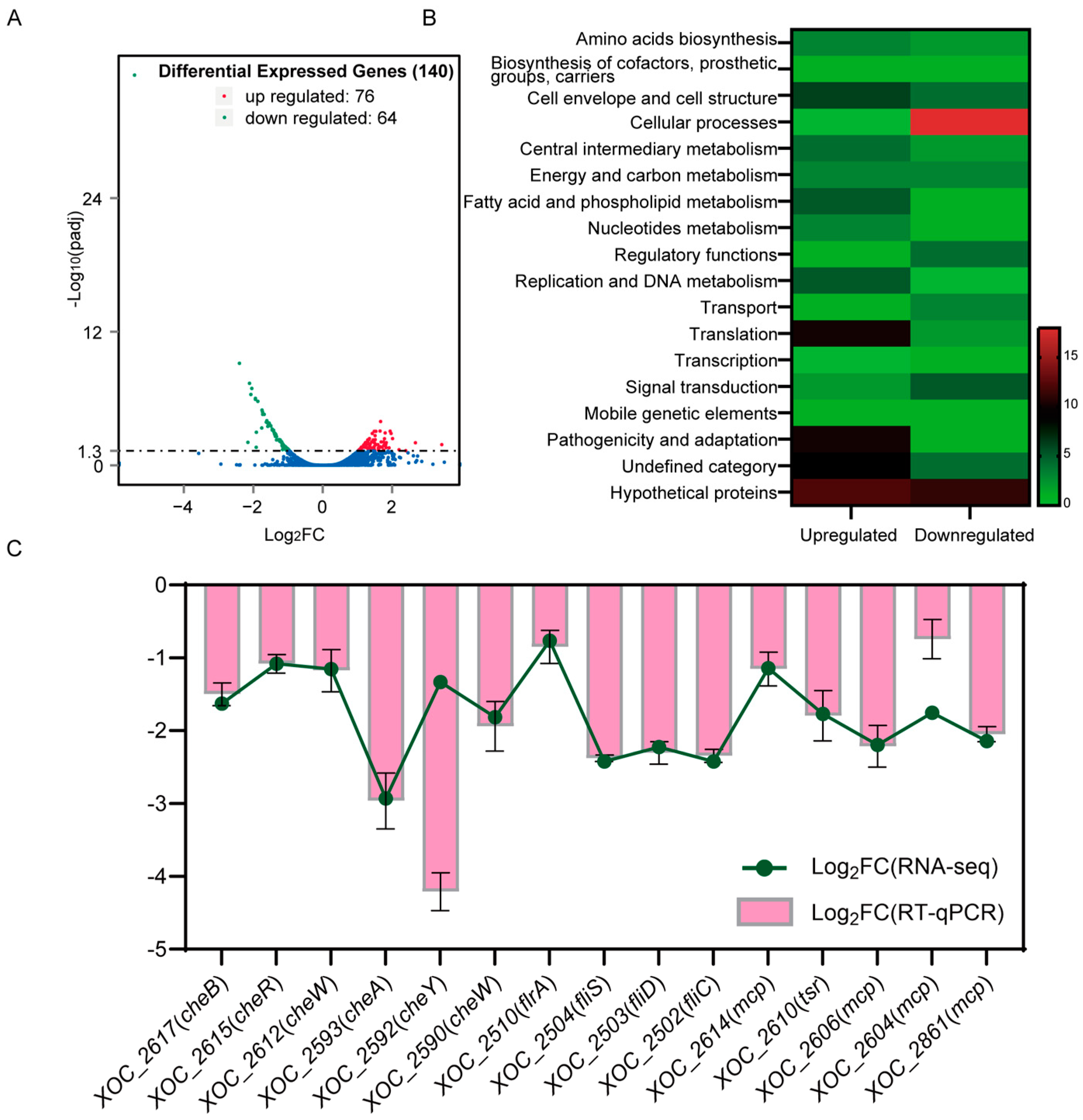
To determine whether VmsR plays a crucial role in virulence, we compared the ∆vmsR with its wild-type strain Xoc GX01 through RNA-Seq. For all six samples analyzed (3 × ∆vmsR and 3 × GX01), more than 80% of the total reads were successfully mapped back to the assembled transcriptome. From the total of 5006 genes obtained in the transcriptome of Xoc GX01, 140 genes were found to be statistically significant differentially expressed genes (DEGs) between ∆vmsR and GX01 using a more stringent threshold (FDR ≤ 0.05 and |Fold Change| ≥ 2). Among these genes, 76 genes were upregulated and 64 genes were downregulated in ∆vmsR (Figure 3A).
To gain a deeper understanding of the functionalities of genes regulated by VmsR, we performed a functional grouping analysis based on the genomic annotations of the Xoc GX01 [34,35]. Based on the clusters of orthologous groups (COGs), out of the 140 DEGs in the ∆vmsR strain, 104 were assigned to 15 various functional categories and 36 were predicted to encode hypothetical proteins or proteins that have not been given a functional category (Figure 3B). The primary functional groupings are “cellular processes” and “pathogenicity and adaption”. In total, 18 and 11 genes fell into these two categories, respectively (Figure 3B). Notably, 105 genes were assigned to “cell envelope and cell structure” (10) and “translation” (12). Table S3 shows a detailed categorization of DEGs.
Consistent with the finding that LuxR regulates cell motility [36,37,38], the identified transcriptional profiles reveal that VmsR has a crucial impact on a number of genes that contribute to cell motility. Upon a thorough examination of the functional classification of the 140 DEGs identified in the ∆vmsR mutant of Xoc GX01, it is evident that the pathways most significantly affected are those associated with cellular processes, which are fundamental to the survival, growth, and virulence of the bacterium. This observation underscores the central role that VmsR plays in regulating vital cellular activities, particularly those related to motility. Consequently, there is a pressing need to delve deeper into the regulatory mechanisms employed by VmsR in this regard. The pertinent gene clusters that are implicated in these processes are outlined as follows: The four flagellar-related DEGs consist of three genes, namely XOC_2502/fliC, XOC_2503/fliD, and XOC_2504/fliS, which encode proteins integral to the formation of flagellar filaments. Additionally, there is a gene encoding the upstream regulator of flagella, flrA. Fourteen of the DEGs encode methyl-accepting chemotaxis proteins: XOC_2212, XOC_2478, XOC_2590, XOC_2602, XOC_2603, XOC_2604, XOC_2605, XOC_2606, XOC_2610, XOC_2612, XOC_2614, XOC_2615, XOC_2617, and XOC_2861. The expression levels of the mRNA for these genes corresponded closely with the RNA-Seq data (Figure 3C). The findings demonstrate a significant role of VmsR in influencing the motility of Xoc. Taken together, the results from the transcriptome analysis reveal that VmsR acts as an essential regulator involved in many cellular processes in Xoc.

2.4. Loss of vmsR in Xoc Enhanced the Swarming Motility but Impaired the Swimming Motility

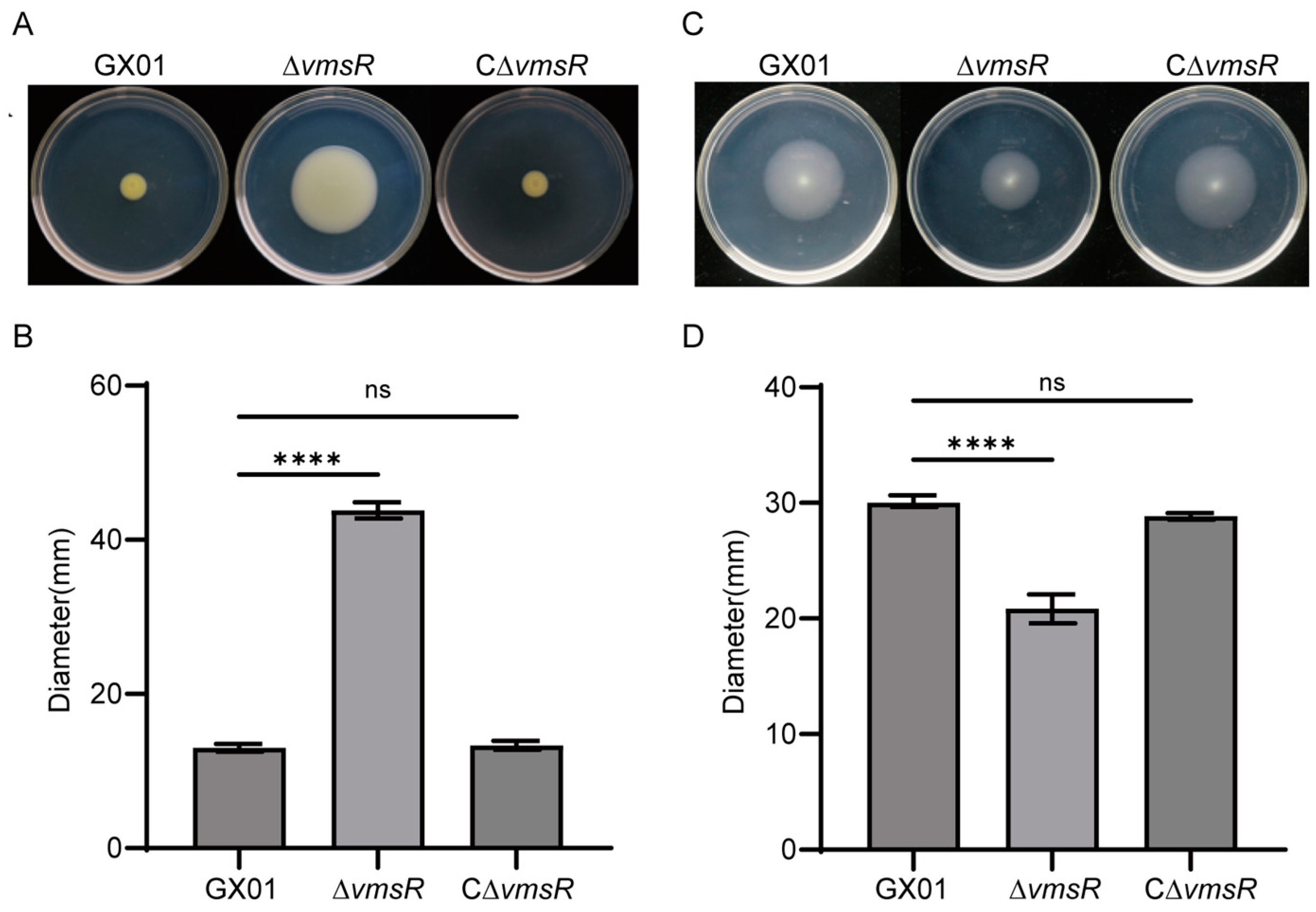
Based on the RNA-Seq analysis, we hypothesize that VmsR is linked to bacterial motility. To validate this prediction, we conducted a study to measure the swarming and swimming motilities of Xoc GX01, the ∆vmsR mutant strain, and the complementary strain C∆vmsR. To assess swarming motility, Xoc strains were inoculated in a semi-solid NA medium containing 0.6% agar and incubated at 28 °C for 3 days. The wild-type strain GX01 exhibited an average bacterial colony diameter of up to 13.00 mm, which was significantly smaller than the 43.83 mm diameter observed for the ∆vmsR strain. Notably, the C∆vmsR strain exhibited a colony diameter averaging 13.33 mm, which is comparable to the wild-type strain GX01 (Figure 4A,B).
Bacterial swimming motility is dependent on the flagellum. To evaluate swimming motility, Xoc strains were inoculated in semi-solid basal medium with 0.25% agar and incubated at 28 °C for 3 days. The wild-type strain GX01 displayed a bacterial colony diameter averaging 40.17 mm, which was comparable to the 39.33 mm observed in the C∆vmsR strain. The ∆vmsR strain exhibited a significantly smaller colony diameter of 29.17 mm, as depicted in Figure 4C,D. This reduction in swimming motility in the ∆vmsR strain may be attributed, in part, to diminished flagellar biogenesis. Collectively, these findings indicate that VmsR plays contrasting roles in regulating swarming and swimming motility in Xoc GX01.

2.5. VmsR Specifically Binds to the fliC and fliS Promoter In Vitro and Facilitates Their Expression

The swimming motility of the ∆vmsR strain has decreased. Bacterial swimming motility is fundamentally reliant on the flagellum, which is a complex organelle responsible for propelling the bacterium through its environment [39]. The model of the flagellum structure is shown in Figure 5A. The RNA-Seq analysis revealed the downregulation of three genes (fliC, fliD, and fliS) that are associated with flagella synthesis. The mRNA expression levels of the fliC, fliD and fliS genes were verified by RT-qPCR. As shown in Figure 5B, the transcriptional levels of these genes were significantly reduced in the ∆vmsR compared to GX01, and the expression patterns of these selected genes are all consistent with that observed in the data obtained from the transcriptome analysis (Figure 3C).
To determine if the transcriptional regulation of fliC, fliD, and fliS is mediated by the direct binding of VmsR to the promoter region, in vitro electrophoretic mobility shift assays (EMSAs) were performed. Purified recombinant VmsR-His6 (Figure 5C) was incubated with 6′-carboxyfluorescein (6′-FAM)-labeled DNAs in binding buffer; then, the protein–DNA complexes were separated by electrophoresis in 4% polyacrylamide gel. The findings conclusively reveal that the recombinant VmsR-His6 exhibits a robust binding affinity to the fliC and fliS promoter regions, significantly impeding their migration within the polyacrylamide gel (Figure 5D,F). Moreover, the observed shifted bands were displaced by an excess of unlabeled probes (10×, 50×, 100×), thus confirming the specificity of VmsR’s binding to the fliC and fliS promoter. To guarantee the specificity of VmsR binding, we incorporated a negative control promoter (the atsE promoter), which demonstrated no affinity for VmsR. As shown in Figure S2A, the EMSA results demonstrate that VmsR can directly bind to the promoter of fliC and fliS while failing to bind to the fliD promoter.
These results supported the hypothesis that VmsR acts as a transcriptional activator of the fliC and fliS promoter. The reduced expression of fliD could been indirectly regulated by VmsR.

2.6. EPS Production and Biofilm Formation of the ∆vmsR Strain Were Increased

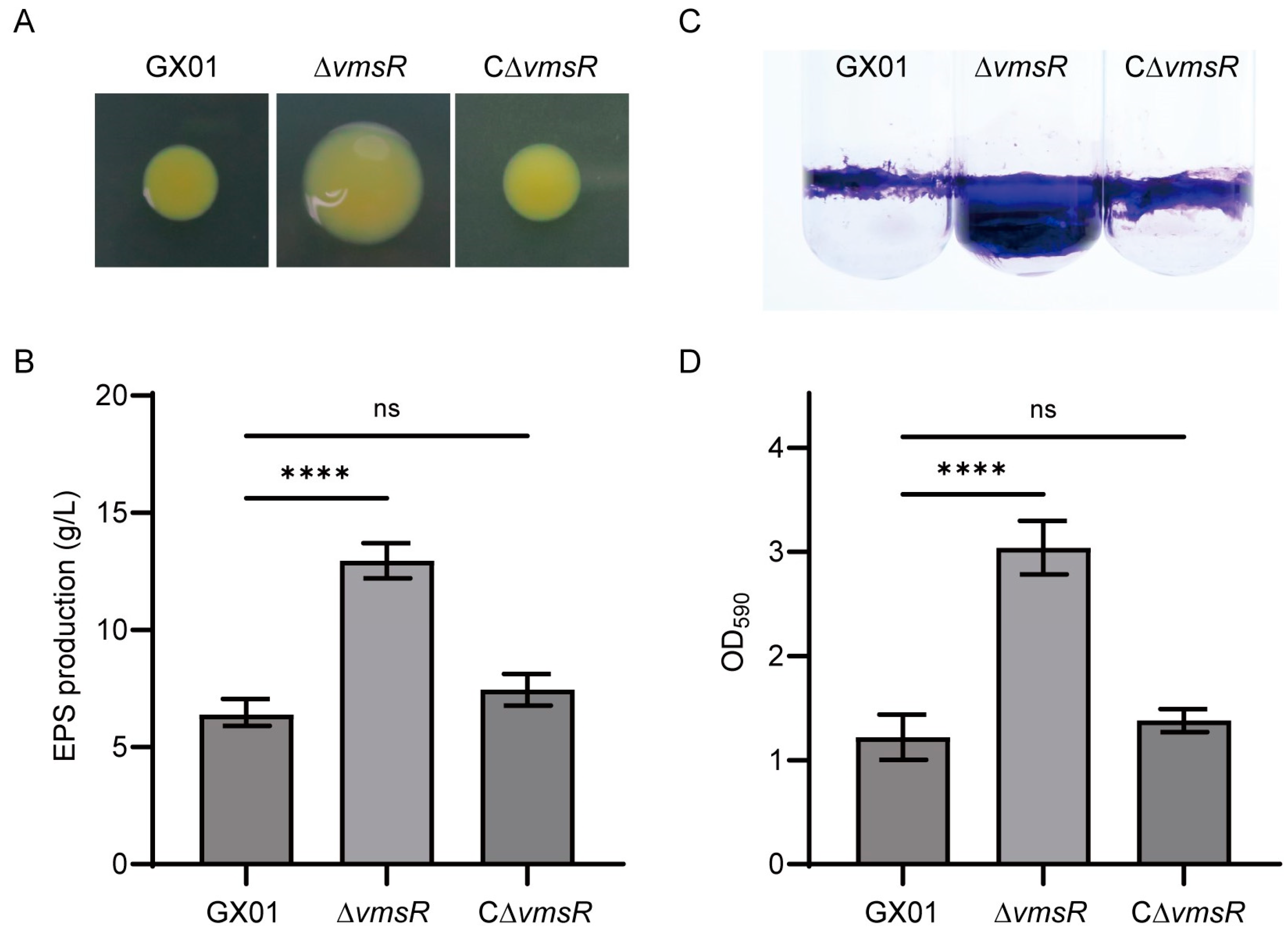
In the above results, we found an interesting phenomenon. The swimming motility and swarming motility of the ∆vmsR strain were opposite. Liu et al. found that the self-secreted EPSs are essential for the swarming motility exhibited by Pseudoalteromonas sp. SM9913 [40]. Consequently, EPS was measured qualitatively and quantitatively in this study. Compared with the wild-type strain, EPS production of the ∆vmsR significantly increased both in qualitative and quantitative analysis. In terms of shape, the colony form of ∆vmsR is fuller and shinier than wild-type strain GX01 and the complementary strain C∆vmsR (Figure 6A). EPS production was up to 12.96 g/L for the vmsR mutant, significantly greater than 6.383 g/L for the wild-type strain Xoc GX01, but similar to 7.45 g/L for the complementary strain C∆vmsR (Figure 6B).
It is firmly established that the biofilm formation process of numerous bacteria necessitates the presence of both EPSs and the type IV pilus (T4P). EPSs occupy a pivotal role in this biofilm formation. EPS constitutes part of the biofilm matrix that maintains and organizes bacterial biofilms, while the T4P facilitates surface attachment as adhesins [41]. Biofilm formation was measured qualitatively and quantitatively in this study. When compared to the Xoc GX01 strain, the biofilm formation capacity of the ∆vmsR mutant was observed to be significantly elevated (Figure 6C). Quantification of the biofilm formation was achieved through the measurement of optical density at 590 nm (OD590). Specifically, the mean OD590 value for the ∆vmsR mutant was 3.04, which significantly surpassed the values of 1.22 recorded for Xoc GX01 and 1.38 for the C∆vmsR strain (Figure 6D).
Taken together, the results indicate that VmsR negatively affects EPS production and biofilm formation in Xoc GX01. Furthermore, we hypothesize that the enhanced biofilm formation and swarming motility could be attributed to the increased production of EPS.

2.7. VmsR Specifically Binds to the gumB Promoter In Vitro and Inhibits Its Expression

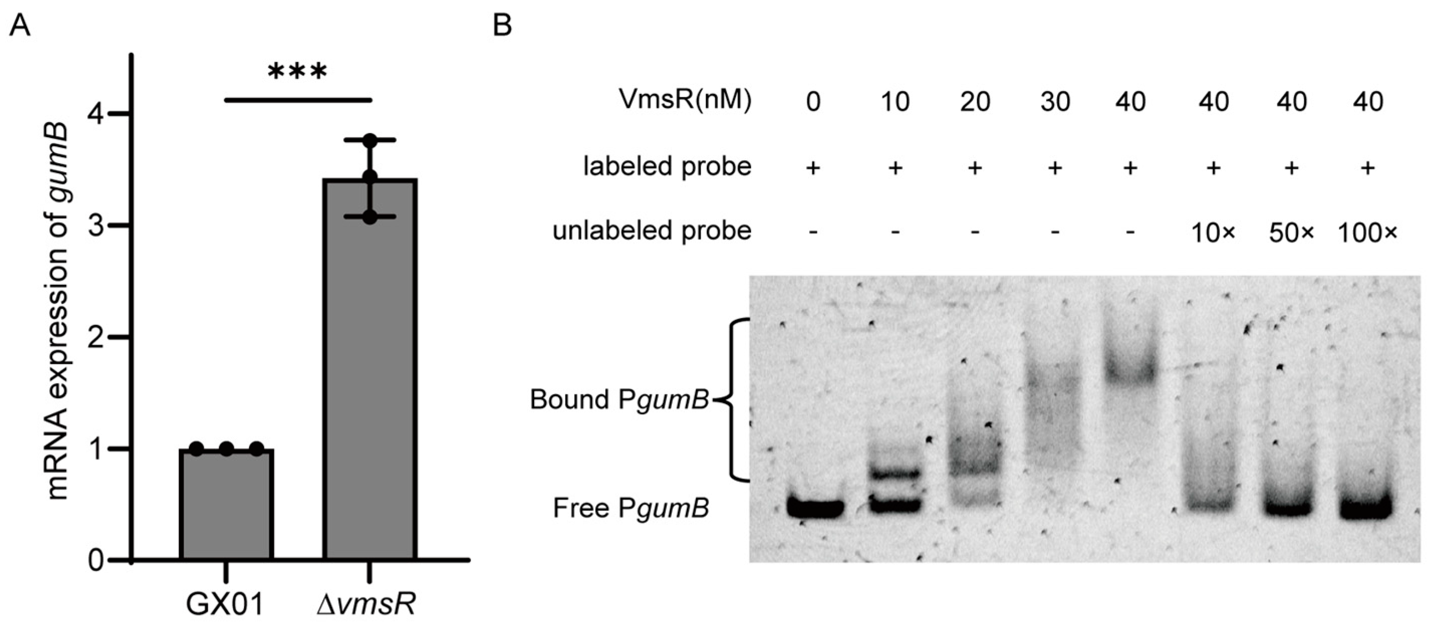
EPS is encoded by a gene cluster with gumB serving as one of the primary genes within this cluster. The gumB gene plays a pivotal role in exporting the xanthan [42]. The mRNA expression levels of gumB gene were verified by RT-qPCR. As shown in Figure 7A, the transcriptional levels of gumB gene were significantly upregulated in the ∆vmsR compared to GX01. To determine whether VmsR could directly regulate the gumB operon, in vitro EMSAs were conducted. Purified recombinant VmsR-His6 (Figure 5C) was incubated with a 6′-FAM-labeled DNA (440 bp) spanning from bp −455 to −16 (Table S2) relative to the translational initiation site (TIS) of the gumB operon. Then, the protein–DNA complexes were separated by electrophoresis in 4% polyacrylamide gel. A distinct shift band was observed with increasing concentrations of recombinant VmsR-His6. Competitive binding assays with unlabeled probes at various concentrations (10×, 50×, 100×) demonstrated competitive interactions, leading to progressively fainter or absent shift bands in the presence of non-fluorescent competitive probes (Figure 7B). Taken together, these results illustrate that VmsR could specifically bind to the gumB operon promoter and repress the expression of gumB gene.
Additionally, the putative consensus sequence recognized by VmsR using the MEME Suite (https://meme-suite.org/meme/), with the promoter regions from fliC, fliS and gumB, identified the core DNA-binding sequence 5′-GTHGTGWWCSMWRWGKYTT-3′ (H: A/C/T; W: A/T; S: C/G; M: A/C; R: A/G; K: G/T; Y: C/T) (Figure S2B).

3. Discussion

In this work, we identified a LuxR-type transcriptional regulator VmsR, which acts as a global regulator influencing diverse cellular processes including cell motility, EPS production and biofilm formation in Xoc GX01. Specifically, we have identified VmsR as a key regulator of flagellum in Xoc. Our findings indicate that VmsR directly promotes the expression of fliC and fliS genes, revealing its crucial role in positively regulating motility and pathogenicity in Xoc.
LuxR-type regulators are known to play a role not only in the QS system but also in other biological functions such as flagellar synthesis. For example, AcrR, a LuxR-type regulator, regulates flagellar assembly and contributes to the virulence, motility, biofilm formation, and growth ability of Acidovorax citrulli [38]. VisN and VisR, two LuxR-type regulators in Sinorhizobium meliloti, act as global regulators of chemotaxis, flagellar, and motility genes [43]. AclR, also a LuxR-type global regular, regulates the motility and virulence of A. citrulli [44]. In accordance with the aforementioned LuxR-type regulators, VmsR similarly performs a crucial function in governing chemotaxis, flagellar assembly, and motility-associated genes. As demonstrated in our study, VmsR is a TCS response regulator, not a traditional QS system regulator. The vmsR mutant strain exhibits a reduced swimming motility but an increased swarming motility, EPS production, and biofilm production. This demonstrated that VmsR either positively or negatively regulated multiple biological functions of Xoc GX01.
The flagellum is involved not only in the movement and chemotaxis of bacteria but also in several functions associated with bacterial pathogenicity, including biofilm formation, protein export, and adhesion [45]. The flagellum also serves as a virulence factor in many bacterial pathogens. Steffens et al. found that the fliC mutant inhibited the motility and showed an increased xanthan production in Xcc JBL007 [46]. In line with the aforementioned findings, the deletion of the vmsR gene in Xoc GX01 resulted in a significantly reduced expression of fliC, which was accompanied by an enhanced EPS production. In our study, the deletion of the vmsR gene led to a diminished expression of the fliC gene, potentially causing a reduction in swimming motility, which contributed significantly to the decreased virulence exhibited by the bacterium on rice. Our RNA-Seq data support the role that VmsR plays in flagellar biosynthesis because three genes involved in flagellar assembly were differentially expressed in the vmsR mutant strain in comparison with the wild-type strain.
In this study, ∆vmsR exhibited a decrease in swimming motility and an increase in swarming motility. The result shows that VmsR plays an important role in the cell motility of Xoc GX01. Swimming motility is an individual movement in liquid or semi-solid media, and it requires flagella, which is a complex organelle responsible for propelling the bacterium through its environment [47]. Song et al. discovered that flagellar instability results in a diminished virulence of Aeromonas veronii and correlates with its pathogenicity [48]. In this study, three flagellar genes, namely fliC, fliD, and fliS, were downregulated through the RT-qPCR analysis. In vitro EMSA assay, VmsR can directly bind to the promoter of fliC and fliS. fliD encodes the HAP2/filament cap protein, which plays a pivotal role in regulating the assembly and stability of bacterial flagella. fliC encodes the H1 flagellin protein, which is a key component of the flagellar filament responsible for motility. FliS functions as a specific export chaperone for FliC, facilitating its translocation and assembly within the bacterial cell [49,50]. In general, the expression of these genes is negatively regulated by the secretion of FlgM, which is a regulatory protein that modulates flagellar biosynthesis and function [51]. FliS, on the other hand, operates as a non-canonical chaperone, fine-tuning FlgM’s activity to regulate the expression of late flagellar genes as well as motility and biofilm formation in Yersinia pseudotuberculosis [51]. Therefore, we speculate that VmsR has positively regulated the expression of fliC and fliS. The deletion of the vmsR gene results in the instability of flagella, causing a decrease in the swimming motility. These results show that the flagellum is very important for pathogenic bacteria. Consequently, the reduction pathogenicity of ∆vmsR may be caused by reduced flagellar synthesis.
Swarming motility is a group movement on semi-solid medium, which requires both flagella and type Ⅳ pili [52]. Swarming motility could increase the expression of virulence genes such as type Ⅲ effector and the type Ⅱ secretory system [53,54]. Swarming motility, regulated by several genes and pathways like the QS system, is a complex bacterial behavior [55]. One study showed that swarming is positively regulated by rhamnolipid production [56]. As in other Xanthomonas [57], the difference of swimming motility and swarming motility remains unknown, requiring further research. Nevertheless, previous research has shown that the self-secreted EPS is positively correlated to the swarming behavior of Pseudoalteromonas sp. SM9913 [40]. Consequently, the enhanced EPS production in the ∆vmsR mutant may offer a plausible explanation for the remarkable swarming motility that has been observed.
Biofilm is a critical virulence factor for many plant pathogens [58,59]. When Xanthomonas citri was impaired in its ability to form a biofilm, it exhibited mitigated disease symptoms [60]. Biofilm formation involves some adhesive molecules, such as polysaccharide or protein. EPS usually contributes to biofilm formation in Xoc [61]. In general, the enhanced biofilm provides a protective barrier that could contribute to increased virulence by facilitating the colonization of host tissues and evasion of host immune responses. The mutant of rbdA in Pseudomonas aeruginosa PAO1 results in increased EPS production and biofilm formation [62]. P. aeruginosa PAO1 has two operons, pel and psl, that have been reported to contribute to EPS production [63]. They proved that the increased expression of the pel operon might contribute to the hyperbiofilm phenotype of the ΔrbdA mutant [62]. Likewise, the mutation of bifA in P. aeruginosa elicits an enhanced biofilm phenotype. Furthermore, BifA has been demonstrated to regulate biofilm formation through modulating the expression of the pel operon [64]. Consistent with the above studies, the EPS production and biofilm formation in the ΔvmsR mutant were increased, which are hypothesized to confer significant advantages to its pathogenicity. Nevertheless, the pathogenicity of the ΔvmsR mutant has decreased. We presume that EPS, being a co-pathogenic factor, does not necessarily exhibit a direct and positive correlation with pathogenicity. High levels of EPS production significantly contribute to the robustness of the biofilm structure, thereby potentially shielding the bacteria from host immune responses [61]. However, this augmentation in robustness also diminishes the bacteria’s capacity to disperse, potentially reducing its virulence in dynamic environments. From an ecological long-term perspective, bacteria excrete substantial quantities of EPS as a strategic defense mechanism to evade the host’s immune system. This temporary state of defense, rather than aggression, facilitates the enhanced survival of the bacterial colony as a whole.
Additionally, flagella are suggested to help E. coli overcome repulsive surface forces to establish initial contact [65]. Flagella contribute to the early stages of biofilm formation in Bacillus cereus by facilitating surface attachment and microcolony formation [66]. However, when E. coli express other surface structures like Curli or conjugative pili, flagella are not necessary for adhesion or biofilm development [67,68]. This suggests that flagellar motility alone is not the key factor for promoting adhesion. On the other hand, flagella may serve a mechanosensory role in detecting surfaces, which triggers a transition from planktonic (free-swimming) to sessile (adherent) states, which are essential for biofilm formation [69,70]. When surfaces are detected, flagella-mediated motility is downregulated, possibly due to the influence of the cellular messenger c-di-GMP, which is linked to both the suppression of motility and the promotion of robust biofilm formation [71,72]. This dual role of flagella—both promoting initial surface contact and then downregulating motility upon surface detection—underscores a complex regulation where motility and biofilm formation are generally mutually exclusive. In this work, biofilm formation of the ∆vmsR strain was increased significantly compared with the wild-type strain GX01, while the swimming of the ∆vmsR strain was reduced. These results are consistent with the above studies. Motility and biofilm are mutually exclusive lifestyles, and shifts between the two are under the strict regulation of bacteria attempting to adapt to the complexity and variability of environmental conditions.
Unexpectedly, EPS production has risen in ∆vmsR, yet the gum gene is conspicuously absent from the DEGs. Similarly, this phenomenon aligns with the observation that the deletion of vemR conspicuously diminished EPS production despite the absence of any gum-related genes in the DEGs [73]. In Xoc, the gum cluster, encompassing 13 genes ranging from gumB to gumN, is vital for EPS assembly, polymerization, and export [74,75]. It has been demonstrated that in Xcc, EPS synthesis is initiated in the late-exponential growth phase and reaches maximal production during the stationary growth phase, and the expression of the gum genes mirrors the time course of EPS production [76]. As described above, our transcriptome analysis used the bacterial cells in the mid-exponential growth phase. Therefore, this inconsistency is probably due to the employment of bacterial cells from different growth phases in the experiments.
Given their central role in bacterial physiology and pathogenesis, HKs represent promising targets for the development of novel antimicrobial agents [77]. While the cognate HKs of VmsR remains unknown, further research efforts are likely to shed light on this important aspect of bacterial signaling. In addition, RR proteins can also be used as direct targets. Identifying the sensor kinase responsible for activating VmsR will not only deepen our understanding of TCS function but also have implications for the development of novel antibacterial strategies and biotechnology applications.
In summary, the vmsR gene occupies a crucial position in the virulence of Xoc GX01, modulating swimming motility and flagellar formation positively, while suppressing EPS production and biofilm formation, either directly or indirectly. Transcriptomic investigations have further illuminated that VmsR adversely affects methyl-accepting chemotaxis proteins, which are integral to motility. The presence of a response regulatory domain at the N-terminus of VmsR suggests its potential to interact with a diverse array of signals regulating biological functions within Xoc GX01. However, the precise nature of these signals remains an enigma. Future research is imperative to decipher these signals and elucidate the underlying molecular mechanisms governing VmsR’s regulation in Xoc GX01, ultimately aiming to devise efficient control strategies against this significant bacterial pathogen.

4. Materials and Methods

4.1. Bacterial Strains, Culture Media and Growth Conditions

Xoc strains were routinely grown in nutrient broth (NB) medium (beef extract, 3 g/L; yeast extract, 1 g/L; polypeptone, 5 g/L) or on NB agar plates at 28 °C. E. coli strains were cultivated in Luria–Bertani (LB) medium or on LB agar plates at 37 °C. When necessary, antibiotics were added to medium at the following concentrations: kanamycin, 25 μg/mL; rifampicin, 50 μg/mL. The Xoc strains and plasmids used in this work are listed in Table S1.

4.2. Construction of the vmsR Deletion Mutant and Its Complementary Strain

The vmsR gene was knocked out in Xoc GX01 using a homologous recombination method. The follow primers used in this study are listed in Table S2. The 492 bp upstream and 522 bp downstream sequences of the vmsR gene were amplified from the Xoc wild-type strain GX01 genome using the vmsR-LF/LR and vmsR-RF/RR primers. The amplified DNA fragments were digested by corresponding restriction enzymes and cloned into pK18mobsacB, which is a conjugative suicide plasmid in Xoc. Then, the resulting recombinant plasmids were created in E. coli strain DH5α and transferred to wild-type strain Xoc GX01 by electroporation (2 mm, 2.5 kV). Transconjugants were screened on NA medium with 10% sucrose and antibiotics (Rifr and Kamr). Transconjugants were identified by external primers (vmsR-LF and vmsR-RR) and vmsR ORF internal primers (vmsR-inF and vmsR-inR). The successful transconjugants should satisfy the following two points at the same time: the combined fragment of upstream and downstream sequences was amplified by external primers, and no PCR product could be amplified by internal primers.
To complement the deletion mutant, the 989 bp sequence of the vmsR gene was amplified using the vmsR-cmF and vmsR-cmR primers. The PCR fragments were digested with the XbaⅠ/HindⅢ restriction enzymes and ligated into XbaⅠ/HindⅢ-digested pXUK, resulting in the recombinant plasmids. pXUK was derived from the endogenous plasmid pXOCgx01 isolated from Xoc GX01 [78]. Recombinant plasmids were created in E. coli strain DH5α and transferred to the vmsR mutant of Xoc by electroporation (2 mm, 2.5 kV). Transconjugants were screened on NA medium with Rifr and Kmr antibiotics. Transconjugants were identified by vmsR-cmF/R primers.

4.3. Virulence Assay

The virulence of Xoc was determined by the pressure inoculation of leaves in rice [79]. Xoc strains were grown in NB medium at 28 °C with shaking at 200 rpm until the value of OD600 reached 0.8–1.0. The value of OD600 was adapted to 0.5 using sterilized ddH2O. The above bacterial cell suspensions were inoculated into the leaves of 6-week-old rice plants using a needleless injector. Water-soaking symptoms were measured 15 days after inoculation. We also recorded the lesion length on at least 30 leaves.

4.4. Assay for Swimming and Swarming Motilities

To determine the swimming and swarming motilities of the vmsR mutant strain and the wild-type strain Xoc GX01, Xoc strains were cultured in NB medium at 28 °C with shaking at 200 rpm until the value of OD600 reached 1.0.
For swimming motility, the value of OD600 was adjusted to 1.0. A volume of 3 μL of each bacterial cell suspension was injected into the center of a basal medium plate containing 0.3% agar and then incubated at 28 °C for 3 days. For swarming motility, the value of OD600 was adjusted to 0.2. A volume of 2 μL of each bacterial cell suspension was precisely dispensed onto the center of an NB medium plate containing 2% sucrose and 0.6% agar and then incubated at 28 °C for 3 days. Due to the low strength of the medium plate, the glass garden should be placed upward. After 3 days of incubation at 28 °C, the diameter of the bacterial colony on each plate was determined. In both swimming and swarming motility assays, each treatment involves three replications, and each experiment was repeated three times.

4.5. Determination of Biofilm Formation and EPS Production

Biofilm formation on glass surfaces was performed as described previously [17]. Briefly, Xoc strains were grown in NB medium at 28 °C with shaking at 200 rpm until the value of OD600 reached 1.0. The value of OD600 was adapted to 0.5, and then 1.0 mL of cultures prepared as aforementioned was added in borosilicate glass tubes (100 mm * 15 mm). These cultures were incubated at 28 °C for 5 days without shaking. We removed the cell suspension from the borosilicate glass tubes and washed the glass tubes with sterile water three times. Then, 2.0 mL of 0.1% (w/v) crystal violet was added to these glass tubes for 30 min. To quantify the biofilm formation, stained biofilm solubilized with 2 mL absolute ethanol was measured with a spectrophotometer under OD600. Each treatment involved three replications, and the same experiment was repeated three times.
For analysis of EPS production on plates, the Xoc strains were cultured in NB medium at 28 °C with shaking at 200 rpm until the value of OD600 reached 0.8–1.0. The value of OD600 was adjusted to 0.2, and then 2 μL of each strain was inoculated onto the surface of NA plates containing 2% sucrose. After 48~72 h of incubation at 28 °C, the colony diameter of the Xoc strains was measured. To estimate EPS production, the Xoc strains were inoculated into 100 mL of NB liquid medium containing sucrose (2% w/v) at 28 °C, 200 rpm for 5 days. EPS was precipitated from the culture supernatant with ethanol, dried at 55 °C and weighed as described. The results were observed after 5 days of incubation at 28 °C. Each treatment involves three replications, and the same experiment was repeated three times.

4.6. Transcriptome Analysis

Xoc strains were cultured in NB medium at 28 °C with shaking at 200 rpm until the OD600 value reached 1.0. Three biological replicates were derived from the bacteria solution. The bacterial cells were collected using high-speed centrifuge and then frozen with liquid nitrogen. The bacteria cells were sent to the PFOMIC Bioinformatics Company (Nanning, China) for library preparation and strand-specific transcriptome sequencing. Sequencing libraries were generated using the NEBNext® UltraTM Directional RNA Library Prep Kit for Illumina® (NEB, USA) following the manufacture’s recommendations, and index codes were added to attribute sequences to each sample. HTSeq v0.6.1 was used to count the read numbers mapped to each gene. And then, the FPKM of each gene was calculated based on the length of the gene and the reads count mapped to this gene. FPKM, the expected number of Fragments Per Kilobase of transcript sequence per Millions base pairs sequenced, considers the effect of sequencing depth and gene length for the reads count at the same time, and it is currently the most commonly used method for estimating gene expression levels [80]. Differential expression analysis of two conditions was performed using the DEGSeq R package (1.20.0). The p values were adjusted using the Benjamini and Hochberg method [81]. A corrected FDR of 0.05 and log2(Fold Change) of 1 were set as the threshold for significantly differential expression.

4.7. RNA Isolation and Quantitative Reverse Transcription PCR Analysis of Gene Expression

Xoc strains were grown in NB medium at 28 °C with shaking at 200 rpm until the value of OD600 reached 1.0. The bacterial cells were collected using high-speed centrifuge and then washed three times with sterilized water. RNA was isolated from the Xoc strains using a TransZol Up Plus RNA Kit (TransGen, Beijing, China). To confirm the reality of the RNA-seq data, two-step quantitative reverse transcription PCR (RT-qPCR) was adapted in this work. First-strand cDNA was synthesized using RT SuperMix for qPCR (Vazyme Biotech, Nanjing, China). The cDNA was added as DNA templates into the PCR system with the 2 × RealStar Green Fast Mixture (GenStar, Beijing, China). The qPCR adapted a three-step amplification procedure. Firstly, the template cDNA was pre-denatured at 95 °C for 2 min. A total of 40 cycles were amplified by qPCR, and each cycle was performed using the following procedure: 95 °C for 15 s, 60 °C for 15 s, and 72 °C for 30 s. The 16S rRNA gene of X. oryzae pv. oryzicola was used as the internal control to verify the absence of significant variation at the cDNA level in the samples.

4.8. In Vitro Electrophoretic Mobility Shift Assay (EMSA)

The EMSA represents a swift and precise technique for detecting interactions between proteins and nucleic acids. The principle is that the electrophoretic mobility of a protein–DNA complex is typically less than that of the free DNA [82,83]. DNA probes were amplified by PCR with the corresponding primers (Figure S1). The 5′-end of the reverse primer was modified with 6′-FAM (Sangon Biotech, Shanghai, China). DNA fragments encoding the putative DNA-binding domain of VmsR were cloned into the plasmid pRSFDuet-1 to generate the recombinant plasmids. The recombinant plasmids were introduced into fresh competent cells of E. coli BL21 (DE3) Chemically Competent Cell (CD601, TransGen, Beijing, China). The recombinant plasmids were incubated in Luria–Bertani medium for 3 to 5 h at 37 °C with a shaking speed of 200 rpm, achieving an OD600 of 0.6 to 0.8. Subsequently, 0.5 mM isopropyl-β-d-thiogalactoside (IPTG) (Solarbio Life Sciences, Beijing, China) was added, and the culture was further maintained for 12 h at 16 °C with a shaking speed of 120 rpm. E. coli BL21 (DE3) cells were collected and disrupted to extract the recombinant polypeptides. The recombinant proteins were purified by ProteinIso® Ni-NTA Resin (TransGen, Beijing, China). The DNA probe (40 ng) was mixed with different amounts of recombinant protein (0–3 μg) in binding buffer (1 mM dithiothreitol, 0.1 mg/mL BSA, 50 mM KCl, 20 mM Tris-HCl, pH 8.0, 5% glycerol) and then reacted at 25 °C for 30 min. The protein–DNA complexes were separated by electrophoresis in 4% polyacrylamide gel (acrylamide/bisacrylamide, 29:1) in 0.5 × Tris-borate-EDTA (TBE) buffer (44.5 mM Tris base, 44.5 mM boric acid, and 1 mM EDTA, pH 8.0) and recorded with a Bio-Rad ChemiDoc™ MP Imaging System (Bio-Rad Laboratories, Hercules, CA, USA). For the competitive EMSA assay, DNA probes without 6′-FAM were substituted.
As shown in Table S2, the fliC probe fragment measures 283 bp, encompassing the region from base pair −245 to +38, relative to the TIS of the fliC promoter. The fliD probe fragment spans 272 bp, extending from base pair −272 to −1 with respect to the TIS of the fliD promoter. Similarly, the fliS probe fragment encompasses 500 bp, covering the sequence from base pair −500 to −1, relative to the TIS of the fliS promoter.

5. Patents

China Invention Patent: Authorization Bulletin Number: CN112852839B (Published: 2 September 2022).

Supplementary Materials

The supporting information can be downloaded at https://www.mdpi.com/article/10.3390/ijms25147595/s1.

Author Contributions

S.H. and Y.H. conceived and designed the research. Y.Z. carried out the experiments. X.Z., J.W., L.L., H.Q., R.Z. and C.L. provided the resources and analyzed the data. Y.Z. wrote the manuscript. S.H. and Y.H. revised the manuscript. All authors have read and agreed to the published version of the manuscript.

Funding

This research was funded by the Guangxi Natural Science Foundation (2020GXNSFDA297026) and the National Natural Science Foundation of China (32360045 and 32060600).

Institutional Review Board Statement

Not applicable.

Informed Consent Statement

Not applicable.

Data Availability Statement

The RNA sequencing data generated in this study are available in the NCBI SRA database under the accession codes SRR28710641-SRR28710646 (https://www.ncbi.nlm.nih.gov/sra/PRJNA1101110, accessed on 16 April 2024). Other data are presented within the manuscript and Supplementary Materials.

Conflicts of Interest

The authors declare no conflicts of interest.

References

  1. NiÑO-Liu, D.O.; Ronald, P.C.; Bogdanove, A.J. Xanthomonas oryzae pathovars: Model pathogens of a model crop. Mol. Plant Pathol. 2006, 7, 303–324. [Google Scholar] [CrossRef] [PubMed]
  2. Ji, Z.Y.; Zakria, M.; Zou, L.F.; Xiong, L.; Li, Z.; Ji, G.H.; Chen, G.Y. Genetic diversity of transcriptional activator-like effector genes in Chinese isolates of Xanthomonas oryzae pv. oryzicola. Phytopathology 2014, 104, 672–682. [Google Scholar] [CrossRef]
  3. Yang, W.F.; Liu, Y.; Chen, L.; Gao, T.C.; Hu, B.S.; Zhang, D.F.; Liu, F.Q. Zinc uptake regulator (zur) gene involved in zinc homeostasis and virulence of Xanthomonas oryzae pv. oryzae in rice. Curr. Microbiol. 2007, 54, 307–314. [Google Scholar] [CrossRef]
  4. Lee, S.W.; Jeong, K.S.; Han, S.W.; Lee, S.E.; Phee, B.K.; Hahn, T.R.; Ronald, P. The Xanthomonas oryzae pv. oryzae PhoPQ two-component system is required for AvrXA21 activity, hrpG expression and virulence. J. Bacteriol. 2008, 190, 2183–2197. [Google Scholar] [CrossRef]
  5. Bender, C.L.; Alarcón-Chaidez, F.; Gross, D.C. Pseudomonas syringae phytotoxins: Mode of action, regulation, and biosynthesis by peptide and polyketide synthetases. Microbiol. Mol. Biol. Rev. 1999, 63, 266–292. [Google Scholar] [CrossRef]
  6. Chan, J.W.Y.F.; Goodwin, P.H. The molecular genetics of virulence of Xanthomonas campestris. Biotechnol. Adv. 1999, 17, 489–508. [Google Scholar] [CrossRef] [PubMed]
  7. Büttner, D.; Bonas, U. Regulation and secretion of Xanthomonas virulence factors. FEMS Microbiol. Rev. 2010, 34, 107–133. [Google Scholar] [CrossRef]
  8. Patankar, A.V.; Gonzalez, J.E. An orphan LuxR homolog of Sinorhizobium meliloti affects stress adaptation and competition for nodulation. Appl. Environ. Microbiol. 2009, 75, 946–955. [Google Scholar] [CrossRef] [PubMed]
  9. Santos, C.L.; Correia-Neves, M.; Moradas-Ferreira, P.; Mendes, M.V. A walk into the LuxR regulators of Actinobacteria: Phylogenomic distribution and functional diversity. PLoS ONE 2012, 7, e46758. [Google Scholar] [CrossRef]
  10. Gonzalez, J.F.; Myers, M.P.; Venturi, V. The inter-kingdom solo OryR regulator of Xanthomonas oryzae is important for motility. Mol. Plant Pathol. 2013, 14, 211–221. [Google Scholar] [CrossRef]
  11. Fuqua, C.; Parsek, M.R.; Greenberg, E.P. Regulation of gene expression by cell-to-cell communication: Acyl-homoserine lactone quorum sensing. Annu. Rev. Genet. 2001, 35, 439–468. [Google Scholar] [CrossRef]
  12. Fuqua, C.; Winans, S.C.; Greenberg, E.P. Census and consensus in bacterial ecosystems: The LuxR-LuxI family of quorum-sensing transcriptional regulators. Annu. Rev. Microbiol. 1996, 50, 727–751. [Google Scholar] [CrossRef] [PubMed]
  13. Nealson, K.H.; Platt, T.; Hastings, J.W. Cellular control of the synthesis and activity of the bacterial luminescent system. J. Bacteriol. 1970, 104, 313–322. [Google Scholar] [CrossRef]
  14. Fuqua, C.; Winans, S.C. Conserved cis-acting promoter elements are required for density-dependent transcription of Agrobacterium tumefaciens conjugal transfer genes. J. Bacteriol. 1996, 178, 435–440. [Google Scholar] [CrossRef]
  15. Qian, W.; Han, Z.J.; He, C.Z. Two-component signal transduction systems of Xanthomonas spp.: A lesson from genomics. Mol. Plant-Microbe Interact. 2008, 21, 151–161. [Google Scholar] [CrossRef]
  16. Ninfa, A.J.; Magasanik, B. Covalent modification of the glnG product, NRI, by the glnL product, NRII, regulates the transcription of the glnALG operon in Escherichia coli. Proc. Natl. Acad. Sci. USA 1986, 83, 5909–5913. [Google Scholar] [CrossRef] [PubMed]
  17. Tang, J.L.; Liu, Y.N.; Barber, C.E.; Dow, J.M.; Wootton, J.C.; Daniels, M.J. Genetic and molecular analysis of a cluster of rpf genes involved in positive regulation of synthesis of extracellular enzymes and polysaccharide in Xanthomonas campestris pathovar campestris. Mol. Gen. Genet. MGG 1991, 226, 409–417. [Google Scholar] [CrossRef]
  18. Guo, Y.P.; Zhang, Y.P.; Li, J.L.; Wang, N.A. Diffusible signal factor-mediated quorum sensing plays a central role in coordinating gene expression of Xanthomonas citri subsp. citri. Mol. Plant-Microbe Interact. 2012, 25, 165–179. [Google Scholar] [CrossRef]
  19. Wengelnik, K.; VandenAckerveken, G.; Bonas, U. HrpG, a key hrp regulatory protein of Xanthomonas campestris pv. vesicatoria is homologous to two-component response regulators. Mol. Plant-Microbe Interact. 1996, 9, 704–712. [Google Scholar] [CrossRef] [PubMed]
  20. Noël, L.; Thieme, F.; Nennstiel, D.; Bonas, U. cDNA-AFLP analysis unravels a genome-wide hrpG-regulon in the plant pathogen Xanthomonas campestris pv. vesicatoria. Mol. Microbiol. 2001, 41, 1271–1281. [Google Scholar] [CrossRef]
  21. Henrichsen, J. Bacterial surface translocation: A survey and a classification. Bacteriol. Rev. 1972, 36, 478–503. [Google Scholar] [CrossRef]
  22. Jarrell, K.F.; McBride, M.J. The surprisingly diverse ways that prokaryotes move. Nat. Rev. Microbiol. 2008, 6, 466–476. [Google Scholar] [CrossRef] [PubMed]
  23. Kearns, D.B. A field guide to bacterial swarming motility. Nat. Rev. Microbiol. 2010, 8, 634–644. [Google Scholar] [CrossRef]
  24. Partridge, J.D.; Harshey, R.M. Swarming: Flexible Roaming Plans. J. Bacteriol. 2013, 195, 909–918. [Google Scholar] [CrossRef] [PubMed]
  25. Mukherjee, S.; Bree, A.C.; Liu, J.; Patrick, J.E.; Chien, P.; Kearns, D.B. Adaptor-mediated Lon proteolysis restricts Bacillus subtilis hyperflagellation. Proc. Natl. Acad. Sci. USA 2015, 112, 250–255. [Google Scholar] [CrossRef] [PubMed]
  26. Harshey, R.M.; Partridge, J.D. Shelter in a swarm. J. Mol. Biol. 2015, 427, 3683–3694. [Google Scholar] [CrossRef] [PubMed]
  27. Tasteyre, A.; Barc, M.-C.; Collignon, A.; Boureau, H.; Karjalainen, T. Role of FliC and FliD flagellar proteins of Clostridium difficile in adherence and gut colonization. Infect. Immun. 2001, 69, 7937–7940. [Google Scholar] [CrossRef]
  28. Dingle Tanis, C.; Mulvey George, L.; Armstrong Glen, D. Mutagenic analysis of the Clostridium difficile flagellar proteins, FliC and FliD, and their contribution to virulence in hamsters. Infect. Immun. 2011, 79, 4061–4067. [Google Scholar] [CrossRef]
  29. Boles, B.R.; Horswill, A.R. Staphylococcal biofilm disassembly. Trends Microbiol. 2011, 19, 449–455. [Google Scholar] [CrossRef]
  30. Fey, P.D.; Olson, M.E. Current concepts in biofilm formation of Staphylococcus epidermidis. Futur. Microbiol. 2010, 5, 917–933. [Google Scholar] [CrossRef]
  31. Kostakioti, M.; Hadjifrangiskou, M.; Hultgren, S.J. Bacterial biofilms: Development, dispersal, and therapeutic strategies in the dawn of the postantibiotic era. Cold Spring Harb. Perspect. Med. 2013, 3, a010306. [Google Scholar] [CrossRef] [PubMed]
  32. Niu, X.N.; Li, Y.M.; Carpenter, S.C.D.; Dan, X.; Li, T.J.; Wu, Q.Z.; Wang, L.; Jiang, W.; Huang, S.; Tang, J.L.; et al. Complete genome resource of Xanthomonas oryzae pv. oryzicola GX01 isolated in South China. Mol. Plant-Microbe Interact. 2022, 35, 357–359. [Google Scholar] [CrossRef] [PubMed]
  33. Xu, H.; Zhao, Y.; Qian, G.; Liu, F. XocR, a LuxR solo required for virulence in Xanthomonas oryzae pv. oryzicola. Front. Cell. Infect. Microbiol. 2015, 5, 37. [Google Scholar] [CrossRef] [PubMed]
  34. He, Y.Q.; Zhang, L.; Jiang, B.L.; Zhang, Z.C.; Xu, R.Q.; Tang, D.J.; Qin, J.; Jiang, W.; Zhang, X.; Liao, J.; et al. Comparative and functional genomics reveals genetic diversity and determinants of host specificity among reference strains and a large collection of Chinese isolates of the phytopathogen Xanthomonas campestris pv. campestris. Genome Biol. 2007, 8, R218. [Google Scholar] [CrossRef] [PubMed]
  35. Qian, W.; Jia, Y.T.; Ren, S.X.; He, Y.Q.; Feng, J.X.; Lu, L.F.; Sun, Q.H.; Ying, G.; Tang, D.J.; Tang, H.; et al. Comparative and functional genomic analyses of the pathogenicity of phytopathogen Xanthomonas campestris pv. campestris. Genome Res. 2005, 15, 757–767. [Google Scholar] [CrossRef] [PubMed]
  36. Lehti, T.A.; Bauchart, P.; Dobrindt, U.; Korhonen, L.K.; Westerlund-Wikström, B. The fimbriae activator MatA switches off motility in Escherichia coli by repression of the flagellar master operon flhDC. Microbiology 2012, 158, 1444–1455. [Google Scholar] [CrossRef]
  37. Cellini, A.; Buriani, G.; Correia, C.; Fiorentini, L.; Vandelle, E.; Polverari, A.; Santos, C.; Vanneste, J.L.; Spinelli, F. Host-specific signal perception by PsaR2 LuxR solo induces Pseudomonas syringae pv. actinidiae virulence traits. Microbiol. Res. 2022, 260, 127048. [Google Scholar] [CrossRef]
  38. Guan, W.; Wang, T.L.; Huang, Q.; Tian, E.Y.; Liu, B.; Yang, Y.W.; Zhao, T.C. A LuxR-type regulator, AcrR, regulates flagellar assembly and contributes to virulence, motility, biofilm formation, and growth ability of Acidovorax citrulli. Mol. Plant Pathol. 2020, 21, 489–501. [Google Scholar] [CrossRef] [PubMed]
  39. Colin, R.; Ni, B.; Laganenka, L.; Sourjik, V. Multiple functions of flagellar motility and chemotaxis in bacterial physiology. FEMS Microbiol. Rev. 2021, 45, fuab038. [Google Scholar] [CrossRef]
  40. Liu, A.; Mi, Z.H.; Zheng, X.Y.; Yu, Y.; Su, H.N.; Chen, X.L.; Xie, B.B.; Zhou, B.C.; Zhang, Y.Z.; Qin, Q.L. Exopolysaccharides play a role in the swarming of the benthic bacterium Pseudoalteromonas sp. SM9913. Front. Microbiol. 2016, 7, 473. [Google Scholar] [CrossRef]
  41. Dye, K.J.; Yang, Z.M. Analysis of vegetative biofilms with microtiter plates. Front. Microbiol. 2022, 13, 894562. [Google Scholar] [CrossRef] [PubMed]
  42. Vorhölter, F.-J.; Schneiker, S.; Goesmann, A.; Krause, L.; Bekel, T.; Kaiser, O.; Linke, B.; Patschkowski, T.; Rückert, C.; Schmid, J.; et al. The genome of Xanthomonas campestris pv. campestris B100 and its use for the reconstruction of metabolic pathways involved in xanthan biosynthesis. J. Biotechnol. 2008, 134, 33–45. [Google Scholar] [CrossRef] [PubMed]
  43. Sourjik, V.; Muschler, P.; Scharf, B.; Schmitt, R. VisN and VisR are global regulators of chemotaxis, flagellar, and motility genes in Sinorhizobium (Rhizobium) meliloti. J. Bacteriol. 2000, 182, 782–788. [Google Scholar] [CrossRef] [PubMed]
  44. Guan, W.; Wang, T.L.; Huang, Q.; Zhao, M.; Tian, E.Y.; Liu, Y.F.; Liu, B.; Yang, Y.W.; Zhao, T.C. Transcriptomic and functional analyses reveal roles of AclR, a luxR-type global regular, in regulating motility and virulence of Acidovorax citrulli. Mol. Plant-Microbe Interact. 2021, 34, 952–961. [Google Scholar] [CrossRef] [PubMed]
  45. Haiko, J.; Westerlund-Wikström, B. The role of the bacterial flagellum in adhesion and virulence. Biology 2013, 2, 1242–1267. [Google Scholar] [CrossRef] [PubMed]
  46. Steffens, T.; Vorhölter, F.J.; Teckentrup, J.; Hublik, G.; Walhorn, V.; Anselmetti, D.; Pühler, A.; Niehaus, K.; Ortseifen, V. Two flagellar mutants of Xanthomonas campestris are characterized by enhanced xanthan production and higher xanthan viscosity. J. Biotechnol. 2022, 347, 9–17. [Google Scholar] [CrossRef] [PubMed]
  47. Nakamura, S.; Minamino, T. Flagella-driven motility of bacteria. Biomolecules 2019, 9, 279. [Google Scholar] [CrossRef]
  48. Song, H.C.; Kang, Y.H.; Zhang, D.X.; Chen, L.; Qian, A.D.; Shan, X.F.; Li, Y. Great effect of porin(aha) in bacterial adhesion and virulence regulation in Aeromonas veronii. Microb. Pathog. 2019, 126, 269–278. [Google Scholar] [CrossRef] [PubMed]
  49. Auvray, F.; Thomas, J.; Fraser, G.M.; Hughes, C. Flagellin polymerisation control by a cytosolic export chaperone. J. Mol. Biol. 2001, 308, 221–229. [Google Scholar] [CrossRef]
  50. Minamino, T.; Kinoshita, M. Structure, assembly, and function of flagella responsible for bacterial locomotion. EcoSal Plus 2023, 11, eesp-0011-2023. [Google Scholar] [CrossRef]
  51. Xu, S.; Peng, Z.; Cui, B.; Wang, T.; Song, Y.; Zhang, L.; Wei, G.; Wang, Y.; Shen, X. FliS modulates FlgM activity by acting as a non-canonical chaperone to control late flagellar gene expression, motility and biofilm formation in Yersinia pseudotuberculosis. Environ. Microbiol. 2014, 16, 1090–1104. [Google Scholar] [CrossRef] [PubMed]
  52. Lakshmanan, D.; Harikrishnan, A.; Jyoti, K.; Ali, M.I.; Jeevaratnam, K. A compound isolated from Alpinia officinarum Hance. inhibits swarming motility of Pseudomonas aeruginosa and down regulates virulence genes. J. Appl. Microbiol. 2020, 128, 1355–1365. [Google Scholar] [CrossRef] [PubMed]
  53. Overhage, J.; Lewenza, S.; Marr, A.K.; Hancock, R.E.W. Identification of genes involved in swarming motility using a Pseudomonas aeruginosa PAO1 mini-Tn5-lux mutant library. J. Bacteriol. 2007, 189, 2164–2169. [Google Scholar] [CrossRef] [PubMed]
  54. Overhage, J.; Bains, M.; Brazas, M.D.; Hancock, R.E.W. Swarming of Pseudomonas aeruginosa is a complex adaptation leading to increased production of virulence factors and antibiotic resistance. J. Bacteriol. 2008, 190, 2671–2679. [Google Scholar] [CrossRef] [PubMed]
  55. Balasubramanian, D.; Schneper, L.; Kumari, H.; Mathee, K. A dynamic and intricate regulatory network determines Pseudomonas aeruginosa virulence. Nucleic Acids Res. 2013, 41, 1–20. [Google Scholar] [CrossRef] [PubMed]
  56. Kohler, T.; Curty, L.K.; Barja, F.; van Delden, C.; Pechere, J.C. Swarming of Pseudomonas aeruginosa is dependent on cell-to-cell signaling and requires flagella and pili. J. Bacteriol. 2000, 182, 5990–5996. [Google Scholar] [CrossRef] [PubMed]
  57. Qi, Y.H.; Huang, L.; Liu, G.F.; Leng, M.; Lu, G.T. PilG and PilH antagonistically control flagellum-dependent and pili-dependent motility in the phytopathogen Xanthomonas campestris pv. campestris. BMC Microbiol. 2020, 20, 37. [Google Scholar] [CrossRef] [PubMed]
  58. Fujishige, N.A.; Kapadia, N.N.; De Hoff, P.L.; Hirsch, A.M. Investigations of biofilm formation. FEMS Microbiol. Ecol. 2006, 56, 195–206. [Google Scholar] [CrossRef] [PubMed]
  59. Das, A.; Rangaraj, N.; Sonti, R.V. Multiple adhesin-like functions of Xanthomonas oryzae pv. oryzae are involved in promoting leaf attachment, entry, and virulence on rice. Mol. Plant-Microbe Interact. 2009, 22, 73–85. [Google Scholar] [CrossRef]
  60. Rigano, L.A.; Siciliano, F.; Enrique, R.; Sendin, L.; Filippone, P.; Torres, P.S.; Qüesta, J.; Dow, J.M.; Castagnaro, A.P.; Vojnov, A.A.; et al. Biofilm formation, epiphytic fitness, and canker development in Xanthomonas axonopodis pv. citri. Mol. Plant-Microbe Interact. 2007, 20, 1222–1230. [Google Scholar] [CrossRef]
  61. Zhang, Y.B.; Wei, C.; Jiang, W.D.; Wang, L.; Li, C.R.; Wang, Y.Y.; Dow, J.M.; Sun, W.X. The HD-GYP domain protein RpfG of Xanthomonas oryzae pv. oryzicola regulates synthesis of extracellular polysaccharides that contribute to biofilm formation and virulence on rice. PLoS ONE 2013, 8, e59428. [Google Scholar] [CrossRef]
  62. An, S.W.; Wu, J.E.; Zhang, L.H. Modulation of Pseudomonas aeruginosa biofilm dispersal by a cyclic-di-GMP phosphodiesterase with a putative hypoxia-sensing domain. Appl. Environ. Microb. 2010, 76, 8160–8173. [Google Scholar] [CrossRef] [PubMed]
  63. Friedman, L.; Kolter, R. Two genetic loci produce distinct carbohydrate-rich structural components of the Pseudomonas aeruginosa biofilm matrix. J. Bacteriol. 2004, 186, 4457–4465. [Google Scholar] [CrossRef] [PubMed]
  64. Kuchma, S.L.; Brothers, K.M.; Merritt, J.H.; Liberati, N.T.; Ausubel, F.M.; O’Toole, G.A. BifA, a cyclic-di-GMP phosphodiesterase, inversely regulates biofilm formation and swarming motility by Pseudomonas aeruginosa PA14. J. Bacteriol. 2007, 189, 8165–8178. [Google Scholar] [CrossRef] [PubMed]
  65. Pratt, L.A.; Kolter, R. Genetic analysis of Escherichia coli biofilm formation: Roles of flagella, motility, chemotaxis and Type I Pili. Mol. Microbiol. 1998, 30, 285–293. [Google Scholar] [CrossRef] [PubMed]
  66. Houry, A.; Briandet, R.; Aymerich, S.; Gohar, M. Involvement of motility and flagella in Bacillus cereus biofilm formation. Microbiology 2010, 156, 1009–1018. [Google Scholar] [CrossRef] [PubMed]
  67. Prigent-Combaret, C.; Prensier, G.; Le Thi, T.T.; Vidal, O.; Lejeune, P.; Dorel, C. Developmental pathway for biofilm formation in curli-producing Escherichia coli strains: Role of flagella, curli and colanic acid. Environ. Microbiol. 2000, 2, 450–464. [Google Scholar] [CrossRef] [PubMed]
  68. Reisner, A.; Haagensen, J.A.J.; Schembri, M.A.; Zechner, E.L.; Molin, S. Development and maturation of Escherichia coli K-12 biofilms. Mol. Microbiol. 2003, 48, 933–946. [Google Scholar] [CrossRef] [PubMed]
  69. Laganenka, L.; López María, E.; Colin, R.; Sourjik, V. Flagellum-mediated mechanosensing and RflP control motility state of pathogenic Escherichia coli. mBio 2020, 11, e02269-19. [Google Scholar] [CrossRef]
  70. Wong, G.C.L.; Antani, J.D.; Lele, P.P.; Chen, J.; Nan, B.Y.; Kuhn, M.J.; Persat, A.; Bru, J.L.; Hoyland-Kroghsbo, N.M.; Siryaporn, A.; et al. Roadmap on emerging concepts in the physical biology of bacterial biofilms: From surface sensing to community formation. Phys. Biol. 2021, 18, 051501. [Google Scholar] [CrossRef]
  71. Valentini, M.; Filloux, A. Biofilms and cyclic di-GMP (c-di-GMP) signaling: Lessons from Pseudomonas aeruginosa and other bacteria. J. Biol. Chem. 2016, 291, 12547–12555. [Google Scholar] [CrossRef] [PubMed]
  72. Jenal, U.; Reinders, A.; Lori, C. Cyclic di-GMP: Second messenger extraordinaire. Nat. Rev. Microbiol. 2017, 15, 271–284. [Google Scholar] [CrossRef] [PubMed]
  73. Li, R.F.; Peng, J.L.; Liu, Q.Q.; Chang, Z.; Huang, Y.X.; Tang, J.L.; Lu, G.T. Xanthomonas campestris VemR enhances the transcription of the T3SS key regulator HrpX via physical interaction with HrpG. Mol. Plant Pathol. 2023, 24, 232–247. [Google Scholar] [CrossRef] [PubMed]
  74. Chaban, B.; Hughes, H.V.; Beeby, M. The flagellum in bacterial pathogens: For motility and a whole lot more. Semin. Cell Dev. Biol. 2015, 46, 91–103. [Google Scholar] [CrossRef] [PubMed]
  75. Katzen, F.; Ferreiro, D.U.; Oddo, C.G.; Ielmini, M.V.; Becker, A.; Pühler, A.; Ielpi, L. Xanthomonas campestris pv. campestris gum mutants: Effects on xanthan biosynthesis and plant virulence. J. Bacteriol. 1998, 180, 1607–1617. [Google Scholar] [CrossRef] [PubMed]
  76. Vojnov, A.A.; Slater, H.; Daniels, M.J.; Dow, J.M. Expression of the gum operon directing xanthan biosynthesis in Xanthomonas campestris and its regulation in planta. Mol. Plant-Microbe Interact. 2001, 14, 768–774. [Google Scholar] [CrossRef]
  77. Chen, H.T.; Yu, C.Q.; Wu, H.; Li, G.Q.; Li, C.R.; Hong, W.; Yang, X.Y.; Wang, H.; You, X.F. Recent advances in histidine kinase-targeted antimicrobial agents. Front. Chem. 2022, 10, 866392. [Google Scholar] [CrossRef]
  78. Zhu, P.C.; Li, Y.M.; Yang, X.; Zou, H.F.; Zhu, X.L.; Niu, X.N.; Xu, L.H.; Jiang, W.; Huang, S.; Tang, J.L.; et al. Type VI secretion system is not required for virulence on rice but for inter-bacterial competition in Xanthomonas oryzae pv. oryzicola. Res. Microbiol. 2020, 171, 64–73. [Google Scholar] [CrossRef] [PubMed]
  79. Yang, B.; Bogdanove, A. Inoculation and Virulence Assay for Bacterial Blight and Bacterial Leaf Streak of Rice. In Rice Protocols; Yang, Y., Ed.; Humana Press: Totowa, NJ, USA, 2013; pp. 249–255. [Google Scholar]
  80. Trapnell, C.; Williams, B.A.; Pertea, G.; Mortazavi, A.; Kwan, G.; van Baren, M.J.; Salzberg, S.L.; Wold, B.J.; Pachter, L. Transcript assembly and quantification by RNA-Seq reveals unannotated transcripts and isoform switching during cell differentiation. Nat. Biotechnol. 2010, 28, 511–515. [Google Scholar] [CrossRef] [PubMed] [PubMed Central]
  81. Benjamini, Y.; Hochberg, Y. Controlling the false discovery rate: A practical and powerful approach to multiple testing. J. R. Stat. Soc. Ser. B 1995, 57, 289–300. [Google Scholar] [CrossRef]
  82. Garner, M.M.; Revzin, A. The use of gel electrophoresis to detect and study nucleic acid-protein interactions. Trends Biochem. Sci. 1986, 11, 395–396. [Google Scholar] [CrossRef]
  83. Hellman, L.M.; Fried, M.G. Electrophoretic mobility shift assay (EMSA) for detecting protein-nucleic acid interactions. Nat. Protoc. 2007, 2, 1849–1861. [Google Scholar] [CrossRef] [PubMed]
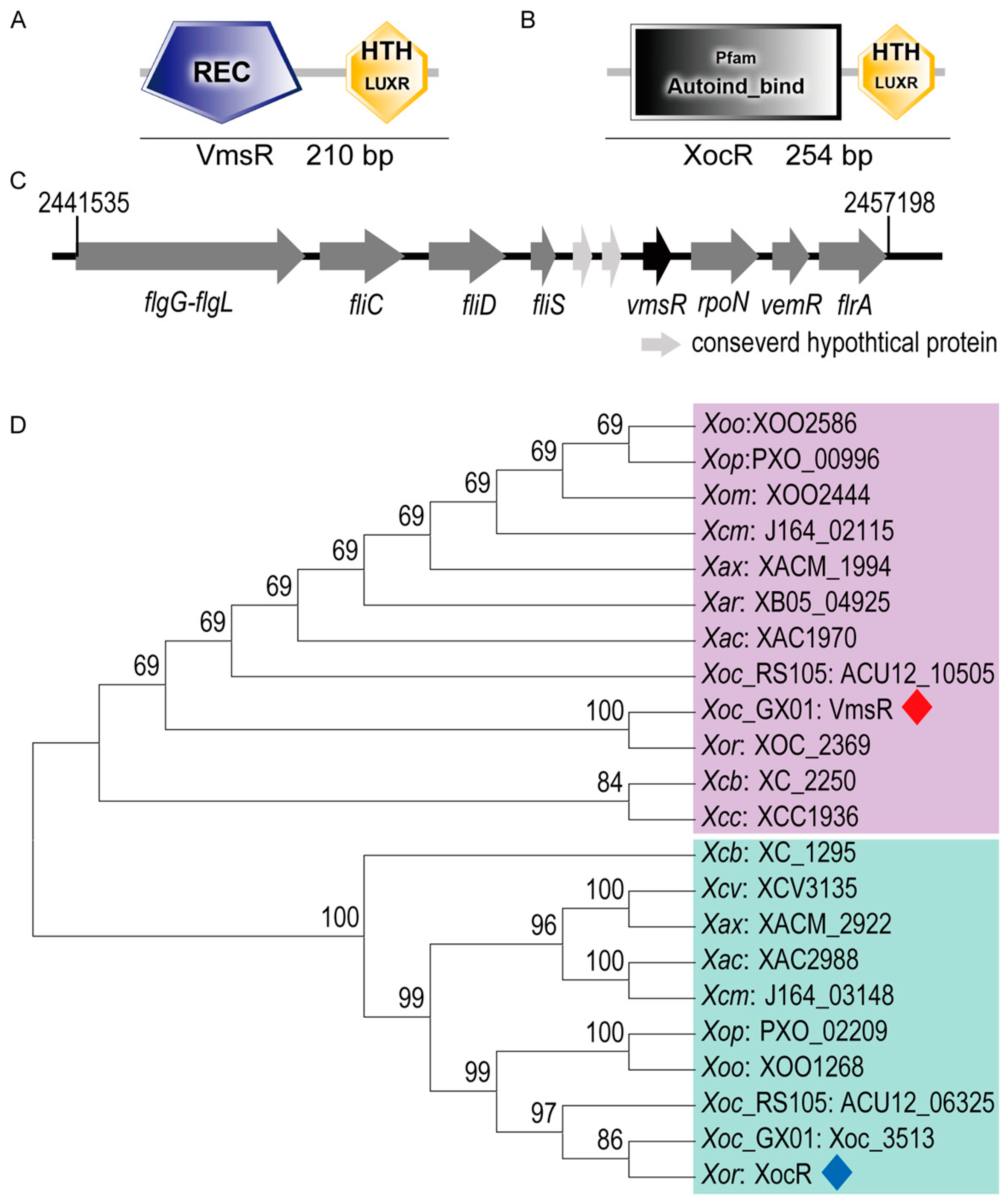
Figure 1. Gene arrangement and phylogenetic trees of VmsR ortholog proteins from Xanthomonas species. (A) Domain structure of VmsR. REC, response regulator domain; HTH LuxR, LuxR family helix–turn–helix domain. (B) Domain structure of XocR. Autoind_bind domain; HTH LuxR, LuxR family helix–turn–helix domain. (C) Arrangement of vmsR and surrounding genes. (D) Phylogenetic trees constructed from amino acid sequence alignments of VmsR ortholog proteins from Xanthomonas species. To highlight VmsR and XocR, VmsR was remarked with a red rhombus, while XocR was distinguished with a blue rhombus. The evolutionary history was inferred using the Neighbor-Joining method. The percentage of replicate trees in which the associated taxa clustered together in the bootstrap test (1000 replicates) are shown next to the branches. The tree is drawn to scale, with branch lengths in the same units as those of the evolutionary distances used to infer the phylogenetic tree. The evolutionary distances were computed using the p-distance method and are in the units of the number of amino acid substitutions per site.
Ijms 25 07595 g001
Figure 2. Virulence of the ∆vmsR was decreased. (A) Fifteen days after inoculation, the lesion length of the wild-type strain GX01, ∆vmsR and complementary strain C∆vmsR were recorded. (B) The virulence of Xoc was impaired when vmsR gene was deleted. Data are displayed as boxplots with individual data points. Error bars represent maximum and minimum values. Values given are the means ± SD of fifteen measurements from a representative experiment. Vertical bars represent standard errors (* p < 0.05, **** p < 0.0001, t = test).
Ijms 25 07595 g002
Figure 3. RNA sequencing and functional classification of differentially expressed genes. (A) Identified DEGs between ∆vmsR and GX01. Volcano plots showing the expression level of each unigene. Limits defined by FDR ≤ 0.05 and |Fold Change| ≥ 2. Red dots represent upregulated genes in ∆vmsR, green dots represent downregulated genes in ∆vmsR, and blue dots represent statistically unchanged genes. (B) Functional classification of the 140 DEGs in the ∆vmsR of Xoc GX01. The transcriptomes of Xoc strains cultured in NB medium were investigated by RNA−Seq. Among the 140 DEGs, 76 genes and 64 genes were upregulated and downregulated, respectively. These genes were broadly categorized according to their biological function. The DEGs list is shown in Table S3. (C) The transcription level of several motility−related genes in ∆vmsR was estimated by quantitative reverse transcription PCR (RT−qPCR). Xoc strains were grown in NB medium to a concentration of OD600 of 0.8−1.0, and RNA was isolated. Relative gene expression with respect to the corresponding transcript levels in the wild−type strain GX01 was calculated. Values given are the means ± SD of triplicate measurements from a representative experiment. Vertical bars represent standard errors (p < 0.01; t = test). Similar results were obtained in three other independent experiments.
Ijms 25 07595 g003
Figure 4. Compared to Xoc GX01, the ∆vmsR exhibited an enhanced swarming motility but a reduced swimming motility. (A,B) The swarming motility of the ∆vmsR strain has increased. Measured data of diameters of the swarming motility on the plate of semi-solid NA medium with 0.6% agar. Each treatment involves three replications, and each experiment was repeated three times. Vertical bars represent standard errors (ns > 0.05, **** p < 0.0001, t = test). (C,D) The swimming motility of the ∆vmsR strain has decreased. Measured data of diameters of the swimming motility on the plate of basal medium with 0.25% agar. Each treatment involves three replications, and each experiment was repeated three times. Vertical bars represent standard errors (ns > 0.05, **** p < 0.0001, t = test).
Ijms 25 07595 g004
Figure 5. VmsR specifically binds to the fliC and fliS promoter in vitro and facilitates their expression. (A) The diagram of flagellar pattern. (B) The transcription level of fliC, fliD and fliS genes in the ∆vmsR strain was estimated by RT−qPCR. The asterisks indicate significant differences compared with the wild−type GX01 strain (mean ± SD, n = 3, * p < 0.05). (C) 12.5% SDS−PAGE gel plots of VmsR−His6 protein purification. M: StarRuler Color Prestained Protein Marker (10−180 kDa) (M221, GenStar, Beijing, China). lane1: cytosol in the absence of IPTG induction. lane2: the precipitates after IPTG induction. lane3: the supernatant after IPTG induction. lane4: purified VmsR−His6 protein. (DF) In vitro electrophoretic mobility shift assay (EMSA) between the recombinant protein VmsR−His6 and the promoter regions of fliC, fliD and fliS genes. Each reaction contained VmsR (10−40 nM) and 6′−FAM-labeled probes (~10 ng).
Ijms 25 07595 g005
Figure 6. EPS production and biofilm formation of the ∆vmsR mutant has increased compared with wild-type strain GX01. (A,B) The EPS production was qualitatively and quantitatively assessed in the wild-type strain GX01, the ∆vmsR mutant, and the complemented strain C∆vmsR. Additionally, the dry weight of each Xoc strain was measured. Vertical bars represent standard errors (ns > 0.05, **** p < 0.0001, t = test). Similar results were obtained in three other independent experiments. (C,D) Biofilm of wild-type strain GX01, ∆vmsR and C∆vmsR was stained with 0.1% (w/v) crystal violet. Quantification of biofilm formed by wild-type strain GX01, ∆vmsR and C∆vmsR. Biofilm formation measured under OD590 using stained biofilm solubilized with absolute ethanol. Vertical bars represent standard errors (ns > 0.05, **** p < 0.0001, t = test).
Ijms 25 07595 g006
Figure 7. VmsR specifically binds to the gumB promoter in vitro and inhibits its expression. (A) The transcription level of gumB in ∆vmsR was estimated by RT−qPCR. The asterisks indicate significant differences compared with the wild−type Xoc GX01 (mean ± SD, n = 3, *** p < 0.001). (B) In vitro electrophoretic mobility shift assay (EMSA) between the recombinant protein VmsR and the promoter regions of gumB genes. Each reaction contained VmsR (10−40 nM) and 6′-FAM-labeled probes (~10 ng).
Ijms 25 07595 g007
Disclaimer/Publisher’s Note: The statements, opinions and data contained in all publications are solely those of the individual author(s) and contributor(s) and not of MDPI and/or the editor(s). MDPI and/or the editor(s) disclaim responsibility for any injury to people or property resulting from any ideas, methods, instructions or products referred to in the content.