Abstract
Chemical pollution poses a significant threat to human health, with detrimental effects on various physiological systems, including the respiratory, cardiovascular, mental, and perinatal domains. While the impact of pollution on these systems has been extensively studied, the intricate relationship between chemical pollution and immunity remains a critical area of investigation. The focus of this study is to elucidate the relationship between chemical pollution and human immunity. To accomplish this task, this study presents a comprehensive review that encompasses in vitro, ex vivo, and in vivo studies, shedding light on the ways in which chemical pollution can modulate human immunity. Our aim is to unveil the complex mechanisms by which environmental contaminants compromise the delicate balance of the body’s defense systems going beyond the well-established associations with defense systems and delving into the less-explored link between chemical exposure and various immune disorders, adding urgency to our understanding of the underlying mechanisms and their implications for public health.
1. Introduction
The exponential growth of the global economy and the accompanying exploitation of resources and industrial production have caused an unprecedented transfer of contaminants to the environment, with multiple impacts on health. Because these human-induced impacts on earth systems are so extensive, a new definition of this geological era has been proposed, the Anthropocene. Although the last few decades have been characterized by marked technological innovation that aims at the concept of sustainability as a way to improve the quality of the environment, anthropogenic activities continue to have a profound effect on human health, mainly due to poor remediation actions [1]. Humans are regularly exposed to many environmental chemicals that may have potentially toxic effects on health. These chemicals can enter the human body through various routes, including the ingestion of chemicals via the consumption of contaminated food or water, inhalation of airborne pollutants through the respiratory system, and dermal absorption through the skin.
The most recent WHO environmental burden of disease estimates that every year, 13% of deaths in the 28 European Member States are attributable to environmental stressors [2]. In terms of the absolute number of deaths attributable to the environment, the European Environment Agency concluded that 90% of deaths attributable to the environment result from non-communicable diseases (NCDs), including cancers, cardiovascular disease, diabetes, and chronic lung illnesses (EEA Report No 21/2019). The interplay between the immune system and non-communicable diseases is complex and multifaceted. Chronic inflammation, immune dysregulation, and lifestyle factors can significantly impact the prevention and management of NCDs. Understanding this relationship can provide insights into the prevention and management of NCDs.
The exposome concept was introduced in the field of epidemiology by Wild in 2005 to encompass “the totality of human environmental exposures from conception onwards, complementing the genome” [3]. Therefore, the exposome concept provides a description of lifelong (from the prenatal period) exposure history. It is now recognized that the exposome poses new challenges in assessing the relationship between exposure to environmental factors (pollution, climate change, lifestyle, and diet) and health. Indeed, increasing evidence indicates that genetic variants account for only a limited fraction of the variability in chronic disease risk, as shown by studies in monozygotic twins, leaving a potentially large role for environmental exposures and interactions between environmental and genetic factors [4]. The molecular processes underlying human health and disease are highly complex. Often, genetic and environmental factors contribute to a given disease or phenotype in a non-additive manner, yielding a gene–environment interaction [5]. Epigenetic mechanisms, including DNA methylation, histone modification, and non-coding RNA, can modulate gene expression levels without changing the underlying DNA sequence. Moreover, many epigenetic modifications are dynamic, reflecting cumulative environmental exposures throughout the lifespan and correlating with aging-related diseases and outcomes [6].
In 2012, Wild enhanced his approach, describing three overlapping domains within the exposome that refer to different factors, such as: (i) the general external environment (including factors such as the urban–rural environment, climate factors, social capital, and education); (ii) the specific external environment (including diet, physical activity, tobacco, infections, occupation, etc.); (iii) the internal environment, including internal biological factors such as metabolic factors, gut microbioflora, inflammation, oxidative stress, and aging [7]. This, together with the results of studies on animal models, would suggest a pathological mechanism of “multiple hits” [8]. According to this model, the rearrangement of gene expression through epigenetic mechanisms can involve different organ systems and is caused by a combination of genetic predisposition and prenatal injury (the “first hit”), which may not be enough to change the adult phenotype on its own. However, tissue imbalances resulting from the perinatal insults and/or adverse stressors/exposures during postnatal life may serve as a “second hit”, which might reveal or accentuate the underlying abnormalities leading to disease states.
All chemicals found in the environment are referred to as pollutants or xenobiotics.
Many such chemicals can persist in the environment over long periods of time and, for this reason, are called persistent organic pollutants (POPs). POPs are characterized by ubiquity and persistence in the environment, and it is known that the derived products can interact with the environment and undergo biotransformation and bioaccumulation processes [9]. Among the anthropogenic environmental pollutants, in addition to certain natural compounds such as heavy metals, pesticides, and drugs, synthetic chemical compounds generated during production processes (e.g., hydrocarbons, dioxins, polychlorinated diphenyls, polybromodiphenyl ethers, microplastics, particulate matter, etc.) can contribute to the pathogenesis of several diseases in living organisms.
The immune system is an ancient defense system formed by all metazoans while struggling with various internal and external factors, whose perturbation may lead to increased susceptibility to pathogens and diseases. Inflammation is a common response to a variety of stressors, including xenobiotics. Under normal conditions, inflammation is a healthy and adaptive process that both combats infection and aids in repairing damage to tissues. However, xenobiotics can elicit prolonged, severe, and/or inappropriate inflammatory responses that play a causal role in the progression of biological events, linking a molecular initiating event to an adverse outcome pathway (AOP) [10]. Ordering relevant and causally linked events in the response cascade is critical for applying this information to Environmental Risk Assessment, which is why the AOP framework was created [11].
In this view, the immune system has a recognized central role in many processes involving chronic diseases, and the frontiers between communicable and non-communicable diseases also require additional exploration [12]. The recognition of altered immune system function in many chronic disease states has proven to be pivotal in non-communicable diseases that are characterized by a chronic low level of inflammation, including autoimmune [13] and neurodegenerative conditions [14], cardiovascular diseases [15], cancer [16], diabetes mellitus, and Metabolic Dysfunction-Associated Steatotic Liver Disease (MASLD) [17,18]. Therefore, compared with other toxicity endpoints (i.e., genotoxicity, endocrine toxicity, or developmental toxicity), there are still many challenges facing the introduction of immunotoxicity endpoint evaluation [19].
So far, there is still insufficient knowledge about the effects of pollutants on the different cell populations of the immune system, and knowledge of related toxicological mechanisms remains elusive. Many findings have suggested that a subset of environmental pollutants can bind the receptors involved in metabolism, oxidative stress, immune response, and inflammation, such as the aryl hydrocarbon receptors (AhRs) [20]. The complexity of AhR signaling arises from multiple factors, including the diverse ligands that activate the receptor, the expression level of the AhR itself, and its interaction with the AhR nuclear translocator [21]. Indeed, the AhR engages in crosstalk with the AhR repressor or other transcription factors and signaling pathways and, in this way, it can also mediate non-genomic effects [22].
In recent decades, the AhR has been increasingly recognized as an important modulator of disease because of its role in regulating the redox system as well as immune and inflammatory responses [23]. The AhR significantly affects the control of adaptive immunity by modulating T cell differentiation and function, both directly and indirectly through its effects on antigen-presenting cells. For instance, 2,3,7,8-tetraclorodibenzo-p-dioxin (TCCD) is a potent xenobiotic ligand of the AhR and was found to inhibit immune responses [24], showing an effect linked to the induction of CD4+ T cells with a regulatory phenotype [25]. Indeed, the ability to activate the AhR has been demonstrated for other contaminants, such as polycyclic aromatic hydrocarbons (PAHs) [26] and polybrominated diphenyl ethers (PBDEs) [27]. The production of reactive oxygen species (ROS) and organic metabolites due to the dysregulation of this receptor can cause serious consequences on biological macromolecules such as proteins, lipids, and DNA [28]. Indeed, oxidative stress, inflammation, and apoptosis can compromise immune response, with the possible development of infections, tumors, and lung diseases [29].
In this review, we analyze how environmental pollutants can modulate immune response, reviewing the biological effects of the most common environmental pollutants on different immune cells in in vitro, ex vivo, and in vivo studies. Indeed, we will devote special attention to pregnancy, describing the effects of pollutants on immune response during different windows of prenatal life (Figure 1).
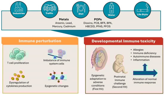
Figure 1.
Schematic representation of the structure of the review.
2. Pollutants and Immune System Modulation: In Vitro, Ex Vivo, and In Vivo Models
A large set of environmental pollutants have been investigated for their impact on immune response. In this paper, we will review the main biological effects of a list of the most widespread chemicals that have been detected in the environment and whose transfer to humans has been documented. From these perspectives, we will focus on the effects of specific classes of toxicants on the immune regulation of human primary and/or immortalized cell lines and in animal models.
2.1. Metals
Heavy metals, such as arsenic, lead, mercury, and cadmium, are a group of highly toxic chemical pollutants that have garnered significant attention due to their detrimental effects on the environment and human health. These elements, which occur naturally in the Earth’s environment, can be released into the environment through various industrial processes, mining activities, and the burning of fossil fuels (anthropogenic activities). One of the most concerning aspects of heavy metals as chemical pollutants is their persistence in the environment. These substances do not degrade over time, accumulating in the soil, water, and air. Consequently, they can enter the food chain, leading to bioaccumulation, where the concentration of these toxic metals increases as one moves up the food web [30,31,32]. Below, we describe the effects of some of the most common heavy metals, analyzing their effects using in vitro, ex vivo, and in vivo models.
2.1.1. Arsenic
Several national and international organizations have classified Arsenic (As) as the most toxic and carcinogenic inorganic environmental pollutant. It exists in nature in both organic and inorganic forms. The inorganic form is more toxic and accumulates in exposed organisms; it is found in the environment in the form of arsenite (AsIII) or arsenate (AsV). The main route of arsenic exposure is the consumption of contaminated drinking water. Many studies have found a positive relationship between arsenic (As) exposure and the onset of human pathologies including cancer (lung and bladder) and liver, respiratory, cardiovascular, and immune diseases [33,34]. In terms of immune system dysregulation, it has been demonstrated that As either reduces the expression level of cytokines (IFN-γ, IL-4, and IL-10) [35,36,37] or induces apoptotic mechanisms in different cell types, such as B and T lymphocytes, macrophages, and neutrophils [38,39]. It has been shown that As can drive M2 polarization in macrophages, triggering the CD206 marker along with TGF-β1 and Arg1 genes that have been shown to be involved in the fibrinogenic process [40]. Arsenic-induced M2 polarization is also favored via miR-21 regulation of phosphatase and tensin homolog deletion on chromosome ten (PTEN), which has been implicated in chronic hepatic fibrosis and in the development of arsenicosis [41].
Microarray analysis of peripheral blood mononuclear cells (PBMCs) purified from subjects exposed to increasing As concentrations has also shown that this pollutant modulates T cell receptor expression, the cell cycle, and apoptotic processes. Furthermore, As increases the expression of inflammatory molecules such as cytokines or growth factors [42] and induces the hypomethylation of leukocyte DNA, which is associated with an increased risk of inflammatory skin lesions [43]. Arsenic also has a negative effect on innate and humoral immunity, altering macrophage and B lymphocyte functions and stimulating oxidative stress signaling, DNA damage, and cytotoxicity in T cells and in human polymorphonuclear neutrophils [44,45,46].
2.1.2. Lead
Lead (Pb) is available in nature in relatively low amounts. Anthropogenic activities, such as manufacturing, mining, and the burning of fossil fuels, have contributed to the increased release of Pb into the environment. Lead induces toxic effects on various human tissues, including the immune system, leading to increased allergies and infectious and autoimmune diseases [47,48]. For instance, in vitro studies have shown that Pb treatments induce the dysregulation of pro-inflammatory cytokine production [49,50,51] and impair THP-1 monocyte/macrophage cell viability at low concentrations. Zhao and coworkers showed that Pb exposure induces the quiescence of hematopoietic stem cells through Wnt3a/β-catenin signaling and decreases the expression of CD70 on bone marrow-resident macrophages [52]. These observations were confirmed by ex vivo studies on a population of chronically exposed workers, in whom higher levels of IFN-γ, IL-2, IL-12, and IL-17 were observed [53,54]. Lead can cross the placenta [55] and can have adverse effects on birth outcomes, possibly by accumulating in the placenta and causing reduced nutrient transfer, oxidative stress, and abnormal functioning [56].
2.1.3. Mercury
Mercury (Hg) is a highly toxic metal, and exposure to its organic and inorganic chemical compounds causes adverse effects on humans, inducing genetic damage and neurological, kidney, cardiac, and immunological diseases [57]. The predominant toxic Hg forms include elemental Hg (Hg0), ionic Hg, and organic (o)Hg, such as methylmercury (MeHg), which is classified as the most toxic among them.
In the innate immune system, Hg can influence the activity of different cell subtypes, such as neutrophils, monocytes/macrophages, NK cells, and dendritic cells. It has been shown that in vitro exposure of neutrophils to low concentrations (≤5 μM) of HgCl2 causes an increase in the production of superoxide ions [58], inhibiting the apoptosis of neutrophils, while at higher concentrations, Hg displays cytotoxic effects. At the same concentrations of pollutant, macrophages show functional impairment of both phagocytic and migratory activity [59,60], showing a reduction in NO production and a greater synthesis of inflammatory cytokines, such as IL-6 and TNF-α. Moreover, after Hg exposure in co-culture assays of intestinal epithelial cells and THP-1 macrophages, mimicking the gastrointestinal tract, the metal induced a pro-inflammatory response with an Increase in IL-8 and IL-1β cytokine production [61].
Mercury derivatives also show genotoxic effects in vitro and in vivo, in terms of both chromosome aberrations and loss of cell proliferative capacity, compromising the metabolic, immune, and nervous systems [62]. Dietary MeHg intake (3.9 μg/g) by mice and rats resulted in a 42–44% suppression of the tumoricidal activity of blood and splenic NK cells, as well as the proliferation of T and B cells of the adaptive immune system [63,64]. These events have been associated with an increase in intracellular oxidative stress [65,66], which causes the activation of transcription factors and the expression of genes encoding pro-inflammatory cytokines.
2.1.4. Cadmium
Cadmium (Cd) is a heavy metal naturally present in the environment (in the soil, water, and air) and is mainly used in the steel industry, in plastics, and as a component of batteries. It is released into the environment through industrial and domestic activities, such as the combustion of fossil fuels (coal, diesel, gasoline, etc.), the incineration of industrial waste (particularly batteries and plastics containing Cd), the production of metal alloys, and the manufacture of fertilizer phosphates. Food, drinking water, and the inhalation of cigarette smoke, in both active and passive forms, are sources of this heavy metal. Target organs for Cd-induced toxicity include the liver, kidneys, lungs, testes, prostate, heart, skeletal system, and the nervous and immune systems.
Following environmental and/or industrial exposure, it has been shown that Cd induces inflammation and oxidative damage in neutrophils and macrophages, leading to an increase in the production of ROS [67,68]. These results have been further confirmed by experimental studies carried out on macrophage cell lines [69]. Cd-induced M1 polarization has been observed in in vitro and in vivo rodent macrophages via the JAK2/STAT3 pathway, contributing to the atherosclerotic process [70]. Furthermore, it has been shown that intrauterine exposure to Cd can provoke a dramatic decrease in the levels of IFN-γ and IL-2 cytokines in offspring and affect the activity of adaptive immunity cell populations in C57Bl/6 mice. In female offspring, CD4+ and CD8+ T cell populations increase after exposure to Cd, while the percentage of NK cells and granulocytes decreases. In addition, a decrease in splenic Treg cells has been shown in both male and female offspring [71,72]. Another study in wild boars highlighted that Cd downregulates the expression of different pro-inflammatory cytokines (TNF-α, IL-12p40, several TLRs, CD14, MD2, BD2, MyD88, p65, and NOS2) in macrophages, negatively affecting their roles in immunity [70].
In a study on workers occupationally exposed to Cd, epigenetic alterations in the expression of miRNAs associated with inflammation and carcinogenesis, as well as their possible correlation with the immune profile, were evaluated. MiRNA profiling showed that miR-221 and miR-155 were modulated in exposed workers with reference to a higher percentage of the Th17 population [73] (See Table 1 for summary).

Table 1.
In vitro, ex vivo, and in vivo effects of Heavy Metals.
2.2. Persistent Organic Pollutants (POPs)
Among the most dangerous organic pollutants for health, persistent polyhalogenated organic compounds or POPs (so-called because they withstand biochemical and photolysis processes by remaining in the environment for a long time) [74] are very stable chemicals whose lipophilic nature determines their bioaccumulation in animal adipose tissues, thus allowing their biomagnification along the food chain. Due to their chemical-physical characteristics, POPs can cross the placenta, causing exposure starting from intrauterine life, but can also accumulate in breast milk, which is particularly rich in lipids, determining their intake in post-natal life [75]. The most common POPs are polycyclic aromatic hydrocarbons (PHAs), organochlorine pesticides (OCPs), polychlorinated biphenyls (PCBs), perfluorinated compounds (PCFs), dioxins, and brominated flame retardants (BFRs), which will be discussed in their dedicated sections.
2.2.1. Dioxins
Dioxins are highly toxic POPs produced from natural processes such as forest fires as well as the burning of trash by anthropic activities. Due to their highly lipophilic nature, they undergo biomagnification along the food chain to eventually accumulate in human tissues. Of all the analyzed congeners, 2,3,7,8-tetrachlorodibenzo-p-dioxin (TCDD) showed the highest toxicity for living organisms [76]. Several in vitro, ex vivo, and in vivo studies have been conducted to determine the adverse effects of dioxins on the various components of the immune system. Regarding the innate response, a compromise in the defense system against pathogenic organisms and cancer cells was observed; in particular, TCDD impairs the correct functioning of macrophages, NK cells, neutrophils, and dendritic cells [77,78,79]. In adaptive response, dioxin modulates the production of antibodies by B lymphocytes and the cytotoxic activity of T lymphocytes, while also attenuating the IgE-mediated hypersensitivity response [80,81]. It has been observed that, in THP-1-derived macrophages, TCDD treatment induces alterations in adherence, adhesion molecule expression, morphology, multiple cytokine/chemokine production, and the expression of total mRNA [82]. Few studies have focused on TCDD’s multigenerational and transgenerational effects on human reproductive health, despite the large amount of evidence in animal models of such effects on male and female reproductive health [83].
2.2.2. Polychlorinated Biphenyls (PCBs)
Polychlorinated biphenyls are environmental pollutants of industrial origin. Although their production was interrupted many years ago, they persist in the environment due to their resistance to chemical, physical, and biological degradation processes. Like other organic pollutants, they accumulate in the soil and can enter the food chain, eventually reaching humans. Numerous studies have shown that PCBs can alter the proper functioning of the immune system at several levels. For example, in macrophage cell lines, treatment with PCB 126, a dioxin-like polybromophenyl, has a pro-inflammatory immunostimulating effect [84]. In vitro, the non-dioxin-like polychlorinated biphenyl (NDL-PCB) congeners, NDL-PCB 153 and NDL-PCB 180, display an immunosuppressive effect by reducing the expression of the pro-inflammatory cytokines TNF-α and IL-6 and the expression of reactive species [85]. PCBs are also capable of altering the physiological antiviral cellular response [86]. In an in vivo mouse model, PCB promoted alterations in collagen in the female bladder compared to the male, indicating an inflammatory event that impairs immune system functions [87]. Moreover, studies from PCB-exposed populations have revealed DNA methylation differences in CpG sites with effects on estradiol CpG site sizes and the immune system [88].
2.2.3. Brominated Flame Retardants (BFRs)
Brominated Flame Retardants (BFRs) are chemicals used in the synthesis of plastics, textile products, and electronic and electrical devices; they are added to industrial products to make them less flammable. It has been shown that the flame retardants contained in various products are released into the environment through combustion, percolation, and degradation [89]. Due to their increasing use, BFRs have become globally widespread pollutants over the years. They have been found both at the level of different environmental matrices (air, aquatic sediments, soil, dust, etc.) and in animal organs and tissues, such as blood, mother’s milk, and adipose tissue [90,91].
There are four main classes of BFRs: (1) polybrominated diphenyl ethers (PBDEs), present in plastics, textiles, electronic devices, and circuits; (2) hexabromocyclododecanes (HBCDDs) used for thermal insulation in buildings; (3) tetrabromobisphenol A (TBBPA) and other phenols used in thermoplastics and televisions; (4) polybrominated biphenyls (PBBs) used in consumer devices and textiles [92]. The immunotoxic effects of the most studied flame retardants will be discussed below.
Polybrominated Diphenyl Ethers (PBDEs)
Polybrominated diphenyl ethers are chemicals consisting of two aromatic rings joined by an ester bond with bromine atoms. The commercial use of PBDEs dates back to 1976 and the three most used blends were penta-BDE, octa-BDE, and deca-BDE. Since 2004, the production of penta- and octa-BDE has been banned, both in the United States and in the European Union, due to their negative effects on the environment and health. The deca-BDEs have been banned in the United States since 2010. On the basis of the number and position of the bromine atoms, we can distinguish 209 congeners with different chemical-physical characteristics that affect their capacity for bioaccumulation and toxicity in the liver, kidney, gut, and thyroid in terms of oxidative and mitochondrial damage and apoptosis [90,93]. Human population studies have shown that PBDEs accumulate in adipose tissue, hair, human serum, breast milk, and the placenta. Among the most toxic and widespread congeners in the environment, BDE-47 (a tetra-BDE) and BDE-209 (a deca-BDE) are the most relevant in terms of health risks. Experimental data on animal models have shown that BDE-47 can have neurotoxic, cardiotoxic, hepatotoxic, and teratogenic effects on zebrafish larvae and adults and in fish [94,95,96].
Several studies have investigated the immunotoxic effects of PBDEs. In in vitro studies, BDE-47 significantly reduced the expression and secretion of the pro-inflammatory response M1 macrophage marker genes IL-6, TNF-α, and IL-1β and the expression of metalloelastase 12, a gene essential in the motility of the macrophage, recruitment of neutrophils, and release of cytokines and chemokines in inflammatory processes [97]. Indeed, the same authors showed that BDE-47 can have diverse action mechanisms on human macrophage cell lines, affecting small extracellular vesicle biogenesis [98] and the content of their miRNA cargo. Furthermore, they demonstrated that BDE-47 treatment can regulate the sEVs’ cargo with purposeful consequences toward downstream events and target bystander macrophages, exacerbating the macrophage LPS-induced pro-inflammatory response [99] as well as altering A549 epithelial lung cells, which modulate the mRNA expression of tight junctions, adhesion molecules, cytokines, and epithelial–mesenchymal transition markers [100]. Finally, it has been shown that BDE-47 can also induce cardiovascular toxicity, activating PPARγ in THP-1 monocytes and inducing the foam cell formation typical of a proatherogenic process [101]. In an animal model, it was observed that they stimulate an increase in the production of ROS in macrophages, leading to the remodeling of the cellular physiology of phagocytes and, therefore, to the destruction of the functional activity of the macrophage [102,103,104]. Recently, Balb/c mice treated with different doses of BDE-47 by gavage showed that BDE-47 significantly reduced antibody response and induced histopathological effects on the liver, spleen, small intestine, and thyroid in the absence of general toxicity, suggesting that exposure to BDE-47 may perturb innate and adaptive immune responses [105].
Hexabromocyclododecane (HBCDD) and Tetrabromobisphenol A (TBBPA)
Hexabromocyclododecane (HBCDD) is a non-aromatic brominated cyclic alkane; it is mainly used in expanded propylene insulation foam [106,107], but also in tapestry fabrics, curtains, and wall coverings [108]. HBCDD is added to plastic material but, since it is not chemically linked to it, it can transfer into the environment and accumulate in the soil, sediments, and dust particles. It is involved in the trophic chain to humans through the ingestion of dust [109,110], bioaccumulating in the blood, adipose tissue, and breast milk [111,112,113]. Immunotoxicity studies on rats have highlighted that HBCDD reduces splenocytes and increases IgG and neutrophils, contributing to immune system imbalance [114]. Tetrabromobisphenol A (TBBPA) is a flame retardant used in thermoplastics production. At the cellular level, Barańska and collaborators found that TBBPA has a genotoxic mechanism because it causes oxidative damage in purines and pyrimidines in PBMCs [114,115,116]. Furthermore, a study conducted on human dendritic cells isolated from healthy subjects by Canbaz and collaborators showed that exposure to this flame retardant can induce an inflammatory phenotype typical of a Th2 response [117]. Furthermore, for the same substance, neurotoxic, nephrotoxic, hepatotoxic, and immunotoxic effects have been reported, which can induce ROS expression, promoting the production of inflammatory cytokines and compromising mitochondrial function [118]. A correlation has also been demonstrated between human exposure to both classes of pollutants and thyroid and neurological disorders, reproductive health, and immunological, oncological, and cardiovascular diseases [119,120].
2.2.4. Bisphenol A
Bisphenol A (BPA) is a chemical that has been synthesized since the 1960s and is widely used in industrialized countries. BPA is used in the production of polycarbonate plastics, in food-grade containers, and in epoxy resins that make up the internal protective coating found in most food and beverage cans. In addition, it is also used for dental devices and in thermal receipt paper. Under the pressure of EFSA, between 2010 and 2015, many studies aimed at understanding the possible effects of BPA on immune system cells and tissues were conducted. Balistreri and collaborators showed that BPA alters the function of neutrophils in a dose-dependent manner, causing an increase in ROS production in these cells through a calcium-dependent process. Transwell chemotaxis assays revealed that BPA exposure reduces the chemotactic capacity of neutrophils in a gradient of the bacterial cell wall component f-Met-Leu-Phe, a potent chemoattractant. Indeed, exposure to BPA also inhibits the ability of neutrophils to kill methicillin-resistant Staphylococcus aureus, a leading human pathogen. These data suggest that BPA could compromise some fundamental functions of the innate immune response [121]. Bisphenol A also promotes the polarization of M1 macrophages In terms of cell number, the gene expression of CD11c and iNOS markers, and the production of pro-inflammatory cytokines via the upregulation of transcription factor IRF5 expression, which is involved in the polarization process [122]. The upregulation of pro-inflammatory cytokines was also induced in the murine macrophage cell line RAW264.7 by the BPA analog BPF (4,4′-Methylenediphenol). Together with the involvement of the JAK2/STAT3 pathway, it contributed to M1 polarization [123,124].
BPA also showed immunotoxic effects on the adaptive immune response. An in vivo study conducted on mice showed that animals treated with a single dose (250 µg/kg) of BPA in the neonatal period and subsequently subjected to breast cancer induction had no significant changes in the population of B lymphocytes or damage to the spleen and peripheral lymph nodes. However, they exhibited a significant difference in IgM reactivity, with a notable decrease in the ability to recognize tumor antigens in guinea pigs [125].
2.2.5. Perfluorooctanesulfonic Acid and Perfluoroctanoic Acid
Perfluorooctanesulfonic acid (PFOS) and perfluorooctanoic acid (PFOA) are perfluoroalkyl organic substances (PFAS). They are chemical compounds containing long carbon chains that are used to increase resistance to high temperatures in numerous products, such as fabrics, carpets, clothing, food-grade paper, non-stick cookware, and firefighting foams. PFOA and PFOS remain in the environment for a long time and, due to their chemical stability, resist degradation processes, accumulating in the soil, air, and water. PFAS are able to provoke the release of the pro-inflammatory cytokines IL-1β and caspase-1 in THP-1 macrophages. They also activate the innate immune response through the AIM2 receptor of inflammasomes, inducing inflammation in the lung, liver, and kidneys of mice [126]. Population studies have shown that PFAS can accumulate in human organs and tissues; for example, PFOS has been found in the liver, kidneys, lungs, hair, breast milk, and urine, but it accumulates predominantly in the blood [127,128]. In vitro and in vivo data have shown that PFOSs and PFOAs have toxic effects on multiple cell types and organs of the immune system. An in vitro study showed that treatment of human T cells with increasing concentrations of PFOS induced a reduction in the expression of IL-2, a cytokine essential for the correct functioning of leukocytes and whose reduction was observed in various autoimmune diseases [129]. An in vivo study conducted in mice showed that PFOS treatment inhibits T cell proliferation. Analysis of the biochemical pathways involved in immune cell signaling revealed the inhibition of genes involved in cell cycle regulation and response to oxidative stress [104]. In vivo studies in mice performed by Torres and collaborators showed that PFOS exposure reduced immune cell populations in some organs and also led to an increase in the number of cells in others, suggesting a possible de novo localization of cells and a decrease in the activity of some organs such as the spleen and liver [130]. Taylor and coworkers showed that PFOA exposure can reduce B-cell subtypes and, consequently, the IgM antibody primary response in female mice [131].
2.3. Volatile Organic Compounds (VOCs)
Volatile organic compounds are a large group of organic chemical compounds that consist of elements of carbon and one or more functional groups containing oxygen, phosphorus, nitrogen, silicon, halogen, and sulfur. VOCs are characterized by high vapor pressure and low water solubility. These molecules are produced by anthropogenic activities such as the manufacture of paints, solvents used in furniture making and electronics, vehicular traffic, refrigerants, and pharmaceuticals. They are also emitted during natural processes, including forest and vegetation fires, volcanic eruptions, and microbial processes [132]. VOCs show high resistance to abiotic and biotic degradation, and by interacting with environmental pollutants (i.e., nitrogen oxide species), they produce tropospheric ozone and secondary environmental pollutants which have hazardous effects on the well-being of humans and ecosystems [132]. Since the 1990s, the relationship between VOC exposure and immune response has been extensively studied, particularly in murine and ex vivo human models. Multiple data sources have shown the ability of different types of compounds to induce an imbalance of reactive oxygen species and the release of pro-inflammatory cytokines from immune cells by activating the inflammatory molecular pathways of NF-kB. Studies have shown that exposing CD3+/CD28+ human PBMCs to toluene decreases the release of IL-4 and IL-13 cytokines while simultaneously increasing the production of the pro-inflammatory cytokine TNF-α [133]. Xylene, another aromatic hydrocarbon, induces mitochondrial damage following oxidative stress in purified human T lymphocytes [134]. Genome-wide analyses have demonstrated that VOCs induce the activation of genes belonging to type I Interferon signaling in human promyelocytic leukemia HL60 cells [135]. A cross-sectional study of workers exposed to formaldehyde suggested a reduction in NK cells, regulatory T cells, and CD8+ effector memory T cells compared to control workers [136]. The aforementioned studies, along with many others related to the effect of different types of volatile organic compounds on the modulation of the immune response, have been thoroughly reviewed by Ogbodo and coworkers in a recently published paper [137], where the authors have also linked the inflammatory response to VOCs to the onset of several autoimmune diseases.
2.4. Particulate Matter (PM)
Airborne particulate matter may be a complex mixture of dust, dirt, soot, and smoke containing both organic and inorganic substances as well as biological components. The airborne particulates can be characterized by their physical attributes, which influence their transport and deposition, and their chemical composition, which influences their effect on health and the immune system. The particles most likely to cause adverse health effects are the fine particulates PM10 and PM2.5 (particles smaller than 10 microns and 2.5 microns in aerodynamic diameter, respectively). PMs play a role in biological pathways related to cell signaling and immune response [138]. Even if the effects of short-term exposure to PM2.5 on innate immunity and its relationship with tissue damage and repair ability have been scarcely studied [139], it has been demonstrated that prolonged exposure to PMs, even at low levels, is associated with the activation of inflammatory pathways (e.g., Nrf2, MAPK, and NF-kB), with an increase in cytokine and chemokine production [140] and immune cell activation [141]. PM can contribute to the development and maintenance of inflammation through epigenetic mechanisms, such as changes in histone tail modifications, microRNA (miRNA) expression, and DNA methylation [141].
By employing ALI (Air Liquid Interface) cultures of various human cells that represent different sections of the respiratory tract, researchers have successfully simulated realistic exposure conditions to airborne particles. This approach has enabled the investigation of toxicity and immune responses following controlled exposure on the first line of immune responder cells. These models are essential for evaluating the effects of PM, including oxidative stress, inflammation, DNA damage, and apoptosis [142]. Recently, Chivé et al. reported that exposure of the Calu-3 cell line to ultrafine particulates in the ALI exposure system induces a pro-inflammatory response and reduces antiviral defenses. Their findings may elucidate the observed association between PM2.5 exposure and the severity of influenza infections in human populations [143].
Alternatively, in vitro studies on the effects of PM exposure on immune cells can be conducted by treating cells with resuspended atmospheric particulates in culture media. In particular, research by Danielsen and collaborators revealed that PM exposure has similar effects on lung epithelial cells (A549) and monocyte cultures (THP-1). The response varied based on the particulate collection site, with the most significant effects observed in areas with high biomass combustion [144]. Marín-Palma and collaborators identified the differential expression of 1196 genes following the exposure of PBMC cells to PM10. The differentially expressed genes related mainly to the inflammatory response, with an upregulation of cytokines and chemokines. Interestingly, this alteration in the inflammatory response was accompanied by the downregulation of genes involved in pathogen defense and antiviral factors [140]. Moreover, an in vivo study on a rat model reported that short-term exposure to high concentrations of PM2.5 induces lung tissue damage and alters innate immune responses. Interestingly, although some repair occurs after 15 days of recovery, the function of innate immunity, including immune defense and lung homeostasis regulation, remains partially impaired [139].
In a study using a rat model, Wang and colleagues found that exposure to traffic-related PM2.5 impairs regulatory T cell (Treg) function and disrupts the balance between T-helper 1 (Th1) and T-helper 2 (Th2) cells. This disruption occurs through differential microRNA (miRNA) expression and DNA methylation modifications, ultimately leading to the exacerbation of asthma symptoms [145].
2.5. Microplastics
Microplastics (MPs) are small plastic particles with diameters less than 1 mm and have become a pervasive environmental pollutant. Originating from the breakdown of larger plastic debris, as well as from products such as cosmetics, textiles, and industrial processes, microplastics become part of the trophic chain in both marine and terrestrial ecosystems, posing a threat to human health [146,147]. So far, MPs have been detected in different biological fluids and cellular compartments as milk, blood, animal excreta, and placenta [148,149]. Microplastics vary significantly in their chemical composition, size, and shape. These variations can affect exposure levels, local or systemic absorption, and interactions at cellular and molecular levels with the plastics [150]. Polystyrene (PS), Polyethylene (PE), Polypropylene (PP), and Polyvinyl chloride (PVC) are among the most frequently found plastics in the environment [151]. These MPs are hydrophobic, making hydrophobicity a key factor in microplastics’ role as vectors for a range of contaminants. It has been observed that microplastics can cause damage to cells in several ways, inducing oxidative stress, inflammation, metabolism disorders, apoptosis, and genotoxicity. PS MPs affect immune function in microglial HMC-3 cells by depositing onto brain microglial cells, altering their cellular morphology and causing immune dysfunction through the alteration of IL-1β, CCL2, and TGF-β levels [152]. In vitro studies using sea bass or seabream fish seem to suggest that the long-term exposure of fish to PVC or PE microplastics could impair fish immune parameters, probably due to the oxidative stress produced in their leucocytes [153]. In addition, in vivo experiments on chickens have demonstrated that exposure to PS MPs causes liver damage and inflammation, autophagy activation, intestinal macrophage infiltration, and lipid accumulation [154]. Additionally, polypropylene (PP) particles induce pro-inflammatory cytokines in a size- and concentration-dependent manner by increasing the levels of cytokines and histamines in PBMCs and RAW 264.7 and HMC-1 cells. Studies on PBMC and THP-1 macrophage-like cell lines have been conducted to investigate metabolic activity, cytotoxicity, ROS production, and macrophage polarization [155]. In addition, it has been shown that UV radiation accelerates the photo-oxidation and aging process of plastics, leading to changes in their chemical structure and physical properties. This process not only contributes to the fragmentation of plastics but also affects their morphology and the release of harmful additives and chemicals, further contaminating ecosystems. Recently, it has been shown that treatment with photoaged PS MPs induced increased malondialdehyde levels [156] in the THP-1 macrophage-like cell line.
Table 2 summarizes the effects of POPs, VOCs, PMs, and MPs on immune response.
Table 2.
In vitro, ex vivo, and in vivo effects of POPs, VOCs, PMs, and microplastics (MPs).
Figure 2 displays the similarities and differences between the main classes of environmental pollutants in the modulation of immune response.
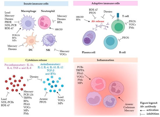
Figure 2.
Schematic representation of the data obtained by in vitro, ex vivo, and in vivo studies on the effects of the main classes of environmental pollutants on immune response.
3. Developmental Immunotoxicology
The term developmental immunotoxicology (DIT) has been used to identify a new approach to understanding the effects of immune development perturbation on adverse outcomes [157]. DIT addresses the impact of xenobiotics on immune responses during the early stages of immune system development/maturation compared to adults. Immunotoxicology studies have defined some critical windows of vulnerability during development [158], when xenobiotics can exert their effects, but sometimes stressors may be applied during rather broad periods which often cover the entire immune developmental period. These effects can be particularly significant in highly polluted areas where environmental stressors are prevalent throughout a large portion of prenatal and postnatal life. Furthermore, considering the accumulation of evidence supporting the hypothesis of the Developmental Origin of Health and Disease (DOHaD) [159,160], disturbances of the immune system during fetal development are of particular interest for possible long-term consequences. According to epidemiological studies, the adverse effects of in utero exposures on the immune system have been associated with higher rates of chronic immune disorders in adults, including autoimmunity, immune deficiency, inflammation, and allergic reactions [161].
Although these associations are of considerable importance, only examining outcomes directly linked to immune system elements fails to capture the complete range of health risks associated with DIT. In fact, from the early stages of life, immune cells are found in all tissues and organs where they serve as homeostatic regulators of the physiological function of the tissue and as sentinels to defend it. In the liver, for example, the functional state of hepatocytes (the majority of cell populations) can be significantly altered by a small number of resident immune cells (i.e., Kupffer cells). This occurs similarly between microglia and astrocytes in the central nervous system and many other organs [162]. For these reasons, the involvement of DIT events underlying behavioral disorders and metabolic dysfunction has been suggested [163].
During pregnancy, major adaptations occur in the maternal immune system to protect the mother and her future baby from pathogens. The perturbation of this balance may have detrimental immune responses against the allogeneic fetus. Pro- and anti-inflammatory stimuli alternate during gestation, and their regulation is the basis of the success of numerous key steps of the pregnancy. For example, the implantation of the embryo on the uterine wall is facilitated by a period of active inflammation, essential for the remodeling of the maternal uterus [164]. On the contrary, the phases of active fetal growth and development are accompanied by a relative inflammatory quiescence [165]. Failure to induce these systemic changes predisposes women to adverse pregnancy outcomes, such as recurrent miscarriage [166], preeclampsia [167], and preterm birth [168]. Additionally, depending on the duration and severity, this inappropriate activation of the maternal immune response may have transgenerational consequences. For instance, the activation of the mother’s immune/inflammatory response has been linked to negative neurobehavioral outcomes in offspring [169,170,171,172]. These findings are noteworthy from an immunological standpoint, as the physiological development of the fetal brain involves a variety of immunological factors at the maternal–fetal interface [173]. For example, in the case of extremely preterm infants, placental methylation of genes related to inflammation has been observed to be associated with a decreased risk of cognitive impairment and decreased neonatal systemic inflammation [174].
Evidence suggests that prenatal exposure to xenobiotics may play a role in altering the epigenetic regulation of immune-related genes that influence the development of regulatory T cell (Treg) networks or the ratio of Th1 and Th2 cells. Because Th1/Th2 immunity is closely linked to disease, epigenetic changes caused by an adverse intrauterine environment could explain the occurrence of disease susceptibility later in life [175]. In parallel with the direct modulation of epigenetic regulation, numerous toxic substances can impact the endocrine system. Given its close collaboration with the immune system in guiding development from gestation to early childhood, endocrine disruptor chemicals may also trigger DIT [176].
In this section, we will provide an overview of the effects of the most relevant xenobiotics during pregnancy, with specific reference to immune dysregulation.
Arsenic toxicity has been linked to many possible pathways, including oxidative stress, epigenetic modification, interference with DNA repair mechanisms, and alterations of the immune system [177,178]. Exposure to As during pregnancy can result in DIT interacting directly and indirectly with the fetal environment. This interference is strongly related to placental function, as the placenta is permeable to As and particularly susceptible to its detrimental effects, including oxidative stress, inflammation, and immune system perturbation [33]. In two separate cohort studies from Bangladesh and the United States, prenatal As exposure was associated with lower percentages of CD4+ T cells and, in particular, activated memory CD4+ T helper cells in cord blood [179,180]. Although a direct comparison of exposure levels between the two populations is not possible, the effects of As exposure appear to be consistent. In the study on the Bangladeshi population, As exposure is determined from the levels found in drinking water samples, whereas in the U.S. study, exposure is assessed from maternal toenail samples. Evidence of the similarity in findings showed that both cohorts demonstrated relatively higher susceptibility to infectious diseases at different stages of childhood, which was associated with prenatal arsenic (As) exposure [181,182]. This suggests that prenatal As exposure causes overall immunosuppression in offspring. Among the mechanisms of action that could explain the negative effects of As exposure on immune system cells, the most widely accepted appears to be the induction of oxidative stress. Arsenic exposure has been shown to trigger oxidative stress in peripheral blood in both adults and children [183,184]. Studies conducted on populations exposed to high levels of As have demonstrated that prenatal exposure to arsenic increases the expression of numerous markers of inflammatory and oxidative stress in umbilical cord blood and placenta [33,185].
The long-term effects of arsenic exposure during pregnancy on the immune system may be mediated by epigenetic mechanisms. Specifically, exposure to arsenic has been shown to induce alterations in DNA methylation patterns, which can influence gene expression and immune cell function [186,187]. Specifically, the authors identified 10 differentially methylated CpGs corresponding to six genes (BAI2, CD1B, TEAD1, SGCZ, ACSS3, and ZMYND19) associated with Gestational Diabetes Mellitus with high Arsenic (As) exposure. Studies on mice have suggested that one possible mechanism linking prenatal As exposure to health effects later in life may be epigenome alteration. Cytosine methylation (e.g., CpG methylation) has been suggested as a valuable candidate for As-related health effects [188].
Toxic effects related to Hg exposure, particularly methylmercury (MetHg), mainly affect the nervous system [189]. However, as previously mentioned, in vitro experimental evidence showed possible associations between Hg exposure and immunotoxicity [61,65,67]. Mercury can be transferred from mother to fetus through the placenta [189], and in utero exposure to Hg has been associated with a higher risk of respiratory infection in infants during the first year of life [190]. Mercury levels during pregnancy and postpartum have been linked to the frequency of natural Treg and NKT cells, as shown in the New Hampshire Birth Cohort Study. Higher natural Treg counts have also been associated with an increased risk of Hg exposure as measured by seafood consumption. Elevated Tregs may represent a counterbalancing mechanism in response to the increased autoimmune pressure associated with low-level Hg exposure [191]. In a study focused on a highly exposed population from the Faroe Islands, Oulhote and colleagues reported a correlation between prenatal methylmercury (MetHg) exposure and a decreased total white blood cell count, specifically affecting monocytes, basophils, and CD3+ T, CD4+ T, and CD19+ B lymphocytes at the age of 5 [192]. It could be hypothesized that such an alteration in the balance of white blood cells could affect inflammatory cytokine levels. In this view, a multicentric cohort study conducted in five European countries (France, Greece, Norway, Spain, and the United Kingdom) showed that in utero exposure to Hg was correlated with poorer childhood metabolic status and higher levels of TNF-α, IL-6, and IL-1 at 6 and 12 years. In this report, the authors suggested that changes in key inflammatory cytokines and the metabolic profile are strictly interconnected [193]. In an Amazonian Brazil population exposed to high levels of mercury, a positive association between mercury levels and serum concentrations of antinuclear antibodies was found, suggesting a possible role of mercury in autoimmune disease [194]. In a previous study conducted in the same population, Pinheiro and colleagues reported elevated glutathione levels and decreased catalase activity in response to Hg exposure [195]. Mercury’s capacity to modulate the synthesis and activity of enzymes within the endogenous antioxidant system may underlie its effects on immune cells. By disrupting the normal function of these enzymes, mercury can impair the antioxidant defenses of immune cells, resulting in altered immune responses and increased vulnerability to infections and diseases.
Studies on the effects of Pb during pregnancy suggest that exposure to the metal can lead to adverse outcomes, such as an increase in enzymes involved in oxidative stress, tissue damage, and the dysregulation of inflammatory pathways [196], and a polarization towards a Th2-type response with elevated serum IgE levels [51,197]. Indeed, lead exposure can affect thymic function, altering the development and maturation of immune cells [198]. This can cause an imbalance in immune functionality, shifting towards a more pro-inflammatory response and increasing the susceptibility to allergies and autoimmune diseases. In utero Pb exposure has been identified as a risk factor for childhood asthma later in life [47]. Indeed, Pb-related immunomodulating effects have also been highlighted by cross-sectional studies that revealed associations between blood Pb levels and biomarkers linked to allergy and infectious diseases in children [199,200].
Cadmium exposure induces oxidative stress by generating ROS, through which it exerts its toxicity. Cd exposure can occur through the consumption of contaminated food as well as through the inhalation of cigarette smoke. Pregnant women who smoke have considerably greater amounts of Cd in their blood, placenta, and umbilical cord blood. Cd concentration was associated with a higher expression of miRNA in cord plasma [201]. Cd exposure was associated with respiratory symptoms in adolescents. One proposed mechanism of action is increased vulnerability to acute respiratory infections in the early years of life [202,203]. The accumulation of Cd in the placenta has been proven in both in vitro and epidemiological studies [204,205]. Cadmium concentrations have been analyzed in cord blood, maternal blood, and placental tissue, with demonstrated Cd-induced oxidative stress that adversely affects birth outcomes [206]. Sanders and coworkers found a significant association between in utero Cd exposure and DNA methylation patterns in the leukocytes of newborns and their mothers. It is worth noting that the methylation pattern was Cd-specific; in fact, when compared to those produced using cotinine rather than Cd levels, the methylation patterns were non-overlapping [207]. Cadmium levels in the urine of pregnant women were associated with a lower absolute number of CD3+ and CD4+ lymphocytes and lower levels of IL-4 and IL-6 in girls, while an Inverse association with the absolute count of CD3+ and CD8+ was only seen in males, according to a recent cohort study done in Wuhan, China [208]. Similarly, a sex-related effect was noted in a study conducted by Nygaard and colleagues. They reported that the concentration of Cd in maternal nails, sampled immediately after delivery, was associated with a decreased number of T helper memory cells in cord blood [180]. Moreover, studies in rats have demonstrated that the sex-specific effects of cadmium on immune system cells might result from its interaction with sexual hormones, particularly 17β-estradiol [209].
Exposure to PFAS and PFOA has been associated with the reduced immunogenicity of vaccines in offspring in two independent cohort studies from Norway and the Faroe Islands [210,211,212]. In the cohort of Faroese children, prenatal exposure to MetHg, PCBs, and PFASs was also associated with higher levels of autoantibodies [213]. However, the authors concluded that more comprehensive cohort studies are needed to determine whether the presence and concentrations of these autoantibodies can predict the occurrence and severity of autoimmune diseases, or whether their presence should be considered merely as a consequence of tissue damage.
PFAS exposure during pregnancy has also been linked to increased risks of asthma and respiratory syncytial virus infection in childhood [214]. Along the same lines, a study conducted using the National Health and Nutrition Examination Survey (NHANES) survey reported an increased risk of asthma in adolescents exposed to high concentrations of PFOA [215,216]. Moreover, Wang and colleagues found a positive relationship between maternal levels of PFOA and PFOS and IgE levels in the cord blood. Interestingly, the authors do not report any association between exposure levels and asthma symptoms when models are adjusted for other possible confounders [217]. However, it is relevant to report that several other studies demonstrated an inverse association between PFAS exposure and the risk of Eczema and Rhinitis [218].
Epidemiological studies in birth cohorts have shown that newborns exposed to high doses of PCBs in maternal blood, or children exposed to higher levels of PCBs in early childhood, have reduced thymus size, increased onset of respiratory disease, and reduced immune response following vaccination [219,220]. PCB exposure during pregnancy has been linked to abnormal cellular immunity development at 6 and 16 months after birth: subjects most exposed to PCBs had significantly higher expression of CD3+ T-lymphocytes, B-lymphocytes, and activated B-lymphocytes, while NK cells were less expressed. In the same study, altered serum immunoglobulins were found in subjects exposed to higher levels of PCBs in utero compared to those less exposed [221].
TCDD exposure during pregnancy has been observed to impact the miRNA profile of the fetal thymus, affecting the regulation of a wide number of genes that may impair immune system development [222]. TCDD also has a harmful effect on reproductive health, inducing epigenetic modifications such as DNA methylation in human germlines. This suggests that if TCDD exposure occurs during initial germ cell development, the alterations can be transmitted to subsequent generations [223]. Few studies have focused on TCDD’s multigenerational and transgenerational effects on human reproductive health, despite the quantity of evidence of such effects on male and female reproductive health in animal models. These studies show that paternal ancestral TCDD exposure substantially contributed to pregnancy outcomes and fetal health, although pregnancy outcomes were considered tightly related to the mother’s health [223].
Cases of occupational occurrence of allergic contact dermatitis in workers exposed to plastic based on BPA, and in some cases to bis-phenol F (BPF), have been reported, and for this reason, such compounds are classified as highly allergenic [224]. Several human birth cohort studies have reported an association between prenatal BPA exposure and allergy symptoms such as asthma and wheezing in children of different ages [225,226]. Although many articles mention allergenic effects as an additional way to demonstrate the immunotoxicity of BPA, there are currently few studies investigating the sensitization effects of BPA analogs [i.e., BPF and bisphenol S (BPS)]. Therefore, Mendyet and coworkers analyzed the NHANES data and found a positive correlation between urinary BPF levels and current asthma and hay fever, while BPS was associated with a higher likelihood of asthma in men [227].
In the United States, blood PBDE levels range from 30–100 ng/g of lipids in adults, but the alarming health concern was mainly based on children who showed blood PBDE levels 3- to 9-fold higher than adults. PBDEs disrupt the endocrine, immune, reproductive, and nervous systems. Studies performed on the Boston Birth Cohort provided evidence that in utero exposure to PBDEs may epigenetically reprogram the offspring’s immunological response through promoter methylation of a pro-inflammatory gene [228]. These data agree with the in vitro results using macrophage-like cell lines, where BDE-47 treatment modulated cytokine expression and secretion and miRNA expression. [97,98,99].
Finally, widening the field of investigation to include potential emerging pollutants, microplastics (MPs) are poised to become one of the next major challenges for research in the field of developmental immunotoxicology. As stated in a previous section of this manuscript, MPs are extraordinarily pervasive in the ecosystem, and their degradation occurs so slowly that a significant reduction in the coming decades seems improbable. Moreover, the use of plastic in current global production systems continues unabated. Single-use plastics constitute 35–40% of current plastic production and represent one of the fastest-growing segments in the plastics industry [229].
Starting from the research by Ragusa and colleagues, which first identified microplastics in the placenta, it has been suggested that these particles may affect the immune system [230]. Specifically, there is concern about their potential to alter maternal immune tolerance mechanisms critical to the success of the pregnancy. According to Zhu and coworkers, eleven different types of microplastics were identified in the placenta samples, with PVC (43.27%), PP (14.55%), and PBS (10.90%) being the three most widely represented types [231]. In addition, both Ragusa and Zhu also reported the presence of MPs in the fetal side of the placenta, suggesting that microplastics can reach the fetal bloodstream. More recently, Xue and colleagues detected MPs in 32 out of 40 samples of amniotic fluid [232]. Experimental studies in animal models have demonstrated that microplastics can interfere with the reproductive system and gametogenesis on multiple levels. These disruptions can range from hormonal imbalances to excessive ROS production in reproductive organs, ultimately affecting fertility and pregnancy outcomes [233]. Hu and colleagues demonstrated that exposure to PS MPs caused fetal losses in an allogeneic mouse mating model by altering the immune microenvironment. Their work highlighted changes in the production of pro/anti-inflammatory cytokines and alterations in the number and ratio of decidual natural killer cells and M1/M2 cells [234].
Although there is a growing body of research indicating the potential risks of exposure to microplastics, these findings are mostly derived from experiments involving animals rather than humans. Consequently, although these animal studies provide crucial information and raise significant concerns about the potential impacts of MPs on human health, there remains a need for more human-oriented research to fully understand the real magnitude of their harmful effects on humans.
It is becoming clear that DIT and prenatal programming may have a crucial role in raising the risk of infectious and non-communicable disease incidence; thus, greater efforts are needed to understand the events leading to DIT in order to effectively counteract them.
An initial intervention could involve evaluating populations exposed to various concentration ranges by standardizing exposure variables and identifying priority outcomes for multicenter studies. Most of the studies mentioned in this paragraph examine the association between exposure and immune system outcomes in highly exposed populations. While these studies provide valuable insights into the effects of high levels of exposure, they do not fully reflect potential health risks. The immune system responses and health outcomes observed at high exposure levels might differ significantly from those at lower levels, potentially leading to an underestimation or overestimation of risks. For example, focusing on highly exposed populations might cause studies to miss subtler effects that occur at lower, more common exposure levels. Conversely, highly exposed populations also represent an important focus for studying co-exposures.
For instance, in a cohort of pregnant women living in a highly contaminated area, Longo and coworkers studied the effects of simultaneous exposure to a multi-pollutant mixture using a WQS regression model, demonstrating the association between the expression levels of immune-relevant miRNAs and levels of a suite of inorganic and organic elements in the sera of pregnant women. The study emphasized the concurrent assessment of essential elements, recognizing their indispensable role in maintaining physiological balance. This approach allowed the interplay between exposure to environmental pollutants and the availability of essential elements to be unraveled, shedding light on potential synergistic or antagonistic effects, with particular reference to epigenetic alterations inherent in the maintenance of the redox state and cellular homeostasis during pregnancy [235].
4. Conclusions
The intricate relationship between chemical pollution and immunity underscores the profound impact of environmental factors on human health. In an era of an increasingly polluted world where chemicals pervade our air, water, food, and soil, safeguarding the resilience of the human immune system is of paramount importance for the well-being of global populations. This situation and the associated risks are greatly amplified in areas where contaminants from industrial (i.e., heavy metals, persistent organic pollutants (POPs), etc.) and hazardous waste pollutants (microplastics, endocrine-disrupting chemicals, etc.) are released into the environment, leading to a complex mixture of substances determined to be harmful to human health. An estimation of the population living near landfills in the EU is about 30 million people, about 6% of the total European population, and it is estimated that there are 2.5 million contaminated sites in Europe, with potentially significant adverse health effects [236]. In this regard, the European Parliament has issued the REACH Regulation [Regulation on the Registration, Evaluation, Authorisation, and Restriction of Chemical Substances (EC) No. 1907/2006] to define the risks that may be posed by chemicals. Indeed, despite some impressive associations demonstrated by epidemiological studies on highly exposed populations, the role of pollutants on pathophysiological mechanisms responsible for the onset of diseases is often unknown. The REACH legislation provides toxicological studies in validated cell and animal model systems to assess the dose response to chemicals, the effects on molecular mechanisms, tissue damage, accumulation, and biomagnification under controlled dose and time exposures. The main outcome of these studies is to evaluate, for each pollutant, the No Observed Adverse Effect Level (NOAEL) and the Lowest Observed Adverse Effect Level (LOAEL), normally used to establish exposure threshold values in risk assessment guidelines. It is also noteworthy that the most recent studies in toxicology are focused on exposure to low-dose pollutant mixtures to better simulate the environmental conditions in real life. The early results suggest that low-dose mixtures could induce dysmetabolism and that pollutants could have a synergistic effect on systemic inflammatory responses, highlighting the need for policymakers to reassess the threshold values in risk assessment and improve strategies to protect human health [237,238,239]. Intervention strategies to alleviate the burden of immune system pathologies involve targeted environmental management and public health initiatives. In 2018, Bourguignon and coworkers proposed an approach aimed at reducing exposure to complex mixtures during pregnancy by implementing global initiatives to mitigate the possible consequences of combinations of adverse lifestyle factors. This theory, similar to the “Hygiene Theory” developed to decrease the burden of communicable diseases, involves reducing environmental exposures regardless of whether a causal link between exposure and illness has been identified. Indeed, considering that the effects of interactions among different pollutants on fetal and neonatal health are often unpredictable, and the long-term consequences are challenging to replicate experimentally, a strategy based on an a priori preventive approach could reduce the disease burden earlier compared to identifying causal links between exposure and illness [240]. Along these lines, Dietert and colleagues proposed another intervention strategy aimed at reducing the burden of immune system pathologies. According to the authors, a wide range of adult-onset chronic conditions could be prevented by an early diagnosis and intervention strategy for “entryway diseases” during the first years of life [162]. The identification of biomarkers of effect emerges as a potent tool for early disease detection for the understanding of the mechanisms underlying pollutant-induced pathologies. Indeed, advancements in scientific knowledge on the evidence-based link between environmental pollution and an altered immune response suggest the importance of (i) improving understanding of exposure risks; (ii) developing policy tools, guidelines, and recommendations for policymakers and authorities; and (iii) enhancing awareness and training for health professionals and citizens. Accurate and continuous information on this issue will promote general knowledge and an improvement of habits in the environmental health domain [241]. By implementing targeted interventions, we can pave the way towards a healthier and more resilient population, with a reduced burden of immune system pathologies.
Author Contributions
Conceptualization, V.L. and F.C.; writing—original draft preparation, G.D., A.L., S.R. and N.A., writing—review and editing, F.M.C., M.L.C., P.C., V.L. and N.A.; supervision, P.C. All authors have read and agreed to the published version of the manuscript.
Funding
This research was funded by the “Progetto Obiettivo Piano Sanitario Nazionale 2019—Linea 4—Piano Nazionale Prevenzione della Regione Siciliana (Italy)”.
Acknowledgments
The authors express their gratitude to Antonina Azzolina and Doriana Lo Piparo for their assistance with writing.
Conflicts of Interest
The authors declare no conflicts of interest.
Abbreviations
| (o)Hg | Organic Mercury |
| A549 | Human lung adenocarcinoma cells |
| AhR | Aryl hydrocarbon receptor |
| AIM2 | Absent in Melanoma 2 receptor |
| AOP | Adverse Outcome Pathway |
| Arg1 | Arginase 1 |
| ARNT | Aryl hydrocarbon receptor nuclear translocator |
| As | Arsenic |
| BD2 | Beta-defensin 2 |
| BDE-209 | Decabromodiphenyl ether |
| BDE-47 | Tetrabromodiphenyl ether |
| BFRs | Brominated flame retardants |
| BPA | Bisphenol A |
| BPF | Bisphenol F |
| BPS | Bisphenol S |
| Cd | Cadmium |
| CD11c | Cluster of Differentiation 11c |
| CD14 | Cluster of Differentiation 14 |
| CD19 | Cluster of Differentiation 19 |
| CD206 | Cluster of Differentiation 206 |
| CD4+ | Cluster of Differentiation 4 positive (T-helper cells) |
| CD70 | Cluster of Differentiation 70 |
| CD8+ | Cluster of Differentiation 8 positive (Cytotoxic T cells) |
| CpG | Cytosine-phosphate-guanine (DNA sequence) |
| deca-BDE | Decabromodiphenyl ether |
| DIT | Developmental Immunotoxicity |
| DNA | Deoxyribonucleic acid |
| DOHaD | Developmental Origins of Health and Disease |
| EEA | European Environment Agency |
| EFSA | European Food Safety Authority |
| EMT | Epithelial-mesenchymal transition |
| HBCDD | Hexabromocyclododecane |
| HBCDDs | Hexabromocyclododecanes |
| Hg | Mercury |
| Hg0 | Elemental Mercury |
| HgCl2 | Mercury (II) chloride |
| IFN-γ | Interferon-gamma |
| IgE | Immunoglobulin E |
| IgG | Immunoglobulin G |
| IgM | Immunoglobulin M |
| IL-10 | Interleukin-10 |
| IL-12 | Interleukin-12 |
| IL-12p40 | Interleukin-12 subunit p40 |
| IL-17 | Interleukin-17 |
| IL-1β | Interleukin-1 beta |
| IL-2 | Interleukin-2 |
| IL-4 | Interleukin-4 |
| IL-6 | Interleukin-6 |
| IL-8 | Interleukin-8 |
| iNOS | Inducible nitric oxide synthase |
| IRF5 | Interferon regulatory factor 5 |
| JAK2 | Janus kinase 2 |
| STAT3 | Signal transducer and activator of transcription 3 |
| LPS | Lipopolysaccharide |
| M1 | Classically activated macrophages |
| M2 | Alternatively activated macrophages |
| MASLD | Metabolic Dysfunction-Associated Steatotic Liver Disease |
| MD2 | Myeloid differentiation factor 2 |
| MeHg | Methylmercury |
| MyD88 | Myeloid differentiation primary response 88 |
| NDL-PCB | Non-dioxin-like polychlorinated biphenyls |
| NHANES | National Health and Nutrition Examination Survey |
| NK cells | Natural killer cells |
| NO | Nitric oxide |
| NOS2 | Nitric oxide synthase 2 |
| OCPs | Organochlorine pesticides |
| p65 | RelA (p65) transcription factor |
| Pb | Lead |
| PBBs | Polybrominated biphenyls |
| PBDEs | Polybrominated diphenyl ethers |
| PBMCs | Peripheral blood mononuclear cells |
| PCBs | Polychlorinated Biphenyls (PCBs) |
| PCFs | Perfluorinated chemicals |
| PE | PolyethylenePFAS—Per- and polyfluoroalkyl substances |
| PFOA | Perfluorooctanoic acid |
| PFOS | Perfluorooctanesulfonic acid |
| PHAs | polycyclic aromatic hydrocarbons |
| PMs | Particulate Matter |
| POPs | Persistent organic pollutants |
| PP | Polypropylene |
| PPARγ | Peroxisome proliferator-activated receptor gamma |
| PS | Polystyrene |
| PTEN | Phosphatase and tensin homolog |
| PVC | Polyvinyl chloride |
| RAW264.7 | Murine macrophage cell line |
| RNA | Ribonucleic acid |
| ROS | Reactive oxygen species |
| sEVs | Small extracellular vesicles |
| TBBPA | Tetrabromobisphenol A |
| TCDD | 2,3,7,8-Tetrachlorodibenzo-p-dioxin |
| TGF-β1 | Transforming growth factor beta 1 |
| Th17 | T-helper 17 cells |
| Th2 | T-helper 2 cells |
| THP-1 | Human monocytic cell line |
| TLRs | Toll-like receptors |
| TNF-α | Tumor necrosis factor alpha |
| Treg | Regulatory T cells |
| VOCs | Volatile Organic Compounds |
| WHO | World Health Organization |
| Wnt3a | Wingless-related integration site 3A |
| WQS | Weighted Quantile Sum regression |
References
- Kuppusamy, S.; Venkateswarlu, K.; Megharaj, M.; Mayilswami, S.; Lee, Y.B. Risk-based remediation of polluted sites: A critical perspective. Chemosphere 2017, 186, 607–615. [Google Scholar] [CrossRef]
- Shrivastava, S.R.; Shrivastava, P.S.; Ramasamy, J. Preventing diseases through promotion of a healthier environment: World Health Organization. Ann. Trop. Med. Public Health 2016, 9, 364. [Google Scholar] [CrossRef]
- Wild, C.P. Complementing the genome with an “exposome”: The outstanding challenge of environmental exposure measurement in molecular epidemiology. Cancer Epidemiol. Biomark. Prev. 2005, 14, 1847–1850. [Google Scholar] [CrossRef] [PubMed]
- Rappaport, S.M. Genetic Factors Are Not the Major Causes of Chronic Diseases. PLoS ONE 2016, 11, e0154387. [Google Scholar] [CrossRef]
- Rappaport, S.M. Redefining environmental exposure for disease etiology. NPJ Syst. Biol. Appl. 2018, 4, 30. [Google Scholar] [CrossRef]
- Sen, P.; Shah, P.P.; Nativio, R.; Berger, S.L. Epigenetic Mechanisms of Longevity and Aging. Cell 2016, 166, 822–839. [Google Scholar] [CrossRef] [PubMed]
- Wild, C.P. The exposome: From concept to utility. Int. J. Epidemiol. 2012, 41, 24–32. [Google Scholar] [CrossRef]
- Padmanabhan, V.; Cardoso, R.C.; Puttabyatappa, M. Developmental Programming, a Pathway to Disease. Endocrinology 2016, 157, 1328–1340. [Google Scholar] [CrossRef]
- Abass, K.; Unguryanu, T.; Junque, E.; Mazej, D.; Tratnik, J.S.; Horvat, M.; Grimalt, J.O.; Myllynen, P.; Rautio, A. Pilot study on the concentrations of organochlorine compounds and potentially toxic elements in pregnant women and local food items from the Finnish Lapland. Environ. Res. 2022, 211, 113122. [Google Scholar] [CrossRef]
- Ankley, G.T.; Bennett, R.S.; Erickson, R.J.; Hoff, D.J.; Hornung, M.W.; Johnson, R.D.; Mount, D.R.; Nichols, J.W.; Russom, C.L.; Schmieder, P.K.; et al. Adverse outcome pathways: A conceptual framework to support ecotoxicology research and risk assessment. Environ. Toxicol. Chem. 2010, 29, 730–741. [Google Scholar] [CrossRef]
- Bajard, L.; Adamovsky, O.; Audouze, K.; Baken, K.; Barouki, R.; Beltman, J.B.; Beronius, A.; Bonefeld-Jorgensen, E.C.; Cano-Sancho, G.; de Baat, M.L.; et al. Application of AOPs to assist regulatory assessment of chemical risks-Case studies, needs and recommendations. Environ. Res. 2023, 217, 114650. [Google Scholar] [CrossRef] [PubMed]
- Furman, D.; Campisi, J.; Verdin, E.; Carrera-Bastos, P.; Targ, S.; Franceschi, C.; Ferrucci, L.; Gilroy, D.W.; Fasano, A.; Miller, G.W.; et al. Chronic inflammation in the etiology of disease across the life span. Nat. Med. 2019, 25, 1822–1832. [Google Scholar] [CrossRef] [PubMed]
- Duan, L.; Rao, X.; Sigdel, K.R. Regulation of Inflammation in Autoimmune Disease. J. Immunol. Res. 2019, 2019, 7403796. [Google Scholar] [CrossRef] [PubMed]
- Heneka, M.T.; Kummer, M.P.; Latz, E. Innate immune activation in neurodegenerative disease. Nat. Rev. Immunol. 2014, 14, 463–477. [Google Scholar] [CrossRef]
- Ferrucci, L.; Fabbri, E. Inflammageing: Chronic inflammation in ageing, cardiovascular disease, and frailty. Nat. Rev. Cardiol. 2018, 15, 505–522. [Google Scholar] [CrossRef] [PubMed]
- Taniguchi, K.; Karin, M. NF-kappaB, inflammation, immunity and cancer: Coming of age. Nat. Rev. Immunol. 2018, 18, 309–324. [Google Scholar] [CrossRef] [PubMed]
- Jin, C.; Henao-Mejia, J.; Flavell, R.A. Innate immune receptors: Key regulators of metabolic disease progression. Cell Metab. 2013, 17, 873–882. [Google Scholar] [CrossRef] [PubMed]
- Kazankov, K.; Jorgensen, S.M.D.; Thomsen, K.L.; Moller, H.J.; Vilstrup, H.; George, J.; Schuppan, D.; Gronbaek, H. The role of macrophages in nonalcoholic fatty liver disease and nonalcoholic steatohepatitis. Nat. Rev. Gastroenterol. Hepatol. 2019, 16, 145–159. [Google Scholar] [CrossRef] [PubMed]
- Mangino, M.; Roederer, M.; Beddall, M.H.; Nestle, F.O.; Spector, T.D. Innate and adaptive immune traits are differentially affected by genetic and environmental factors. Nat. Commun. 2017, 8, 13850. [Google Scholar] [CrossRef]
- Huang, W.; Rui, K.; Wang, X.; Peng, N.; Zhou, W.; Shi, X.; Lu, L.; Hu, D.; Tian, J. The aryl hydrocarbon receptor in immune regulation and autoimmune pathogenesis. J. Autoimmun. 2023, 138, 103049. [Google Scholar] [CrossRef]
- Gutierrez-Vazquez, C.; Quintana, F.J. Regulation of the Immune Response by the Aryl Hydrocarbon Receptor. Immunity 2018, 48, 19–33. [Google Scholar] [CrossRef] [PubMed]
- Grosskopf, H.; Walter, K.; Karkossa, I.; von Bergen, M.; Schubert, K. Non-Genomic AhR-Signaling Modulates the Immune Response in Endotoxin-Activated Macrophages After Activation by the Environmental Stressor BaP. Front. Immunol. 2021, 12, 620270. [Google Scholar] [CrossRef] [PubMed]
- Opitz, C.A.; Holfelder, P.; Prentzell, M.T.; Trump, S. The complex biology of aryl hydrocarbon receptor activation in cancer and beyond. Biochem. Pharmacol. 2023, 216, 115798. [Google Scholar] [CrossRef] [PubMed]
- Kerkvliet, N.I.; Baecher-Steppan, L.; Smith, B.B.; Youngberg, J.A.; Henderson, M.C.; Buhler, D.R. Role of the Ah locus in suppression of cytotoxic T lymphocyte activity by halogenated aromatic hydrocarbons (PCBs and TCDD): Structure-activity relationships and effects in C57Bl/6 mice congenic at the Ah locus. Fundam. Appl. Toxicol. 1990, 14, 532–541. [Google Scholar] [CrossRef] [PubMed]
- Marshall, N.B.; Vorachek, W.R.; Steppan, L.B.; Mourich, D.V.; Kerkvliet, N.I. Functional characterization and gene expression analysis of CD4+ CD25+ regulatory T cells generated in mice treated with 2,3,7,8-tetrachlorodibenzo-p-dioxin. J. Immunol. 2008, 181, 2382–2391. [Google Scholar] [CrossRef]
- O’Driscoll, C.A.; Gallo, M.E.; Fechner, J.H.; Schauer, J.J.; Mezrich, J.D. Real-world PM extracts differentially enhance Th17 differentiation and activate the aryl hydrocarbon receptor (AHR). Toxicology 2019, 414, 14–26. [Google Scholar] [CrossRef] [PubMed]
- Jung, A.M.; Beitel, S.C.; Gutenkunst, S.L.; Billheimer, D.; Jahnke, S.A.; Littau, S.R.; White, M.; Hoppe-Jones, C.; Cherrington, N.; Burgess, J.L. Excretion of polybrominated diphenyl ethers and AhR activation in breastmilk among firefighters. Toxicol. Sci. 2023, 192, 223–232. [Google Scholar] [CrossRef] [PubMed]
- Checa, J.; Aran, J.M. Reactive Oxygen Species: Drivers of Physiological and Pathological Processes. J. Inflamm. Res. 2020, 13, 1057–1073. [Google Scholar] [CrossRef] [PubMed]
- Bezerra, F.S.; Lanzetti, M.; Nesi, R.T.; Nagato, A.C.; Silva, C.P.E.; Kennedy-Feitosa, E.; Melo, A.C.; Cattani-Cavalieri, I.; Porto, L.C.; Valenca, S.S. Oxidative Stress and Inflammation in Acute and Chronic Lung Injuries. Antioxidants 2023, 12, 548. [Google Scholar] [CrossRef]
- Ahmed, A.S.S.; Sultana, S.; Habib, A.; Ullah, H.; Musa, N.; Hossain, M.B.; Rahman, M.M.; Sarker, M.S.I. Bioaccumulation of heavy metals in some commercially important fishes from a tropical river estuary suggests higher potential health risk in children than adults. PLoS ONE 2019, 14, e0219336. [Google Scholar] [CrossRef]
- Onakpa, M.M.; Njan, A.A.; Kalu, O.C. A Review of Heavy Metal Contamination of Food Crops in Nigeria. Ann. Glob. Health 2018, 84, 488–494. [Google Scholar] [CrossRef] [PubMed]
- Orosun, M.M.; Nwabachili, S.; Alshehri, R.F.; Omeje, M.; Alshdoukhi, I.F.; Okoro, H.K.; Ogunkunle, C.O.; Louis, H.; Abdulhamid, F.A.; Osahon, S.E.; et al. Potentially toxic metals in irrigation water, soil, and vegetables and their health risks using Monte Carlo models. Sci. Rep. 2023, 13, 21220. [Google Scholar] [CrossRef] [PubMed]
- Ahmed, S.; Mahabbat-e Khoda, S.; Rekha, R.S.; Gardner, R.M.; Ameer, S.S.; Moore, S.; Ekstrom, E.C.; Vahter, M.; Raqib, R. Arsenic-associated oxidative stress, inflammation, and immune disruption in human placenta and cord blood. Environ. Health Perspect. 2011, 119, 258–264. [Google Scholar] [CrossRef] [PubMed]
- Banerjee, N.; Banerjee, S.; Sen, R.; Bandyopadhyay, A.; Sarma, N.; Majumder, P.; Das, J.K.; Chatterjee, M.; Kabir, S.N.; Giri, A.K. Chronic arsenic exposure impairs macrophage functions in the exposed individuals. J. Clin. Immunol. 2009, 29, 582–594. [Google Scholar] [CrossRef] [PubMed]
- Cho, Y.; Ahn, K.H.; Back, M.J.; Choi, J.M.; Ji, J.E.; Won, J.H.; Fu, Z.; Jang, J.M.; Kim, D.K. Age-related effects of sodium arsenite on splenocyte proliferation and Th1/Th2 cytokine production. Arch. Pharm. Res. 2012, 35, 375–382. [Google Scholar] [CrossRef] [PubMed]
- Conde, P.; Acosta-Saavedra, L.C.; Goytia-Acevedo, R.C.; Calderon-Aranda, E.S. Sodium arsenite-induced inhibition of cell proliferation is related to inhibition of IL-2 mRNA expression in mouse activated T cells. Arch. Toxicol. 2007, 81, 251–259. [Google Scholar] [CrossRef] [PubMed]
- Morzadec, C.; Bouezzedine, F.; Macoch, M.; Fardel, O.; Vernhet, L. Inorganic arsenic impairs proliferation and cytokine expression in human primary T lymphocytes. Toxicology 2012, 300, 46–56. [Google Scholar] [CrossRef] [PubMed]
- Binet, F.; Antoine, F.; Girard, D. Interaction between arsenic trioxide and human primary cells: Emphasis on human cells of myeloid origin. Inflamm. Allergy Drug Targets 2009, 8, 21–27. [Google Scholar] [CrossRef][Green Version]
- Binet, F.; Cavalli, H.; Moisan, E.; Girard, D. Arsenic trioxide (AT) is a novel human neutrophil pro-apoptotic agent: Effects of catalase on AT-induced apoptosis, degradation of cytoskeletal proteins and de novo protein synthesis. Br. J. Haematol. 2006, 132, 349–358. [Google Scholar] [CrossRef]
- Xiao, T.; Zou, Z.; Xue, J.; Syed, B.M.; Sun, J.; Dai, X.; Shi, M.; Li, J.; Wei, S.; Tang, H.; et al. LncRNA H19-mediated M2 polarization of macrophages promotes myofibroblast differentiation in pulmonary fibrosis induced by arsenic exposure. Environ. Pollut. 2021, 268 Pt A, 115810. [Google Scholar] [CrossRef]
- Xue, J.; Xiao, T.; Wei, S.; Sun, J.; Zou, Z.; Shi, M.; Sun, Q.; Dai, X.; Wu, L.; Li, J.; et al. miR-21-regulated M2 polarization of macrophage is involved in arsenicosis-induced hepatic fibrosis through the activation of hepatic stellate cells. J. Cell. Physiol. 2021, 236, 6025–6041. [Google Scholar] [CrossRef] [PubMed]
- Wu, M.M.; Chiou, H.Y.; Ho, I.C.; Chen, C.J.; Lee, T.C. Gene expression of inflammatory molecules in circulating lymphocytes from arsenic-exposed human subjects. Environ. Health Perspect. 2003, 111, 1429–1438. [Google Scholar] [CrossRef] [PubMed]
- Pilsner, J.R.; Liu, X.; Ahsan, H.; Ilievski, V.; Slavkovich, V.; Levy, D.; Factor-Litvak, P.; Graziano, J.H.; Gamble, M.V. Folate deficiency, hyperhomocysteinemia, low urinary creatinine, and hypomethylation of leukocyte DNA are risk factors for arsenic-induced skin lesions. Environ. Health Perspect. 2009, 117, 254–260. [Google Scholar] [CrossRef] [PubMed]
- Dangleben, N.L.; Skibola, C.F.; Smith, M.T. Arsenic immunotoxicity: A review. Environ. Health to Environ Health 2013, 12, 73–88. [Google Scholar] [CrossRef] [PubMed]
- Dashner-Titus, E.J.; Schilz, J.R.; Simmons, K.A.; Duncan, T.R.; Alvarez, S.C.; Hudson, L.G. Differential response of human T-lymphocytes to arsenic and uranium. Toxicol. Lett. 2020, 333, 269–278. [Google Scholar] [CrossRef] [PubMed]
- Huang, X.L.; Khan, M.I.; Wang, J.; Ali, R.; Ali, S.W.; Zahra, Q.U.; Kazmi, A.; Lolai, A.; Huang, Y.L.; Hussain, A.; et al. Role of receptor tyrosine kinases mediated signal transduction pathways in tumor growth and angiogenesis-New insight and futuristic vision. Int. J. Biol. Macromol. 2021, 180, 739–752. [Google Scholar] [CrossRef] [PubMed]
- Dietert, R.R.; Lee, J.E.; Hussain, I.; Piepenbrink, M. Developmental immunotoxicology of lead. Toxicol. Appl. Pharmacol. 2004, 198, 86–94. [Google Scholar] [CrossRef] [PubMed]
- Hsiao, C.L.; Wu, K.H.; Wan, K.S. Effects of environmental lead exposure on T-helper cell-specific cytokines in children. J. Immunotoxicol. 2011, 8, 284–287. [Google Scholar] [CrossRef] [PubMed]
- Heo, Y.; Parsons, P.J.; Lawrence, D.A. Lead differentially modifies cytokine production in vitro and in vivo. Toxicol. Appl. Pharmacol. 1996, 138, 149–157. [Google Scholar] [CrossRef]
- Krocova, Z.; Macela, A.; Kroca, M.; Hernychova, L. The immunomodulatory effect(s) of lead and cadmium on the cells of immune system in vitro. Toxicol. Vitr. 2000, 14, 33–40. [Google Scholar] [CrossRef]
- Mishra, K.P.; Singh, V.K.; Rani, R.; Yadav, V.S.; Chandran, V.; Srivastava, S.P.; Seth, P.K. Effect of lead exposure on the immune response of some occupationally exposed individuals. Toxicology 2003, 188, 251–259. [Google Scholar] [CrossRef] [PubMed]
- Zhao, Y.; Wu, J.; Xu, H.; Li, Q.; Zhang, Y.; Zhai, Y.; Tang, M.; Liu, Y.; Liu, T.; Ye, Y.; et al. Lead exposure suppresses the Wnt3a/beta-catenin signaling to increase the quiescence of hematopoietic stem cells via reducing the expression of CD70 on bone marrow-resident macrophages. Toxicol. Sci. 2023, 195, 123–142. [Google Scholar] [CrossRef] [PubMed]
- Dobrakowski, M.; Boron, M.; Czuba, Z.P.; Kasperczyk, A.; Machon-Grecka, A.; Kasperczyk, S. Cytokines related to three major types of cell-mediated immunity in short- and long-term exposures to lead compounds. J. Immunotoxicol. 2016, 13, 770–774. [Google Scholar] [CrossRef]
- Metryka, E.; Kupnicka, P.; Kapczuk, P.; Aszakiewicz, B.; Piotrowska, K.; Tkacz, M.; Gutowska, I.; Chlubek, D.; Baranowska-Bosiacka, I. Lead (Pb) Accumulation in Human THP-1 Monocytes/Macrophages In Vitro and the Influence on Cell Apoptosis. Biol. Trace Elem. Res. 2021, 199, 955–967. [Google Scholar] [CrossRef]
- Osman, K.; Akesson, A.; Berglund, M.; Bremme, K.; Schutz, A.; Ask, K.; Vahter, M. Toxic and essential elements in placentas of Swedish women. Clin. Biochem. 2000, 33, 131–138. [Google Scholar] [CrossRef]
- Llanos, M.N.; Ronco, A.M. Fetal growth restriction is related to placental levels of cadmium, lead and arsenic but not with antioxidant activities. Reprod. Toxicol. 2009, 27, 88–92. [Google Scholar] [CrossRef]
- Zahir, F.; Rizwi, S.J.; Haq, S.K.; Khan, R.H. Low dose mercury toxicity and human health. Environ. Toxicol. Pharmacol. 2005, 20, 351–360. [Google Scholar] [CrossRef]
- Jansson, G.; Harms-Ringdahl, M. Stimulating effects of mercuric- and silver ions on the superoxide anion production in human polymorphonuclear leukocytes. Free Radic. Res. Commun. 1993, 18, 87–98. [Google Scholar] [CrossRef]
- Christensen, M.M.; Ellermann-Eriksen, S.; Rungby, J.; Mogensen, S.C. Comparison of the interaction of methyl mercury and mercuric chloride with murine macrophages. Arch. Toxicol. 1993, 67, 205–211. [Google Scholar] [CrossRef]
- InSug, O.; Datar, S.; Koch, C.J.; Shapiro, I.M.; Shenker, B.J. Mercuric compounds inhibit human monocyte function by inducing apoptosis: Evidence for formation of reactive oxygen species, development of mitochondrial membrane permeability transition and loss of reductive reserve. Toxicology 1997, 124, 211–224. [Google Scholar] [CrossRef]
- Rodriguez-Viso, P.; Domene, A.; Velez, D.; Devesa, V.; Monedero, V.; Zuniga, M. Mercury toxic effects on the intestinal mucosa assayed on a bicameral in vitro model: Possible role of inflammatory response and oxidative stress. Food Chem. Toxicol. 2022, 166, 113224–113236. [Google Scholar] [CrossRef] [PubMed]
- Sanchez-Alarcon, J.; Milic, M.; Bustamante-Montes, L.P.; Isaac-Olive, K.; Valencia-Quintana, R.; Ramirez-Duran, N. Genotoxicity of Mercury and Its Derivatives Demonstrated In Vitro and In Vivo in Human Populations Studies. Systematic Review. Toxics 2021, 9, 326–348. [Google Scholar] [CrossRef] [PubMed]
- Ilback, N.G. Effects of methyl mercury exposure on spleen and blood natural killer (NK) cell activity in the mouse. Toxicology 1991, 67, 117–124. [Google Scholar] [CrossRef] [PubMed]
- Ilback, N.G.; Sundberg, J.; Oskarsson, A. Methyl mercury exposure via placenta and milk impairs natural killer (NK) cell function in newborn rats. Toxicol. Lett. 1991, 58, 149–158. [Google Scholar] [CrossRef] [PubMed]
- Migdal, C.; Foggia, L.; Tailhardat, M.; Courtellemont, P.; Haftek, M.; Serres, M. Sensitization effect of thimerosal is mediated in vitro via reactive oxygen species and calcium signaling. Toxicology 2010, 274, 1–9. [Google Scholar] [CrossRef] [PubMed]
- Migdal, C.; Tailhardat, M.; Courtellemont, P.; Haftek, M.; Serres, M. Responsiveness of human monocyte-derived dendritic cells to thimerosal and mercury derivatives. Toxicol. Appl. Pharmacol. 2010, 246, 66–73. [Google Scholar] [CrossRef] [PubMed]
- Hussain, T.; Shukla, G.S.; Chandra, S.V. Effects of cadmium on superoxide dismutase and lipid peroxidation in liver and kidney of growing rats: In vivo and in vitro studies. Pharmacol. Toxicol. 1987, 60, 355–358. [Google Scholar] [CrossRef] [PubMed]
- Mates, J.M.; Segura, J.A.; Alonso, F.J.; Marquez, J. Roles of dioxins and heavy metals in cancer and neurological diseases using ROS-mediated mechanisms. Free Radic. Biol. Med. 2010, 49, 1328–1341. [Google Scholar] [CrossRef]
- Garcia-Mendoza, D.; Han, B.; van den Berg, H.; van den Brink, N.W. Cell-specific immune-modulation of cadmium on murine macrophages and mast cell lines in vitro. J. Appl. Toxicol. 2019, 39, 992–1001. [Google Scholar] [CrossRef]
- Wan, Y.; Mo, L.; Huang, H.; Mo, L.; Zhu, W.; Li, W.; Yang, G.; Chen, L.; Wu, Y.; Song, J.; et al. Cadmium contributes to atherosclerosis by affecting macrophage polarization. Food Chem. Toxicol. 2023, 173, 113603–113614. [Google Scholar] [CrossRef]
- Hanson, M.L.; Holaskova, I.; Elliott, M.; Brundage, K.M.; Schafer, R.; Barnett, J.B. Prenatal cadmium exposure alters postnatal immune cell development and function. Toxicol. Appl. Pharmacol. 2012, 261, 196–203. [Google Scholar] [CrossRef]
- Holaskova, I.; Elliott, M.; Hanson, M.L.; Schafer, R.; Barnett, J.B. Prenatal cadmium exposure produces persistent changes to thymus and spleen cell phenotypic repertoire as well as the acquired immune response. Toxicol. Appl. Pharmacol. 2012, 265, 181–189. [Google Scholar] [CrossRef]
- Goyal, T.; Mitra, P.; Singh, P.; Ghosh, R.; Sharma, S.; Sharma, P. Association of microRNA expression with changes in immune markers in workers with cadmium exposure. Chemosphere 2021, 274, 129615. [Google Scholar] [CrossRef]
- El-Shahawi, M.S.; Hamza, A.; Bashammakh, A.S.; Al-Saggaf, W.T. An overview on the accumulation, distribution, transformations, toxicity and analytical methods for the monitoring of persistent organic pollutants. Talanta 2010, 80, 1587–1597. [Google Scholar] [CrossRef]
- Haddad, S.; Ayotte, P.; Verner, M.A. Derivation of exposure factors for infant lactational exposure to persistent organic pollutants (POPs). Regul. Toxicol. Pharmacol. 2015, 71, 135–140. [Google Scholar] [CrossRef]
- Zhao, Y.Y.; Tao, F.M.; Zeng, E.Y. Theoretical study on the chemical properties of polybrominated diphenyl ethers. Chemosphere 2008, 70, 901–907. [Google Scholar] [CrossRef]
- Lawrence, B.P.; Warren, T.K.; Luong, H. Fewer T lymphocytes and decreased pulmonary influenza virus burden in mice exposed to 2,3,7,8-tetrachlorodibenzo-p-dioxin (TCDD). J. Toxicol. Environ. Health A 2000, 61, 39–53. [Google Scholar]
- Marshall, N.B.; Kerkvliet, N.I. Dioxin and immune regulation: Emerging role of aryl hydrocarbon receptor in the generation of regulatory T cells. Ann. N. Y. Acad. Sci. 2010, 1183, 25–37. [Google Scholar] [CrossRef]
- Warren, T.K.; Mitchell, K.A.; Lawrence, B.P. Exposure to 2,3,7,8-tetrachlorodibenzo-p-dioxin (TCDD) suppresses the humoral and cell-mediated immune responses to influenza A virus without affecting cytolytic activity in the lung. Toxicol. Sci. 2000, 56, 114–123. [Google Scholar] [CrossRef]
- Kerkvliet, N.I. Recent advances in understanding the mechanisms of TCDD immunotoxicity. Int. Immunopharmacol. 2002, 2, 277–291. [Google Scholar] [CrossRef]
- Torti, M.F.; Giovannoni, F.; Quintana, F.J.; Garcia, C.C. The Aryl Hydrocarbon Receptor as a Modulator of Anti-viral Immunity. Front. Immunol. 2021, 12, 624293. [Google Scholar] [CrossRef] [PubMed]
- Li, X.; Li, N.; Han, Y.; Rao, K.; Ji, X.; Ma, M. 2,3,7,8-Tetrachlorodibenzo-p-dioxin (TCDD)-induced suppression of immunity in THP-1-derived macrophages and the possible mechanisms. Environ. Pollut. 2021, 287, 117302. [Google Scholar] [CrossRef] [PubMed]
- Bruner-Tran, K.L.; Gnecco, J.; Ding, T.; Glore, D.R.; Pensabene, V.; Osteen, K.G. Exposure to the environmental endocrine disruptor TCDD and human reproductive dysfunction: Translating lessons from murine models. Reprod. Toxicol. 2017, 68, 59–71. [Google Scholar] [CrossRef] [PubMed]
- Wang, C.; Petriello, M.C.; Zhu, B.; Hennig, B. PCB 126 induces monocyte/macrophage polarization and inflammation through AhR and NF-kappaB pathways. Toxicol. Appl. Pharmacol. 2019, 367, 71–81. [Google Scholar] [CrossRef] [PubMed]
- Santoro, A.; Ferrante, M.C.; Di Guida, F.; Pirozzi, C.; Lama, A.; Simeoli, R.; Clausi, M.T.; Monnolo, A.; Mollica, M.P.; Mattace Raso, G.; et al. Polychlorinated Biphenyls (PCB 101, 153, and 180) Impair Murine Macrophage Responsiveness to Lipopolysaccharide: Involvement of NF-kappaB Pathway. Toxicol. Sci. 2015, 147, 255–269. [Google Scholar] [CrossRef] [PubMed]
- Waugh, C.A.; Arukwe, A.; Jaspers, V.L.B. Deregulation of microRNA-155 and its transcription factor NF-kB by polychlorinated biphenyls during viral infections. APMIS 2018, 126, 234–240. [Google Scholar] [CrossRef] [PubMed]
- Kennedy, C.L.; Spiegelhoff, A.; Wang, K.; Lavery, T.; Nunez, A.; Manuel, R.; Hillers-Ziemer, L.; Arendt, L.M.; Stietz, K.P.K. The Bladder Is a Novel Target of Developmental Polychlorinated Biphenyl Exposure Linked to Increased Inflammatory Cells in the Bladder of Young Mice. Toxics 2021, 9, 214–231. [Google Scholar] [CrossRef] [PubMed]
- Curtis, S.W.; Cobb, D.O.; Kilaru, V.; Terrell, M.L.; Marder, M.E.; Barr, D.B.; Marsit, C.J.; Marcus, M.; Conneely, K.N.; Smith, A.K. Genome-wide DNA methylation differences and polychlorinated biphenyl (PCB) exposure in a US population. Epigenetics 2021, 16, 338–352. [Google Scholar] [CrossRef] [PubMed]
- Sjodin, A.; Patterson, D.G., Jr.; Bergman, A. A review on human exposure to brominated flame retardants--particularly polybrominated diphenyl ethers. Environ. Int. 2003, 29, 829–839. [Google Scholar] [CrossRef]
- McGrath, T.J.; Ball, A.S.; Clarke, B.O. Critical review of soil contamination by polybrominated diphenyl ethers (PBDEs) and novel brominated flame retardants (NBFRs); concentrations, sources and congener profiles. Environ. Pollut. 2017, 230, 741–757. [Google Scholar] [CrossRef]
- Shaw, S.D.; Blum, A.; Weber, R.; Kannan, K.; Rich, D.; Lucas, D.; Koshland, C.P.; Dobraca, D.; Hanson, S.; Birnbaum, L.S. Halogenated flame retardants: Do the fire safety benefits justify the risks? Rev. Environ. Health 2010, 25, 261–305. [Google Scholar] [CrossRef] [PubMed]
- EFSA Panel on Contaminants in the Food Chain (CONTAM); Schrenk, D.; Bignami, M.; Bodin, L.; Chipman, J.K.; Del Mazo, J.; Grasl-Kraupp, B.; Hogstrand, C.; Ron Hoogenboom, L.; Leblanc, J.-C.; et al. Update of the risk assessment of polybrominated diphenyl ethers (PBDEs) in food. EFSA J. Eur. Food Saf. Auth. 2024, 22, e8497. [Google Scholar]
- Xue, J.; Xiao, Q.; Zhang, M.; Li, D.; Wang, X. Toxic Effects and Mechanisms of Polybrominated Diphenyl Ethers. Int. J. Mol. Sci. 2023, 24, 13487–13529. [Google Scholar] [CrossRef] [PubMed]
- Alzualde, A.; Behl, M.; Sipes, N.S.; Hsieh, J.H.; Alday, A.; Tice, R.R.; Paules, R.S.; Muriana, A.; Quevedo, C. Toxicity profiling of flame retardants in zebrafish embryos using a battery of assays for developmental toxicity, neurotoxicity, cardiotoxicity and hepatotoxicity toward human relevance. Neurotoxicol. Teratol. 2018, 70, 40–50. [Google Scholar] [CrossRef]
- Lema, S.C.; Schultz, I.R.; Scholz, N.L.; Incardona, J.P.; Swanson, P. Neural defects and cardiac arrhythmia in fish larvae following embryonic exposure to 2,2′,4,4′-tetrabromodiphenyl ether (PBDE 47). Aquat. Toxicol. 2007, 82, 296–307. [Google Scholar] [CrossRef]
- Parsons, A.; Lange, A.; Hutchinson, T.H.; Miyagawa, S.; Iguchi, T.; Kudoh, T.; Tyler, C.R. Molecular mechanisms and tissue targets of brominated flame retardants, BDE-47 and TBBPA, in embryo-larval life stages of zebrafish (Danio rerio). Aquat. Toxicol. 2019, 209, 99–112. [Google Scholar] [CrossRef]
- Longo, V.; Longo, A.; Di Sano, C.; Cigna, D.; Cibella, F.; Di Felice, G.; Colombo, P. In vitro exposure to 2,2′,4,4′-tetrabromodiphenyl ether (PBDE-47) impairs innate inflammatory response. Chemosphere 2019, 219, 845–854. [Google Scholar] [CrossRef] [PubMed]
- Longo, V.; Longo, A.; Adamo, G.; Fiannaca, A.; Picciotto, S.; La Paglia, L.; Romancino, D.; La Rosa, M.; Urso, A.; Cibella, F.; et al. 2,2′4,4′-Tetrabromodiphenyl Ether (PBDE-47) Modulates the Intracellular miRNA Profile, sEV Biogenesis and Their miRNA Cargo Exacerbating the LPS-Induced Pro-Inflammatory Response in THP-1 Macrophages. Front. Immunol. 2021, 12, 664534. [Google Scholar] [CrossRef]
- Longo, V.; Aloi, N.; Lo Presti, E.; Fiannaca, A.; Longo, A.; Adamo, G.; Urso, A.; Meraviglia, S.; Bongiovanni, A.; Cibella, F.; et al. Impact of the flame retardant 2,2′4,4′-tetrabromodiphenyl ether (PBDE-47) in THP-1 macrophage-like cell function via small extracellular vesicles. Front. Immunol. 2022, 13, 1069207. [Google Scholar]
- Albano, G.D.; Longo, V.; Montalbano, A.M.; Aloi, N.; Barone, R.; Cibella, F.; Profita, M.; Colombo, P. Extracellular vesicles from PBDE-47 treated M(LPS) THP-1 macrophages modulate the expression of markers of epithelial integrity, EMT, inflammation and muco-secretion in ALI culture of airway epithelium. Life Sci. 2023, 322, 121616. [Google Scholar] [CrossRef]
- Ren, Q.; Xie, X.; Zhao, C.; Wen, Q.; Pan, R.; Du, Y. 2,2′,4,4′-Tetrabromodiphenyl Ether (PBDE 47) Selectively Stimulates Proatherogenic PPARgamma Signatures in Human THP-1 Macrophages to Contribute to Foam Cell Formation. Chem. Res. Toxicol. 2022, 35, 1023–1035. [Google Scholar] [CrossRef]
- Babior, B.M. Phagocytes and oxidative stress. Am. J. Med. 2000, 109, 33–44. [Google Scholar] [CrossRef] [PubMed]
- Jin, S.; Yang, F.; Hui, Y.; Xu, Y.; Lu, Y.; Liu, J. Cytotoxicity and apoptosis induction on RTG-2 cells of 2,2′,4,4′-tetrabromodiphenyl ether (BDE-47) and decabrominated diphenyl ether (BDE-209). Toxicol. Vitr. 2010, 24, 1190–1196. [Google Scholar] [CrossRef] [PubMed]
- Lv, Q.Y.; Wan, B.; Guo, L.H.; Yang, Y.; Ren, X.M.; Zhang, H. In vivo immunotoxicity of perfluorooctane sulfonate in BALB/c mice: Identification of T-cell receptor and calcium-mediated signaling pathway disruption through gene expression profiling of the spleen. Chem. Biol. Interact. 2015, 240, 84–93. [Google Scholar] [CrossRef]
- Barletta, B.; Corinti, S.; Maranghi, F.; Tait, S.; Tassinari, R.; Martinelli, A.; Longo, A.; Longo, V.; Colombo, P.; Di Felice, G.; et al. The environmental pollutant BDE-47 modulates immune responses in invitro and in vivo murine models. Chemosphere 2024, 349, 140739. [Google Scholar] [CrossRef]
- Birnbaum, L.S.; Staskal, D.F. Brominated flame retardants: Cause for concern? Environ. Health Perspect. 2004, 112, 9–17. [Google Scholar] [CrossRef]
- Schecter, A.; Szabo, D.T.; Miller, J.; Gent, T.L.; Malik-Bass, N.; Petersen, M.; Paepke, O.; Colacino, J.A.; Hynan, L.S.; Harris, T.R.; et al. Hexabromocyclododecane (HBCD) stereoisomers in U.S. food from Dallas, Texas. Environ. Health Perspect. 2012, 120, 1260–1264. [Google Scholar] [CrossRef][Green Version]
- Kajiwara, N.; Sueoka, M.; Ohiwa, T.; Takigami, H. Determination of flame-retardant hexabromocyclododecane diastereomers in textiles. Chemosphere 2009, 74, 1485–1489. [Google Scholar] [CrossRef]
- Abdallah, M.A.; Harrad, S.; Covaci, A. Hexabromocyclododecanes and tetrabromobisphenol-A in indoor air and dust in Birmingham, U.K: Implications for human exposure. Environ. Sci. Technol. 2008, 42, 6855–6861. [Google Scholar] [CrossRef]
- Remberger, M.; Sternbeck, J.; Palm, A.; Kaj, L.; Stromberg, K.; Brorstrom-Lunden, E. The environmental occurrence of hexabromocyclododecane in Sweden. Chemosphere 2004, 54, 9–21. [Google Scholar] [CrossRef]
- Kakimoto, K.; Akutsu, K.; Konishi, Y.; Tanaka, Y. Time trend of hexabromocyclododecane in the breast milk of Japanese women. Chemosphere 2008, 71, 1110–1114. [Google Scholar] [CrossRef]
- Knutsen, H.K.; Kvalem, H.E.; Thomsen, C.; Froshaug, M.; Haugen, M.; Becher, G.; Alexander, J.; Meltzer, H.M. Dietary exposure to brominated flame retardants correlates with male blood levels in a selected group of Norwegians with a wide range of seafood consumption. Mol. Nutr. Food Res. 2008, 52, 217–227. [Google Scholar] [CrossRef]
- Pulkrabova, J.; Hradkova, P.; Hajslova, J.; Poustka, J.; Napravnikova, M.; Polacek, V. Brominated flame retardants and other organochlorine pollutants in human adipose tissue samples from the Czech Republic. Environ. Int. 2009, 35, 63–68. [Google Scholar] [CrossRef]
- EFSA Panel on Contaminants in the Food Chain (CONTAM); Schrenk, D.; Bignami, M.; Bodin, L.; Chipman, J.K.; Del Mazo, J.; Grasl-Kraupp, B.; Hogstrand, C.; Hoogenboom, L.R.; Leblanc, J.-C.; et al. Update of the risk assessment of hexabromocyclododecanes (HBCDDs) in food. EFSA J. Eur. Food Saf. Auth. 2021, 19, e06421. [Google Scholar]
- Baranska, A.; Bukowska, B.; Michalowicz, J. Determination of Apoptotic Mechanism of Action of Tetrabromobisphenol A and Tetrabromobisphenol S in Human Peripheral Blood Mononuclear Cells: A Comparative Study. Molecules 2022, 27, 6052–6069. [Google Scholar] [CrossRef]
- Baranska, A.; Wozniak, A.; Mokra, K.; Michalowicz, J. Genotoxic Mechanism of Action of TBBPA, TBBPS and Selected Bromophenols in Human Peripheral Blood Mononuclear Cells. Front. Immunol. 2022, 13, 869741. [Google Scholar] [CrossRef]
- Canbaz, D.; Lebre, M.C.; Logiantara, A.; van Ree, R.; van Rijt, L.S. Indoor pollutant hexabromocyclododecane enhances house dust mite-induced activation of human monocyte-derived dendritic cells. J. Immunotoxicol. 2016, 13, 810–816. [Google Scholar] [CrossRef]
- Yu, Y.; Yu, Z.; Chen, H.; Han, Y.; Xiang, M.; Chen, X.; Ma, R.; Wang, Z. Tetrabromobisphenol A: Disposition, kinetics and toxicity in animals and humans. Environ. Pollut. 2019, 253, 909–917. [Google Scholar] [CrossRef]
- Feiteiro, J.; Mariana, M.; Cairrao, E. Health toxicity effects of brominated flame retardants: From environmental to human exposure. Environ. Pollut. 2021, 285, 117475. [Google Scholar] [CrossRef]
- Miao, B.; Yakubu, S.; Zhu, Q.; Issaka, E.; Zhang, Y.; Adams, M. A Review on Tetrabromobisphenol A: Human Biomonitoring, Toxicity, Detection and Treatment in the Environment. Molecules 2023, 28, 2505–2539. [Google Scholar] [CrossRef]
- Balistrieri, A.; Hobohm, L.; Srivastava, T.; Meier, A.; Corriden, R. Alterations in human neutrophil function caused by bisphenol A. Am. J. Physiol. Cell Physiol. 2018, 315, C636–C642. [Google Scholar] [CrossRef]
- Lu, X.; Li, M.; Wu, C.; Zhou, C.; Zhang, J.; Zhu, Q.; Shen, T. Bisphenol A promotes macrophage proinflammatory subtype polarization via upregulation of IRF5 expression in vitro. Toxicol. Vitr. 2019, 60, 97–106. [Google Scholar] [CrossRef]
- Kodila, A.; Franko, N.; Sollner Dolenc, M. A review on immunomodulatory effects of BPA analogues. Arch. Toxicol. 2023, 97, 1831–1846. [Google Scholar] [CrossRef]
- Shi, M.; Lin, Z.; Ye, L.; Chen, X.; Zhang, W.; Zhang, Z.; Luo, F.; Liu, Y.; Shi, M. Estrogen receptor-regulated SOCS3 modulation via JAK2/STAT3 pathway is involved in BPF-induced M1 polarization of macrophages. Toxicology 2020, 433–434, 152404. [Google Scholar] [CrossRef]
- Hernandez Avila, R.; Palacios-Arreola, M.I.; Nava-Castro, K.E.; Morales-Montor, J.; Ostoa-Saloma, P. Neonatal Bisphenol A Exposure Affects the IgM Humoral Immune Response to 4T1 Breast Carcinoma Cells in Mice. Int. J. Environ. Res. Public Health 2019, 16, 1784–1792. [Google Scholar] [CrossRef]
- Wang, L.Q.; Liu, T.; Yang, S.; Sun, L.; Zhao, Z.Y.; Li, L.Y.; She, Y.C.; Zheng, Y.Y.; Ye, X.Y.; Bao, Q.; et al. Perfluoroalkyl substance pollutants activate the innate immune system through the AIM2 inflammasome. Nat. Commun. 2021, 12, 2915. [Google Scholar] [CrossRef]
- Jian, J.M.; Chen, D.; Han, F.J.; Guo, Y.; Zeng, L.; Lu, X.; Wang, F. A short review on human exposure to and tissue distribution of per- and polyfluoroalkyl substances (PFASs). Sci. Total Environ. 2018, 636, 1058–1069. [Google Scholar] [CrossRef]
- Perez, F.; Nadal, M.; Navarro-Ortega, A.; Fabrega, F.; Domingo, J.L.; Barcelo, D.; Farre, M. Accumulation of perfluoroalkyl substances in human tissues. Environ. Int. 2013, 59, 354–362. [Google Scholar] [CrossRef]
- Midgett, K.; Peden-Adams, M.M.; Gilkeson, G.S.; Kamen, D.L. In vitro evaluation of the effects of perfluorooctanesulfonic acid (PFOS) and perfluorooctanoic acid (PFOA) on IL-2 production in human T-cells. J. Appl. Toxicol. 2015, 35, 459–465. [Google Scholar] [CrossRef]
- Torres, L.; Redko, A.; Limper, C.; Imbiakha, B.; Chang, S.; August, A. Effect of Perfluorooctanesulfonic acid (PFOS) on immune cell development and function in mice. Immunol. Lett. 2021, 233, 31–41. [Google Scholar] [CrossRef]
- Taylor, K.D.; Woodlief, T.L.; Ahmed, A.; Hu, Q.; Duncker, P.C.; DeWitt, J.C. Quantifying the impact of PFOA exposure on B-cell development and antibody production. Toxicol. Sci. 2023, 194, 101–108. [Google Scholar] [CrossRef]
- David, E.; Niculescu, V.-C. Volatile Organic Compounds (VOCs) as Environmental Pollutants: Occurrence and Mitigation Using Nanomaterials. Int. J. Environ. Res. Public Health 2021, 18, 13147–13162. [Google Scholar] [CrossRef]
- Wichmann, G.; Muhlenberg, J.; Fischader, G.; Kulla, C.; Rehwagen, M.; Herbarth, O.; Lehmann, I. An experimental model for the determination of immunomodulating effects by volatile compounds. Toxicol. Vitr. 2005, 19, 685–693. [Google Scholar] [CrossRef]
- Salimi, A.; Talatappe, B.S.; Pourahmad, J. Xylene Induces Oxidative Stress and Mitochondria Damage in Isolated Human Lymphocytes. Toxicol. Res. 2017, 33, 233–238. [Google Scholar] [CrossRef]
- Sarma, S.N.; Kim, Y.-J.; Ryu, J.-C. Gene expression profiles of human promyelocytic leukemia cell lines exposed to volatile organic compounds. Toxicology 2010, 271, 122–130. [Google Scholar] [CrossRef]
- Hosgood, H.D., 3rd; Zhang, L.; Tang, X.; Vermeulen, R.; Hao, Z.; Shen, M.; Qiu, C.; Ge, Y.; Hua, M.; Ji, Z.; et al. Occupational exposure to formaldehyde and alterations in lymphocyte subsets. Am. J. Ind. Med. 2013, 56, 252–257. [Google Scholar] [CrossRef]
- Ogbodo, J.O.; Arazu, A.V.; Iguh, T.C.; Onwodi, N.J.; Ezike, T.C. Volatile organic compounds: A proinflammatory activator in autoimmune diseases. Front. Immunol. 2022, 13, 928379. [Google Scholar] [CrossRef]
- Vlaanderen, J.; Vermeulen, R.; Whitaker, M.; Chadeau-Hyam, M.; Hottenga, J.J.; de Geus, E.; Willemsen, G.; Penninx, B.; Jansen, R.; Boomsma, D.I. Impact of long-term exposure to PM(2.5) on peripheral blood gene expression pathways involved in cell signaling and immune response. Environ. Int. 2022, 168, 107491. [Google Scholar] [CrossRef]
- Liu, Y.; Xu, J.; Shi, J.; Zhang, Y.; Ma, Y.; Zhang, Q.; Su, Z.; Zhang, Y.; Hong, S.; Hu, G.; et al. Effects of short-term high-concentration exposure to PM(2.5) on pulmonary tissue damage and repair ability as well as innate immune events. Environ. Pollut. 2023, 319, 121055. [Google Scholar] [CrossRef]
- Marin-Palma, D.; Fernandez, G.J.; Ruiz-Saenz, J.; Taborda, N.A.; Rugeles, M.T.; Hernandez, J.C. Particulate matter impairs immune system function by up-regulating inflammatory pathways and decreasing pathogen response gene expression. Sci. Rep. 2023, 13, 12773. [Google Scholar] [CrossRef]
- Li, F.; An, Z.; Li, H.; Gao, X.; Wang, G.; Wu, W. Involvement of Oxidative Stress and the Epidermal Growth Factor Receptor in Diesel Exhaust Particle-Induced Expression of Inflammatory Mediators in Human Mononuclear Cells. Mediat. Inflamm. 2019, 2019, 3437104. [Google Scholar] [CrossRef] [PubMed]
- Yan, Z.; Ge, P.; Lu, Z.; Liu, X.; Cao, M.; Chen, W.; Chen, M. The Cytotoxic Effects of Fine Particulate Matter (PM(2.5)) from Different Sources at the Air-Liquid Interface Exposure on A549 Cells. Toxics 2023, 12, 21–35. [Google Scholar] [CrossRef] [PubMed]
- Chive, C.; Martiotan-Faivre, L.; Eon-Bertho, A.; Alwardini, C.; Degrouard, J.; Albinet, A.; Noyalet, G.; Chevaillier, S.; Maisonneuve, F.; Sallenave, J.M.; et al. Exposure to PM(2.5) modulate the pro-inflammatory and interferon responses against influenza virus infection in a human 3D bronchial epithelium model. Environ. Pollut. 2024, 348, 123781. [Google Scholar] [CrossRef] [PubMed]
- Danielsen, P.H.; Moller, P.; Jensen, K.A.; Sharma, A.K.; Wallin, H.; Bossi, R.; Autrup, H.; Molhave, L.; Ravanat, J.L.; Briede, J.J.; et al. Oxidative stress, DNA damage, and inflammation induced by ambient air and wood smoke particulate matter in human A549 and THP-1 cell lines. Chem. Res. Toxicol. 2011, 24, 168–184. [Google Scholar] [CrossRef]
- Wang, C.; Wang, J.; Zheng, X.; Zhang, J.; Zhang, J.; Qiao, G.; Liu, H.; Zhao, H.; Bai, J.; Zhang, H.; et al. Epigenetic regulation is involved in traffic-related PM(2.5) aggravating allergic airway inflammation in rats. Clin. Immunol. 2022, 234, 108914. [Google Scholar] [CrossRef] [PubMed]
- Khan, A.; Jia, Z. Recent insights into uptake, toxicity, and molecular targets of microplastics and nanoplastics relevant to human health impacts. iScience 2023, 26, 106061. [Google Scholar] [CrossRef] [PubMed]
- Zhao, B.; Rehati, P.; Yang, Z.; Cai, Z.; Guo, C.; Li, Y. The potential toxicity of microplastics on human health. Sci. Total Environ. 2024, 912, 168946. [Google Scholar] [CrossRef]
- Barcelo, D.; Pico, Y.; Alfarhan, A.H. Microplastics: Detection in human samples, cell line studies, and health impacts. Environ. Toxicol. Pharmacol. 2023, 101, 104204. [Google Scholar] [CrossRef]
- Leslie, H.A.; van Velzen, M.J.M.; Brandsma, S.H.; Vethaak, A.D.; Garcia-Vallejo, J.J.; Lamoree, M.H. Discovery and quantification of plastic particle pollution in human blood. Environ. Int. 2022, 163, 107199. [Google Scholar] [CrossRef]
- Kadac-Czapska, K.; Osko, J.; Knez, E.; Grembecka, M. Microplastics and Oxidative Stress-Current Problems and Prospects. Antioxidants 2024, 13, 579–614. [Google Scholar] [CrossRef]
- Lian, Y.; Shi, R.; Liu, J.; Zeb, A.; Wang, Q.; Wang, J.; Yu, M.; Li, J.; Zheng, Z.; Ali, N.; et al. Effects of polystyrene, polyethylene, and polypropylene microplastics on the soil-rhizosphere-plant system: Phytotoxicity, enzyme activity, and microbial community. J. Hazard. Mater. 2024, 465, 133417. [Google Scholar] [CrossRef] [PubMed]
- Kwon, W.; Kim, D.; Kim, H.Y.; Jeong, S.W.; Lee, S.G.; Kim, H.C.; Lee, Y.J.; Kwon, M.K.; Hwang, J.S.; Han, J.E.; et al. Microglial phagocytosis of polystyrene microplastics results in immune alteration and apoptosis in vitro and in vivo. Sci. Total Environ. 2022, 807 Pt 2, 150817. [Google Scholar] [CrossRef] [PubMed]
- Espinosa, C.; Garcia Beltran, J.M.; Esteban, M.A.; Cuesta, A. In vitro effects of virgin microplastics on fish head-kidney leucocyte activities. Environ. Pollut. 2018, 235, 30–38. [Google Scholar] [CrossRef] [PubMed]
- Yin, K.; Wang, D.; Zhang, Y.; Lu, H.; Hou, L.; Guo, T.; Zhao, H.; Xing, M. Polystyrene microplastics promote liver inflammation by inducing the formation of macrophages extracellular traps. J. Hazard. Mater. 2023, 452, 131236. [Google Scholar] [CrossRef] [PubMed]
- Adler, M.Y.; Issoual, I.; Ruckert, M.; Deloch, L.; Meier, C.; Tschernig, T.; Alexiou, C.; Pfister, F.; Ramsperger, A.F.; Laforsch, C.; et al. Effect of micro- and nanoplastic particles on human macrophages. J. Hazard. Mater. 2024, 471, 134253. [Google Scholar] [CrossRef] [PubMed]
- La Maestra, S.; Benvenuti, M.; Alberti, S.; Ferrea, L.; D’Agostini, F. UVB-Aged Microplastics and Cellular Damage: An in Vitro Study. Arch. Environ. Contam. Toxicol. 2024. [Google Scholar] [CrossRef] [PubMed]
- Dietert, R.R. Developmental Immunotoxicity, Perinatal Programming, and Noncommunicable Diseases: Focus on Human Studies. Adv. Med. 2014, 2014, 867805. [Google Scholar] [CrossRef] [PubMed]
- Holsapple, M.P.; West, L.J.; Landreth, K.S. Species comparison of anatomical and functional immune system development. Birth Defects Res. B Dev. Reprod. Toxicol. 2003, 68, 321–334. [Google Scholar] [CrossRef] [PubMed]
- Barker, D.J. The fetal and infant origins of adult disease. BMJ 1990, 301, 1111. [Google Scholar] [CrossRef]
- Fleming, T.P.; Watkins, A.J.; Velazquez, M.A.; Mathers, J.C.; Prentice, A.M.; Stephenson, J.; Barker, M.; Saffery, R.; Yajnik, C.S.; Eckert, J.J.; et al. Origins of lifetime health around the time of conception: Causes and consequences. Lancet 2018, 391, 1842–1852. [Google Scholar] [CrossRef]
- Hardy, A.; Benford, D.; Halldorsson, T.; Jeger, M.J.; Knutsen, H.K.; More, S.; Naegeli, H.; Noteborn, H.; Ockleford, C.; Ricci, A.; et al. Guidance on the risk assessment of substances present in food intended for infants below 16 weeks of age. EFSA J. 2017, 15, e04849. [Google Scholar]
- Dietert, R.R.; DeWitt, J.C.; Germolec, D.R.; Zelikoff, J.T. Breaking patterns of environmentally influenced disease for health risk reduction: Immune perspectives. Environ. Health Perspect. 2010, 118, 1091–1099. [Google Scholar] [CrossRef]
- Popescu, M.; Feldman, T.B.; Chitnis, T. Interplay Between Endocrine Disruptors and Immunity: Implications for Diseases of Autoreactive Etiology. Front. Pharmacol. 2021, 12, 626107. [Google Scholar] [CrossRef]
- Griffith, O.W.; Chavan, A.R.; Protopapas, S.; Maziarz, J.; Romero, R.; Wagner, G.P. Embryo implantation evolved from an ancestral inflammatory attachment reaction. Proc. Natl. Acad. Sci. USA 2017, 114, E6566–E6575. [Google Scholar] [CrossRef] [PubMed]
- Jarmund, A.H.; Giskeodegard, G.F.; Ryssdal, M.; Steinkjer, B.; Stokkeland, L.M.T.; Madssen, T.S.; Stafne, S.N.; Stridsklev, S.; Moholdt, T.; Heimstad, R.; et al. Cytokine Patterns in Maternal Serum From First Trimester to Term and Beyond. Front. Immunol. 2021, 12, 752660. [Google Scholar] [CrossRef]
- Kaislasuo, J.; Simpson, S.; Petersen, J.F.; Peng, G.; Aldo, P.; Lokkegaard, E.; Paidas, M.; Pal, L.; Guller, S.; Mor, G. IL-10 to TNFalpha ratios throughout early first trimester can discriminate healthy pregnancies from pregnancy losses. Am. J. Reprod. Immunol. 2020, 83, e13195. [Google Scholar] [CrossRef] [PubMed]
- Harmon, A.C.; Cornelius, D.C.; Amaral, L.M.; Faulkner, J.L.; Cunningham, M.W., Jr.; Wallace, K.; LaMarca, B. The role of inflammation in the pathology of preeclampsia. Clin. Sci. 2016, 130, 409–419. [Google Scholar] [CrossRef]
- Prairie, E.; Cote, F.; Tsakpinoglou, M.; Mina, M.; Quiniou, C.; Leimert, K.; Olson, D.; Chemtob, S. The determinant role of IL-6 in the establishment of inflammation leading to spontaneous preterm birth. Cytokine Growth Factor Rev. 2021, 59, 118–130. [Google Scholar] [CrossRef] [PubMed]
- Atladottir, H.O.; Thorsen, P.; Ostergaard, L.; Schendel, D.E.; Lemcke, S.; Abdallah, M.; Parner, E.T. Maternal infection requiring hospitalization during pregnancy and autism spectrum disorders. J. Autism Dev. Disord. 2010, 40, 1423–1430. [Google Scholar] [CrossRef]
- Bell, M.J.; Hallenbeck, J.M. Effects of intrauterine inflammation on developing rat brain. J. Neurosci. Res. 2002, 70, 570–579. [Google Scholar] [CrossRef]
- Ponzio, N.M.; Servatius, R.; Beck, K.; Marzouk, A.; Kreider, T. Cytokine levels during pregnancy influence immunological profiles and neurobehavioral patterns of the offspring. Ann. N. Y. Acad. Sci. 2007, 1107, 118–128. [Google Scholar] [CrossRef] [PubMed]
- Smith, S.E.; Li, J.; Garbett, K.; Mirnics, K.; Patterson, P.H. Maternal immune activation alters fetal brain development through interleukin-6. J. Neurosci. 2007, 27, 10695–10702. [Google Scholar] [CrossRef] [PubMed]
- Lu-Culligan, A.; Iwasaki, A. The Role of Immune Factors in Shaping Fetal Neurodevelopment. Annu. Rev. Cell Dev. Biol. 2020, 36, 441–468. [Google Scholar] [CrossRef] [PubMed]
- Tomlinson, M.S.; Bommarito, P.A.; Martin, E.M.; Smeester, L.; Fichorova, R.N.; Onderdonk, A.B.; Kuban, K.C.K.; O’Shea, T.M.; Fry, R.C. Microorganisms in the human placenta are associated with altered CpG methylation of immune and inflammation-related genes. PLoS ONE 2017, 12, e0188664. [Google Scholar] [CrossRef]
- Schaub, B.; Liu, J.; Hoppler, S.; Schleich, I.; Huehn, J.; Olek, S.; Wieczorek, G.; Illi, S.; von Mutius, E. Maternal farm exposure modulates neonatal immune mechanisms through regulatory T cells. J. Allergy Clin. Immunol. 2009, 123, 774–782.e5. [Google Scholar] [CrossRef]
- Schjenken, J.E.; Green, E.S.; Overduin, T.S.; Mah, C.Y.; Russell, D.L.; Robertson, S.A. Endocrine Disruptor Compounds-A Cause of Impaired Immune Tolerance Driving Inflammatory Disorders of Pregnancy? Front. Endocrinol. 2021, 12, 607539. [Google Scholar] [CrossRef]
- Attreed, S.E.; Navas-Acien, A.; Heaney, C.D. Arsenic and Immune Response to Infection During Pregnancy and Early Life. Curr. Environ. Health Rep. 2017, 4, 229–243. [Google Scholar] [CrossRef]
- Xu, H.; Wang, X.; Burchiel, S.W. Toxicity of environmentally-relevant concentrations of arsenic on developing T lymphocyte. Environ. Toxicol. Pharmacol. 2018, 62, 107–113. [Google Scholar] [CrossRef]
- Kile, M.L.; Houseman, E.A.; Baccarelli, A.A.; Quamruzzaman, Q.; Rahman, M.; Mostofa, G.; Cardenas, A.; Wright, R.O.; Christiani, D.C. Effect of prenatal arsenic exposure on DNA methylation and leukocyte subpopulations in cord blood. Epigenetics 2014, 9, 774–782. [Google Scholar] [CrossRef] [PubMed]
- Nygaard, U.C.; Li, Z.; Palys, T.; Jackson, B.; Subbiah, M.; Malipatlolla, M.; Sampath, V.; Maecker, H.; Karagas, M.R.; Nadeau, K.C. Cord blood T cell subpopulations and associations with maternal cadmium and arsenic exposures. PLoS ONE 2017, 12, e0179606. [Google Scholar] [CrossRef]
- Farzan, S.F.; Li, Z.; Korrick, S.A.; Spiegelman, D.; Enelow, R.; Nadeau, K.; Baker, E.; Karagas, M.R. Infant Infections and Respiratory Symptoms in Relation to in Utero Arsenic Exposure in a U.S. Cohort. Environ. Health Perspect. 2016, 124, 840–847. [Google Scholar] [CrossRef] [PubMed]
- Rahman, A.; Vahter, M.; Ekstrom, E.C.; Persson, L.A. Arsenic exposure in pregnancy increases the risk of lower respiratory tract infection and diarrhea during infancy in Bangladesh. Environ. Health Perspect. 2011, 119, 719–724. [Google Scholar] [CrossRef] [PubMed]
- Escobar, J.; Varela-Nallar, L.; Coddou, C.; Nelson, P.; Maisey, K.; Valdes, D.; Aspee, A.; Espinosa, V.; Rozas, C.; Montoya, M.; et al. Oxidative damage in lymphocytes of copper smelter workers correlated to higher levels of excreted arsenic. Mediat. Inflamm. 2010, 2010, 403830. [Google Scholar] [CrossRef] [PubMed]
- Luna, A.L.; Acosta-Saavedra, L.C.; Lopez-Carrillo, L.; Conde, P.; Vera, E.; De Vizcaya-Ruiz, A.; Bastida, M.; Cebrian, M.E.; Calderon-Aranda, E.S. Arsenic alters monocyte superoxide anion and nitric oxide production in environmentally exposed children. Toxicol. Appl. Pharmacol. 2010, 245, 244–251. [Google Scholar] [CrossRef]
- Fry, R.C.; Navasumrit, P.; Valiathan, C.; Svensson, J.P.; Hogan, B.J.; Luo, M.; Bhattacharya, S.; Kandjanapa, K.; Soontararuks, S.; Nookabkaew, S.; et al. Activation of inflammation/NF-kappaB signaling in infants born to arsenic-exposed mothers. PLoS Genet. 2007, 3, e207. [Google Scholar] [CrossRef]
- Gao, Y.; Wang, H.; Fu, G.; Feng, Y.; Wu, W.; Yang, H.; Zhang, Y.; Wang, S. DNA methylation analysis reveals the effect of arsenic on gestational diabetes mellitus. Genomics 2023, 115, 110674. [Google Scholar] [CrossRef] [PubMed]
- Martinez, V.D.; Lam, W.L. Health Effects Associated With Pre- and Perinatal Exposure to Arsenic. Front. Genet. 2021, 12, 664717. [Google Scholar] [CrossRef] [PubMed]
- Xie, Y.; Liu, J.; Benbrahim-Tallaa, L.; Ward, J.M.; Logsdon, D.; Diwan, B.A.; Waalkes, M.P. Aberrant DNA methylation and gene expression in livers of newborn mice transplacentally exposed to a hepatocarcinogenic dose of inorganic arsenic. Toxicology 2007, 236, 7–15. [Google Scholar] [CrossRef] [PubMed]
- Grandjean, P.; Landrigan, P.J. Neurobehavioural effects of developmental toxicity. Lancet Neurol. 2014, 13, 330–338. [Google Scholar] [CrossRef]
- Emeny, R.T.; Korrick, S.A.; Li, Z.; Nadeau, K.; Madan, J.; Jackson, B.; Baker, E.; Karagas, M.R. Prenatal exposure to mercury in relation to infant infections and respiratory symptoms in the New Hampshire Birth Cohort Study. Environ. Res. 2019, 171, 523–529. [Google Scholar] [CrossRef]
- Movassagh, H.; Halchenko, Y.; Sampath, V.; Nygaard, U.C.; Jackson, B.; Robbins, D.; Li, Z.; Nadeau, K.C.; Karagas, M.R. Maternal gestational mercury exposure in relation to cord blood T cell alterations and placental gene expression signatures. Environ. Res. 2021, 201, 111385. [Google Scholar] [CrossRef] [PubMed]
- Oulhote, Y.; Shamim, Z.; Kielsen, K.; Weihe, P.; Grandjean, P.; Ryder, L.P.; Heilmann, C. Children’s white blood cell counts in relation to developmental exposures to methylmercury and persistent organic pollutants. Reprod. Toxicol. 2017, 68, 207–214. [Google Scholar] [CrossRef] [PubMed]
- Stratakis, N.; Conti, D.V.; Borras, E.; Sabido, E.; Roumeliotaki, T.; Papadopoulou, E.; Agier, L.; Basagana, X.; Bustamante, M.; Casas, M.; et al. Association of Fish Consumption and Mercury Exposure During Pregnancy with Metabolic Health and Inflammatory Biomarkers in Children. JAMA Netw. Open 2020, 3, e201007. [Google Scholar] [CrossRef] [PubMed]
- Nyland, J.F.; Fillion, M.; Barbosa, F., Jr.; Shirley, D.L.; Chine, C.; Lemire, M.; Mergler, D.; Silbergeld, E.K. Biomarkers of methylmercury exposure immunotoxicity among fish consumers in Amazonian Brazil. Environ. Health Perspect. 2011, 119, 1733–1738. [Google Scholar] [CrossRef] [PubMed]
- Pinheiro, M.C.N.; Macchi, B.M.; Vieira, J.L.F.; Oikawa, T.; Amoras, W.W.; Guimaraes, G.A.; Costa, C.A.; Crespo-Lopez, M.E.; Herculano, A.M.; Silveira, L.C.L.; et al. Mercury exposure and antioxidant defenses in women: A comparative study in the Amazon. Environ. Res. 2008, 107, 53–59. [Google Scholar] [CrossRef]
- Boskabady, M.; Marefati, N.; Farkhondeh, T.; Shakeri, F.; Farshbaf, A.; Boskabady, M.H. The effect of environmental lead exposure on human health and the contribution of inflammatory mechanisms, a review. Environ. Int. 2018, 120, 404–420. [Google Scholar] [CrossRef]
- Lawrence, D.A.; McCabe, M.J., Jr. Immunomodulation by metals. Int. Immunopharmacol. 2002, 2, 293–302. [Google Scholar] [CrossRef]
- Lee, J.-E.; Dietert, R.R. Developmental immunotoxicity of lead: Impact on thymic function. Birth Defects Res. A Clin. Mol. Teratol. 2003, 67, 861–867. [Google Scholar] [CrossRef] [PubMed]
- Annesi-Maesano, I.; Pollitt, R.; King, G.; Bousquet, J.; Hellier, G.; Sahuquillo, J.; Huel, G. In utero exposure to lead and cord blood total IgE. Is there a connection? Allergy 2003, 58, 589–594. [Google Scholar] [CrossRef]
- Wells, E.M.; Bonfield, T.L.; Dearborn, D.G.; Jackson, L.W. The relationship of blood lead with immunoglobulin E, eosinophils, and asthma among children: NHANES 2005–2006. Int. J. Hyg. Environ. Health 2014, 217, 196–204. [Google Scholar] [CrossRef]
- Sekovanic, A.; Dorotic, A.; Pasalic, D.; Orct, T.; Kljakovic-Gaspic, Z.; Grgec, A.S.; Stasenko, S.; Mioc, T.; Piasek, M.; Jurasovic, J. The effects of maternal cigarette smoking on cadmium and lead levels, miRNA expression and biochemical parameters across the feto-placental unit. Heliyon 2022, 8, e12568. [Google Scholar] [CrossRef]
- Canepari, S.; Astolfi, M.L.; Drago, G.; Ruggieri, S.; Tavormina, E.E.; Cibella, F.; Perrino, C. PM(2.5) elemental composition in indoor residential environments and co-exposure effects on respiratory health in an industrial area. Environ. Res. 2023, 216 Pt 2, 114630. [Google Scholar] [CrossRef]
- Drago, G.; Perrino, C.; Canepari, S.; Ruggieri, S.; L’Abbate, L.; Longo, V.; Colombo, P.; Frasca, D.; Balzan, M.; Cuttitta, G.; et al. Relationship between domestic smoking and metals and rare earth elements concentration in indoor PM(2.5). Environ. Res. 2018, 165, 71–80. [Google Scholar] [CrossRef]
- Breen, J.G.; Eisenmann, C.; Horowitz, S.; Miller, R.K. Cell-specific increases in metallothionein expression in the human placenta perfused with cadmium. Reprod. Toxicol. 1994, 8, 297–306. [Google Scholar] [CrossRef]
- Kippler, M.; Hoque, A.M.; Raqib, R.; Ohrvik, H.; Ekstrom, E.C.; Vahter, M. Accumulation of cadmium in human placenta interacts with the transport of micronutrients to the fetus. Toxicol. Lett. 2010, 192, 162–168. [Google Scholar] [CrossRef]
- Chandravanshi, L.; Shiv, K.; Kumar, S. Developmental toxicity of cadmium in infants and children: A review. Environ. Anal. Health Toxicol. 2021, 36, e2021003. [Google Scholar] [CrossRef]
- Sanders, A.P.; Smeester, L.; Rojas, D.; DeBussycher, T.; Wu, M.C.; Wright, F.A.; Zhou, Y.H.; Laine, J.E.; Rager, J.E.; Swamy, G.K.; et al. Cadmium exposure and the epigenome: Exposure-associated patterns of DNA methylation in leukocytes from mother-baby pairs. Epigenetics 2014, 9, 212–221. [Google Scholar] [CrossRef]
- Zeng, Q.; Zhang, W.X.; Zheng, T.Z.; Zhou, B.; Li, J.X.; Zhang, B.; Xia, W.; Li, Y.Y.; Xu, S.Q. Prenatal and postnatal cadmium exposure and cellular immune responses among pre-school children. Environ. Int. 2020, 134, 105282. [Google Scholar] [CrossRef]
- Pillet, S.; Rooney, A.A.; Bouquegneau, J.-M.; Cyr, D.G.; Fournier, M. Sex-specific effects of neonatal exposures to low levels of cadmium through maternal milk on development and immune functions of juvenile and adult rats. Toxicology 2005, 209, 289–301. [Google Scholar] [CrossRef]
- Grandjean, P.; Heilmann, C.; Weihe, P.; Nielsen, F.; Mogensen, U.B.; Budtz-Jorgensen, E. Serum Vaccine Antibody Concentrations in Adolescents Exposed to Perfluorinated Compounds. Environ. Health Perspect. 2017, 125, 077018. [Google Scholar] [CrossRef]
- Grandjean, P.; Heilmann, C.; Weihe, P.; Nielsen, F.; Mogensen, U.B.; Timmermann, A.; Budtz-Jorgensen, E. Estimated exposures to perfluorinated compounds in infancy predict attenuated vaccine antibody concentrations at age 5-years. J. Immunotoxicol. 2017, 14, 188–195. [Google Scholar] [CrossRef] [PubMed]
- Granum, B.; Haug, L.S.; Namork, E.; Stolevik, S.B.; Thomsen, C.; Aaberge, I.S.; van Loveren, H.; Lovik, M.; Nygaard, U.C. Pre-natal exposure to perfluoroalkyl substances may be associated with altered vaccine antibody levels and immune-related health outcomes in early childhood. J. Immunotoxicol. 2013, 10, 373–379. [Google Scholar] [CrossRef] [PubMed]
- Osuna, C.E.; Grandjean, P.; Weihe, P.; El-Fawal, H.A. Autoantibodies associated with prenatal and childhood exposure to environmental chemicals in Faroese children. Toxicol. Sci. 2014, 142, 158–166. [Google Scholar] [CrossRef] [PubMed]
- Ait Bamai, Y.; Goudarzi, H.; Araki, A.; Okada, E.; Kashino, I.; Miyashita, C.; Kishi, R. Effect of prenatal exposure to per- and polyfluoroalkyl substances on childhood allergies and common infectious diseases in children up to age 7years: The Hokkaido study on environment and children’s health. Environ. Int. 2020, 143, 105979. [Google Scholar] [CrossRef] [PubMed]
- Impinen, A.; Longnecker, M.P.; Nygaard, U.C.; London, S.J.; Ferguson, K.K.; Haug, L.S.; Granum, B. Maternal levels of perfluoroalkyl substances (PFASs) during pregnancy and childhood allergy and asthma related outcomes and infections in the Norwegian Mother and Child (MoBa) cohort. Environ. Int. 2019, 124, 462–472. [Google Scholar] [CrossRef] [PubMed]
- Stein, C.R.; McGovern, K.J.; Pajak, A.M.; Maglione, P.J.; Wolff, M.S. Perfluoroalkyl and polyfluoroalkyl substances and indicators of immune function in children aged 12–19 y: National Health and Nutrition Examination Survey. Pediatr. Res. 2016, 79, 348–357. [Google Scholar] [CrossRef] [PubMed]
- Wang, I.J.; Hsieh, W.-S.; Chen, C.-Y.; Fletcher, T.; Lien, G.-W.; Chiang, H.-L.; Chiang, C.-F.; Wu, T.-N.; Chen, P.-C. The effect of prenatal perfluorinated chemicals exposures on pediatric atopy. Environ. Res. 2011, 111, 785–791. [Google Scholar] [CrossRef] [PubMed]
- von Holst, H.; Nayak, P.; Dembek, Z.; Buehler, S.; Echeverria, D.; Fallacara, D.; John, L. Perfluoroalkyl substances exposure and immunity, allergic response, infection, and asthma in children: Review of epidemiologic studies. Heliyon 2021, 7, e08160. [Google Scholar] [CrossRef] [PubMed]
- Heilmann, C.; Grandjean, P.; Weihe, P.; Nielsen, F.; Budtz-Jorgensen, E. Reduced antibody responses to vaccinations in children exposed to polychlorinated biphenyls. PLoS Med. 2006, 3, e311. [Google Scholar] [CrossRef]
- Park, H.Y.; Hertz-Picciotto, I.; Petrik, J.; Palkovicova, L.; Kocan, A.; Trnovec, T. Prenatal PCB exposure and thymus size at birth in neonates in Eastern Slovakia. Environ. Health Perspect. 2008, 116, 104–109. [Google Scholar] [CrossRef]
- Horvathova, M.; Jahnova, E.; Palkovicova, L.; Trnovec, T.; Hertz-Picciotto, I. The kinetics of cell surface receptor expression in children perinatally exposed to polychlorinated biphenyls. J. Immunotoxicol. 2011, 8, 367–380. [Google Scholar] [CrossRef]
- Singh, N.P.; Singh, U.P.; Guan, H.; Nagarkatti, P.; Nagarkatti, M. Prenatal exposure to TCDD triggers significant modulation of microRNA expression profile in the thymus that affects consequent gene expression. PLoS ONE 2012, 7, e45054. [Google Scholar] [CrossRef]
- Gaspari, L.; Paris, F.; Kalfa, N.; Soyer-Gobillard, M.O.; Sultan, C.; Hamamah, S. Experimental Evidence of 2,3,7,8-Tetrachlordibenzo-p-Dioxin (TCDD) Transgenerational Effects on Reproductive Health. Int. J. Mol. Sci. 2021, 22, 9091–9108. [Google Scholar] [CrossRef]
- O’Boyle, N.M.; Delaine, T.; Luthman, K.; Natsch, A.; Karlberg, A.T. Analogues of the epoxy resin monomer diglycidyl ether of bisphenol F: Effects on contact allergenic potency and cytotoxicity. Chem. Res. Toxicol. 2012, 25, 2469–2478. [Google Scholar] [CrossRef]
- Donohue, K.M.; Miller, R.L.; Perzanowski, M.S.; Just, A.C.; Hoepner, L.A.; Arunajadai, S.; Canfield, S.; Resnick, D.; Calafat, A.M.; Perera, F.P.; et al. Prenatal and postnatal bisphenol A exposure and asthma development among inner-city children. J. Allergy Clin. Immunol. 2013, 131, 736–742. [Google Scholar] [CrossRef]
- Gascon, M.; Casas, M.; Morales, E.; Valvi, D.; Ballesteros-Gomez, A.; Luque, N.; Rubio, S.; Monfort, N.; Ventura, R.; Martinez, D.; et al. Prenatal exposure to bisphenol A and phthalates and childhood respiratory tract infections and allergy. J. Allergy Clin. Immunol. 2015, 135, 370–378. [Google Scholar] [CrossRef]
- Mendy, A.; Salo, P.M.; Wilkerson, J.; Feinstein, L.; Ferguson, K.K.; Fessler, M.B.; Thorne, P.S.; Zeldin, D.C. Association of urinary levels of bisphenols F and S used as bisphenol A substitutes with asthma and hay fever outcomes. Environ. Res. 2020, 183, 108944. [Google Scholar] [CrossRef]
- Dao, T.; Hong, X.; Wang, X.; Tang, W.Y. Aberrant 5’-CpG Methylation of Cord Blood TNFalpha Associated with Maternal Exposure to Polybrominated Diphenyl Ethers. PLoS ONE 2015, 10, e0138815. [Google Scholar] [CrossRef]
- Landrigan, P.J.; Raps, H.; Cropper, M.; Bald, C.; Brunner, M.; Canonizado, E.M.; Charles, D.; Chiles, T.C.; Donohue, M.J.; Enck, J.; et al. Correction: The Minderoo-Monaco Commission on Plastics and Human Health. Ann. Glob. Health 2023, 89, 71. [Google Scholar] [CrossRef]
- Ragusa, A.; Svelato, A.; Santacroce, C.; Catalano, P.; Notarstefano, V.; Carnevali, O.; Papa, F.; Rongioletti, M.C.A.; Baiocco, F.; Draghi, S.; et al. Plasticenta: First evidence of microplastics in human placenta. Environ. Int. 2021, 146, 106274. [Google Scholar] [CrossRef]
- Zhu, L.; Zhu, J.; Zuo, R.; Xu, Q.; Qian, Y.; An, L. Identification of microplastics in human placenta using laser direct infrared spectroscopy. Sci. Total Environ. 2023, 856 Pt 1, 159060. [Google Scholar] [CrossRef]
- Liu, S.; Guo, J.; Liu, X.; Yang, R.; Wang, H.; Sun, Y.; Chen, B.; Dong, R. Detection of various microplastics in placentas, meconium, infant feces, breastmilk and infant formula: A pilot prospective study. Sci. Total Environ. 2023, 854, 158699. [Google Scholar] [CrossRef]
- Dubey, I.; Khan, S.; Kushwaha, S. Developmental and reproductive toxic effects of exposure to microplastics: A review of associated signaling pathways. Front. Toxicol. 2022, 4, 901798. [Google Scholar] [CrossRef]
- Hu, J.; Qin, X.; Zhang, J.; Zhu, Y.; Zeng, W.; Lin, Y.; Liu, X. Polystyrene microplastics disturb maternal-fetal immune balance and cause reproductive toxicity in pregnant mice. Reprod. Toxicol. 2021, 106, 42–50. [Google Scholar] [CrossRef]
- Longo, V.; Drago, G.; Longo, A.; Ruggieri, S.; Sprovieri, M.; Cibella, F.; Colombo, P. A multipollutant low-grade exposure regulates the expression of miR-30b, Let-7a and miR-223 in maternal sera: Evidence from the NEHO cohort. Sci. Total Environ. 2022, 844, 157051. [Google Scholar] [CrossRef] [PubMed]
- Shaddick, G.; Thomas, M.L.; Amini, H.; Broday, D.; Cohen, A.; Frostad, J.; Green, A.; Gumy, S.; Liu, Y.; Martin, R.V.; et al. Data Integration for the Assessment of Population Exposure to Ambient Air Pollution for Global Burden of Disease Assessment. Environ. Sci. Technol. 2018, 52, 9069–9078. [Google Scholar] [CrossRef]
- Chen, Y.; Liu, S.; Xu, H.; Zheng, H.; Bai, C.; Pan, W.; Zhou, H.; Liao, M.; Huang, C.; Dong, Q. Maternal exposure to low dose BDE209 and Pb mixture induced neurobehavioral anomalies in C57BL/6 male offspring. Toxicology 2019, 418, 70–80. [Google Scholar] [CrossRef] [PubMed]
- Hernandez-Mesa, M.; Narduzzi, L.; Ouzia, S.; Soetart, N.; Jaillardon, L.; Guitton, Y.; Le Bizec, B.; Dervilly, G. Metabolomics and lipidomics to identify biomarkers of effect related to exposure to non-dioxin-like polychlorinated biphenyls in pigs. Chemosphere 2022, 296, 133957. [Google Scholar] [CrossRef]
- Naville, D.; Gaillard, G.; Julien, B.; Vega, N.; Pinteur, C.; Chanon, S.; Vidal, H.; Le Magueresse-Battistoni, B. Chronic exposure to a pollutant mixture at low doses led to tissue-specific metabolic alterations in male mice fed standard andhigh-fat high-sucrose diet. Chemosphere 2019, 220, 1187–1199. [Google Scholar] [CrossRef]
- Bourguignon, J.P.; Parent, A.S.; Kleinjans, J.C.S.; Nawrot, T.S.; Schoeters, G.; Van Larebeke, N. Rationale for Environmental Hygiene towards global protection of fetuses and young children from adverse lifestyle factors. Environ. Health 2018, 17, 42–53. [Google Scholar] [CrossRef]
- Cori, L.; Bianchi, F.; Sprovieri, M.; Cuttitta, A.; Ruggieri, S.; Alessi, A.L.; Biondo, G.; Gorini, F. Communication and Community Involvement to Support Risk Governance. Int. J. Environ. Res. Public Health 2019, 16, 4356–4366. [Google Scholar] [CrossRef] [PubMed]
Disclaimer/Publisher’s Note: The statements, opinions and data contained in all publications are solely those of the individual author(s) and contributor(s) and not of MDPI and/or the editor(s). MDPI and/or the editor(s) disclaim responsibility for any injury to people or property resulting from any ideas, methods, instructions or products referred to in the content. |
© 2024 by the authors. Licensee MDPI, Basel, Switzerland. This article is an open access article distributed under the terms and conditions of the Creative Commons Attribution (CC BY) license (https://creativecommons.org/licenses/by/4.0/).