Article

Evaluating the Therapeutic Effect of Hispidin on Prostate Cancer Cells

1
Graduate Institute of Cell Biology, College of Life Sciences, China Medical University, Taichung 40402, Taiwan
2
Department of Medical Laboratory Science and Biotechnology, China Medical University, Taichung 40402, Taiwan
3
International Master’s Program of Biomedical, School of Medicine, China Medical University, Taichung 40402, Taiwan
4
Department of Chinese Pharmaceutical Sciences and Chinese Medicine Resources, College of Chinese Medicine, China Medical University, Taichung 40402, Taiwan
5
Department of Food Nutrition and Healthy Biotechnology, Asia University, Taichung 41354, Taiwan
*
Author to whom correspondence should be addressed.
Int. J. Mol. Sci. 2024, 25(14), 7857; https://doi.org/10.3390/ijms25147857
Submission received: 22 May 2024 / Revised: 5 July 2024 / Accepted: 16 July 2024 / Published: 18 July 2024
(This article belongs to the Special Issue Natural Products and Synthetic Compounds for Drug Development)

Abstract

:
Androgen deprivation therapy (ADT) is the primary treatment for advanced prostate cancer (PCa). However, prolonged ADT inevitably results in therapy resistance with the emergence of the castration-resistant PCa phenotype (CRPC). Hence, there is an urgent need to explore new treatment options capable of delaying PCa progression. Hispidin (HPD) is a natural polyketide primarily derived from plants and fungi. HPD has been shown to have a diverse pharmacological profile, exhibiting anti-inflammatory, antiviral, cardiovascular and neuro-protective activities. However, there is currently no research regarding its properties in the context of PCa treatment. This research article seeks to evaluate the anti-cancer effect of HPD and determine the underlying molecular basis in both androgen-sensitive PCa and CRPC cells. Cell growth, migration, and invasion assays were performed via the MTS method, a wound healing assay and the transwell method. To investigate if HPD affected the expression of proteins, Western blot analysis was conducted. Furthermore, apoptosis was assessed by Annexin V-FITC/PI staining and Western blot analyses. HPD exhibited a favorable pharmaceutical profile to inhibit cell growth; disrupt the cell cycle; attenuate wound healing, migration and invasion; and induce apoptosis in PCa cells in vitro. The mechanistic results demonstrated that HPD reduced AR, MMP-2 and MMP-9 expression and activated the caspase-related pathway, leading to programmed cell death in PCa cells. We showed the anti-cancer effect of HPD on PCa cells and confirmed its feasibility as a novel therapeutic agent. This study provides significant insights into the delineation of the molecular mechanism of HPD in PCa cells and the development of an effective and safe therapy using HPD to eliminate PCa progression.

1. Introduction

The prostate is a vital component of the male reproductive system located within the pelvic cavity, positioned below the bladder and anterior to the rectum. The main function of the prostate lies in the production and reservoir of prostatic fluid [1]. However, prostate cancer (PCa) is the unstrained growth of cells in the prostate, with the highest incidence rate of new cases being found in the USA and Western countries [2,3]. PCa could be propelled by the global adoption of Westernized diets, an aging demographic and refined diagnostic technologies. Due to its often slow progression, the early detection of PCa poses a formidable challenge. As such, the diagnosis of this malignancy mostly occurs only subsequent to metastasis to critical sites, including bone, which significantly heightens the complexity of therapeutic intervention [4]. Common treatment strategies include radical prostatectomy [5] and radiation therapy [6] for local PCa, systemic androgen deprivation therapy (ADT) [7] for advanced PCa and chemotherapy [8] for terminal PCa. While pharmacological ADT is effective for managing symptoms and slowing the progression of advanced PCa, it is associated with severe adverse effects [9] and inevitably leads to the development of the castration-resistant phenotype (CRPC), typically within a five-year frame [10]. Therefore, there is a critical need for the exploration of new and promising agents that promise to hinder the transition of PCa cells into CRPC or directly kill PCa cells, coupled with an elucidation of their molecular regulatory mechanisms, for future application in clinical treatment.
Hispidin (HPD) is a naturally occurring polyketide product ubiquitously found in several plants and fungi [9,10,11,12], and it exhibits a wide array of pharmacological activities. Previously, the antioxidant [13], anti-inflammatory [14], antiviral [15] and cardiovascular- [16] and neuro-protective [17] capabilities of HPD have been evaluated. Polyketide compounds, but not HPD, have been reported to display cellular toxic activity in PCa cells [18]. Additionally, studies of murine models have affirmed the biological safety profile of HPD, with no adverse effects [9,19]. Intriguingly, HPD has also demonstrated a substantial inhibitory effect on growth and survival in gastrointestinal cancer models, including stomach [20], pancreatic [21] and colorectal cancer [22]. However, the specific anti-cancer function and the underlying molecular mechanism of HPD in PCa remain to be uncovered.
In the present study, we evaluated the anti-cancer effect and determined the molecular basis of HPD in PCa cells. HPD affected cell cycle progression at the S phase, resulting in a substantial inhibition of cell growth in both LNCaP (androgen-sensitive) and C4-2 (CRPC) PCa cells. Furthermore, HPD significantly suppressed migration and invasion by interference with the protein expression of matrix metalloproteinase (MMP)-2 and MMP-9. Moreover, HPD induced PCa cell death through activation of caspase-associated apoptosis. As such, this study demonstrates, for the first time, that HPD exhibits emerging anti-cancer efficacy in PCa. These findings offer a new insight into the molecular mechanism of HPD in PCa cells and provides the basis for potential future application of HPD in the management of PCa progression.

2. Results

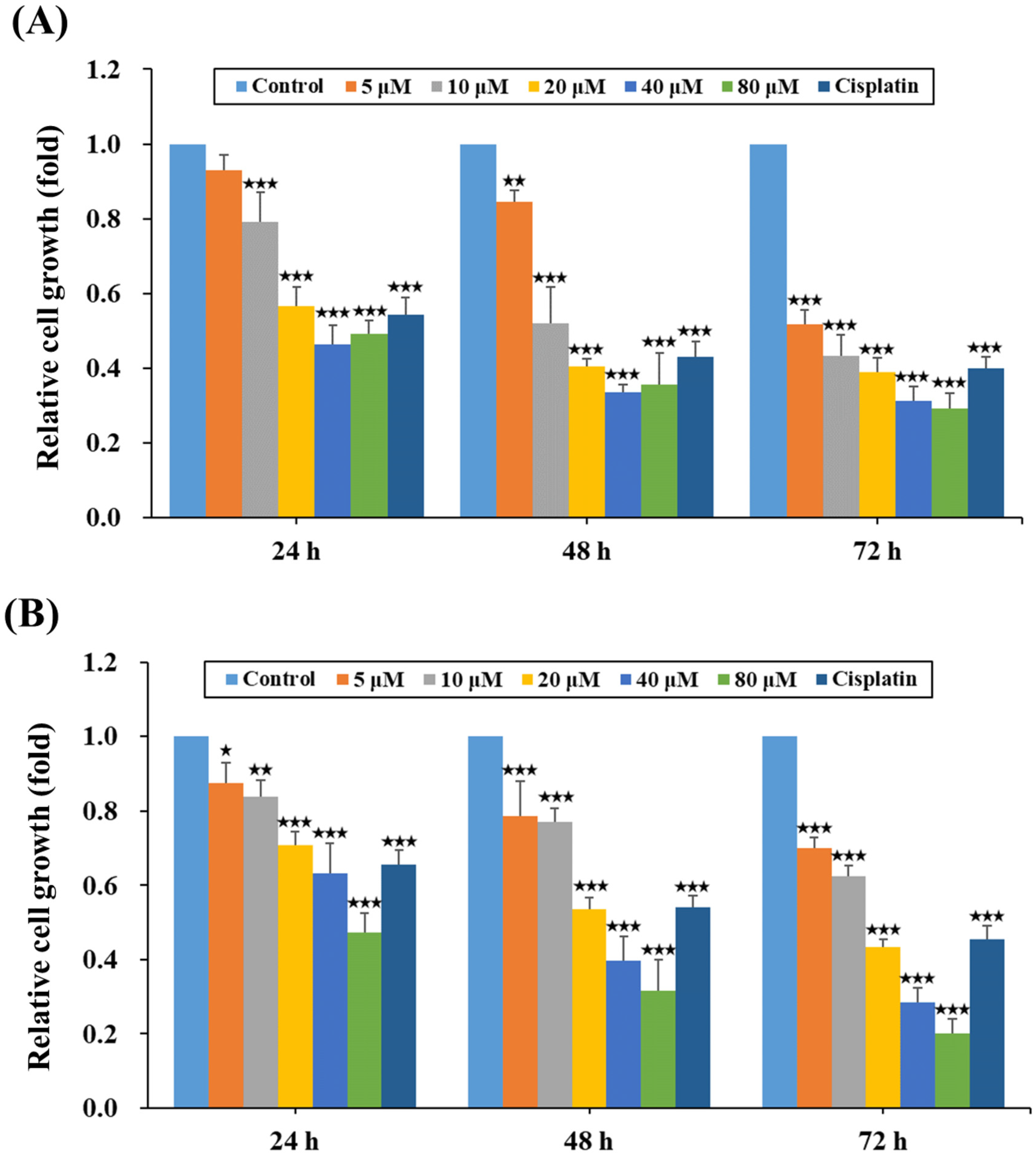
2.1. HPD Attenuated the Growth of PCa Cells

HPD is a natural polyketide and has been reported to display multiple favorable pharmacological functions with low toxicity in normal cells [9,19]. To evaluate the therapeutic impact of HPD on the cellular properties of PCa, several bio-functionality assays were performed, including cell growth, migration and invasion, as well as cell death analysis. Firstly, LNCaP and C4-2 PCa cells were subjected to dose-dependent HPD treatment to study its effects on cell growth, utilizing an MTS proliferation assay at 24-, 48- and 72-h time points. The results revealed a profound impact of HPD on the growth rates of LNCaP (Figure 1A) and C4-2 (Figure 1B) cells across different concentrations in a dose-dependent manner, with an achieved IC50 (half-maximal inhibitory concentration) of 6.09 μM for LNCaP cells and 16.63 μM for C4-2 cells, respectively, at 72 h of treatment. Moreover, we considered that hormones (such as androgens) in fetal bovine serum (FBS) might affect the efficacy of HPD on androgen-sensitive LNCaP PCa cells. An additional experiment was included using charcoal-stripped FBS, which had been stripped of hormones and non-polar materials (lipophilic compounds). As shown in Supplemental Figure S1, HPD presented the similar effects on LNCaP cell growth in regular FBS and in charcoal-stripped FBS. It suggested that HPD would not change the efficacy of cell growth in androgen-sensitive PCa cells with or without the androgen condition.

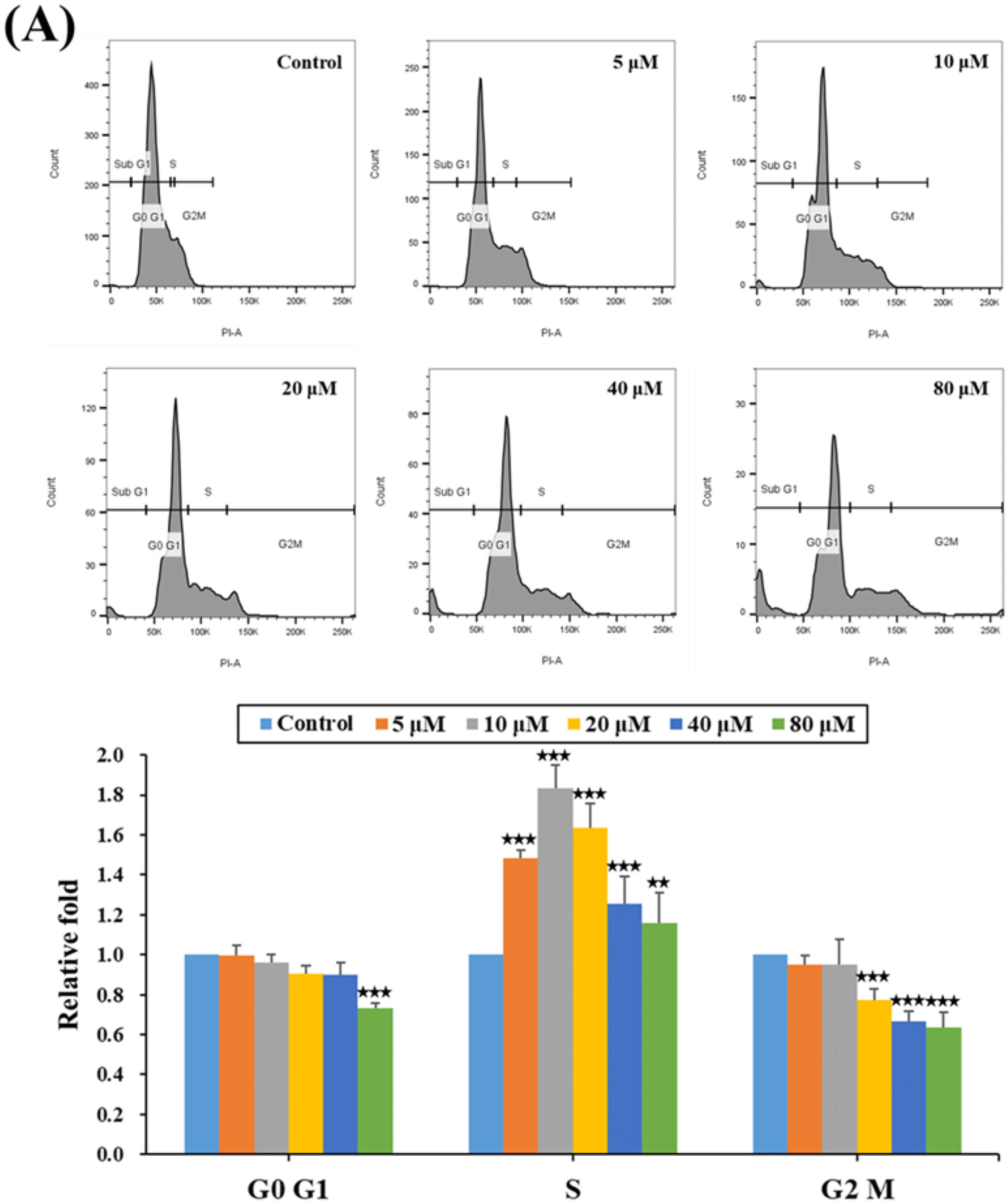
2.2. HPD Impeded Cell Cycle Progression at the S Phase in PCa Cells

Given the importance of the cell cycle for cell growth, we conducted a thorough examination of the cell cycle phase distribution by employing flow cytometry analysis after treating PCa cells with HPD at various concentrations. As shown in Figure 2A (LNCaP) and Figure 2B (C4-2), a significant dose-dependent increase in the relative fold of cell populations at the S phase was observed after HPD treatment. Concurrently, the relative fold of cell populations at the G2/M phase was reduced. Notably, an additional cell population in the sub-G1 phase was detected at high concentrations of HPD (Supplemental Figure S2), indicating possible cell death induction by HPD. Collectively, the data suggested that HPD affected cell cycle progression at the S phase and induced a sub-G1 cell population in PCa cells, resulting in the substantial inhibition of PCa cell growth and induction of cell death.

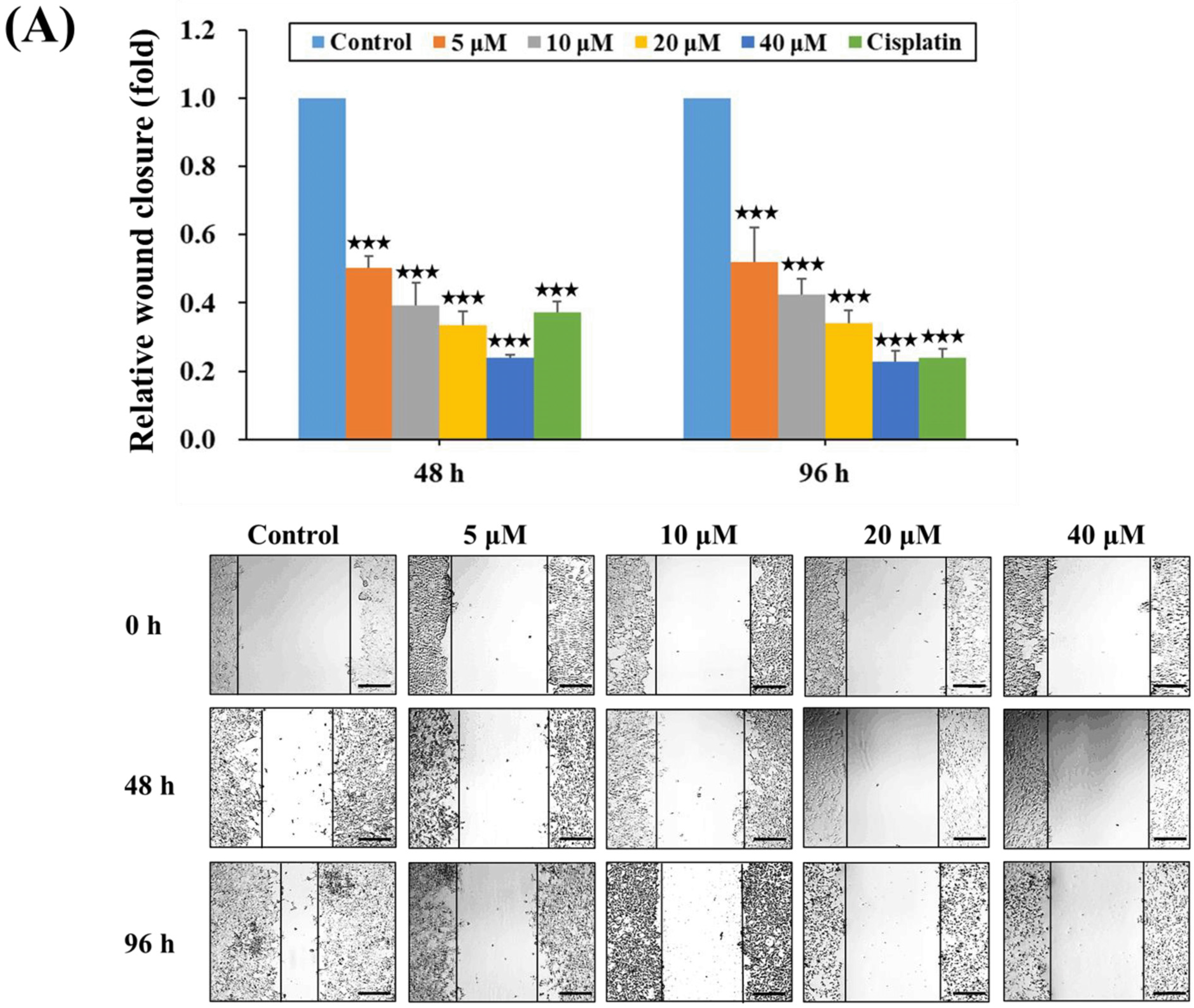
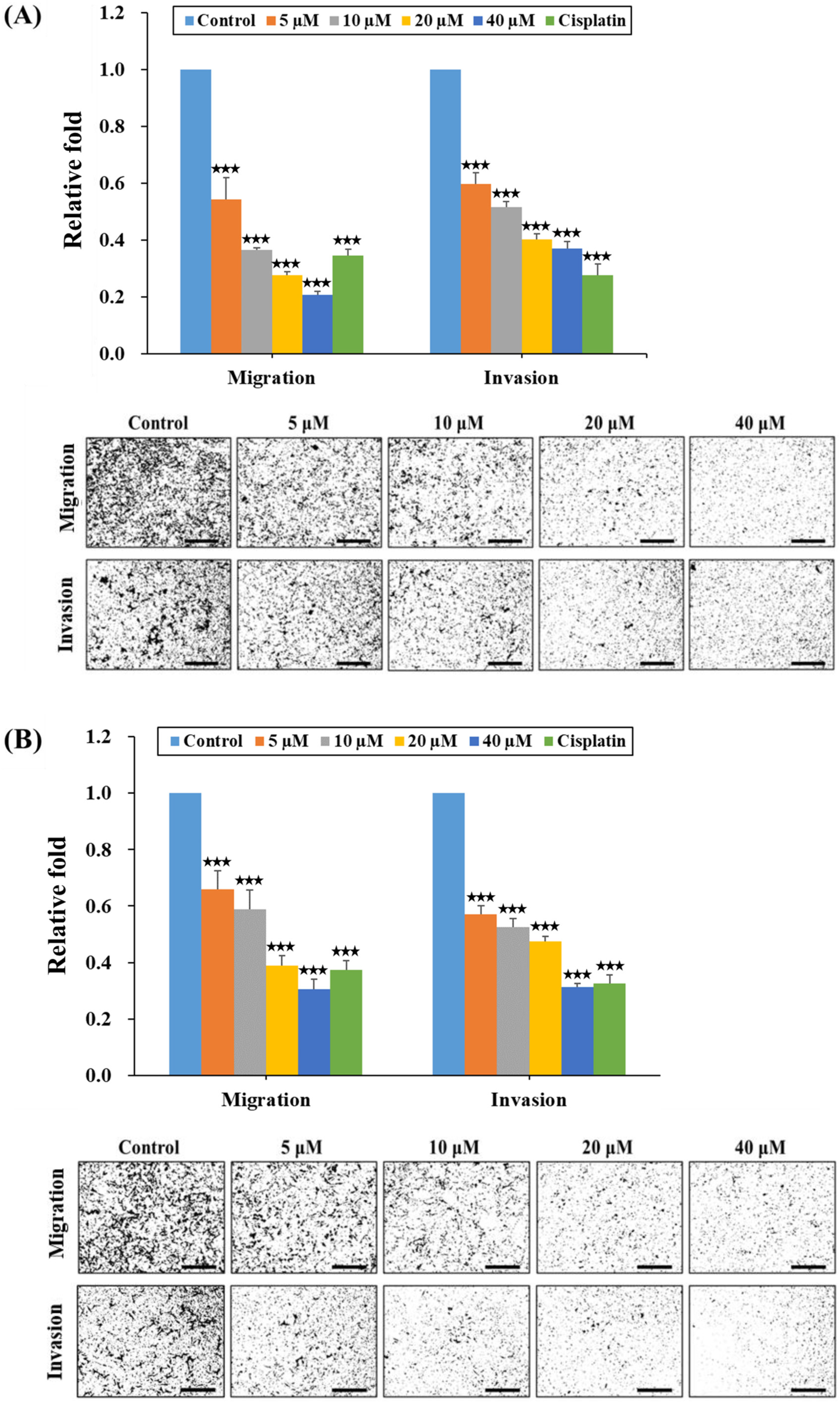
2.3. HPD Suppressed Migration and Invasion of PCa Cells

We subsequently employed a wound healing assay to determine the influence of HPD on the migratory ability of PCa cells. Comparing the migratory patterns of HPD-treated LNCaP (Figure 3A) and C4-2 (Figure 3B) cells, the results revealed significant reductions in the relative migratory distances compared to their respective controls. Thus, HPD possessed the capability to affect migration in PCa cells in a concentration-dependent manner.
Next, we assessed the effects of HPD on the migration and invasion dynamics of PCa cells by employing the transwell method. Equal numbers of LNCaP and C4-2 cells were seeded into the transwell chambers, being subjected to either the control condition or various concentrations of HPD treatment for 48 h. As shown in Figure 4A (LNCaP) and Figure 4B (C4-2), HPD significantly reduced both the migratory and invasive capabilities of PCa cells, aligning with the observations shown in Figure 3A,B. Remarkably, the inhibitory effects of HPD commenced at a concentration as low as 5 μM. The combined data indicated that HPD greatly suppressed the migration and invasion of LNCaP and C4-2 cells.

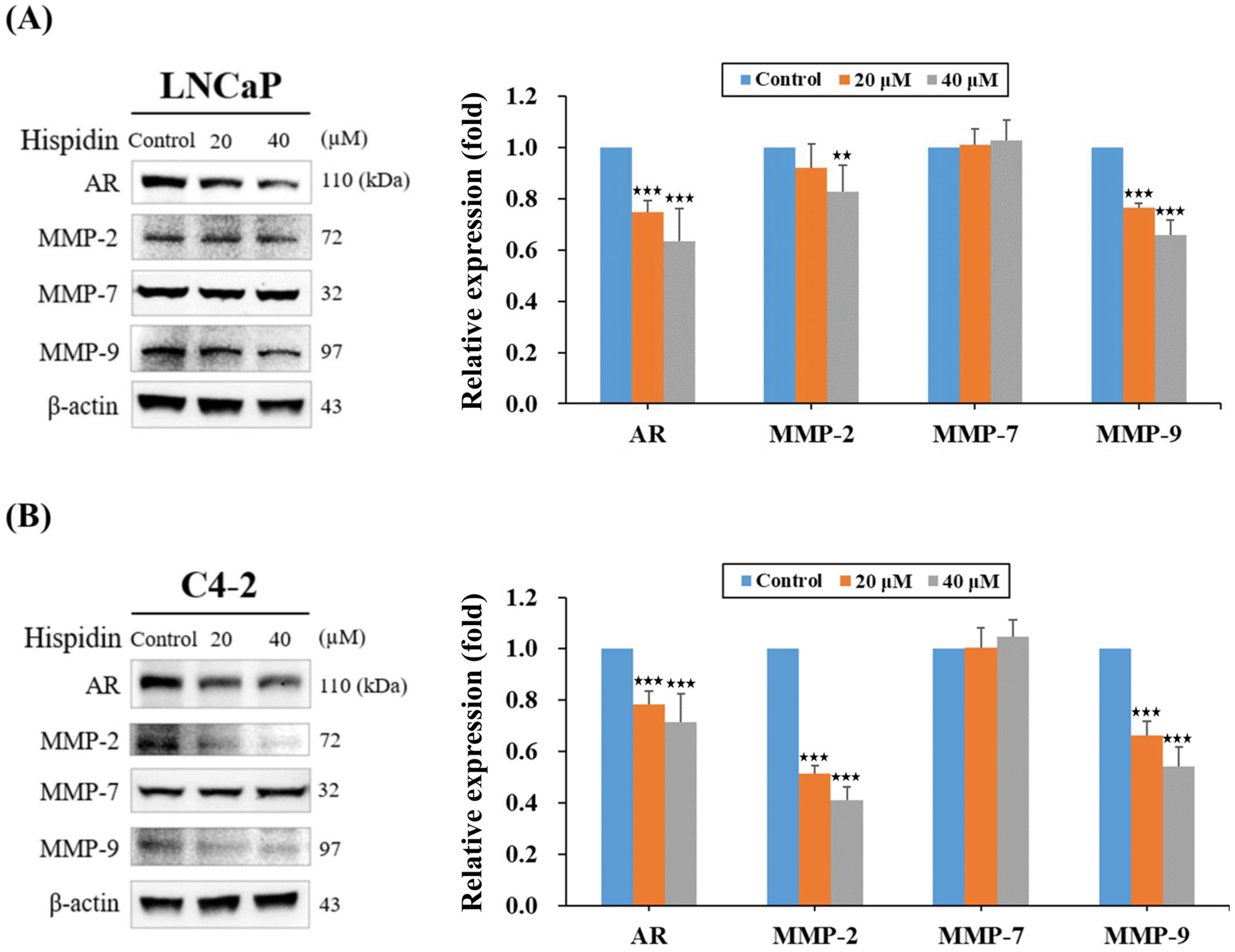
2.4. HPD Reduced Protein Expression Related to Cell Growth and Cancer Progression in PCa Cells

AR and MMPs have been well established as essential protein factors associated with cell growth, cell cycle regulation, and migration and invasion in PCa [23,24,25,26]. To delineate whether HPD affected these key proteins, Western blot analysis was conducted. The results revealed that HPD had inhibitory effects on AR expression in a dose-dependent manner in both LNCaP and C4-2 cells (Figure 5A,B). Additionally, concentration-dependent reductions in the expression levels of MMP-2 and MMP-9 proteins in PCa cells after HPD treatment was observed (Figure 5A,B). However, no significant change in the expression levels of MMP-7 was detected in both cells. Taken together, the results of Western blot analysis suggested that HPD reduced the expression levels of AR, MMP-2 and MMP-9 proteins, all linked to cell progression leading to the inhibition of growth (Figure 1), migration and invasion (Figure 3 and Figure 4) in PCa cells.

2.5. HPD Induced Cell Death via Activation of Caspase-Dependent Apoptosis in PCa Cells

Previously, we demonstrated that HPD induced the sub-G1-phase cell population linked to cell death in PCa cells. To further unravel the underlying molecular basis re-sponsible for HPD-mediated cell death, we first utilized flow cytometry with Annexin V and PI staining. As shown in Figure 6A, the dual-staining results showed that the percentages of apoptotic LNCaP and C4-2 cells were as low as 7.7 ± 0.5% and 6.6 ± 1.4% in the control groups, respectively. Significantly, HPD raised the apoptotic cell fractions in a dose-dependent manner (Figure 6A,B). Subsequently, the expression levels of caspase-3 as an essential executioner caspase for apoptosis and its downstream factor, poly (ADP-ribose) polymerase (PARP), were analyzed by Western blotting to evaluate if caspase-dependent apoptosis was activated by HPD treatment. As shown in Figure 6C, in comparison with the control group, HPD decreased the expression levels of caspase-3 (Casp-3; 35 kDa) and PARP (116 kDa) proteins. However, the activated forms of Casp-3 and PARP, cleaved Casp-3 (c-Casp-3; 19 kDa) and cleaved PARP (c-PARP; 89 kDa), were substantially increased after HPD treatment (Figure 6C). These findings suggested that HPD was able to induce cell death via activation of caspase-dependent apoptosis in PCa cells, culminating in the cleavage of Casp-3 into c-Casp-3 and subsequent cleavage of downstream PARP into c-PARP, thereby fostering the breakdown of PCa cells and inducing apoptosis.

3. Discussion

PCa progression, including CRPC, poses a challenging and unresolved clinical issue in PCa research. Currently, there is no effective remedy available to successfully cure this deadly disease. Crude extracts and/or pure compounds directly isolated from herbal plants and fungi belonging to the Phellinus and Inonotus genera have been demonstrated to be effective at treating diseases, including cardiovascular and hepatic diseases, as well as diabetes [27,28]. Specifically, HPD is purified from Phellinus and Inonotus and displays pharmacological effects with antioxidant [13], anti-inflammatory [14] and cardiovascular- [16] and neuro-protective [17] activities. In this study, we elucidated the efficacy of HPD in the treatment of PCa in vitro for the first time and provided the underlying molecular basis in both androgen-sensitive LNCaP and CRPC C4-2 cells. Interestingly, a very recent paper reported that HPD also substantially influenced metastatic PCa cells though alternations of the PI3K and the MAPK signaling pathways, as well as changes in the status of mitochondrial reactive oxygen species (ROS) in PC-3 and DU145 cells [29]. These findings particularly strengthened our study and enriched the potential and multiple biological activities of HPD in PCa. But, few possible limitations associated with the use of HPD have recently been identified, such as the fact that the chemical instability of HPD easily lost its antioxidant activity because it is susceptible to air oxidation [30]. Additionally, HPD showed lower cell permeability for stage 2 human colorectal carcinoma and blood–brain barrier penetration [31]. It needs to further investigate the potential challenges of HPD pharmacokinetics for the clinical application.
One prominent cancer hallmark is uncontrolled cell growth and division [32] linked to the cell cycle. Specific blockade of the cell cycle elements could be considered and employed as a possible therapeutic strategy in the treatment of cancer [33,34,35]. The cell cycle is restrictively regulated to ensure that cells undergo DNA replication and division at appropriate times, typically including the G0, G1, S, G2, and M phases. The G0 phase refers to the period when cells remain dormant; they are metabolically active, but not proliferating. After re-entering the cell cycle, in the G1 phase, cells begin to proliferate, marking the period of highest DNA content as they prepare for entry into the S phase. During the S phase, DNA replication occurs, resulting in a doubling of the diploid content. Afterwards, in the G2 phase and the M phase, cells enter the division process into two daughter cells. Our data revealed that HPD significantly raised the percentage of PCa cells in the S phase of the cell cycle (Figure 2). Concurrently, there was a decrease in the percentage of PCa cells in the G2/M phases. These results demonstrated that the HPD-treated PCa cells accumulated in the S phase, impeding progression towards the G2/M phase, thus disrupting the completion of the cell cycle to inhibit PCa cell growth.
Advanced PCa often forms metastasis [4], making the inhibition of their migration and invasion crucial in PCa treatment. In particular, our results demonstrated that HPD significantly suppressed migration and invasion in LNCaP and C4-2 cells as low as 5 μM in a dose-dependent pattern (Figure 3 and Figure 4). MMPs have been demonstrated to play a pivotal role in mediating the synthesis, degradation, and remodeling of the extracellular matrix (ECM) [36,37]. The ECM contains several protein factors, including collagen, fibronectin, gelatin, and other structural proteins, which are fundamental for the maintenance of cellular morphology, movement and intercellular communication, as well as the functions of tumor microenvironment [38]. Importantly, MMPs can degrade and disrupt adhesion between cells and the ECM, promoting the migration and invasion of tumor cells into other tissues and further facilitating the development of fatal metastasis. Among MMPs, MMP-2, -7 and -9 have been reported to promote cancer cell migration and invasion, with MMP-2 and MMP-9 being especially prevalent in PCa progression [39,40,41,42,43]. Thus, targeting these cancer-associated proteases offers an intriguing opportunity to prevent PCa progression under the alternation of the tumor microenvironment. Notably, our data confirmed that HPD significantly reduced the protein expression of MMP-2 and MMP-9 (Figure 5), demonstrating its potential to attenuate the migratory and the invasive capabilities of PCa cells (Figure 3 and Figure 4). Further research is needed to elucidate the specific inhibitory mechanisms and the related signaling pathways.
The activation of programmed cell death/apoptosis has been found to be employed as an attractive therapeutic strategy to treat cancer [44,45,46]. Caspases are a class of cysteine proteases controlling the cellular process of programmed death. This family intricately regulates apoptosis through precise interactions with numerous protein factors [47,48]. Under normal conditions, Casp-3 remains inactive in its original form within the cellular matrix. When cells receive apoptotic stimuli, such as drug treatment, Casp-3 undergoes cleavage, transforming into c-Casp-3, an activated state responsible for cellular apoptosis. Subsequently, PARP is cleaved. Research indicated that c-Casp-3 can cleave PARP (116 kDa), producing c-PARP (89 kDa) as a catalyst for cell disintegration, ultimately leading to cellular apoptosis [49]. Our Western blot data revealed that the expression levels of precursor Casp-3 and PARP proteins were decreased through HPD treatment in LNCaP and C4-2 cells, while the levels of activated c-Casp-3 and c-PARP proteins were increased (Figure 6C). Moreover, flow cytometry analysis confirmed that HPD effectively increased the numbers of apoptotic cells in PCa cells (Figure 6A,B). Taken together, the apoptotic data implied that HPD could serve as a potential therapeutic agent for treating malignant PCa. An additional pre-clinical study will be warranted to evaluate the anti-PCa/CRPC effect of HPD in vivo.

4. Materials and Methods

4.1. Cell Lines and Cell Culture

PCa cell lines and LNCaP (androgen-sensitive) and C4-2 (CRPC) cells were kindly offered by Dr. Leland W.K. Chung (Cedars-Sinai Medical Center, Los Angeles, CA, USA). PCa cells were cultured and maintained in RPMI 1640 medium (Thermo Fisher Scientific/GIBCO, Waltham, MA, USA) with 10% FBS (GE Healthcare/Hyclone, Pittsburgh, PA, USA) in a humidified 37 °C incubator (Thermo Fisher Scientific) under 5% CO2 atmosphere.

4.2. Cell Growth Assay

Cell growth was examined by the CellTiter 96® AQueous One Solution Cell Proliferation assay (Promega, Madison, WI, USA) [44,50]. Both LNCaP and C4-2 cells were seeded in 96-well plates (1 × 10⁴ cells/well) and incubated overnight. Subsequently, cells were treated with increasing concentrations of HPD (5, 10, 20, 40 and 80 μM) or control (0.08% DMSO) for 24, 48 and 72 h. The HPD (Supplemental Figure S3) was provided by Dr. Guan-Jhong Huang (China Medical University, Taiwan) [51]. Originally, HPD was dissolved in DMSO (a stock solution; 100 mM) and then diluted by RPMI 1640 medium in different working concentrations. Moreover, a common anti-cancer drug, cisplatin (Sigma-Aldrich, St. Louis, MO, USA), was utilized as a positive control [44].

4.3. Cell Migration Assay

The migratory ability of PCa cells treated with HPD (5, 10, 20 and 40 μM) or control (0.08% DMSO) was evaluated by a wound healing assay [45,46]. Additionally, cisplatin (10 µM) as utilized as a positive control. An artificial “wound” was created using a 200 μL pipette tip on the confluent cell monolayers of the 6-well plates. Cells were recorded under a microscope equipped with a camera at different time points. Quantitative analysis of the wound closure was calculated by the distance traveled by cells using the ImageJ 1.54g software.

4.4. Cell Progression Assays In Vitro

Cell progression assays in vitro were conducted by transwell chambers [45,46]. The membranes of the transwell chamber were either used natively (for cell migration assay) or coated with growth factor-reduced Matrigel basement membrane matrix (Coring, Bedford, MA, USA) (for cell invasion assay). Subsequently, PCa cells (1 × 10⁵ cells/well) were seeded in the transwell chamber and then treated with HPD (5, 10, 20 and 40 μM) or the control (0.08% DMSO). Additionally, cisplatin (10 µM) was utilized as a positive control. After 48 h, the migrated or invaded cells were fixed with 4% formaldehyde and stained with a crystal violet solution. After dehydration, an inverted microscope was used to count the numbers of PCa migrated and invaded cells.

4.5. Investigation of Cell Cycle Arrest

To understand the HPD-mediated effect on the cell cycle, the phase distribution in PCa cells was detected by flow cytometry. PCa cells were incubated with different concentrations of HPD (5, 10, 20, 40 and 80 μM) or control (0.08% DMSO) for 24 h. Subsequently, the treated PCa cells were centrifuged and cell pellets were fixed with chilled ethanol through 24 h incubation at −20 °C. Eventually, the cells were stained with 20 μg/mL of Propidium Iodide (PI) solution (BioLegend, San Diego, CA, USA) at 37 °C for 30 min and analyzed using FACSCanto (BD Biosciences, San Diego, CA, USA).

4.6. Apoptosis Analysis

After being treated for 48 h with HPD or the control, PCa cells were assayed by an Annexin V-FITC and PI Detection Kit (Biolegend, San Diego, CA, USA) based on the manufacturer’s instructions. The percentage of apoptotic PCa cells were detected by a flow cytometer with FCS Express v2.0 software (BD FACSCanto, BD Bioscience, San Diego, CA, USA).

4.7. Western Blot Analysis

To examine the protein expression levels, Western blot analysis was performed. Total protein preparation, SDS-PAGE electrophoresis, blotting and protein signal detection were described previously [52]. Additionally, the primary antibodies used in this study were as follows: anti-AR (Santa Cruz Biotechnology, Dallas, TX, USA), MMP-2, MMP-7, MMP-9 (Cell Signaling Technology, Danvers, MA, USA), caspase-3 (Novus Biologicals, Centennial, CO, USA), cleaved caspase-3 (Biovision, Waltham, MA, USA), PARP and β-actin (GeneTex, Irvin, CA, USA).

4.8. Statistical Analysis

All data underwent analysis based on a minimum of at least three independent experiments utilizing a two-tailed unpaired Student’s t-test to compare independent means. Statistically significant results were defined as p-values less than 0.05: * p < 0.05, ** p < 0.01, and *** p < 0.001.

5. Conclusions

Our research data provided evidence that HPD exhibited a favorable pharmaceutical profile to inhibit cell growth, disrupt the cell cycle, attenuate migration and invasion and induce apoptosis in PCa cells (Figure 7). Through the inhibition of AR expression, HPD suppressed PCa cell growth and influenced cell cycle. By decreasing MMP-2 and MMP-9 expression, HPD attenuated migration and invasion in PCa cells. Furthermore, HPD led to apoptosis through the activation of the Casp-3 and PARP pathway in PCa cells. We showed the anti-cancer effect of HPD on PCa cells in vitro and confirmed its feasibility as a novel therapeutic agent. Additionally, a promising safety profile of HPD has been previously reported in the murine models with the low adverse effects [9,19]. Ultimately, we aim to develop an effective and safe therapy using HPD to eliminate PCa and prevent the progression to CRPC.

Supplementary Materials

The following supporting information can be downloaded at: https://www.mdpi.com/article/10.3390/ijms25147857/s1.

Author Contributions

Conceptualization, K.-C.C. and W.-C.H.; methodology, K.-C.C., P.B. and W.-C.H.; validation, K.-C.C., P.B., J.-C.T., J.V., B.-Y.H. and X.-Y.L.; formal analysis, K.-C.C. and W.-C.H.; investigation, P.B., J.-C.T., J.V., G.-J.H. and W.-C.H.; resources, G.-J.H. and W.-C.H.; data curation, K.-C.C., P.B. and W.-C.H.; writing—original draft preparation, K.-C.C.; writing—review and editing, J.V. and W.-C.H.; visualization, K.-C.C.; supervision, W.-C.H.; project administration, W.-C.H.; funding acquisition, W.-C.H. All authors have read and agreed to the published version of the manuscript.

Funding

This research was funded by grants from National Science and Technology Council (MOST 111-2314-B-039-035-MY3) (W.-C.H.) and China Medical University, Taiwan (CMU112-MF-23) (W.-C.H.).

Institutional Review Board Statement

Not applicable.

Informed Consent Statement

Not applicable.

Data Availability Statement

Data were contained within the article.

Acknowledgments

Experiments and data analyses were performed in part through the use of the Medical Research Core Facilities, the Office of Research and Development at China Medical University, Taichung, Taiwan.

Conflicts of Interest

The authors declare no conflicts of interest.

References

  1. Verze, P.; Cai, T.; Lorenzetti, S. The role of the prostate in male fertility, health and disease. Nat. Rev. Urol. 2016, 13, 379–386. [Google Scholar] [CrossRef] [PubMed]
  2. Bracarda, S.; de Cobelli, O.; Greco, C.; Prayer-Galetti, T.; Valdagni, R.; Gatta, G.; de Braud, F.; Bartsch, G. Cancer of the prostate. Crit. Rev. Oncol. Hematol. 2005, 56, 379–396. [Google Scholar] [CrossRef] [PubMed]
  3. Siegel, R.L.; Giaquinto, A.N.; Jemal, A. Cancer statistics, 2024. CA Cancer J. Clin. 2024, 74, 12–49. [Google Scholar] [CrossRef] [PubMed]
  4. Yamamichi, G.; Kato, T.; Uemura, M.; Nonomura, N. Diagnosing and Prognosing Bone Metastasis in Prostate Cancer: Clinical Utility of Blood Biomarkers. Anticancer Res. 2023, 43, 283–290. [Google Scholar] [CrossRef] [PubMed]
  5. Mellman, I.; Coukos, G.; Dranoff, G. Cancer immunotherapy comes of age. Nature 2011, 480, 480–489. [Google Scholar] [CrossRef] [PubMed]
  6. Baskar, R.; Lee, K.A.; Yeo, R.; Yeoh, K.W. Cancer and radiation therapy: Current advances and future directions. Int. J. Med. Sci. 2012, 9, 193–199. [Google Scholar] [CrossRef] [PubMed]
  7. Heidenreich, A.; Aus, G.; Bolla, M.; Joniau, S.; Matveev, V.B.; Schmid, H.P.; Zattoni, F. EAU guidelines on prostate cancer. Eur. Urol. 2008, 53, 68–80. [Google Scholar] [CrossRef] [PubMed]
  8. Jain, K.K. Personalised medicine for cancer: From drug development into clinical practice. Expert. Opin. Pharmacother. 2005, 6, 1463–1476. [Google Scholar] [CrossRef] [PubMed]
  9. Li, I.C.; Chen, C.C.; Sheu, S.J.; Huang, I.H.; Chen, C.C. Optimized production and safety evaluation of hispidin-enriched Sanghuangporus sanghuang mycelia. Food Sci. Nutr. 2020, 8, 1864–1873. [Google Scholar] [CrossRef]
  10. Tian, L.W.; Feng, Y.; Tran, T.D.; Shimizu, Y.; Pfeifer, T.; Vu, H.T.; Quinn, R.J. Achyrodimer F, a tyrosyl-DNA phosphodiesterase I inhibitor from an Australian fungus of the family Cortinariaceae. Bioorg. Med. Chem. Lett. 2017, 27, 4007–4010. [Google Scholar] [CrossRef]
  11. Yousfi, M.; Djeridane, A.; Bombarda, I.; Duhem, B.; Gaydou, E.M. Isolation and characterization of a new hispolone derivative from antioxidant extracts of Pistacia atlantica. Phytother. Res. 2009, 23, 1237–1242. [Google Scholar] [CrossRef] [PubMed]
  12. Jung, J.Y.; Lee, I.K.; Seok, S.J.; Lee, H.J.; Kim, Y.H.; Yun, B.S. Antioxidant polyphenols from the mycelial culture of the medicinal fungi Inonotus xeranticus and Phellinus linteus. J. Appl. Microbiol. 2008, 104, 1824–1832. [Google Scholar] [CrossRef] [PubMed]
  13. Zhang, H.; Chen, R.; Zhang, J.; Bu, Q.; Wang, W.; Liu, Y.; Li, Q.; Guo, Y.; Zhang, L.; Yang, Y. The integration of metabolome and proteome reveals bioactive polyphenols and hispidin in ARTP mutagenized Phellinus baumii. Sci. Rep. 2019, 9, 16172. [Google Scholar] [CrossRef] [PubMed]
  14. Wangun, H.V.; Härtl, A.; Tam Kiet, T.; Hertweck, C. Inotilone and related phenylpropanoid polyketides from Inonotus sp. and their identification as potent COX and XO inhibitors. Org. Biomol. Chem. 2006, 4, 2545–2548. [Google Scholar] [CrossRef] [PubMed]
  15. Awadh Ali, N.A.; Mothana, R.A.; Lesnau, A.; Pilgrim, H.; Lindequist, U. Antiviral activity of Inonotus hispidus. Fitoterapia 2003, 74, 483–485. [Google Scholar] [CrossRef]
  16. Lee, Y.S.; Kang, Y.H.; Jung, J.Y.; Kang, I.J.; Han, S.N.; Chung, J.S.; Shin, H.K.; Lim, S.S. Inhibitory constituents of aldose reductase in the fruiting body of Phellinus linteus. Biol. Pharm. Bull. 2008, 31, 765–768. [Google Scholar] [CrossRef] [PubMed]
  17. Park, I.H.; Jeon, S.Y.; Lee, H.J.; Kim, S.I.; Song, K.S. A beta-secretase (BACE1) inhibitor hispidin from the mycelial cultures of Phellinus linteus. Planta Med. 2004, 70, 143–146. [Google Scholar] [CrossRef] [PubMed]
  18. Smetanina, O.F.; Yurchenko, A.N.; Ivanets, E.V.; Kalinovsky, A.I.; Khudyakova, Y.V.; Dyshlovoy, S.A.; von Amsberg, G.; Yurchenko, E.A.; Afiyatullov, S.S. Unique prostate cancer-toxic polyketides from marine sediment-derived fungus Isaria felina. J. Antibiot. 2017, 70, 856–858. [Google Scholar] [CrossRef]
  19. Li, I.C.; Chang, F.C.; Kuo, C.C.; Chu, H.T.; Li, T.J.; Chen, C.C. Pilot Study: Nutritional and Preclinical Safety Investigation of Fermented Hispidin-Enriched Sanghuangporus sanghuang Mycelia: A Promising Functional Food Material to Improve Sleep. Front. Nutr. 2021, 8, 788965. [Google Scholar] [CrossRef]
  20. Lv, L.X.; Zhou, Z.X.; Zhou, Z.; Zhang, L.J.; Yan, R.; Zhao, Z.; Yang, L.Y.; Bian, X.Y.; Jiang, H.Y.; Li, Y.D.; et al. Hispidin induces autophagic and necrotic death in SGC-7901 gastric cancer cells through lysosomal membrane permeabilization by inhibiting tubulin polymerization. Oncotarget 2017, 8, 26992–27006. [Google Scholar] [CrossRef]
  21. Chandimali, N.; Huynh, D.L.; Jin, W.Y.; Kwon, T. Combination Effects of Hispidin and Gemcitabine via Inhibition of Stemness in Pancreatic Cancer Stem Cells. Anticancer Res. 2018, 38, 3967–3975. [Google Scholar] [CrossRef] [PubMed]
  22. Lim, J.H.; Lee, Y.M.; Park, S.R.; Kim, D.H.; Lim, B.O. Anticancer activity of hispidin via reactive oxygen species-mediated apoptosis in colon cancer cells. Anticancer Res. 2014, 34, 4087–4093. [Google Scholar] [PubMed]
  23. Agoulnik, I.U.; Weigel, N.L. Androgen receptor action in hormone-dependent and recurrent prostate cancer. J. Cell Biochem. 2006, 99, 362–372. [Google Scholar] [CrossRef] [PubMed]
  24. Shafi, A.A.; Yen, A.E.; Weigel, N.L. Androgen receptors in hormone-dependent and castration-resistant prostate cancer. Pharmacol. Ther. 2013, 140, 223–238. [Google Scholar] [CrossRef] [PubMed]
  25. Tang, Q.; Cheng, B.; Dai, R.; Wang, R. The Role of Androgen Receptor in Cross Talk Between Stromal Cells and Prostate Cancer Epithelial Cells. Front. Cell Dev. Biol. 2021, 9, 729498. [Google Scholar] [CrossRef] [PubMed]
  26. El-Chaer, W.K.; Moraes, C.F.; Nóbrega, O.T. Diagnosis and Prognosis of Prostate Cancer from Circulating Matrix Metalloproteinases and Inhibitors. J. Aging Res. 2018, 2018, 7681039. [Google Scholar] [CrossRef] [PubMed]
  27. Lee, I.K.; Yun, B.S. Styrylpyrone-class compounds from medicinal fungi Phellinus and Inonotus spp., and their medicinal importance. J. Antibiot. 2011, 64, 349–359. [Google Scholar] [CrossRef] [PubMed]
  28. He, P.; Zhang, Y.; Li, N. The phytochemistry and pharmacology of medicinal fungi of the genus Phellinus: A review. Food Funct. 2021, 12, 1856–1881. [Google Scholar] [CrossRef] [PubMed]
  29. Wang, Y.J.; Hao, Y.Y.; Lee, D.H.; Guo, X.Y.; Sun, H.N.; Kwon, T. Hispidin Increases Cell Apoptosis and Ferroptosis in Prostate Cancer Cells Through Phosphatidylinositol-3-Kinase and Mitogen-activated Protein Kinase Signaling Pathway. Anticancer Res. 2024, 44, 2533–2544. [Google Scholar] [CrossRef]
  30. Yuan, W.; Yuan, W.; Zhou, R.; Lv, G.; Sun, M.; Zhao, Y.; Zheng, W. Production of hispidin polyphenols from medicinal mushroom Sanghuangporus vaninii in submerged cultures. Chin. Herb. Med. 2023, 15, 594–602. [Google Scholar] [CrossRef]
  31. Benarous, K.; Serseg, T.; Mermer, A.; Tahmasebifar, A.; Boulebd, H.; Linani, A. Exploring the Anti-Cancer Potential of Hispidin: A Comprehensive in Silico and in Vitro Study on Human Osteosarcoma Saos2 Cells. Chem. Biodivers. 2024, 21, e202301833. [Google Scholar] [CrossRef] [PubMed]
  32. Hanahan, D. Hallmarks of Cancer: New Dimensions. Cancer Discov. 2022, 12, 31–46. [Google Scholar] [CrossRef] [PubMed]
  33. Vakili-Samiani, S.; Khanghah, O.J.; Gholipour, E.; Najafi, F.; Zeinalzadeh, E.; Samadi, P.; Sarvarian, P.; Pourvahdani, S.; Kelaye, S.K.; Hamblin, M.R.; et al. Cell cycle involvement in cancer therapy; WEE1 kinase, a potential target as therapeutic strategy. Mutat. Res. 2022, 824, 111776. [Google Scholar] [CrossRef] [PubMed]
  34. Almalki, S.G. The pathophysiology of the cell cycle in cancer and treatment strategies using various cell cycle checkpoint inhibitors. Pathol. Res. Pract. 2023, 251, 154854. [Google Scholar] [CrossRef] [PubMed]
  35. Petroni, G.; Formenti, S.C.; Chen-Kiang, S.; Galluzzi, L. Immunomodulation by anticancer cell cycle inhibitors. Nat. Rev. Immunol. 2020, 20, 669–679. [Google Scholar] [CrossRef] [PubMed]
  36. Verma, R.P.; Hansch, C. Matrix metalloproteinases (MMPs): Chemical-biological functions and (Q)SARs. Bioorg. Med. Chem. 2007, 15, 2223–2268. [Google Scholar] [CrossRef] [PubMed]
  37. Iyer, R.P.; Patterson, N.L.; Fields, G.B.; Lindsey, M.L. The history of matrix metalloproteinases: Milestones, myths, and misperceptions. Am. J. Physiol. Heart Circ. Physiol. 2012, 303, H919–H930. [Google Scholar] [CrossRef] [PubMed]
  38. Page-McCaw, A.; Ewald, A.J.; Werb, Z. Matrix metalloproteinases and the regulation of tissue remodelling. Nat. Rev. Mol. Cell Biol. 2007, 8, 221–233. [Google Scholar] [CrossRef] [PubMed]
  39. Stearns, M.; Stearns, M.E. Evidence for increased activated metalloproteinase 2 (MMP-2a) expression associated with human prostate cancer progression. Oncol. Res. 1996, 8, 69–75. [Google Scholar]
  40. Liao, H.Y.; Da, C.M.; Liao, B.; Zhang, H.H. Roles of matrix metalloproteinase-7 (MMP-7) in cancer. Clin. Biochem. 2021, 92, 9–18. [Google Scholar] [CrossRef]
  41. Aalinkeel, R.; Nair, B.B.; Reynolds, J.L.; Sykes, D.E.; Mahajan, S.D.; Chadha, K.C.; Schwartz, S.A. Overexpression of MMP-9 contributes to invasiveness of prostate cancer cell line LNCaP. Immunol. Investig. 2011, 40, 447–464. [Google Scholar] [CrossRef] [PubMed]
  42. Pego, E.R.; Fernández, I.; Núñez, M.J. Molecular basis of the effect of MMP-9 on the prostate bone metastasis: A review. Urol. Oncol. 2018, 36, 272–282. [Google Scholar] [CrossRef] [PubMed]
  43. Justulin, L.A., Jr.; Della-Coleta, H.H.; Taboga, S.R.; Felisbino, S.L. Matrix metalloproteinase (MMP)-2 and MMP-9 activity and localization during ventral prostate atrophy and regrowth. Int. J. Androl. 2010, 33, 696–708. [Google Scholar] [CrossRef] [PubMed]
  44. Hsieh, P.F.; Jiang, W.P.; Basavaraj, P.; Huang, S.Y.; Ruangsai, P.; Wu, J.B.; Huang, G.J.; Huang, W.C. Cell suspension culture extract of Eriobotrya japonica attenuates growth and induces apoptosis in prostate cancer cells via targeting SREBP-1/FASN-driven metabolism and AR. Phytomedicine 2021, 93, 153806. [Google Scholar] [CrossRef] [PubMed]
  45. Basavaraj, P.; Ruangsai, P.; Hsieh, P.F.; Jiang, W.P.; Bau, D.T.; Huang, G.J.; Huang, W.C. Alpinumisoflavone Exhibits the Therapeutic Effect on Prostate Cancer Cells by Repressing AR and Co-Targeting FASN- and HMGCR-Mediated Lipid and Cholesterol Biosynthesis. Life 2022, 12, 1769. [Google Scholar] [CrossRef] [PubMed]
  46. Basavaraj, P.; Hsieh, P.F.; Jiang, W.P.; Bau, D.T.; Huang, G.J.; Huang, W.C. Elucidation of scandenolone as anti-cancer activity through impairment of the metabolic and signaling vulnerabilities in prostate cancer. Biomed. Pharmacother. 2023, 164, 114948. [Google Scholar] [CrossRef] [PubMed]
  47. Van Opdenbosch, N.; Lamkanfi, M. Caspases in Cell Death, Inflammation, and Disease. Immunity 2019, 50, 1352–1364. [Google Scholar] [CrossRef]
  48. Fan, T.J.; Han, L.H.; Cong, R.S.; Liang, J. Caspase family proteases and apoptosis. Acta Biochim. Biophys. Sin. 2005, 37, 719–727. [Google Scholar] [CrossRef]
  49. Agarwal, A.; Mahfouz, R.Z.; Sharma, R.K.; Sarkar, O.; Mangrola, D.; Mathur, P.P. Potential biological role of poly (ADP-ribose) polymerase (PARP) in male gametes. Reprod. Biol. Endocrinol. 2009, 7, 143. [Google Scholar] [CrossRef]
  50. Fang, C.W.; Yang, J.S.; Chiang, J.H.; Shieh, P.C.; Tsai, F.J.; Tsai, C.W.; Chang, W.S. Metformin induces autophagy of cisplatin-resistant human gastric cancer cells in addition to apoptosis. Biomedicine 2023, 13, 14–23. [Google Scholar] [CrossRef]
  51. Lin, W.-C.; Deng, J.-S.; Huang, S.-S.; Wu, S.-H.; Lin, H.-Y.; Huang, G.-J. Evaluation of antioxidant, anti-inflammatory and anti-proliferative activities of ethanol extracts from different varieties of Sanghuang species. RSC Adv. 2017, 7, 7780–7788. [Google Scholar] [CrossRef]
  52. Huang, S.Y.; Huang, G.J.; Hsieh, P.F.; Wu, H.C.; Huang, W.C. Osajin displays potential antiprostate cancer efficacy via impairment of fatty acid synthase and androgen receptor expression. Prostate 2019, 79, 1543–1552. [Google Scholar] [CrossRef] [PubMed]
Figure 1. HPD inhibited cell growth in PCa cells. (A) LNCaP and (B) C4-2 cells were treated with control or HPD for 24, 48 and 72 h. The relative cell growth was assigned as the fold of the control-treated cells for 24, 48 and 72 h, respectively. The relative fold was defined as the ratio of each dose of HPD to the control group (as 1.0). Results were shown as the mean ± SD of three independent experiments. Cisplatin (10 µM) was employed as a positive control. * p < 0.05, ** p < 0.01, *** p < 0.001.
Ijms 25 07857 g001
Figure 2. HPD induced cell cycle arrest, as indicated through increases in the S-phase PCa cell numbers. (A) LNCaP and (B) C4-2 cells were treated with HPD and control for 24 h, followed by flow cytometry. The relative fold was assigned relative to the control-treated cells for each phase. The representative images of flow cell cycle are displayed in the top panels and the quantification of the results are in the bottom panels. The relative fold was defined as the ratio of each dose of HPD to the control group (as 1.0). Results are represented as the mean ± SD of three independent experiments. * p < 0.05, ** p < 0.01, *** p < 0.001.
Ijms 25 07857 g002aIjms 25 07857 g002b
Figure 3. HPD inhibited the migratory ability of PCa cells. (A) LNCaP and (B) C4-2 cells were treated with control or HPD for 48 and 96 h. The migratory distances of PCa cells at each concentration of HPD or the control were compared to the baseline at 0 h. The relative cell migratory distances were calculated to assess the wound healing capabilities of cells at 48 and 96 h (control as 1.0). The quantification of wound closure (the top panels) and the representative images of wound closure (the bottom panels; Scale bar = 100 µm) are shown. Results represented as the mean ± SD of three independent experiments. Cisplatin (10 µM) was used as a positive control. *** p < 0.001.
Ijms 25 07857 g003aIjms 25 07857 g003b
Figure 4. HPD inhibited the migratory and invasive potentials of PCa cells. The transwell migration and invasion assays were performed to determine migration and invasion in both (A) LNCaP and (B) C4-2 cells in accordance with the control and HPD treatment. The relative migration and invasion were assigned as 1.0 in the control-treated cells. The relative cell numbers for migration and invasion were calculated (control as 1.0). The quantification of results (the top panels) and the representative images of transwell migration and invasion (the bottom panels; Scale bar = 100 µm) were shown. Results are represented as the mean ± SD of three independent experiments. Cisplatin (10 µM) was applied as a positive control. *** p < 0.001.
Ijms 25 07857 g004
Figure 5. HPD reduced the expression of AR, MMP-2 and MMP-9 proteins in PCa cells. (A) LNCaP and (B) C4-2 cells were treated with HPD or control for 48 h. The expression levels of AR and MMP proteins were subsequently detected by Western blot analysis. The relative protein expression (fold) was assigned as 1.0 in the control-treated cells. The results of Western blot analysis (the left panels) and the quantitative assays of protein expression (the right panels) were shown. Results are represented as the mean ± SD of three independent experiments. β-actin was used an internal control. ** p < 0.01, *** p < 0.001.
Ijms 25 07857 g005
Figure 6. HPD induced caspase-dependent cell death in PCa cells. (A) PCa cells were exposed to control or HPD for 48 h, and apoptotic cells were subsequently analyzed by a flow cytometry-based Annexin V/PI dual-staining assay. Results represented as the mean ± SD of five independent experiments. (B) Representative red dot plots of PCa cells treated with control or HPD (160 μM) from (A). (C) Western blot analysis of caspase-3 (Casp-3), cleaved caspase-3 (c-Casp-3), PARP and cleaved PARP (c-PARP) expression in PCa cells after HPD or control treatment. β-actin was used as an internal control. * p < 0.05, ** p < 0.01, *** p < 0.001.
Ijms 25 07857 g006
Figure 7. HPD displays the promising therapeutic effect on PCa cells. HPD inhibited cell growth, migration and invasion while simultaneously reducing AR, MMP-2 and MMP-9 expression and activating the Casp-3 associated pathway leading to apoptosis.
Ijms 25 07857 g007
Disclaimer/Publisher’s Note: The statements, opinions and data contained in all publications are solely those of the individual author(s) and contributor(s) and not of MDPI and/or the editor(s). MDPI and/or the editor(s) disclaim responsibility for any injury to people or property resulting from any ideas, methods, instructions or products referred to in the content.

