Article

Photocatalytic Degradation of Organic Dyes by Magnetite Nanoparticles Prepared by Co-Precipitation

by
Thandi B. Mbuyazi
and
Peter A. Ajibade
*
School of Chemistry and Physics, University of KwaZulu-Natal, Private Bag X01, Scottsville, Pietermaritzburg 3209, South Africa
*
Author to whom correspondence should be addressed.
Int. J. Mol. Sci. 2024, 25(14), 7876; https://doi.org/10.3390/ijms25147876
Submission received: 4 June 2024 / Revised: 6 July 2024 / Accepted: 15 July 2024 / Published: 18 July 2024
(This article belongs to the Special Issue Functional Nanomaterial: Design, Synthesis and Applications)

Abstract

:
Iron oxide nanoparticles were synthesized by co-precipitation using three different iron salt stoichiometric mole ratios. Powder X-ray diffraction patterns revealed the inverse cubic spinel structure of magnetite iron oxide. Transmission electron microscopic images showed Fe3O4 nanoparticles with different shapes and average particle sizes of 5.48 nm for Fe3O4-1:2, 6.02 nm for Fe3O4-1.5:2, and 6.98 nm for Fe3O4-2:3 with an energy bandgap of 3.27 to 3.53 eV. The as-prepared Fe3O4 nanoparticles were used as photocatalysts to degrade brilliant green (BG), rhodamine B (RhB), indigo carmine (IC), and methyl red (MR) under visible light irradiation. The photocatalytic degradation efficiency of 80.4% was obtained from Fe3O4-1:2 for brilliant green, 61.5% from Fe3O4-1.5:2 for rhodamine B, and 77.9% and 73.9% from Fe3O4-2:3 for both indigo carmine and methyl red. This indicates that Fe3O4-2:3 is more efficient in the degradation of more than one dye. This study shows that brilliant green degrades most effectively at pH 9, rhodamine B degrades best at pH 6.5, and indigo carmine and methyl red degrade most efficiently at pH 3. Recyclability experiments showed that the Fe3O4 photocatalysts can be recycled four times and are photostable.

1. Introduction

Azo dyes are the most prevalent and biggest group of artificial dyes that are extensively used in the textiles, beauty products, pharmaceutical, and food industries [1,2]. The use of azo dyes has seen a dramatic increase in the textile industry over the past decades for coloring cotton and wool because of their vibrant colors, affordability, and excellent durability upon exposure to light and washing [3]. Most of these dyes have mutagenic, cancer-causing, genotoxic, and teratogenic properties [4]. Furthermore, their presence in water bodies inhibits solar radiation from penetrating, lowering oxygen quantity, interfering with photosynthesis, and damaging terraqueous ecosystems [5,6]. As a result, researchers have long been involved in developing an efficient, cost-effective, and dependable method for eliminating dyes from wastewater [7,8].
Photodegradation is commonly used to remove contaminants from the environment [9]. It involves subjecting pollutants to light, which causes their photochemical breakdown into simpler, non-toxic compounds. This method is frequently preferred due to its economic viability, sustainability, absence of secondary waste, and broad-spectrum degradation of different organic dyes into CO2 and H2O [10,11]. The practicality of the procedure can be improved by using a photocatalyst, which is a semiconducting material that is excited by absorbing light photons. Iron oxide is one of the materials that show potential for photocatalytic applications because of its cost-effective synthesis, magnetic properties, and n-type semiconducting effects [12].
Previous studies have shown the properties of superparamagnetic iron oxide nanoparticles synthesized by co-precipitation could be influenced by variations of the preparative parameters [3,13,14]. Alibeigi and Vaezi investigated the effect of Fe2+/Fe3+ molar ratios and observed that increasing the iron salt ratio increases crystallinity and specific saturation magnetization [15]. A study by Dehghanpour also demonstrated that adjusting the mole ratio of Fe2+ to Fe3+ ions, pH, and capping agents may influence saturation magnetization and the size of the nanoparticles [16]. Another study reported that changing the stoichiometric ratio of 1:2 for Fe2+ and Fe3+ salts decreased electrical conductivity and led to the phase change of the nanoparticles from Fe3O4 to γ-Fe2O3 [17]. Iida et al. [18] synthesized Fe3O4 nanoparticles by hydrolysis in an aqueous solution comprising ferrous and ferric salts in varying ratios, with 1,6-hexane-diamine as the base. An increase in the ratio of Fe2+ to Fe3+ ions resulted in an increase in the size of Fe3O4 nanoparticles from 9 to approximately 37 nm. Lee et al. [19] studied the antioxidant properties and physiological effects of maghemite nanoparticles (γ-Fe2O3) prepared by varying Fe2+/Fe3+ molar ratios using ferrous/ferric salt co-precipitation. Thus, studies have shown that the physical and chemical changes that occur in iron oxide nanoparticles when the Fe2+/Fe3+ molar ratio varies throughout the co-precipitation process. However, no work has studied the photocatalytic potential of Fe3O4 with respect to different Fe2+ to Fe3+ molar ratios.
The surface properties, crystalline structure, and morphology of the nanoparticles, which are all controlled by the synthetic technique, could influence the photocatalytic degradation efficiency of magnetite nanoparticles [20]. Studies have shown that a change in dye pH, light source, and surface charge influences the photocatalytic activity of Fe3O4 nanoparticles against organic dyes such as rhodamine B [21], methyl red [22], brilliant green [23], and indigo carmine [24]. In the current study, we evaluate the influence of the Fe2+/Fe3+ salt ratios on the structural and morphological optical and photocatalytic properties of Fe3O4 nanoparticles synthesized via the co-precipitation method. The potential of the as-synthesized iron oxide nanoparticles as photocatalysts for the photodegradation of brilliant green, rhodamine B, indigo carmine, and methyl red under visible light was studied. Additionally, the photostability of the nanoparticles, the effect of pH, and scavengers on the photodegradation of the organic dyes by the iron oxide nanoparticles were evaluated.

2. Results

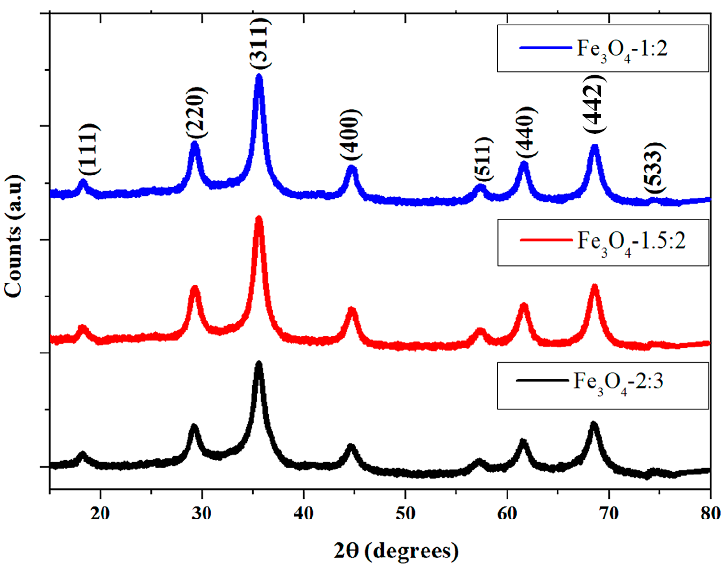
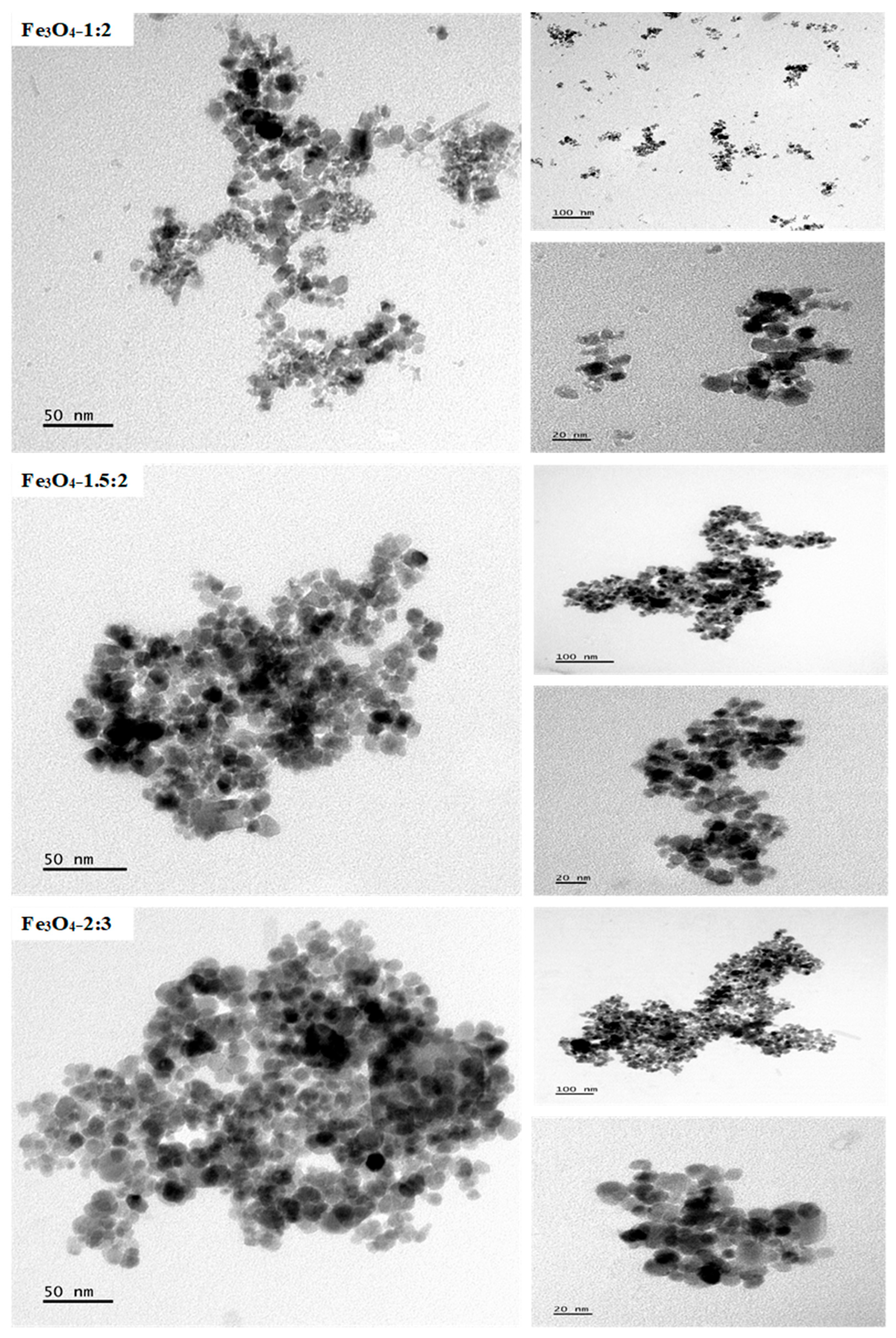
2.1. Morphological Studies of the Iron Oxide Nanoparticles

Figure 1 displays the diffractograms of the iron oxide nanoparticles prepared using different iron salt ratios. The 2θ peaks observed at 18.23°, 29.31°, 35.73°, 44.66°, 57.27°, 61.69°, 68.65, and 74.38° correlate to (111), (220), (311), (400), (511), (440), (442), and (533) reflections of inverse cubic spinel structure of magnetite (JCPDS no: 19-0629) [25,26]. The crystal growth direction inclination is toward the (311) plane. The diffraction patterns suggest that the iron salt ratios used have no apparent impact on the crystalline phases of the as-synthesized iron oxide nanoparticles.
The average crystallite sizes of the magnetite nanoparticles were calculated using the Debye–Scherrer equation [27]:
D = K λ / β c o s θ
where D, K, λ, β, and θ stand for crystallite diameter (nm), Scherrer constant, X-ray wavelength, full width at half-maximum (FWHM), and Bragg angle, respectively. The FWHM and θ were calculated by fitting all diffraction peaks to a Gaussian function. The calculated crystallite sizes were 4.45 nm, 5.89 nm, and 6.50 nm for Fe3O4-1:2, Fe3O4-1.5:2, and Fe3O4-2:3, respectively. The lattice parameter is another significant piece of information that may be retrieved from XRD patterns. The lattice parameter of magnetite nanoparticles was derived using Bragg’s rule and the following equation [28].
a = d h k l h 2 + k 2 + l 2
where a, dhkl, and hkl are the lattice constant, inter-planer distance, and Miller indices, respectively. Magnetite (JCPDS 19-0629) has a standard lattice constant of 8.3960 Å. The calculated lattice constants of the as-prepared magnetite nanoparticles are 8.3686 Å, 8.3579 Å, and 8.3574 Å for Fe3O4-1:2, Fe3O4-1.5:2, and Fe3O4-2:3, respectively. The results show that the calculated lattice constants of the as-prepared magnetite nanoparticles slightly vary from one another, and they are less than that of standard magnetite. This discrepancy could be attributed to the effect of various crystal size variations on the crystal lattice [29].
TEM images in Figure 2 revealed that Fe3O4-1:2 nanoparticles are a combination of square-like, oval, rod, and rectangular shapes with a mean size of 5.48 nm, while Fe3O4-1.5:2 nanoparticles are agglomerated square-like, rectangular, and oval-shaped with a mean size of 6.02 nm. Fe3O4-2:3 nanoparticles are also agglomerated and quasi-spherical, with an average size of 6.98 nm. There is no drastic change in average particle size with an increase in the stoichiometric ratios of the iron salts, as reported in other studies [16,18]. However, there is apparent agglomeration for Fe3O4-1.5:2 and Fe3O4-2:3 nanoparticles and larger particle size distributions compared to Fe3O4-1:2 (Figure S1).
FTIR was used to study the interactions between octylamine and the as-synthesized Fe3O4 nanoparticles (Figure S2). The weak ν(N–H)asy and ν(N–H)sym vibrations at 3376 and 3291 cm−1 are assigned to the amine group of the capping agent [30]. The C–H characteristic peaks of the octylamine alkyl chain are at 2919 and 2851 cm−1 [31]. The C–N bending vibrations appear at 1460 cm−1. The capping agent vibrational bands observed also appear in the spectra of the iron oxide nanoparticles but with low intensity, which suggests the presence of an octylamine capping agent on the surface of iron oxide nanoparticles. However, the N–H bands were not observed in the Fe3O4 nanoparticles spectra, signifying that the capping of the nanoparticles took place through the -NH2 group of octylamine [32]. Furthermore, the Fe–O peak at 550 cm−1 confirms the synthesis of iron oxide nanoparticles [33].

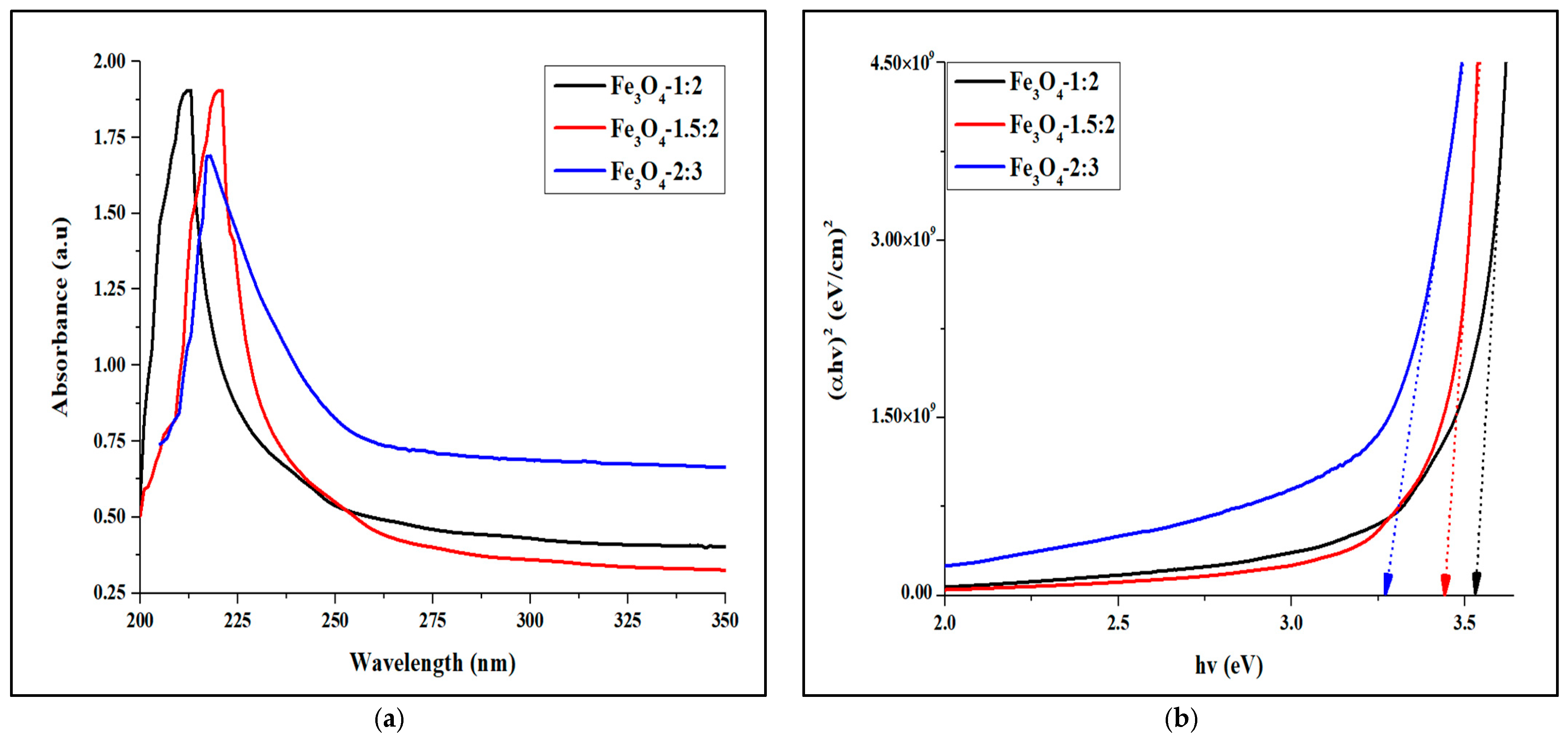
2.2. Optical Studies of the Magnetite Nanoparticles

Figure 3a displays the UV-Vis spectra of the as-prepared iron oxide nanoparticles, which exhibit sharp absorption peaks at 212.5 nm, 220.4, and 217.6 nm for Fe3O4-1:2, Fe3O4-1.5:2, and Fe3O4-2:3, respectively. To further investigate the optical properties of the magnetite nanoparticles, the Tauc plot was used to calculate the energy gap [34]. The estimated energy gaps were 3.53 eV for Fe3O4-1:2, 3.44 eV for Fe3O4-1.5:2, and 3.27 eV for Fe3O4-2:3 (Figure 3b). The bandgap is observed to decrease with an increase in iron salt ratios, with Fe3O4-2:3 nanoparticles having a larger average particle size and the smallest bandgap. The energy gap of the as-synthesized magnetite nanoparticles is greater than that of bulk magnetite (0.20 eV) due to the quantum confinement effect [35]. The optical bandgap of Fe3O4 nanoparticles has been reported to range from 3.23 to 3.9 eV [36,37]. Moreover, Shamaila et al. synthesized Fe3O4 nanoparticles with an energy bandgap of 2.5 eV and titanium-coated Fe3O4 nanoparticles with a bandgap of 3.22 eV [38]. The bare Fe3O4 demonstrated poor photocatalytic activity, whereas the titanium-coated Fe3O4 exhibited improved performance under visible light. The optical bandgaps of the magnetite nanoparticles in this study are comparable to previously reported bandgaps [36,37,38], and the nanoparticles have the potential to be used as photocatalysts for dye degradation under visible light.

2.3. Surface Properties

The point of zero charge (PZC) of the magnetite nanoparticles was determined, as shown in Figure 4. The pHpzc values of Fe3O4-1:2, Fe3O4-1.5:2, and Fe3O4-2:3 were found to be 6.22, 5.56, and 4.55, respectively. The pHpzc values obtained in this study are comparable to some pHpzc values for magnetite found in the literature [39,40]. At the PZC, the catalyst surface consists of an equal proportion of positive and negative functional groups, yielding a zero net charge. The surface accumulates a negative charge above the PZC and a positive charge below the PZC. This is due to the presence of FeOH groups on the surface of magnetite iron oxide nanoparticles in aqueous solution [41]. These hydroxyl groups can protonate or deprotonate to produce FeOH2+ or FeO, which changes in response to pH values below or above the pHPZC.

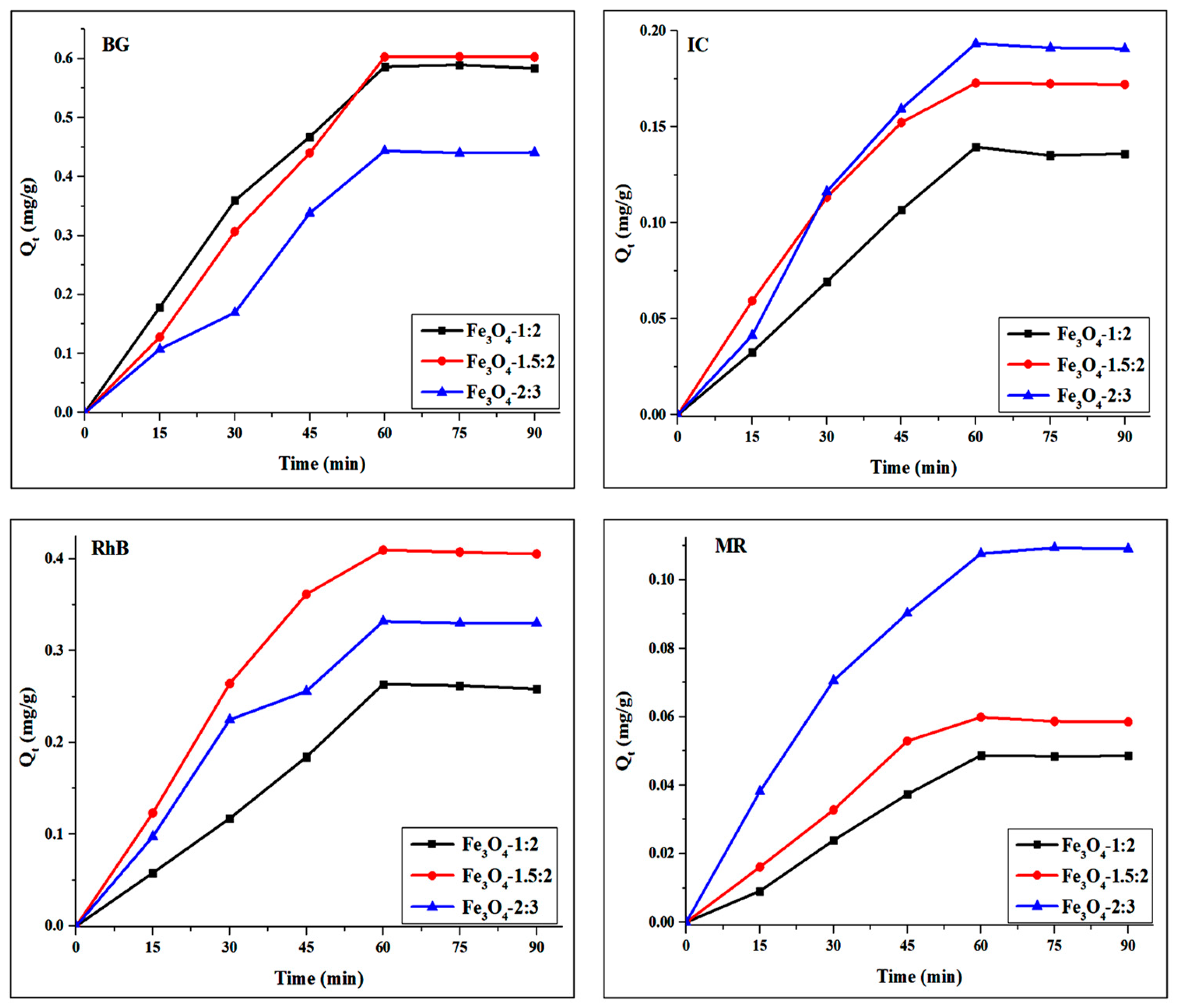
3. Adsorption Studies of Magnetite Nanoparticles

The adsorption capacity of Fe3O4 nanoparticles over BG, RhB, IC, and MR dyes was evaluated. The impact of contact time on dye adsorption on Fe3O4 nanoparticles was investigated to determine the equilibrium interval (Figure 5). Significant adsorption efficiency was noted during the first 45 min, indicating the availability of accessible sites [42,43]. Maximum adsorption capacity was attained at 60 min; beyond this time, adsorption remained constant. Therefore, a 60 min equilibration time was decided upon for the adsorption period prior to visible light exposure for the photocatalytic studies.

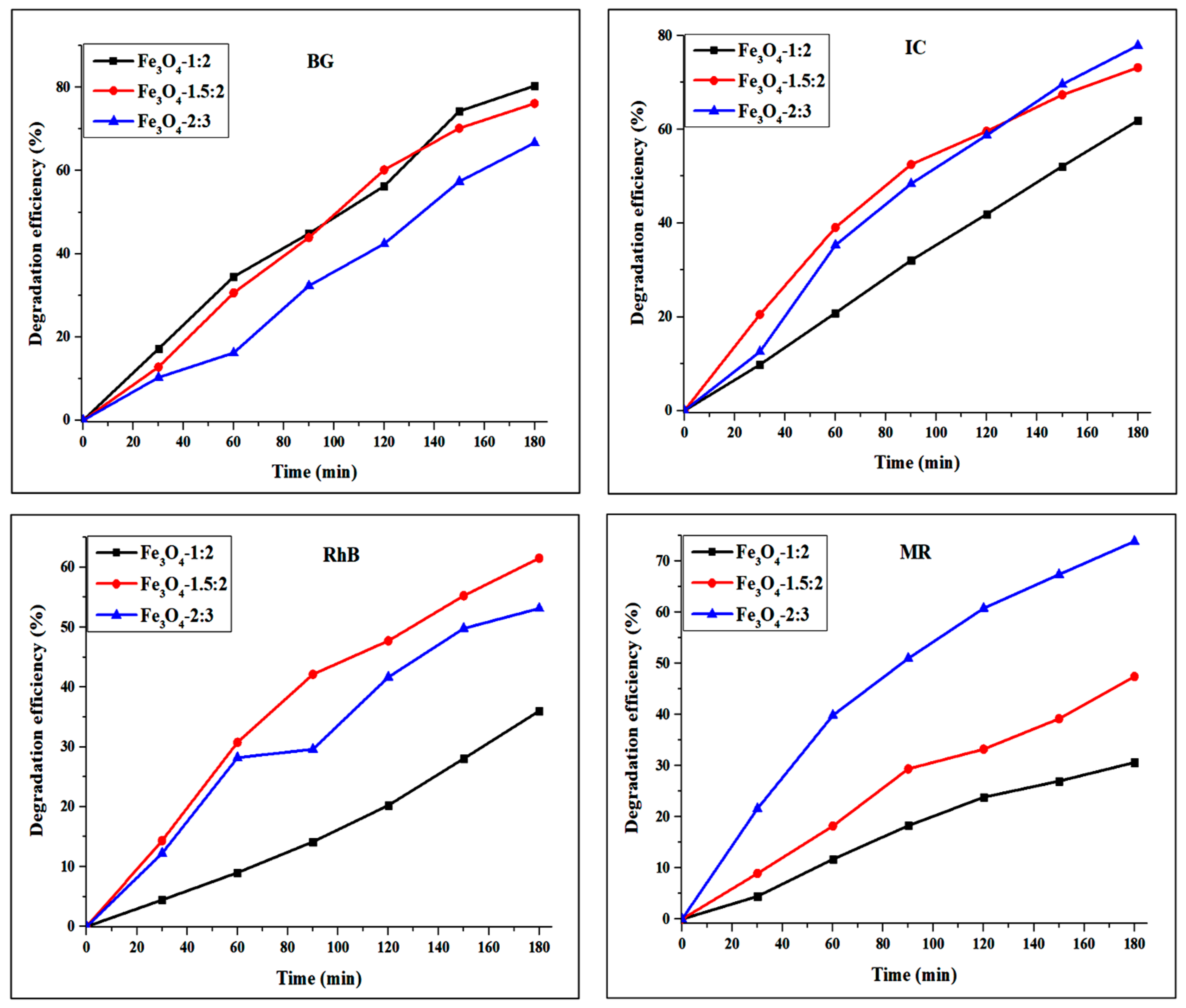
4. Photocatalytic Studies of Magnetite Nanoparticles

The photocatalytic degradation efficiencies of the Fe3O4 nanoparticles were evaluated using BG, RhB, IC, and MR under visible light (Figure 6). The percentage degradation of BG after 180 min irradiation is 80.4%, 76.1%, and 66.7% by Fe3O4-1:2, Fe3O4-1.5:2, and Fe3O4-2:3, respectively. The degradation efficiency of 36.0%, 61.5%, and 53.1% was obtained for Fe3O4-1:2, Fe3O4-1.5:2, and Fe3O4-2:3, respectively, against RhB dye. Different degradation efficiencies may be attributed to the morphologies of the iron oxide nanoparticles. The low percentage degradation of RhB by the as-prepared iron oxide nanoparticles could be ascribed to the dye’s intricate structure [44]. The photocatalytic degradation efficiencies of IC by Fe3O4-1:2, Fe3O4-1.5:2, and Fe3O4-2:3 was 61.9%, 73.2%, and 77.9%. Meanwhile, the efficiencies of 30.6%, 47.4%, and 73.9% were obtained for Fe3O4-1:2, Fe3O4-1.5:2, and Fe3O4-2:3 against MR dye, respectively. The results show that Fe3O4-2:3 exhibited high degradation efficiency for both IC and MR. The enhanced efficiency of Fe3O4-2:3 nanoparticles can be ascribed to their small particle size, leading to a greater surface area that facilitates more interactions with dye molecules. It has been demonstrated that size is a critical factor influencing the activity of photocatalysts. The catalyst’s size and shape affect its surface structure, which subsequently influences its interaction with dye molecules [45,46].
The comparisons between the current work and previous studies on the degradation of BG, IC, MR, and RhB dyes are presented in Table 1. In most studies, doped or supported nanoparticles are used under UV light or high-wattage visible light. However, in this study, high degradation efficiency was achieved using a light source with lower wattage and unsupported magnetite nanoparticles. The high degradation efficiency of the magnetite nanoparticles may be attributed to the presence of both Fe2+ and Fe3+ ions that can facilitate the generation of ·OH radicals, improving the photocatalytic reaction [47].
The photodegradation rate constants of organic dyes by the as-prepared Fe3O4 nanoparticles were further studied using the Langmuir–Hinshelwood pseudo-first-order kinetic model [59,60]. The reaction rate constants were calculated from the gradient of the ln(Ct/C0) versus time plot (Figure S3) and presented in Table 2. The photodegradation rate constants were calculated to be in the range of 0.0061–0.0091 min−1 for BG, 0.0024–0.0053 min−1 for RhB, 0.0053–0.0084 min−1 for IC, and 0.0021–0.0074 min−1 for MR. In accordance with the degradation efficiency curves, the Fe3O4-2:3 degradation rate constant was higher for anionic dyes but lower for cationic dyes. The correlation coefficient (R2) values of <0.97 indicate that the photocatalytic decomposition of all dyes fitted the pseudo-first-order model [61].

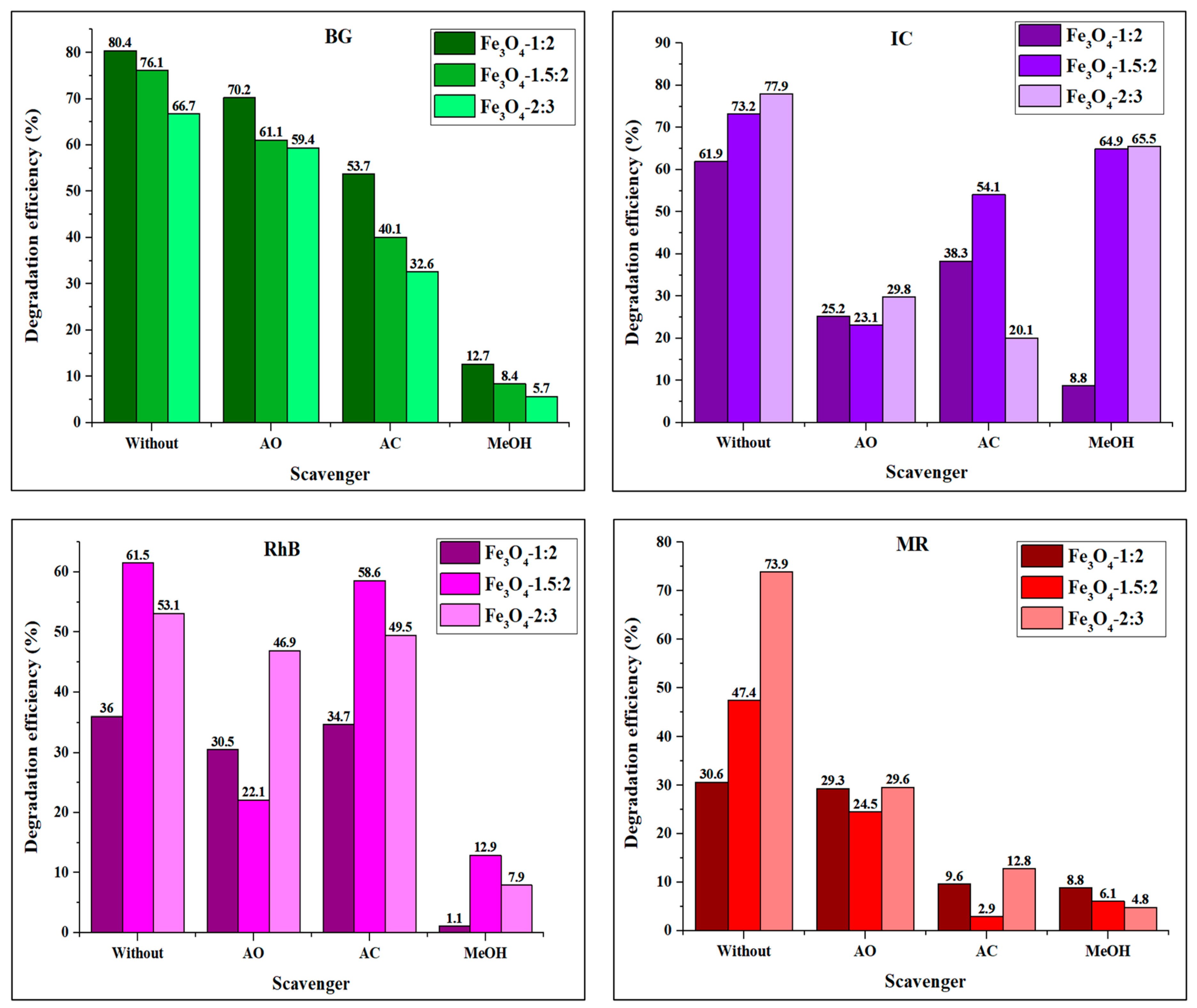
4.1. Effect of Scavengers on the Degradation of Organic Dyes by Fe3O4 Nanoparticles

The photocatalytic degradation reaction is regulated by a movement of electron–hole pairs photogenerated on the surface of the catalyst. Therefore, it is crucial to establish the effect of the reactive species in the photodegradation reaction [62,63]. Hence, methanol alcohol, acrylamide, and ammonium oxalate were introduced as scavengers to quench ·OH, ·O2, and h+ in the photocatalytic reaction, respectively. Figure 7 illustrates the percentage degradation of the organic dyes when different scavengers are added to the reaction system. The bar graphs show that the photodegradation efficiency of Fe3O4 nanoparticles was significantly reduced upon the introduction of methanol (MeOH), which suggests that ·OH plays a major part in the degradation of BG, RhB, and MR. The photodegradation efficiency of BG decreased to 32.6–53.7% and 59.4–70.2% after adding AC and AO, respectively. This indicates that ·OH and ·O2 are the major active species in the degradation of BG, with h+ serving as a secondary species. This trend is consistent with the literature [64,65]. Adding MeOH and AO inhibited the photodegradation of rhodamine B drastically, indicating that h+ and ·OH are the major active species, while ·O2 plays a limited part in the photodegradation process [66,67]. For indigo carmine, the order of reactive oxidative species responsible for photocatalytic degradation by the Fe3O4 was different for all nanoparticles. The observed trend for Fe3O4-1:2 was ·OH > h+ > ·O2, which is the same trend observed for all nanoparticles against rhodamine B. Fe3O4-1.5:2 followed the order ·O2 > h+ > ·OH, while Fe3O4-2:3 follows the sequence of h+ > ·O2 > ·OH [68,69,70,71]. The addition of MeOH in methyl red reaction media inhibited degradation efficiency from 30.6 to 8.8% (for Fe3O4-1:2), 73.9 to 4.8% (for Fe3O4-2:3), and 47.4 to 6.1% (for Fe3O4-1.5:2). The significant reduction in efficiency suggests that the hydroxyl radicals are major reactive species during the photodegradation. Similarly, the addition of AC reduced the performance of Fe3O4-1:2 (9.6–30.6%), Fe3O4-1.5:2 (2.9–47.4%), and Fe3O4-2:3 (12.8–73.9%), suggesting that ·O2 also has a major role in the photocatalytic degradation of methyl red. The results show that Fe3O4-1:2 and Fe3O4-2:3 follow a reactive species sequence of ·OH > ·O2 > h+, while Fe3O4-1.5:2 follows an ·O2 > ·OH > h+ trend [72,73].

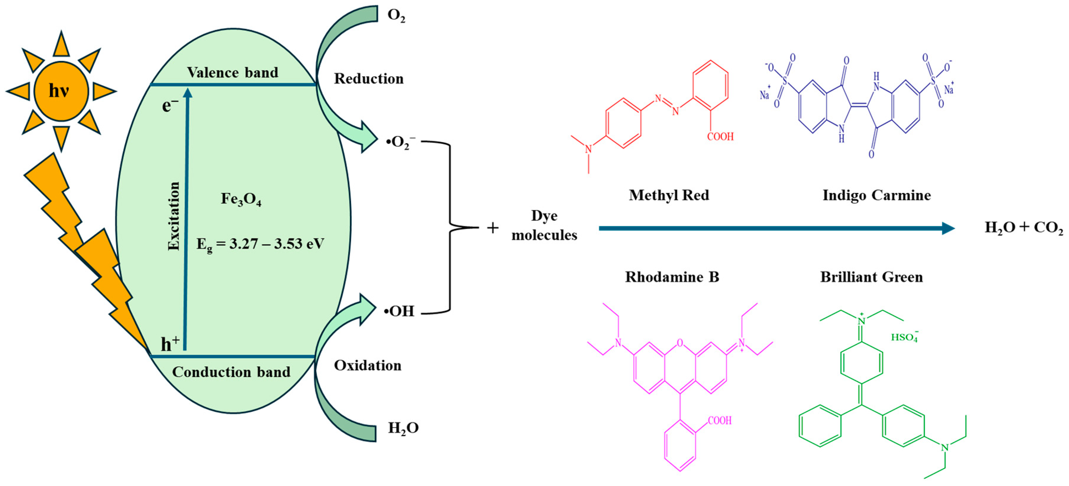
4.2. Proposed Mechanism
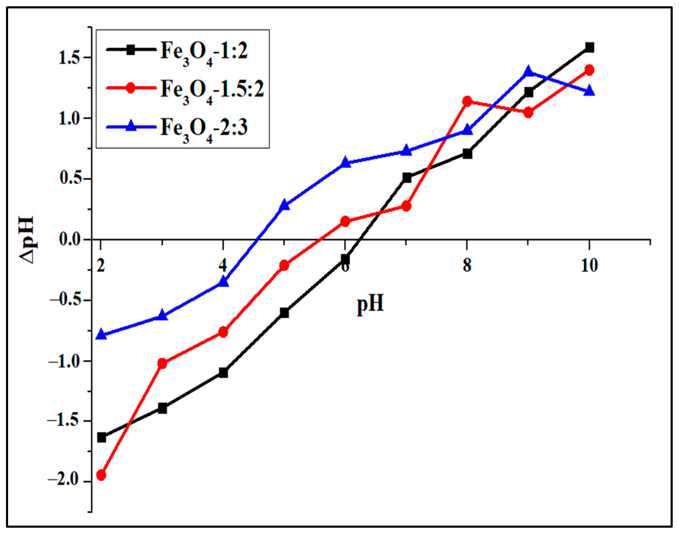
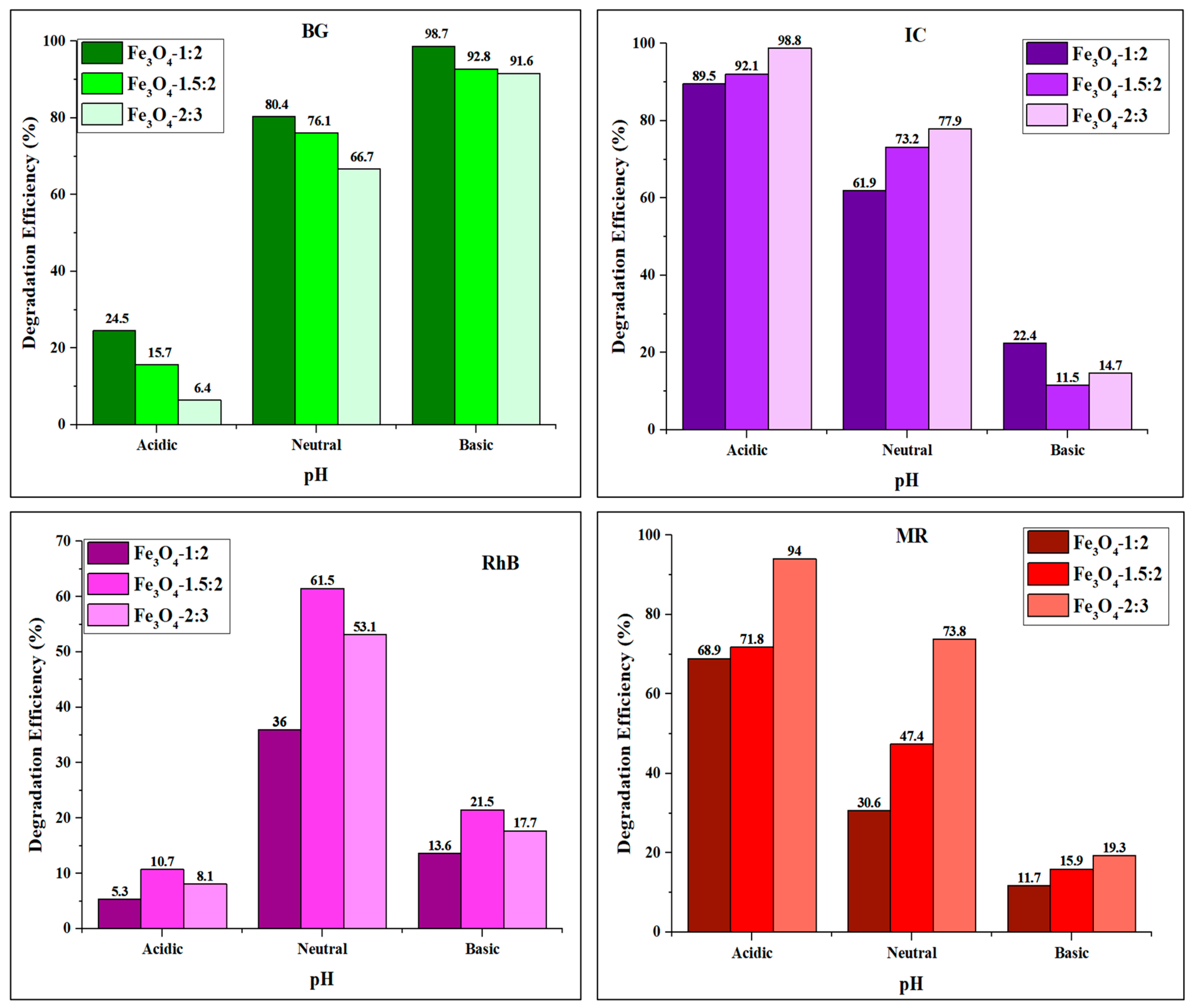
The proposed mechanism for the photocatalytic degradation of organic dyes by the as-prepared Fe3O4 nanoparticles is illustrated in Scheme 1. When the mixture of the dye and iron oxide (Fe3O4) nanoparticles is subjected to visible light, electron–hole pairs are generated as electrons (e) transition from the valence band to the conduction band. The electrons (e) react with oxygen molecules to generate oxygen peroxide radicals (·O2), while the positive charge holes (h+) react with H2O to produce hydroxyl radicals (·OH). The oxygen peroxide radical undergoes protonation to form hydroperoxyl radicals (·O2H) and eventually forms H2O2, which also dissociates into hydroxyl radicals [55]. The organic dye molecules are degraded by oxidation reactions with ·O2 and ·OH radicals, resulting in CO2, H2O, and some mineralization products.

4.3. Effect of pH

The surface charge of a photocatalyst and the charge of organic dyes are both strongly impacted by pH [74,75,76]. Figure 8 illustrates the findings of a study conducted in neutral, acidic, and alkaline dye solutions to examine the impact of pH on degradation efficiency. The degradation percentage of the BG dye by the iron oxide nanoparticles was lower at a pH of 3 and higher at a pH of 9. The high degradation efficiency observed in the alkaline solution can be attributed to the tertiary carbon of brilliant green, making the dye molecule a carbon-centered electrophile and the excess OH ions available, making the degradation process more favorable [77]. Rhodamine B degrades most efficiently at neutral pH. When the dye solution is adjusted to either alkaline or acidic, the degradation efficiency is drastically reduced. The dye is cationic (RhB+) at pH 3, and high H+ concentration reduces the catalyst’s accessible sites for interactions with positively charged RhB dye molecules [78,79]. The RhB+ undergoes deprotonation at pH 9 and forms a zwitter ion, which results in the repelling of negatively charged dye molecules and the catalysts due to coulombic forces [80]. A similar phenomenon was observed in other studies using different catalysts [78,81,82]. For indigo carmine, the results show that an increase in the pH of the reaction media decreases the photocatalytic degradation efficiency. At a pH of 3, the highest degradation efficiency was 98.8% after 180 min for Fe3O4-2:3, while at a pH of 9, the lowest degradation of 11.5% for Fe3O4-1.5:2 was obtained. Indigo carmine is negatively charged over wide pH ranges, and its structure breaks into hydrocarbon anion fragments with negatively charged sulfonate groups and sodium ions [83,84]. The drastic differences in the degradation efficiency observed over the 3–9 pH range could be due to the coulomb interactions between the surface of the catalysts and the IC dye molecules [85]. Similar results were obtained in the study by Bhakpar et al. using ZnO nanoparticles [75]. The degradation percentage was also found to be higher in an acidic medium than in neutral and alkaline pH for methyl red. Low degradation efficiency (11.7% for Fe3O4-1:2, 15.9% for Fe3O4-1.5:2, and 19.3% for Fe3O4-2:3) were observed at pH 9, while high degradation efficiency of 68.9%, 71.8% and 94.0% for Fe3O4-1:2, Fe3O4-1.5:2, and Fe3O4-2:3 were obtained at pH 3. This is mainly because pH has a considerable effect on the molecular structure of methyl red. In an acidic medium, the structure of methyl red transforms into quinoid form, which is unstable and easily degradable. In contrast, the dye molecules in basic media are in the benzenoid form, which is stable and difficult to degrade [86,87]. The findings are consistent with those of previous studies [88,89].

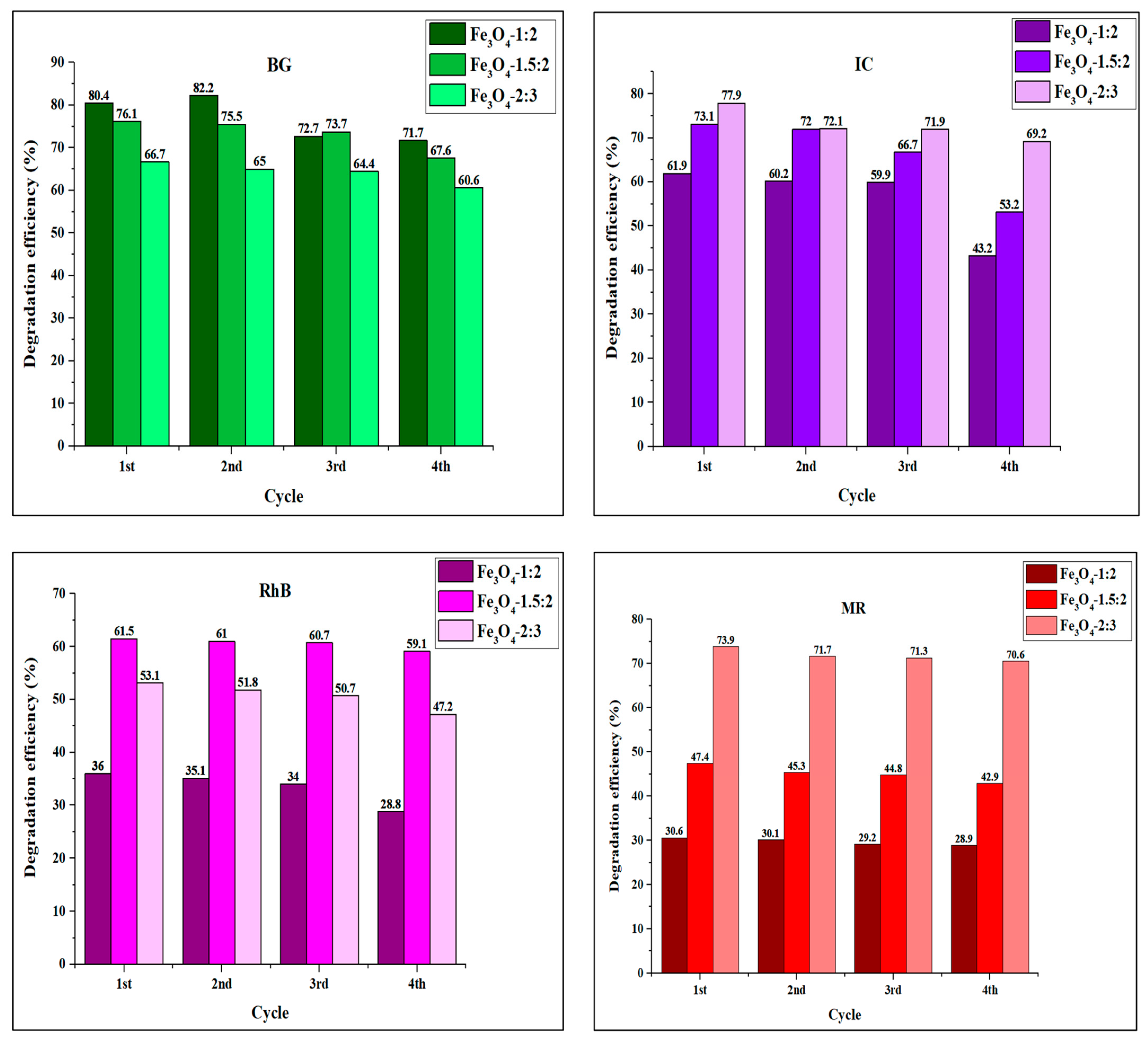
4.4. Recyclability of Iron Oxide Nanoparticles

The stability of the photocatalyst is a significant parameter for its potential practical application in industry [90]. As a result, the recyclability of the Fe3O4 photocatalysts was assessed in four runs. A degradation efficiency reduction of about 6.1–8.7%, 2.4–7.2%, 8.7–18.7%, and 1.7–4.5% was observed for BG, RhB, IC, and MR, respectively, as shown in Figure 9. This suggests that the as-synthesized iron oxide nanoparticles exhibit good photostability and can be recycled in dye photodegradation reactions (Figures S4–S7). The decrease in degradation percentage may be attributed to the adsorption of the intermediates produced by the degradation of organic dyes onto the active sites of the nanoparticle’s active sites [91].

5. Materials and Methods

5.1. Materials

FeCl2·4H2O (≥99%), Fe2(SO4)3·H2O (≥97%), ammonia solution (25%), octylamine (≥99%), ethanol (≥96%), brilliant green (≥90%), rhodamine B (95%), methyl red (95%), indigo carmine (~85%), sodium hydroxide (≥98%), HCl (37%), ammonium oxalate (≥99%), acrylamide (99%), and methanol (99.9%) were obtained from Sigma-Aldrich (St. Louis, MO, USA) and were utilized as supplied.

5.2. Preparation of Iron Oxide Nanoparticles

Iron oxide nanoparticles were prepared using the co-precipitation approach [92]. Two separate solutions of FeCl2·4H2O and Fe2(SO4)3·H2O were made using 50 mL of water and co-precipitated with 15 mL 25% ammonia solution at 90 °C under nitrogen flow. After adding base to the solution, the reaction mixture was stirred for 30 min; thereafter, 20 mL of octylamine was introduced into the solution, and the resulting mixture was further stirred for 1 h. Three different Fe2+:Fe3+ mole ratios of 1:2, 1.5:2, and 2:3 were used to prepare Fe3O4-1:2, Fe3O4-1.5:2, and Fe3O4-2:3, respectively. The nanoparticles were separated using a magnet, rinsed with distilled water and ethanol, and then oven-dried at 70 °C for 3 h.

5.3. Physical Characterization

Transmission electron microscopy (TEM) micrographs were taken using a JEOL JEM-1400 electron microscope (JEOL, Tokyo, Japan) and Gatan digital microgram software https://www.gatan.com/products/tem-analysis/gatan-microscopy-suite-software accessed on 15 June 2024, and the size distributions were calculated using ImageJ 1.53t software. A Bruker D8 Advance diffractometer (Bruker, Billerica, MA, USA) was employed to record the diffractograms. FTIR spectra were recorded using the Cary 630 FTIR spectrometer (Agilent Technologies, Santa Clara, CA, USA) in the 4000–500 cm−1 range. Absorption spectra were recorded on a Perkin Elmer lambda 25 UV–Vis spectrophotometer (PerkinElmer, Waltham, MA, USA) in the 200–700 nm range.

5.4. Adsorption Studies

Adsorption studies were conducted by dispersing 5 mg of the as-prepared Fe3O4 nanoparticles in 5 mL of 10 mg/L dye solution in seven test tubes. The dye–adsorbent mixture was agitated for 90 min with a magnetic stirrer bar. At 15 min intervals, sample aliquots were collected, and a magnet was used to separate the adsorbent from the solution. The dye adsorption was monitored using a UV-Visible spectrometer. The dye adsorption Qt (mg/g) was determined by Equation (3) [93].
Q = ((C0 − Ct)/W) × V
C0 represents the initial dye concentration (mg/L), Ct represents the dye concentration (mg/L) at the time ‘t’, V represents the dye volume (L), and W represents the adsorbent mass (g).

5.5. Photocatalysis Experiment

The photocatalytic potential of the iron oxide nanoparticles was studied by evaluating the degradation of brilliant green (BG), rhodamine B (RhB), indigo carmine (IC), and methyl red. A total of 5 mg of the as-prepared Fe3O4 nanoparticles was dispersed in 5 mL of 10 mg/L dye solution in seven test tubes. The heterogeneous solution was sonicated for 30 min. and then magnetically agitated in the dark for 60 min to reach adsorption–desorption equilibrium. The dye–photocatalyst mixture was then subjected to an OSRAM VIALOX 70 W mercury lamp (OSRAM, Munich, Germany) with continuous agitation for 180 min at room temperature. At 30 min intervals, 5 mL aliquots were collected, magnet-separated, and the dye was analyzed with a UV-Vis spectrophotometer. The degradation efficiency was determined using Equation (4) [94]:
D = (C0 − Ct)/C0 × 100
D denotes degradation efficiency, and C0 and Ct denote the dye concentrations at t0 and time t, respectively.
The catalysts were recycled four times to assess their photostability and reusability. Following each run, the catalyst was magnetically removed from the dye solution, rinsed with distilled water and ethanol, and oven-dried at 70 °C for 3 h.

5.6. Effect of pH

The effect of pH on the photodegradation of BG, RhB, IC, and MR by Fe3O4 was investigated at pH 3, pH 6.5, and pH 9. 0.1 M aqueous hydrochloric acid and sodium hydroxide solutions were used to adjust the pH of the dye solutions. A 0.01 M NaCl solution was used for the point of zero charge (pHzpc) test for magnetite nanoparticles, which was conducted between the pH values of 2 and 10. Fe3O4 nanoparticles (0.03 g) were dispersed in 30 mL of NaCl solution and shaken for 24 h at the ambient temperature. The pH of the supernatant was determined. The change in the solution pH was calculated and plotted over the initial solution pH to determine the pHzpc.

5.7. Effect of Scavengers

The quenching experiment was used to examine the reactive species responsible for the photocatalytic degradation of the BG, RhB, IC, and MR dyes by the magnetite nanoparticles. Acrylamide (AC), ammonium oxalate (AO), and methanol (MeOH) were used as ·O2, h+, and ·OH quenchers. Scavengers at a concentration of 10 mM were added to the dye solutions, and the reaction was carried out using the same reaction parameters as the photocatalysis experiment.

6. Conclusions

Magnetite iron oxide nanoparticles were synthesized by co-precipitating using three different iron (II) and iron (III) salt mole ratios. The p-XRD patterns correspond to the magnetite crystalline phase of iron oxide. The as-synthesized iron oxide nanoparticles consist of nanoparticles with different mixed shapes with average sizes of 5.48 nm, 6.02 nm, and 6.98 nm for Fe3O4-1:2, Fe3O4-1.5:2, and Fe3O4-2:3. The energy gaps are 3.53, 3.44, and 3.27 eV for Fe3O4-1:2, Fe3O4-1.5:2, and Fe3O4-2:3, respectively. The results show the iron salt ratio influences the morphology and energy band gap of the as-prepared Fe3O4 nanoparticles. Photocatalytic degradation of BG by the Fe3O4 nanoparticles after 180 min are 80.4% (Fe3O4-1:2), 76.1% (Fe3O4-1.5:2), and 66.7% (Fe3O4-2:3). Degradation efficiencies of 36.61.5% (Fe3O4-1:2), 61.5% (Fe3O4-1.5:2), and 53.1% (Fe3O4-2:3) were obtained against RhB. While degradation efficiencies of 61.9% for Fe3O4-1:2, 73.2% for Fe3O4-1.5:2, and 77.9% for Fe3O4-2:3 were obtained against IC, and the maximum percentage degradation of 73.9% was attained for Fe3O4-2:3 against MR. The results of this study revealed that the morphology; ·OH, ·O2, and h+ reactive species; and pH influence the photodegradation efficiency of BG, RhB, IC, and MR by the as-prepared Fe3O4 nanoparticles. The as-prepared Fe3O4 nanoparticles are recyclable and are photostable for the degradation of the organic dyes. Photocatalytic degradation efficiencies of over 70% were obtained against some of the dyes, and their general photostability indicates that the synthetic magnetite iron oxide nanoparticles could be fine-tuned for practical applications.

Supplementary Materials

The following supporting information can be downloaded at https://www.mdpi.com/article/10.3390/ijms25147876/s1.

Author Contributions

P.A.A.: conceptualization, supervision, writing—review and editing, and funding acquisition; T.B.M.: data curation, formal analysis, and writing—draft preparation. All authors have read and agreed to the published version of the manuscript.

Funding

This research was funded by the National Research Foundation, South Africa.

Institutional Review Board Statement

Not applicable.

Informed Consent Statement

Not applicable.

Data Availability Statement

All relevant data have been included in the manuscript.

Conflicts of Interest

The authors declare no conflicts of interest.

References

  1. Benkhaya, S.; M’Rabet, S.; El Harfi, A. Classifications, properties, recent synthesis and applications of azo dyes. Heliyon 2020, 6, e03271. [Google Scholar] [CrossRef] [PubMed]
  2. Slama, H.B.; Chenari Bouket, A.; Pourhassan, Z.; Alenezi, F.N.; Silini, A.; Cherif-Silini, H.; Oszako, T.; Luptakova, L.; Golińska, P.; Belbahri, L. Diversity of Synthetic dyes from textile industries, discharge impacts and treatment methods. Appl. Sci. 2021, 11, 6255. [Google Scholar] [CrossRef]
  3. Alsantali, R.; Raja, Q.; Alzahrani, A.; Sadiq, A.; Naeem, N.; Mughal, E.; Al-Rooqi, M.; El Guesmi, N.; Moussa, Z.; Ahmed, S. Miscellaneous azo dyes: A comprehensive review on recent advancements in biological and industrial Applications. Dyes Pigm. 2021, 199, 110050. [Google Scholar] [CrossRef]
  4. John, A.; Yang, H.-H.; Muhammad, S.; Khan, Z.I.; Yu, H.; Luqman, M.; Tofail, M.; Hussain, M.I.; Awan, M.U. Cross Talk between Synthetic Food Colors (Azo Dyes), Oral Flora, and Cardiovascular Disorders. Appl. Sci. 2022, 12, 7084. [Google Scholar] [CrossRef]
  5. Sharma, J.; Sharma, S.; Soni, V. Classification and impact of synthetic textile dyes on Aquatic Flora: A review. Reg. Stud. Mar. Sci. 2021, 45, 101802. [Google Scholar] [CrossRef]
  6. Alsukaibi, A.K.D. Various approaches for the detoxification of toxic dyes in wastewater. Processes 2022, 10, 1968. [Google Scholar] [CrossRef]
  7. Kubra, K.T.; Salman, M.S.; Hasan, M.N. Enhanced toxic dye removal from wastewater using biodegradable polymeric natural adsorbent. J. Mol. Liq. 2021, 328, 115468. [Google Scholar] [CrossRef]
  8. Shin, J.-H.; Yang, J.E.; Park, J.E.; Jeong, S.-W.; Choi, S.-J.; Choi, Y.J.; Jeon, J. Rapid and efficient removal of anionic dye in water using a chitosan-coated iron oxide-immobilized polyvinylidene fluoride membrane. ACS Omega 2022, 7, 8759–8766. [Google Scholar] [CrossRef] [PubMed]
  9. Al-Nuaim, M.A.; Alwasiti, A.A.; Shnain, Z.Y. The photocatalytic process in the treatment of polluted water. Chem. Pap. 2023, 77, 677–701. [Google Scholar] [CrossRef] [PubMed]
  10. Alahmadi, N. Recent progress in photocatalytic removal of environmental pollution hazards in water using nanostructured materials. Separations 2022, 9, 264. [Google Scholar] [CrossRef]
  11. Van Thuan, D.; Ngo, H.L.; Thi, H.P.; Chu, T.T.H. Photodegradation of hazardous organic pollutants using titanium oxides-based photocatalytic: A review. Environ. Res. 2023, 229, 116000. [Google Scholar] [CrossRef] [PubMed]
  12. Pal, S.; Kumar, S.; Verma, A.; Kumar, A.; Ludwig, T.; Frank, M.; Mathur, S.; Prakash, R.; Sinha, I. Development of magnetically recyclable visible light photocatalysts for hydrogen peroxide production. Mater. Sci. Semicond. Process. 2020, 112, 105024. [Google Scholar] [CrossRef]
  13. Ganapathe, L.S.; Kazmi, J.; Mohamed, M.A.; Berhanuddin, D.D. Molarity effects of Fe and NaOH on synthesis and characterisation of magnetite (Fe3O4) nanoparticles for potential application in magnetic hyperthermia therapy. Magnetochemistry 2022, 8, 161. [Google Scholar] [CrossRef]
  14. Granath, T.; Mandel, K.; Löbmann, P. The Significant influence of the pH value on citrate coordination upon modification of superparamagnetic iron oxide nanoparticles. Part. Syst. Charact. 2022, 39, 2100279. [Google Scholar] [CrossRef]
  15. Alibeigi, S.; Vaezi, M. Phase transformation of iron oxide nanoparticles by varying the molar ratio of Fe2+:Fe3+. Chem. Eng. Technol. 2008, 31, 1591–1596. [Google Scholar] [CrossRef]
  16. Dehghanpour, H.R. pH, molar ratio of ferrous to ferric ions and surfactant presence effects on physical properties of iron oxide nanoparticles generated by co-precipitation method. J. Coord. Chem. 2020, 73, 3452–3464. [Google Scholar] [CrossRef]
  17. Šutka, A.; Bitina, S.; Smits, K.; Sutka, A.; Ignatane, L.; Mairov, M.; Käämbre, T.; Timusk, M.; Laipniece, L.; Lazdovica, K. Rapid, high-yield aqueous synthesis of ultrafine magnetite nanoparticles from Fe(III) precursor at room temperature. J. Mater. Sci. 2023, 59, 447–457. [Google Scholar] [CrossRef]
  18. Majidi, S.; Zeinali Sehrig, F.; Farkhani, S.M.; Soleymani Goloujeh, M.; Akbarzadeh, A. Current methods for synthesis of magnetic nanoparticles. Artif. Cells Nanomed. Biotechnol. 2016, 44, 722–734. [Google Scholar] [CrossRef] [PubMed]
  19. Lee, K.-J.; N’Deh, K.P.U.; Kim, G.-J.; Choi, J.; Kim, J.; Kim, E.-K.; An, J. Fe2+:Fe3+ molar ratio influences the immunomodulatory properties of maghemite (γ-Fe2O3) nanoparticles in an atopic dermatitis model. ACS Appl. Bio Mater. 2021, 4, 1252–1267. [Google Scholar] [CrossRef]
  20. Terna, A.D.; Elemike, E.E.; Mbonu, J.I.; Osafile, O.E.; Ezeani, R.O. The future of semiconductors nanoparticles: Synthesis, properties and applications. Mater. Sci. Eng. 2021, 272, 115363. [Google Scholar] [CrossRef]
  21. Madima, N.; Kefeni, K.K.; Mishra, S.B.; Mishra, A.K.; Kuvarega, A.T. Fabrication of magnetic recoverable Fe3O4/TiO2 heterostructure for photocatalytic degradation of rhodamine B dye. Inorg. Chem. Commun. 2022, 145, 109966. [Google Scholar] [CrossRef]
  22. Khan, I.; Zada, N.; Khan, I.; Sadiq, M.; Saeed, K. Enhancement of photocatalytic potential and recoverability of Fe3O4 nanoparticles by decorating over monoclinic zirconia. J. Environ. Health Sci. Eng. 2020, 18, 1473–1489. [Google Scholar] [CrossRef] [PubMed]
  23. Sriramulu, M.; Balaji; Sumathi, S. Photocatalytic, antimicrobial and antifungal activity of biogenic iron oxide nanoparticles synthesised using aegle marmelos Extracts. J. Inorg. Organomet. Polym. 2021, 31, 1738–1744. [Google Scholar] [CrossRef]
  24. Ravelo-Nieto, E.; Ovalle-Serrano, S.A.; Gutiérrez-Pineda, E.A.; Blanco-Tirado, C.; Combariza, M.Y. Textile wastewater depuration using a green cellulose based Fe3O4 bionanocomposite. J. Environ. Chem. Eng. 2023, 11, 109516. [Google Scholar] [CrossRef]
  25. Accogli, A.; Bertoli, L.; Panzeri, G.; Gibertini, E.; Pesce, R.; Bussetti, G.; Magagnin, L. Electrochemical characterization of magnetite (Fe3O4) nanoaggregates in acidic and alkaline solutions. ACS Omega 2021, 6, 26880–26887. [Google Scholar] [CrossRef] [PubMed]
  26. Sosun; Ali, A.; Mannan, A.; Ali Shah, U.; Zia, M. Removal of of toxic metal ions (Ni2+ and Cd2+) from wastewater by using TOPO decorated iron oxide nanoparticles. Appl. Water Sci. 2022, 12, 86. [Google Scholar] [CrossRef]
  27. Ahmouda, K.; Boudiaf, M.; Barani, D.; Benhaoua, B. The effect of mediating plant extract on the photocatalytic activity of different eco-friendly synthesized magnetite nanoparticles against cresol red photodegradation. J. Photochem. Photobiol. 2024, 450, 115442. [Google Scholar] [CrossRef]
  28. Spivakov, A.; Lin, C.-R.; Chang, Y.-C.; Wang, C.-C.; Sarychev, D. Magnetic and magneto-optical oroperties of iron oxides nanoparticles synthesized under atmospheric pressure. Nanomaterials 2020, 10, 1888. [Google Scholar] [CrossRef] [PubMed]
  29. Syahida, A.N.; Sutanto, H.; Alkian, I.; Irianti, F.D.D.; Wibowo, A.A.; Priyono, P. Synthesized and characterization nanosized synthesis Fe3O4 powder from natural iron sand. J. Phys. Conf. Ser. 2021, 1943, 012013. [Google Scholar] [CrossRef]
  30. Gaur, R.; Shahabuddin, S.; Ahmad, I.; Sridewi, N. Role of alkylamines in tuning the morphology and optical properties of SnS2 nanoparticles synthesized by via facile thermal decomposition approach. Nanomaterials 2022, 12, 3950. [Google Scholar] [CrossRef]
  31. Chuburu, F.; Malystkyi, V.; Moreau, J.; Callewaert, M.; Henoumont, C.; Cadiou, C.; Feuillie, C.; Laurent, S.; Molinari, M. Synthesis and characterization of conjugated hyaluronic acids. Application to stability studies of chitosan-hyaluronic acid nanogels based on fluorescence resonance energy transfer. Gels 2022, 8, 182. [Google Scholar] [CrossRef] [PubMed]
  32. Sanmartín-Matalobos, J.; Bermejo-Barrera, P.; Aboal-Somoza, M.; Fondo, M.; García-Deibe, A.M.; Corredoira-Vázquez, J.; Alves-Iglesias, Y. Semiconductor quantum dots as target analytes: Properties, surface chemistry and detection. Nanomaterials 2022, 12, 2501. [Google Scholar] [CrossRef] [PubMed]
  33. Abu-Noqta, O.A.; Aziz, A.A.; Usman, A.I. Colloidal stability of iron oxide nanoparticles coated with different capping agents. Mater. Today Proc. 2019, 17, 1072–1077. [Google Scholar] [CrossRef]
  34. Sakthi Sri, S.P.; Taj, J.; George, M. Facile synthesis of magnetite nanocubes using deep eutectic solvent: An insight to anticancer and photo-Fenton efficacy. Surf. Interf. 2020, 20, 100609. [Google Scholar] [CrossRef]
  35. Jain, R. Effect of gadolinium doping on the structural, magnetic, electrical, optical, and elastic properties of magnetite nanoparticles. Mater. Sci. Eng. 2023, 296, 116631. [Google Scholar] [CrossRef]
  36. León-Flores, J.; Pérez-Mazariego, J.L.; Marquina, M.; Gómez, R.; Escamilla, R.; Tehuacanero-Cuapa, S.; Reyes-Damián, C.; Arenas-Alatorre, J. Controlled formation of hematite—Magnetite nanoparticles by a biosynthesis method and its photocatalytic removal potential against methyl orange dye. J. Cluster Sci. 2023, 34, 2381–2395. [Google Scholar] [CrossRef]
  37. Nnadozie, E.C.; Ajibade, P.A. Preparation, phase analysis and electrochemistry of magnetite (Fe3O4) and maghemite (γ-Fe2O3) nanoparticles. Int. J. Electrochem. Sci. 2022, 17, 22124. [Google Scholar] [CrossRef]
  38. Shamaila, S.; Bano, T.; Sajjad, A.K.L. Efficient visible light magnetic modified iron oxide photocatalysts. Ceram. Int. 2017, 43, 14672–14677. [Google Scholar] [CrossRef]
  39. Bayer, T.; Wei, R.; Kappler, A.; Byrne, J.M. Cu(II) and Cd(II) removal efficiency of microbially redox-activated magnetite nanoparticles. ACS Earth Space Chem. 2023, 7, 1837–1847. [Google Scholar] [CrossRef] [PubMed]
  40. Silva, J.M.M.; Feuser, P.E.; Cercená, R.; Peterson, M.; Dal-Bó, A.G. Obtention of magnetite nanoparticles via the hydrothermal method and effect of synthesis parameters. J. Magn. Magn. Mater. 2023, 580, 170925. [Google Scholar] [CrossRef]
  41. Zhang, J.; Lin, S.; Han, M.; Su, Q.; Xia, L.; Hui, Z. Adsorption properties of magnetic magnetite nanoparticle for coexistent Cr(VI) and Cu(II) in mixed solution. Water 2020, 12, 446. [Google Scholar] [CrossRef]
  42. Saxena, M.; Sharma, N.; Saxena, R. Highly efficient and rapid removal of a toxic dye: Adsorption kinetics, isotherm, and mechanism studies on functionalized multiwalled carbon nanotubes. Surf. Interf. 2020, 21, 100639. [Google Scholar] [CrossRef]
  43. Medhat, A.; El-Maghrabi, H.H.; Abdelghany, A.; Abdel Menem, N.M.; Raynaud, P.; Moustafa, Y.M.; Elsayed, M.A.; Nada, A.A. Efficiently activated carbons from corn cob for methylene blue adsorption. Appl. Surf. Sci. 2021, 3, 100037. [Google Scholar] [CrossRef]
  44. Ajibade, P.A.; Oluwalana, A.E. Photocatalytic degradation of single and binary mixture of brilliant green and rhodamine B dyes by zinc sulfide quantum dots. Molecules 2021, 26, 7686. [Google Scholar] [CrossRef] [PubMed]
  45. Cao, S.; Tao, F.; Tang, Y.; Li, Y.; Yu, J. Size- and shape-dependent catalytic performances of oxidation and reduction reactions on nanocatalysts. Chem. Soc. Rev. 2016, 45, 4747–4765. [Google Scholar] [CrossRef] [PubMed]
  46. Li, D.; Song, H.; Meng, X.; Shen, T.; Sun, J.; Han, W.; Wang, X. Effects of particle size on the structure and photocatalytic performance by alkali-treated TiO2. Nanomaterials 2020, 10, 546. [Google Scholar] [CrossRef] [PubMed]
  47. Gonçalves, A.H.A.; Siciliano, P.H.C.; Alves, O.C.; Cesar, D.V.; Henriques, C.A.; Gaspar, A.B. Synthesis of a magnetic Fe3O4/RGO composite for the rapid photo-fenton discoloration of indigo carmine dye. Top. Catal. 2020, 63, 1017–1029. [Google Scholar] [CrossRef]
  48. Sancheti, S.V.; Saini, C.; Ambati, R.; Gogate, P.R. Synthesis of ultrasound assisted nanostuctured photocatalyst (NiO supported over CeO2) and its application for photocatalytic as well as sonocatalytic dye degradation. Catal. Today 2018, 300, 50–57. [Google Scholar] [CrossRef]
  49. Soltani-nezhad, F.; Saljooqi, A.; Mostafavi, A.; Shamspur, T. Synthesis of Fe3O4/CdS–ZnS nanostructure and its application for photocatalytic degradation of chlorpyrifos pesticide and brilliant green dye from aqueous solutions. Ecotoxicol. Environ. Saf. 2020, 189, 109886. [Google Scholar] [CrossRef] [PubMed]
  50. Sunkara, J.R.; Botsa, S.M. SnO2/Fe2O3/Ag Nanocomposite via hydrothermal approach: A novel highly efficient photodegradation of eosin yellow and brilliant green dyes under visible light irradiation. Chem. Afr. 2019, 2, 635–644. [Google Scholar] [CrossRef]
  51. AbouSeada, N.; Ahmed, M.A.; Elmahgary, M.G. Synthesis and characterization of novel magnetic nanoparticles for photocatalytic degradation of indigo carmine dye. Mater. Sci. Energy Technol. 2022, 5, 116–124. [Google Scholar] [CrossRef]
  52. Dayana, P.N.; Abel, M.J.; Inbaraj, P.F.H.; Sivaranjani, S.; Thiruneelakandan, R.; Prince, J.J. Zirconium doped copper ferrite (CuFe2O4) nanoparticles for the enhancement of visible light-responsive photocatalytic degradation of rose bengal and indigo carmine dyes. J. Cluster Sci. 2022, 33, 1739–1749. [Google Scholar] [CrossRef]
  53. Güy, N.; Özacar, M. Visible light-induced degradation of indigo carmine over ZnFe2O4/Tannin/ZnO: Role of tannin as a modifier and its degradation mechanism. Int. J. Hydrogen Energy 2018, 43, 8779–8793. [Google Scholar] [CrossRef]
  54. Gupta, N.K.; Ghaffari, Y.; Bae, J.; Kim, K.S. Synthesis of coral-like α-Fe2O3 nanoparticles for dye degradation at neutral pH. J. Mol. Liq. 2020, 301, 112473. [Google Scholar] [CrossRef]
  55. Pegu, B.; Konwer, S. Facile green synthesis of reduced graphene oxide/iron nanocomposites using tea leaf extract as efficient photocatalyst for methyl red degradation. Graphene 2D Mater. 2023, 8, 121–133. [Google Scholar] [CrossRef]
  56. Bilgic, A. Fabrication of monoBODIPY-functionalized Fe3O4@SiO2@TiO2 nanoparticles for the photocatalytic degradation of rhodamine B under UV irradiation and the detection and removal of Cu(II) ions in aqueous solutions. J. Alloys Compd. 2022, 899, 163360. [Google Scholar] [CrossRef]
  57. Maqbool, S.; Ahmed, A.; Mukhtar, A.; Jamshaid, M.; Rehman, A.U.; Anjum, S. Efficient photocatalytic degradation of Rhodamine B dye using solar light-driven La-Mn co-doped Fe2O3 nanoparticles. Environ. Sci. Pollut. Res. 2023, 30, 7121–7137. [Google Scholar] [CrossRef] [PubMed]
  58. Muraro, P.C.L.; Mortari, S.R.; Vizzotto, B.S.; Chuy, G.; dos Santos, C.; Brum, L.F.W.; da Silva, W.L. Iron oxide nanocatalyst with titanium and silver nanoparticles: Synthesis, characterization and photocatalytic activity on the degradation of rhodamine B dye. Sci. Rep. 2020, 10, 3055. [Google Scholar] [CrossRef] [PubMed]
  59. Abbasi, Z.; Farrokhnia, A.; Garcıa-Lopez, E.I.; Zargar Shoushtari, M. Superparamagnetic recoverable flowerlike Fe3O4@Bi2O3 core–shell with g-C3N4 sheet nanocomposite: Synthesis, characterization, mechanism and kinetic study of photo-catalytic activity. J. Mater. Sci. Mater. Electron. 2020, 31, 1022–1033. [Google Scholar] [CrossRef]
  60. Kaushik, J.; Saluja, T.; Tisha; Nisha; Baig, A.; Sonal; Dubey, P.; Sonkar, S.K. Photoactive Fe3O4@Fe2O3 synthesized from industrial iron oxide dust for Fenton-free degradation of multiple organic dyes. Ind. Eng. Chem. Res. 2023, 62, 10487–10497. [Google Scholar] [CrossRef]
  61. Purkait, P.K.; Majumder, S.; Roy, S.; Maitra, S.; Chandra Das, G.; Chaudhuri, M.G. Enhanced heterogeneous photocatalytic degradation of florasulam in aqueous media using green synthesized TiO2 nanoparticle under UV light irradiation. Inorg. Chem. Commun. 2023, 155, 111017. [Google Scholar] [CrossRef]
  62. El Sharkawy, H.M.; Shawky, A.M.; Elshypany, R.; Selim, H. Efficient photocatalytic degradation of organic pollutants over TiO2 nanoparticles modified with nitrogen and MoS2 under visible light irradiation. Sci. Rep. 2023, 13, 8845. [Google Scholar] [CrossRef] [PubMed]
  63. Pavel, M.; Anastasescu, C.; State, R.-N.; Vasile, A.; Papa, F.; Balint, I. Photocatalytic degradation of organic and inorganic pollutants to harmless end products: Assessment of practical application potential for water and air cleaning. Catalysts 2023, 13, 380. [Google Scholar] [CrossRef]
  64. Mishra, S.; Sahu, T.K.; Verma, P.; Kumar, P.; Samanta, S.K. Microwave-assisted catalytic degradation of brilliant green by spinel zinc ferrite sheets. ACS Omega 2019, 4, 10411–10418. [Google Scholar] [CrossRef] [PubMed]
  65. Ritika; Kaur, M.; Umar, A.; Mehta, S.K.; Kansal, S.K.; Khan, M.A.; Algarni, H. Enhanced solar light-mediated photocatalytic degradation of brilliant green dye in aqueous phase using BiPO4 nanospindles and MoS2/BiPO4 nanorods. J. Mater. Sci. Mater. Electron. 2019, 30, 20741–20750. [Google Scholar] [CrossRef]
  66. Diao, Z.-H.; Liu, J.-J.; Hu, Y.-X.; Kong, L.-J.; Jiang, D.; Xu, X.-R. Comparative study of rhodamine B degradation by the systems pyrite/H2O2 and pyrite/persulfate: Reactivity, stability, products and mechanism. Sep. Purif. Technol. 2017, 184, 374–383. [Google Scholar] [CrossRef]
  67. Zhu, B.; Cheng, H.; Ma, J.; Kong, Y.; Komarneni, S. Efficient degradation of rhodamine B by magnetically separable ZnS–ZnFe2O4 composite with the synergistic effect from persulfate. Chemosphere 2019, 237, 124547. [Google Scholar] [CrossRef] [PubMed]
  68. Toghan, A.; Abd El-Lateef, H.M.; Taha, K.K.; Modwi, A. Mesoporous TiO2@g-C3N4 composite: Construction, characterization, and boosting indigo carmine dye destruction. Diam. Relat. Mater. 2021, 118, 108491. [Google Scholar] [CrossRef]
  69. Toghan, A.; Modwi, A. Boosting unprecedented indigo carmine dye photodegradation via mesoporous MgO@g-C3N4 nanocomposite. J. Photochem. Photobiol. 2021, 419, 113467. [Google Scholar] [CrossRef]
  70. Bakry, A.M.; Alamier, W.M.; El-Shall, M.S.; Awad, F.S. Facile synthesis of amorphous zirconium phosphate graphitic carbon nitride composite and its high performance for photocatalytic degradation of indigo carmine dye in water. J. Mater. Res. Technol. 2022, 20, 1456–1469. [Google Scholar] [CrossRef]
  71. Chaudhary, R.G.; Sonkusare, V.; Bhusari, G.; Mondal, A.; Potbhare, A.; Juneja, H.; Abdala, A.; Sharma, R. Preparation of mesoporous ThO2 nanoparticles: Influence of calcination on morphology and visible-light-driven photocatalytic degradation of indigo carmine and methylene blue. Environ. Res. 2023, 222, 115363. [Google Scholar] [CrossRef] [PubMed]
  72. Sonkusare, V.N.; Chaudhary, R.G.; Bhusari, G.S.; Mondal, A.; Potbhare, A.K.; Mishra, R.K.; Juneja, H.D.; Abdala, A.A. Mesoporous octahedron-shaped tricobalt tetroxide nanoparticles for photocatalytic degradation of toxic dyes. ACS Omega 2020, 5, 7823–7835. [Google Scholar] [CrossRef] [PubMed]
  73. Riaz, U.; Farooq, A.; Alam, J. Microwave-assisted rapid degradation of methyl red dye using polyfuran/polythiophene and its co-oligomers as catalysts. Spectrochim. Acta A Mol. Biomol. Spectrosc. 2023, 302, 123106. [Google Scholar] [CrossRef] [PubMed]
  74. Ziyaadini, M.; Ghashang, M. Sunlight-induced photocatalytic degradation of indigo carmine using Bi5Ti3FeO15 layered structure. Optik 2021, 228, 166207. [Google Scholar] [CrossRef]
  75. Bhapkar, A.R.; Geetha, M.; Jaspal, D.; Gheisari, K.; Laad, M.; Cabibihan, J.-J.; Sadasivuni, K.K.; Bhame, S. Aluminium doped ZnO nanostructures for efficient photodegradation of indigo carmine and azo carmine G in solar irradiation. Appl. Nanosci. 2023, 13, 5777–5793. [Google Scholar] [CrossRef]
  76. Nandisha, P.S.; Sowbhagya, S.; Yallappa, S. Synthesis and characterization of ternary NiO@Bi2MoO6–MoS heterojunction with enhanced photodegradation efficiency towards indigo carmine dye. Solid State Sci. 2023, 139, 107157. [Google Scholar] [CrossRef]
  77. Rao, C.V.; Giri, A.S.; Goud, V.V.; Golder, A.K. Studies on pH-dependent color variation and decomposition mechanism of brilliant green dye in Fenton reaction. Int. J. Ind. Chem. 2016, 7, 71–80. [Google Scholar] [CrossRef]
  78. Abadi, R.W.; Setiawan, C.M.; Santoso, S.P.; Bundjaja, V.; Angkawijaya, A.E.; Jiang, Y.-F.; Wijaya, C.J.; Ismadji, S.; Retnoningtyas, E.S.; Soetaredjo, F.E.; et al. Polystyrene-templated hollow mesoporous magnetite as a bifunctional adsorbent for the removal of rhodamine B via simultaneous adsorption and degradation. J. Environ. Chem. Eng. 2022, 10, 108194. [Google Scholar] [CrossRef]
  79. Ahmad, M.; Rehman, W.; Khan, M.M.; Qureshi, M.T.; Gul, A.; Haq, S.; Ullah, R.; Rab, A.; Menaa, F. Phytogenic fabrication of ZnO and gold decorated ZnO nanoparticles for photocatalytic degradation of Rhodamine B. J. Environ. Chem. Eng. 2021, 9, 104725. [Google Scholar] [CrossRef]
  80. Singh, S.; Kumar, A.; Gupta, H. Activated banana peel carbon: A potential adsorbent for rhodamine B decontamination from aqueous system. Appl. Water Sci. 2020, 10, 185. [Google Scholar] [CrossRef]
  81. Fatimah, I.; Purwiandono, G.; Sahroni, I.; Wijayana, A.; Faraswati, M.; Dwi Putri, A.; Oh, W.-C.; Doong, R.-A. Magnetically-separable photocatalyst of magnetic biochar from snake fruit peel for rhodamine B photooxidation. Environ. Nanotechnol. Monit. Manag. 2022, 17, 100669. [Google Scholar] [CrossRef]
  82. Zhang, D.; Cui, S.; Yang, J. Preparation of Ag2O/g-C3N4/Fe3O4 composites and the application in the photocatalytic degradation of rhodamine B under visible light. J. Alloys Compd. 2017, 708, 1141–1149. [Google Scholar] [CrossRef]
  83. Adel, M.; Ahmed, M.A.; Mohamed, A.A. Effective removal of indigo carmine dye from wastewaters by adsorption onto mesoporous magnesium ferrite nanoparticles. Environ. Nanotechnol. Monit. Manag. 2021, 16, 100550. [Google Scholar] [CrossRef]
  84. Yazdi, M.G.; Ivanic, M.; Mohamed, A.; Uheida, A. Surface modified composite nanofibers for the removal of indigo carmine dye from polluted water. RSC Adv. 2018, 8, 24588–24598. [Google Scholar] [CrossRef] [PubMed]
  85. Hadjltaief, H.B.; Bairq, Z.A.S.; Shi, C.; Benzina, M. Evaluation of sono-assisted solar/Fenton process for indigo carmine degradation over magnetic ZnO-Fe3O4 supported Tunisian kaolinite clay. Surf. Interf. 2021, 26, 101395. [Google Scholar] [CrossRef]
  86. Anjum, F.; Asiri, A.M.; Khan, M.A.; Khan, M.I.; Khan, S.B.; Akhtar, K.; Bakhsh, E.M.; Alamry, K.A.; Alfifi, S.Y.; Chakraborty, S. Photo-degradation, thermodynamic and kinetic study of carcinogenic dyes via zinc oxide/graphene oxide nanocomposites. J. Mater. Res. Technol. 2021, 15, 3171–3191. [Google Scholar] [CrossRef]
  87. Chen, X.; Wu, Z.; Liu, D.; Gao, Z. Preparation of ZnO photocatalyst for the efficient and rapid photocatalytic degradation of azo dyes. Nanoscale Res. Lett. 2017, 12, 143. [Google Scholar] [CrossRef] [PubMed]
  88. Saha, B.; Debnath, A.; Saha, B. Fabrication of PANI@Fe–Mn–Zr hybrid material and assessments in sono-assisted adsorption of methyl red dye: Uptake performance and response surface optimization. J. Indian Chem. Soc. 2022, 99, 100635. [Google Scholar] [CrossRef]
  89. Sharma, S.C.D.; Sun, Q.; Li, J.; Wang, Y.; Suanon, F.; Yang, J.; Yu, C.-P. Decolorization of azo dye methyl red by suspended and co-immobilized bacterial cells with mediators anthraquinone-2,6-disulfonate and Fe3O4 nanoparticles. Int. Biodeterior. Biodegrad. 2016, 112, 88–97. [Google Scholar] [CrossRef]
  90. Pervez, M.N.; Stylios, G.K.; Liang, Y.; Ouyang, F.; Cai, Y. Low-temperature synthesis of novel polyvinylalcohol (PVA) nanofibrous membranes for catalytic dye degradation. J. Clean. Prod. 2020, 262, 121301. [Google Scholar] [CrossRef]
  91. Cheng, S.; Zhao, S.; Xing, B.; Liu, Y.; Zhang, C.; Xia, H. Preparation of magnetic adsorbent-photocatalyst composites for dye removal by synergistic effect of adsorption and photocatalysis. J. Clean. Prod. 2022, 348, 131301. [Google Scholar] [CrossRef]
  92. Ye, L.; Zhou, L.; Lu, Y. Direct continuous synthesis of oleic acid-modified Fe3O4 nanoparticles in a microflow system. Ind. Eng. Chem. Res. 2022, 61, 4320–4328. [Google Scholar] [CrossRef]
  93. Mirzaei, K.; Jafarpour, E.; Shojaei, A.; Khasraghi, S.S.; Jafarpour, P. An investigation on the influence of highly acidic media on the microstructural stability and dye adsorption performance of UiO-66. Appl. Surf. Sci. 2023, 618, 156531. [Google Scholar] [CrossRef]
  94. Meena, P.L.; Poswal, K.; Surela, A.K.; Saini, J.K. Synthesis of graphitic carbon nitride/zinc oxide (g-C3N4/ZnO) hybrid nanostructures and investigation of the effect of ZnO on the photodegradation activity of g-C3N4 against the brilliant cresyl blue (BCB) dye under visible light irradiation. Adv. Compos. Hybrid Mater. 2022, 6, 16. [Google Scholar] [CrossRef]
Figure 1. XRD patterns of Fe3O4 nanoparticles were prepared using different iron salt stochiometric ratios.
Ijms 25 07876 g001
Figure 2. TEM images of Fe3O4-1:2, Fe3O4-1.5:2, and Fe3O4-2:3 nanoparticles at different magnifications.
Ijms 25 07876 g002
Figure 3. (a) Fe3O4 nanoparticles absorption spectra and (b) Tauc plots.
Ijms 25 07876 g003
Figure 4. pHpzc of magnetite nanoparticles.
Ijms 25 07876 g004
Figure 5. Adsorption capacity of Fe3O4 nanoparticles.
Ijms 25 07876 g005
Figure 6. Degradation efficiency plots of BG, RhB, IC, and MR by Fe3O4 nanoparticles.
Ijms 25 07876 g006
Figure 7. Effect of scavengers on the photocatalytic degradation of brilliant green (BG), indigo carmine (IC), rhodamine B (RhB), and methyl red (MR) by Fe3O4 nanoparticles.
Ijms 25 07876 g007
Scheme 1. Mechanism of the photocatalytic degradation of MR, IC, RhB, and BG dyes.
Ijms 25 07876 sch001
Figure 8. The effect of pH on brilliant green (BG), indigo carmine (IC), rhodamine B (RhB), and methyl red (MR) dyes over Fe3O4 nanoparticles.
Ijms 25 07876 g008
Figure 9. The recyclability of Fe3O4 nanophotocatalysts.
Ijms 25 07876 g009
Table 1. Photocatalytic degradation efficiency of magnetite nanoparticles compared to other photocatalysts.
DyeCatalystMethod of
Synthesis
Size (nm)Catalyst Load (mg)Dye Concentration (ppm)Degradation
Efficiency (%)
Time (min)Light SourceReference
BGNiO/CeO2ultrasound-2001081.92408 W UV light[48]
Fe3O4/
CdS–ZnS
ultrasound25–451.5 1092.93360300 W tungsten lamp[49]
SnO2/Fe2O3hydrothermal21.4150107475300 W visible light[50]
Fe3O4-1:2co-precipitation5.4851080.418070 W visible lightThis study
ICCoFe2O4/
SnO2
sol-gel/
sonochemical
1310055512015 W UV-A lamp[51]
Zr/CuFe2O4chemical
precipitation
421002071120150 W tungsten lamp[52]
ZnFe2O4solvothermal-501666.3300128 W visible light[53]
Fe3O4-2:3co-precipitation6.9851077.918070 W visible lightThis study
MRα-Fe2O3co-precipitation541001076532 W UV/H2O2[54]
Fe3O4/ZrO2chemical
reduction
803.54 ± 5.1120708440UV-light[22]
GO/Fein situ-0.03107030250 W Xenon lamp[55]
Fe3O4-2:3co-precipitation6.9851073.918070 W visible lightThis study
RhBFe3O4@SiO2@TiO2co-precipitation/Stöber8–18503029.4960UV 8/18[56]
Fe2O3co-precipitation42–49405058.21240Sunlight[57]
Fe2O3chemical
precipitation
3141024.2538.03120202 W visible light[58]
Fe3O4-1.5:2co-precipitation6.0251061.518070 W visible lightThis study
Table 2. Degradation rate constants of BG, RhB, IC, and MR by Fe3O4 nanoparticles.
CatalystBGRhBICMR
k (min−1)R2k (min−1)R2k (min−1)R2k (min−1)R2
Fe3O4-1:20.00910.98360.00240.98250.00530.99250.00210.9955
Fe3O4-1.5:20.00830.99420.00530.99660.00730.99770.00350.9961
Fe3O4-2:30.00610.97970.00430.99120.00840.99590.00740.9987
Disclaimer/Publisher’s Note: The statements, opinions and data contained in all publications are solely those of the individual author(s) and contributor(s) and not of MDPI and/or the editor(s). MDPI and/or the editor(s) disclaim responsibility for any injury to people or property resulting from any ideas, methods, instructions or products referred to in the content.

