
Multifunctional Sr,Mg-Doped Mesoporous Bioactive Glass Nanoparticles for Simultaneous Bone Regeneration and Drug Delivery

 ,
,  , ,
, ,  and
and
Abstract
1. Introduction
2. Results and Discussion
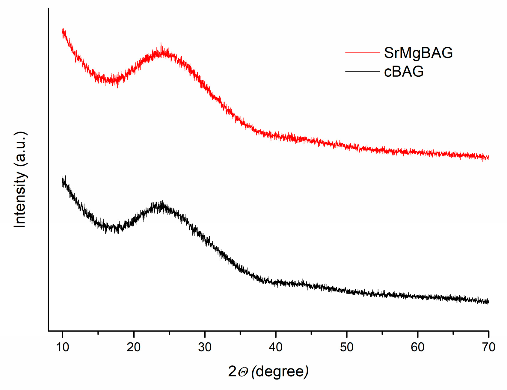
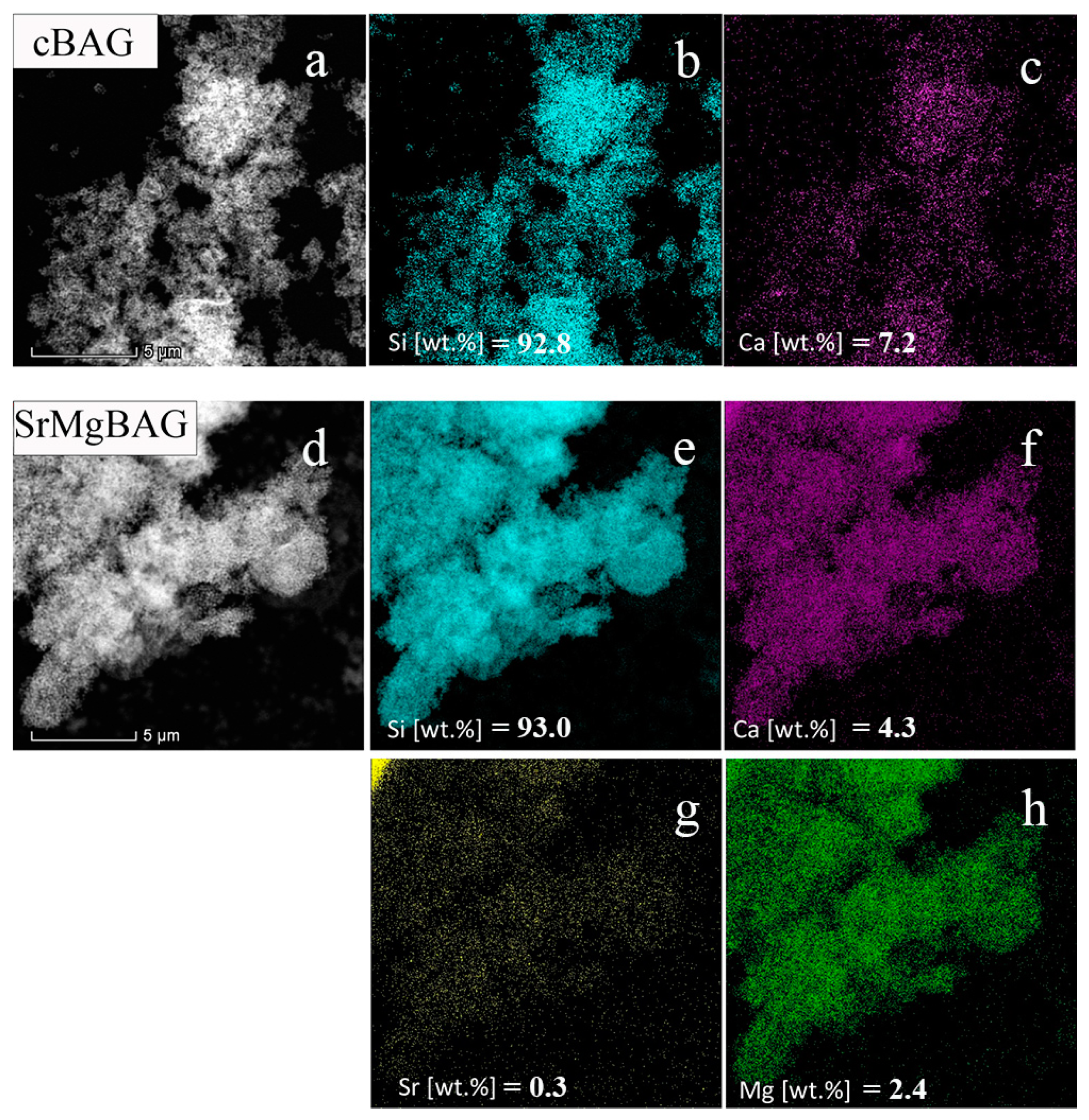
2.1. Compositional Analysis
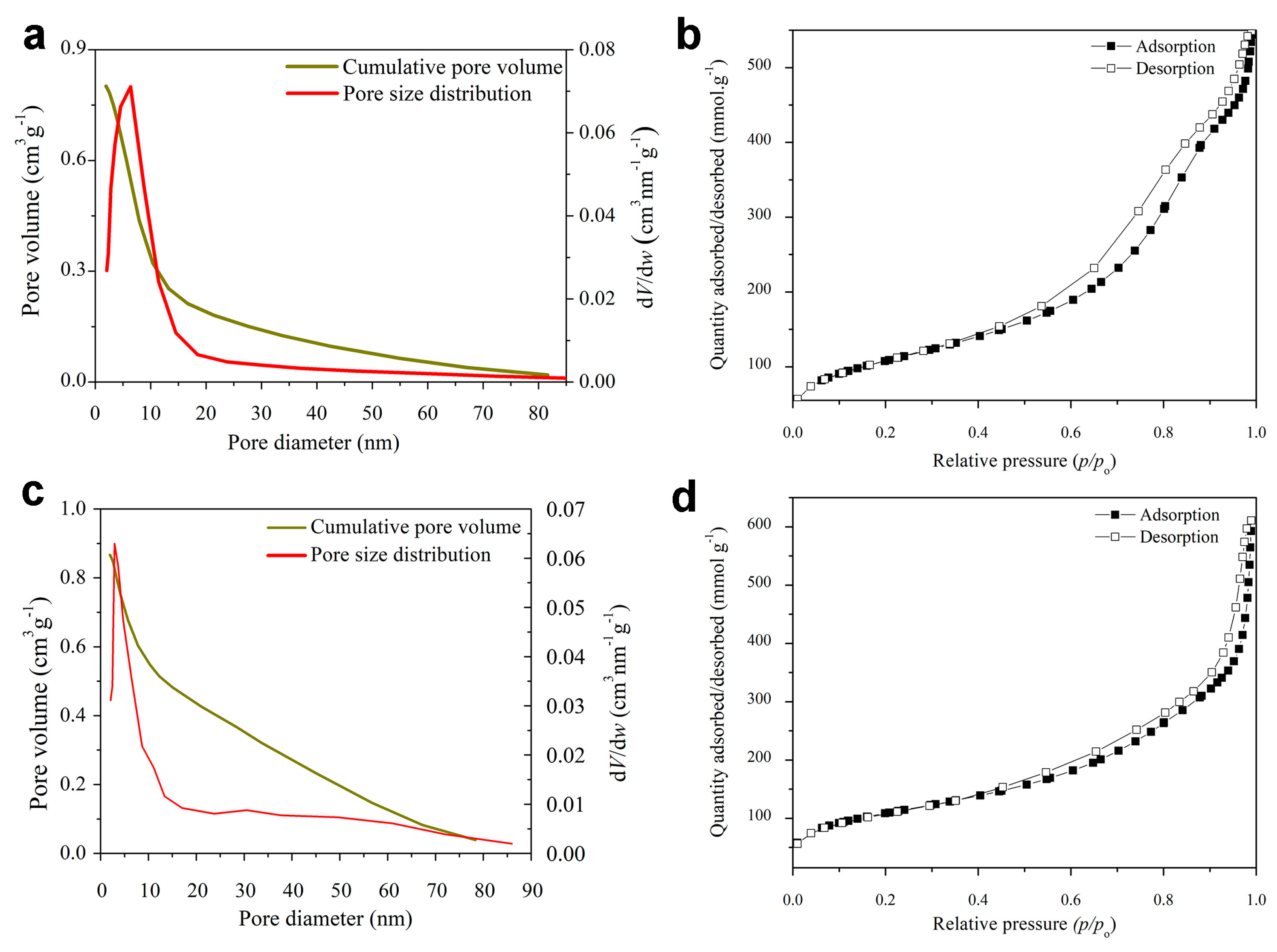
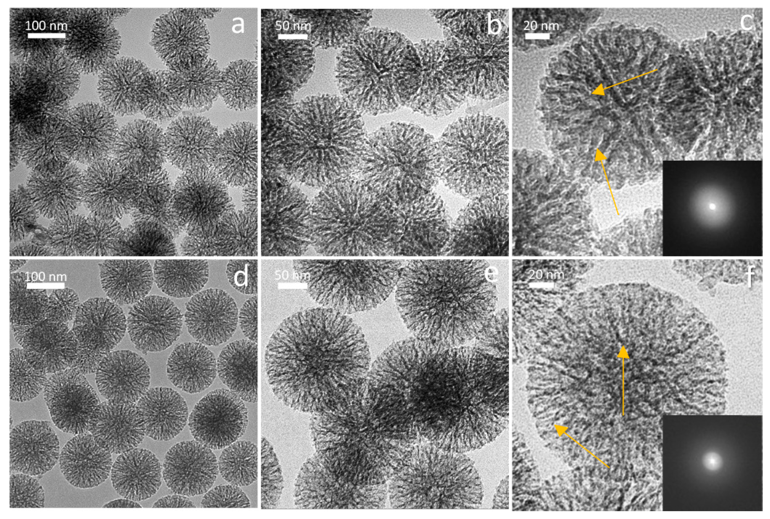
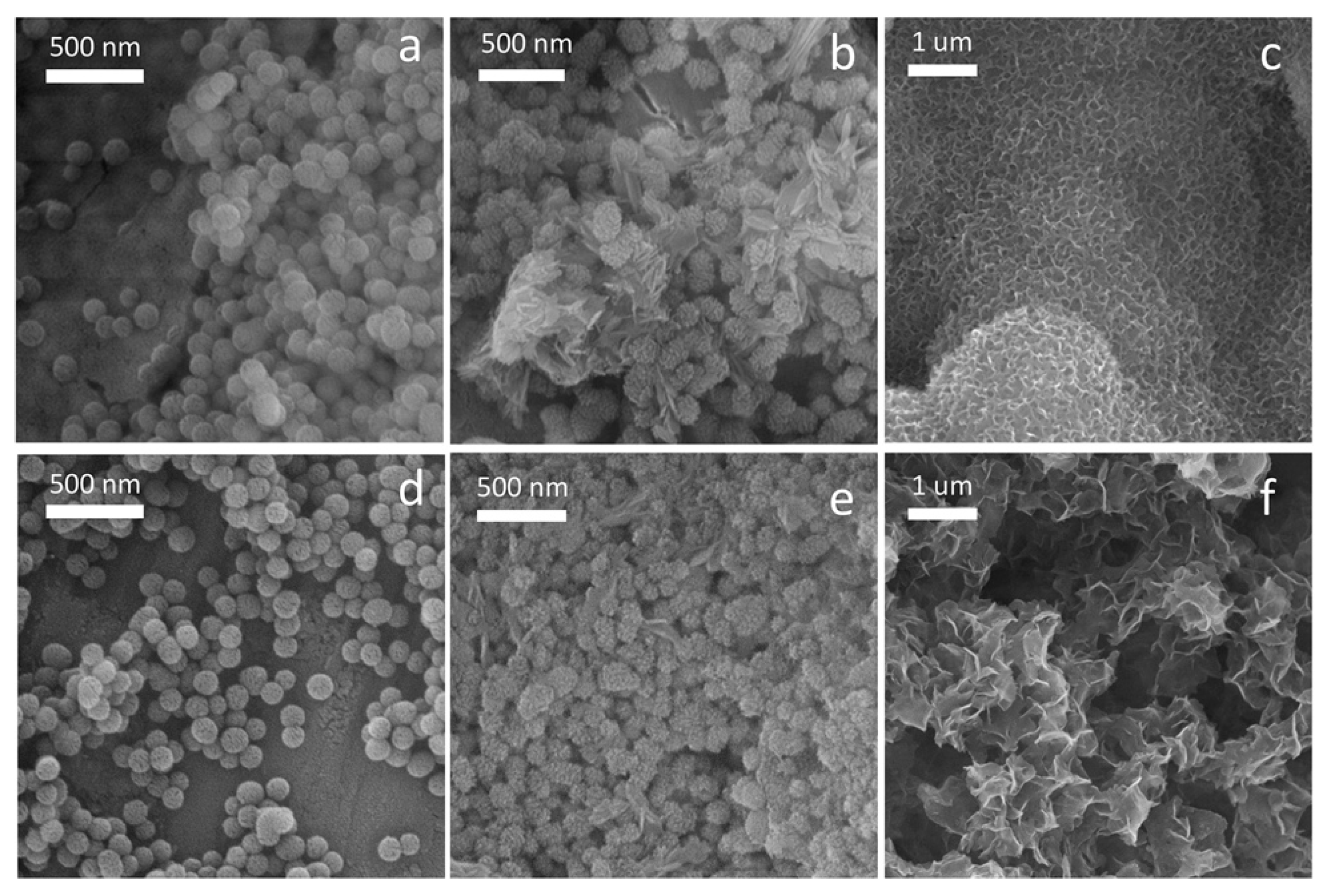
2.2. Morphology and Textural Properties
2.3. In Vitro Bioactivity
2.4. Dissolution and Ion Release Evaluation
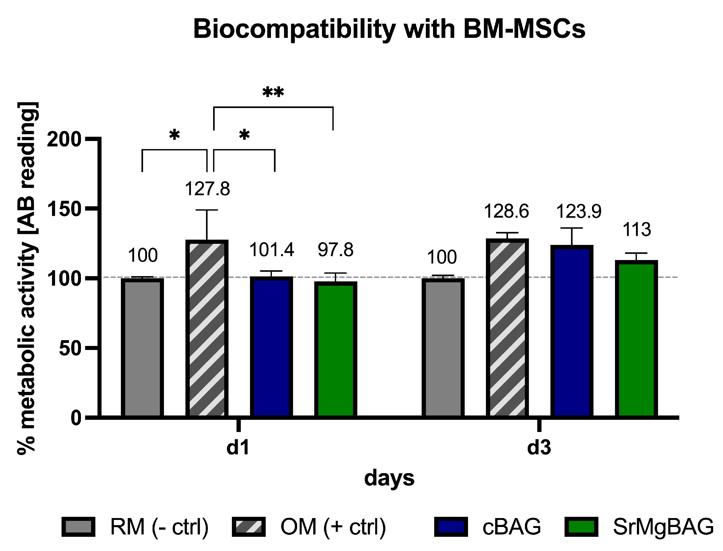
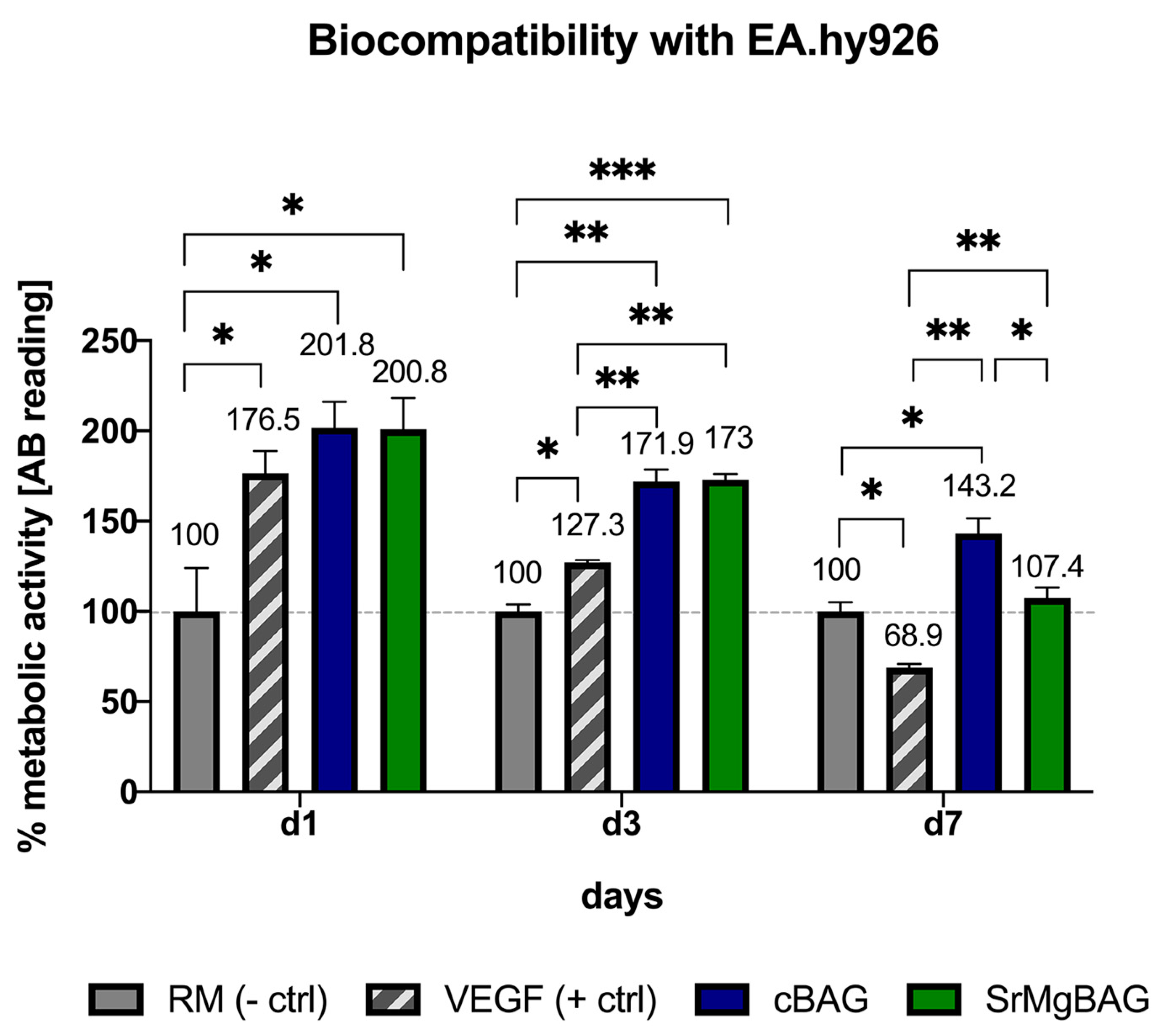
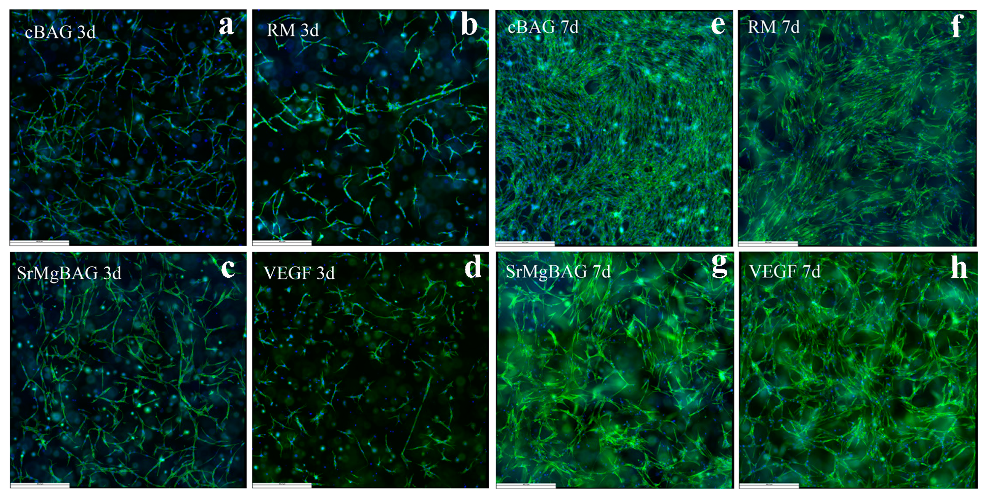
2.5. Assessment of Biological Properties
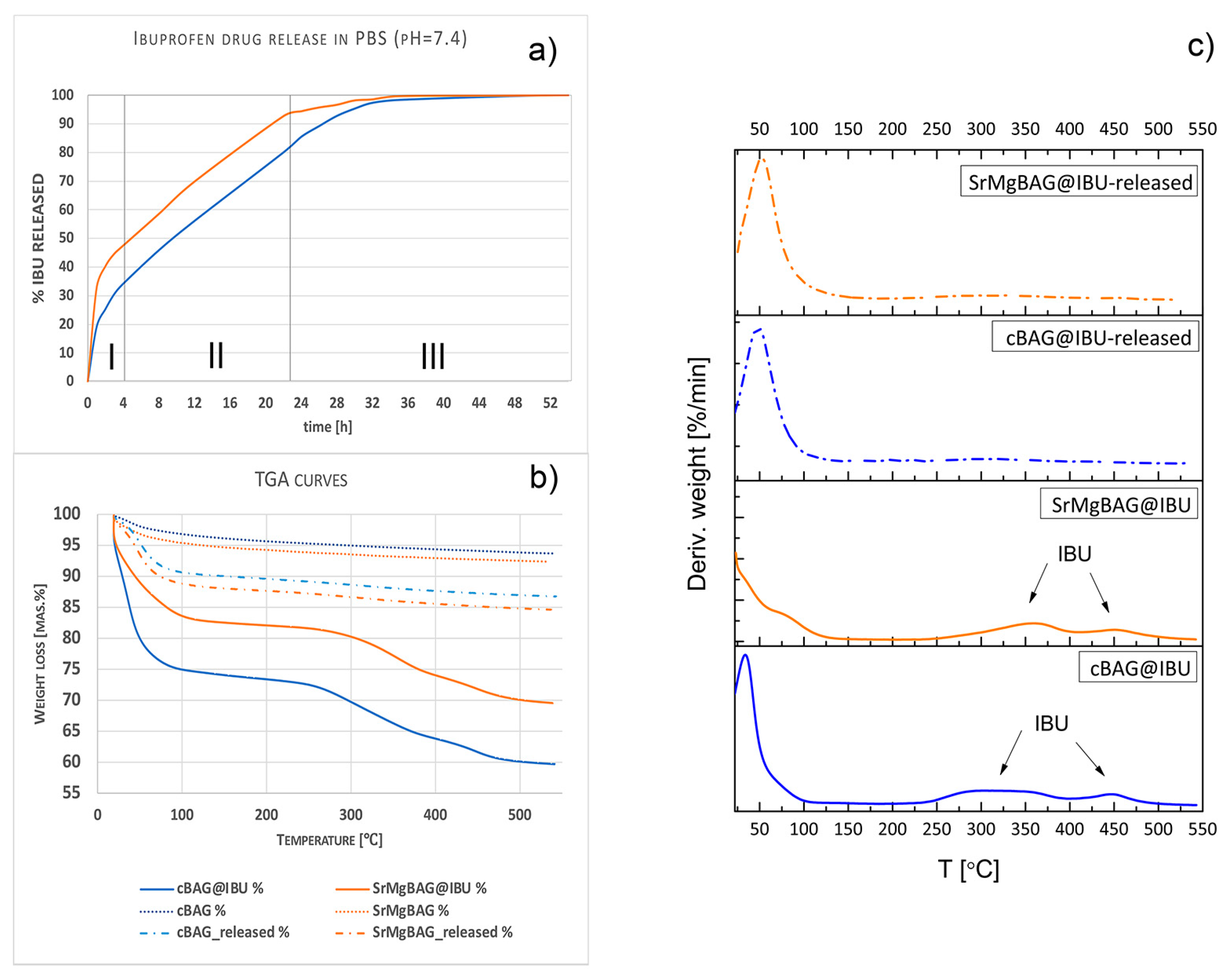
2.6. Drug Loading and Release
3. Materials and Methods
3.1. Synthesis of Mesoporous Bioactive Glass Particles
3.2. Characterization of Obtained Particles
3.2.1. Compositional Analysis
3.2.2. Morphology and Textural Properties
3.2.3. In Vitro Bioactivity
3.2.4. Dissolution Extracts Preparation and Ion Release Evaluation
3.3. Assessment of Biological Properties of Mesoporous Bioactive Glass Particles
3.3.1. Two-Dimensional Experiments with Stem Cells
Two-Dimensional Cell Cultures with Human Bone Marrow-Derived Mesenchymal Stem Cells (BM-MSCs)
Cell Seeding
Resazurin Assay
Alkaline Phosphatase (ALP) Analysis
Alizarin Red S Staining
3.3.2. Three-Dimensional Cell Culture Model with Human Endothelial Cells
Cell Culture Conditions
Three-Dimensional Model Preparation
Resazurin Reduction Assay
Fluorescence Staining
Statistical Analysis
3.4. Drug Loading and Release Evaluation
4. Conclusions
Author Contributions
Funding
Data Availability Statement
Acknowledgments
Conflicts of Interest
References
- Hench, L.L. The Story of Bioglass®. J. Mater. Sci. Mater. Med. 2006, 17, 967–978. [Google Scholar] [CrossRef]
- Li, R.; Clark, A.E.; Hench, L.L. An Investigation of Bioactive Glass Powders by Sol-Gel Processing. J. Appl. Biomater. 1991, 2, 231–239. [Google Scholar] [CrossRef]
- Kong, C.H.; Steffi, C.; Shi, Z.; Wang, W. Development of Mesoporous Bioactive Glass Nanoparticles and Its Use in Bone Tissue Engineering. J. Biomed. Mater. Res. B Appl. Biomater. 2018, 106, 2878–2887. [Google Scholar] [CrossRef]
- Vallet-Regi, M.; Rámila, A.; Del Real, R.P.; Pérez-Pariente, J. A New Property of MCM-41: Drug Delivery System. Chem. Mater. 2001, 13, 308–311. [Google Scholar] [CrossRef]
- Vallet-Regí, M. Nanostructured Mesoporous Silica Matrices in Nanomedicine. J. Intern. Med. 2010, 267, 22–43. [Google Scholar] [CrossRef]
- Theivendran, S.; Lazarev, S.; Yu, C. Mesoporous Silica/Organosilica Nanoparticles for Cancer Immunotherapy. Exploration 2023, 3, 20220086. [Google Scholar] [CrossRef]
- Yan, X.; Yu, C.; Zhou, X.; Tang, J.; Zhao, D. Highly Ordered Mesoporous Bioactive Glasses with Superior in Vitro Bone-Forming Bioactivities. Angew. Chem. Int. Ed. 2004, 43, 5980–5984. [Google Scholar] [CrossRef]
- Vallet-Regí, M.; Colilla, M.; Izquierdo-Barba, I.; Vitale-Brovarone, C.; Fiorilli, S. Achievements in Mesoporous Bioactive Glasses for Biomedical Applications. Pharmaceutics 2022, 14, 2636. [Google Scholar] [CrossRef]
- Zhu, H.; Zheng, K.; Boccaccini, A.R. Multi-Functional Silica-Based Mesoporous Materials for Simultaneous Delivery of Biologically Active Ions and Therapeutic Biomolecules. Acta Biomater. 2021, 129, 1–17. [Google Scholar] [CrossRef]
- Sharifi, E.; Bigham, A.; Yousefiasl, S.; Trovato, M.; Ghomi, M.; Esmaeili, Y.; Samadi, P.; Zarrabi, A.; Ashrafizadeh, M.; Sharifi, S.; et al. Mesoporous Bioactive Glasses in Cancer Diagnosis and Therapy: Stimuli-Responsive, Toxicity, Immunogenicity, and Clinical Translation. Adv. Sci. 2022, 9, 2102678. [Google Scholar] [CrossRef]
- Tabia, Z.; El Mabrouk, K.; Bricha, M.; Nouneh, K. Mesoporous Bioactive Glass Nanoparticles Doped with Magnesium: Drug Delivery and Acellular: In Vitro Bioactivity. RSC Adv. 2019, 9, 12232–12246. [Google Scholar] [CrossRef]
- Heras, C.; Jiménez-Holguín, J.; Doadrio, A.L.; Vallet-Regí, M.; Sánchez-Salcedo, S.; Salinas, A.J. Multifunctional Antibiotic- and Zinc-Containing Mesoporous Bioactive Glass Scaffolds to Fight Bone Infection. Acta Biomater. 2020, 114, 395–406. [Google Scholar] [CrossRef]
- Zheng, K.; Balasubramanian, P.; Paterson, T.E.; Stein, R.; MacNeil, S.; Fiorilli, S.; Vitale-Brovarone, C.; Shepherd, J.; Boccaccini, A.R. Ag Modified Mesoporous Bioactive Glass Nanoparticles for Enhanced Antibacterial Activity in 3D Infected Skin Model. Mater. Sci. Eng. C 2019, 103, 109764. [Google Scholar] [CrossRef]
- Kurtuldu, F.; Mutlu, N.; Michálek, M.; Zheng, K.; Masar, M.; Liverani, L.; Chen, S.; Galusek, D.; Boccaccini, A.R. Cerium and Gallium Containing Mesoporous Bioactive Glass Nanoparticles for Bone Regeneration: Bioactivity, Biocompatibility and Antibacterial Activity. Mater. Sci. Eng. C 2021, 124, 112050. [Google Scholar] [CrossRef]
- Liu, X.; Sun, Y.; Shen, J.; Min, H.S.; Xu, J.; Chai, Y. Strontium Doped Mesoporous Silica Nanoparticles Accelerate Osteogenesis and Angiogenesis in Distraction Osteogenesis by Activation of Wnt Pathway. Nanomedicine 2022, 41, 102496. [Google Scholar] [CrossRef]
- Westhauser, F.; Wilkesmann, S.; Nawaz, Q.; Hohenbild, F.; Rehder, F.; Saur, M.; Fellenberg, J.; Moghaddam, A.; Ali, M.S.; Peukert, W.; et al. Effect of Manganese, Zinc and Copper on the Biological and Osteogenic Properties of Mesoporous Bioactive Glass Nanoparticles. J. Biomed. Mater. Res. A 2020, 109, 1457–1467. [Google Scholar] [CrossRef]
- Zheng, K.; Torre, E.; Bari, A.; Taccardi, N.; Cassinelli, C.; Morra, M.; Fiorilli, S.; Vitale-Brovarone, C.; Iviglia, G.; Boccaccini, A.R. Antioxidant Mesoporous Ce-Doped Bioactive Glass Nanoparticles with Anti-Inflammatory and pro-Osteogenic Activities. Mater. Today Bio 2020, 5, 100041. [Google Scholar] [CrossRef]
- Li, Y.J.; Wong, K.W.; Huang, Y.C.; Chien, C.S.; Shih, C.J. Evaluation of in Vitro Bioactivity and Angiogenesis-Promoting Effect for Mesoporous Bioactive Glass Codoped with Copper and Silver. J. Non-Cryst. Solids 2023, 613, 122371. [Google Scholar] [CrossRef]
- Zhang, Y.; Hu, M.; Zhang, W.; Zhang, X. Homology of Selenium (Se) and Tellurium (Te) Endow the Functional Similarity of Se-Doped and Te-Doped Mesoporous Bioactive Glass Nanoparticles in Bone Tissue Engineering. Ceram. Int. 2022, 48, 3729–3739. [Google Scholar] [CrossRef]
- Simila, H.O.; Boccaccini, A.R. Sol-Gel Synthesis of Lithium Doped Mesoporous Bioactive Glass Nanoparticles and Tricalcium Silicate for Restorative Dentistry: Comparative Investigation of Physico-Chemical Structure, Antibacterial Susceptibility and Biocompatibility. Front. Bioeng. Biotechnol. 2023, 11, 1065597. [Google Scholar] [CrossRef]
- Baino, F.; Hamzehlou, S.; Kargozar, S. Bioactive Glasses: Where Are We and Where Are We Going? J. Funct. Biomater. 2018, 9, 25. [Google Scholar] [CrossRef] [PubMed]
- Arcos, D.; Portolés, M.T. Mesoporous Bioactive Nanoparticles for Bone Tissue Applications. Int. J. Mol. Sci. 2023, 24, 3249. [Google Scholar] [CrossRef] [PubMed]
- Zheng, K.; Sui, B.; Ilyas, K.; Boccaccini, A.R. Porous Bioactive Glass Micro- And Nanospheres with Controlled Morphology: Developments, Properties and Emerging Biomedical Applications. Mater. Horiz. 2021, 8, 300–335. [Google Scholar] [CrossRef] [PubMed]
- Wu, A.M.; Bisignano, C.; James, S.L.; Abady, G.G.; Abedi, A.; Abu-Gharbieh, E.; Alhassan, R.K.; Alipour, V.; Arabloo, J.; Asaad, M.; et al. Global, Regional, and National Burden of Bone Fractures in 204 Countries and Territories, 1990–2019: A Systematic Analysis from the Global Burden of Disease Study 2019. Lancet Healthy Longev. 2021, 2, e580–e592. [Google Scholar] [CrossRef] [PubMed]
- Huang, E.E.; Zhang, N.; Shen, H.; Li, X.; Maruyama, M.; Utsunomiya, T.; Gao, Q.; Guzman, R.A.; Goodman, S.B. Novel Techniques and Future Perspective for Investigating Critical-Size Bone Defects. Bioengineering 2022, 9, 171. [Google Scholar] [CrossRef] [PubMed]
- Archunan, M.W.; Petronis, S. Bone Grafts in Trauma and Orthopaedics. Cureus 2021, 13, e17705. [Google Scholar] [CrossRef] [PubMed]
- Li, H.; Chang, J. Bioactive Silicate Materials Stimulate Angiogenesis in Fibroblast and Endothelial Cell Co-Culture System through Paracrine Effect. Acta Biomater. 2013, 9, 6981–6991. [Google Scholar] [CrossRef] [PubMed]
- Zheng, K.; Niu, W.; Lei, B.; Boccaccini, A.R. Immunomodulatory Bioactive Glasses for Tissue Regeneration. Acta Biomater. 2021, 133, 168–186. [Google Scholar] [CrossRef]
- O’Neill, E.; Awale, G.; Daneshmandi, L.; Umerah, O.; Lo, K.W.H. The Roles of Ions on Bone Regeneration. Drug Discov. Today 2018, 23, 879–890. [Google Scholar] [CrossRef]
- Naruphontjirakul, P.; Porter, A.E.; Jones, J.R. In Vitro Osteogenesis by Intracellular Uptake of Strontium Containing Bioactive Glass Nanoparticles. Acta Biomater. 2018, 66, 67–80. [Google Scholar] [CrossRef]
- Naruphontjirakul, P.; Tsigkou, O.; Li, S.; Porter, A.E.; Jones, J.R. Human Mesenchymal Stem Cells Differentiate into an Osteogenic Lineage in Presence of Strontium Containing Bioactive Glass Nanoparticles. Acta Biomater. 2019, 90, 373–392. [Google Scholar] [CrossRef]
- Liu, W.; Guo, S.; Tang, Z.; Wei, X.; Gao, P.; Wang, N.; Li, X.; Guo, Z. Magnesium Promotes Bone Formation and Angiogenesis by Enhancing MC3T3-E1 Secretion of PDGF-BB. Biochem. Biophys Res. Commun. 2020, 528, 664–670. [Google Scholar] [CrossRef] [PubMed]
- Bose, S.; Fielding, G.; Tarafder, S.; Bandyopadhyay, A. Understanding of Dopant-Induced Osteogenesis and Angiogenesis in Calcium Phosphate Ceramics. Trends Biotechnol. 2013, 31, 594–605. [Google Scholar] [CrossRef] [PubMed]
- Yoshizawa, S.; Brown, A.; Barchowsky, A.; Sfeir, C. Magnesium Ion Stimulation of Bone Marrow Stromal Cells Enhances Osteogenic Activity, Simulating the Effect of Magnesium Alloy Degradation. Acta Biomater. 2014, 10, 2834–2842. [Google Scholar] [CrossRef] [PubMed]
- Lin, S.; Yang, G.; Jiang, F.; Zhou, M.; Yin, S.; Tang, Y.; Tang, T.; Zhang, Z.; Zhang, W.; Jiang, X. A Magnesium-Enriched 3D Culture System That Mimics the Bone Development Microenvironment for Vascularized Bone Regeneration. Adv. Sci. 2019, 6, 1900209. [Google Scholar] [CrossRef] [PubMed]
- Wang, J.L.; Xu, J.K.; Hopkins, C.; Chow, D.H.K.; Qin, L. Biodegradable Magnesium-Based Implants in Orthopedics—A General Review and Perspectives. Adv. Sci. 2020, 7, 1902443. [Google Scholar] [CrossRef] [PubMed]
- Wu, C.; Zhou, Y.; Lin, C.; Chang, J.; Xiao, Y. Strontium-Containing Mesoporous Bioactive Glass Scaffolds with Improved Osteogenic/Cementogenic Differentiation of Periodontal Ligament Cells for Periodontal Tissue Engineering. Acta Biomater. 2012, 8, 3805–3815. [Google Scholar] [CrossRef] [PubMed]
- Lee, J.H.; Mandakhbayar, N.; El-Fiqi, A.; Kim, H.W. Intracellular Co-Delivery of Sr Ion and Phenamil Drug through Mesoporous Bioglass Nanocarriers Synergizes BMP Signaling and Tissue Mineralization. Acta Biomater. 2017, 60, 93–108. [Google Scholar] [CrossRef] [PubMed]
- Fiorilli, S.; Molino, G.; Pontremoli, C.; Iviglia, G.; Torre, E.; Cassinelli, C.; Morra, M.; Vitale-Brovarone, C. The Incorporation of Strontium to Improve Bone-Regeneration Ability of Mesoporous Bioactive Glasses. Materials 2018, 11, 678. [Google Scholar] [CrossRef]
- Amudha, S.; Ramya, J.R.; Arul, K.T.; Deepika, A.; Sathiamurthi, P.; Mohana, B.; Asokan, K.; Dong, C.L.; Kalkura, S.N. Enhanced Mechanical and Biocompatible Properties of Strontium Ions Doped Mesoporous Bioactive Glass. Compos. B Eng. 2020, 196, 108099. [Google Scholar] [CrossRef]
- Shoaib, M.; Bahadur, A.; Iqbal, S.; AL-Anazy, M.M.; Laref, A.; Tahir, M.A.; Channar, P.A.; Noreen, S.; Yasir, M.; Iqbal, A.; et al. Magnesium Doped Mesoporous Bioactive Glass Nanoparticles: A Promising Material for Apatite Formation and Mitomycin c Delivery to the MG-63 Cancer Cells. J. Alloys Compd. 2021, 866, 159013. [Google Scholar] [CrossRef]
- Pouroutzidou, G.K.; Liverani, L.; Theocharidou, A.; Tsamesidis, I.; Lazaridou, M.; Christodoulou, E.; Beketova, A.; Pappa, C.; Triantafyllidis, K.S.; Anastasiou, A.D.; et al. Synthesis and Characterization of Mesoporous Mg-and Sr-Doped Nanoparticles for Moxifloxacin Drug Delivery in Promising Tissue Engineering Applications. Int. J. Mol. Sci. 2021, 2021, 577. [Google Scholar] [CrossRef] [PubMed]
- Neščáková, Z.; Zheng, K.; Liverani, L.; Nawaz, Q.; Galusková, D.; Kaňková, H.; Michálek, M.; Galusek, D.; Boccaccini, A.R. Multifunctional Zinc Ion Doped Sol—Gel Derived Mesoporous Bioactive Glass Nanoparticles for Biomedical Applications. Bioact. Mater. 2019, 4, 312–321. [Google Scholar] [CrossRef] [PubMed]
- Vallet-Regi, M.; Salinas, A.J. Mesoporous Bioactive Glasses for Regenerative Medicine. Mater. Today Bio 2021, 11, 100121. [Google Scholar] [CrossRef] [PubMed]
- Zheng, K.; Boccaccini, A.R. Sol-Gel Processing of Bioactive Glass Nanoparticles: A Review. Adv. Colloid. Interface Sci. 2017, 249, 363–373. [Google Scholar] [CrossRef] [PubMed]
- Liang, Q.; Hu, Q.; Miao, G.; Yuan, B.; Chen, X. A Facile Synthesis of Novel Mesoporous Bioactive Glass Nanoparticles with Various Morphologies and Tunable Mesostructure by Sacrificial Liquid Template Method. Mater. Lett. 2015, 148, 45–49. [Google Scholar] [CrossRef]
- Li, X.; Zhang, L.; Dong, X.; Liang, J.; Shi, J. Preparation of Mesoporous Calcium Doped Silica Spheres with Narrow Size Dispersion and Their Drug Loading and Degradation Behavior. Microporous Mesoporous Mater. 2007, 102, 151–158. [Google Scholar] [CrossRef]
- Wang, Y.; Song, H.; Yang, Y.; Liu, Y.; Tang, J.; Yu, C. Kinetically Controlled Dendritic Mesoporous Silica Nanoparticles: From Dahlia- to Pomegranate-like Structures by Micelle Filling. Chem. Mater. 2018, 30, 5770–5776. [Google Scholar] [CrossRef]
- Hochstrasser, J.; Juère, E.; Kleitz, F.; Wang, W.; Kübel, C.; Tallarek, U. Insights into the Intraparticle Morphology of Dendritic Mesoporous Silica Nanoparticles from Electron Tomographic Reconstructions. J. Colloid. Interface Sci. 2021, 592, 296–309. [Google Scholar] [CrossRef]
- Wang, Y.B.; Liu, Z.; Shi, S.H.; Hu, K.K.; Zhang, Y.T.; Guo, M. Research Progress of Dendritic Fibrous Nano-Silica (DFNS). Wuji Cailiao Xuebao/J. Inorg. Mater. 2018, 33, 1274–1288. [Google Scholar]
- El Baakili, S.; El Mabrouk, K.; Bricha, M. Acellular Bioactivity and Drug Delivery of New Strontium Doped Bioactive Glasses Prepared through a Hydrothermal Process. RSC Adv. 2022, 12, 15361–15372. [Google Scholar] [CrossRef] [PubMed]
- Kalim, D.; Kovarık, T.; Krenek, T.; Docheva, D.; Stich, T.; Pola, J. Recent Advances and Future Perspectives of Sol-Gel Derived Porous Bioactive Glasses: A Review. RSC Adv. 2020, 10, 33782–33835. [Google Scholar] [CrossRef]
- Massera, J.; Hupa, L.; Hupa, M. Influence of the Partial Substitution of CaO with MgO on the Thermal Properties and in Vitro Reactivity of the Bioactive Glass S53P4. J. Non-Cryst. Solids 2012, 358, 2701–2707. [Google Scholar] [CrossRef]
- Soulié, J.; Nedelec, J.M.; Jallot, E. Influence of Mg Doping on the Early Steps of Physico-Chemical Reactivity of Sol-Gel Derived Bioactive Glasses in Biological Medium. Phys. Chem. Chem. Phys. 2009, 11, 10473–10483. [Google Scholar] [CrossRef] [PubMed]
- Ma, J.; Chen, C.Z.; Wang, D.G.; Jiao, Y.; Shi, J.Z. Effect of Magnesia on the Degradability and Bioactivity of Sol-Gel Derived SiO2-CaO-MgO-P2O5 System Glasses. Colloids Surf. B Biointerfaces 2010, 81, 87–95. [Google Scholar] [CrossRef] [PubMed]
- Schumacher, M.; Habibovic, P.; van Rijt, S. Mesoporous Bioactive Glass Composition Effects on Degradation and Bioactivity. Bioact. Mater. 2021, 6, 1921–1931. [Google Scholar] [CrossRef] [PubMed]
- Villanueva-Flores, F.; Castro-Lugo, A.; Ramírez, O.T.; Palomares, L.A. Understanding Cellular Interactions with Nanomaterials: Towards a Rational Design of Medical Nanodevices. Nanotechnology 2020, 31, 132002. [Google Scholar] [CrossRef] [PubMed]
- Díaz-Tocados, J.M.; Herencia, C.; Martínez-Moreno, J.M.; Montes de Oca, A.; Rodríguez-Ortiz, M.E.; Vergara, N.; Blanco, A.; Steppan, S.; Almadén, Y.; Rodríguez, M.; et al. Magnesium Chloride Promotes Osteogenesis through Notch Signaling Activation and Expansion of Mesenchymal Stem Cells. Sci. Rep. 2017, 7, 7839. [Google Scholar] [CrossRef] [PubMed]
- Damian-Buda, A.I.; Ghițulică, C.D.; Cucuruz, A.; Voicu, G.; Culita, D.; Fruth-Oprișan, V.; Ciocan, L.T. Mesoporous Bioactive Glass Nanoparticles in the SiO2-P2O5-CaO-MO (M = Mg, Zn) System: Synthesis and Properties. J. Funct. Biomater. 2022, 13, 180. [Google Scholar] [CrossRef]
- Tahir, M.A.; Saif ur Rahman, M.; Nisha, F.; Shahzad, F.; Jawad, M.T.; Bahadur, A.; Qamar, M.A.; Shoaib, M. Hydroxycarbonate Apatite Formation and 5-Fluorouracil Delivery by Strontium Containing Mesoporous Bioactive Glass Nanoparticles. Ceram. Int. 2022, 48, 15862–15867. [Google Scholar] [CrossRef]
- Taghvaei, A.H.; Danaeifar, F.; Gammer, C.; Eckert, J.; Khosravimelal, S.; Gholipourmalekabadi, M. Synthesis and Characterization of Novel Mesoporous Strontium-Modified Bioactive Glass Nanospheres for Bone Tissue Engineering Applications. Microporous Mesoporous Mater. 2020, 294, 109889. [Google Scholar] [CrossRef]
- El-Fiqi, A.; Kim, T.H.; Kim, M.; Eltohamy, M.; Won, J.E.; Lee, E.J.; Kim, H.W. Capacity of Mesoporous Bioactive Glass Nanoparticles to Deliver Therapeutic Molecules. Nanoscale 2012, 4, 7475–7488. [Google Scholar] [CrossRef]
- Yao, H.; Luo, J.; Deng, Y.; Li, Z.; Wei, J. Alginate-Modified Mesoporous Bioactive Glass and Its Drug Delivery, Bioactivity, and Osteogenic Properties. Front. Bioeng. Biotechnol. 2022, 10, 994925. [Google Scholar] [CrossRef]
- Inocêncio, S.; Cordeiro, T.; Matos, I.; Danède, F.; Sotomayor, J.C.; Fonseca, I.M.; Correia, N.T.; Corvo, M.C.; Dionísio, M. Ibuprofen Incorporated into Unmodified and Modified Mesoporous Silica: From Matrix Synthesis to Drug Release. Microporous Mesoporous Mater. 2021, 310, 110541. [Google Scholar] [CrossRef]
- El-Fiqi, A.; Bakry, M. Facile and Rapid Ultrasound-Mediated Synthesis of Spherical Mesoporous Silica Submicron Particles with High Surface Area and Worm-like Mesoporosity. Mater. Lett. 2020, 281, 128620. [Google Scholar] [CrossRef]
- George, A.; Stephen Brunauer, B. Adsorption of Gases in Multimolecular Layers. J. Am. Chem. Soc. 1938, 60, 309–319. [Google Scholar]
- Sing, K.S.W. Adsorption Methods for the Characterization of Porous Materials. Adv. Colloid Interface Sci. 1998, 76–77, 3–11. [Google Scholar] [CrossRef]
- James, S.; Fox, J.; Afsari, F.; Lee, J.; Clough, S.; Knight, C.; Ashmore, J.; Ashton, P.; Preham, O.; Hoogduijn, M.; et al. Multiparameter Analysis of Human Bone Marrow Stromal Cells Identifies Distinct Immunomodulatory and Differentiation-Competent Subtypes. Stem Cell Rep. 2015, 4, 1004–1015. [Google Scholar] [CrossRef] [PubMed]
- R Core Team R. A Language and Environment for Statistical Computing. R Foundation for Statistical Computing. Available online: https://www.R-project.org (accessed on 20 February 2024).
- Posit Team RStudio: Integrated Development 3 Environment for R. Posit Software. Available online: https://github.com/rstudio/rstudio (accessed on 20 February 2024).
- Wickham, H.; Averick, M.; Bryan, J.; Chang, W.; McGowan, L.; François, R.; Grolemund, G.; Hayes, A.; Henry, L.; Hester, J.; et al. Welcome to the Tidyverse. J. Open Source Softw. 2019, 4, 1686. [Google Scholar] [CrossRef]


| Sample | Composition of Bioactive Glass (mol.%) | |||
|---|---|---|---|---|
| SiO2 | CaO | MgO | SrO | |
| cBAG | 91.61 | 8.39 | / | / |
| SrMgBAG | 88.96 | 6.04 | 4.74 | 0.26 |
| Sample | SSA (m2/g) | Vtot (cm3/g) | Vmeso (cm3) | Vmicro (cm3) | Dp (nm) | Dmax (nm) | davg (nm) SEM | ζ—Potential (mV) | |
|---|---|---|---|---|---|---|---|---|---|
| Ethanol | H2O | ||||||||
| cBAG | 386.17 | 0.8382 | 0.801 | 0.114 | 7.356 | 6.376 | 117.97 ± 14.86 | −10 | −11.5 |
| SrMgBAG | 383.40 | 0.9235 | 0.867 | 0.119 | 9.530 | 2.919 | 112.46 ± 14.72 | 0.17 | −9.49 |
Disclaimer/Publisher’s Note: The statements, opinions and data contained in all publications are solely those of the individual author(s) and contributor(s) and not of MDPI and/or the editor(s). MDPI and/or the editor(s) disclaim responsibility for any injury to people or property resulting from any ideas, methods, instructions or products referred to in the content. |
© 2024 by the authors. Licensee MDPI, Basel, Switzerland. This article is an open access article distributed under the terms and conditions of the Creative Commons Attribution (CC BY) license (https://creativecommons.org/licenses/by/4.0/).
Share and Cite
Matic, T.; Daou, F.; Cochis, A.; Barac, N.; Ugrinovic, V.; Rimondini, L.; Veljovic, D. Multifunctional Sr,Mg-Doped Mesoporous Bioactive Glass Nanoparticles for Simultaneous Bone Regeneration and Drug Delivery. Int. J. Mol. Sci. 2024, 25, 8066. https://doi.org/10.3390/ijms25158066
Matic T, Daou F, Cochis A, Barac N, Ugrinovic V, Rimondini L, Veljovic D. Multifunctional Sr,Mg-Doped Mesoporous Bioactive Glass Nanoparticles for Simultaneous Bone Regeneration and Drug Delivery. International Journal of Molecular Sciences. 2024; 25(15):8066. https://doi.org/10.3390/ijms25158066
Chicago/Turabian StyleMatic, Tamara, Farah Daou, Andrea Cochis, Nemanja Barac, Vukasin Ugrinovic, Lia Rimondini, and Djordje Veljovic. 2024. "Multifunctional Sr,Mg-Doped Mesoporous Bioactive Glass Nanoparticles for Simultaneous Bone Regeneration and Drug Delivery" International Journal of Molecular Sciences 25, no. 15: 8066. https://doi.org/10.3390/ijms25158066
APA StyleMatic, T., Daou, F., Cochis, A., Barac, N., Ugrinovic, V., Rimondini, L., & Veljovic, D. (2024). Multifunctional Sr,Mg-Doped Mesoporous Bioactive Glass Nanoparticles for Simultaneous Bone Regeneration and Drug Delivery. International Journal of Molecular Sciences, 25(15), 8066. https://doi.org/10.3390/ijms25158066