Abstract
Despite successful vaccination efforts, the emergence of new SARS-CoV-2 variants poses ongoing challenges to control COVID-19. Understanding humoral responses regarding SARS-CoV-2 infections and their impact is crucial for developing future vaccines that are effective worldwide. Here, we identified 41 immunodominant linear B-cell epitopes in its spike glycoprotein with an SPOT synthesis peptide array probed with a pool of serum from hospitalized COVID-19 patients. The bioinformatics showed a restricted set of epitopes unique to SARS-CoV-2 compared to other coronavirus family members. Potential crosstalk was also detected with Dengue virus (DENV), which was confirmed by screening individuals infected with DENV before the COVID-19 pandemic in a commercial ELISA for anti-SARS-CoV-2 antibodies. A high-resolution evaluation of antibody reactivity against peptides representing epitopes in the spike protein identified ten sequences in the NTD, RBD, and S2 domains. Functionally, antibody-dependent enhancement (ADE) in SARS-CoV-2 infections of monocytes was observed in vitro with pre-pandemic Dengue-positive sera. A significant increase in viral load was measured compared to that of the controls, with no detectable neutralization or considerable cell death, suggesting its role in viral entry. Cross-reactivity against peptides from spike proteins was observed for the pre-pandemic sera. This study highlights the importance of identifying specific epitopes generated during the humoral response to a pathogenic infection to understand the potential interplay of previous and future infections on diseases and their impact on vaccinations and immunodiagnostics.
1. Introduction
The COVID-19 pandemic has imposed a high global burden of disease, with millions of lives lost and a tremendous economic cost. SARS-CoV-2 continues to adapt, and despite the rapid development of vaccines and successful vaccination strategies, several variants of concern have emerged. Most recently, the Omicron XBB.1.5. variant has gained attention, but other variants have been reported as well, including B.1.1.7 (Alpha), B.1.351 (Beta), P.1 (Gamma), and B.1.617.2 (Delta) [1]. The variants spread dynamically, and the dominant Omicron variants (VoCs) are the most important variants. Mutations especially in the spike RBD have displayed greater levels of infectivity and transmissibility [2]. Also, these variants have demonstrated a resistance to neutralization and escape neutralizing antibodies (nAbs) induced by vaccinations or prior infections [3,4]. These outcomes have led to the development of an updated vaccine to include mutations in the Omicron variant
The study of humoral response to SARS-CoV-2 is essential to mapping population and individual responses to viral infections and vaccinations. The antibody recognition of linear B-cell epitopes varies among individuals [5,6], but an immunodominant antibody analysis is important for serodiagnosis and prognosis [7].
Cross-reactivity is a major concern in developing diagnostic tests, and SARS-CoV-2 serological assays mostly use the glycoprotein spike (S) or N (nucleoprotein) full-length proteins or subdomains. Although this strategy increases the tests’ sensibility, it also increases the chance of false-positive results. Despite the low similarity of SARS-CoV-2 proteins to other human coronaviruses (HCoVs), some conserved regions can present cross-reactivity [4]. In addition, there is evidence that the response to SARS-CoV-2 infections appears to be shaped by previous HCoV exposure, which has the potential to lead to broadly neutralizing responses [4]. Interestingly, cross-reactivity with other endemic viruses has been uncovered, including with Dengue virus (DENV) and other respiratory viruses [4,7,8].
Antibodies against the SARS-CoV-2 spike protein and its receptor-binding domain (S1-RBD) were significantly increased in DENV-infected patients compared to normal controls [8]. In addition, anti-S1-RBD IgG antibodies purified from S1-RBD hyperimmune rabbit sera could cross-react with the DENV envelope protein (E) and non-structural protein 1 (NS1) [8]. Functionally, in vitro assays demonstrated that DENV infection and DENV NS1-induced endothelial hyperpermeability were inhibited in the presence of anti-S1-RBD IgG, and the passive transfer of anti-S1-RBD IgG induced some protection against DENV infection in mice [8]. Also, an in vitro analysis using COVID-19 patient sera showed neutralizing activity against Dengue infections [8].
Cross-reactive antibodies represent a double-edged sword; some can induce neutralization, but others can drive antibody-dependent enhancement (ADE). ADE is a phenomenon in which antibodies increase viral infection, as observed with DENV, yellow fever virus (YFV), West Nile virus (WNV), Ebola virus, influenza A virus [9,10,11], human immunodeficiency virus (HIV) [12,13,14], measles, and respiratory syncytial virus (RSV), but the exact mechanism is still uncertain [15].
Among other examples of ADE, secondary infection with a Dengue virus of the heterologous serotype has been associated with immunopathologic vascular leakage and hemorrhagic syndrome, Dengue hemorrhagic fever/Dengue shock syndrome (DHF/DSS) [16].
Virus–antibody complexes bind to Fc receptors (FcRs), expressed on immune cells, by the antibody’s fragment crystallizable (Fc) portion. Human mAbs against SARS-CoV-2 spike protein were found to enhance the viral infection in vitro by the FcγR-mediated pathway [17]. Another study using serum samples of acute and convalescent COVID-19 patients demonstrated ADE by FcγR-mediated and C1q-mediated pathways [18,19,20,21].
ADE also can induce enhanced immune activation [9]. SARS-CoV-2 ADE has been under debate since the beginning of the pandemic, especially due to concerns related to vaccination and the emergence of new variants. Although in vitro evidence supports a potential risk, no conclusive data have been reported that ADE is related to disease severity [22,23]. ADE is a rare event that requires many conditions associated with antibodies, viruses, and hosts [24,25]. Some events involved with antibody responses to SARS-CoV-2 are still unclear, such as the duration of humoral response and the pre-formed antibody repertoire due to previous infections. Each individual has a collection of memory B-cells and antibodies that can drive the immune response against SARS-CoV-2.
In this study, we mapped the immunodominant IgG linear B-cell epitopes and evaluated their cross-reactivity profile against pre-pandemic Dengue-positive serum samples. Several epitopes identified in the SARS-CoV-2 spike protein showed a similarity in terms of residue to DENV proteins, and we hypothesized that pre-formed DENV antibodies could interact with the SARS-CoV-2 spike protein. The data suggest that some sites in spike proteins cross-react with DENV pre-pandemic sera, and it could potentiate SARS-CoV-2 infections of monocytes through ADE in vitro. Many other studies have evaluated the cross-reactivity of coronaviruses and SARS-CoV-2 [26,27,28,29,30,31,32], and it is broadly distributed across the viral proteome, including spike proteins, with the recognition of the spike RBD [33,34,35,36,37,38,39,40,41].
2. Results
2.1. Mapping of IgG Epitopes within SARS-CoV-2 Spike Protein
The coding sequence of the spike protein of SARS-CoV-2 was represented by a library of 15 mer peptides offset by five amino acids synthesized directly onto a cellulose membrane. Immunodominant epitopes were identified by an SPOT synthesis analysis using a pool of hospitalized individuals with severe COVID-19 infections and chemiluminescent imaging of bound IgG (Figure 1). Signal intensities were normalized to the maximum signal, and an intensity level cut-off of 30% was used to define the epitopes. Forty-one linear B-cell epitopes ranging from five to fifteen amino acids were identified (Table 1). Nineteen epitopes were located in the S1 domain, seven in the N-terminal domain (NTD), seven in the receptor binding domain (RBD), two in SD1, and another three in SD2. One epitope, TQTNSPRRAR, was detected in the core region of the furin cleavage site (685RS686). The S2 fragment housed twenty-two (22) epitopes that included an epitope encompassing the TMPRSS2 cleavage site, LPDPSKPSKRSFIED (815RS816), and fusion peptide 1 (816–837).
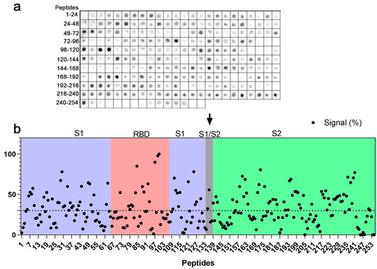
Figure 1.
Map of linear IgG B-cell epitopes in SARS-CoV-2 spike protein. An SPOT synthesis analysis using a library of 15 mer peptides with an overlap of 5 residues to represent the S protein-coding sequences synthesized directly onto a cellulose membrane. (a) Chemiluminescent image of peptides recognized by antibodies after probing in a serum pool from hospitalized individuals with severe COVID-19 infections. Each spot represents a peptide. (b) Graphical representation of the signals measured at each peptide spot and normalized to the maximum signal. Peptide sequences are displayed with an intensity level above 30% (dashed line), defined as a positive reaction. Peptides were synthesized following the primary sequence of spike protein and highlighted in receptor binding domain (RBD), as well as S1, S1/S2 (furin cleavage site; arrow), and S2 domains.

Table 1.
IgG epitopes mapped in SARS-CoV-2 spike protein using COVID-19 patient sera.
Three epitopes were found in the first Heptad repeat (HR1; 920–970) and two in Heptad repeat 2 (HR2; 1163–1202). No epitopes were localized to the transmembrane domain (TMD), and there was only one in the C-terminal after the TMD. Among the epitopes with signals greater than 80%, two (2) were located in the RBD (GKIADYNYKL and PLQSYGFQPTGVGY) and one (1) in the S2 domain (LPPLL). Following their localization, each epitope sequence was searched using the Basic Local Alignment Search Tool for proteins (BLASTp) and multiple sequence alignments restricted to the coronavirus family. Using four consecutive and identical amino acids as the minimum binding site for an epitope, most epitopes showed no commonality to endemic coronaviruses, suggesting that some epitopes are specific to SARS-CoV-2 (Table S1).
2.2. Cross-Reactivity with Anti-DENV Antibodies
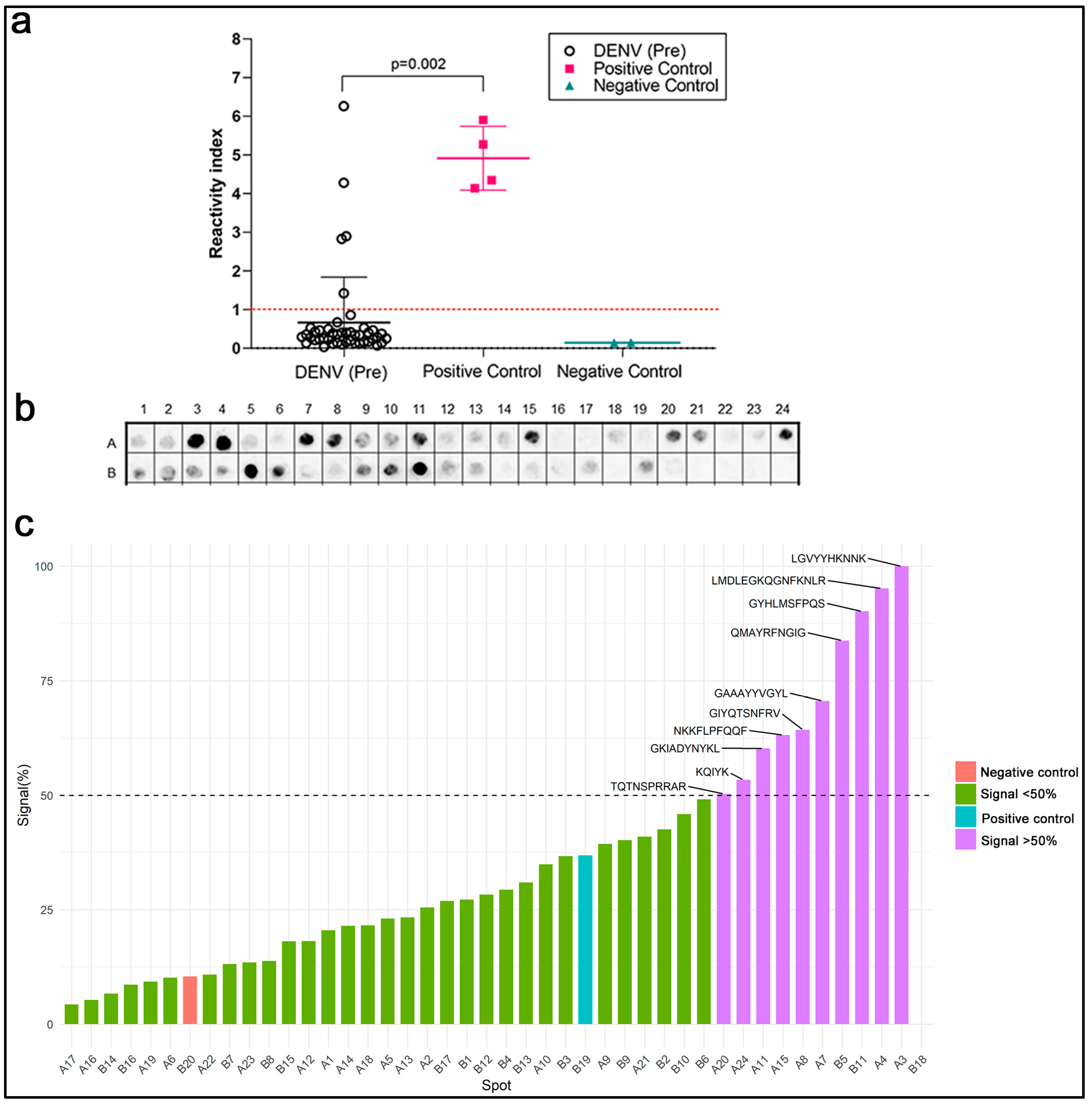
Previous evidence has suggested that the SARS-CoV-2 S1-RBD could be recognized by antibodies in patients infected with DENV. When we expanded the BLASTp parameters to include the Dengue virus (Taxid: 12,637), twenty-one sequences presented a cross-reactivity potential based on the above criteria (Table S1). Next, the reactivity of serum samples (n = 45) from patients with antibodies from DENV infections before the COVID-19 pandemic were evaluated in a commercial ELISA utilizing the spike protein and nucleoprotein of SARS-CoV-2 as targets for capturing antibodies. Figure 2a presents the reactivity index of the individual samples and shows that five samples from DENV patients were positive (11%). Pre-pandemic serum samples were also tested in a commercial ELISA for DENV (1–4). DENV antibodies (IgG) were highly seropositive among healthy blood donors (20; 71.4%), and, as expected, the highest seropositive sera were found in the small library of DENV-positive pre-pandemic sera (37; 92.5%) (Figure S3).

Figure 2.
Cross-reactivity between DENV and SARS-CoV2- infections. (a) Commercial SARS-CoV-2 ELISAs using sera from pre-pandemic DENV-positive patients and kit negative and positive controls. (b) Chemiluminescent image of an SPOT synthesis analysis of the 41 epitopes identified in the SARS-CoV-2 spike protein using a serum pool from pre-pandemic DENV-infected patients (n = 8), revealing reactive IgG antibodies. Each spot represents one of the 41 identified epitopes displayed in rows (A and B) and columns (1–24). (c) Graphic representation of quantifying signal intensities normalized to the maximum signal. An intensity level above 50% was defined as reactive. A Kruskal–Walli’s test was applied to identify statistical differences, followed by Dunn’s multiple comparison tests. A p < 0.05 was considered to indicate a significant difference.
A SPOT synthesis analysis was used to determine if the reactivity of the pre-pandemic DENV serum could bind to the SARS-CoV-2-specific epitopes in its spike protein (Figure 2c). From the forty-one previously identified epitopes, ten presented a normalized signal intensity >50% to pre-pandemic DENV serum, defined to be cross-reactive based on a statistical analysis. Three cross-reactive regions were located in S1-NTD, two in the RBD region, one in S1/SD1, one in the furin cleavage site, and the remaining three in the S2 domain.
2.3. Bioinformatic Analysis
An open question was whether the sequence similarity between the SARS-CoV-2 spike protein and DENV proteins alone could explain the observed cross-reactivity. Additional BLASTp searches were performed with the ten epitope sequences displaying cross-reactivity to identify DENV-1–4 proteins or polyproteins (Table 2). Searches were conducted for short input sequences of at least four consecutive amino acids to identify mimetic peptides in the DENV. Most spike epitopes (n = eight) had a sequence of at least four amino acids identical to a Dengue protein. The other two epitopes presented sequence gaps with a lower level of sequence identity. The identified sequences have a high level of conservation among the VoCs (Figure S1) and a low level of identification against the endemic coronaviruses (Figure S2).
Table 2.
BLASTp analysis of cross-reactive IgG epitopes for sequence identification to DENV proteins.
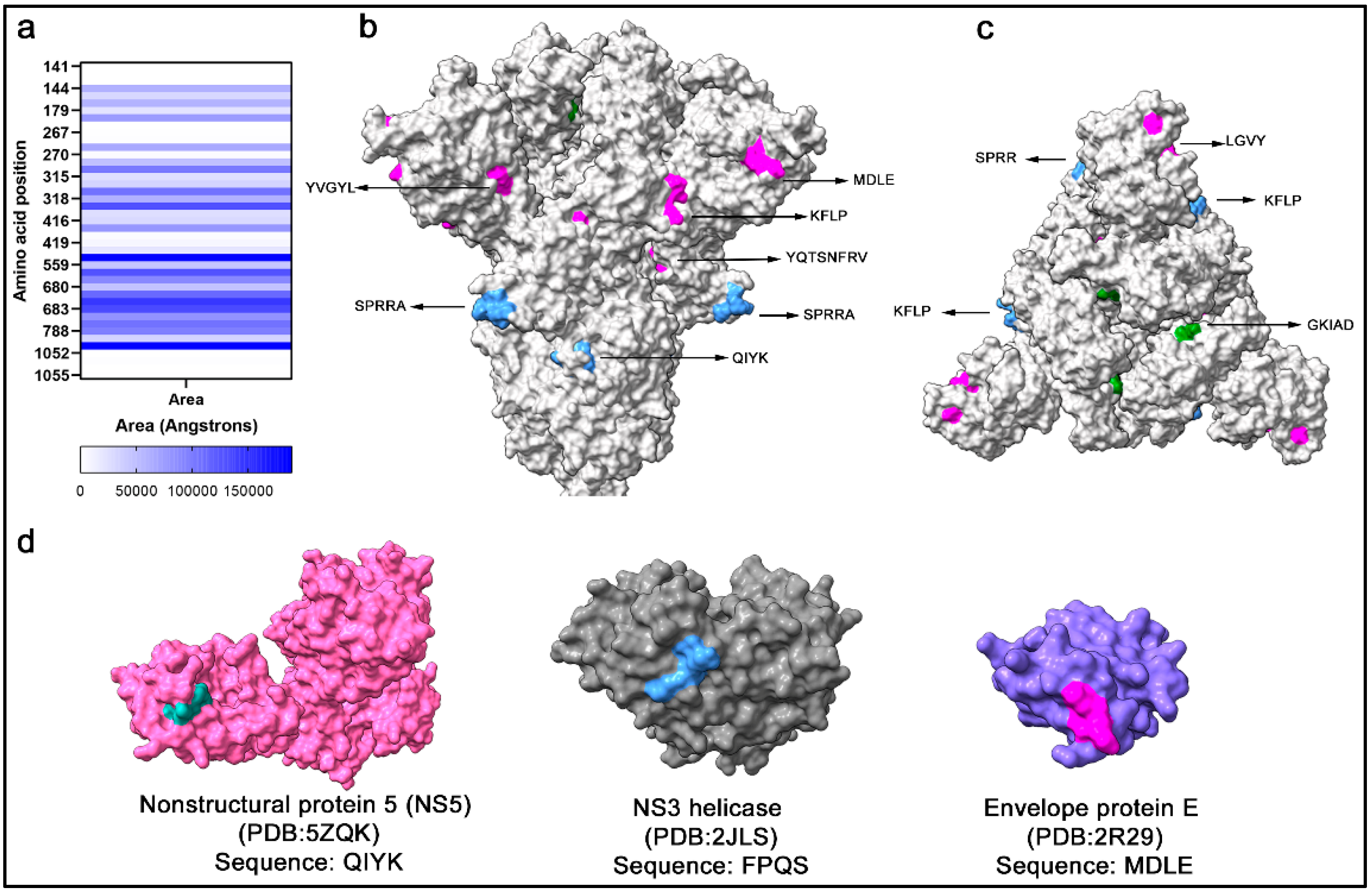
Sequence identity per se does not signify cross-reactivity, as antibody interaction also depends on structure conformation and accessibility to antigens. Therefore, potential cross-reactive sequences were located within the spike protein monomer and trimer, and the solvent accessibility area (SASA) was calculated (Figure 3a), which is correlated to the spatial arrangement and exposure of residues to the solvent. In a side view of the spike trimer (Figure 3b), several surface-exposed residues in cross-reactive epitopes were localized, which supported their potential to interact with antibodies (Figure 3b). Likewise, highly surface-exposed areas were present at the top of the trimeric spike protein (Figure 3c). The FPQS residues are in a buried surface area of the spike protein with low solvent accessibility and are unlikely to represent a cross-reactive site. Within the Ns5 and envelope protein E of DENV2 and the NS3 helicase of DENV4, searches of the epitope sequences in the protein databank revealed similar epitope sequences with surface exposure (Figure 3d).
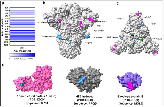
Figure 3.
Solvent accessible area of cross-reactive SARS-CoV-2 spike peptides. (a) Data represent the exposed area for the calculation of SASA for each residue. (b) Cross-reactive residues are marked in spike structures in the S1 domain (magenta), RBD (green), and S2 domain (blue). (c) The top view of the spike trimer shows clusters of cross-reactive residue sites in the RBD, S1 domain, and S2 domain. (d) Structural model of DENV-2 NS5 (pink), NS3 helicase (gray), and envelope protein E (blue) with the identical residues in SARS-CoV-2 spike QIYK (cyan), FPQS (blue), and MDLE (magenta), respectively.
2.4. Pre-Pandemic DENV Sera Display Antibody-Dependent Enhancement In Vitro
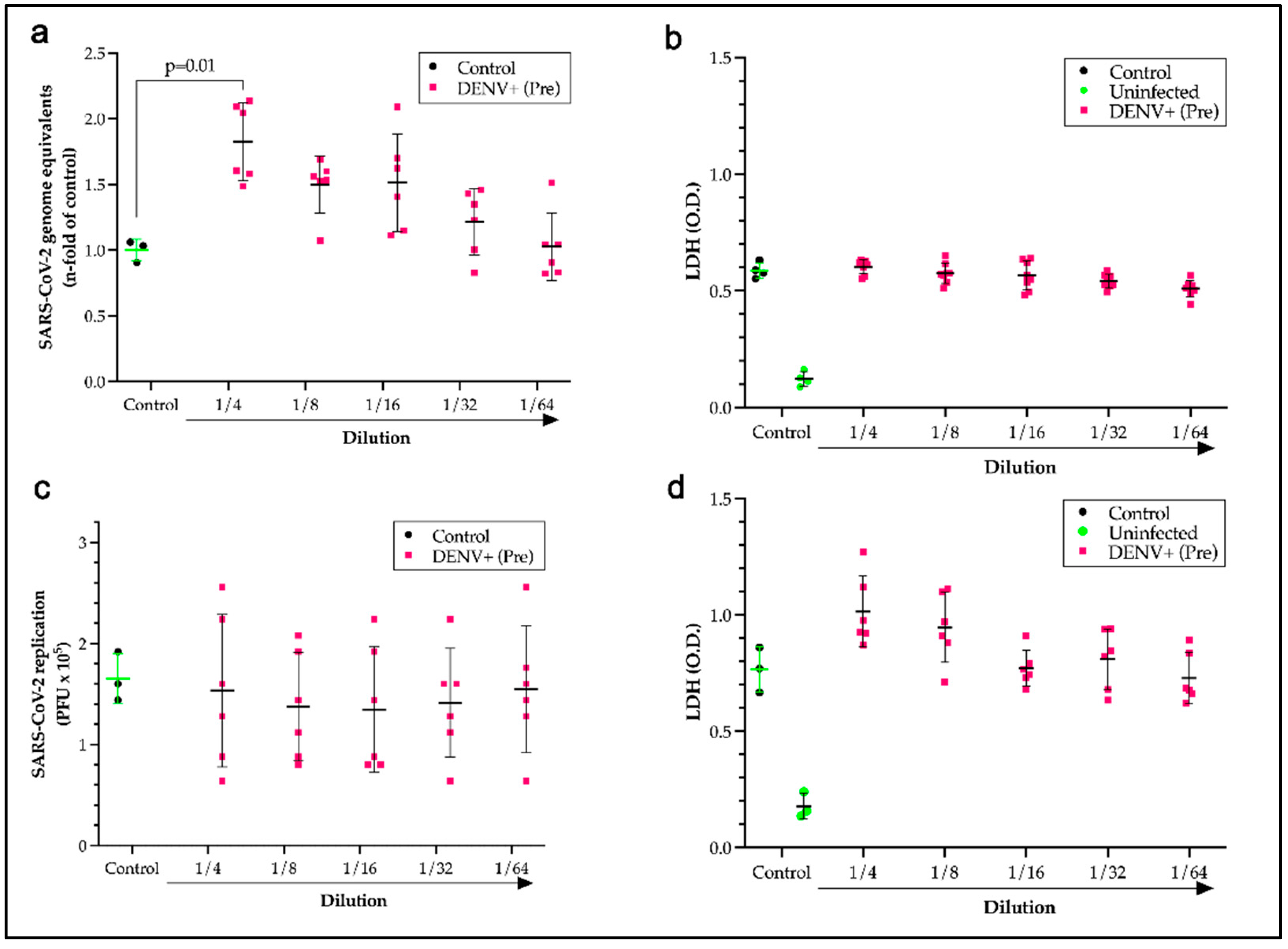
After identifying cross-reactive sites, the question remained as to whether the presence of antibodies formed during a DENV infection could potentiate antibody-dependent enhancement of SARS-CoV-2 infections. To test this hypothesis, monocytes that express Fc receptors (FcRs) were used as a model system for infection with SARS-CoV-2 in the presence of a pool of pre-pandemic DENV-positive sera over a two-fold serial dilution series. Incubation of the virus with pre-pandemic DENV-positive sera with a dilution ratio of one to four induced a significant increase (p = 0.014) in the viral load of monocytes that was 1.7 times the control loads (Figure 4a). Measurements of lactate dehydrogenase for cell death showed no significant loss of monocytes from the increase in viral entry, which was expected since SARS-CoV-2 replication is aborted in monocytes (Figure 4b). The neutralization capacity of a DENV patient serum pool was also evaluated by a plaque assay using Calu-3 cells. No neutralization (Figure 4c) or significant cell death (Figure 4d) was measured for the pre-pandemic Dengue-positive pool compared to the COVID-19 control and healthy human sera.
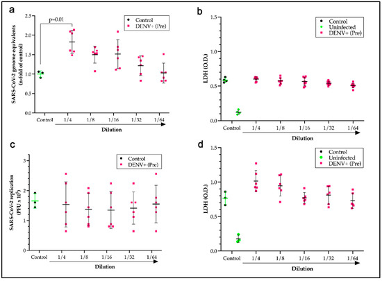
Figure 4.
Antibody-dependent enhancement of SARS-CoV-2 infections of monocytes without cell death or neutralization by pre-pandemic DENV-positive sera. A monocyte in vitro infection assay with SARS-CoV-2 virus preincubated with vehicle (control), showing a 2-fold serial dilution series of a pool of pre-pandemic DENV patient sera or normal sera (uninfected) measuring viral load by qPCR (a) and lactate dehydrogenase (LDH) for cell death (b). An in vitro infection assay with Calu-3 cells with SARS-CoV-2 virus preincubated with vehicle (control), showing a 2-fold serial dilution series of a pool of pre-pandemic DENV patient sera or normal sera (uninfected) measuring: (c) SARS-CoV-2 replication levels as plaque-forming units (PFUs) or LDH levels as a cell death indicator (d). Data show the individual data from six experiments and the mean and standard deviation. Kruskal–Walli’s test was applied to identify statistical differences, followed by Dunn’s multiple comparison tests. Significant differences were considered with p < 0.05.
2.5. Antibody Binding to Peptides from Spike Protein
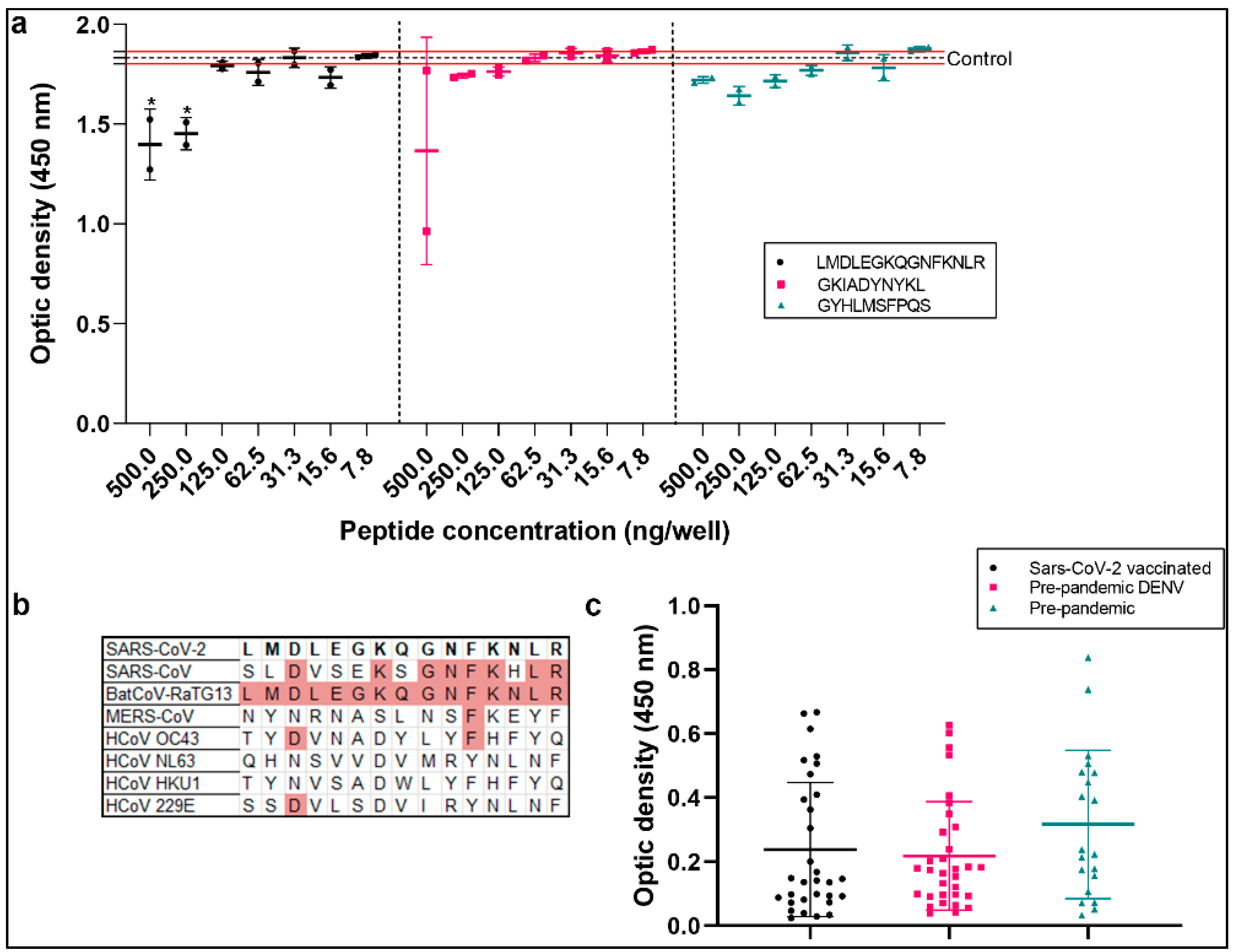
To evaluate the cross-reactivity sequences in spike proteins, we synthesized three peptides with a strong signal in the SPOT synthesis and solvent accessibility analyses (LMDLEGKQG NFKNLR, LGVYYHKNNK, and GKIADYNYKL). The peptides were used in a competitive ELISA, and different concentrations were pre-incubated with a pool of DENV-positive samples (Figure 5). The samples were used in the NovaLisa Dengue IgG kit. The only peptide that significantly reduced antibody binding was LMDLEGKQGNFKNLR, which contained the MDLE sequence from the DENV envelope protein. Compared to the untreated control, the peptide concentrations of 500 and 250 ng/well were reduced by approximately 23% and 20%, respectively. Cross-reactivity with other coronaviruses is plausible. However, multiple sequence alignments of spike proteins with other coronaviruses and endemic viruses were performed. Similarities were found for Bat-Cov and Sars-CoV, but not for other coronaviruses. A peptide ELISA using the LMDLEGKQGNFKN LR sequence showed no significant difference between the pre-pandemic, DENV-positive pre-pandemic, and COVID-19-vaccinated samples (Table S2). The results highlight that this sequence elicits non-specific antibody reactions.
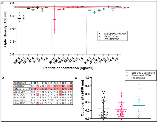
Figure 5.
Analysis of spike cross-reactive peptides. (a) Competitive ELISA using DENV IgG test performed using a pool of DENV-positive samples pre-incubated with peptides from spike protein. The control group was tested in triplicate at 1:100 without peptides, and the data show the mean (dotted line) and standard deviation (red lines). (b) Multiple sequence alignment of spike protein (P0DTC2) and other coronaviruses (SARS-CoV-1 (P59594), MERS-CoV (K9N5Q8), HCoV-OC43 (P36334), HCoV-NL63 (Q6Q1S2), HCoV-HKU1 (Q14EB0), HCoV-229E (P15423), and BatCoV-RaTG13 (A0A6B9 WHD3)); similar amino acids are highlighted in red. (c) In-house peptide ELISA performed with peptide LMDLEGKQGNFKFKNLR and a cohort of pre-pandemic sera (n = 17), individuals vaccinated with the first dose of Oxford–AstraZeneca and heterologous doses (n = 31), and DENV-positive pre-pandemic group (n = 32). A one-way ANOVA was applied to identify statistical differences, followed by Dunnet’s multiple comparison tests. p < 0.05 was considered a significant difference (* p < 0.05).
3. Discussion
The World Health Organization recently declared the end of the COVID-19 public health emergency. Yet, the emergence of variants that display increased transmissibility, are refractory to previously neutralizing antibodies, or both shows the importance of continued studies on the humoral response to SARS-CoV-2 and other pathogens to develop next-generation vaccines and therapies. Here, we began by identifying the immunodominant epitopes in the spike protein of SARS-CoV-2 that are recognized by antibodies in the serum of hospitalized COVID-19 patients. Employing an SPOT synthesis peptide microarray, a total of 41 epitopes were identified. These epitopes were distributed across different domains of the spike protein, including S1, NTD, RBD, SD1, SD2, furin cleavage site, S2 fragments, and heptad repeats 1 and 2. Some of these epitopes show partial or total sequence similarities to some epitopes reported in previous studies on SARS-CoV-2 [42,43,44,45,46,47], indicating consistency between the techniques and serum panels used, which is important for defining immunodominant epitopes across multiple populations stimulated by natural infections and vaccinations. Screening immunodominant epitopes is important to understanding the immune response, supporting vaccine development by identifying key antigenic targets that elicit strong and specific immune responses, making accurate diagnoses, and understanding the immune escape. The epitopes of other viral proteins are equally crucial; while mutations in spike proteins are linked to immune evasion and infectivity in Omicron variants, their pathogenicity is also correlated with NSP6 [48].
Epitope mapping is also a strategic method for developing broadly neutralizing antibodies or nanobodies for prophylactic, diagnostic, and therapeutic use [49]. Neutralizing epitopes are predominantly located within the NTD and RBD, and antibodies binding to the RBD can account for over 90% of neutralizing activity in convalescent sera [50]. Neutralizing antibodies (nAbs) can also target the S2 stem helix (SH) and S2 fusion peptide (FP) regions [51,52,53]. The immune evasion of variants of concern is driven by mutations in the spike protein that threaten natural and vaccine-induced immunity. However, conserved pan-variant epitopes that confer broad neutralization have great potential for therapy and vaccine design [26,54,55,56,57,58].
The binding mode of nAbs can be divided into four main classes depending on the location of their epitopes in the spike protein [50]. The RBD region is targeted by class 1 and 2 antibodies which include the RBM and can compete with ACE2 binding. The major IgG epitopes CV19/SG/13huG and CV19/SG/14huG are located within this region. Class 3 RBD antibodies bind to regions flanking the ACE2-binding region, and the epitopes CV19/09huG and CV19/SG/12huG could be involved in the binding of nAbs. Importantly, this region contains highly conserved residues in the SARS-CoV and SARS-CoV-2 RBDs which could confer broad cross-reactivity as observed with antibody S30924, which also neutralized the SARS-CoV-2 VoC Omicron B.1.1.529 [59]. The epitope recognized by class 4 antibodies is highly conserved in the RBD but does not directly block ACE2–RBD binding [50]. This cryptic region spans from residues 369 to 385, but our study found low reactivity for any epitope in this region.
Changes to epitope sequences in the NTD and RBD will likely contribute to immune escape by variants. In contrast, the neutralizing epitopes in the S2 subunit are more conserved across variants [60]. However, low reactivity was observed for the S2 SH region, which spans 14 residues (1146–1159) and is conserved across beta-CoVs [61]. The S2 FPs are also highly conserved among all coronaviruses, suggesting an antibody targeting this region could display broad-spectrum activity. The epitope CV19/SG/26huG (LPDPSKP SKRSFIED) was identified in the S2 FP1, which overlaps with the ‘RSFIEDLLF’ motif bound by several human monoclonal antibodies isolated from convalescent patients that bind the [62,63]. The R815 conserved residue is the S2′ site of TMPRSS2, and by targeting this region, antibodies would interfere with cleavage and inhibit the membrane fusion of S protein [63].
SPOT synthesis analyses are also useful for identifying cross-reactive epitopes, which are a significant concern in developing serological tests, and SARS-CoV-2 is no exception. Although SARS-CoV-2 proteins exhibit low sequence similarity to other human coronaviruses (HCoVs), previous evidence has suggested that the response to SARS-CoV-2 may be influenced by earlier exposures to other coronaviruses and other endemic viruses, including respiratory viruses [64]. Fifteen highly antigenic epitopes against pathogens, self-proteins, and common human viruses in a cohort of pre-pandemic individuals naïve to SARS-CoV-2 showed cross-reactivity with the spike protein of SARS-CoV-2 [65]. This points to a potential limitation of linear B-cell epitope mapping with the presence of cross-reactive antibodies in serum samples obtained from individuals with unknown clinical histories. Only eight epitopes identified in this study were non-specific for SARS-CoV-2 [65].
By confirming the results with bioinformatics, it is possible to identify cross-reactive epitope sequences based on a minimum antibody binding site of four consecutive amino acids. Here, ten sequences exhibited potential cross-reactivity with the Dengue virus. Dengue fever is a vector-borne viral disease caused by the flavivirus Dengue virus which has four serotypes, each with three structural and seven non-structural proteins [66,67]. It is endemic to tropical regions worldwide which are home to approximately 2.5 billion people, and there are estimated to be 400 million yearly cases globally. Previous data have shown that pre-pandemic Dengue patient sera contain cross-reactive antibodies for SARS-CoV-2 that can cause false-positive SARS-CoV-2 serology results and could partially explain the low specificity of some serological tests [68]. A recent study highlighted the antigenic similarity between the SARS-CoV-2 S1-RBD regions and DENV proteins (E and NS1), which would allow us to induce cross-reactive antibodies after SARS-CoV-2 S1-RBD immunization in vivo using an experimental animal model.
The cross-reactivity between DENV and SARS-CoV-2 occurs in diagnostic tests, and some studies have shown a minimal risk of serological cross-reactivity between DENV and SARS-CoV-2 IgG antibodies when employing serological assays [69]. However, other works have demonstrated a higher profile of cross-reactivity of SARS-CoV-2 antibodies, but not neutralizing antibodies, among individuals previously exposed to DENV. Interestingly, assays utilizing spike S1 as the antigen detected more positives among primary DENV infections than in-house ELISAs using receptor binding domain (RBD) proteins [70]. It was demonstrated that DENV cross-reactive antibodies might cause false-positive results in Dengue serological tests [71] and potentially hinder Dengue infections [8].
Using commercial ELISA kits for COVID-19 and DENV, our results demonstrated that cross-reactivity exists between antibodies generated in response to SARS-CoV-2 and DENV. These findings highlight the weakness of full-length viral proteins in serological tests containing a mixture of definable specific and non-specific linear B-cell epitopes.
This issue is particularly relevant in SARS-CoV-2 and DENV due to the potential for cross-reactive DENV antibodies to facilitate antibody-dependent enhancement (ADE). ADE has been observed in various viral infections, including DENV, West Nile virus, Ebola virus, measles, respiratory syncytial virus, and human immunodeficiency virus (HIV) [12,15,20]. Also, in SARS-CoV ADE, vaccine preclinical trials in animal models have been reported [72]. In the context of SARS-CoV-2, ADE has been debated since the pandemic’s beginning, particularly concerning the potential risk associated with vaccination, convalescent serum therapy, and the emergence of new variants.
ADE has been extensively studied in Dengue fever. Individuals with pre-existing immunity against one serotype of DENV are at a higher risk of developing severe Dengue shock syndrome (DSS) or Dengue hemorrhagic fever (DHF) when infected with a different serotype [73]. This fact is attributed to antibodies with low neutralizing activity but that can facilitate viral entry into macrophages via Fc receptors [74]. ADE-driven infection of macrophages can increase viral replication and load [75]. This clinical concern has challenged the development of a safe and effective Dengue vaccine that induces a balanced immune response against all serotypes to avoid ADE [76].
Fortunately, unlike DENV, SARS-CoV-2 does not replicate productively in macrophages. Macrophages can use phagocytosis to counter the virus, but this does not result in productive infection. However, SARS-CoV-2’s entry into immune cells leads to an abortive infection followed by host cell pyroptosis, the release of proinflammatory cytokines, and the activation of inflammasomes [77,78].
Apart from FcR-dependent ADE, there is also evidence of FcR-independent ADE in SARS-CoV-2 infections. Antibodies that recognize specific binding domains on the SARS-CoV-2 spike protein can induce structural changes that facilitate viral entry and infection. Some antibodies can mediate the ADE of diseases in vitro in an FcR-independent manner [79]. Human mAbs against SARS-CoV-2 spike protein were found to enhance the virus infection in vitro by the FcγR-mediated pathway [17]. In addition, ADE was reported to neutralize mAbs using SARS-CoV-2 pseudovirus infections on FcγRIIB-expressing B-cells; the bivalent interaction of antibodies with S-trimer RBDs enhanced the activity of the SARS-CoV-2 pseudovirus on FcγRIIB-expressing B-cells [80]. A study revealed that FcγR- and/or C1q-mediated ADE was detected in 50% of IgG-positive sera.
Interestingly, most of these sera also exhibited neutralizing activity without FcγR and C1q. ADE antibodies were found in 41.4% of acute COVID-19 patients, suggesting the potential for ADE to promote virus replication even in the early phase of infection [18]. The authors proposed that C1q-mediated ADE may occur in the respiratory tissues of COVID-19 patients, and possibly ADE plays a role in the exacerbation of the disease [18].
Different factors contribute to antibody efficacy, depending on titers, affinity viral epitopes, and stoichiometry. The production of neutralizing antibodies is considered the primary goal of vaccination. Still, cross-reactive or non-neutralizing antibodies may also be produced, which can impact the severity of the infection. The presence of cross-reactive antibodies or non-neutralizing antibodies may increase disease severity through ADE [81]. Although ADE has not been observed on a widespread scale in COVID-19 cases, the risks associated with ADE include the potential for more severe disease outcomes, increased viral replication, and exacerbated immune responses.
In vitro ADE does not always correlate with enhanced infection in vivo because other antibody functions could play a role in viral clearance, such as antibody-dependent cell-mediated cytotoxicity (ADCC) and complement-dependent cytotoxicity (CDC). Previous studies with SARS-CoV-2 antibodies have shown an in vitro enhancement of infections but in vivo protection in animal models [72]. Although a small effect of ADE was evidenced on pre-pandemic DENV-positive serum in vitro, there is no proof that this event could have happened in vivo, enhancing the infection.
Evidence of a rare effect of ADE has been reported in preclinical animal models infused with antibodies [82]. Additionally, a mouse monoclonal antibody (mAb RS2) against spike protein increased the viral load in the respiratory tracts of animals in post-exposure prevention studies [83]. Interestingly, a competitive ELISA showed that antibodies competing with the mAb RS2 epitope are significantly higher in the plasma of patients with severe COVID-19 infections than those in patients with mild infections or vaccinated individuals [83]. This finding underscores the potential urgency in understanding the role of antibodies with a similar binding of mAb RS2 in developing more severe COVID-19 infections. Recently, a study analyzed ADE in patients exposed to Middle East respiratory syndrome coronavirus (MERS-CoV); 56% demonstrated ADE against an SARS-CoV-2 pseudo-virus. However, subsequent exposure to SARS-CoV-2 vaccination diminished this ADE activity [84]. However, despite the concern raised in vaccine development, it is important to note that ADE has not been significantly demonstrated in COVID-19 vaccines [85]. This finding provides reassurance and alleviates any concerns about ADE in the context of COVID-19 vaccination.
Highly fucosylated antibodies of vaccines do not cause ADE, but fucosylated antibodies produced in acute primary infections or convalescent sera can induce it [77]. It is essential for vaccine development to balance ADE and neutralizing antibodies. Candidates for vaccines must elicit an immune response that produces neutralizing antibodies capable of successfully preventing viral entry and the specific recognition of pathogen antigens. Epitope selection and studies of cross-reactive antibodies are essential for the rational design of vaccines, antibody therapy, and serological diagnostic tests. ELISA tests have demonstrated cross-reactivity sequences in spike proteins, which could elicit the non-specific binding of antibodies that are not restricted to DENV infections. These findings may contribute to the development of recombinant proteins that lack these cross-reactive sites to increase the specificity of diagnostic tests and epitope-based vaccines.
Similarly, other studies have highlighted that pre-existing cross-reactive IgA [45] and IgG serum antibodies against spike proteins were detectable in pre-pandemic cohorts [86]. Our results highlight that IgG spike cross-reactive sequences could be recognized non-specifically by antibodies drawn from pre-pandemic sera. This result opens new avenues for studying the influence of cross-reactive sites on immunodiagnostics, disease, and vaccine development.
4. Materials and Methods
4.1. Patient Samples
Serum panels were collected by venipuncture using vacuum tubes containing gel separators. After being collected, the tubes were gently inverted to mix the blood with the gel. Subsequently, the samples were centrifuged at 3.000 rpm for 10 min, and serum was separated using a pipette. Serum samples (30) were obtained from individuals who received a single dose of the Oxford/AstraZeneca vaccine, and 7 serum samples were collected from individuals who received four heterologous doses, all primed with Oxford/AstraZeneca and booster of Pfizer Spike mRNA vaccine or Janssen (Ad26.COV2-S). Also, Dengue (1–4) pre-pandemic serum (n = 45) samples were utilized. Additional negative controls (n = 28) included sera collected before the pandemic from healthy individuals from blood bank donors (HEMORIO, Rio de Janeiro, Brazil). Patient privacy was maintained by excluding identifying information. Serum samples from individuals with Dengue were collected before the onset of the COVID-19 pandemic and generously provided by the Laboratory of Flavivirus of the Oswaldo Cruz Institute (FIOCRUZ, Rio de Janeiro, Brazil).
4.2. B-Linear Epitope Mapping
The complete sequences of the spike protein of SARS-CoV-2 were retrieved from the UniProt database (http://www.uniprot.org/: accessed on 27 January 2020). Microarrays of peptides and a pool of human COVID-19 patient sera (n = 12) were used to map linear B-cell IgG epitopes using Auto-Spot Robot ASP-222 synthesizer (Intavis Bioanalytical Instruments AG, Köln, Germany) according to a previous SPOT synthesis protocol [87].
4.3. Peptide Synthesis
SARS-CoV-2 10 mer and 15 mer peptides (LMDLEGKQGNFKNLR, LGVYYHKNN K, and GKIADYNYKL) were chosen for synthesis using the F-moc strategy in a synthesizer machine (MultiPep-1 CEM Corp, Charlotte, NC, USA), using methods previously described [86].
4.4. Enzyme-Linked Immunosorbent Assay (ELISA)
Using the manufacturer’s instructions, ELISA assays were performed using a confirmatory commercial COVID-19 IgG kit (Dia. Pro, Giovanni Milan, Italy). Each microplate strip has its wells coated with nucleocapsid and spike-specific antigens, making it possible to map the serological response to the different IgG types produced. To detect DENV (1–4) IgG antibodies, a SERION ELISA classic/antigen Dengue Virus IgG commercial kit (#ESR114G; Virion/Serion GmbH, Würzburg, Germany) was used. Tests were performed following the manufacturer’s instructions. Results are represented as reactivity index calculated using the formula (IR = sample absorbance/cut-off).
Competitive ELISA was performed using NovaLisa ELISA IgG kit (DENG0120) to detect DENV antibodies. A pooled serum sample from eight DENV-positive patients was prepared and diluted to a 1:100 concentration utilizing the kit’s diluent. The serum was preincubated for 1 h at room temperature with each of the three SARS-CoV-2 spike peptides (LMDLEGKQGNFKNLR, LGVYYHKNNK, and GKIA DYNYKL) at six different concentrations: 500, 250, 125, 62.5, 31.2, 15.6, and 7.8 ng/well. Serum without any peptide added served as the control. Following preincubation, the ELISA was performed according to the manufacturer’s instructions. The values were recorded and the control values (serum without peptides) were compared to values from serum preincubated with different peptides and concentrations. This comparison allowed for the evaluation of the binding efficiency and inhibition potential of the peptides.
In-house peptide ELISA was performed as described previously [88]. Briefly, 500 ng of peptides in a coating buffer (50 mM carbonate-bicarbonate buffer, pH 9.6) was added to Immunolon 4HB plates (Immunochemistry Technologies, Bloomington, MN, USA) overnight at 4 °C. After washing with PBS-T (phosphate-buffered saline plus 0.05% Tween® 20), plates were incubated for 1 h at 37 °C with 1% bovine serum albumin (BSA) (200 µL) in PBS-T to block free binding sites. Subsequently, following the dilution, sera were diluted (1:25) in coating buffer and PBS-T with 1% BSA (1:25). Then, 100 µL of the diluted sera was applied to plates, which were incubated for 1 h at 37 °C. Following washing with PBS-T, plates were incubated with 100 µL of anti-human IgG HRP (1:10.000) for 1 h. Binding antibodies were revealed by adding 3,3′,5,5′-tetramethyl benzidine (1-Step™ Ultra TMB-ELISA, Science Biotech Ltd. Lages, Brazil), which was added for 15 min, and the reaction was stopped after a few minutes with 0.5 M sulfuric acid. Optic density was evaluated at 450 nm within 2 h of adding the benzindine.
4.5. In Silico Analysis
BLASTp (http://blast.ncbi.nlm.nih.gov) was used to find cross-reactive sequences in SARS-CoV-2 epitopes that match the Dengue virus (Taxid: 12,637). The website was accessed on 16 May 2023. The BLASTp algorithm parameters were set as follows: expect threshold of 30,000, word size of 2, and PAM30 matrix. Moreoever, gap cost was set to existence = nine and extension = 1, the compositional parameter was set to no adjustment, and the low-complexity filter was disabled and adjusted for short input sequences. The results from BLASTp analysis were screened and filtered for at least four contiguous and identical amino acid residues with the DENV (1–4) peptides with no gap and no mismatched residues. Additionally, peptides were screened using Protein Information Resource (PIR; https://research.bioinformatics.udel.edu/peptidematch/index.jsp, accessed on 10 September 2023). Spike trimer protein in a closed state was retrieved from I-Tasser (https://zhanggroup.org/I-TASSER/, accessed on 5 November 2022). Annotation of epitopes and cross-reactive sites was performed using Chimera X [89]. The accessible surface area was performed by using the accessibility calculation for protein (ver. 1.2) online server that calculates the solvent-accessible surface areas of spike protein (P0DTC2) amino acids (http://cib.cf.ocha.ac.jp/bitool/ASA/, accessed on 10 October 2023).
4.6. Cells, Viruses, and Reagents
African green monkey kidney cells (Vero, E6 cell; ThermoFisher, Waltham, MA, USA) and human lung epithelial cell lines (Calu-3) were expanded in high-glucose DMEM with 10% fetal bovine serum (FBS; Sigma-Aldrich, St Louis, MO, USA), with 100 U/mL penicillin and 100 μg/mL streptomycin (Pen/Strep; ThermoFisher, Waltham, MA, USA) at 37 °C in a humidified atmosphere with 5% CO2. Peripheral blood mononuclear cells (PBMCs) were isolated by density gradient centrifugation (Ficoll-Paque, GE HealthCare, Chicago, IL, USA) from buffy coat blood preparations from healthy donors. PBMCs (2 × 10⁶ cells) were plated into 48-well plates (NalgeNunc Int Corrp, Rochester, NY, USA) in RPMI-1640 with 5% inactivated male human AB serum (Sigma-Aldrich, St Louis, MO, USA) for 3 h. Non-adherent cells were removed, and monocytes were maintained in (low-glucose) DMEM with 5% human serum, 100 U/mL penicillin, and 100 μg/mL streptomycin. The purity of monocytes was above 90%, as determined by flow cytometry (FACScan; Becton Dickinson, Juiz de Fora, Brazil) using anti-CD3 (BD Biosciences, Mississauga, ON, Canada) and anti-CD14 (BD Biosciences, Mississauga, ON, Canada) antibodies. SARS-CoV-2 (GenBank accession no. MT710714) was expanded in Vero E6 cells. Viral isolation was performed after a single passage in cell culture in 150 cm2 flasks with high-glucose DMEM plus 2% FBS. Observations for cytopathic effects were performed daily and peaked 4 to 5 days after infection. All procedures related to virus culture were handled in biosafety level 3 (BSL3) multiuser facilities, according to WHO guidelines. Virus titers were determined as plaque-forming units (PFU/mL), and virus stocks were kept in −80 °C ultra-low-temperature freezers.
4.7. Infections and Virus Titration
Infections were initiated with SARS-CoV-2 at MOI of 0.1 in low (monocytes)- or high (Calu-3)-glucose DMEM without serum. Viral input was incubated with diluted serum samples for 15 min before exposure to cell culture. After 1 h, the unbound virus was removed, and cells were washed and incubated with a complete medium. For virus titration, monolayers of Vero E6 cells (2 × 10⁴ cells/well) in 96-well plates were infected with serial dilutions of supernatants containing SARS-CoV-2 for 1 h at 37 °C. Semi-solid high-glucose DMEM medium containing 2% FBS and 2.4% carboxymethylcellulose was added, and cultures were incubated for three days at 37 °C. Then, the cells were fixed with 10% formalin for 2 h at room temperature. The cell monolayer was stained with 0.04% solution of crystal violet in 20% ethanol for 1 h. Plaque numbers were scored with at least three replicates per dilution by independent readers blinded to the experimental group, and the virus titers were determined by plaque-forming units (PFU) per milliliter. Experimental procedures involving human cells from healthy donors were performed with samples obtained after their written informed consent was given.
4.8. Molecular Detection of Viral RNA Levels
According to the manufacturer’s instructions, total RNA was extracted from cells using QIAamp Viral RNA kit (Qiagen Sciences Inc., Germantown, MD, USA). Quantitative RT-PCR was performed using QuantiTect Probe RT-PCR Kit (Qiagen Sciences Inc., Germantown, MD, USA) in a StepOnePlus™ Real-Time PCR System (ThermoFisher, Waltham, MA, USA). Amplifications were performed in 15 µL reaction mixtures containing 2X reaction mix buffer, 50 µM of each primer, 10 µM of the probe, and 5 µL of RNA template. Primers, probes, and cycling conditions followed the protocol the Centers for Disease Control and Prevention (CDC, Atlanta, GA, USA) recommended to detect SARS-CoV-2. A standard curve method was employed for virus quantification. The housekeeping gene RNAse P was amplified to reference the cell quantities assayed. The Ct values for this target were compared to calibrations obtained from 102 to 107 cells.
4.9. LDH Measurement
Cell death was determined by proxy using the liberated lactate dehydrogenase (LDH) activity level in supernatants using CytoTox® Kit (Promega, Madison, WI, USA). Supernatants were centrifuged at 5000 rpm for 1 min before an assay to remove cellular debris.
5. Conclusions
We utilized an SPOT synthesis analysis to map IgG immunodominant epitopes in the spike protein of SARS-CoV-2. The distribution of these epitopes across different domains of the spike protein highlights the complexity of the humoral immune response and the potential for diverse antibody interactions. Further, our findings suggest that individuals with a previous DENV infection may harbor cross-reactive antibodies that can lead to ADE in vitro. The observations that pre-formed antibodies in pre-pandemic serum against peptides from spike proteins have many implications in immunodiagnostics. Also, the sequence similarities to previously reported epitopes in SARS-CoV-2 highlight the importance of understanding the immunodominance of epitopes in the population. This knowledge can aid in the development of broadly neutralizing antibodies or nanobodies for prophylactic, diagnostic, and therapeutic purposes. The presence of conserved pan-variant epitopes provides opportunities to design effective vaccines and therapeutics that can target multiple SARS-CoV-2 variants. In conclusion, our study sheds light on the cross-reactivity of IgG epitopes and the potential for antibody-mediated enhancement in vitro in SARS-CoV-2 and previous viral infections. This knowledge has important implications for understanding the immune response to SARS-CoV-2 and developing strategies to combat the ongoing COVID-19 pandemic.
6. Patents
Patent applications were filed on 5 June 2020 for the epitope targeting of SARS-CoV-2 and the construction of chimeric proteins (provisional patent applications BR1120210214011 (Brazil), US17638108 (USA), EP4039696 (Europa), CN114258399 (China), and IN202217 004847 (India) to D.W.P-Jr., A.M.D., P.N.-P., and S.G.D.-S., Oswaldo Cruz Foundation).
Supplementary Materials
The following supporting information can be downloaded at: https://www.mdpi.com/article/10.3390/ijms25158180/s1.
Author Contributions
Conceptualization, S.G.D.-S. and C.M.M.; Methodology, G.C.L., L.R.G., J.R.T., J.P.R.S.C. and P.N.-P.; Data analysis and curation, G.C.L., D.C.B.-H., T.M.L.S. and S.G.D.-S.; Software, G.C.L.; Original draft, G.C.L.; Review and editing, D.W.P.J. and S.G.D.-S.; Project administration, C.M.M. and S.G.D.-S.; Funding acquisition, C.M.M., D.C.B.-H. and S.G.D.-S. All authors have read and agreed to the published version of the manuscript.
Funding
This research was funded by the Carlos Chagas Filho Foundation for Research Support of the State of Rio de Janeiro/FAPERJ (#110.198-13), and Brazilian Council for Scientific Research (CNPq, #467.488/2014-2 and 301744/2019-0) to S.G.D-S. and Mercosur Structural Convergence Fund (FOCEM, Mercosur, #03/11 to D.C.B-H. FAPERJ (#210.003/2018) also provided funding to Carlos M. Morel (INCT-IDPN) through the National Institutes of Science and Technology Program (INCT).
Institutional Review Board Statement
The study was approved by the Oswaldo Cruz Institute/FIOCRUZ (CAAE:49971421.8.0000.5248), the University of Estacio de Sá (CAAE: 3309 0820.8. 0000.5284), and UNIGRANRIO (CAAE: 21362220.1.0000.5283) Study Center Ethics Committee and conducted according to good clinical practice and applicable regulatory requirements, including the Declaration of Helsinki.
Informed Consent Statement
Not applicable.
Data Availability Statement
The data presented in this study are available upon request from the corresponding author.
Acknowledgments
We would like to thank the National Institute of Quality Control, FIOCRUZ, RJ, Brazil, for the MALDI-TOF analysis. P.N-P. and G.C.L. are fellows from CAPES/CDTS.
Conflicts of Interest
The authors not included in the patents declare no competing interests. The funding agencies had no role in the study design, data collection, data analysis, publication decision, or manuscript preparation.
References
- Hadj Hassine, I. Covid-19 vaccines and variants of concern: A review. Rev. Med. Virol. 2022, 32, e2313. [Google Scholar] [CrossRef]
- Liu, L.; Iketani, S.; Guo, Y.; Chan, J.F.W.; Wang, M.; Liu, L.; Luo, Y.; Chu, H.; Huang, Y.; Nair, M.S.; et al. Striking antibody evasion manifested by the omicron variant of SARS-CoV-2. Nature 2022, 602, 676–681. [Google Scholar] [CrossRef]
- Hoffmann, M.; Krüger, N.; Schulz, S.; Cossmann, A.; Rocha, C.; Kempf, A.; Nehlmeier, I.; Graichen, L.; Moldenhauer, A.S.; Winkler, M.S.; et al. The Omicron variant is highly resistant against antibody-mediated neutralization: Implications for control of the COVID-19 pandemic. Cell 2022, 185, 447–456.e11. [Google Scholar] [CrossRef]
- Garcia-Beltran, W.F.; Lam, E.C.; St Denis, K.; Nitido, A.D.; Garcia, Z.H.; Hauser, B.M.; Feldman, J.; Pavlovic, M.N.; Gregory, D.J.; Poznansky, M.C.; et al. Multiple SARS-CoV-2 variants escape neutralization by vaccine-induced humoral immunity. Cell 2021, 184, 2372–2383. [Google Scholar] [CrossRef]
- Ladner, J.T.; Henson, S.N.; Boyle, A.S.; Engelbrektson, A.L.; Fink, Z.W.; Rahee, F.; D’ambrozio, J.; Schaecher, K.E.; Stone, M.; Dong, W.; et al. Epitope-resolved profiling of the SARS-CoV-2 antibody response identifies cross-reactivity with endemic human coronaviruses. Cell Rep. Med. 2021, 2, 100189. [Google Scholar] [CrossRef]
- Devaux, C.A.; Fantini, J. Unravelling antigenic cross-reactions toward the world of coronaviruses: Extent of the stability of shared epitopes and SARS-CoV-2 anti-spike cross-neutralizing antibodies. Pathogens 2023, 12, 713. [Google Scholar] [CrossRef]
- Polyiam, K.; Phoolcharoen, W.; Butkhot, N.; Srisaowakarn, C.; Thitithanyanont, A.; Auewarakul, P.; Hoonsuwan, T.; Ruengjitchatchawalya, M.; Mekvichitsaeng, P.; Roshorm, Y.M. Immunodominant linear B cell epitopes in the spike and membrane proteins of SARS-CoV-2 identified by immunoinformatics prediction and immunoassay. Sci. Rep. 2021, 11, 20383. [Google Scholar] [CrossRef]
- Cheng, Y.L.; Chao, C.H.; Lai, Y.C.; Hsieh, K.H.; Wang, J.R.; Wan, S.W.; Huang, H.J.; Chuang, Y.C.; Chuang, W.J.; Yeh, T.M. Antibodies against the SARS-CoV-2 S1-RBD cross-react with dengue virus and hinder dengue pathogenesis. Front. Immunol. 2022, 13, 941923. [Google Scholar] [CrossRef]
- Murray, S.M.; Ansari, A.M.; Frater, J.; Klenerman, P.; Dunachie, S.; Barnes, E.; Ogbe, A. The impact of pre-existing cross-reactive immunity on SARS-CoV-2 infection and vaccine responses. Nat. Rev. Immunol. 2022, 23, 304–316. [Google Scholar] [CrossRef]
- Takada, A.; Kawaoka, Y. Antibody-dependent enhancement of viral infection: Molecular mechanisms and in vivo implications. Rev. Med. Virol. 2003, 13, 387–398. [Google Scholar] [CrossRef]
- Tirado, S.M.C.; Yoon, K.J. Antibody-dependent enhancement of virus infection and disease. Viral Immunol. 2003, 16, 69–86. [Google Scholar] [CrossRef]
- Thomas, S.; Smatti, M.K.; Ouhtit, A.; Cyprian, F.S.; Almaslamani, M.A.; Thani, A.A.; Yassine, H.M.; Thomas, S. Antibody-Dependent Enhancement (ADE) and the role of complement system in disease pathogenesis. Mol. Immunol. 2022, 152, 172–182. [Google Scholar] [CrossRef]
- Gorlani, A.; Forthal, D.N. Antibody-dependent enhancement and the risk of HIV infection. Curr. HIV Res. 2013, 11, 421–426. [Google Scholar] [CrossRef]
- Sullivan, N.; Sun, Y.; Binley, J.; Lee, J.; Barbas, C.F., 3rd; Parren, P.W.; Burton, D.R.; Sodroski, J. Determinants of human immunodeficiency virus type 1 envelope glycoprotein activation by soluble CD4 and monoclonal antibodies. J. Virol. 1998, 72, 6332–6338. [Google Scholar] [CrossRef]
- Guillon, C.; Schutten, M.; Boers, P.H.; Gruters, R.A.; Osterhaus, A.D. Antibody-mediated enhancement of human immunodeficiency virus type 1 infectivity is determined by the structure of gp120 and depends on modulation of the gp120-CCR5 interaction. J. Virol. 2002, 76, 2827–2834. [Google Scholar] [CrossRef]
- Halstead, S.B. Dengue. Lancet 2007, 370, 1644–1652. [Google Scholar] [CrossRef]
- Zhou, Y.; Liu, Z.; Li, S.; Xu, W.; Zhang, Q.; Silva, I.T.; Li, C.; Wu, Y.; Jiang, Q.; Liu, Z.; et al. Enhancement versus neutralization by SARS-CoV-2 antibodies from a convalescent donor associate with distinct epitopes on the RBD. Cell Rep. 2021, 34, 108699. [Google Scholar] [CrossRef]
- Okuya, K.; Hattori, T.; Saito, T.; Takadate, Y.; Sasaki, M.; Furuyama, W.; Marzi, A.; Ohiro, Y.; Konno, S.; Hattori, T.; et al. Multiple routes of antibody-dependent enhancement of SARS-CoV-2 infection. Microbiol. Spectr. 2022, 10, e0155321. [Google Scholar] [CrossRef]
- Goncalvez, A.P.; Engle, R.E.; St. Claire, M.; Purcell, R.H.; Lai, C.J. Monoclonal antibody-mediated enhancement of dengue virus infection in vitro and in vivo and strategies for prevention. Proc. Natl. Acad. Sci. USA 2007, 104, 9422–9427. [Google Scholar] [CrossRef]
- Pierson, T.C.; Xu, Q.; Nelson, S.; Oliphant, T.; Nybakken, G.E.; Fremont, D.H.; Diamond, M.S. The stoichiometry of antibody-mediated neutralization and enhancement of West Nile virus infection. Cell Host Microbe 2007, 1, 135–145. [Google Scholar] [CrossRef]
- Taylor, A.; Foo, S.S.; Bruzzone, R.; Dinh, L.V.; King, N.J.; Mahalingam, S. Fc receptors in antibody-dependent enhancement of viral infections. Immunol. Rev. 2015, 268, 340–364. [Google Scholar] [CrossRef]
- Mu, S.; Song, S.; Hao, Y.; Luo, F.; Wu, R.; Wang, Y.; Han, X.; Li, T.; Hu, C.; Li, S.; et al. Neutralizing antibodies from the rare convalescent donors elicited antibody-dependent enhancement of SARS-CoV-2 variants infection. Front. Med. 2022, 9, 952697. [Google Scholar] [CrossRef]
- Arvin, A.M.; Fink, K.; Schmid, M.A.; Cathcart, A.; Spreafico, R.; Havenar-Daughton, C.; Lanzavecchia, A.; Corti, D.; Virgin, H.W. A perspective on potential antibody-dependent enhancement of SARS-CoV-2. Nature 2020, 584, 353–363. [Google Scholar] [CrossRef]
- Cloutier, M.; Nandi, M.; Ihsan, A.U.; Chamard, H.A.; Ilangumaran, S.; Ramanathan, S. ADE and hyperinflammation in SARS-CoV2 infection—Comparison with dengue hemorrhagic fever and feline infectious peritonitis. Cytokine 2020, 136, 155256. [Google Scholar] [CrossRef]
- Yang, Y.; Xu, F. Evolving understanding of antibody-dependent enhancement (ADE) of SARS-CoV-2. Front. Immunol. 2022, 13, 1008285. [Google Scholar] [CrossRef]
- Pinto, D.; Park, Y.J.; Beltramello, M.; Walls, A.C.; Tortorici, M.A.; Bianchi, S.; Jaconi, S.; Culap, K.; Zatta, F.; De Marco, A.; et al. Cross-neutralization of SARS-CoV-2 by a human monoclonal SARS-CoV antibody. Nature 2020, 583, 290–295. [Google Scholar] [CrossRef]
- Bates, T.A.; Weinstein, J.B.; Farley, S.; Leier, H.C.; Messer, W.B.; Tafesse, F.G. Cross-reactivity of SARS-CoV structural protein antibodies against SARS-CoV-2. Cell Rep. 2021, 34, 108737. [Google Scholar] [CrossRef]
- Ou, X.; Liu, Y.; Lei, X.; Li, P.; Mi, D.; Ren, L.; Guo, L.; Guo, R.; Chen, T.; Hu, J.; et al. Characterization of spike glycoprotein of SARS-CoV-2 on virus entry and its immune cross-reactivity with SARS-CoV. Nat. Commun. 2020, 11, 1620. [Google Scholar] [CrossRef]
- Jiang, W.; Wang, J.; Jiao, S.; Gu, C.; Xu, W.; Chen, B.; Wang, R.; Chen, H.; Xie, Y.; Wang, A.; et al. Characterization of MW06, a human monoclonal antibody with cross-neutralization activity against both SARS-CoV-2 and SARS-CoV. MAbs 2021, 13, 1953683. [Google Scholar] [CrossRef]
- Watanabe, Y.; Hosokawa, N.; Yoshida, M.; Miura, T.; Kawano, M. Identification of closed linear epitopes in S1-RBD and S2-HR1/2 of SARS-CoV-2 spike protein able to induce neutralizing Abs. Vaccines 2023, 11, 287. [Google Scholar] [CrossRef]
- Hicks, J.; Klumpp-Thomas, C.; Kalish, H.; Shunmugavel, A.; Mehalko, J.; Denson, J.P.; Snead, K.R.; Drew, M.; Corbett, K.S.; Graham, B.S.; et al. Serologic cross-reactivity of SARS-CoV-2 with endemic and seasonal betacoronaviruses. J. Clin. Immunol. 2021, 41, 906–913. [Google Scholar] [CrossRef] [PubMed]
- Holenya, P.; Lange, P.J.; Reimer, U.; Woltersdorf, W.; Panterodt, T.; Glas, M.; Wasner, M.; Eckey, M.; Drosch, M.; Hollidt, J.M.; et al. Peptide microarray-based analysis of antibody responses to SARS-CoV-2 identifies unique epitopes with potential for diagnostic test development. Eur. J. Immunol. 2021, 51, 1839–1849. [Google Scholar] [CrossRef]
- Wang, Z.; Ren, L.; Hao, Y.; Zhu, M.; Jiang, H.; Wang, S.; Li, D.; Shao, Y. Pre-existing anti-HCoV-OC43 immunity influences the durability and cross-reactivity of humoral response to SARS-CoV-2 vaccination. Front. Cell Infect. Microbiol. 2022, 12, 978440. [Google Scholar] [CrossRef]
- Ng, K.W.; Faulknerm, N.; Cornishm, G.H.; Rosam, A.; Harveym, R.; Hussainm, S.; Ulfertsm, R.; Earlm, C.; Wrobelm, A.G.; Benton, D.J.; et al. Preexisting and de novo humoral immunity to SARS-CoV-2 in humans. Science 2020, 370, 1339–1343. [Google Scholar] [CrossRef] [PubMed]
- To, K.K.W.; Cheng, V.C.C.; Cai, J.P.; Chan, K.H.; Chen, L.L.; Wong, L.H.; Choi, C.Y.K.; Fong, C.H.Y.; Ng, A.C.K.; Lu, L.; et al. Seroprevalence of SARS-CoV-2 in Hong Kong and in residents evacuated from Hubei province, China: A multicohort study. Lancet Microbe 2020, 1, e111–e118. [Google Scholar] [CrossRef] [PubMed]
- Khan, A.A.; Alahmari, A.A.; Almuzaini, Y.; Alamri, F.; Alsofayan, Y.M.; Aburas, A.; Al-Muhsen, S.; Van Kerkhove, M.; Yezli, S.; Ciottone, G.R.; et al. Potential cross-reactive immunity to COVID-19 infection in individuals with laboratory-confirmed MERS-CoV infection: A national retrospective cohort study from Saudi Arabia. Front. Immunol. 2021, 12, 727989. [Google Scholar] [CrossRef] [PubMed]
- Klompus, S.; Leviatan, S.; Vogl, T.; Mazor, R.D.; Kalka, I.N.; Stoler-Barak, L.; Nathan, N.; Peres, A.; Moss, L.; Godneva, A.; et al. Cross-reactive antibodies against human coronaviruses and the animal coronavirome suggest diagnostics for future zoonotic spillovers. Sci. Immunol. 2021, 6, eabe9950. [Google Scholar] [CrossRef] [PubMed]
- Wang, J.; Guo, C.; Cai, L.; Liao, C.; Yi, H.; Li, Q.; Hu, H.; Deng, Q.; Lu, Y.; Guo, Z.; et al. Pre-existing cross-reactive antibody responses do not significantly impact inactivated COVID-19 vaccine-induced neutralization. Front. Immunol. 2021, 12, 772511. [Google Scholar] [CrossRef] [PubMed]
- Majdoubi, A.; Michalski, C.; O’Connell, S.E.; Dada, S.; Narpala, S.; Gelinas, J.; Mehta, D.; Cheung, C.; Winkler, D.F.; Basappa, M.; et al. A majority of uninfected adults show preexisting antibody reactivity against SARS-CoV-2. JCI Insight 2021, 6, e146316. [Google Scholar] [CrossRef] [PubMed]
- Rak, A.; Donina, S.; Zabrodskaya, Y.; Rudenko, L.; Isakova-Sivak, I. Cross-reactivity of SARS-CoV-2 nucleocapsid-binding antibodies and its implication for COVID-19 serology tests. Viruses 2022, 14, 2041. [Google Scholar] [CrossRef] [PubMed]
- Souris, M.; Tshilolo, L.; Parzy, D.; Ingoba, L.L.; Ntoumi, F.; Kamgaing, R.; Ndour, M.; Mbongi, D.; Phoba, B.; Tshilolo, M.A.; et al. Pre-pandemic cross-reactive immunity against SARS-CoV-2 among Central and West African populations. Viruses 2022, 14, 2259. [Google Scholar] [CrossRef] [PubMed]
- Vigan-Womas, I.; Spadoni, J.L.; Poiret, T.; Taïeb, F.; Randrianarisaona, F.; Faye, R.; Mbow, A.A.; Gaye, A.; Dia, N.; Loucoubar, C.; et al. Linear epitope mapping of the humoral response against SARS-CoV-2 in two independent African cohorts. Sci. Rep. 2023, 13, 782. [Google Scholar] [CrossRef] [PubMed]
- Haynes, W.A.; Kamath, K.; Bozekowski, J.; Baum-Jones, E.; Campbell, M.; Casanovas-Massana, A.; Daugherty, P.S.; Dela Cruz, C.S.; Dhal, A.; Farhadian, S.F.; et al. High-resolution epitope mapping and characterization of SARS-CoV-2 antibodies in large cohorts of subjects with COVID-19. Commun. Biol. 2021, 4, 1317. [Google Scholar] [CrossRef]
- Camerini, D.; Randall, A.Z.; Trappl-Kimmons, K.; Oberai, A.; Hung, C.; Edgar, J.; Shandling, A.; Huynh, V.; Teng, A.A.; Hermanson, G.; et al. Mapping SARS-CoV-2 antibody epitopes in COVID-19 patients with a multi-coronavirus protein microarray. Microbiol. Spectr. 2021, 9, e0141621. [Google Scholar] [CrossRef] [PubMed]
- De-Simone, S.G.; Napoleão-Pêgo, P.; Lechuga, G.C.; Carvalho, J.P.R.S.; Monteiro, M.E.; Morel, C.M.; Provance, D.W., Jr. Mapping IgA epitope and cross-reactivity between Severe acute Respiratory Syndrome-Associated Coronavirus 2 and DENV. Vaccines 2023, 11, 1749. [Google Scholar] [CrossRef] [PubMed]
- Lorenz, P.; Steinbeck, F.; Mai, F.; Reisinger, E.C.; Müller-Hilke, B. A linear B-cell epitope close to the furin cleavage site within the S1 domain of SARS-CoV-2 Spike protein discriminates the humoral immune response of nucleic acid- and protein-based vaccine cohorts. Front. Immunol. 2023, 14, 1192395. [Google Scholar] [CrossRef] [PubMed]
- Elko, E.A.; Nelson, G.A.; Mead, H.L.; Zaia, J.A.; Ladner, J.T.; Altin, J.A. COVID-19 vaccination elicits an evolving, cross- reactive antibody response to epitopes conserved with endemic coronavirus spike proteins. CellReports 2022, 40, 111022. [Google Scholar] [CrossRef] [PubMed]
- Chen, D.Y.; Chin, C.V.; Kenney, D.; Tavares, A.H.; Khan, N.; Conway, H.L.; Liu, G.; Choudhary, M.C.; Gertje, H.P.; O’Connell, A.K.; et al. Spike and nsp6 are key determinants of SARS-CoV-2 Omicron BA.1 attenuation. Nature 2023, 615, 143–150. [Google Scholar] [CrossRef] [PubMed]
- Du, L.; Yang, Y.; Zhang, X.; Li, F. Recent advances in nanotechnology-based COVID-19 vaccines and therapeutic antibodies. Nanoscale 2022, 14, 1054–1074. [Google Scholar] [CrossRef] [PubMed]
- Chen, Y.; Zhao, X.; Zhou, H.; Zhu, H.; Jiang, S.; Wang, P. Broadly neutralizing antibodies to SARS-CoV-2 and other human coronaviruses. Nat. Rev. Immunol. 2023, 23, 189–199. [Google Scholar] [CrossRef] [PubMed]
- Huang, Q.; Han, X.; Yan, J. Structure-based neutralizing mechanisms for SARS-CoV-2 antibodies. Emerg. Microbes Infect. 2022, 11, 2412–2422. [Google Scholar] [CrossRef] [PubMed]
- Piccoli, L.; Park, Y.J.; Tortorici, M.A.; Czudnochowski, N.; Walls, A.C.; Beltramello, M.; Silacci-Fregni, C.; Pinto, D.; Rosen, L.E.; Bowen, J.E.; et al. Mapping neutralizing and immunodominant sites on the SARS-CoV-2 Spike receptor-binding domain by structure-guided high-resolution serology. Cell 2020, 183, 1024–1042. [Google Scholar] [CrossRef] [PubMed]
- Wang, L.; Zhao, J.; Schank, M.; Khanal, S.; Dang, X.; Cao, D.; Nguyen, L.N.T.; Zhang, Y.; Wu, X.Y.; Adkins, J.L.; et al. Identification of virus-specific B-cell epitopes by convalescent plasma from COVID-19 patients. Mol. Immunol. 2022, 152, 215–223. [Google Scholar] [CrossRef] [PubMed]
- Wang, C.Y.; Peng, W.J.; Kuo, B.S.; Ho, Y.H.; Wang, M.S.; Yang, Y.T.; Chang, P.Y.; Shen, Y.H.; Hwang, K.P. Toward a pan-SARS-CoV-2 vaccine targeting conserved epitopes on Spike and non-spike proteins for potent, broad and durable immune responses. PLoS Pathog. 2023, 19, e1010870. [Google Scholar] [CrossRef] [PubMed]
- Xiong, H.; Sun, H.; Wang, S.; Yuan, L.; Liu, L.; Zhu, Y.; Zhang, J.; Huang, Y.; Qi, R.; Jiang, Y.; et al. The neutralizing breadth of antibodies targeting diverse conserved epitopes between SARS-CoV and SARS-CoV-2. Proc. Natl. Acad. Sci. USA 2022, 119, e2215628119. [Google Scholar] [CrossRef] [PubMed]
- AlKhalifah, J.M.; Seddiq, W.; Alshehri, M.A.; Alhetheel, A.; Albarrag, A.; Meo, S.A.; Al-Tawfiq, J.A.; Barry, M. Impact of MERS-CoV and SARS-CoV-2 viral infection on immunoglobulin-IgG cross-reactivity. Vaccines 2023, 11, 552. [Google Scholar] [CrossRef]
- Mannar, D.; Saville, J.W.; Sun, Z.; Zhu, X.; Marti, M.M.; Srivastava, S.S.; Berezuk, A.M.; Zhou, S.; Tuttle, K.S.; Sobolewski, M.D.; et al. SARS-CoV-2 variants of concern: Spike protein mutational analysis and epitope for broad neutralization. Nat. Commun. 2022, 13, 4696. [Google Scholar] [CrossRef]
- Prakash, S.; Dhanushkodi, N.R.; Zayou, L.; Ibraim, I.C.; Quadiri, A.; Coulon, P.G.; Tifrea, D.F.; Suzer, B.; Shaik, A.M.; Chilukuri, A.; et al. Cross-protection induced by highly conserved human B, CD4+, and CD8+ T-cell epitopes-based vaccine against severe infection, disease, and death caused by multiple SARS-CoV-2 variants of concern. Front. Immunol. 2024, 15, 1328905. [Google Scholar] [CrossRef] [PubMed]
- Zhou, T.; Wang, L.; Misasi, J.; Pegu, A.; Zhang, Y.; Harris, D.R.; Olia, A.S.; Talana, C.A.; Yang, E.S.; Chen, M.; et al. Structural basis for potent antibody neutralization of SARS-CoV-2 variants including B.1.1.529. Science 2022, 376, eabn8897. [Google Scholar] [CrossRef] [PubMed]
- Shrestha, L.B.; Tedla, N.; Bull, R.A. Broadly neutralizing antibodies against emerging SARS-CoV-2 variants. Front. Immunol. 2021, 12, 752003. [Google Scholar] [CrossRef]
- Pinto, D.; Sauer, M.M.; Czudnochowski, N.; Low, J.S.; Tortorici, M.A.; Housley, M.P.; Noack, J.; Walls, A.C.; Bowen, J.E.; Guarino, B.; et al. Broad betacoronavirus neutralization by a stem helix–specific human antibody. Science 2021, 373, 1109–1116. [Google Scholar] [CrossRef] [PubMed]
- Low, J.S.; Jerak, J.; Tortorici, M.A.; McCallum, M.; Pinto, D.; Cassotta, A.; Foglierini, M.; Mele, F.; Abdelnabi, R.; Weynand, B.; et al. ACE2-binding exposes the SARS-CoV-2 fusion peptide to broadly neutralizing coronavirus antibodies. Science 2022, 377, 735–742. [Google Scholar] [CrossRef] [PubMed]
- Dacon, C.; Tucker, C.; Peng, L.; Lee, C.D.; Lin, T.H.; Yuan, M.; Cong, Y.; Wang, L.; Purser, L.; Williams, J.K.; et al. Broadly neutralizing antibodies target the coronavirus fusion peptide. Science 2022, 377, 728–735. [Google Scholar] [CrossRef] [PubMed]
- Shrock, E.; Fujimura, E.; Kula, T.; Timms, R.T.; Lee, I.H.; Leng, Y.; Robinson, M.L.; Sie, B.M.; Li, M.Z.; Chen, Y.; et al. Viral epitope profiling of COVID-19 patients reveals cross-reactivity and correlates of severity. Science 2022, 370, eabd4250. [Google Scholar] [CrossRef] [PubMed]
- Jaago, M.; Rähni, A.; Pupina, N.; Pihlak, A.; Sadam, H.; Tuvikene, J.; Avarlaid, A.; Planken, A.; Planken, M.; Haring, L.; et al. Differential patterns of cross-reactive antibody response against SARS-CoV-2 spike protein detected for chronically ill and healthy COVID-19 naïve individuals. Sci. Rep. 2022, 12, 16817. [Google Scholar] [CrossRef] [PubMed]
- Roy, S.K.; Bhattacharjee, S. Dengue virus: Epidemiology, biology, and disease etiology. Can. J. Microbiol. 2021, 67, 687–70231. [Google Scholar] [CrossRef] [PubMed]
- Nasar, S.; Rashid, N.; Iftikhar, S. Dengue proteins with their role in pathogenesis, and strategies for developing an effective anti-dengue treatment: A review. J. Med. Virol. 2020, 92, 941–955. [Google Scholar] [CrossRef] [PubMed]
- Vanroye, F.; Bossche, D.V.D.; Brosius, I.; Tack, B.; Esbroeck, M.V.; Jacobs, J. COVID-19 antibody detecting rapid diagnostic tests show high cross-reactivity when challenged with pre-pandemic malaria, schistosomiasis, and dengue samples. Diagnostics 2021, 11, 1163. [Google Scholar] [CrossRef]
- Shurrab, F.M.; Al-Sadeq, D.W.; Amanullah, F.H.; Al-Absi, E.S.; Qotba, H.; Yassine, H.M.; Abu-Raddad, L.J.; Nasrallah, G.K. Low Risk of Serological Cross-Reactivity between the Dengue Virus and SARS-CoV-2-IgG antibodies using advanced detection assays. Intervirology 2022, 65, 224–229. [Google Scholar] [CrossRef]
- Hunsawong, T.; Buddhari, D.; Rungrojcharoenkit, K.; Suthangkornkul, R.; Mongkolsirichaikul, D.; Lohachanakul, J.; Tayong, K.; Sirikajornpan, K.; Rodpradit, P.; Poolpanichupatam, Y. Anti-Arbovirus Antibodies Cross-React With Severe Acute Respiratory Syndrome Coronavirus 2. Microbiol. Spectr. 2022, 10, e0263922. [Google Scholar] [CrossRef] [PubMed]
- Khairunisa, S.Q.; Amarullah, I.H.; Churrotin, S.; Fitria, A.L.; Amin, M.; Lusida, M.I.; Soegijanto, S. Potential misdiagnosis between COVID-19 and dengue infection using rapid serological test. Infect. Dis. Rep. 2021, 13, 540–551. [Google Scholar] [CrossRef]
- Jeyanathan, M.; Afkhami, S.; Smaill, F.; Miller, M.S.; Lichty, B.D.; Xing, Z. Immunological considerations for COVID-19 vaccine strategies. Nat. Rev. Immunol. 2020, 20, 615–632. [Google Scholar] [CrossRef] [PubMed]
- Nath, H.; Mallick, A.; Roy, S.; Sukla, S.; Basu, K.; De, A.; Biswas, S. Archived dengue serum samples produced false-positive results in SARS-CoV-2 lateral flow-based rapid antibody tests. J. Med. Microbiol. 2021, 70, 001369. [Google Scholar] [CrossRef] [PubMed]
- Katzelnick, L.C.; Gresh, L.; Halloran, M.E.; Mercado, J.C.; Kuan, G.; Gordon, A.; Balmaseda, A.; Harris, E. Antibody-dependent enhancement of severe dengue disease in humans. Science 2017, 358, 929–932. [Google Scholar] [CrossRef] [PubMed]
- Yamanaka, A.; Imad, H.A.; Phumratanaprapin, W.; Phadungsombat, J.; Konishi, E.; Shioda, T. Antibody-dependent enhancement representing in vitro infective progeny virus titer correlates with the viremia level in dengue patients. Sci. Rep. 2021, 11, 12354. [Google Scholar] [CrossRef] [PubMed]
- Sridhar, S.; Luedtke, A.; Langevin, E.; Zhu, M.; Bonaparte, M.; Machabert, T.; Savarino, S.; Zambrano, B.; Moureau, A.; Khromava, A.; et al. Effect of dengue serostatus on dengue vaccine safety and efficacy. N. Engl. J. Med. 2018, 379, 327–340. [Google Scholar] [CrossRef] [PubMed]
- Matveeva, O.; Nechipurenko, Y.; Lagutkin, D.; Yegorov, Y.E.; Kzhyshkowska, J. SARS-CoV-2 infection of phagocytic immune cells and COVID-19 pathology: Antibody-dependent as well as independent cell entry. Front. Immunol. 2022, 13, 1050478. [Google Scholar] [CrossRef] [PubMed]
- Junqueira, C.; Crespo, Â.; Ranjbar, S.; de Lacerda, L.B.; Lewandrowski, M.; Ingber, J.; Parry, B.; Ravid, S.; Clark, S.; Schrimpf, M.R.; et al. FcγR-mediated SARS-CoV-2 infection of monocytes activates inflammation. Nature 2022, 606, 576–584. [Google Scholar] [CrossRef] [PubMed]
- Nakayama, E.E.; Shioda, T. SARS-CoV-2 Related antibody-dependent enhancement phenomena in vitro and in vivo. Microorganisms 2023, 11, 1015. [Google Scholar] [CrossRef] [PubMed]
- Wang, S.; Wang, J.; Yu, X.; Jiang, W.; Chen, S.; Wang, R.; Wang, M.; Jiao, S.; Yang, Y.; Wang, W.; et al. Antibody-dependent enhancement (ADE) of SARS-CoV-2 pseudoviral infection requires FcγRIIB and virus-antibody complex with bivalent interaction. Commun. Biol. 2022, 5, 262. [Google Scholar] [CrossRef] [PubMed]
- Ajmeriya, S.; Kumar, A.; Karmakar, S.; Rana, S.; Singh, H. Neutralizing antibodies and antibody-dependent enhancement in COVID-19: A perspective. J. Indian. Inst. Sci. 2022, 102, 671–687. [Google Scholar] [CrossRef] [PubMed]
- Li, D.; Edwards, R.J.; Manne, K.; Martinez, D.R.; Schäfer, A.; Alam, S.M.; Wiehe, K.; Lu, X.; Parks, R.; Sutherland, L.L.; et al. In vitro and in vivo functions of SARS-CoV-2 infection-enhancing and neutralizing antibodies. Cell 2021, 184, 4203–4219. [Google Scholar] [CrossRef] [PubMed]
- Matveev, A.; Pyankov, O.; Khlusevich, Y.; Tyazhelkova, O.; Emelyanova, L.; Timofeeva, A.; Shipovalov, A.; Chechushkov, A.; Zaitseva, N.; Kudrov, G.; et al. Antibodies Capable of Enhancing SARS-CoV-2 Infection Can Circulate in Patients with Severe COVID-19. Int. J. Mol. Sci. 2023, 24, 10799. [Google Scholar] [CrossRef] [PubMed]
- Thomas, S.; Smatti, M.K.; Alsulaiti, H.; Zedan, H.T.; Eid, A.H.; Hssain, A.A.; Abu Raddad, L.J.; Gentilcore, G.; Ouhtit, A.; Althani, A.A. Antibody-dependent enhancement (ADE) of SARS-CoV-2 in patients exposed to MERS-CoV and SARS-CoV-2 antigens. J. Med. Virol. 2024, 96, e29628. [Google Scholar] [CrossRef]
- Ikewaki, N.; Kurosawa, G.; Levy, G.A.; Preethy, S.; Abraham, S.J.K. Antibody-dependent disease enhancement (ADE) after COVID-19 vaccination and beta-glucans as a safer strategy in management. Vaccine 2023, 41, 2427–2429. [Google Scholar] [CrossRef] [PubMed]
- Monteiro, M.E.S.; Lechuga, G.C.; Napoleão-Pêgo, P.; Carvalho, J.P.R.S.; Gomes, L.R.; Morel, C.M.; Provance, D.W.; De-Simone, S.G. Humoral immune response to SARS-CoV-2 spike protein receptor-binding motif linear epitopes. Vaccines 2024, 12, 342. [Google Scholar] [CrossRef]
- De-Simone, S.G.; Gomes, L.R.; Napoleão-Pêgo, P.; Lechuga, G.C.; Pina, J.C.; Silva, F.R. Identification of linear B epitopes liable for the protective immunity of diphtheria toxin. Vaccines 2021, 9, 313. [Google Scholar] [CrossRef]
- Napoleao-Pego, P.; Carneiro, F.G.; Durans, A.M.; Gomes, L.R.; Morel, C.M.; Provance, D.W., Jr.; De-Simone, S.G. Performance assessment of a multi-epitope chimeric antigen for the serodiagnosis of acute phase of Mayaro fever. Sci. Rep. 2021, 11, 15374. [Google Scholar] [CrossRef]
- Pettersen, E.F.; Goddard, T.D.; Huang, C.C.; Meng, E.C.; Couch, G.S.; Croll, T.I.; Morris, J.H.; Ferrin, T.E. UCSF ChimeraX: Structure visualization for researchers, educators, and developers. Protein Sci. 2021, 30, 70–82. [Google Scholar] [CrossRef] [PubMed]

Disclaimer/Publisher’s Note: The statements, opinions and data contained in all publications are solely those of the individual author(s) and contributor(s) and not of MDPI and/or the editor(s). MDPI and/or the editor(s) disclaim responsibility for any injury to people or property resulting from any ideas, methods, instructions or products referred to in the content. |
© 2024 by the authors. Licensee MDPI, Basel, Switzerland. This article is an open access article distributed under the terms and conditions of the Creative Commons Attribution (CC BY) license (https://creativecommons.org/licenses/by/4.0/).