Article

Integrated Bioinformatics-Based Identification and Validation of Neuroinflammation-Related Hub Genes in Primary Open-Angle Glaucoma

Department of Anatomy and Neurobiology, School of Basic Medical Sciences, Central South University, Changsha 410013, China
*
Author to whom correspondence should be addressed.
Int. J. Mol. Sci. 2024, 25(15), 8193; https://doi.org/10.3390/ijms25158193
Submission received: 25 June 2024 / Revised: 24 July 2024 / Accepted: 24 July 2024 / Published: 26 July 2024
(This article belongs to the Section Molecular Pathology, Diagnostics, and Therapeutics)

Abstract

:
Glaucoma is a leading cause of permanent blindness, affecting 80 million people worldwide. Recent studies have emphasized the importance of neuroinflammation in the early stages of glaucoma, involving immune and glial cells. To investigate this further, we used the GSE27276 dataset from the GEO (Gene Expression Omnibus) database and neuroinflammation genes from the GeneCards database to identify differentially expressed neuroinflammation-related genes associated with primary open-angle glaucoma (POAG). Subsequently, these genes were submitted to Gene Ontology and the Kyoto Encyclopedia of Genes and Genomes for pathway enrichment analyses. Hub genes were picked out through protein-protein interaction networks and further validated using the external datasets (GSE13534 and GSE9944) and real-time PCR analysis. The gene–miRNA regulatory network, receiver operating characteristic (ROC) curve, genome-wide association study (GWAS), and regional expression analysis were performed to further validate the involvement of hub genes in glaucoma. A total of 179 differentially expressed genes were identified, comprising 60 upregulated and 119 downregulated genes. Among them, 18 differentially expressed neuroinflammation–related genes were found to overlap between the differentially expressed genes and neuroinflammation–related genes, with six genes (SERPINA3, LCN2, MMP3, S100A9, IL1RN, and HP) identified as potential hub genes. These genes were related to the IL-17 signaling pathway and tyrosine metabolism. The gene–miRNA regulatory network showed that these hub genes were regulated by 118 miRNAs. Notably, GWAS data analysis successfully identified significant single nucleotide polymorphisms (SNPs) corresponding to these six hub genes. ROC curve analysis indicated that our genes showed significant accuracy in POAG. The expression of these genes was further confirmed in microglia, Müller cells, astrocytes, and retinal ganglion cells in the Spectacle database. Moreover, three hub genes, SERPINA3, IL1R1, and LCN2, were validated as potential diagnostic biomarkers for high-risk glaucoma patients, showing increased expression in the OGD/R-induced glaucoma model. This study suggests that the identified hub genes may influence the development of POAG by regulation of neuroinflammation, and it may offer novel insights into the management of POAG.

1. Introduction

Glaucoma is characterized by the gradual loss of the retinal ganglion cells (RGCs) and alterations in the neuro-retinal tissue surrounding the head of the optic nerve, ultimately leading to an impairment in the visual field [1]. It is the leading cause of permanent blindness, affecting 80 million people worldwide. Approximately 64.3 million people lived with glaucoma in 2013, and this number is expected to reach 111.8 million by 2040 [2]. Its occurrence is higher among African and Asian groups, with males showing a higher risk than females. Glaucoma is subdivided into two types; open-angle glaucoma affects about 3.1% of the population, whereas angle-closure glaucoma affects around 0.5% [3]. Glaucoma is usually associated with an elevated intraocular pressure (IOP), as an obstruction in the trabecular meshwork in the eye results in the accumulation of aqueous humor, which exerts more force on the internal surface and results in elevated IOP [4]. However, elevated IOP is not the sole causative factor for glaucoma; other factors also contribute to the loss of RGCs, such as oxidative stress, excitotoxicity, mitochondrial dysfunction, and neuroinflammation [2,5,6]. The primary and most widely used method for preventing glaucoma progression is to reduce IOP, which can be achieved through topical drugs, laser therapy, and surgical interventions [7]. However, IOP treatment with topical therapies requires frequent administrations and might lead to side effects [8]. As a result, researchers have been focusing on alternative strategies, such as gene therapy [9], cell therapy [10], and inflammation modulation [11].
The role of neuroinflammation in glaucoma is gaining significance due to the involvement of the immune and glial cells. Although, the exact role of neuroinflammation leading to the degeneration of RGCs needs to be fully understood [12]. In the central nervous system (CNS), astrocytes and microglial cells are the primary responders to immune challenges. These cells defend neural tissue against pathogens and facilitate recovery from damage and stress. Additionally, these glial cells activate the inflammatory response in the retina and optic nerve. When exposed to glaucoma-related stimuli, such as optic nerve transection, ocular hypertension, or excitotoxicity, these glial cells become reactive and release mediators like cytokines and interleukins, which can have both neuroprotective and detrimental effects on neuronal survival [13,14]. Microglial activation has been linked to the optic nerve, retinal ischemia/reperfusion injury, and glaucoma [15,16]. Amongst the factors released by actuated microglia, TNF-α and IL-1β play crucial roles in several models of RGC injury and the progression of clinical glaucoma [16,17,18,19]. The initial neuroinflammatory responses by the astrocytes and microglia in glaucoma become more harmful as the disease progresses to a chronic stage. However, there is a lack of evidence identifying which stressors or damage-associated molecules trigger these inflammatory reactions [20]. Therefore, identifying the initiators of inflammatory events is a crucial area of study in glaucoma research.
Furthermore, it has been reported that these inflammatory responses and their pathogenic role in glaucoma may be linked to the regulation of multiple genes [21]. Thus, identifying and validating potential genes using computational tools and bioinformatics enables us to understand the neuroinflammation-related molecular mechanisms involved in glaucoma more deeply. In this study, we investigated datasets that included POAG, normal individuals, and neuroinflammation-related genes to identify the differentially expressed neuroinflammation-related genes (DENIGs) associated with POAG, and their biological functions. This study will further extend our knowledge of neuroinflammation-related gene expression in glaucoma, promising candidate biomarkers and therapeutic targets.

2. Results

2.1. Identification of DEGs and DENIGs in POAG

The GSE27276 dataset compared the genome-wide expression profiles between normal (n = 19) and POAG (n = 17) groups. Threshold values of logFC > 1 and p < 0.05 were used to detect the potential DEGs. In total, 179 DEGs were identified, comprising 60 upregulated genes and 119 downregulated genes. Figure 1A,C,D show the volcano plot, heatmap, and boxplot of DEGs, respectively. A total of 1299 neuroinflammation-related genes were obtained from the GeneCards database. The Venn diagram revealed that 18 DENIGs (IL1RN, HP, LGALS1, PTGDS, MT1A, DDIT4, S100A9, MAOA, LCN2, SERPINA3, SLC2A3, MMP3, CEBPD, GRP, ADH1B, LAMB3, KRT19, and ALOX5AP) were found to overlap between the DEGs and neuroinflammation-related genes (Table 1, Figure 1B).

2.2. Pathway Enrichment Analysis

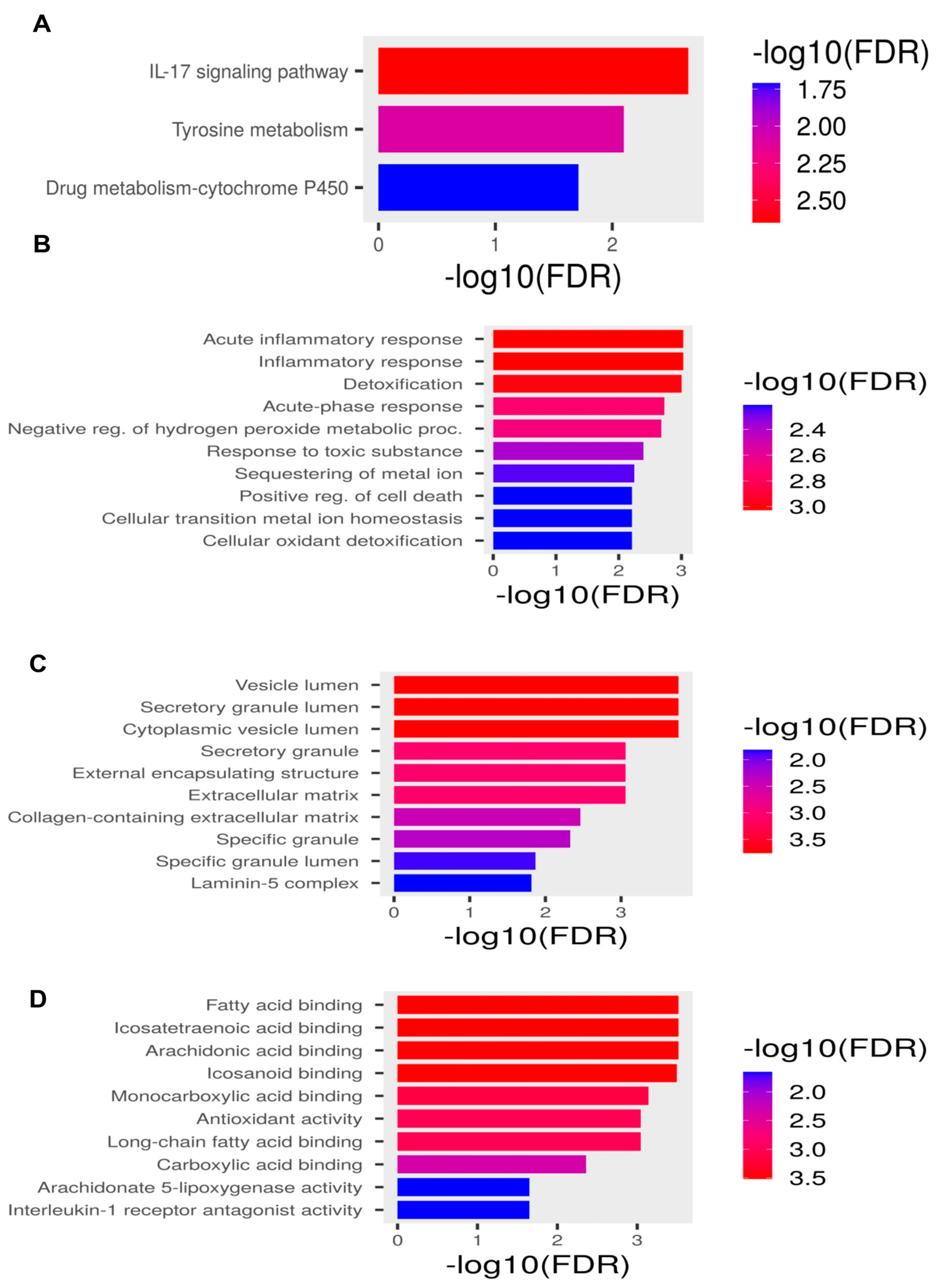
The GO analysis identified that DENIGs were enriched in acute anti-inflammatory responses, inflammatory responses, detoxification, vesicle lumina, secretory granule lumina, and fatty acid binding (Figure 2B–D). The KEGG analysis also showed that the DENIGs were associated with the IL-17 signaling pathway and tyrosine metabolism, which play crucial roles in the inflammatory response (Figure 2A). The details of the functional and pathway enrichment analyses of DENIGs are summarized in Table 2.

2.3. Identification of Hub Genes Associated with Neuroinflammation in POAG

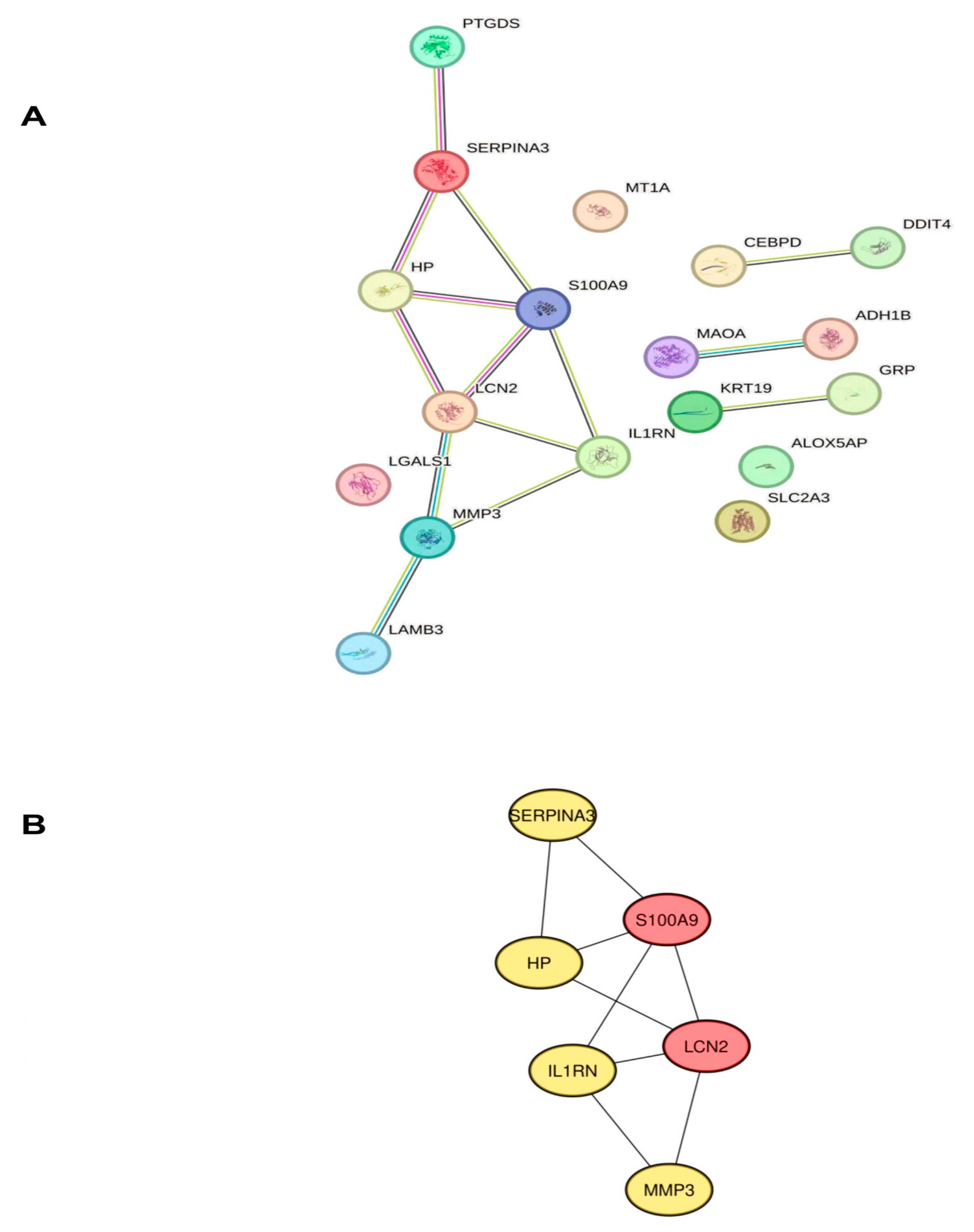
To investigate the link between the proteins encoded by DENIGs in glaucoma and to identify hub genes, we examined the PPI network of the DENIGs using STRING with a PPI enrichment p-value < 0.0150 (Figure 3A). The PPI network was further evaluated with the Cytoscape plugin cytoHubba to identify hub genes, as shown in Figure 3B. The top six genes (SERPINA3, LCN2, MMP3, S100A9, IL1RN, and HP) were identified as potential hub genes.

2.4. Validation of Hub Genes

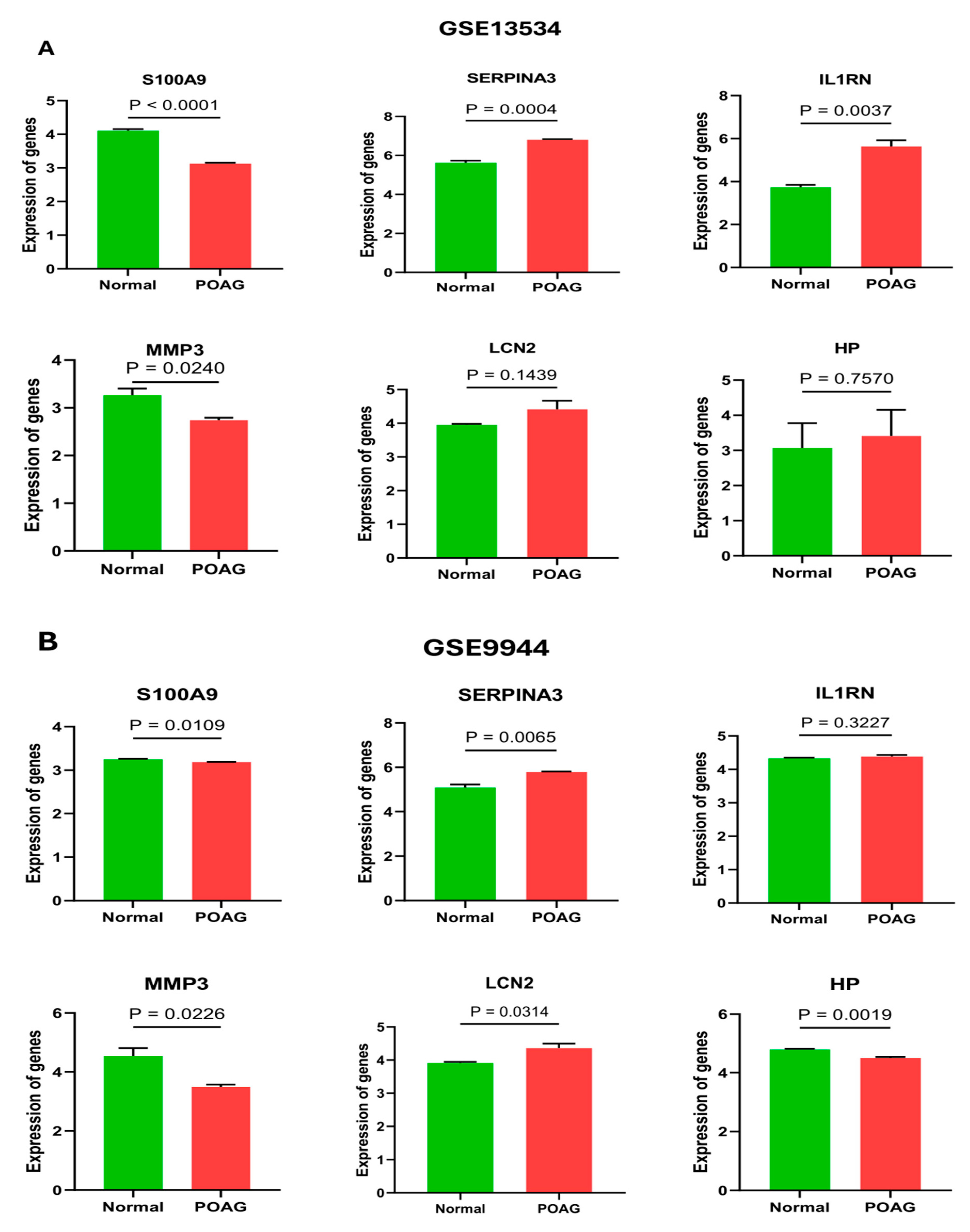
The GSE13534 and GSE9944 datasets were used to confirm the expression of the identified hub genes. The mRNA expression levels of SERPINA3 and IL1RN were elevated in POAG patients relative to the normal group, while the gene LCN2 was elevated in dataset GSE9944 and was non-significant in dataset GSE13534. Furthermore, the expression levels of genes S100A9 and MMP3 were found to be reduced in POAG patients (Figure 4).

2.5. Estimation of Possible miRNA Regulatory Networks of Hub Genes

MicroRNAs (miRNAs) are potent regulators of gene expression at the post-transcriptional level [22]. To assess the miRNAs regulating our hub genes, we predicted the potential miRNA regulatory networks of the hub genes. Figure 5 depicts the interaction networks of six hub genes and 118 miRNAs. Specifically, SERPINA3 and MMP3 were found to be modulated by 54 and 28 miRNAs, respectively. Additionally, 18 miRNAs were identified as targeting LCN2 and S100A9, while IL1RN and HP were targeted by 15 and 10 miRNAs, respectively. The analysis revealed that hsa-mir-34a-5p, hsa-mir-1343-3p, and hsa-mir-124-3p regulate a more significant number of hub genes, each with a connectivity degree of n = 4. It is noteworthy that the highest number of miRNAs regulate SERPINA3, a member of the serine protease inhibitor superfamily, which has been described to be intricate in the pathological inflammatory processes of many neurological disease processes, such as ischemic stroke, intracerebral hemorrhage, and Alzheimer’s disease [23].

2.6. Regional Expression

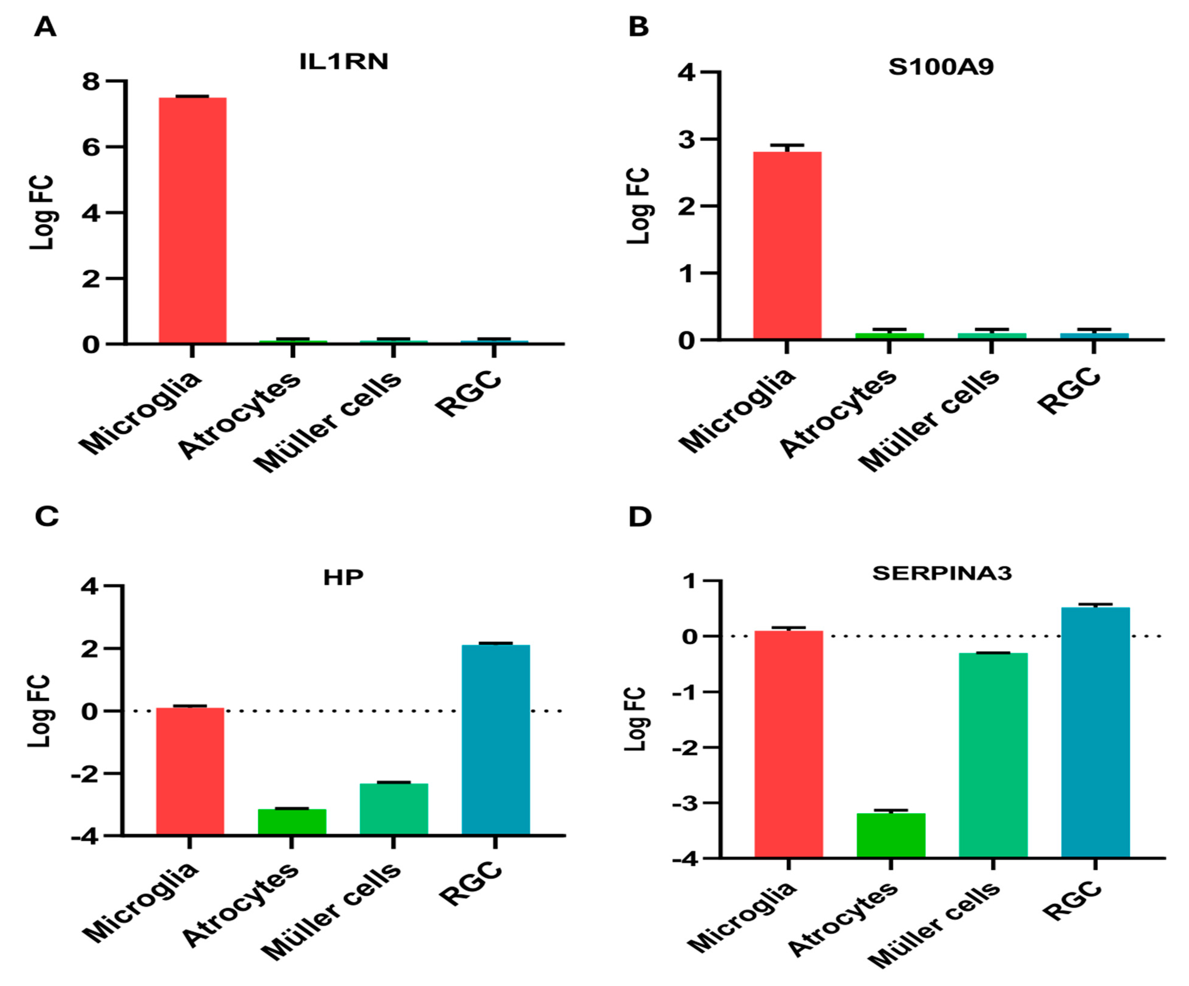
Single-cell RNA sequencing has transformed ocular gene expression research. This technique has allowed scientists to pinpoint the expression profiles of many cell types and to detail how gene expression varies across different biological conditions, such as specific topographical areas or medical disorders. We analyzed the regional expression of our hub genes across multiple cell types using Spectacle. The logFC value of each gene across the cluster of cells is shown in Figure 6. SERPINA3 and HP showed upregulated expression in RGCs, while downregulation was observed in Müller cells and astrocytes. S100A9 and IL1RN were upregulated in microglial cells and had no expression in other cells. The expressions of the other two genes (LCN2 and MMP3) were not reported in any datasets using Spectacle.

2.7. GWAS Analysis

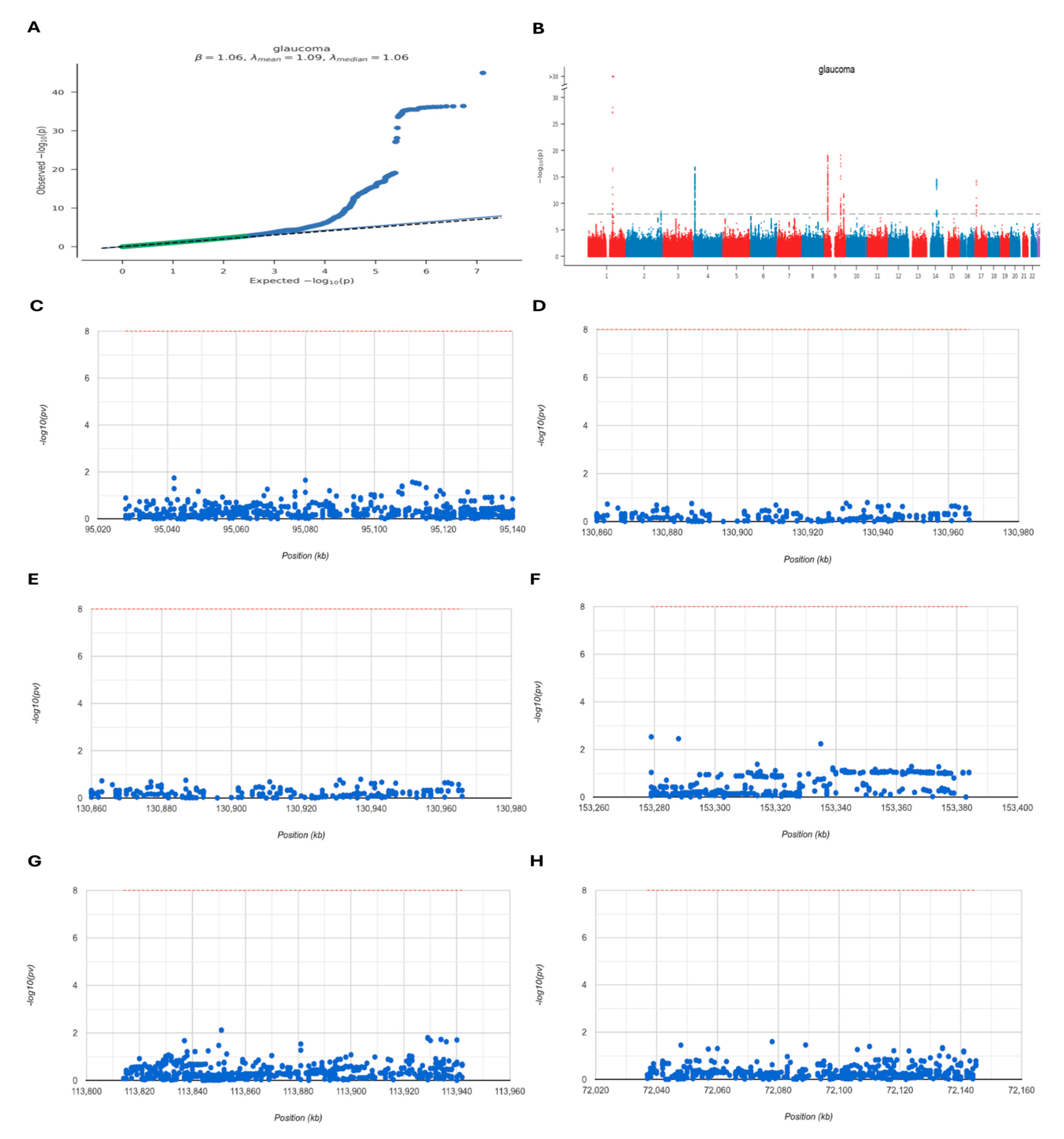
The GWAS data on glaucoma was assessed to determine which pathogenic regions were linked to the six neuroinflammation-related hub genes associated with POAG. A Q-Q plot illustrated SNP loci acquired from the GWAS data, revealing significant associations with glaucoma Figure 7A. Furthermore, performing precise site analysis of the GWAS data outlined the critical SNP loci dispersed within regions of genetic richness Figure 7B. Notably, the pathogenic regions aligned to our hub genes were identified; SERPINA3, LCN2, MMP3, S100A9, IL1RN, and HP were situated within the pathogenic region of chromosomes 16, 9, 11, 1, 2, and 16, respectively. Figure 7C–H.

2.8. Diagnostic Value Validation

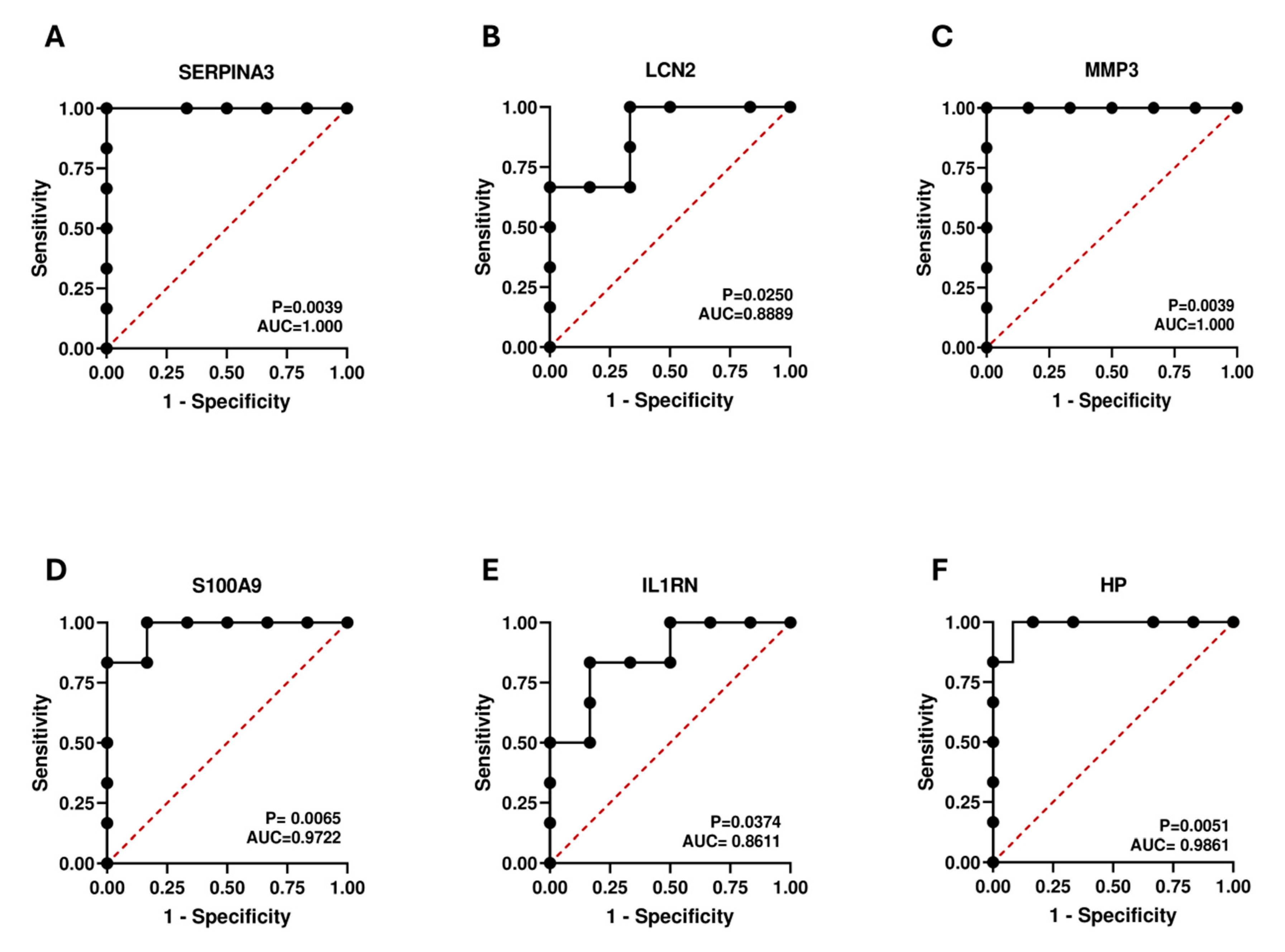
The efficacy of the hub genes was evaluated by constructing ROC curves using the dataset GSE9944 (Figure 8). AUC values and p-values were calculated for six hub genes. All our hub genes exceeded an AUC value of 0.7, signifying a robust diagnostic value in POAG diagnosis. The AUC values of SERPINA3, LCN2, MMP3, S100A9, IL1RN, and HP were 1.00, 0.88, 1.00, 0.97, 0.86, and 0.98, respectively. These findings indicated that our hub genes can accurately diagnose patients with POAG.

2.9. Validation of Hub Genes Using a Glaucoma Model

To verify the expression of hub genes, we induced a simulated glaucoma model in R28 cells using OGD/R injury. We then conducted real-time PCR experiments to validate the expression of the hub genes. Our findings revealed a significant decrease in the mRNA levels of SA1009 and MMP3 in the glaucoma model group compared to the normal group, while SERPINA3, IL1RN, and LCN2 expression increased (Figure 9). These results were similar to our bioinformatics analysis. There were no significant differences in the expression of HP between the two groups. These findings suggest that SERPINA3, IL1RN, and LCN2, and their associated signaling pathways, may play a role in the progression of POAG via the regulation of neuroinflammation.

3. Discussion

Glaucoma is usually associated with an elevated IOP, although it is not the sole causative factor. Other factors also contribute to the loss of RGCs, such as oxidative stress, excitotoxicity, mitochondrial dysfunction, and neuroinflammation [2,5,6]. The pathogenesis of POAG still needs to be fully elucidated. Evidence increasingly suggests that the immune system and neuroinflammation play crucial roles in the onset and progression of neurodegeneration in glaucoma [24]. Previous reports have indicated that neuronal dysfunction and immune surveillance by the glial cells contributes to retinal neurodegeneration [14]. In this study, 179 DEGs (60 upregulated genes and 119 downregulated genes) were intersected with neuroinflammation-related genes to obtain DENIGs (n = 18). Next, PPI network analysis identified six hub genes: SERPINA3, LCN2, MMP3, S100A9, IL1RN, and HP.
SERPINA3, a serine protease inhibitor superfamily member, functions primarily as a protease inhibitor to maintain cellular homeostasis [25]. SERPINA3 is mainly associated with the acute phase response and the inflammatory response [26]. Multiple studies have identified SERPINA3 and its subfamily SerpinA3N/SerpinA3 as common factors in neurodegenerative diseases, suggesting that they play essential roles in disease development [27,28]. Additionally, SERPINA3N has been found to reduce neuronal apoptosis and neuroinflammation by activating the Akt–mTOR pathway [29] (Zhang et al., 2022). However, another study found that SERPINA3 promotes neuroinflammation by activating the NF-κB signaling pathway [30]. IL1RN, belonging to the IL-1 family, has been documented to reduce inflammatory signaling by inhibiting the expression of other pro-inflammatory molecules and preventing further neuronal loss [31]. Interleukin-1 receptor antagonism has been recognized as a significant therapeutic approach to address neuroinflammation [31]. S100A9 is involved in various disorders, including neurodegenerative [32,33,34] and ocular inflammatory diseases [35]. S100A9 is important in leukocyte trafficking and arachidonic acid metabolism [36]. Additionally, the gene expression of S100A9 is stimulated by the TLR (Toll-like receptor)/IL-1 pathway, and its expression is boosted by IL-10 and glucocorticoids [37]. In Alzheimer’s disease, increased levels of S100A9 during inflammation could lead to amyloid formation and deposition [32]. Moreover, another study found elevated levels of S100A9 in the tears of glaucoma patients, indicating surface inflammatory biomarkers [38]. LCN2 is a secreted protein with many functions, including immune regulation, iron transport, cell proliferation, differentiation, and cell death [39]. Many studies have linked the expression of LCN2 to Alzheimer’s disease [40,41], Parkinson’s disease [42], and multiple sclerosis [43]. Exposure to amyloid beta triggers the production of LCN2 in primary astrocytes, and LCN2 increases the susceptibility of primary neurons to amyloid beta toxicity [44]. Various studies using whole transcriptome analyses in rodent glaucoma models have indicated that LCN2 is upregulated in glaucomatous retinae [45,46,47]. It has also been demonstrated that LCN2 is significantly upregulated shortly after optic nerve injury, suggesting that LCN2 may contribute to RGC loss [46]. Matrix metalloproteinases (MMPs) are essential for modifying the extracellular matrix (ECM) and other signaling molecules, which are vital for facilitating various functions such as embryonic development, cell survival, blood vessel formation, immune response, and wound healing [48]. Potentially, corneal endothelial cells release MMP-3 on the apical side of the cell, which then passes with the natural flow of aqueous humor into the trabecular meshwork and outflow channels, where it breaks down some of the ECM, making the tissue more permeable to aqueous humor flow [49,50]. A recent study used MMP3 as a gene therapy option for glaucoma treatment [51].
Further, the DENIGs were submitted for GO and KEGG enrichment analysis to illustrate their biological functions. The DENIGs are involved in the acute anti-inflammatory response, inflammatory response, detoxification, vesicle lumina, secretory granule lumina, fatty acid binding, and IL-17 signaling. Our study observed that three hub genes were associated with IL-17 signaling. IL-17 is a cytokine primarily secreted by the stimulation of Th-17 cells, and it triggers inflammatory responses by inducing other cytokines and inflammatory chemokines [52,53]. Prolonged inflammation can lead to the release of IL-17, which may exacerbate inflammation by stimulating the release of cytokines and chemoattractant proteins. Moreover, IL-17 may also have anti-inflammatory and neuroprotective effects when astrocytes are exposed to high levels of IL-17 [54]. IL-17A, a subfamily of IL-17, regulates the retinal immune response and RGC death in experimental glaucoma by promoting retinal microglial activation [55]. Recent studies using RNA sequencing have revealed significant alterations in the expression of genes associated with immune activities in the retinae of mice with ocular hypertension, and it has been suggested that IL-17A may be involved in the pathological process of glaucomatous neuropathy by influencing the phenotype switching of retinal microglia [56,57]. Mice with experimental glaucoma have shown a significant increase in IL-17A expression by microglia, induced by acute IOP elevation [58]. Furthermore, suppressing IL-17A shows promise as an innovative glaucoma therapeutic target [55]. However, the exact role of IL-17 in glaucoma has yet to be fully elucidated, and further studies are needed to clarify the role of IL-17A in glaucoma.
Next, we analyzed the potential miRNA regulatory networks of hub genes. When they are dysregulated, miRNAs—which are potent regulators of gene expression at the post-transcriptional level [22]—can influence the pathways and processes associated with glaucoma, such as apoptosis, autophagy, neurogenesis, aging, ECM remodeling, oxidative stress, inflammation, and angiogenesis [59]. Notably, our analysis revealed that SERPINA3 was regulated by the highest number of miRNAs. This gene has been implicated in the pathological inflammatory processes of various neurological diseases [23]. Several miRNAs have been recognized as biomarkers to aid in the early detection of POAG, and may be potential candidates for diagnosing and treating glaucoma [60]. Our analysis identified that hsa-mir-34a-5p, hsa-mir-1343-3p, and hsa-mir-124-3p regulate a more significant number of hub genes, each with a connectivity degree of n = 4. The upregulation of hsa-mir-124-3p was identified in pseudoexfoliation glaucoma [61], targeting three specific pathways: TGF-β1, fibrosis/ECM, and proteoglycan metabolism [62]. Furthermore, hsa-mir-124-3p was significantly upregulated in human trabecular meshwork cells [63]. Both miR-155-5p and miR-146a regulated the gene LCN2 in our analysis. These miRNAs are recognized as primary regulators in inflammation-related processes, especially in neuroinflammation [60]. Expression of miR-155 is found in activated immune cells, playing a pivotal role in the immune responses of B cells, macrophages, and microglia [64].
In the CNS, miR-155 enhances microglial triggering and inflammation. A study discovered that knocking out the miR-155 gene in mice considerably decreases the amount and activation of inflammatory factors in the retinal region, indicating that miR-155 is essential for optic nerve inflammation [65,66]. Conversely, miR-146a prevents inflammation by reducing microglial activation and suppressing the inflammatory cascade. The NF-κB signaling pathway is a pivotal pro-inflammatory pathway, regulated by miR-155 and miR-146a through positive and negative responses. NF-κB stimulates miR-155 expression, and miR-155, in turn, enhances NF-κB activation [67]. On the other hand, miR-146a inhibits interleukin-1 receptor-associated kinases-1 and TNF receptor-associated factor 6 in the NFκB signaling pathway, thereby inhibiting inflammation [68].
We submitted the genes to the Spectacle database to analyze the expression of our hub genes in RGCs and other cell types involved in neuroinflammation. SERPINA3 and HP showed upregulated expression in the RGCs, while downregulation was observed in the Müller cells and astrocytes. S100A9 and IL1RN were upregulated in the microglial cells and had no expression in other cell types. Microglial activation is the first response in the neurodegeneration associated with glaucoma [69]. Elevated IOP has been observed to trigger the activation of retinal microglia, leading to the release of pro-inflammatory cytokines that can detrimentally affect RGCs. Microglia consistently monitor and maintain the homeostasis of their microenvironment through their scavenging and phagocytosing functions, and provide neurotrophic support [70]. Microglia can migrate to an injury site upon receiving signaling cues, and this phenomenon has been observed in both experimental models and in the eyes of human donors with glaucoma [71,72,73]. Despite the critical role of microglia in maintaining synaptic plasticity through pruning and elimination, these functions can also contribute to harmful processes in the context of glaucoma [74]. A study demonstrated that S100A9 induces the activation of BV-2 microglial cells and enhances the production of pro-inflammatory factors by activating the NF-κB signaling pathway [75]. This, in turn, exacerbates damage to oligodendrocyte precursor cells, confirming the findings of our analysis regarding the expression of S100A9 in microglial cells. The same observation was noted in association with Alzheimer’s disease, where a high concentration of S100A9 resulted in the impaired mobility and proliferation of immune cells, indicating neurotoxicity during acute inflammatory conditions [76]. Our analysis also reveals the upregulation of the IL1RN gene in microglial cells. The IL-1 family includes an additional member, IL-1Ra (the IL-1 receptor antagonist), encoded by the IL1RN gene. It is well-documented that the activation of the microglial inflammasome and the production of IL-1 can lead to neuroinflammation and neurodegeneration [77]. Studies in rodents lacking NLRP3 or caspase-1 have shown protection from neuroinflammation and cognitive decline. The activation of microglia and the expression of the inflammatory cytokine IL-1 in the central nervous system are closely associated with neuroinflammation. Furthermore, evidence suggests that inhibiting IL-1 signaling via pharmacological means or genetic manipulation in various central nervous system disease models could reduce neuroinflammation or slow disease progression [78].
The GWAS emerged as a valuable tool for interpreting the genetic basis of disease susceptibility, enabling the identification of vulnerability genes across various disorders. We performed GWAS analysis to validate the presence of pathogenic regions associated with our six neuroinflammation-related hub genes (SERPINA3, LCN2, MMP3, S100A9, IL1RN, and HP) in POAG. We found hub gene localization on chromosomes 16, 9, 11, 1, 2, and 16, respectively. Significant single SNPs corresponding to six of these hub genes were identified. Furthermore, according to our ROC curve analysis, the AUC values of SERPINA3, LCN2, MMP3, S100A9, IL1RN, and HP were 1.00, 0.88, 1.00, 0.97, 0.86, and 0.98, respectively. This suggests that the identified hub genes possess significant identification and potential biomarker capabilities. Typically, an AUC of 0.5 signifies no discrimination, 0.7 to 0.8 is acceptable, 0.8 to 0.9 is excellent, and an AUC above 0.9 is outstanding. The three hub genes SERPINA3, IL1R1, and LCN2 were validated as potential diagnostic biomarkers through real-time PCR, showing increased expression in the OGD/R-induced glaucoma model. Our GWAS, ROC curve, and real-time PCR analyses provide evidence supporting the association of our hub genes with glaucoma susceptibility, indicating their potential as solid biomarkers for the disease.

Therapeutic Potential of Hub Genes

There are two main therapeutic approaches to treat glaucoma. The first domain focuses on the use of conventional and widely used antiglaucoma medications, which mainly focus on the reduction of IOP. Conversely, the second domain has recently evolved and focuses on alternative neuroprotective options for RGC loss prevention. Recently, studies have been focusing on immunomodulation as a potential neuroprotective strategy. Similarly, many studies have explored the role of our hub genes in neuroinflammation and targeted these genes for immunomodulation therapy. Certain studies have observed that S100A9 is a promising candidate for developing novel therapeutic interventions [79,80]. The inhibition of S100A9 with drugs and inhibitors markedly attenuates brain injury after subarachnoid hemorrhage [81], reduces mortality rates in murine models of sepsis [82] and traumatic brain injury [83], and improves the viability and survival rate of neurons after spinal cord injury [84]. S100A9 has also been reported to regulate TNFα production by activating the TLR4 pathway in acute herpetic neuralgia, a possible target for glaucoma therapy [85,86]. Furthermore, SERPINA3 exerts a protective effect in traumatic brain injury by regulating MAPK signaling pathways [87]. Moreover, the administration of recombinant SERPINA3 reduced cornea neovascularization and inflammation in a murine model of corneal alkaline burn [88]. SERPINA3 has been shown to increase the activity of superoxide dismutase and catalase, protecting retinal neuronal and Müller cells against reactive oxygen species (ROS) [89]. Additionally, the SERPINA3 protein participates in the propagation of the formation of β-amyloid or prion proteins in neurodegenerative diseases, and the glycosylation of SERPINA3 can be a therapeutic tool [90]. Pro-inflammatory cytokines from microglia alter the transcriptome profiles of inactive astrocytes and change them to an active state, which further releases a high level of LCN2. Several approaches have been studied to alleviate the neurotoxic effects of LCN2, such as inhibiting LCN2 expression, blocking the function of secreted LCN2 and its receptor 24p3R, suppressing LCN2 receptor signaling, and reducing neurodegeneration [91,92]. MMP activation or expression inhibition has been observed as a therapeutic approach in many neurodegenerative diseases. Consequently, synthetic broad-spectrum inhibitors and agents that interfere with the substrate of the MMPs could serve as potential targets or have medicinal value in inhibiting MMPs [93].

4. Methods

4.1. Data Sources

The gene expression dataset (GSE27276) of the genome-wide expression of the trabecular meshwork in POAG patients was downloaded from the GEO database (https://www.ncbi.nlm.nih.gov/geo/ Retrieved on 5 January 2024). Neuroinflammation-related genes (n = 1299, relevance score > 1) were attained from the Gene-Cards database (https://www.genecards.org/ accessed on 5 January 2024) [94]. The methods used for the analysis are summarized in Figure 10.

4.2. Data Processing and DEGs Analysis

The raw data from the GSE27276 dataset were processed by the Geoexplorer online tool (https://geoexplorer.rosalind.kcl.ac.uk/ Retrieved on 5 January 2024) [95]. The DEGs of the control and POAG groups were identified. A p-value < 0.05 and a logFC value > 1 were set as the threshold values for DEGs screening. The DENIGs were screened by assessing the intersection of DEGs and neuroinflammation-related genes using the freely available online Venn diagram website. The heatmap used to visualize the expression of DEGs was generated using TBtools (Version 2.042) [96].

4.3. Pathway Enrichment Analyses

The identified DENIGs were submitted to GO and KEGG for pathway enrichment analysis. An online gene-set enrichment tool, ShinyGO 0.80 (http://bioinformatics.sdstate.edu/go/ accessed on 12 January 2024) [97], was used for the GO and KEGG analyses. GO encompasses three different kinds of terms: MF (molecular function), BP (biological process), and CC (cellular component). It is often used to investigate the functional aspects of genomic or transcriptome data [98]. KEGG is a data resource for the systematic study of gene functions in the context of the networks of genes or proteins. The threshold value was set to p < 0.05 for this analysis.

4.4. Protein–Protein Interaction (PPI), Network Construction, and Gene Identification

The PPI network of the DENIGs was analyzed using STRING (http://string-db.org/ Retrieved on 12 January 2024) [99]. STRING is an online database that integrates predicted and experimental interactions among proteins. The interactions between DEGs with a score > 0.4 were chosen for the PPI network construction using Cytoscape software (version 3.9.0). Furthermore, hub genes were identified using cytoHubba in Cytoscape [100].

4.5. External Validation of Hub Genes

The hub genes were further validated using two other external datasets (GSE13534 and GSE9944) acquired from the GEO database. The GSE13534 dataset contains data on the optic nerve tissue of normal and POAG human donors. Similarly, the dataset GSE9944 includes data on the gene expression of human optic nerve head astrocytes with or without glaucoma.

4.6. Estimation of the Gene–miRNA Regulatory Network

The gene–miRNA regulatory network was built using the miRNet database [101]. The green round dots represent hub genes within the network, while the dark yellow dots represent miRNA.

4.7. Regional Expression

Spectacle (https://singlecell-eye.org/app/spectacle/ accessed on 14 January 2024) was used to analyze the expression of our hub genes across the clusters of immune and ocular cells. This freely available online platform offers various visualization tools to explore gene expression patterns within different cell types [102]. This helps us to identify the specific cell types expressing a gene of interest. We analyzed our hub genes against four types of cells (microglia, Müller cells, astrocytes, and RGCs).

4.8. Genome-Wide Association Study (GWAS) Analysis

Data for the GWAS analysis were taken from the Gene Atlas database (http://geneatlas.roslin.ed.ac.uk/ Retrieved on 12 June 2024). This diverse repository contains correlations among hundreds of traits, and vast arrays of genetic variations within the UK Biobank dataset. This dataset encompasses data from 452,264 individuals of British origin, comprising 778 distinct phenotypic traits and covering 30 million genetic loci.

4.9. Receiver Operating Characteristic (ROC) Curve Analysis

To validate the diagnostic precision of our hub genes, we executed the ROC curve and the area under the curve (AUC) evaluations using GraphPad Prism (Version 9.5.1). Following previous studies, an AUC > 0.7 was selected as the threshold value for genes that were indicative of a disease diagnosis [103]. The dataset GSE9944 was used for expression pattern analysis.

4.10. Cell Culture

R28 cells are immortalized retinal precursor cells frequently utilized in in vitro research to investigate the physiological functions of RGCs. The cells were provided by the Department of Anatomy and Neurobiology at Central South University, China, and cultured in a low-glucose DMEM medium (Biosharp, Hefei, China, Catalog Number SH30021.01). The media was supplemented with 10% fetal bovine serum (Meilun Biotechnology, Shanghai, China, MA0015) and 1% penicillin/streptomycin (Beyotime Biotechnology, Shanghai, China, Catalog Number 25200-056). The cells were cultured in an incubator with 5% CO2 at 37 °C. Following previous studies, the oxygen and glucose deprivation/reoxygenation (OGD/R) cell model was established to mimic experimental glaucoma [104]. In brief, R28 cells were cultured in low-glucose DMEM for 24 h. Subsequently, the culture medium was replaced with serum- and glucose-free DMEM, and the cells were transported to a gas-tight instrument flushed with a mixture of 5% CO2 and 95% N2 gas. The in and out valves of the instrument were sealed after flushing, and cells were kept in OGD condition for 2 1/2 h. Following this, reoxygenation was performed using low-glucose DMEM under normal conditions (5% CO2) at 37 °C for 24 h.

4.11. RNA Extraction and Real-Time PCR

Total RNA was isolated from the cells with the SteadyPure Rapid RNA Extraction Kit (Accurate Biology, Changsha, China), and cDNA was synthesized using the PrimeScript RT reagent Kit (Accurate Biology). We utilized the Bio-Rad CFX Connect Real-Time PCR Detection System. The primer sequences are listed in Table 3. The 2−ΔΔct method was employed to measure the mRNA expression.

4.12. Statistical Analysis

Graphical representations were generated utilizing GraphPad Prism (Version 9.5.1). Comparison between the two groups was determined using the Student’s t-test. Statistical significance was attributed to data disparities when p < 0.05.

5. Conclusions

Our study identified and validated six neuroinflammation-related hub genes in glaucoma (SERPINA3, LCN2, MMP3, S100A9, IL1RN, and HP). We explored these hub genes’ involvement in molecular pathways leading to glaucoma, including the anti-inflammatory response, IL-17 signaling, and tyrosine metabolism. GWAS analysis identified significant SNPs corresponding to these hub genes. Further, we explored the diagnostic accuracy of our hub genes by ROC curve analysis and validated them using external datasets and real-time PCR analysis. We found that all our hub genes may have a significant role in the regulation of POAG. We also added several pieces of evidence from the literature regarding the role of our hub genes in the disease mechanisms and their therapeutic potential in glaucoma and many other neurodegenerative diseases. Therefore, these findings may provide early diagnosis biomarkers and lay the foundations for possible therapeutic options to treat glaucoma. However, future research is warranted to investigate the specific mechanisms through which these hub genes regulate neuroinflammation, to exploit them for therapeutic purposes and assess the significance of neuroinflammation-related biomarkers in POAG.

Author Contributions

Methodology, Z.U. and Y.T.; software, Z.U. and Y.T.; validation, J.H.; formal analysis, Z.U.; writing—original draft, Z.U. and Y.T.; writing—review and editing, J.H.; supervision, J.H. All authors have read and agreed to the published version of the manuscript.

Funding

This work was supported by the Central South University School-Enterprise Joint Innovation Project (no. 2021XQLH084).

Institutional Review Board Statement

Not applicable.

Informed Consent Statement

Not applicable.

Data Availability Statement

The corresponding author can provide raw and processed data upon request.

Conflicts of Interest

The authors declare no conflicts of interest.

Abbreviations

PPIProtein-protein interactions
ECMExtracellular matrix
OGD/ROxygen and glucose deprivation/reoxygenation
DMEMDulbecco’s Modified Eagle’s Medium
MFMolecular function
BPBiological process
CCCellular component
GOGene Ontology
DENIGsDifferentially expressed neuroinflammation-related genes
DEGsDifferentially expressed genes
IOPIntraocular pressure
RGCsRetinal ganglion cells
SNPsSingle nucleotide polymorphisms
GWASGenome-wide association study
ROCReceiver operating characteristic
KEGGKyoto Encyclopedia of Genes and Genomes
POAGPrimary open-angle glaucoma
SERPINA3serine protease inhibitor, clade A, member 3
LCN2Lipocalin 2
MMP3Matrix Metalloproteinase-3
S100A9S100 Calcium-Binding Protein A9
IL1RNInterleukin-1 Receptor Antagonist
HPHaptoglobin
GEOGene Expression Omnibus

References

  1. Wu, Y.; Szymanska, M.; Hu, Y.; Fazal, M.I.; Jiang, N.; Yetisen, A.K.; Cordeiro, M.F. Measures of disease activity in glaucoma. Biosens. Bioelectron. 2022, 196, 113700. [Google Scholar] [CrossRef] [PubMed]
  2. Allison, K.; Patel, D.; Alabi, O. Epidemiology of Glaucoma: The Past, Present, and Predictions for the Future. Cureus 2020, 12, e11686. [Google Scholar] [CrossRef] [PubMed]
  3. Zhang, N.; Wang, J.; Li, Y.; Jiang, B. Prevalence of primary open angle glaucoma in the last 20 years: A meta-analysis and systematic review. Sci. Rep. 2021, 11, 13762. [Google Scholar] [CrossRef] [PubMed]
  4. Macanian, J.; Sharma, S.C. Pathogenesis of Glaucoma. Encyclopedia 2022, 2, 1803–1810. [Google Scholar] [CrossRef]
  5. Williams, P.A.; Marsh-Armstrong, N.; Howell, G.R.; Bosco, A.; Danias, J.; Simon, J.; Di Polo, A.; Kuehn, M.H.; Przedborski, S.; Raff, M.; et al. Neuroinflammation in glaucoma: A new opportunity. Exp. Eye Res. 2017, 157, 20–27. [Google Scholar] [CrossRef] [PubMed]
  6. Baudouin, C.; Kolko, M.; Melik-Parsadaniantz, S.; Messmer, E.M. Inflammation in Glaucoma: From the back to the front of the eye, and beyond. Prog. Retin. Eye Res. 2021, 83, 100916. [Google Scholar] [CrossRef] [PubMed]
  7. Schuster, A.K.; Erb, C.; Hoffmann, E.M.; Dietlein, T.; Pfeiffer, N. The Diagnosis and Treatment of Glaucoma. Dtsch. Arzteblatt Int. 2020, 117, 225–234. [Google Scholar] [CrossRef]
  8. Loftsson, T.; Stefánsson, E. Aqueous eye drops containing drug/cyclodextrin nanoparticles deliver therapeutic drug concentrations to both anterior and posterior segment. Acta Ophthalmol. 2022, 100, 7–25. [Google Scholar] [CrossRef] [PubMed]
  9. Hakim, A.; Guido, B.; Narsineni, L.; Chen, D.-W.; Foldvari, M. Gene therapy strategies for glaucoma from IOP reduction to retinal neuroprotection: Progress towards non-viral systems. Adv. Drug Deliv. Rev. 2023, 196, 114781. [Google Scholar] [CrossRef]
  10. Tao, Y.; Zhang, Q.; Meng, M.; Huang, J. A bibliometric analysis of the application of stem cells in glaucoma research from 1999 to 2022. Front. Cell Dev. Biol. 2023, 11, 1081898. [Google Scholar] [CrossRef]
  11. Choi, S.; Choi, S.-H.; Bastola, T.; Park, Y.; Oh, J.; Kim, K.-Y.; Hwang, S.; Miller, Y.I.; Ju, W.-K. AIBP: A New Safeguard against Glaucomatous Neuroinflammation. Cells 2024, 13, 198. [Google Scholar] [CrossRef] [PubMed]
  12. Duarte, J.N. Neuroinflammatory Mechanisms of Mitochondrial Dysfunction and Neurodegeneration in Glaucoma. J. Ophthalmol. 2021, 2021, 4581909. [Google Scholar] [CrossRef] [PubMed]
  13. Tezel, G. Immune regulation toward immunomodulation for neuroprotection in glaucoma. Curr. Opin. Pharmacol. 2013, 13, 23–31. [Google Scholar] [CrossRef] [PubMed]
  14. Ramirez, A.I.; de Hoz, R.; Salobrar-Garcia, E.; Salazar, J.J.; Rojas, B.; Ajoy, D.; López-Cuenca, I.; Rojas, P.; Triviño, A.; Ramírez, J.M. The Role of Microglia in Retinal Neurodegeneration: Alzheimer’s Disease, Parkinson, and Glaucoma. Front. Aging Neurosci. 2017, 9, 214. [Google Scholar] [CrossRef]
  15. Bordone, M.P.; González Fleitas, M.F.; Pasquini, L.A.; Bosco, A.; Sande, P.H.; Rosenstein, R.E.; Dorfman, D. Involvement of microglia in early axoglial alterations of the optic nerve induced by experimental glaucoma. J. Neurochem. 2017, 142, 323–337. [Google Scholar] [CrossRef] [PubMed]
  16. Evangelho, K.; Mogilevskaya, M.; Losada-Barragan, M.; Vargas-Sanchez, J.K. Pathophysiology of primary open-angle glaucoma from a neuroinflammatory and neurotoxicity perspective: A review of the literature. Int. Ophthalmol. 2019, 39, 259–271. [Google Scholar] [CrossRef] [PubMed]
  17. Adornetto, A.; Russo, R.; Parisi, V. Neuroinflammation as a target for glaucoma therapy. Neural Regen. Res. 2019, 14, 391. [Google Scholar] [CrossRef]
  18. Fernández-Albarral, J.A.; Salazar, J.J.; De Hoz, R.; Marco, E.M.; Martín-Sánchez, B.; Flores-Salguero, E.; Salobrar-García, E.; López-Cuenca, I.; Barrios-Sabador, V.; Avilés-Trigueros, M.; et al. Retinal Molecular Changes Are Associated with Neuroinflammation and Loss of RGCs in an Experimental Model of Glaucoma. Int. J. Mol. Sci. 2021, 22, 2066. [Google Scholar] [CrossRef]
  19. Zhao, X.; Sun, R.; Luo, X.; Wang, F.; Sun, X. The Interaction Between Microglia and Macroglia in Glaucoma. Front. Neurosci. 2021, 15, 610788. [Google Scholar] [CrossRef]
  20. Vernazza, S.; Tirendi, S.; Bassi, A.M.; Traverso, C.E.; Saccà, S.C. Neuroinflammation in Primary Open-Angle Glaucoma. J. Clin. Med. 2020, 9, 3172. [Google Scholar] [CrossRef]
  21. Wang, H.; Sun, P.; Chen, Y.; Jiang, L.; Wu, H.; Zhang, W.; Gao, F. Research progress on human genes involved in the pathogenesis of glaucoma (Review). Mol. Med. Rep. 2018, 18, 656–674. [Google Scholar] [CrossRef]
  22. Yin, J.; Qian, Z.; Chen, Y.; Li, Y.; Zhou, X. MicroRNA regulatory networks in the pathogenesis of sarcopenia. J. Cell. Mol. Med. 2020, 24, 4900–4912. [Google Scholar] [CrossRef] [PubMed]
  23. Hu, X.; Xiao, Z.; Shen, Y.; Yang, W.; Wang, P.; Li, P.; Wang, Z.; Pu, M.; Zhao, L.; Xie, P.; et al. SERPINA3: A novel inflammatory biomarker associated with cerebral small vessel disease burden in ischemic stroke. CNS Neurosci. Ther. 2023, 30, e14472. [Google Scholar] [CrossRef] [PubMed]
  24. Chen, H.; Cho, K.-S.; Vu, T.H.K.; Shen, C.-H.; Kaur, M.; Chen, G.; Mathew, R.; McHam, M.L.; Fazelat, A.; Lashkari, K.; et al. Commensal microflora-induced T cell responses mediate progressive neurodegeneration in glaucoma. Nat. Commun. 2018, 9, 3209. [Google Scholar] [CrossRef]
  25. Zattoni, M.; Mearelli, M.; Vanni, S.; Colini Baldeschi, A.; Tran, T.H.; Ferracin, C.; Catania, M.; Moda, F.; Di Fede, G.; Giaccone, G.; et al. Serpin Signatures in Prion and Alzheimer’s Diseases. Mol. Neurobiol. 2022, 59, 3778–3799. [Google Scholar] [CrossRef] [PubMed]
  26. Aslam, M.S.; Yuan, L. Serpina3n: Potential drug and challenges, mini review. J. Drug Target. 2020, 28, 368–378. [Google Scholar] [CrossRef] [PubMed]
  27. Li, M.D.; Burns, T.C.; Morgan, A.A.; Khatri, P. Integrated multi-cohort transcriptional meta-analysis of neurodegenerative diseases. Acta Neuropathol. Commun. 2014, 2, 93. [Google Scholar] [CrossRef]
  28. Schrötter, A.; Park, Y.M.; Marcus, K.; Martins-de-Souza, D.; Nilsson, P.; Magraoui, F.E.; Meyer, H.E.; Grinberg, L.T. Key players in neurodegenerative disorders in focus—New insights into the proteomic profile of Alzheimer’s disease, schizophrenia, ALS, and multiple sclerosis—24th HUPO BPP Workshop: September 29, 2015, Vancouver, Canada. Proteomics 2016, 16, 1047–1050. [Google Scholar] [CrossRef] [PubMed]
  29. Yuan, Q.; Wang, S.; Zhang, G.; He, J.; Liu, Z.; Wang, M.; Cai, H.; Wan, J. Highly expressed of SERPINA3 indicated poor prognosis and involved in immune suppression in glioma. Immun. Inflamm. Dis. 2021, 9, 1618–1630. [Google Scholar] [CrossRef]
  30. Liu, C.; Zhao, X.-M.; Wang, Q.; Du, T.-T.; Zhang, M.-X.; Wang, H.-Z.; Li, R.-P.; Liang, K.; Gao, Y.; Zhou, S.-Y.; et al. Astrocyte-derived SerpinA3N promotes neuroinflammation and epileptic seizures by activating the NF-κB signaling pathway in mice with temporal lobe epilepsy. J. Neuroinflammation 2023, 20, 161. [Google Scholar] [CrossRef]
  31. Yamanaka, G.; Ishida, Y.; Kanou, K.; Suzuki, S.; Watanabe, Y.; Takamatsu, T.; Morichi, S.; Go, S.; Oana, S.; Yamazaki, T.; et al. Towards a Treatment for Neuroinflammation in Epilepsy: Interleukin-1 Receptor Antagonist, Anakinra, as a Potential Treatment in Intractable Epilepsy. Int. J. Mol. Sci. 2021, 22, 6282. [Google Scholar] [CrossRef] [PubMed]
  32. Wang, C.; Klechikov, A.G.; Gharibyan, A.L.; Wärmländer, S.K.T.S.; Jarvet, J.; Zhao, L.; Jia, X.; Shankar, S.K.; Olofsson, A.; Brännström, T.; et al. The role of pro-inflammatory S100A9 in Alzheimer’s disease amyloid-neuroinflammatory cascade. Acta Neuropathol. 2014, 127, 507–522. [Google Scholar] [CrossRef] [PubMed]
  33. Horvath, I.; Iashchishyn, I.A.; Moskalenko, R.A.; Wang, C.; Wärmländer, S.K.T.S.; Wallin, C.; Gräslund, A.; Kovacs, G.G.; Morozova-Roche, L.A. Co-aggregation of pro-inflammatory S100A9 with α-synuclein in Parkinson’s disease: Ex vivo and in vitro studies. J. Neuroinflammation 2018, 15, 172. [Google Scholar] [CrossRef] [PubMed]
  34. Xia, P.; Ji, X.; Yan, L.; Lian, S.; Chen, Z.; Luo, Y. Roles of S100A8, S100A9 and S100A12 in infection, inflammation and immunity. Immunology 2023, 171, 365–376. [Google Scholar] [CrossRef] [PubMed]
  35. Wang, Y.-Q.; Dai, X.-D.; Ran, Y.; Cao, Y.; Lan, C.-L.; Guan, J.-T.; Liu, C.; Yang, F.-M.; Gan, Y.-J.; Liu, B.-J.; et al. Circulating S100A8/A9 Levels Reflect Intraocular Inflammation in Uveitis Patients. Ocul. Immunol. Inflamm. 2020, 28, 133–141. [Google Scholar] [CrossRef]
  36. Tong, L.; Lan, W.; Lim, R.R.; Chaurasia, S.S. S100A Proteins as Molecular Targets in the Ocular Surface Inflammatory Diseases. Ocul. Surf. 2014, 12, 23–31. [Google Scholar] [CrossRef] [PubMed]
  37. Singh, P.; Ali, S.A. Multifunctional Role of S100 Protein Family in the Immune System: An Update. Cells 2022, 11, 2274. [Google Scholar] [CrossRef] [PubMed]
  38. Wong, T.T.; Zhou, L.; Li, J.; Tong, L.; Zhao, S.Z.; Li, X.R.; Yu, S.J.; Koh, S.K.; Beuerman, R.W. Proteomic Profiling of Inflammatory Signaling Molecules in the Tears of Patients on Chronic Glaucoma Medication. Investig. Opthalmol. Vis. Sci. 2011, 52, 7385. [Google Scholar] [CrossRef] [PubMed]
  39. Ferreira, A.C.; Dá Mesquita, S.; Sousa, J.C.; Correia-Neves, M.; Sousa, N.; Palha, J.A.; Marques, F. From the periphery to the brain: Lipocalin-2, a friend or foe? Prog. Neurobiol. 2015, 131, 120–136. [Google Scholar] [CrossRef]
  40. Dekens, D.W.; Naudé, P.J.W.; Engelborghs, S.; Vermeiren, Y.; Van Dam, D.; Oude Voshaar, R.C.; Eisel, U.L.M.; De Deyn, P.P. Neutrophil Gelatinase-Associated Lipocalin and its Receptors in Alzheimer’s Disease (AD) Brain Regions: Differential Findings in AD with and without Depression. J. Alzheimers Dis. 2016, 55, 763–776. [Google Scholar] [CrossRef]
  41. Llorens, F.; Hermann, P.; Villar-Piqué, A.; Diaz-Lucena, D.; Nägga, K.; Hansson, O.; Santana, I.; Schmitz, M.; Schmidt, C.; Varges, D.; et al. Cerebrospinal fluid lipocalin 2 as a novel biomarker for the differential diagnosis of vascular dementia. Nat. Commun. 2020, 11, 619. [Google Scholar] [CrossRef]
  42. Kim, B.-W.; Jeong, K.H.; Kim, J.-H.; Jin, M.; Kim, J.-H.; Lee, M.-G.; Choi, D.-K.; Won, S.-Y.; McLean, C.; Jeon, M.-T.; et al. Pathogenic Upregulation of Glial Lipocalin-2 in the Parkinsonian Dopaminergic System. J. Neurosci. 2016, 36, 5608–5622. [Google Scholar] [CrossRef] [PubMed]
  43. Al Nimer, F.; Elliott, C.; Bergman, J.; Khademi, M.; Dring, A.M.; Aeinehband, S.; Bergenheim, T.; Romme Christensen, J.; Sellebjerg, F.; Svenningsson, A.; et al. Lipocalin-2 is increased in progressive multiple sclerosis and inhibits remyelination. Neurol. Neuroimmunol. Neuroinflammation 2016, 3, e191. [Google Scholar] [CrossRef]
  44. Mesquita, S.D.; Ferreira, A.C.; Falcao, A.M.; Sousa, J.C.; Oliveira, T.G.; Correia-Neves, M.; Sousa, N.; Marques, F.; Palha, J.A. Lipocalin 2 modulates the cellular response to amyloid beta. Cell Death Differ. 2014, 21, 1588–1599. [Google Scholar] [CrossRef]
  45. Yasuda, M.; Tanaka, Y.; Omodaka, K.; Nishiguchi, K.M.; Nakamura, O.; Tsuda, S.; Nakazawa, T. Transcriptome profiling of the rat retina after optic nerve transection. Sci. Rep. 2016, 6, 28736. [Google Scholar] [CrossRef] [PubMed]
  46. Ueno, S.; Yoneshige, A.; Koriyama, Y.; Hagiyama, M.; Shimomura, Y.; Ito, A. Early Gene Expression Profile in Retinal Ganglion Cell Layer After Optic Nerve Crush in Mice. Investig. Opthalmol. Vis. Sci. 2018, 59, 370. [Google Scholar] [CrossRef]
  47. Yoneshige, A.; Hagiyama, M.; Takashima, Y.; Ueno, S.; Inoue, T.; Kimura, R.; Koriyama, Y.; Ito, A. Elevated Hydrostatic Pressure Causes Retinal Degeneration Through Upregulating Lipocalin-2. Front. Cell Dev. Biol. 2021, 9, 664327. [Google Scholar] [CrossRef]
  48. Tokito, A.; Jougasaki, M. Matrix Metalloproteinases in Non-Neoplastic Disorders. Int. J. Mol. Sci. 2016, 17, 1178. [Google Scholar] [CrossRef] [PubMed]
  49. O’Callaghan, J.; Crosbie, D.E.; Cassidy, P.S.; Sherwood, J.M.; Flügel-Koch, C.; Lütjen-Drecoll, E.; Humphries, M.M.; Reina-Torres, E.; Wallace, D.; Kiang, A.-S.; et al. Therapeutic potential of AAV-mediated MMP-3 secretion from corneal endothelium in treating glaucoma. Hum. Mol. Genet. 2017, 26, 1230–1246. [Google Scholar] [CrossRef]
  50. Weinreb, R.N.; Robinson, M.R.; Dibas, M.; Stamer, W.D. Matrix Metalloproteinases and Glaucoma Treatment. J. Ocul. Pharmacol. Ther. 2020, 36, 208–228. [Google Scholar] [CrossRef]
  51. O’Callaghan, J.; Delaney, C.; O’Connor, M.; Van Batenburg-Sherwood, J.; Schicht, M.; Lütjen-Drecoll, E.; Hudson, N.; Ni Dhubhghaill, S.; Humphries, P.; Stanley, C.; et al. Matrix metalloproteinase-3 (MMP-3)–mediated gene therapy for glaucoma. Sci. Adv. 2023, 9, eadf6537. [Google Scholar] [CrossRef] [PubMed]
  52. Zenobia, C.; Hajishengallis, G. Basic biology and role of interleukin-17 in immunity and inflammation. Periodontol. 2000 2015, 69, 142–159. [Google Scholar] [CrossRef] [PubMed]
  53. Chen, J.; Liu, X.; Zhong, Y. Interleukin-17A: The Key Cytokine in Neurodegenerative Diseases. Front. Aging Neurosci. 2020, 12, 566922. [Google Scholar] [CrossRef] [PubMed]
  54. Milovanovic, J.; Arsenijevic, A.; Stojanovic, B.; Kanjevac, T.; Arsenijevic, D.; Radosavljevic, G.; Milovanovic, M.; Arsenijevic, N. Interleukin-17 in Chronic Inflammatory Neurological Diseases. Front. Immunol. 2020, 11, 947. [Google Scholar] [CrossRef] [PubMed]
  55. Chen, J.; Zhong, H.; Yu, H.; Sun, J.; Shen, B.; Xu, X.; Huang, S.; Huang, P.; Zhong, Y. Interleukin-17A modulates retinal inflammation by regulating microglial activation via the p38 MAPK pathway in experimental glaucoma neuropathy. FASEB J. 2023, 37, e22945. [Google Scholar] [CrossRef] [PubMed]
  56. Kwon, H.S.; Koh, S.-H. Neuroinflammation in neurodegenerative disorders: The roles of microglia and astrocytes. Transl. Neurodegener. 2020, 9, 42. [Google Scholar] [CrossRef] [PubMed]
  57. Scimone, C.; Granata, F.; Longo, M.; Mormina, E.; Turiaco, C.; Caragliano, A.A.; Donato, L.; Sidoti, A.; D’Angelo, R. Germline Mutation Enrichment in Pathways Controlling Endothelial Cell Homeostasis in Patients with Brain Arteriovenous Malformation: Implication for Molecular Diagnosis. Int. J. Mol. Sci. 2020, 21, 4321. [Google Scholar] [CrossRef] [PubMed]
  58. Zhong, H.; Sun, X. Contribution of Interleukin-17A to Retinal Degenerative Diseases. Front. Immunol. 2022, 13, 847937. [Google Scholar] [CrossRef] [PubMed]
  59. Dobrzycka, M.; Sulewska, A.; Biecek, P.; Charkiewicz, R.; Karabowicz, P.; Charkiewicz, A.; Golaszewska, K.; Milewska, P.; Michalska-Falkowska, A.; Nowak, K.; et al. miRNA Studies in Glaucoma: A Comprehensive Review of Current Knowledge and Future Perspectives. Int. J. Mol. Sci. 2023, 24, 14699. [Google Scholar] [CrossRef]
  60. Wang, Y.; Niu, L.; Zhao, J.; Wang, M.; Li, K.; Zheng, Y. An update: Mechanisms of microRNA in primary open-angle glaucoma. Brief. Funct. Genomics 2021, 20, 19–27. [Google Scholar] [CrossRef]
  61. Kondkar, A.A.; Azad, T.A.; Sultan, T.; Radhakrishnan, R.; Osman, E.A.; Almobarak, F.A.; Lobo, G.P.; Al-Obeidan, S.A. Polymorphism rs3742330 in microRNA Biogenesis Gene DICER1 Is Associated with Pseudoexfoliation Glaucoma in Saudi Cohort. Genes 2022, 13, 489. [Google Scholar] [CrossRef]
  62. Rao, A.; Chakraborty, M.; Roy, A.; Sahay, P.; Pradhan, A.; Raj, N. Differential miRNA Expression: Signature for Glaucoma in Pseudoexfoliation. Clin. Ophthalmol. 2020, 14, 3025–3038. [Google Scholar] [CrossRef]
  63. Kathirvel, K.; Fan, X.; Haribalaganesh, R.; Bharanidharan, D.; Sharmila, R.; Krishnadas, R.; Muthukkaruppan, V.; Willoughby, C.E.; Senthilkumari, S. Small RNA Sequencing Reveals a Distinct MicroRNA Signature between Glucocorticoid Responder and Glucocorticoid Non-Responder Primary Human Trabecular Meshwork Cells after Dexamethasone Treatment. Genes 2023, 14, 2012. [Google Scholar] [CrossRef]
  64. Zingale, V.D.; Gugliandolo, A.; Mazzon, E. MiR-155: An Important Regulator of Neuroinflammation. Int. J. Mol. Sci. 2021, 23, 90. [Google Scholar] [CrossRef]
  65. Yan, L.; Lee, S.; Lazzaro, D.R.; Aranda, J.; Grant, M.B.; Chaqour, B. Single and Compound Knock-outs of MicroRNA (miRNA)-155 and Its Angiogenic Gene Target CCN1 in Mice Alter Vascular and Neovascular Growth in the Retina via Resident Microglia. J. Biol. Chem. 2015, 290, 23264–23281. [Google Scholar] [CrossRef]
  66. Yin, H.; Song, S.; Pan, X. Knockdown of miR-155 protects microglia against LPS-induced inflammatory injury via targeting RACK1: A novel research for intracranial infection. J. Inflamm. 2017, 14, 17. [Google Scholar] [CrossRef]
  67. Wu, D.; Cerutti, C.; Lopez-Ramirez, M.A.; Pryce, G.; King-Robson, J.; Simpson, J.E.; Van Der Pol, S.M.; Hirst, M.C.; De Vries, H.E.; Sharrack, B.; et al. Brain Endothelial miR-146a Negatively Modulates T-Cell Adhesion through Repressing Multiple Targets to Inhibit NF-κB Activation. J. Cereb. Blood Flow Metab. 2015, 35, 412–423. [Google Scholar] [CrossRef]
  68. Chen, L.; Dong, R.; Lu, Y.; Zhou, Y.; Li, K.; Zhang, Z.; Peng, M. MicroRNA-146a protects against cognitive decline induced by surgical trauma by suppressing hippocampal neuroinflammation in mice. Brain. Behav. Immun. 2019, 78, 188–201. [Google Scholar] [CrossRef]
  69. Ishikawa, M.; Izumi, Y.; Sato, K.; Sato, T.; Zorumski, C.F.; Kunikata, H.; Nakazawa, T. Glaucoma and microglia-induced neuroinflammation. Front. Ophthalmol. 2023, 3, 1132011. [Google Scholar] [CrossRef]
  70. Tezel, G. Molecular regulation of neuroinflammation in glaucoma: Current knowledge and the ongoing search for new treatment targets. Prog. Retin. Eye Res. 2022, 87, 100998. [Google Scholar] [CrossRef] [PubMed]
  71. Howell, G.R.; Macalinao, D.G.; Sousa, G.L.; Walden, M.; Soto, I.; Kneeland, S.C.; Barbay, J.M.; King, B.L.; Marchant, J.K.; Hibbs, M.; et al. Molecular clustering identifies complement and endothelin induction as early events in a mouse model of glaucoma. J. Clin. Investig. 2011, 121, 1429–1444. [Google Scholar] [CrossRef]
  72. Tribble, J.R.; Kokkali, E.; Otmani, A.; Plastino, F.; Lardner, E.; Vohra, R.; Kolko, M.; André, H.; Morgan, J.E.; Williams, P.A. When Is a Control Not a Control? Reactive Microglia Occur Throughout the Control Contralateral Pathway of Retinal Ganglion Cell Projections in Experimental Glaucoma. Transl. Vis. Sci. Technol. 2021, 10, 22. [Google Scholar] [CrossRef]
  73. Rutigliani, C.; Tribble, J.R.; Hagström, A.; Lardner, E.; Jóhannesson, G.; Stålhammar, G.; Williams, P.A. Widespread retina and optic nerve neuroinflammation in enucleated eyes from glaucoma patients. Acta Neuropathol. Commun. 2022, 10, 118. [Google Scholar] [CrossRef]
  74. Williams, P.A.; Tribble, J.R.; Pepper, K.W.; Cross, S.D.; Morgan, B.P.; Morgan, J.E.; John, S.W.M.; Howell, G.R. Inhibition of the classical pathway of the complement cascade prevents early dendritic and synaptic degeneration in glaucoma. Mol. Neurodegener. 2016, 11, 26. [Google Scholar] [CrossRef]
  75. Wu, M.; Xu, L.; Wang, Y.; Zhou, N.; Zhen, F.; Zhang, Y.; Qu, X.; Fan, H.; Liu, S.; Chen, Y.; et al. S100A8/A9 induces microglia activation and promotes the apoptosis of oligodendrocyte precursor cells by activating the NF-κB signaling pathway. Brain Res. Bull. 2018, 143, 234–245. [Google Scholar] [CrossRef]
  76. Bai, Q.; Sun, D.; Zeng, Y.; Zhu, J.; Zhang, C.; Zhang, X.; Chen, L.; Zhou, X.; Ye, L.; Tang, Y.; et al. Effect of Proinflammatory S100A9 Protein on Migration and Proliferation of Microglial Cells. J. Mol. Neurosci. 2023, 73, 983–995. [Google Scholar] [CrossRef]
  77. Matt, S.M. Epigenetic Regulation of Neuroinflammation in the Aged Mouse Brain; University of Illinois: Champaign, IL, USA, 2018. [Google Scholar]
  78. Retinasamy, T.; Shaikh, M.F. Interleukin 1 receptor antagonist and neurodegenerative diseases: The future treatment strategy. Neurosci. Res. Notes 2023, 6, 164. [Google Scholar] [CrossRef]
  79. Naaman, E.; Qarawani, A.; Ben-Zvi Elimelech, R.; Harel, M.; Sigal-Dror, S.; Safuri, S.; Smirnovas, V.; Baronaite, I.; Romanova, N.V.; Morozova-Roche, L.A.; et al. The Surprising Nonlinear Effects of S100A9 Proteins in the Retina. ACS Chem. Neurosci. 2024, 15, 735–744. [Google Scholar] [CrossRef]
  80. Zhang, X.; Sun, D.; Zhou, X.; Zhang, C.; Yin, Q.; Chen, L.; Tang, Y.; Liu, Y.; Morozova-Roche, L.A. Proinflammatory S100A9 stimulates TLR4/NF-κB signaling pathways causing enhanced phagocytic capacity of microglial cells. Immunol. Lett. 2023, 255, 54–61. [Google Scholar] [CrossRef]
  81. Wang, G.; Huang, K.; Tian, Q.; Guo, Y.; Liu, C.; Li, Z.; Yu, Z.; Zhang, Z.; Li, M. S100A9 aggravates early brain injury after subarachnoid hemorrhage via inducing neuroinflammation and inflammasome activation. iScience 2024, 27, 109165. [Google Scholar] [CrossRef]
  82. Liao, Y.-L.; Zhou, X.-Y.; Ji, M.-H.; Qiu, L.-C.; Chen, X.-H.; Gong, C.-S.; Lin, Y.; Guo, Y.-H.; Yang, J.-J. S100A9 Upregulation Contributes to Learning and Memory Impairments by Promoting Microglia M1 Polarization in Sepsis Survivor Mice. Inflammation 2021, 44, 307–320. [Google Scholar] [CrossRef]
  83. Shi, M.; Cao, Y.; Zhang, X.; Zhang, Y.; Zhu, Y.; Liu, L.; Mi, L.; Liu, X.; Zhou, Y.; Li, S.; et al. Inhibition of S100a8/A9 Ameliorates Neuroinflammation by Blocking NET Formation Via AMPK/Nrf2/HO-1 Pathway after Traumatic Brain Injury in Mice; SSRN: Rochester, NY, USA, 2023. [Google Scholar]
  84. Sun, F.; Zhang, H.; Huang, T.; Shi, J.; Wei, T.; Wang, Y. S100A9 blockade improves the functional recovery after spinal cord injury via mediating neutrophil infiltration. Exp. Ther. Med. 2022, 23, 291. [Google Scholar] [CrossRef]
  85. Rolle, T.; Ponzetto, A.; Malinverni, L. The Role of Neuroinflammation in Glaucoma: An Update on Molecular Mechanisms and New Therapeutic Options. Front. Neurol. 2021, 11, 612422. [Google Scholar] [CrossRef]
  86. Silva, C.R.; Melo, B.M.S.; Silva, J.R.; Lopes, A.H.; Pereira, J.A.; Cecilio, N.T.; Berlink, J.; Souza, G.G.; Lucas, G.; Vogl, T.; et al. S100A9 plays a pivotal role in a mouse model of herpetic neuralgia via TLR4/TNF pathway. Brain. Behav. Immun. 2020, 88, 353–362. [Google Scholar] [CrossRef]
  87. Jing, Y.; Yang, D.; Fu, Y.; Wang, W.; Yang, G.; Yuan, F.; Chen, H.; Ding, J.; Chen, S.; Tian, H. Neuroprotective Effects of Serpina3k in Traumatic Brain Injury. Front. Neurol. 2019, 10, 1215. [Google Scholar] [CrossRef]
  88. Zhou, T.; Chen, L.; Huang, C.; Lin, Z.; Zong, R.; Zhu, C.; Pan, F.; Ma, J.; Liu, Z.; Zhou, Y. Serine Proteinase Inhibitor SERPINA3K Suppresses Corneal Neovascularization Via Inhibiting Wnt Signaling and VEGF. Investig. Opthalmol. Vis. Sci. 2014, 55, 4863. [Google Scholar] [CrossRef]
  89. Sanchez-Navarro, A.; González-Soria, I.; Caldiño-Bohn, R.; Bobadilla, N.A. Integrative view of serpins in health and disease: The contribution of serpinA3. Am. J. Physiol.-Cell Physiol. 2020, 320, C106–C118. [Google Scholar] [CrossRef]
  90. De Mezer, M.; Rogaliński, J.; Przewoźny, S.; Chojnicki, M.; Niepolski, L.; Sobieska, M.; Przystańska, A. SERPINA3: Stimulator or Inhibitor of Pathological Changes. Biomedicines 2023, 11, 156. [Google Scholar] [CrossRef]
  91. Jung, B.-K.; Ryu, K.-Y. Lipocalin-2: A therapeutic target to overcome neurodegenerative diseases by regulating reactive astrogliosis. Exp. Mol. Med. 2023, 55, 2138–2146. [Google Scholar] [CrossRef]
  92. Jung, B.-K.; Park, Y.; Yoon, B.; Bae, J.-S.; Han, S.-W.; Heo, J.-E.; Kim, D.-E.; Ryu, K.-Y. Reduced secretion of LCN2 (lipocalin 2) from reactive astrocytes through autophagic and proteasomal regulation alleviates inflammatory stress and neuronal damage. Autophagy 2023, 19, 2296–2317. [Google Scholar] [CrossRef]
  93. Raeeszadeh-Sarmazdeh, M.; Do, L.; Hritz, B. Metalloproteinases and Their Inhibitors: Potential for the Development of New Therapeutics. Cells 2020, 9, 1313. [Google Scholar] [CrossRef] [PubMed]
  94. Stelzer, G.; Rosen, N.; Plaschkes, I.; Zimmerman, S.; Twik, M.; Fishilevich, S.; Stein, T.I.; Nudel, R.; Lieder, I.; Mazor, Y.; et al. The GeneCards Suite: From Gene Data Mining to Disease Genome Sequence Analyses. Curr. Protoc. Bioinforma. 2016, 54, 1.30.1–1.30.33. [Google Scholar] [CrossRef] [PubMed]
  95. Hunt, G.P.; Grassi, L.; Henkin, R.; Smeraldi, F.; Spargo, T.P.; Kabiljo, R.; Koks, S.; Ibrahim, Z.; Dobson, R.J.B.; Al-Chalabi, A.; et al. GEOexplorer: A webserver for gene expression analysis and visualisation. Nucleic Acids Res. 2022, 50, W367–W374. [Google Scholar] [CrossRef] [PubMed]
  96. Chen, C.; Wu, Y.; Li, J.; Wang, X.; Zeng, Z.; Xu, J.; Liu, Y.; Feng, J.; Chen, H.; He, Y.; et al. TBtools-II: A “one for all, all for one” bioinformatics platform for biological big-data mining. Mol. Plant 2023, 16, 1733–1742. [Google Scholar] [CrossRef] [PubMed]
  97. Ge, S.X.; Jung, D.; Yao, R. ShinyGO: A graphical gene-set enrichment tool for animals and plants. Bioinformatics 2020, 36, 2628–2629. [Google Scholar] [CrossRef] [PubMed]
  98. Manzoni, C.; Kia, D.A.; Vandrovcova, J.; Hardy, J.; Wood, N.W.; Lewis, P.A.; Ferrari, R. Genome, transcriptome and proteome: The rise of omics data and their integration in biomedical sciences. Brief. Bioinform. 2018, 19, 286–302. [Google Scholar] [CrossRef] [PubMed]
  99. Szklarczyk, D.; Kirsch, R.; Koutrouli, M.; Nastou, K.; Mehryary, F.; Hachilif, R.; Gable, A.L.; Fang, T.; Doncheva, N.T.; Pyysalo, S.; et al. The STRING database in 2023: Protein–protein association networks and functional enrichment analyses for any sequenced genome of interest. Nucleic Acids Res. 2023, 51, D638–D646. [Google Scholar] [CrossRef] [PubMed]
  100. Shannon, P.; Markiel, A.; Ozier, O.; Baliga, N.S.; Wang, J.T.; Ramage, D.; Amin, N.; Schwikowski, B.; Ideker, T. Cytoscape: A Software Environment for Integrated Models of Biomolecular Interaction Networks. Genome Res. 2003, 13, 2498–2504. [Google Scholar] [CrossRef] [PubMed]
  101. Chang, L.; Zhou, G.; Soufan, O.; Xia, J. miRNet 2.0: Network-based visual analytics for miRNA functional analysis and systems biology. Nucleic Acids Res. 2020, 48, W244–W251. [Google Scholar] [CrossRef]
  102. Voigt, A.P.; Whitmore, S.S.; Lessing, N.D.; DeLuca, A.P.; Tucker, B.A.; Stone, E.M.; Mullins, R.F.; Scheetz, T.E. Spectacle: An interactive resource for ocular single-cell RNA sequencing data analysis. Exp. Eye Res. 2020, 200, 108204. [Google Scholar] [CrossRef]
  103. Li, S.; Xiao, J.; Huang, C.; Sun, J. Identification and validation of oxidative stress and immune-related hub genes in Alzheimer’s disease through bioinformatics analysis. Sci. Rep. 2023, 13, 657. [Google Scholar] [CrossRef] [PubMed]
  104. Yao, F.; Peng, J.; Zhang, E.; Ji, D.; Gao, Z.; Tang, Y.; Yao, X.; Xia, X. Pathologically high intraocular pressure disturbs normal iron homeostasis and leads to retinal ganglion cell ferroptosis in glaucoma. Cell Death Differ. 2023, 30, 69–81. [Google Scholar] [CrossRef] [PubMed]
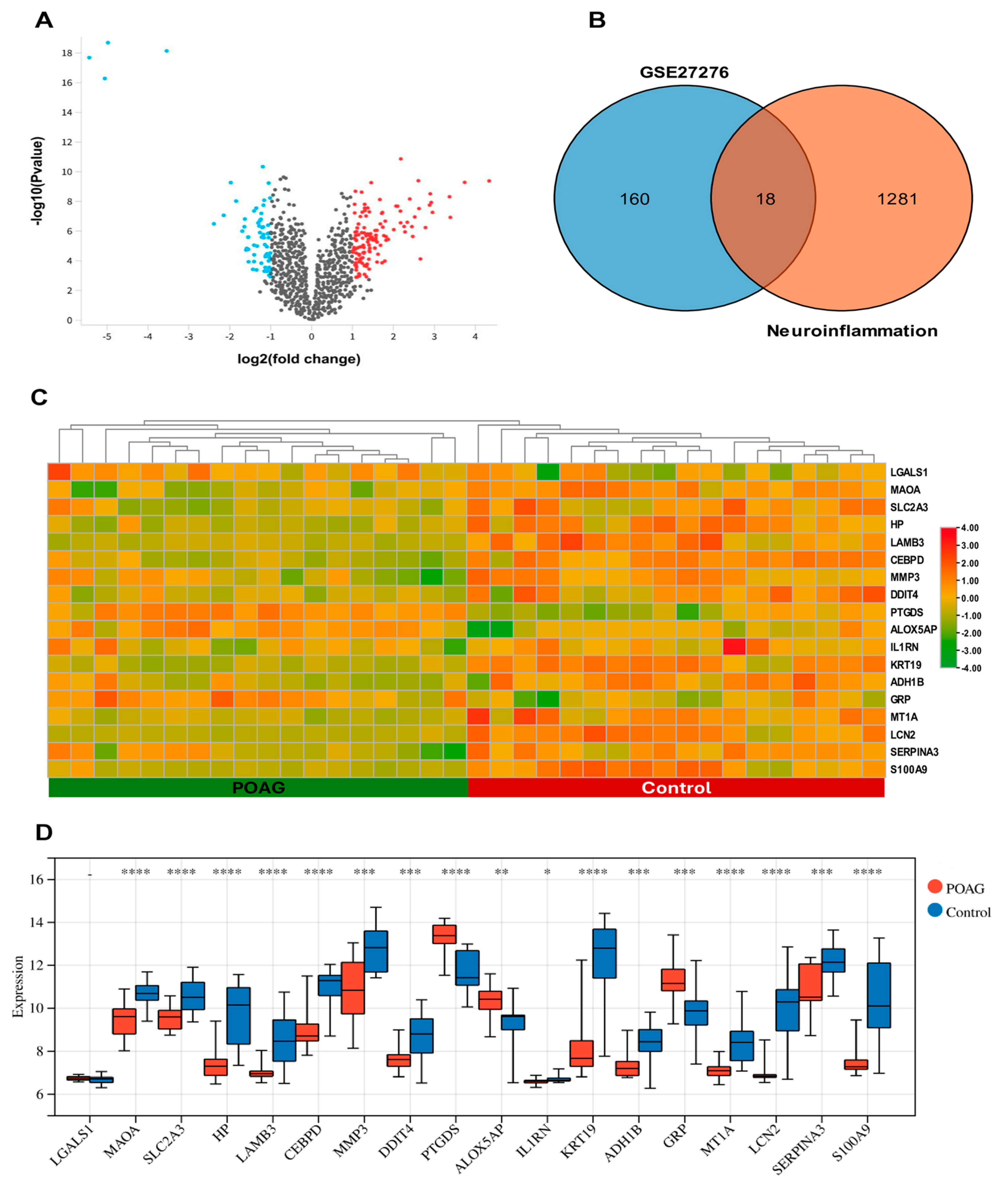
Figure 1. (A) Volcano plot for DEGs between the normal and POAG groups. The red dots indicate upregulated genes, while the blue dots signify downregulated genes. (B) There is an overlap of 18 DENIGs between DEGs and neuroinflammation-related genes in the Venn diagram. (C) Heatmap of DENIGs analyzed from the dataset of GSE27276 and the GeneCards database. (D) The boxplot of 18 DENIGs in the POAG and normal control groups. * p < 0.05, ** p < 0.01, *** p < 0.001, and **** p < 0.0001.
Ijms 25 08193 g001
Figure 2. Pathway enrichment analysis of DENIGs. (A) KEGG analysis. (B) GO biological processes. (C) GO cellular components. (D) GO molecular functions.
Ijms 25 08193 g002
Figure 3. The PPI network and identification of hub genes. (A) The key modules of the DENIGs were identified through PPI network analysis. The PPI networks of the DENIGs in POAG patients was constructed using STRING. (B) SERPINA3, LCN2, MMP3, S100A9, IL1RN, and HP were identified as hub genes by cytoHubba.
Ijms 25 08193 g003
Figure 4. Comparison of hub gene (S100A9, SERPINA3, IL1RN, MMP3, LCN2, and HP) expression between the POAG and normal groups of the external validation dataset. The mRNA expression levels of SERPINA3 and IL1RN were elevated in POAG patients relative to the normal group. In contrast, the gene LCN2 was elevated in dataset GSE9944 and was non-significant in dataset GSE13534. (A) GSE13534; (B) GSE9944.
Ijms 25 08193 g004
Figure 5. Potential miRNA regulatory networks. The interaction networks consisted of six hub genes and 118 miRNAs. SERPINA3 and MMP3 are the most modulated genes, by 54 and 28 miRNAs, respectively.
Ijms 25 08193 g005
Figure 6. Expression of six hub genes in microglia, astrocytes, Müller cells, and RGC. SERPINA3 and HP showed upregulated expression in RGCs, while downregulation was observed in Müller cells and astrocytes. S100A9 and IL1RN were upregulated in microglial cells and had no expression in other cells. (A) SERPINA3; (B) HP; (C) S100A9; (D) IL1RN.
Ijms 25 08193 g006
Figure 7. The GWAS analysis was performed to explore the pathogenic regions linked to hub genes in POAG. (A) A Q-Q plot illustrated the SNP loci recognized by GWAS data, showing significant associations with glaucoma. The blue dots represent the p-values for each SNP (B) Critical SNPs are dispersed within genetically rich regions. Each dot represents an SNP. The x-axis indicates the chromosome, and the y-axis shows the link with glaucoma. Red Dots: SNPs on odd-numbered chromosomes. Blue Dots: SNPs on even-numbered chromosomes. The SNP pathogenic regions correspond to (C) SERPINA3, (D) LCN2, (E) MMP3, (F) S100A9, (G) IL1RN, and (H) HP.
Ijms 25 08193 g007
Figure 8. The ROC curve confirmed the diagnostic importance of hub genes for the POAG dataset GSE9944. The AUC areas of the six hub genes (A) SERPINA3, (B) LCN2, (C) MMP3, (D) S100A9, (E) IL1RN, and (F) HP were >0.7.
Ijms 25 08193 g008
Figure 9. Experimental validation of hub genes by real-time PCR experiments. Findings revealed a significant decrease in the mRNA levels of SA1009, IL1RN, and MMP3 in the glaucoma model group compared to the normal group, while SERPINA3 and LCN2 expression increased. (A) S100A9 (B) SERPINA3, (C) IL1RN, (D) MMP3, (E) LCN2, and (F) HP. ns = non-significant,* p < 0.05, ** p < 0.01 and *** p < 0.001.
Ijms 25 08193 g009
Figure 10. The overall process of identifying hub genes associated with neuroinflammation in POAG. GO: gene ontology, KEGG: Kyoto encyclopedia of genes and genomes, DEGs: differentially expressed genes, DENIGs: differentially expressed neuroinflammation genes, PPI: protein–protein interaction.
Ijms 25 08193 g010
Table 1. The 18 DENIGs in POAG patients compared to control patients.
IDGene SymbolGene TitleChangelog2 (Fold Change)log10
(p Value)
GI_13027803-SMMP3matrix metallopeptidase 3down−1.8984.308
GI_15718674-SALOX5AParachidonate 5-lipoxygenase activating proteinup1.1753.247
GI_27894318-AIL1RNinterleukin 1 receptor antagonistdown−1.5354.606
GI_28866959-SMT1Ametallothionein 1Adown−1.4145.486
GI_28872797-SCEBPDCCAAT/enhancer binding protein deltadown−2.25611.354
GI_33469954-SMAOAmonoamine oxidase Adown−1.4527.383
GI_34222290-SGRPgastrin releasing peptideup1.43.175
GI_34577060-SADH1Balcohol dehydrogenase 1B (class I), beta polypeptidedown−1.1024.372
GI_38455401-SLCN2lipocalin 2down−2.9017.469
GI_38505192-SPTGDSprostaglandin D2 synthaseup1.6366.538
GI_40217850-SKRT19keratin 19down−4.3168.929
GI_4557712-SLAMB3laminin subunit beta 3down−1.5545.392
GI_4826761-SHPhaptoglobindown−2.467.126
GI_5902089-SSLC2A3solute carrier family 2 member 3down−1.0214.429
GI_6006015-SLGALS1galectin 1up1.5984.509
GI_9506686-SDDIT4DNA damage inducible transcript 4down−1.2064.006
GI_9665246-SSERPINA3serpin family A member 3down−1.4744.358
GI_9845520-SS100A9S100 calcium binding protein A9down−2.7996.001
Table 2. GO and KEGG enrichment analyses for DENIGs in POAG.
TermDescriptionEnrichment FDR (p-Value)Gene
Count
Fold EnrichmentGenes
KEGG pathway
Path:hsa04657IL-17 signaling pathway0.0022836341.00537634LCN2 MMP3 S100A9
Path:hsa00350Tyrosine metabolism0.008155124270.62037037ADH1B MAOA
Path:hsa00982Drug metabolism-cytochrome P4500.019936646236.84541063ADH1B MAOA
Path:hsa04151PI3K-Akt signaling pathway0.13232693927.18173258LAMB3 DDIT4
Biological processes
GO:0010727negative reg. of hydrogen peroxide metabolic proc.0.0001619442953.375MMP3 HP
GO:0006953acute-phase response7.84 × 10−53200.7105263IL1RN SERPINA3 HP
GO:0002526acute inflammatory response0.000268135388.00384615HP IL1RN SERPINA3
GO:0098869cellular oxidant detoxification0.009726848271.28037383S100A9 HP
GO:0046916cellular transition metal ion homeostasis0.009977053265.18803419LCN2 S100A9
GO:1990748cellular detoxification0.009977053262.51639344S100A9 HP
GO:0097237cellular response to a toxic substance0.010245208258.66923077S100A9 HP
GO:0098754detoxification0.011070075251.88435374S100A9 HP
GO:0042742defense response to bacterium0.002732388332.13623596LCN2 HP S100A9
GO:0006954inflammatory response7.84 × 10−5521.37612108IL1RN S100A9 HP SERPINA3 MMP3
Cellular components
GO:0031838haptoglobin-hemoglobin complex0.0065484981346.6818182HP
GO:0071682endocytic vesicle lumen0.0120723761158.8958333HP
GO:0035580specific granule lumen0.000785589293.01219512LCN2 HP
GO:1904724tertiary granule lumen0.030363329156.91791045HP
GO:0031093platelet alpha granule lumen0.030363329154.47857143SERPINA3
GO:0072562blood microparticle0.001545822254.09219858SERPINA3 HP
GO:0034774secretory granule lumen8.47 × 10−6441.45108696LCN2 S100A9 SERPINA3 HP
GO:0060205cytoplasmic vesicle lumen8.47 × 10−6441.11590296LCN2 S100A9 SERPINA3 HP
GO:0031983vesicle lumen8.47 × 10−6440.89544236LCN2 S100A9 SERPINA3 HP
GO:0035578azurophil granule lumen0.040033456136.31904762SERPINA3
GO:0042581specific granule0.003126567235.97641509LCN2 HP
GO:0031012extracellular matrix0.001072635319.00415282S100A9 SERPINA3 MMP3
GO:0030312external encapsulating structure0.001072635318.97263682S100A9 SERPINA3 MMP3
GO:0062023collagen-containing extracellular matrix0.011593959216.83664459S100A9 SERPINA3
Molecular functions
GO:0005152interleukin-1 receptor antagonist activity0.01002467811271.166667IL1RN
GO:0035662Toll-like receptor 4 binding0.0100246781953.375S100A9
GO:0050543icosatetraenoic acid binding0.0100246781635.5833333S100A9
GO:0050544arachidonic acid binding0.0100246781635.5833333S100A9
GO:0050542icosanoid binding0.0103948261544.7857143S100A9
GO:0035325Toll-like receptor binding0.0114492661317.7916667S100A9
GO:0036041long-chain fatty acid binding0.0133501811254.2333333S100A9
GO:0005149interleukin-1 receptor binding0.0133501811224.3235294IL1RN
GO:0019966interleukin-1 binding0.0133501811224.3235294IL1RN
GO:0048019receptor antagonist activity0.0155684061181.5952381IL1RN
GO:0030547signaling receptor inhibitor activity0.027296208193.01219512IL1RN
GO:0016209antioxidant activity0.010024678279.44791667S100A9 HP
GO:0017171serine hydrolase activity0.010024678233.01731602HP MMP3
GO:0004175endopeptidase activity0.017891408215.34607646MMP3 HP
Table 3. List of primers used for qPCR.
GeneForward (5′-3′)Reverse (5′-3′)
LCN2CGTCCTAAATGGCCAACCCTTAGGAAGAGGGGGAGAAGCC
S100A9TGGCTGCCAAAACAGGATCTGCCCCAGAACCAAGGTCATT
SERPINA3GCTGAACTGCACTGTTGTGGATGCACACAGAGACCCACAG
MMP3ATCCCTTTTGATGGGCCTGGGGATGGAAGAGACGGCCAAA
IL1R1CTGATCATCCCGTGAGCCTCGTCTGGACTGTGGACATGCA
HPAGTGAGAATGCGACAGCCAATCAGTTGCCCTCACGTACAC
GAPDHAGGTCGGTGTGAACGGATTTGGGGGTCGTTGATGGCAACA
Disclaimer/Publisher’s Note: The statements, opinions and data contained in all publications are solely those of the individual author(s) and contributor(s) and not of MDPI and/or the editor(s). MDPI and/or the editor(s) disclaim responsibility for any injury to people or property resulting from any ideas, methods, instructions or products referred to in the content.