Article

Mutation Spectrum of GAA Gene in Pompe Disease: Current Knowledge and Results of an Italian Study

Institute for Biomedical Research and Innovation (IRIB), National Research Council (CNR), 90146 Palermo, Italy
*
Author to whom correspondence should be addressed.
These authors contributed equally to this work.
Int. J. Mol. Sci. 2024, 25(17), 9139; https://doi.org/10.3390/ijms25179139
Submission received: 12 June 2024 / Revised: 25 July 2024 / Accepted: 4 August 2024 / Published: 23 August 2024

Abstract

:
Studying a patient with Pompe disease (PD) is like opening Pandora’s box. The specialist is faced with numerous clinical features similar to those of several diseases, and very often the symptoms are well hidden and none is associated with this rare disease. In recent years, scientific interest in this disease has been growing more and more, but still no symptom is recognized as key to a correct diagnosis of it, nor is there any specific disease marker to date. New diagnostic/therapeutic proposals on disease allow for the diffusion of knowledge of this pathology for timely diagnosis of the patient. Due to unawareness and difficulty in diagnosis, many adults with PD are diagnosed with great delay. In this article, we report and discuss current knowledge of PD and provide new data from work conducted on a cohort of 2934 Italian subjects recruited in recent years. A genetic analysis of the GAA gene was performed on patients with significant clinical signs and pathological enzyme activity to define the genetic profile of subjects. This identified 39 symptomatic PD subjects with low acid alpha-glucosidase enzyme activity and the presence of two causative mutations in GAA gene regions. Furthermore, 22 subjects with genetic variants of uncertain significance (GVUS) were identified.

1. Introduction

The lives of patients and families facing the challenges of rare and/or severely disabling diseases have changed compared to 20 years ago. Clinicians spend a lot of time studying their symptoms and trying to understand how to approach patients’ problems.
But sometimes that is not so easy. Time has a significant impact on clinician’s efforts and attention. This restriction could result in the clinician identifying the disease incorrectly and misdiagnosing it. Glycogenosis type II, also known as Pompe disease (PD), is an autosomal recessive disorder caused by mutations in the GAA gene, localized on chromosome 17, that lead to a deficiency in the acid alpha-glucosidase enzyme (GAA-enzyme). This disease leads to an accumulation of glycogen in lysosomes [1,2,3], which injures the heart, leg and arm muscles, and the muscles of respiration [4]. Pompe disease is classified by age of onset, organ involvement, and severity and rate of progression. It presents two spectra of phenotypes named infantile-onset Pompe disease (IOPD) and late-onset Pompe disease (LOPD), with an incidence of 40,000–50,000 in European and American populations [5,6,7,8]. The infantile form, the most severe, is characterized by early-age onset, severe hypertrophic cardiomyopathy, marked hypotonia, macroglossia, hepatomegaly, it usually presents in the first days or weeks of life, and it has an invariably fatal outcome within one year of age. The late-onset form (juvenile or adult) is characterized by skeletal myopathy, fatigue, muscle weakness, hyperCKemia and possible respiratory failure [9,10,11].
The time of onset and phenotypes of LOPD are variable, and patients are likely to have manifestations from the fifth decade of life [4,12,13,14,15]. Mutations in the GAA gene, inherited in an autosomal recessive way, are very heterogeneous. They can be point mutations that can affect the functionality and stability of the protein or the splicing process, or small and large deletions and insertions [16,17,18]. Taverna et al. reported that GAA variants are clustered in three critical regions of the gene: exon 2, which contains the start codon exons 10 and 11, which encode for the catalytic site, and exon 14 which encodes for a highly conserved region of GAA protein [17]. The Pompe disease GAA variants database http://www.pompevariantdatabase.nl, (accessed on 26 April 2024) which was last updated in 2023, reports more than 911 disease-associated mutations [19].
Currently, enzyme replacement therapy (ERT) with GAA-enzyme is the first and unique treatment available for PD, and was approved in 2006 [20]. In order to achieve optimal results, ERT should be started before symptoms are evident: indeed, it is well established that early initiation of ERT in IOPD improves survival, reduces the need for mechanical ventilation, results in early independent ambulation and improves the patient’s overall quality of life [21]. In 2014, during the 208th International Workshop of the European Neuromuscular Center, the European Pompe Consortium was formed, bringing together a group of experts from 11 different European countries, all with long experience in the treatment and follow-up of patients with PD, to outline recommendations for the initiation and discontinuation of ERT in adult patients [22]. It was observed that patients receiving ERT present significant differences in their responses. These responses are heterogeneous and influenced by many factors, such as age at treatment initiation, cross-reactive immune material (CRIM) status, ACE genotype, sustained intermediate titers (SIT), muscle fiber type involvement, extent of pre-existing pathology, high and sustained antibody titers (HSAT), and ACTN genotype [23]. Cross-reactive immunological material (CRIM) is a measurement of natural GAA production and an important factor influencing patients’ responses to ERT. In general, CRIM-positive subjects tolerate ERT well. However, CRIM-negative subjects and some classified as CRIM-positive have a low response to ERT due to complications from the immune response to the drug [23,24,25].
To update the status of Italian subjects with both childhood onset (IOPD) and late onset (LOPD) in Italy, we present the results collected in recent years on samples that came to our attention. We evaluated the enzyme activity and genetic study of mutations in the GAA gene. We employed a fluorimetric test to evaluate alpha-glucosidase activity on blood spots in 2934 patients who presented with symptoms linked to PD, or for newborn screening. In the whole cohort of patients analyzed with reduced or borderline activity, genetic testing was done, and we found 39 patients with two causative mutations in GAA gene connected to PD, and 22 subjects with GVUS variants. These results not only underscore the critical importance of early and accurate diagnosis but also highlight the essential collaboration between clinicians and geneticists. The integration of enzyme activity screening and genetic testing represents a major advancement in our ability to identify and manage PD, ensuring patients receive timely and appropriate care. By presenting these data, we aim to emphasize the substantial progress made in the field of PD research and diagnosis in Italy, showcasing how interdisciplinary efforts are pivotal to advancing medical knowledge and patient outcomes. Our research indicates that Italian patients with Pompe disease present a diverse genetic landscape, with specific mutations in the GAA gene contributing to disease severity and progression. The variability in symptom onset, ranging from childhood to adulthood, underlines the complexity of genotype–phenotype correlations in this population. In addition, enzymatic and genetic testing over the years has allowed more precise diagnoses and management strategies tailored to the individual profiles of our patients. In conclusion, early diagnosis remains critical for optimizing outcomes in Pompe disease. In Italy, efforts to enhance awareness among healthcare providers have facilitated timely identification of symptoms such as muscle weakness, respiratory insufficiency, and cardiac involvement. Diagnostic modalities including enzyme assays, genetic testing, and imaging techniques play pivotal roles in confirming the disease and monitoring its progression.

Sentinel Symptoms of PD, Diagnostic Tests, and Therapeutic Procedures

The PD challenge for clinicians begins when manifestations are found in smooth and skeletal muscle, endothelial cells and motor neurons, and in heart in the IOPD form.
The study conducted by Thurberg et al. showed that muscle cells at the start of the disease have small glycogen-filled lysosomes that grow in size and number over time. The clinical symptoms are either not visible or absent at this stage [26]. As the disease progresses, the lysosomal membranes break, thus allowing glycogen to enter into cytoplasm. Muscle damage begins and the patient experiences more visible symptoms, such as severe myopathy, which results in decreased muscle function. These can be defined as sentinel symptoms accompanied by features such as respiratory failure, high hyperCKemia (CK) values, etc. But how to begin to identify the disease?
If PD is clinically suspected, diagnosis typically involves thorough enzymatic and genetic analyses. These assessments are conducted using dry blood samples (DBS), cultured fibroblasts, or muscle tissue. In DBS samples, the presence of polymorphonuclear neutrophil-derived α-glucosidase (maltase-glucoamylase, MGA) can interfere with the accurate measurement of enzymatic activity at acidic pH levels. Techniques such as immunocapture or the competitive inhibition of MGA activity with maltose or acarbose have been utilized to eliminate the interfering effects of MGA activity in DBS extracts [27,28]. Alpha acid glucosidase has optimal activity at pH 3.7–4.5 and hydrolyzes alpha 1-4 and alpha 1-6 bonds in glucose polymers. Only if enzyme activity is below normal, borderline, or absent is a genetic assay through Sanger sequencing conducted. The study of the entire portion of the GAA gene for mutation research is important to the prediction of disease risk, identification of healthy carriers, and for phenotype–genotype correlations.
Because PD is an autosomal recessive disease, it is critical to investigate and know the family background after the proband is identified. Family members of an affected person have a higher-than-average chance of having the disease themselves or of being disease carriers. It is very important to verify this possibility through family screening because early diagnosis is necessary for the effective treatment and management of symptoms. It is, however, necessary that further surveys be carried out because the association between genetic factors and symptom profile is not well understood [29]. Genetic heterogeneity associated with a phenotype, and the phenotypic pleiotropy associated with a genotype, can mislead clinical presentation [30].
In the study by Salunkhe et al., the basic concepts of genetic variations and available genetic tests were explained [30]. In the study by Taverna et al., an overview of PD was presented, focusing on pathogenesis, clinical phenotypes, molecular genetics, and the role of miRNAs as potential biomarkers for PD [17]. Another group of international experts met in 2006 to discuss different aspects of PD, including genetic counseling [14]. The guideline aims to increase public awareness of this disease and introduce the broad spectrum of related symptoms in order to accelerate diagnosis and take advantage of emerging therapies. In our study, all infants with infantile-onset Pompe disease (IOPD) displayed a clinical presentation characterized by severe and progressive hypotonia (“floppy baby”- or “rag doll”-type), hypertrophic cardiomyopathy, respiratory insufficiency, and delays or regression in motor development milestones. For those with late-onset Pompe disease (LOPD), we observed elevated creatine kinase (CK) levels, feeding difficulties, and progressive motor dysfunction such as difficulty raising the upper limbs, climbing stairs, and rising from a low chair, often leading to respiratory failure. Additionally, moderate hepatomegaly was noted in a few cases. Some subjects had previous muscle biopsy results available.
Various other symptoms were also identified to varying extents, including dysphagia, osteoporosis, sleep apnea, small-fiber neuropathy, impaired gastric function, urinary tract involvement (monitored by Glucose Tetrasaccharide (Glc4) Urine Testing), pain, and fatigue. These findings underscore the systemic nature of Pompe disease and emphasize symptoms that unfortunately may not have been promptly recognized as related to the disease. Regarding the genetic analysis of our samples, we did not identify a clear genotype–phenotype correlation. We believe that other factors beyond genetic mutations may influence the clinical manifestations of Pompe disease, factors that are not yet fully understood. Therefore, all subjects investigated in our study represent hypothetical Pompe disease patients. This comprehensive characterization highlights the diverse clinical spectrum and challenges in diagnosing Pompe disease, emphasizing the critical need for increased awareness and early intervention strategies to improve patient outcomes.
The interest of the scientific community in the treatment of inherited diseases has focused on the continuous development of drugs and therapies that use molecules to modify gene expression, and correct or compensate for an abnormal phenotype. In particular, strategies include resolution of disease enzyme deficiency (enzyme replacement therapy-ERT), replacement of the gene encoding the deficient enzyme (gene therapy), and cell dispensing that produces the deficient protein (hematopoietic stem cell transplantation) [31,32,33,34]. Other therapeutic strategies include administering drugs that enhance the activity of mutated proteins (enzyme-enhancement therapy), or reducing substrate biosynthesis, the accumulation of which results in toxicity (substrate reduction therapy) [35,36,37,38] (Figure 1).
To date, the only specific treatment available for PD patients is enzyme replacement therapy, which aims to arrest the natural course of the disease [28,39,40]. Both the success and limitations of ERT have provided new insights into the disease’s pathophysiology and motivated the scientific community to develop the next generation of therapies that will be introduced into clinical procedure [21,41,42,43].
The replacement enzyme is produced biotechnologically and administered intravenously. ERT with GAA-enzyme significantly prolonged the survival of IOPD patients, with marked improvements in symptomatology. In LOPD, it is only effective in slowing the progression of the disease. Consequently, this treatment is more effective in the IOPD than the LOPD form. In addition, systemic administration through the blood does not effectively target all affected organs [44,45]. However, it is unquestionable that ERT has provided many benefits, adding years and quality of life to PD patients, but it is equally clear that there is a need to improve and optimize the current drugs, which still have several limitations [31,40,46,47]. In recent years, there has been a significant increase in the number of research projects, both in vitro and in vivo mouse models, with the goal of improving the PD patient’s treatment [48]. In our study of 39 patients diagnosed with late-onset Pompe disease (LOPD), Italian clinicians monitored the effects of enzyme replacement therapy (ERT) on their motor function over time. Upon initiating therapy, patients generally exhibited a partial improvement in their motor abilities within the first year. This initial phase of treatment showed encouraging signs, with noticeable gains in muscle strength and mobility. As therapy continued beyond the first year, the trajectory of disease progression changed. Rather than continuing to show significant improvements, the patients’ motor functions stabilized. This stabilization indicates that, while the rapid initial improvements tapered off, the therapy played a crucial role in maintaining the gains achieved during the first year and in slowing the overall progression of LOPD. These findings suggest that enzyme replacement therapy provides substantial early benefits in terms of motor function, and while it may not lead to continuous improvement, it is effective in preventing further decline. This highlights the importance of early and sustained treatment in managing LOPD and improving patients’ quality of life.

2. Results

We evaluated the enzyme activity of 2934 subjects, 1670 males and 1264 females, divided by age. Mean age was 31 years at time of analysis: 165 subjects aged 0 to 1 years old suspected to have IOPD, and 2769 subjects over 1 year old suspected to have LOPD. The typical clinical features reported in Toscano et al. were found in all our examined subjects [49]. Due to the importance of scientific health research to date, a study of subjects from different hospital departments was conducted to delineate the clinical phenomenon in an observational and analytical view. DBS received with suspected PD came from: 1380 neurology (47.03%), 40 metabolic diseases (1.36%), 473 pediatrics (16.12%), 116 pneumology (3.95%), 177 internal medicine (6.03%), 345 rheumatology (11.76%), 322 from other departments such as cardiology, anesthesia and reanimation, hematology, and medical genetics (10.97%), and 81 from unspecified departments (2.76%) (Figure 2).
The first step was to assess the outcome of enzyme activity. Of the 2934 individuals studied by DBS, 2489 showed normal acid α-glucosidase activity (≥6 nmol/mL/h), 105 individuals had no or reduced activity of the enzyme (≤3 nmol/mL/h), and 340 had an enzyme activity between 3 and 6 nmol/mL/h.
In subjects with enzyme activity <6 nmol/mL/h, we performed genetic analysis on 445 subjects to research and identify any change in allelic variants of the GAA gene. In 105 subjects with none or severely reduced enzyme activity, we identified 39 patients who had either homozygosity for one mutation, or compound heterozygosity for two mutations in the GAA gene, known in the literature to be responsible for PD. Segregation analysis in the patients’ family with Sanger sequencing confirmed that, in the 31 cases of compound heterozygote, mutations were on both homologous chromosomes. This confirmed the diagnosis of PD from a genetic point of view for all patients analyzed.
In the remaining 51 of the 105 subjects were false positives in which the enzymatic analysis showed values below normal, but no causative mutations in the GAA gene were found in the genetic analysis. The remaining 15 out of the 105 subjects had a causative mutation in simple heterozygosity. Therefore, the reduced enzyme activity could be due to a leukopenia condition and/or improper storage/transport of the sample.
The most common mutation reported in our analyzed samples was the pathogenic c.-32-13 T>G variant (homozygous or with a second pathogenic allele), located in intron 1 documented in the literature as a causative mutation of PD. All variants found in our molecular study associated with the c.-32-13 T>G mutation were reported in the bibliographies and public databases of PD mutations (Pompe variant database, clinvar). Specifically, 25 of 39 patients reported this causative mutation. Of these 25, 5 patients had the mutation in homozygosity and 20 patients in compound heterozygosity (Table 1).
In addition, the study was further investigated by conducting MLPA in patients who had only one mutation on Sanger sequencing analysis. The assay was performed in 149 subjects. This method revealed in four subjects the presence of exonic deletions in the GAA gene causative of PD in association with other causative point mutations. In detail, the deletions found in our patients were located in exon 9, exon 17, exon 18, and exon 19, respectively, in the first four cases.
In the other four, three investigations were conducted for a family segregation study in which, in addition to the deletion, benign polymorphisms were found. One patient showed a deletion in exon 12.
Four of these eight subjects had the deletion in association with other causative point mutations, as shown in Table 1, and for the remaining four no additional mutations in the GAA gene were found.
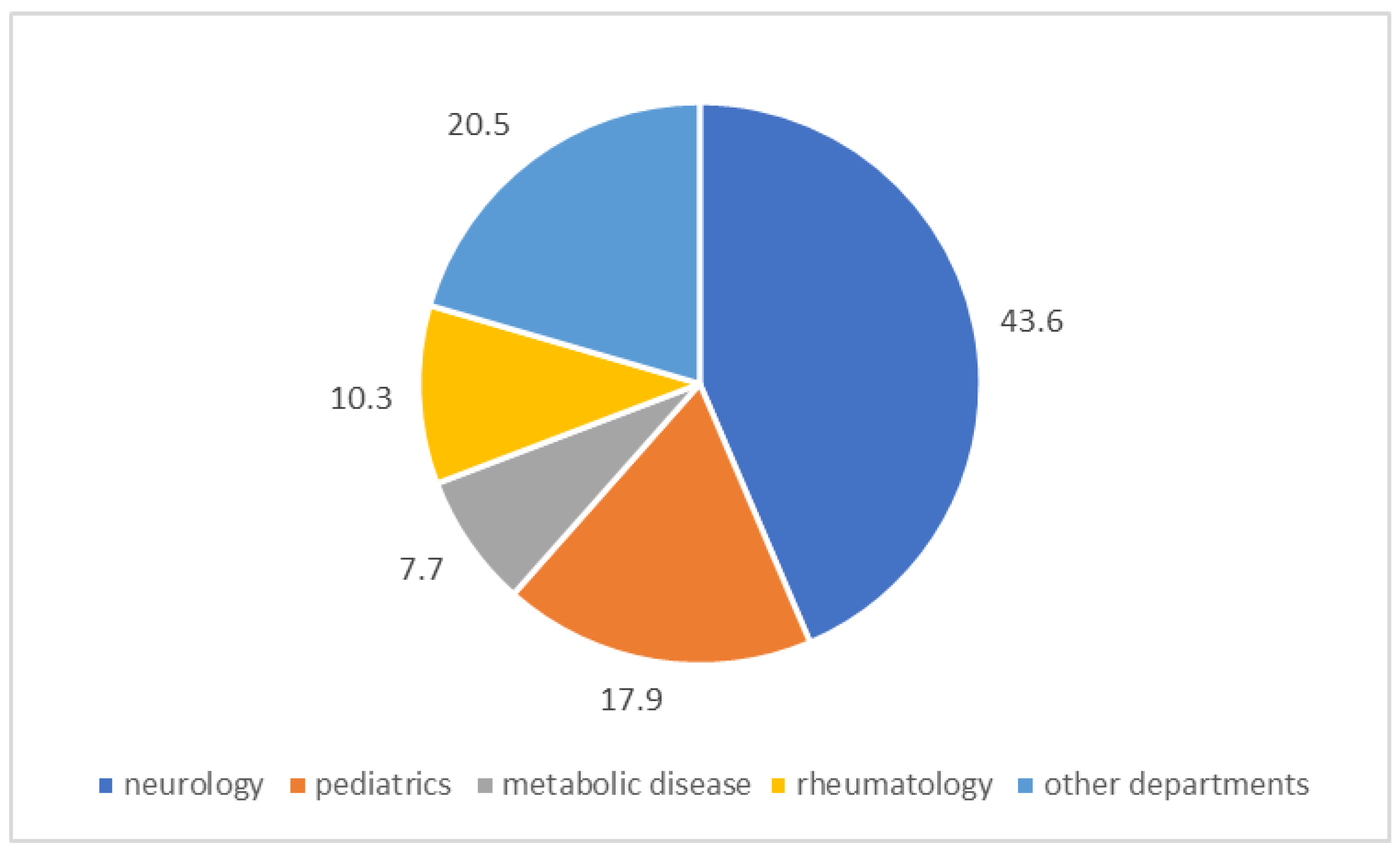
In our study, we identified a total of 39 subjects with PD, including 27 with two classic mutations in compound heterozygosity, 8 with mutations in homozygosity, and 4 with one causative mutation associated with a large deletion. Specifically, as shown in Figure 3, these subjects came from the following departments: 17 from neurology (43.6%), 4 from rheumatology (10.3%), 7 from pediatrics (17.9%), 3 from metabolic diseases (7.7%), and 8 from other departments (20.5%).
Furthermore, our study identified five allelic variants in the sequence of the GAA gene (c.547-67C>G, c.547-39T>G, R437H, L641V, and L705P) that had not been previously described in the literature. In the entire cohort of patients analyzed with enzyme activity between 3 and 6 nmol/mL/h, we also detected 22 patients with variants of uncertain significance (GVUS) mutations in heterozygosity (Figure 4).
In our study, we investigated all genetic polymorphisms as a possible factor contributing to PD. These were classified according to the type of genetic polymorphism investigated and whether the result favored or did not favor association with PD. As asserted by Ravaglia et al., genetic variations impacting muscle structure and metabolism could potentially influence the phenotype of metabolic myopathies [50].
We found in our subjects frequently benign polymorphisms, as reported in databases of PD-associated mutations and annotated as benign variants, and which were thus unrelated to PD.
These statistical results collected over a recent period geographically cover the entire Italian demographic area. Therefore, this clinical study plays an important role in knowing and being able to identify typical signs and symptoms of PD.

3. Discussion

The diagnosis of PD poses challenges due to its diverse clinical presentations among patients, the rarity of the condition, and the complexities involved in recognizing its symptoms. Consequently, it often remains underdiagnosed. This article aimed to highlight the increasing number of PD Italian cases identified through dried blood spot (DBS) testing in recent years. Additionally, this study incorporated the assessment of quantitative variations in the GAA gene using the multiple ligation-dependent probe amplification (MLPA) assay to enhance diagnostic accuracy. As discussed, for autosomal recessive diseases, the presence of two causative mutations in compound heterozygosity or homozygosity defines an individual with PD. To determine whether the two mutations are in cis or trans positions, a genetic investigation was conducted within the family to see how the mutations were transmitted in their chromosomes and thus inherited by the individual under study. The cis configuration occurs when two mutant alleles are on the same homologous chromosome, while the trans configuration occurs when the two mutant alleles are on different homologous chromosomes.
Our study concluded that Italian patients exhibited a characteristic genetic profile similar to that of the broader European population, with the c.-32-13T>G variant being the most prevalent. Genetic analysis also indicated that the functional significance and potential interrelationships of numerous benign polymorphisms, as well as other genetic variants of uncertain significance (GVUS), are still relatively unknown. We can conclude that our results allow us to set the cutoff for enzyme activity at <3 nmol/mL/h for the identified cases of PD.
For MLPA, large deletions make up only 1.5% of known variants, and the deletion of exon 18 was a common variant in Caucasian patients. A more systematic approach, as previously described in our study, could lead to the identification of new variants associated with PD and allow for a definitive diagnosis of patients. The implementation of MLPA should be considered to complete the genetic analysis of the most challenging cases, where there is a strong clinical suspicion supported by signs and symptoms associated with pathological enzyme activity values. Given the extensive heterogeneity of the GAA gene, both in enzymatic and genetic characteristic terms, additional efforts are required to investigate the potential influence of environmental and epigenetic factors on phenotype modulation. Furthermore, for improved patient classification, future studies should be conducted through close collaboration between neuropsychiatry divisions and genetic laboratories. In summary, prompt diagnosis is crucial for maximizing outcomes of Pompe disease. In Italy, initiatives to raise awareness among healthcare providers have facilitated timely recognition of symptoms such as muscle weakness, respiratory insufficiency, and cardiac involvement. Diagnostic methods such as enzyme assays, genetic testing, and imaging techniques are essential for confirming the disease and tracking its progression. This information could be useful for developing new biomarkers for this disease. The advent of enzyme replacement therapy (ERT) has revolutionized the management of Pompe disease, offering a targeted approach to supplementing deficient GAA enzyme activity. Italian patients have benefited from access to ERT, which aims to alleviate symptoms, improve muscle function, and enhance quality of life. Ongoing research explores novel therapeutic avenues, including gene therapy and pharmacological chaperones, to further optimize treatment outcomes and address challenges such as immune response and sustained efficacy. In conclusion, Pompe disease presents complex clinical and genetic landscapes among Italian patients, underscoring the importance of ongoing research and multidisciplinary collaboration in enhancing diagnostic accuracy, therapeutic efficacy, and overall patient outcomes. Continued efforts are essential to improve disease-management strategies, expand treatment options, and ultimately improve the lives of individuals living with Pompe disease, both in Italy and globally.

4. Materials and Methods

4.1. Patients

Our data collected in recent years allowed the identification of patients with a pathological value of alpha-acid glucosidase enzyme activity and the presence of GAA mutations. The biochemical and genetic investigations that led to PD diagnosis were performed at the Centre for Research and Diagnosis of Lysosomal Storage Disorders at IRIB-CNR in Palermo, and were approved by the Hospital Ethics Committee of the University of Palermo (Sicily, Italy). Signed informed consent was obtained from patients. For each patient, clinical history and laboratory data were collected in a case report form.

4.2. Sample Collection

The peripheral blood of the patient was collected using EDTA as an anticoagulant, and dried in a specific adsorbent paper spot (DBS). Blood was dropped and allowed to dry for 24 h at room temperature before being sent (with 1–2 days of transport) to the centralized analytical laboratory in Palermo. Sample storage after use was conducted at a temperature of 4 °C. The same samples from analyzed subjects were used for multiple analyses (enzymatic and genetic assay).

4.3. Enzymatic Assay

The analysis of GAA enzyme activity was conducted on dried blood spots. The method used was described by Chamoles et al. [27], with modifications to improve the precision of the enzymatic activity measurement. DBS samples were examined by fluorometry techniques. The fluorometric method uses the substrate 4-methylumbelliferyl-α-D-glucoside [51]. Genetic analysis was performed in patients with GAA activity less than 6 nmol/mL/h.

4.4. DNA Extraction

DNA extraction was performed by the whole DBS spot, corresponding to approximately 3 µL of blood per punch, followed by the addition of 310 microliters of the digestion buffer (Buffer G2—Qiagen GmbH, Hilden, Germany) and 15 microliters of the Proteinase K solution (Qiagen GmbH lot No. 175025821) per each individual sample. DNA extraction was performed using a Qiagen EZ1 advanced XL (QIAGEN Redwood City-1001 Marshall Street-Redwood City, CA, USA) automatic extractor in combination with the EZ1 DNA investigator kit.

4.5. Genetic Analysis-PCR, Sanger Sequencing, and MPLA

Eleven pairs of primers were designed to analyze twenty target regions containing the twenty exons of the GAA gene. PCR products were purified and analyzed by Sanger sequencing, using an automated DNA sequencer (Eppendorf-Donau-City-Straße 11-13, 3. OG 1220 Wien, Austria) to identify mutations. Multiplex ligation-dependent probe amplification (MLPA) is a relatively new diagnostic method to evaluate large deletions and duplications undetectable by GAA gene sequencing, using a very small amount of DNA. This analysis was conducted using the kit “SALSA MLPA Probemix P453 GAA” MRC-Holland. Nomenclature of the variants in this article refer to NM_000152.5(GAA) (https://www.ncbi.nlm.nih.gov/clinvar, accessed on 1 August 2024).

5. Conclusions

To satisfy the ongoing need for knowledge and to promote research, a study was conducted in our laboratories to describe biochemical findings and the mutation spectrum of the GAA gene for PD patients. Although this metabolic disorder is caused by a unique enzyme deficiency, it appears with a wide range of clinical phenotypes and with considerable variability in progression speed, degree of organ involvement, and disease severity. This is related to the number of known pathogenic mutations, their kind (nonsense, missense, framesahift), and their mutual interaction with the rest of the genome [52,53,54,55,56,57]. This report presents a study of Italian subjects with PD, highlighting the importance of knowledge of the disease so that the clinician can recognize it and request enzyme and genetic counselling to avoid misdiagnosis.
This report presents a work on Pompe disease, highlighting the importance of genetic counseling and enzyme testing to avoid misdiagnosis. Diagnostic delay or misdiagnosis in a patient with PD signs and symptoms are the most critical points in determining the evolution of the patient’s clinical and therapeutic history. The right strategic approach is the most effective solution to improve the course of the disease and patient’s outcome. In addition, the possibility of extending the genetic study to family members of identified affected probands promotes not only early diagnosis but also timely therapeutic intervention.

Author Contributions

Conceptualization, M.M., A.L.C., G.D. and P.C.; methodology, M.M. and A.L.C.; formal analysis, C.Z. and P.C.; investigation, D.F., M.G. and E.M.; writing—original draft preparation, M.M. and A.L.C.; reviewing original draft, G.D. and P.C.; supervision, G.D. and P.C. All authors have read and agreed to the published version of the manuscript.

Funding

This research received no external funding.

Institutional Review Board Statement

The study was conducted in accordance with the Declaration of Helsinki and approved by the Hospital Ethics Committee of the University of Palermo.

Informed Consent Statement

Informed consent was obtained from all subjects involved in the study.

Data Availability Statement

The raw data supporting the conclusions of this article will be made available by the authors on request.

Acknowledgments

We thank Marco Zora and Emanuela Maria Marsana (IRIB-CNR) for their excellent work on enzymatic activity assays.

Conflicts of Interest

The authors declare no conflicts of interest.

References

  1. Raben, N.; Plotz, P.; Byrne, B.J. Acid alpha-glucosidase deficiency (glycogenosis type II, Pompe disease). Curr. Mol. Med. 2002, 2, 145–166. [Google Scholar] [CrossRef]
  2. Herzog, A.; Hartung, R.; Reuser, A.J.; Hermanns, P.; Runz, H.; Karabul, N.; Gökce, S.; Pohlenz, J.; Kampmann, C.; Lampe, C.; et al. A cross-sectional single-centre study on the spectrum of Pompe disease, German patients: Molecular analysis of the GAA gene, manifestation and genotype-phenotype correlations. Orphanet J. Rare Dis. 2012, 7, 35. [Google Scholar] [CrossRef] [PubMed]
  3. van den Hout, H.M.; Hop, W.; van Diggelen, O.P.; Smeitink, J.A.; Smit, G.P.; Poll-The, B.T.; Bakker, H.D.; Loonen, M.C.; de Klerk, J.B.; Reuser, A.J.; et al. The natural course of infantile Pompe’s disease: 20 original cases compared with 133 cases from the literature. Pediatrics 2003, 112, 332–340. [Google Scholar] [CrossRef] [PubMed]
  4. Hirschhorn, R.; Huie, M.L. Frequency of mutations for glycogen storage disease type II in different populations: The delta525T and deltaexon 18 mutations are not generally "common" in white populations. J. Med. Genet. 1999, 36, 85–86. [Google Scholar]
  5. De Filippi, P.; Saeidi, K.; Ravaglia, S.; Dardis, A.; Angelini, C.; Mongini, T.; Morandi, L.; Moggio, M.; Di Muzio, A.; Filosto, M.; et al. Genotype-phenotype correlation in Pompe disease, a step forward. Orphanet J. Rare. Dis. 2014, 9, 102. [Google Scholar] [CrossRef] [PubMed]
  6. Chien, Y.H.; Lee, N.C.; Thurberg, B.L.; Chiang, S.C.; Zhang, X.K.; Keutzer, J.; Huang, A.C.; Wu, M.H.; Huang, P.H.; Tsai, F.J.; et al. Pompe disease in infants: Improving the prognosis by newborn screening and early treatment. Pediatrics 2009, 124, e1116–e1125. [Google Scholar] [CrossRef]
  7. Er, T.K.; Chen, C.C.; Chien, Y.H.; Liang, W.C.; Kan, T.M.; Jong, Y.J. Development of a feasible assay for the detection of GAA mutations in patients with Pompe disease. Clin. Chim. Acta 2014, 429, 18–25. [Google Scholar] [CrossRef]
  8. Mechtler, T.P.; Stary, S.; Metz, T.F.; De Jesús, V.R.; Greber-Platzer, S.; Pollak, A.; Herkner, K.R.; Streubel, B.; Kasper, D.C. Neonatal screening for lysosomal storage disorders: Feasibility and incidence from a nationwide study in Austria. Lancet 2012, 379, 335–341. [Google Scholar] [CrossRef]
  9. Montalvo, A.L.; Bembi, B.; Donnarumma, M.; Filocamo, M.; Parenti, G.; Rossi, M.; Merlini, L.; Buratti, E.; De Filippi, P.; Dardis, A.; et al. Mutation profile of the GAA gene in 40 Italian patients with late onset glycogen storage disease type II. Hum. Mutat. 2006, 27, 999–1006. [Google Scholar] [CrossRef]
  10. Martiniuk, F.; Chen, A.; Mack, A.; Arvanitopoulos, E.; Chen, Y.; Rom, W.N.; Codd, W.J.; Hanna, B.; Alcabes, P.; Raben, N.; et al. Carrier frequency for glycogen storage disease type II in New York and estimates of affected individuals born with the disease. Am. J. Med. Genet. 1998, 79, 69–72. [Google Scholar] [CrossRef]
  11. Tang, H.; Feuchtbaum, L.; Sciortino, S.; Matteson, J.; Mathur, D.; Bishop, T.; Olney, R.S. The First Year Experience of Newborn Screening for Pompe Disease in California. Int. J. Neonatal. Screen. 2020, 6, 9. [Google Scholar] [CrossRef] [PubMed]
  12. Sawada, T.; Kido, J.; Nakamura, K. Newborn Screening for Pompe Disease. Int. J. Neonatal. Screen 2020, 6, 31. [Google Scholar] [CrossRef]
  13. Rairikar, M.V.; Case, L.E.; Bailey, L.A.; Kazi, Z.B.; Desai, A.K.; Berrier, K.L.; Coats, J.; Gandy, R.; Quinones, R.; Kishnani, P.S. Insight into the phenotype of infants with Pompe disease identified by newborn screening with the common c.-32-13T>G "late-onset" GAA variant. Mol. Genet. Metab. 2017, 122, 99–107. [Google Scholar] [CrossRef]
  14. Kishnani, P.S.; Steiner, R.D.; Bali, D.; Berger, K.; Byrne, B.J.; Case, L.E.; Crowley, J.F.; Downs, S.; Howell, R.R.; Kravitz, R.M.; et al. Pompe disease diagnosis and management guideline. Genet. Med. 2006, 8, 267–288. [Google Scholar] [CrossRef]
  15. Slonim, A.E.; Bulone, L.; Ritz, S.; Goldberg, T.; Chen, A.; Martiniuk, F. Identification of two subtypes of infantile acid maltase deficiency. J. Pediatr. 2000, 137, 283–285. [Google Scholar] [CrossRef] [PubMed]
  16. Anna, A.; Monika, G. Splicing mutations in human genetic disorders: Examples, detection, and confirmation. J. Appl. Genet. 2018, 59, 253–268. [Google Scholar] [CrossRef]
  17. Taverna, S.; Cammarata, G.; Colomba, P.; Sciarrino, S.; Zizzo, C.; Francofonte, D.; Zora, M.; Scalia, S.; Brando, C.; Curto, A.L.; et al. Pompe disease: Pathogenesis, molecular genetics and diagnosis. Aging 2020, 12, 15856–15874. [Google Scholar] [CrossRef]
  18. El Marabti, E.; Abdel-Wahab, O. Therapeutic Modulation of RNA Splicing in Malignant and Non-Malignant Disease. Trends Mol. Med. 2021, 27, 643–659. [Google Scholar] [CrossRef]
  19. Malekkou, A.; Theodosiou, A.; Alexandrou, A.; Papaevripidou, I.; Sismani, C.; Jacobs, E.H.; Ruijter, G.J.G.; Anastasiadou, V.; Ourani, S.; Athanasiou, E.; et al. Variants associated with reduced enzymatic activity but lack of Pompe-related symptoms, incidentally identified by exome sequencing. Mol. Genet. Metab. Rep. 2023, 36, 100997. [Google Scholar] [CrossRef]
  20. Jastrzębska, A.; Kostera-Pruszczyk, A. Multisystem presentation of Late Onset Pompe Disease: What every consulting neurologist should know. Neurol. Neurochir. Pol. 2023, 57, 143–150. [Google Scholar] [CrossRef]
  21. Sun, B.; Bird, A.; Young, S.P.; Kishnani, P.S.; Chen, Y.T.; Koeberl, D.D. Enhanced response to enzyme replacement therapy in Pompe disease after the induction of immune tolerance. Am. J. Hum. Genet. 2007, 81, 1042–1049. [Google Scholar] [CrossRef]
  22. van der Ploeg, A.T.; Kruijshaar, M.E.; Toscano, A.; Laforêt, P.; Angelini, C.; Lachmann, R.H.; Pascual Pascual, S.I.; Roberts, M.; Rösler, K.; Stulnig, T.; et al. European consensus for starting and stopping enzyme replacement therapy in adult patients with Pompe disease: A 10-year experience. Eur. J. Neurol. 2017, 24, 768-e31. [Google Scholar] [CrossRef]
  23. Desai, A.K.; Kazi, Z.B.; Bali, D.S.; Kishnani, P.S. Characterization of immune response in Cross-Reactive Immunological Material (CRIM)-positive infantile Pompe disease patients treated with enzyme replacement therapy. Mol. Genet. Metab. Rep. 2019, 20, 100475. [Google Scholar] [CrossRef]
  24. Bali, D.S.; Goldstein, J.L.; Banugaria, S.; Dai, J.; Mackey, J.; Rehder, C.; Kishnani, P.S. Predicting cross-reactive immunological material (CRIM) status in Pompe disease using GAA mutations: Lessons learned from 10 years of clinical laboratory testing experience. Am. J. Med. Genet. Part C Semin. Med. Genet. 2012, 160, 40–49. [Google Scholar] [CrossRef]
  25. Curelaru, S.; Desai, A.K.; Fink, D.; Zehavi, Y.; Kishnani, P.S.; Spiegel, R. A favorable outcome in an infantile-onset Pompe patient with cross reactive immunological material (CRIM) negative disease with high dose enzyme replacement therapy and adjusted immunomodulation. Mol. Genet. Metab. Rep. 2022, 32, 100893. [Google Scholar] [CrossRef]
  26. Thurberg, B.L.; Lynch Maloney, C.; Vaccaro, C.; Afonso, K.; Tsai, A.C.; Bossen, E.; Kishnani, P.S.; O’Callaghan, M. Characterization of pre- and post-treatment pathology after enzyme replacement therapy for Pompe disease. Lab. Invest. 2006, 86, 1208–1220. [Google Scholar] [CrossRef]
  27. Chamoles, N.A.; Niizawa, G.; Blanco, M.; Gaggioli, D.; Casentini, C. Glycogen storage disease type II: Enzymatic screening in dried blood spots on filter paper. Clin. Chim. Acta 2004, 347, 97–102. [Google Scholar] [CrossRef]
  28. Kishnani, P.S.; Beckemeyer, A.A. New therapeutic approaches for Pompe disease: Enzyme replacement therapy and beyond. Pediatr. Endocrinol. Rev. 2014, 12, 114–124. [Google Scholar]
  29. Huie, M.L.; Chen, A.S.; Brooks, S.S.; Grix, A.; Hirschhorn, R. A de novo 13 nt deletion, a newly identified C647W missense mutation and a deletion of exon 18 in infantile onset glycogen storage disease type II (GSDII). Hum. Mol. Genet. 1994, 3, 1081–1087. [Google Scholar] [CrossRef]
  30. Salunkhe, M.; Agarwal, A.; Faruq, M.; Srivastava, A.K. Genetic Testing in Neurology: What Every Neurologist Must Know. Ann. Indian Acad. Neurol. 2022, 25, 350–353. [Google Scholar]
  31. Meena, N.K.; Raben, N. Pompe Disease: New Developments in an Old Lysosomal Storage Disorder. Biomolecules 2020, 10, 1339. [Google Scholar] [CrossRef] [PubMed]
  32. Brady, R.O. Emerging strategies for the treatment of hereditary metabolic storage disorders. Rejuvenation Res. 2006, 9, 237–244. [Google Scholar] [CrossRef]
  33. Magadum, A.; Kaur, K.; Zangi, L. mRNA-Based Protein Replacement Therapy for the Heart. Mol. Ther. 2019, 27, 785–793. [Google Scholar] [CrossRef]
  34. Coutinho, M.F.; Santos, J.I.; Matos, L.; Alves, S. Genetic Substrate Reduction Therapy: A Promising Approach for Lysosomal Storage Disorders. Diseases 2016, 4, 33. [Google Scholar] [CrossRef]
  35. Cox-Brinkman, J.; van Breemen, M.J.; van Maldegem, B.T.; Bour, L.; Donker, W.E.; Hollak, C.E.; Wijburg, F.A.; Aerts, J.M. Potential efficacy of enzyme replacement and substrate reduction therapy in three siblings with Gaucher disease type III. J. Inherit. Metab. Dis. 2008, 31, 745–752. [Google Scholar] [CrossRef]
  36. Coutinho, M.F.; Santos, J.I.; Alves, S. Less Is More: Substrate Reduction Therapy for Lysosomal Storage Disorders. Int. J. Mol. Sci. 2016, 17, 1065. [Google Scholar] [CrossRef]
  37. Desnick, R.J.; Schuchman, E.H. Enzyme replacement therapy for lysosomal diseases: Lessons from 20 years of experience and remaining challenges. Annu. Rev. Genomics Hum. Genet. 2012, 13, 307–335. [Google Scholar] [CrossRef]
  38. Wraith, J.E.; Beck, M.; Lane, R.; van der Ploeg, A.; Shapiro, E.; Xue, Y.; Kakkis, E.D.; Guffon, N. Enzyme replacement therapy in patients who have mucopolysaccharidosis I and are younger than 5 years: Results of a multinational study of recombinant human alpha-L-iduronidase (laronidase). Pediatrics 2007, 120, e37–e46. [Google Scholar] [CrossRef]
  39. Chen, M.; Zhang, L.; Quan, S. Enzyme replacement therapy for infantile-onset Pompe disease. Cochrane Database Syst. Rev. 2017, 11, CD011539. [Google Scholar] [CrossRef]
  40. Güngör, D.; Kruijshaar, M.E.; Plug, I.; D’Agostino, R.B.; Hagemans, M.L.; van Doorn, P.A.; Reuser, A.J.; van der Ploeg, A.T. Impact of enzyme replacement therapy on survival in adults with Pompe disease: Results from a prospective international observational study. Orphanet J. Rare Dis. 2013, 8, 49. [Google Scholar] [CrossRef]
  41. Baruteau, J.; Broomfield, A.; Crook, V.; Finnegan, N.; Harvey, K.; Burke, D.; Burch, M.; Shepherd, G.; Vellodi, A. Successful Desensitisation in a Patient with CRIM-Positive Infantile-Onset Pompe Disease. JIMD Rep. 2014, 12, 99–102. [Google Scholar]
  42. Messinger, Y.H.; Mendelsohn, N.J.; Rhead, W.; Dimmock, D.; Hershkovitz, E.; Champion, M.; Jones, S.A.; Olson, R.; White, A.; Wells, C.; et al. Successful immune tolerance induction to enzyme replacement therapy in CRIM-negative infantile Pompe disease. Genet. Med. 2012, 14, 135–142. [Google Scholar] [CrossRef]
  43. Lieberman, A.P.; Puertollano, R.; Raben, N.; Slaugenhaupt, S.; Walkley, S.U.; Ballabio, A. Autophagy in lysosomal storage disorders. Autophagy 2012, 8, 719–730. [Google Scholar] [CrossRef]
  44. Byrne, B.J.; Fuller, D.D.; Smith, B.K.; Clement, N.; Coleman, K.; Cleaver, B.; Vaught, L.; Falk, D.J.; McCall, A.; Corti, M. Pompe disease gene therapy: Neural manifestations require consideration of CNS directed therapy. Ann. Transl. Med. 2019, 7, 290. [Google Scholar] [CrossRef]
  45. Stevens, D.; Milani-Nejad, S.; Mozaffar, T. Pompe Disease: A Clinical, Diagnostic, and Therapeutic Overview. Curr. Treat. Options Neurol. 2022, 24, 573–588. [Google Scholar] [CrossRef]
  46. Desai, A.K.; Baloh, C.H.; Sleasman, J.W.; Rosenberg, A.S.; Kishnani, P.S. Benefits of Prophylactic Short-Course Immune Tolerance Induction in Patients With Infantile Pompe Disease: Demonstration of Long-Term Safety and Efficacy in an Expanded Cohort. Front. Immunol. 2020, 11, 1727. [Google Scholar] [CrossRef]
  47. Banugaria, S.G.; Prater, S.N.; Patel, T.T.; Dearmey, S.M.; Milleson, C.; Sheets, K.B.; Bali, D.S.; Rehder, C.W.; Raiman, J.A.; Wang, R.A.; et al. Algorithm for the early diagnosis and treatment of patients with cross reactive immunologic material-negative classic infantile pompe disease: A step towards improving the efficacy of ERT. PLoS ONE 2013, 8, e67052. [Google Scholar] [CrossRef]
  48. Xu, S.; Lun, Y.; Frascella, M.; Garcia, A.; Soska, R.; Nair, A.; Ponery, A.S.; Schilling, A.; Feng, J.; Tuske, S.; et al. Improved efficacy of a next-generation ERT in murine Pompe disease. JCI Insight 2019, 4, e125358. [Google Scholar] [CrossRef]
  49. Toscano, A.; Rodolico, C.; Musumeci, O. Multisystem late onset Pompe disease (LOPD): An update on clinical aspects. Ann. Transl. Med. 2019, 7, 284. [Google Scholar] [CrossRef] [PubMed]
  50. Ravaglia, S.; Malovini, A.; Cirio, S.; Danesino, C.; De Filippi, P.; Moggio, M.; Mongini, T.; Maggi, L.; Servidei, S.; Vianello, A.; et al. Polymorphism in exercise genes and respiratory function in late-onset Pompe disease. J. Appl. Physiol. 2021, 131, 1762–1771. [Google Scholar] [CrossRef]
  51. Spada, M.; Porta, F.; Vercelli, L.; Pagliardini, V.; Chiadò-Piat, L.; Boffi, P.; Pagliardini, S.; Remiche, G.; Ronchi, D.; Comi, G.; et al. Screening for later-onset Pompe’s disease in patients with paucisymptomatic hyperCKemia. Mol. Genet. Metab. 2013, 109, 171–173. [Google Scholar] [CrossRef]
  52. Thirumal Kumar, D.; Umer Niazullah, M.; Tasneem, S.; Judith, E.; Susmita, B.; George Priya Doss, C.; Selvarajan, E.; Zayed, H. A computational method to characterize the missense mutations in the catalytic domain of GAA protein causing Pompe disease. J. Cell Biochem. 2019, 120, 3491–3505. [Google Scholar] [CrossRef] [PubMed]
  53. Zhong, N.; Martiniuk, F.; Tzall, S.; Hirschhorn, R. Identification of a missense mutation in one allele of a patient with Pompe disease, and use of endonuclease digestion of PCR-amplified RNA to demonstrate lack of mRNA expression from the second allele. Am. J. Hum. Genet. 1991, 49, 635–645. [Google Scholar] [PubMed]
  54. Peruzzo, P.; Pavan, E.; Dardis, A. Molecular genetics of Pompe disease: A comprehensive overview. Ann. Transl. Med. 2019, 7, 278. [Google Scholar] [CrossRef] [PubMed]
  55. Löscher, W.N.; Huemer, M.; Stulnig, T.M.; Simschitz, P.; Iglseder, S.; Eggers, C.; Moser, H.; Möslinger, D.; Freilinger, M.; Lagler, F.; et al. Pompe disease in Austria: Clinical, genetic and epidemiological aspects. J. Neurol. 2018, 265, 159–164. [Google Scholar] [CrossRef]
  56. Arslan, A.; Poyrazoğlu, H.G.; Kiraz, A.; Özcan, A.; Işık, H.; Ergul, A.B.; Mungan, N.; Streubel, B.; Ceylaner, S.; Altuner Torun, Y. Combination of two different homozygote mutations in Pompe disease. Pediatr. Int. 2016, 58, 241–243. [Google Scholar] [CrossRef]
  57. Turaça, L.T.; de Faria, D.O.; Kyosen, S.O.; Teixeira, V.D.; Motta, F.L.; Pessoa, J.G.; Rodrigues E Silva, M.; de Almeida, S.S.; D’Almeida, V.; Munoz Rojas, M.V.; et al. Novel GAA mutations in patients with Pompe disease. Gene 2015, 561, 124–131. [Google Scholar] [CrossRef]
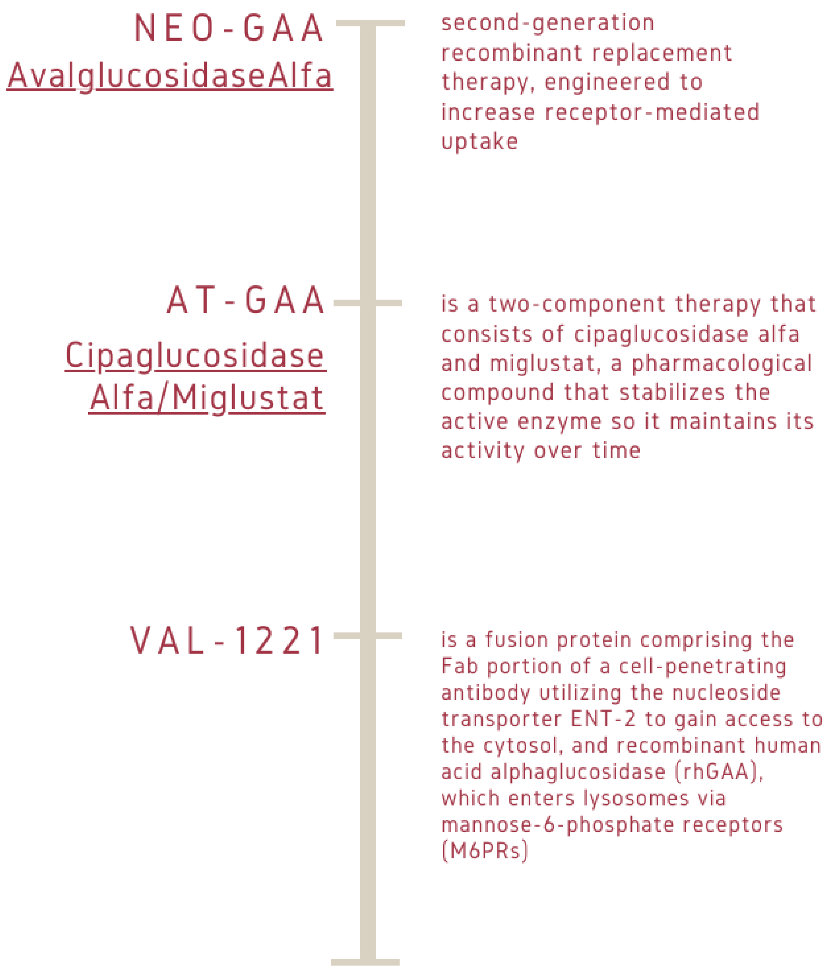
Figure 1. Different types of molecular drugs involved in gene-expression regulation. Avalglucosidase alpha, or NeoGAA, is a drug for enzyme replacement therapy, specifically designed for Pompe disease. Avalglucosidase alpha is a recombinant form of GAA that restores deficient enzyme levels. First developed by Sanofi Genzyme, avalglucosidase alpha is a chemically modified version of alglucosidase alpha, where synthetic bis-phosphorylated oligosaccharides were attached to the structure to improve cellular uptake of the drug and better muscle targeting. Pombiliti + Opfolda (cipaglucosidase alpha/miglustat), formerly known as AT-GAA, is an approved combination therapy for adults with late-onset Pompe disease, or LOPD. VAL-1221 is a fusion protein comprising the Fab portion of a cell-penetrating antibody utilizing the nucleoside transporter ENT-2 to gain access to the cytosol, and recombinant human acid alpha glucosidase (rhGAA), which enters lysosomes via mannose-6- phosphate receptors (M6PRs).
Ijms 25 09139 g001
Figure 2. Sources of the 2934 subjects studied.
Ijms 25 09139 g002
Figure 3. Sources of the 39 PD subjects.
Ijms 25 09139 g003
Figure 4. Flowchart diagram: reference value of activity enzyme is ≥6 nmol/mL/h.
Ijms 25 09139 g004
Table 1. Genetic analysis information of 39 Italian patients with late-onset glycogen storage disease. Reference value of activity enzyme is (≥6 nmol/mL/h).
SubjectAgeFamily HistoryGender
F Female
M Male
α-Glucosidase Activity
(DBS)
Variant 1Variant 2
#157yesF2.1c.-32-13T>Gc.1655 T>C L552P
#254yesM0.7c.-32-13T>Gc.1655 T>C L552P
#338noM0.8c.-32-13T>Gc.2431 delC
#445noM0.6c.-32-13T>Gc.2242dupG
#521noM1.6c.-32-13T>Gc.1655 T>C L552P
#62noM0.6c.-32-13T>Gc.1927 G>A G643R
#7NSnoM0.8c.-32-13T>Gc.1465 G>A D489N
#810noM1.4c.-32-13T>Gc.2238 G>A W746
#954noM1.5c.-32-13T>Gc.2560 C>T R854
#1049noF1.3c.-32-13T>Gc.1210 G>A D404N
#1163yesM0.9c.-32-13T>Gc.526 delG E176R fs45
#12NSnoNS0.9c.-32-13T>Gc.784 G>A E262K
#1382yesM1c.-32-13T>Gc.784 G>A E262K
#1433yesF1.3c.-32-13T>Gc.525 delT E176R fs45
#159yesM1.5c.-32-13T>GDel ex18
#1635noF5.4c.670 C>Tc.861C>T
#175siM1.1c.-32-13T>G
homozygous
#182 monthnoM0.4c.1465 G>A D489Nc.2238 G>A W746
#1978noM1.5c.-32-13T>G
homozygous
#2024 daysnoM0c. 1565 C>G P522R homozygous
#2158noM0.8c.-32-13T>G
homozygous
#2259yesM0.7c.-32-13T>G
homozygous
#2352yesM1.6c.-32-13T>G
homozygous
#243 monthnoM0.9c.526 delG E176R fs45c.2104 C>T R702C
#2511 monthnoF0.5c.1655 T>C L552Pc.2161 ins G E721G fs16
#261 monthnoF0.9c.784 G>A E262KDeletion on exon 9 and 19
#2742yesM5c.784 G>A E262KDeletion on exon 9 and 17
#287noF0.4c.784 G>A E262KC.1979 G>A R660H
#295 monthnoM0.4c.1327-18 A>Gc.1655 T>C L552P
#307noF1.1c.784 G>A E262Kc.1979 G>A R660H
#3112noF0.5c.2012 T>A M671K homozygous
#326 mouthnoM0.8c.1437+2 T>Cc.1655 T>C L552P
#3353noM20.3c.-32-13T>Gc.1551 +1 G>C
#3447yesF5.4c.-32-13T>Gc.655 G>A G219R
#3515noM4.2c.2332-40 C>G homozygous
#3670yesF3.3c.-32-13T>Gc.1551+1 G>C etero
#3745noM0.6c.-32-13T>Gc.2242dupG E748G
#3824yesM3.3c.-32-13T>GDeletion on exon 18
#3954yesF0.6c.118 C>T R40c.2647-7 G>A
Disclaimer/Publisher’s Note: The statements, opinions and data contained in all publications are solely those of the individual author(s) and contributor(s) and not of MDPI and/or the editor(s). MDPI and/or the editor(s) disclaim responsibility for any injury to people or property resulting from any ideas, methods, instructions or products referred to in the content.